
Submitted:
03 July 2026
Posted:
06 July 2026
You are already at the latest version
Abstract
Numerous types of environmental toxicants alter human metabolomics, directly or indirectly, through gut microbial dysbiosis, upsetting immune homeostasis. Environmental toxicants capable of these direct and indirect effects include heavy metals, pesticides, herbicides, polychlorinated biphenyl (PCB) congeners, and endocrine-disrupting chemicals. The cell and molecular events induced by these diverse toxicants are reviewed, along with their potential additive, synergistic, and antagonistic effects on immune homeostasis (increasing proinflammatory immune cell activation and suppressing immunoregulation), which leads to systemic oxidative stress (OS). As people are exposed in varying degrees to countless chemicals and environmental factors over a lifetime, it is challenging to correlate specific diagnoses to any single toxicant or exposure, often a key challenge in linking environmental exposure to health outcomes. However, many toxicants have the common mechanistic effect of OS. The effects of toxicants are more pronounced with aging due to cumulative exposures and immunoaging (immunosenescence), with chronic low-grade inflammation referred to as “inflammaging”. The cell and molecular mechanisms of toxicants include altered calcium flux, mitochondrial dysfunction, and increased levels of damage-associated molecular patterns (alarmins) that trigger an inflammatory response and possibly promote autoimmune and neurological disorders. OS skews type-1 immunity for defenses against pathogens and cancers more toward type-2 immune responses to self-antigens (autoimmunity). The toxicants may directly affect the innate and adaptive immune cells inducing this skewing or they may modify the portions of gut microbial species and strains and their production of metabolites that indirectly affect systemic immunity. These latter toxicant influences require metabolomic analysis of the differential structures and activities of the microbial metabolites. The damaging effects of OS and inflammation disrupting immune homeostasis leading to disorders are reviewed and discussed. The need for well designed studies that allow for standardized comparison of exposures and related effects are emphasized, and their real world limitations noted.
Keywords:
thiols
; oxidative stress
; inflammation
; autoimmunity
; aging
1. Introduction
There tends to be a decline in protective immunity and an increase in autoimmune disease incidence with increasing advanced age, and exposure to environmental toxicants may accelerate aging related mechanisms. The acquisition of differential immune changes with advanced age may be an intrinsic property of host innate and adaptive immune cells, as well as the cumulative effects of environmental toxicants on the immune cells and the microbiome gut brain axis (MGBA), which has regulatory effects on the cardiovascular, endocrine, and immune systems. An additional complexity is that toxic insults can come from many types of toxic insults including chemical, physical, and psychological stresses, e.g., in a mouse report, cold (physical)-restraint (psychological) stress inhibited immunity to a Listeria monocytogenes infection [1,2,3] in a manner similar to that of the heavy metal effects of lead [4,5].
Additionally, Pb and psychological stress have been shown to generate additive, synergistic or potentiated effects [6]. Exogenous physical and psychological stresses also may relate to the increased prevalence of anxiety and depression in adolescents due to their induction of endogenous metabolites such as inflammasome products [7] and the kynurenine and serotonin pathways of tryptophan metabolism [8]. Although the decline of immune functions is associated with accumulated toxicant exposures and aging-related pathologies, including increased incidence of infectious diseases, autoimmune diseases, and cancers of the immune and other organ systems, it is likely that the cause of cellular and organism ageing is due to genetics and lifelong environmental exposure histories, which affect cellular metabolism and regulatory inter-organ communications.
Certainly, toxic and infectious exposures and physical/psychological stresses can affect longevity, and immunosuppression as well as exaggerated, unregulated immune responses, including chronic inflammation, can shorten life expectancy; however, the particular cellular and molecular changes occurring with ageing that lessen both quality of life and life span have not been fully delineated. This review focuses primarily on the cellular and molecular parameters of OS and the neuroendocrine immune (NEI) network.
Environmental toxicants influence the thiols and functions of immune cells altering immunity via loss of reducing agents like glutathione (GSH) and the induction of oxidants and stress-related neurotransmitters. Figure 1 partially shows the inclusion of “NO uncoupling”, oxidants/antioxidants and the tryptophan–kynurenine pathway causing immunosuppression via indoleamine-2,3-dioxygenase (IDO) depletion of tryptophan. Environmental toxicant-induced stresses and oxidants skew and diminish immunity.
In general, the practical study of toxic and infectious exposures and related physiological stresses is challenging. In a human population, basic findings obtained in the study of mice or basic cell lines, are often difficult to assess due to issues of study design, sample size, dosage, the accurate measurement of exposures and the timing of such exposures. The issue of causation versus correlation arises, and the accurate assessment of associated physiological stresses in the presence of mediating, secondary variables often require statistical models to determine significance. The importance of replication and adequate sample size, along with related statistical power provide challenges for researchers.
1.1. Methods and Materials
A systematic review of the literature was conducted using Google and PubMed sources. The set of keywords used were “environmental toxins and oxidative stress”, “Redox changes (Ox>Red) and aging”, “toxins” and “autoimmunity”. These were then organized by three general themes. These were inflammation and aging, OS and immunity, and Environmental stresses and redox effects on immune cells and health.
1.2. Inflammation and Aging
The cause of the decline of antigen-specific immunity is whether the immune dysfunction is due to the increased presence of inflammatory mediators in older persons due to age alone and/or whether inflammation is a consequence of age-related accumulation of toxic metabolites. Numerous changes in body composition and physiology have been noted with increasing age. Many diseases, including those associated with inflammatory processes, are age-related in terms of prevalence, but how these diseases relate to the primary aging processes remain to be defined, requiring investigation.
For example, the proinflammatory cytokine IL-6 has correlated positively with aging [9,10,11] and upon infection of mice at higher bacterial burdens, the serum IL-6 concentration increases exponentially along with the stress-related hormone corticosterone, and the number of lymphocytes decline [12]; however, this is not evidence that IL-6 causes immune cell senescence or lowered responsiveness. The determination of causation requires careful study and appropriate study design and a controlled experimental context.
Proinflammatory cytokines increase in centenarians (long-lived persons) [13,14] suggests that it may be the ability to cope with inflammation that is more critical than the ability to control inflammation. Since inflammation is closely associated with OS [15], it-is important to evaluate the mechanistic connections between inflammation, OS, and aging, because lymphocytes are highly sensitive to OS, and monocytes and neutrophils, which produce oxidants, are relatively resistant to OS. Additionally, innate immunity is intimately involved in inflammation. OS mechanisms influence the decline of antigen-specific immunity, including the availability of naïve T cells and their response to new antigens.
Innate immunity is designed to assist host defenses by rapidly lowering pathogen propagation and invasion prior to development of adaptive immunity. However, it’s posited that the positive aspects of innate immunity decline in the elderly due to a shift toward production of molecules such as IDO, which deplete molecules necessary for enhanced immunity such as tryptophan (an amino acid needed for lymphocyte activity), and the production of reactive oxygen species (ROS) instead of nitric oxide (NO) (“the uncoupling of NO production”) [16], which leads to interference with T and B cell development and activation.
The availability of tetrahydrobiopterin (BH4), a co-factor for NO synthases and tyrosine and tryptophan hydroxylase, is linked with glutathione (GSH) levels and both decline due to the redox changes. Loss of BH4 is, in part, responsible for the uncoupling of NO and increased IDO activity, and BH4 loss is associated with cardiovascular diseases and neuropsychiatric disorders [17]. The age-associated decline in cellular energy with less ATP and a concomitant increase in intracellular oxidants (H2O2 and ˙NO) suggests that mitochondria malfunctioning may be central to the biology and chemistry of ageing [18,19]. The connection of mitochondrial dysfunction as a key aspect of aging again implicates environmental toxins since many are known to affect mitochondrial functions [20].
1.3. OS and Immunity
Cellular thiol proteins influence membranal lipid domains, membranal lipids can influence the number of cellular thiols, and OS alters the lipid composition of the plasma membrane (PM) via lipid peroxidation [21], which can be initiated by secondary organic aerosol (SOA) toxicants [22]. Thus, depletion of cellular thiols can result in immunomodulation due to a restructuring of PM lipids and proteins (receptors and intracellular signal transduction circuits) of lymphocytes and antigen-presenting cells (APC), which can alter early signal transduction events (G0 G1) associated with APC.
The existence of protein-protein, protein-lipid and lipid-lipid domains within the PM and the influence of cellular thiols on these domains have been previously reported [23,24]. Interestingly, rearrangement of membrane lipids after OS is often accompanied with expression of extracellular expression of heat shock proteins (HSPs), which have immunomodulatory properties. HSP70 has been shown to increase Treg activity [25] and inhibit antigen-presentation [26]. However, HSP70 also activates innate immune cells to release inflammatory cytokines [27]. Additionally, protein disulfide exchanges, which are affected by redox changes, can influence enzymatic activities associated with different immune activities, e.g., modifying the metalloprotease activity of TACE/ADAM-17, which affects release of TNF, TNFR, and L-selectin [28,29]. However, as for the effects of HSP70, not all immune activities uniformly decline with increased induction of stress proteins or increasing age [30]. CD8+ T cells tend to decline more rapidly than CD4+ T cells, and innate and humoral immunity (mainly B cell and Th2 cell activities) appear to outlast some other T cell functions [31].
In fact, enhanced innate cell induction of inflammation is, at least partially, responsible for the declining antigen-specific immunity. Additionally, macrophages with different amounts of GSH differentially activate lymphoid subsets, which produce different cytokines and immune responses [32,33,34,35,36] Accumulated toxicants with age, and age itself, are posited to cause redox changes in the elderly, which involve an imbalance of NO and ROS, decline of BH4 and GSH, stress-induced norepinephrine, and lower oxidoreductase activity. Each of these changes are responsible for the decline of antigen-specific immunity.
Numerous signaling events required for T cell activation are also thiol-modulated, including signaling via CD4 through Lck, phospholipase Cγ1 (PLCγl) and various kinases [37,38,39]. The conformation of the protein transmembrane domains of the TCR/CD3/CD4 or CD8 complex likely will affect their association with intra-membranal enzymes, cytoplasmic associates (e.g., Lck), and the lipid-lipid domains. CD4 has intra- [39] and extra-cellular thiol influences on activation [40,41]. Likewise, the CD45 family [42], and L-selectin [43] have known involvement in T cell activation via thiol changes. A change in membrane structure/fluidity also influences lymphocyte activation. Note that cholesterol, a known modulator of membrane structure, can modify antigen-specific presentation [44], and its availability is dependent on conditions known to vary with aging-related HDL/LDL changes and metabolic syndrome.
Numerous enzymes with immunomodulatory potential are thiol-regulated, including adenylate cyclase [45], 5-nucleotidase [46], insulin receptor [47], and Na+/K+ ATPase [48]. Changes in the lymphocytes’ transport functions start early in G1 (Na+, K+, and Ca++ changes occur within 1 min; glucose by 10 min; amino acids and nucleosides by 1 hr). Since cell transport mechanisms directly utilize thiol-sensitive proteins [49,50,51,52], thiol- modulation has regulatory influence on multiple early events involved in lymphocyte activation. Na/K-ATPase activity decreases in lymphocytes from aged people [53]; membrane potential changes are less responsive with aged cells [55,514]; and nucleotide pools [56], kinase activity [57], protein phosphorylation patterns [58], and G protein involvements [59] have been reported to be modified by aging. Many of these processes are interconnected in the activation of T cells [60].
Previously published studies have shown that lymphocyte activation and proliferation are affected by cellular thiols (R-SH), which includes surface thiols [61,62,63] as well as total thiols [64] and GSH [65,66,67,68]. Polymorphisms of GLCLC and GLCLR, the two genes encoding glutamate cysteine ligase (GCL) [69], the rate-limiting enzyme in GSH biosynthesis may be partially responsible for differential aging rates among the elderly. However, cellular thiols also influence the phospholipid and nucleotide composition of lymphocytes [68,70]. Since the toxic mechanism of action includes the ability to dysregulate the redox status, production of inflammatory mediators, epigenetic alterations, barrier deterioration, and alteration of mitochondrial function [71], the role of thiols classically fits into the modulatory scheme. These cellular, biochemical, and genetic modifications may lead to development of autoimmune diseases [72].
Toxins dysregulate enzymatic processes due to cellular damage or toxins, themselves, may bind to proteins to create new epitopes tricking the immune system into treating the body’s own constituents as foreign invaders. Examples are dysregulated production of citrullinated proteins [73] and redox-mediated carbamylation [74] generating autoantibodies and immune complexes in the joints of rheumatoid arthritis patients. Note that an indicator of redox is the shortening of telomere length [75]
1.4. Environmental Stresses and Redox Effects on Immune Cells and Health
Chemical, physical, and psychological stressors are posited to cause an imbalance of the neuroendocrine immune (NEI) network, which then results in oxidative stress and inflammation; this imbalance occurs more frequently in older persons, who are, in general, more sensitive to these changes. Environmental stresses exacerbate age-related metabolic changes, including lowered production of neurotransmitters and altered regulation of nitric oxide synthase (NOS) activity, which are intrinsic aspects of NEI mechanisms, cause increased production of reactive oxygen species and loss of cellular thiols [76]. These changes to redox status have been posited to lower antigen-specific immunity and to be implicated in age-related comorbidities. These comorbidities include cardiovascular disease, type-2 diabetes, and arthritis.
In a study with middle-aged mice [77], there was a decline in activation of nuclear factor erythroid 2-related factor 2 (Nrf2)–dependent antioxidant enzymes in lungs, liver and cerebellum in response to airborne nanoparticulate matter (nPM). The suppressed inducibility of the Nrf2-regulated antioxidant enzymes were glutamate cysteine ligase (both the GCLC and GCLM subunits), heme oxygenase 1 (HO-1) and NADPH:quinone oxidoreductase 1 (NQO1). The nPM effect was associated with an increased or exaggerated inflammatory response, which is suggested to play a central role in nPM-associated cardiopulmonary diseases [78,79,80,81]. NF-kB activation in response to particulates is observed in both human macrophages and NHBE epithelial cells [82,83]. The Nrf2-dependent antioxidant response is also a protective mechanism against oxidative stimuli in macrophages [84], including against particles [85].
Oxidative stress and aging have been linked to increased autoimmune activities, especially humoral (antibody-mediated) autoimmunity [86]. As indicated B cells, in general, have greater thiol sensitivity than T cells (see Table 1) and [87], thus, it’s surprising that B cell and Th2 cell activities appear to outlast some T cell functions [88], which emphasizes the need to investigate the molecular basis for aging and the oxidative sensitivities. The mechanisms involved in the differential activation of Th1 and Th2 cells with aging, HIV infection, or exposure to thiol-reactive chemicals [87] have not been fully delineated.
It has been reported that oxidative stress mainly on APC allows Th2 development to predominate. This outcome was suggested to be related mainly to loss of IL-12 production; however, in addition to less IL-12 for Th1 cell activation, regulatory effects dependent on the ratio of macrophages with high (M1) or low (M2) levels of GSH, as described [34], need to be further considered regarding aging. This scheme is shown (see Figure 1) with incorporation of the influence of stress and involvement of NO uncoupling. IL-4 is a cytokine promoting Th2 activities, which lowers the GSH level of macrophages [89,90], thus enhancing the number of M2 (oxMφ or alternatively activated) macrophages with less GSH. These GSH influences on Mφs further implicate thiol chemistry in immune modulations.
Resting macrophages have been suggested to aid T cell proliferation via release of proton- donors, such as cysteine or GSH, whereas activated macrophages release more oxidants and are more inhibitory (Figure 2) and [91]. Toxic metabolites are shown to disrupt the redox influences of macrophages on T cells.
1.5. Exposures, Context and Study Design Issues
The practical detection of various types of exposures, oxidative stress, and related immunological responses and toxicity is challenging. Many of the results reported here are based on carefully curated mouse cohorts or cell lines, studied in highly controlled experimental settings. To develop these results into practical understanding relevant to the human population requires an appreciation of study design and related statistical modeling. The level of control possible in mouse and lab populations, to the level of genetic similarity and careful control of exposures and length of exposures, is not typically possible in humans.
When developing a cohort or set of experimental subjects, ensuring they all share a similar environment, source of exposure and timing of exposure, is very challenging and often results in limited sample size and lower statistical power to detect defined effect sizes. Often, a case-control study is the most reasonable approach, beginning with a set of cases whose physiological condition and previous exposures are validated. To each case, a set of controls is identified having similar exposure history but with no resulting physiological condition. Typically, a 2 to 1 or 3 to 1 ratio of controls to cases is taken. Methods for the analysis of case-control studies have been developed [ref: epi methods] and often rely on detection of a significant odds ratio or relative risk when comparing cases versus controls. Cohort studies are also of relevance here [ref: epi methods].
Epidemiologic and environmental studies provide a context for more general interpretation of results, but are challenging to conduct as they often require baseline assessments of exposures and follow-up based assessment of the onset of, for example, autism or infectious diseases. For illnesses examined in older populations, socio economic bias, age cohort effects and underlying genetic susceptibility or immunity may affect the interpretation of the observed data and summary statistics such as rates, odds ratios and relative risks [ref: epi methods].
In the case of exposures to environmental risk factors, accumulated exposures and lifetime exposures, for example to chemical agents which remain in the body, may also have an impact in regard to the onset of disease, and yet are challenging to measure.
2. Discussion
Oxidative stress and aging have been identified in basic research as affecting the immune system and other physiologic conditions, especially in regard to older individuals. Using these findings as a basis for (i) replication of these results in human subjects subject to various mediating variables and (ii) determining possible treatments or preventative practices in human populations, is an ongoing challenge. To interpret results based on small sample sizes or mouse and cell lines-based studies more generally, requires carefully designed studies.
If focusing on age cohorts within the human population, the need to identify and model mediating variables such as socio-economic effects, lifestyle, previous exposures and illnesses requires careful study design. This is challenging with limited resources. Further, the presence of genetic effects related to aging, the immune system, sensitivity to toxic exposures, are complex and may also play a role.
Many of the basic studies reported here were often limited in scope, identifying individual risk factors without consideration of possible correlation with environmental or genetic factors when applying the results in human populations. Future studies, taking these results and examining them in broader, controlled settings, will be useful in extending the results to more practical settings.
Ref: epi Methods:
Clayton D. and Hills M. (1993) Statistical Methods in Epidemiology. Oxford University Press, Oxford.
References
- Cao, L.; Filipov, N.M.; Lawrence, D.A. Sympathetic nervous system plays a major role in acute cold/restraint stress inhibition of host resistance to Listeria monocytogenes. J. Neuroimmunol. 2002, 125(1-2), 94–102. [Google Scholar] [CrossRef] [PubMed]
- Cao, L.; Hudson, C.A.; Lawrence, D.A. Acute cold/restraint stress inhibits host resistance to Listeria monocytogenes via beta1-adrenergic receptors. Brain Behav. Immun. 2003, 17(2), 121–33. [Google Scholar] [CrossRef] [PubMed]
- Emeny, R.T.; Lawrence, D.A. Chapter 48: Cold-restraint-induced immune and biochemical changes inhibit host resistance to Listeria. In Psychoneuroimmunology Vol. IV; Ader, et al., Eds.; Academic Press, 2006; pp. 1035–1051. [Google Scholar]
- Lawrence, D.A.; McCabe, M.J. Immune modulation by toxic metals. In Organ- Specific Metal Toxicology; Waalkes, M., Goyer, R., Klaassen, C., Eds.; Academic Press: NY, 1995; pp. 305–337. [Google Scholar]
- Dyatlov, V.A.; Lawrence, D.A. Neonatal lead exposure potentiates sickness behavior induced by Listeria monocytogenes infection of mice. Brain Behav. Immun. 2002, 16(4), 477–92. [Google Scholar] [CrossRef] [PubMed]
- Cory-Slechta, D.A.; Virgolini, M.B.; Rossi-George, A.; et al. Lifetime consequences of combined maternal lead and stress. Basic Clin. Pharmacol. Toxicol. 2008, 102, 218–227. [Google Scholar] [CrossRef] [PubMed]
- Iwata, M.; Ota, K.T.; Duman, R.S. The inflammasome: pathways linking psychological stress, depression, and systemic illnesses. Brain Behav. Immun. 2013, 31, 105–14. [Google Scholar] [CrossRef] [PubMed]
- Miura, H.; Ozaki, N.; Sawada, M.; Isobe, K.; Ohta, T.; Nagatsu, T. A link between stress and depression: shifts in the balance between the kynurenine and serotonin pathways of tryptophan metabolism and the etiology and pathophysiology of depression. Stress. 2008, 11(3), 198–209. [Google Scholar] [CrossRef] [PubMed]
- Tylutka, A.; Walas, Ł.; Zembron-Lacny, A. Level of IL-6, TNF, and IL-1β and age-related diseases: a systematic review and meta-analysis. Front Immunol. 2024, 15, 1330386. [Google Scholar] [CrossRef] [PubMed]
- Fagiolo, U.; Cossarizza, A.; Scala, E.; Fanales-Belasio, E.; Ortolani, C.; Cozzi, E.; Monti, D.; Franceschi, C.; Paganelli, R. Increased cytokine production in mononuclear cells of healthy elderly people. Eur. J. Immunol. 1993, 23, 2375–2378. [Google Scholar] [CrossRef] [PubMed]
- Singh, T.; Newman, A.B. Inflammatory markers in population studies of aging. Ageing Res. Rev. 2011, 10(3), 319–329. [Google Scholar] [CrossRef] [PubMed]
- Kim, D.; Reilly, A.; Lawrence, D.A. Relationships between IFNgamma, IL-6, corticosterone, and Listeria monocytogenes pathogenesis in BALB/c mice. Cell Immunol. 2001, 207(1), 13–8. [Google Scholar] [CrossRef] [PubMed]
- Pinti, M.; Gibellini, L.; Lo Tartaro, D.; De Biasi, S.; Nasi, M.; Borella, R.; Fidanza, L.; Neroni, A.; Troiano, L.; Franceschi, C.; Cossarizza, A. A Comprehensive Analysis of Cytokine Network in Centenarians. Int. J. Mol. Sci. 2023, 24(3), 2719. [Google Scholar] [CrossRef] [PubMed]
- Franceschi, C.; Bonafe, M.; Valensin, S.; Olivieri, F.; De Luca, M.; Ottaviani, E.; De Benedictis, G. Inflammaging. An evolutionary perspective on immunosenescence. Ann. N Y Acad. Sci. 2000, 908, 244–54. [Google Scholar] [CrossRef] [PubMed]
- Steven, S.; Frenis, K.; Oelze, M.; Kalinovic, S.; Kuntic, M.; Bayo Jimenez, M.T.; Vujacic-Mirski, K.; Helmstädter, J.; Kröller-Schön, S.; Münzel, T.; Daiber, A. Vascular Inflammation and Oxidative Stress: Major Triggers for Cardiovascular Disease. Oxid. Med. Cell Longev. 2019, 2019, 7092151. [Google Scholar] [CrossRef] [PubMed]
- Oxenkrug, G.F. Interferon-gamma-inducible kynurenines/pteridines inflammation cascade: implications for aging and aging-associated psychiatric and medical disorders. J. Neural Transm. 2011, 118(1), 75–85. [Google Scholar] [CrossRef] [PubMed]
- Fanet, H.; Capuron, L.; Castanon, N.; Calon, F.; Vancassel, S. Tetrahydrobioterin (BH4) Pathway: From Metabolism to Neuropsychiatry. Curr. Neuropharmacol. 2021, 19(5), 591–609. [Google Scholar] [CrossRef] [PubMed]
- Yap, L.P.; Garcia, J.V.; Han, D.; Cadenas, E. The energy-redox axis in aging and age-related neurodegeneration. Adv. Drug Deliv. Rev. 2009, 61(14), 1283–98. [Google Scholar] [CrossRef] [PubMed]
- Storz, P. Reactive oxygen species-mediated mitochondria-to-nucleus signaling: a key to aging and radical-caused diseases. Sci. STKE 2006, re3. [Google Scholar] [CrossRef] [PubMed]
- Jia, G.; Aroor, A.R.; Martinez-Lemus, L.A.; Sowers, J.R. Mitochondrial functional impairment in response to environmental toxins in the cardiorenal metabolic syndrome. Arch. Toxicol. 2015, 89(2), 147–53. [Google Scholar] [CrossRef] [PubMed]
- Higdon, A.; Diers, A.R.; Oh, J.Y.; Landar, A.; Darley-Usmar, V.M. Cell signalling by reactive lipid species: new concepts and molecular mechanisms. Biochem J. 2012, 442(3), 453–64. [Google Scholar] [CrossRef] [PubMed]
- Shiraiwa, M.; Fang, T.; Wei, J.; Lakey, P.; Hwang, B.; Edwards, K.C.; Kapur, S.; Mena, J.; Huang, Y.K.; Digman, M.A.; Weichenthal, S.A.; Nizkorodov, S.; Kleinman, M.T. Chemical and Cellular Formation of Reactive Oxygen Species from Secondary Organic Aerosols in Epithelial Lining Fluid. Res. Rep. Health Eff. Inst. 2023, 2023(215), 1–56. [Google Scholar] [PubMed]
- Busija, A.R.; Patel, H.H.; Insel, P.A. Caveolins and cavins in the trafficking, maturation, and degradation of caveolae: implications for cell physiology. Am. J. Physiol. Cell Physiol. 2017, 312(4), C459–C477. [Google Scholar] [CrossRef] [PubMed]
- Torres, M.; Rosselló, C.A.; Fernández-García, P.; Lladó, V.; Kakhlon, O.; Escribá, P.V. The Implications for Cells of the Lipid Switches Driven by Protein-Membrane Interactions and the Development of Membrane Lipid Therapy. Int. J. Mol. Sci. 2020, 21(7), 2322. [Google Scholar] [CrossRef] [PubMed]
- Wachstein, J.; Tischer, S.; Figueiredo, C.; Limbourg, A.; Falk, C.; Immenschuh, S.; Blasczyk, R.; Eiz-Vesper, B. HSP70 enhances immunosuppressive function of CD4(+)CD25(+)FoxP3(+) T regulatory cells and cytotoxicity in CD4(+)CD25(-) T cells. PLoS ONE 2012, 7(12), e51747. [Google Scholar] [CrossRef] [PubMed]
- Kettern, N.; Rogon, C.; Limmer, A.; Schild, H.; Höhfeld, J. The Hsc/Hsp70 co-chaperone network controls antigen aggregation and presentation during maturation of professional antigen presenting cells. PLoS ONE 2011, 6(1), e16398. [Google Scholar] [CrossRef] [PubMed]
- Radons, J.; Multhoff, G. Immunostimulatory functions of membrane-bound and exported heat shock protein 70. Exerc Immunol. Rev. 2005, 11, 17–33. [Google Scholar] [PubMed]
- Srour, N.; Lebel, A.; McMahon, S.; Fournier, I.; Fugère, M.; Day, R.; Dubois, C.M. TACE/ADAM-17 maturation and activation of sheddase activity require proprotein convertase activity. FEBS Lett. 2003, 554(3), 275–83. [Google Scholar] [CrossRef] [PubMed]
- Scott, A.J.; O’Dea, K.P.; O’Callaghan, D.; Williams, L.; Dokpesi, J.O.; Tatton, L.; Handy, J.M.; Hogg, P.J.; Takata, M. Reactive oxygen species and p38 mitogen-activated protein kinase mediate tumor necrosis factor α-converting enzyme (TACE/ADAM-17) activation in primary human monocytes. J. Biol. Chem. 2011, 286, 35466–35476. [Google Scholar] [PubMed]
- Gomez, C.R. Role of heat shock proteins in aging and chronic inflammatory diseases. Geroscience 2021, 43(5), 2515–2532. [Google Scholar] [CrossRef] [PubMed]
- Goronzy, J.J.; Lee, W.W.; Weyand, C.M. Aging and T-cell diversity. Exp. Gerontol. 2007, 42(5), 400–6. [Google Scholar] [CrossRef] [PubMed]
- Rasheed, A.; Rayner, K.J. Macrophage Responses to Environmental Stimuli During Homeostasis and Disease. Endocr. Rev. 2021, 42(4), 407–435. [Google Scholar] [CrossRef] [PubMed]
- Peterson, J.D.; Herzenberg, L.A.; Vasquez, K.; Waltenbaugh, C. Glutathione levels in antigen-presenting cells modulate Th1 versus Th2 response patterns. Proc. Natl. Acad. Sci. USA 1998, 95, 3071–3076. [Google Scholar] [CrossRef] [PubMed]
- Murata, Y.; Shimamura, T.; Hamuro, J. The polarization of T(h)1/T(h)2 balance is dependent on the intracellular thiol redox status of macrophages due to the distinctive cytokine production. Int. Immunol. 2002, 14, 201–12. [Google Scholar] [PubMed]
- Dobashi, K.; Aihara, M.; Araki, T.; Shimizu, Y.; Utsugi, M.; lizuka, K.; Murata, Y.; Hamuro, J.; Nakazawa, T.; Mori, M. Regulation of LPS induced IL-12 production by IFN-gamma and IL-4 through intracellular glutathione status in human alveolar macrophages. Clin. Exp. Immunol. 2001, 124, 290–6. [Google Scholar] [PubMed]
- Bozzo, C.; Goria, M.; Marengo, C.; Marena, S.; Veglia, F.; Pagano, G. Lymphocyte Na,K-ATPase is reduced in aged people. Metabolism 1990, 39(8), 808–14. [Google Scholar] [CrossRef] [PubMed]
- Kanner, S.B.; Kavanagh, T.J.; Grossmann, A.; Hu, S.-K.; Bolen, J.B.; Rabinovitch, P.S.; Ledbetter, J.A. Sulfhydryl oxidation down-regulates T-cell signaling and inhibits tyrosine phosphorylation of phospholipase C71. Proc.Nat. Acad. Sci. 1992, 89, 300–304. [Google Scholar] [CrossRef] [PubMed]
- Cemerski, S.; Cantagrel, A.; Van Meerwijk, J.P.; Romagnoli, P. Reactive oxygen species differentially affect T cell receptor-signaling pathways. JBiol Chem. 2002, 277, 19585–19593. [Google Scholar] [CrossRef]
- Huse, M.; Eck, M.J.; Harrison, S.C. A Zn2+ ion links the cytoplasmic tail of CD4 and the N-terminal region of Lck. J. Biol. Chem. 1998, 273, 18729–18733. [Google Scholar] [CrossRef] [PubMed]
- Barbouche, R.; Miquelis, R.; Jones, I.M.; Fenouillet, E. Protein-disulfide isomerase-mediated reduction of two disulfide bonds of HIV envelope glycoprotein 120 occurs post-CXCR4 binding and is required for fusion. J. Biol. Chem. 2003, 278, 3131–3136. [Google Scholar] [PubMed]
- Matthias, L.J.; Azimi, I.; Tabrett, C.A.; Hogg, P.J. Reduced monomeric CD4 is the preferred receptor for HIV. J. Biol. Chem. 2010, 285(52), 40793–40799. [Google Scholar] [CrossRef] [PubMed]
- Stahls, A. The sulfhydryl reagent N-ethylmaleimide induces hyperphosphorylation on tyrosine residues in the Jurkat T-cell line. Biochem Biophys. Res. Commun. 1992, 187(1), 73–8. [Google Scholar] [PubMed]
- Bennett, T.A.; Edwards, B.S.; Sklar, L.A.; Rogelj, S. Sulfhydryl regulation of L-selectin shedding: phenylarsine oxide promotes activation-independent L-selectin shedding from leukocytes. J. Lmmunol. 2000, 164, 4120–4129. [Google Scholar]
- Hughes, D.A.; Townsend, P.J.; Haslam, P.L. Enhancement of the antigen-presenting function of monocytes by cholesterol: possible relevance to inflammatory mechanisms in extrinsic allergic alveolitis and athersclerosis. Clin.Exp. Lmmunol. 1992, 87, 279286. [Google Scholar] [CrossRef]
- Houslay, M.D. Regulation of adenylate cyclase (EC 4.6.1.1) activity by itslipid environment. Proc. Nutr. Soc. 1985, 44, 157–165. [Google Scholar] [CrossRef] [PubMed]
- Dipple, I.; Gordon, L.M.; Houslay, M.D. The activity of 5′-nucleotidase in liver plasma membranes is affected by the increase in bilayer fluidity achieved by anionic drugs but not by cationic drugs. J. Biol. Chem. 1982, 257, 1811–1815. [Google Scholar] [CrossRef] [PubMed]
- Ginsberg, B.H.; Jabour, J.; Spector, A.A. Effect of alterations in membrane lipid unsaturation on the properties of the insulin receptor of ehrlich ascites cells. Biochem Biophys. Acta. 1982, 690, 157–164. [Google Scholar] [CrossRef] [PubMed]
- Sandermann, H.; Gottwald, B.A. Cooperative lipid activation of (Na++K+)-ATPase as a consequence of non-cooperative lipid-protein interactions. Biochim. Biophys. Acta 1983, 732, 332–335. [Google Scholar] [CrossRef] [PubMed]
- Boumendil-Podevin, E.G.; Podevin, R.A. Stimulatory and inhibitory effects of sulfhydryl reagent on p-aminobippuric acid transport by isolated renal tubules. Biochem. Biophys. Acta 1977, 467, 364–375. [Google Scholar] [CrossRef] [PubMed]
- School, B.M.; Emst-DeVries, S.E.; VanHaard, P.M.M.; DePont, J.J.H.H.M.; Bonting, S.L. Studies on (NaVK+)-activated ATPase. XLVI. Effect of cation-induced conformational changes on sulfhydryl group modification. Biochem. Biophys. Acta 1980, 602, 144–154. [Google Scholar]
- Kwock, L.; Wallach, D.F.H.; Hefter, K. Involvement of sulfhydryl groups in the action of insulin and radiation on thymocyte Na+ -dependent animo acid transport. Biochem. Biophys. Acta 1976, 419, 93–103. [Google Scholar] [PubMed]
- Hare, J.D. Distinctive alterations of nucleoside, sugar and amino acid uptake by sulfhydryl reagents in cultured mouse cells. Arch. Biochem. Biophys. 1975, 170, 347–352. [Google Scholar] [CrossRef] [PubMed]
- Witkowski, J.M.; Mysliwski, A.; Mysliwska, J. Decrease of lymphocyte (Na/K)ATPase activity in aged people. Mech. Ageing Dev. 1985, 33, 11–17. [Google Scholar] [PubMed]
- Damjanovich, S.; Pieri, C. Electroimmunology: membrane potential, ion channel activities, and stimulatory signal transduction in human T lymphocytes from young and elderly. Ann. NYAcad Sci. 1991, 621, 29–39. [Google Scholar]
- Varga, Z.; Bressani, N.; Zaia, A.M.; Bene, L.; Fulop, T.; Leovey, A.; Fabris, N.; Damjanovich, S.; Zaia, A.M. Cell surface markers, inositol phosphate levels and membrane potential of lymphocytes from young and old human patients. Immunol. Lett. 1990, 23, 275–280. [Google Scholar] [CrossRef] [PubMed]
- Borzi, R.M.; DalMonte, P.; Uguccioni, M.; Meliconi, R.; Facchini, A. Intracellular nucleotides of lymphocytes and granulocytes from normal ageing subjects. Mech. Ageing Dev. 1992, 64, 1–11. [Google Scholar] [CrossRef] [PubMed]
- Shi, J.; Miller, R.A. Tyrosine-specific protein phosphorylation in response to anti-CD3 antibody is diminished in old mice. J. Gerontol. 1992, 47, 6147–53. [Google Scholar] [CrossRef]
- Patel, H.R.; Miller, R.A. Age-associated changes in mitogen-induced protein phosphorylation in murine T lymphocytes. Eur. J. Immunol. 1992, 22, 253–60. [Google Scholar] [CrossRef] [PubMed]
- Fulop, T.; Barabas, G.; Varga, Z.; CsongorJ; Hauck, M.; SzucsS; Seres, I.; Mohacsi, A.; Kekessy, D.; Despont, J.P. Tranmembrane signaling changes with aging. Ann. NYAcad Sci. 1992, 673, 165–71. [Google Scholar]
- Rudd, C.E.; Janssen, O.; Cai, Y.-C.; daSilva, A.J.; Raab, M.; Prasad, K.V.S. Two-step TCRZ/CD3-CD4 and CD28 signaling in T cells: SH2/SH3 domains, protein-tyrosine and lipid kinases. Immunol. Today 1994, 15, 225–234. [Google Scholar] [PubMed]
- Lawrence, D.A.; Song, R.; Weber, P. Surface thiols of human lymphocytes and their changes after in vitro and in vivo activation. J. Leuk. Biol. 1996, 60, 611–618. [Google Scholar] [CrossRef]
- Duncan, D.D.; Lawrence, D.A. Four sulfhydryl-modifying compounds cause different structural damage. Chem. Biol. Lnter. 1988, 68, 453–469. [Google Scholar] [CrossRef]
- Noelle, R.J.; Lawrence, D.A. Modulation of T cell functions II. Cell. Lmmunol. 1981, 68, 453–469. [Google Scholar] [CrossRef]
- Ayers, F.C.; Warner, G.L.; Smith, K.L.; Lawrence, D.A. Fluorometric quantitation of cellular and nonprotein thiols. Anal. Biochem. 1986, 154, 186–93. [Google Scholar] [CrossRef] [PubMed]
- Messina, J.; Lawrence, D.A. Effects of 2ME and BSO on cystine metabolism by mitogen stimulated human PBMC. Lnternatl J. Lmmunopharm 1992, 14, 1221–34. [Google Scholar]
- Noelle, R.J.; Lawrence, D.A. Determination of GSH in lymphocyte and possible association of redox state and proliferative capacity of lymphocytes. Biochem. J. 1981, 198, 571–579. [Google Scholar] [CrossRef] [PubMed]
- Messina, J.; Lawrence, D.A. Cell cycle progression of GSH-depleted human PBMC is inhibited at S phase. J. Lmmunol. 1989, 143, 1974–1981. [Google Scholar]
- Palace, G.P.; Lawrence, D.A. Phospholipid metabolism of lymphocytes with inhibited glutathione synthesis using buthionine sulfoximine. Free Rad. Biol. Med. 1995, 18, 709–720. [Google Scholar] [PubMed]
- Walsh, A.C.; Li, W.; Rosen, D.R.; Lawrence, D.A. Genetic mapping of GLCLC, the human gene encoding the catalytic subunit of gamma-glutamyl-cysteine synthetase, to chromosome band 6p12 and characterization of a polymorphic trinucleotide repeat within its 5′ untranslated region. Cytogenet Cell Genet. 1996, 75(1), 14–16. [Google Scholar] [PubMed]
- Palace, G.P.; Lawrence, D.A. Nucleotide changes in oxidatively stressed lymphocytes. J. Biochem Toxicol. 1995, 10(3), 137–42. [Google Scholar] [CrossRef] [PubMed]
- Wallace, D.R.; Taalab, Y.M.; Heinze, S.; Tariba Lovaković, B.; Pizent, A.; Renieri, E.; Tsatsakis, A.; Farooqi, A.A.; Javorac, D.; Andjelkovic, M.; Bulat, Z.; Antonijević, B.; Buha Djordjevic, A. Toxic-Metal-Induced Alteration in miRNA Expression Profile as a Proposed Mechanism for Disease Development. Cells 2020, 9(4), 901. [Google Scholar] [CrossRef] [PubMed]
- Kharrazian, D. Exposure to Environmental Toxins and Autoimmune Conditions. Integr. Med. 2021, 20(2), 20–24. [Google Scholar]
- Auger, I.; Balandraud, N.; Massy, E.; Hemon, M.F.; Peen, E.; Arnoux, F.; Mariot, C.; Martin, M.; Lafforgue, P.; Busnel, J.M.; Roudier, J. Peptidylarginine Deiminase Autoimmunity and the Development of Anti-Citrullinated Protein Antibody in Rheumatoid Arthritis: The Hapten-Carrier Model. Arthritis Rheumatol. 2020, 72(6), 903–911. [Google Scholar] [CrossRef] [PubMed]
- Trejo-Zambrano, M.I.; Gómez-Bañuelos, E.; Andrade, F. Redox-Mediated Carbamylation As a Hapten Model Applied to the Origin of Antibodies to Modified Proteins in Rheumatoid Arthritis. Antioxid. Redox Signal. 2022, 36(7-9), 389–409. [Google Scholar] [CrossRef] [PubMed]
- Pedersen-Lane, J. R.B.; Zurier; Lawrence, D.A. Analysis of the thiol status of peripheral blood leukocytes in rheumatoid arthritis patients. J. Leukoc. Biol. 2007, 81, 934–941. [Google Scholar] [CrossRef] [PubMed]
- Zhang, C.; Liu, S.; Chen, G. Environmental and Climate Impacts on Physiological Aging: Comprehensive Pathways to Enhanced Age-related Resilience. Aging Dis. 2024, 16(3), 1218–1224. [Google Scholar] [CrossRef] [PubMed]
- Zhang, H.; Liu, H.; Davies, K.J.; Sioutas, C.; Finch, C.E.; Morgan, T.E.; Forman, H.J. Nrf2-regulated phase II enzymes are induced by chronic ambient nanoparticle exposure in young mice with age-related impairments. Free Radic. Biol. Med. 2012, 52(9), 2038–46. [Google Scholar] [CrossRef] [PubMed]
- Tsai, D.H.; Amyai, N.; Marques-Vidal, P.; Wang, J.L.; Riediker, M.; Mooser, V.; Paccaud, F.; Waeber, G.; Vollenweider, P.; Bochud, M. Effects of particulate matter on inflammatory markers in the general adult population. Part Fibre Toxicol. 2012, 9, 24. [Google Scholar] [CrossRef] [PubMed]
- Ristovski, Z.D.; Miljevic, B.; Surawski, N.C.; Morawska, L.; Fong, K.M.; Goh, F.; Yang, I.A. Respiratory health effects of diesel particulate matter. Respirology 2012, 17, 201–12. [Google Scholar] [CrossRef] [PubMed]
- Anderson, J.O.; Thundiyil, J.G.; Stolbach, A. Clearing the air: a review of the effects of particulate matter air pollution on human health. J. Med. Toxicol. 2012, 8, 166–75. [Google Scholar] [PubMed]
- Frampton, M.W. Inflammation and airborne particles. Clin. Occup. Env. Med. 2006, 5, 797–815. [Google Scholar] [CrossRef]
- Soukup, J.M.; Becker, S. Human alveolar macrophage responses to air pollution particulates are associated with insoluble components of coarse material, including particulate endotoxin. Toxicol. Appl. Pharmacol. 2001, 171, 20–6. [Google Scholar] [CrossRef] [PubMed]
- Becker, S.; Mundandhara, S.; Devlin, R.B.; Madden, M. Regulation of cytokine production in human alveolar macrophages and airway epithelial cells in response to ambient air pollution particles: further mechanistic studies. Toxicol. Appl. Pharmacol. 2005, 207, 269–75. [Google Scholar] [CrossRef] [PubMed]
- Ishii, T.; Itoh, K.; Takahashi, S.; Sato, H.; Yanagawa, T.; Katoh, Y.; Bannai, S.; Yamamoto, M. Transcription factor Nrf2 coordinately regulates a group of oxidative stress-inducible genes in macrophages. J. Biol. Chem. 2000, 275, 16023–9. [Google Scholar] [CrossRef] [PubMed]
- Li, N.; Alam, J.; Venkatesan, M.I.; Eiguren-Fernandez, A.; Schmitz, D.; Di Stefano, E.; Slaughter, N.; Killeen, E.; Wang, X.; Huang, A.; Wang, M.; Miguel, A.H.; Cho, A.; Sioutas, C.; Nel, A.E. Nrf2 is a key transcription factor that regulates antioxidant defense in macrophages and epithelial cells: protecting against the proinflammatory and oxidizing effects of diesel exhaust chemicals. J. Immunol. 2004, 173, 3467–81. [Google Scholar] [CrossRef] [PubMed]
- Boren, E.; Gershwin, M.E. Inflamm-aging: autoimmunity, and the immune-risk phenotype. Autoimmun. Rev. 2004, 3, 401–406. [Google Scholar] [CrossRef] [PubMed]
- Krieger, J.A.; Heo, Y.; Lawrence, D.A. Oxidative stress and heavy metal modification of T lymphocyte subsets. In T Lymphocyte Subpopulations in Immunotoxicology; Kimber, I., Selgrade, M.J.K., Eds.; John Wiley & Sons: Chicester, England, 1998; pp. 103–119. [Google Scholar]
- Pawelec, G.; Ouyang, Q.; Wagner, W.; Biol, D.; Wikby, A. Pathways to a robust immune response in the elderly. Immunol. Allergy Clin. North Am. 2003, 23, 1–13. [Google Scholar] [CrossRef] [PubMed]
- Dobashi, K.; Aihara, M.; Araki, T.; Shimizu, Y.; Utsugi, M.; Iizuka, K.; Murata, Y.; Hamuro, J.; Nakazawa, T.; Mori, M. Regulation of LPS induced IL-12 production by IFN-gamma and IL-4 through intracellular glutathione status in human alveolar macrophages. Clin. Exp. Immunol. 2001, 124, 290–6. [Google Scholar] [PubMed]
- Dobashi, K.; Aihara, M.; Araki, T.; Shimizu, Y.; Utsugi, M.; Iizuka, K.; Murata, Y.; Hamuro, J.; Nakazawa, T.; Mori, M. Regulation of LPS induced IL-12 production by IFN-gamma and IL-4 through intracellular glutathione status in human alveolar macrophages. Clin. Exp. Immunol. 2001, 124(2), 290–6. [Google Scholar] [CrossRef]
- Lawrence DA. (1981). Antigen activation of T cells. Handbook of Cancer Immunology 8:257‐320.mmunol. 124:290‐6.
- Yang, S.N.; Hsieh, C.C.; Kuo, H.F.; Lee, M.S.; Huang, M.Y.; Kuo, C.H.; Hung, C.H. The effects of environmental toxins on allergic inflammation. Allergy Asthma Immunol. Res. 2014, 6(6), 478–84. [Google Scholar] [CrossRef] [PubMed]
- Kharrazian, D. Exposure to Environmental Toxins & Autoimmune Conditions. Integr. Med. 2024, 23(1), 22–26. [Google Scholar]
Figure 1.
Environmental toxicants induce stress and oxidative metabolites that alter cellular thiols and skew immunity of type-1 helper T (Th1) cells to type-2 Th2 cells and antigen-presenting macrophages (RM or M1) with less glutathione (GSH) to OM or M2, which decreases responses to pathogens and cancers and enhances autoimmune responses with interleukin (IL) -4, 6, and 10.
Figure 1.
Environmental toxicants induce stress and oxidative metabolites that alter cellular thiols and skew immunity of type-1 helper T (Th1) cells to type-2 Th2 cells and antigen-presenting macrophages (RM or M1) with less glutathione (GSH) to OM or M2, which decreases responses to pathogens and cancers and enhances autoimmune responses with interleukin (IL) -4, 6, and 10.
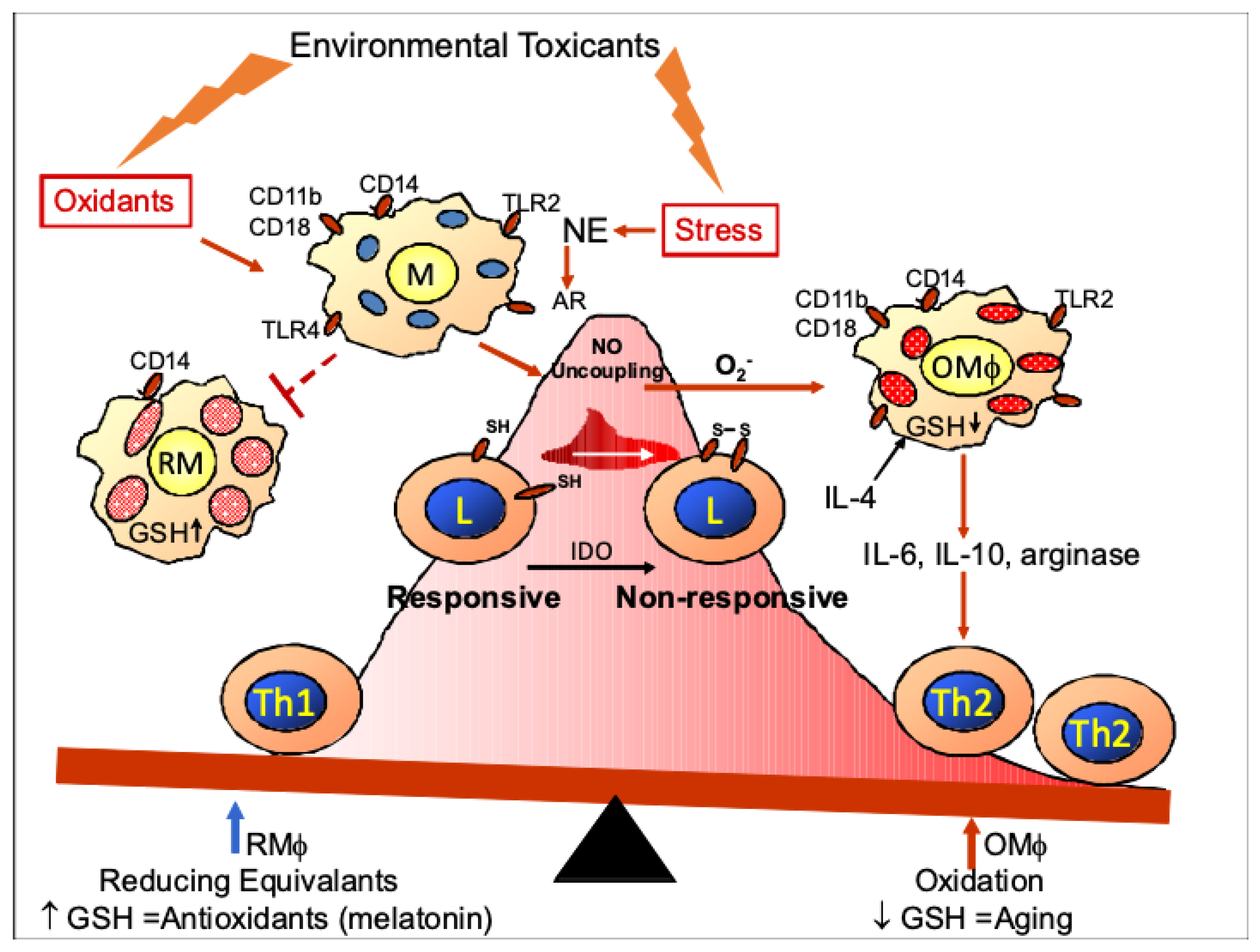
Figure 2.
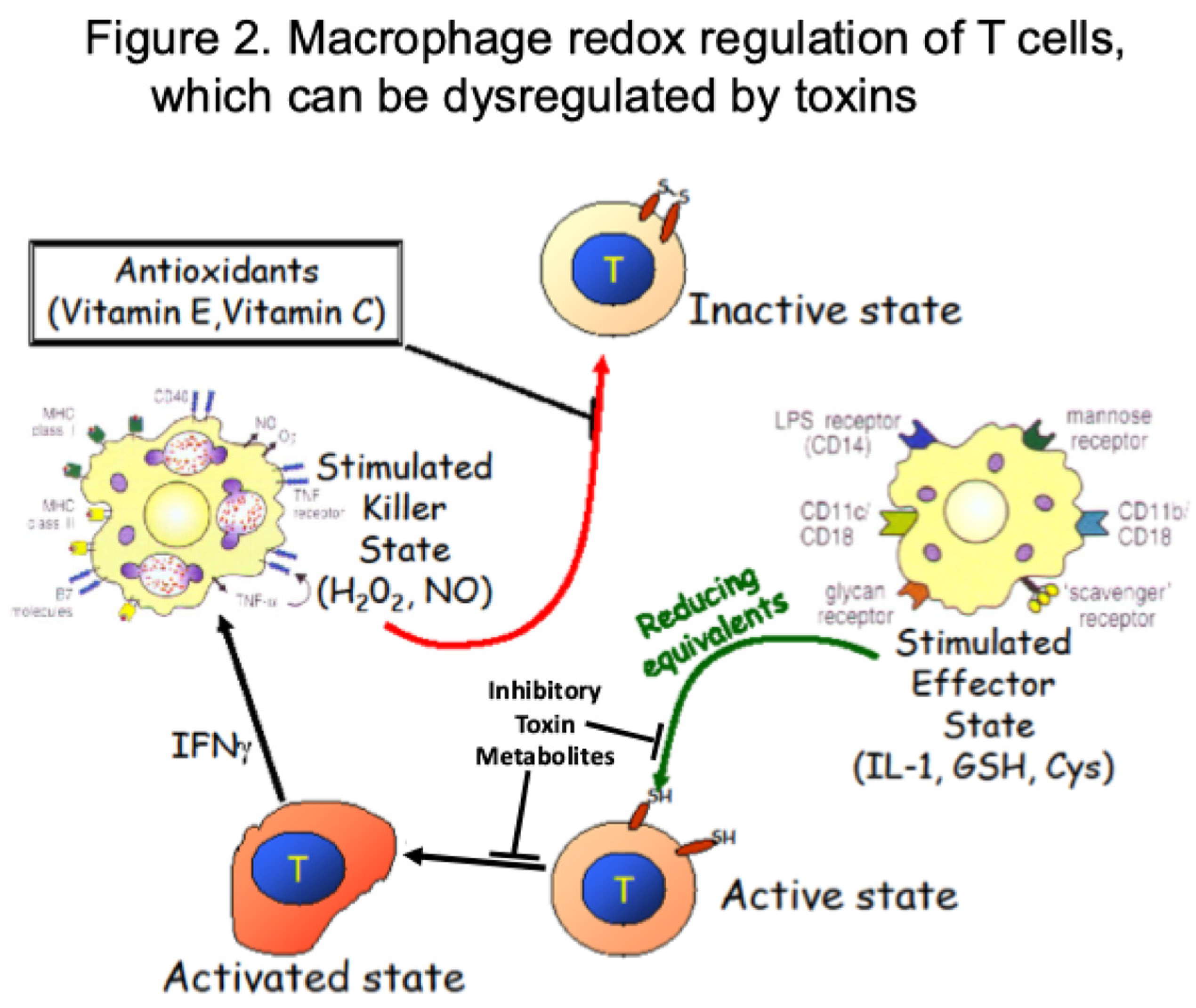

Table 1.
Thiol/Lipid Characteristics of Human Lymphoid Subsets.
| Parameter Assessed | Relative Lymphoid Ranking |
|---|---|
| Radiosensitivity | CD19 > CD8 > CD4 |
| Sensitivity to sulfhydryl-reactive compounds, e.g., N-ethylmaleimide |
CD19 > CD8>CD4 |
| Sensitivity to oxidants, e.g., H2O2 | B cells > T cells |
| GSH content | CD8 = CD4 > CD19 |
| Exofacial thiols | CD19 > CD8 > CD4 |
| Membrane fluidity | B cells > T cells |
| Ratio unsaturated: saturated lipids | B cells > T cells |
| Lipid packing (M540 insertion) | CD19 > CD8 > CD4 |
| Associated methods and correlations between lipid composition, cellular thiols, and cellular sensitivities [87]. | |
Disclaimer/Publisher’s Note: The statements, opinions and data contained in all publications are solely those of the individual author(s) and contributor(s) and not of MDPI and/or the editor(s). MDPI and/or the editor(s) disclaim responsibility for any injury to people or property resulting from any ideas, methods, instructions or products referred to in the content. |
© 2026 by the authors. Licensee MDPI, Basel, Switzerland. This article is an open access article distributed under the terms and conditions of the Creative Commons Attribution (CC BY) license (http://creativecommons.org/licenses/by/4.0/).
Copyright: This open access article is published under a Creative Commons CC BY 4.0 license, which permit the free download, distribution, and reuse, provided that the author and preprint are cited in any reuse.


