

Submitted:
03 July 2026
Posted:
06 July 2026
You are already at the latest version
Abstract
In this paper, we present our discovery of physicochemical purine-pyrimidine symmetries in DNA genomes and the Supersymmetry genetic code (SSyGC) table but now extend both systems to symmetries of the hydrogen-bonds of bases and the free energy symmetry of trinucleotides/codons which are important for the energy stability of all species. For the first time, we show mirror symmetry in some quadruplets of single-strand RNA coronaviruses and DNA viruses such as acellular infectious agents and obligate intracellular parasites. However, complete quadruplet symmetries are present in all DNA species from protist and prokaryote Cyanobacteria (Blue-green Algae) and eukaryotes. We discovered that DNA quadruplet symmetries and free energy symmetries present a fundamental difference between free-living organisms which have autonomous replication, and non-free-living organisms which are parasites and need a host for replication: only free-living organisms have DNA and free energy symmetries while non-free-living organisms do not. On the other hand, the SSyGC table, with its physicochemical purine-pyrimidine symmetries, is common for all RNA and DNA species as non-free-living and free-living organisms, according to their important symmetry role in the translation process without misreading. After the discovery of the same physicochemical DNA and genetic code symmetries, and the Natural law of DNA creation and conservation as dominant concepts in biological systems responsible for the origin of life and evolution, it is necessary to extend theory of evolution to three balanced, basic core principles of evolution - symmetries, mutations and natural selection.
Keywords:
DNA quadruplet symmetry
; trinucleotide/codon quadruplet classification
; genetic code symmetries
; proteinogenesis translation process
; RNA coronaviruses and DNA viruses
; cyanobacteria
; eukaryotes
1. Introduction
In Darwin’s book written in 1859: “On the origin of species by means of natural selection” he noticed that some differences, such as the shape, size and colours of plants or animals, may have been inherited from their ancestors or caused by mutations. During his time, when genetics and their fundamental biological systems, such as the genome, DNA molecule and genetic code were undiscovered, Darwin’s theory regarding the evolution of living species was based on two principles: mutations and natural selection. After 1930, evolutionary biology completed Darwin’s work with genetics and emerged as Darwinism.
The principle of genetic inheritance was discovered by Gregor Mendel in 1866, by conducting hybridisation experiments with pea plants [Mendel, 1896], but at the time, were not broadly known or seen as generally applicable. In physics, the universe is observed and analysed as a material system in perpetual motion and governed exclusively by natural laws. The idea that natural laws are also associated with symmetry was presented and explained mathematically by Emmy Nöther who, in 1918, postulated her famous theorem by correlating it to the law of energy conservation [Nöther, 1918]. At the same time, Einstein’s and Schrödinger’s great advancement was to place symmetries as a dominant concept in the fundamental laws of physics and a primary feature of nature [Schrödinger, 1944]. Gross expressed the idea that the symmetry principle is present as a feature of natural laws [Gross, 1996]. Jacques Monod put great significance on symmetry in biology [Monod, 1978].
The symmetry concept has also been used to propose models for genomics. In 1951, when the structure of DNA molecule was yet unknown, Chargaffʼs first parity rule on nucleotide pairing with the equality of frequencies of nucleotides A and T, as well as C and G in the whole DNA molecule was postulated [Chargaff, 1951]. In 1953, DNA structure was fully explained by Watson and Crick. In 1968, Chargaffʼs unexpected second parity rule (synonymous with strand symmetry) showed a similarity of frequencies of nucleotides A and T, as well C and G in the same DNA strand. This rule was extended to the similarity of frequencies of oligonucleotides as direct to their respective reverse complement within one DNA strand in long enough segments (˃100kb for trinucleotides). The most accepted hypothesis is the inversion and inverted transposition which could be major contributing factors and sufficient time to reach their equilibrium [Albrecht-Buehler, 2007].
To discover common symmetries in DNA molecules in all living species, in 2016 we postulated quadruplet DNA symmetry which includes both strands of DNA with dominant mirror symmetry and Watson-Crick pairing between them. The same symmetry is present in the free energy of trinucleotides/codons [Rosandić and Paar, 2023B]. We also discovered the concept of the Natural law of DNA creation and conservation [Rosandić et al., 2016, 2022]. The Natural law is activated according to which the same mono- or oligonucleotide insertion must be inserted simultaneously into both strands of DNA. Consequently, only mutations on the principle of the Natural law of DNA creation and conservation could have been incorporated into the genome during evolution without violating symmetries in the creation of new species.
In 1961, M.W. Nirenberg and J.H. Matthaei published their discovery in which codons code natural amino acids [Nirenberg and Matthaei, 1961]. F.H. Crick intensively traced how to find genetic code symmetries, and, in 1968, he published the Universal Genetic Code table [Crick, 1968]. After the discovery of more than thirty alternative genetic codes, its name changed to the Standard Genetic Code. It was constructed on a horizontal and vertical alphabetical U-C-A-G array of bases with a complete inability to show the physicochemical symmetry between codons. In 2009, Koonin and Novozhilov summarised the relationship between symmetry in the genetic code and evolution: “Why is the genetic code the way it is and how did it come to be, that was asked over fifty years ago at the dawn of molecular biology and might remain pertinent even in another fifty years” [Koonin and Novozhilov, 2009]. The problem was extremely difficult because there are about 1084 possible codon combinations of the genetic code [Freeland and Hurst, 1998].
Earlier, in 2022 but 60 years after Nirenberg’s discovery of the genetic code, we developed the Supersymmetry Genetic Code (SSyGC) model with multifaceted physicochemical symmetries between bases and codons and with a purine-pyrimidine symmetry net as a central position and core of the genetic code. The SSyGC table is common to all RNA and DNA species, as well as to all alternative codes which differ from the Standard Genetic Code [Rosandić and Paar, 2022]. We also discovered that the symmetries in the SSyGC table play a fundamental role in recognising and differentiating codons from mRNA and the anticodon tRNA without misreading in the translation processes of proteinogenesis. These symmetries also support the wobble hypothesis with non-Watson-Crick base-pairing from mRNA to tRNA within the translation process [Rosandić and Paar, 2024].
In the historical overview of this important discovery from Darwin’s evolutionary theory until the present, the goal of this study was to answer the following questions:
- Have the physicochemical symmetries evolved during evolution from RNA to DNA species?
- Are there identical symmetries in the DNA molecule and the genetic code?
- Has the genetic code evolved over time?
- How have mutations and symmetries been simultaneously connected in the DNA genome during evolution?
- Is Darwin’s theory of evolution contradictory to genome physicochemical symmetries?
2. Materials and Methods
Trinucleotide frequency analysis was performed using a custom-designed computational tool implemented in C#. The algorithm applies a sliding window technique to traverse DNA sequences, systematically quantifying the occurrence of each trinucleotide while excluding those containing ambiguous nucleotides (i.e., ‘N’ bases). The analytical procedure is based on the CLT - Find method, which is publicly available at: http://genom.hazu.hr/tools.html. The DNA sequence datasets used in this analysis are presented in Supplementary Material, Table 1 and Table 2 and 3.
The species analysed in this manuscript (S1): Subgroup I are single-strand RNA coronaviruses; subgroup II are DNA viruses; subgroup III is DNA circovirida; subgroup IV are bacteria; subgroup V are eukaryotes.
In 2016, we discovered DNA quadruplets with basic mirror symmetry between both strands of the DNA molecule. [Rosandić et al., 2016, 2022]. Based on the conducted analysis, we constructed novel 3D diagrams with quadruplets according to our classification for 10 A+T rich and 10 C+G rich trinucleotides/codons [Rosandić et al., 2013; Rosandić and Paar, 2023B] with frequencies of different DNA species. The combined DNA quadruplet frequencies are nearly identical for trinucleotides within each quadruplet for bacteria and eukaryotes (Supplementary Table S2 and S3).
Figure 1.
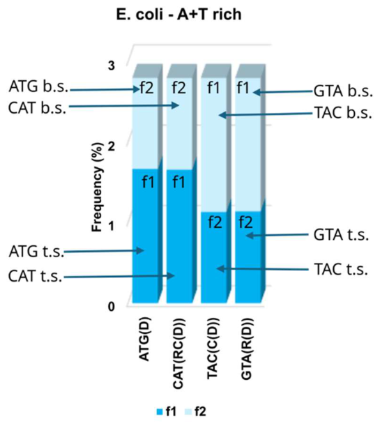
Matrix from enlarged 3D diagram for the presentation of A+T rich quadruplet symmetry from E. coli. Frequencies f1 and f2 and combined two-strand frequencies f1 + f2 are displayed for a quadruplet generated by ATG: ATG direct (D), CAT reverse complement (RC(D)), GTA reverse (R(D)), TAC complement (C(D)); t.s. top strand, b.s. bottom strand. Summary values of four trinucleotides from both DNA strands of quadruplet with quadruplet symmetry reflected in the plateau on the upper edge of the symmetrical quadruplet: (fD = fRC = fC = fR).
Figure 1.
Matrix from enlarged 3D diagram for the presentation of A+T rich quadruplet symmetry from E. coli. Frequencies f1 and f2 and combined two-strand frequencies f1 + f2 are displayed for a quadruplet generated by ATG: ATG direct (D), CAT reverse complement (RC(D)), GTA reverse (R(D)), TAC complement (C(D)); t.s. top strand, b.s. bottom strand. Summary values of four trinucleotides from both DNA strands of quadruplet with quadruplet symmetry reflected in the plateau on the upper edge of the symmetrical quadruplet: (fD = fRC = fC = fR).
3. Results
3.1. Relationship Between the DNA Quadruplet, Energy Symmetry and H-Bonds
Analysing symmetries in the DNA molecule, we show for the first time a relationship between purine-pyrimidine trinucleotide quadruplets and energy structures such as the hydrogen bonds of bases and free energy of trinucleotides/codons (Figure 2b). Namely, two chains of DNA are in an antiparallel direction 5ʼ3ʼ↔3ʼ5ʼ, and bases are connected between two chains by hydrogen bonds: A and T (weak bases) with two hydrogen bonds, and C and G (strong bases) with three hydrogen bonds, creating Watson-Crick pairing between purines and pyrimidines. Hydrogen bonds ensure that a very long DNA molecule cannot break (e. g. a chimpanzee’s 1st chromosome has 231299448 bases). Chargaffʼs second parity rule, also called strand symmetry, showed a marked similarity of frequencies of A↔T, and C↔G nucleotides within only one strand of DNA. This rule was extended to the similarity of frequencies of oligonucleotides such as trinucleotides to those of their respective reverse complements within one DNA strand in long enough segments (˃ 100kb for trinucleotides) [Chargaff, 1951] (Figure 2a).
Natural systems strive to be in a state of minimal energy. Breslauer et al. 1986 and Klump et al. 2020 measured the value of free energy of each codon and concluded that it is equal to the free energy of its reverse complement. We show that all 10 A+T rich and 10 C+G rich quadruplets from our classification of trinucleotides/codons, as well as the SSyGC table, are completely symmetrically synchronised with free energy mapping [Rosandić and Paar, 2023A] (Figure 2b).
3.2. The Natural Symmetry Law of DNA Creation and Conservation Is Occam’s Razor as to How DNA Symmetries Originated
Symmetry plays an important role with respect to the law of nature. Indeed, it is hard to imagine that much progress could be made in deducing the laws of nature without the existence of certain symmetries [Gross, 1996].
In 1968, Chargaffʼs second parity rule (strand symmetry) showed a similarity of frequencies in nucleotides A and T, as well as of C and G in the same DNA strand. This rule was extended to the similarity of frequencies of oligonucleotides as direct to their respective reverse complement within one DNA strand in long enough segments (˃100kb for trinucleotides). The most accepted hypotheses to explain this rule were made Albrecht-Buehler 2007: “In a genome duplex that exceeds 100 kb the frequency distributions of their trinucleotides (triplet profiles) are the same in both strands. This remarkable symmetry, sometimes called Chargaffʼs second parity rule, is not the result of base pairing, but can be explained as the result of countless inversions and inverted transpositions that occurred throughout evolution. Only a relatively small number of some 10,000 inversions/transpositions were required to render the strands of their genome duplexes symmetrical and to create a majority profile”. There is not a single case discovered of the transitional symmetrical form of DNA species without Chargaffʼs second parity rule. Evolution also would be much slower.
Unexpectedly, trinucleotide’s quadruplet symmetries and the concept of the Natural symmetry law of DNA creation ensue Chargaff’s first and second parity rule (Figure 2b) [Rosandić et al. 2016, 2022]. According to natural law, all mono/oligonucleotides which enter one strand (top strand) of the DNA must enter the second strand (bottom strand) regardless of their location. Binding with a complementary pair, the quadruplet structures with mirror symmetry between both strands automatically created quartic symmetry in a bidirectional 5′3′↔3′5′ manner. It is not important in which location of both DNA strands the mono/oligonucleotides enter and in which combination of nucleotides (Figure 2b, d). It is extremely important that in each DNA strand the same number of similar mono/oligonucleotides enter. The result is a new sequence with quadruplet symmetries. Each nucleotide or oligonucleotides enter in both DNA strands as a quartic structure with superior mirror symmetry between them as in the SSyGC table. Due to mirror symmetry, direct transformation from the SSyGC table into DNA sequence is possible. Simultaneously, the Natural symmetry law does not disrupt Watson-Crick pairing of the DNA sequence. However, Figure 2c clearly show that only Watson-Crick pairing between bases cannot create quadruplet nor strand symmetry sequence. Only quadruplets with the Natural law of DNA creation and conservation do not break symmetries during evolution.
The Natural law of DNA creation and conservation can accelerate evolution without countless inversions/inverted transpositions. Namely, we show that the free energy value and hydrogen bonds (H-bonds) follow quadruplet symmetries and together decrease disorder (entropy) in the DNA molecule.
3.3. Have Symmetries Evolved from RNA to DNA Species During Evolution?
We investigated two novel symmetries: hydrogen-bond symmetries in weak (A, T) and strong (C, T) bases, and free energy symmetry in DNA trinucleotides and the genetic codons (Figure 2b and Figure 6), [Rosandić and Paar, 2023A].
3.3.1. Symmetries in Single-Strand RNA Coronaviruses
Viruses are acellular infectious agents and obligate intracellular parasites. They cannot replicate without a host as they lack the structures of cell such as cytoplasm and membrane with an independent metabolism, ribosomes etc. Viruses cannot create their own energy - outside a host cell they are inactive particles. However, they possess RNA and DNA genetic materials, can mutate and evolve to adapt to their environments. The newly expanded taxonomic classification system certainly supports the hypothesis that viruses are both obligate intracellular parasites, as well as living entities that are sustained by complementary matching and conformational recognition with respect to both their intrinsic processes as well as those involved in their survival and interactions with their targeted hosts (Stefano and Kream, 2022).
We show that viruses are very much alive due to acquired genetic advantages such as purine-pyrimidine incomplete mirror symmetries (to group bases according symmetries) of their genomes that permit them to overtake some cell processes (Figure 3 and Figure 4).
The single-strand RNA species make up 70% of all viruses, including coronaviruses. Their single-strand genomes have quadruplets, but not quadruplet symmetries, because their genomes do not have a second (bottom) strand and are shorter than 100 kb to make quadruplet symmetries. In our previous article [Rosandić et al., 2022] we analysed all chromosomes of the human genome (non-coding DNA, coding DNA, random sequence of 200000 bp), as well as the entire thermodynamic system with our Supersymmetry genetic code, which is common for all RNA and DNA species and has remained unchanged during evolution. The logarithmic relationship between the oligonucleotide order and minimal DNA sequence length, to establish the validity of strand symmetry (Chargaffʼs second parity rule), automatically follows from the DNA quadruplet structure of the genomic sequence >100 kb. We show that the structure and stability of DNA are influenced by Watson-Crick pairing and the Natural law of DNA creation and conservation, according to which the same mono-oligonucleotide insertion must be inserted simultaneously into both strands of DNA during evolution (Figure 2c). In this case, quadruplet symmetries of each species from bacteria to eukaryotes are conserved.
After our statistical analysis, we concluded that strand symmetry is not created for trinucleotide sequences shorter than approximately 100 kb. By further decreasing the sequence length to about 50 kb, the frequency identity of symmetries completely disappears. We discovered that:
- -
- Single strand RNA viruses do have not Watson-Crick A↔T and C↔G nucleotide pairing (Figure 3).
- -
- They cannot create 10 A+T rich and 10 C+G rich quadruplet symmetries because they have only one strand and a complete genome <100 bp.
- -
- However, they have quadruplets in form direct (D) – reverse complement (RC) – complement (C) – reverse (R) (Figure 3).
- -
- Double strand DNA viruses also do have not the quadruplet symmetries as their genomes are shorter than 100 kb (Figure 4).
- -
- All DNA genomes of bacteria and eukaryotes have much more than 100 kb and can create 10 A+T rich and 10 C+G rich quadruplet symmetries according to our classification of trinucleotides/codons with fD = fRC = fC = fR (Figure 5).
- -
- DNA quadruplet symmetries show that the structure and stability of DNA are influenced by Watson-Crick pairing and the Natural law of DNA creation and conservation, according to which the same mono-oligonucleotide insertion must be inserted simultaneously into both strands of DNA (Figure 2c).
- -
- We also showed that about 98% of non-coding human DNA have quadruplet symmetries, and less than 2% of coding DNA have not [Rosandić et al., 2022].
Figure 5.
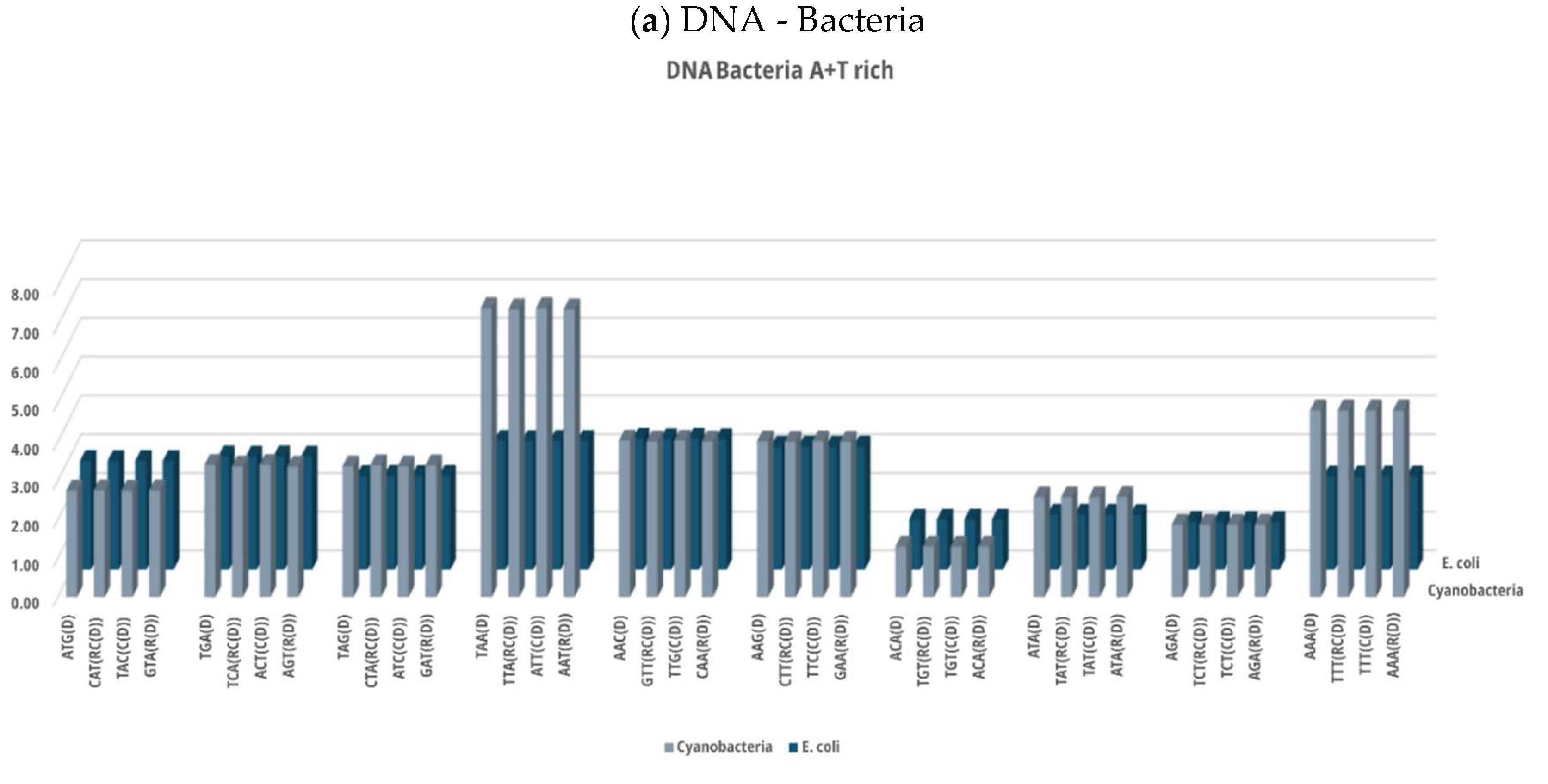
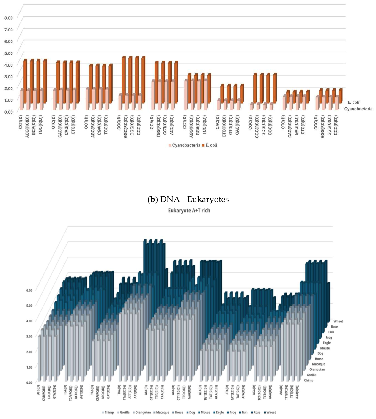
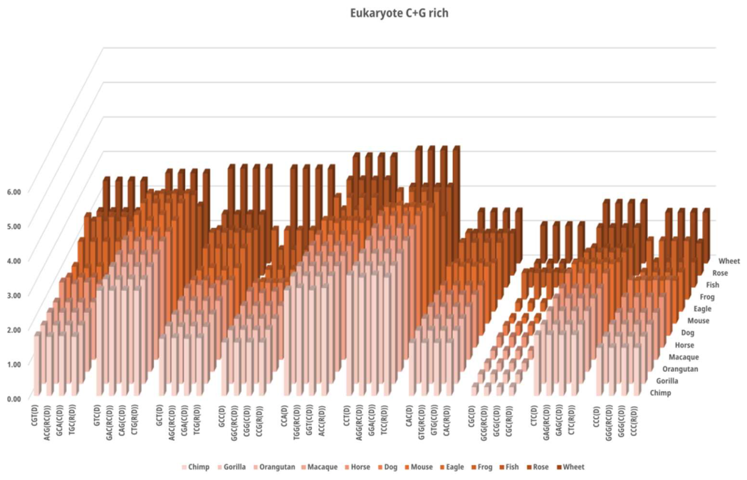
Examples of 10 A+T rich and 10 C+G rich quadruplet symmetry matrices between the DNA genome of prokaryotes (Cyanobacteria, Escherichia coli) and eukaryotes (wheat, rose, fish, frog, eagle, mouse, dog, horse, macaque, orangutan, gorilla and chimpanzee. a) The protists Cyanobacteria with the ability to replicate evolved around 2.7 billion years ago and their metabolism would have been anaerobic (see the text), compared to the well-known E. coli. Their A+T rich and C+G rich quadruplets are completely symmetrical. b) All species in the group of eukaryotes have perfect symmetries in both A+T rich and C+G rich quadruplets. Summary values of four trinucleotides from both DNA strands of each quadruplet reflected in the plateau on the upper edge of the symmetrical quadruplet (fD = fRC = fC = fR). The numerical values can be seen in Supplementary Table S3. The numerical values for the same bacteria in Supplementary Material in Preprint are correct. Bacteria and eukaryotes have trinucleotide quadruplet symmetries with Watson-Crick pairing and a very precise symmetrical structure between purine-pyrimidine symmetry, hydrogen-bonds symmetry and free energy symmetry.
Figure 5.
Examples of 10 A+T rich and 10 C+G rich quadruplet symmetry matrices between the DNA genome of prokaryotes (Cyanobacteria, Escherichia coli) and eukaryotes (wheat, rose, fish, frog, eagle, mouse, dog, horse, macaque, orangutan, gorilla and chimpanzee. a) The protists Cyanobacteria with the ability to replicate evolved around 2.7 billion years ago and their metabolism would have been anaerobic (see the text), compared to the well-known E. coli. Their A+T rich and C+G rich quadruplets are completely symmetrical. b) All species in the group of eukaryotes have perfect symmetries in both A+T rich and C+G rich quadruplets. Summary values of four trinucleotides from both DNA strands of each quadruplet reflected in the plateau on the upper edge of the symmetrical quadruplet (fD = fRC = fC = fR). The numerical values can be seen in Supplementary Table S3. The numerical values for the same bacteria in Supplementary Material in Preprint are correct. Bacteria and eukaryotes have trinucleotide quadruplet symmetries with Watson-Crick pairing and a very precise symmetrical structure between purine-pyrimidine symmetry, hydrogen-bonds symmetry and free energy symmetry.
To discover some symmetry in the group of 56 single-strand coronaviruses, we analysed the relative frequencies of 10 A+T rich and 10 C+G rich quadruplets according to our trinucleotide/codon quadruplet’s classification [Rosandić and Paar, 2023B] (Figure 3A, B). For the first time ever, we analysed hydrogen bonds (H-bonds) in relation to weak (A, T) and strong (C, G) bases, although RNA single-strand viruses have only one strand (Figure 2b).
Unexpectedly, our results show that even single-strand viruses have some group according mirror symmetry of energy mapping between the hydrogen bonds of bases, as well as the free energy of trinucleotides.
3.3.2. The Double Stranded DNA Viruses and DNA Circoviridae
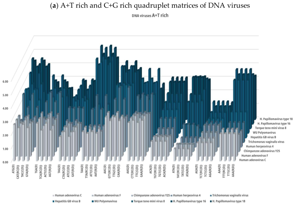
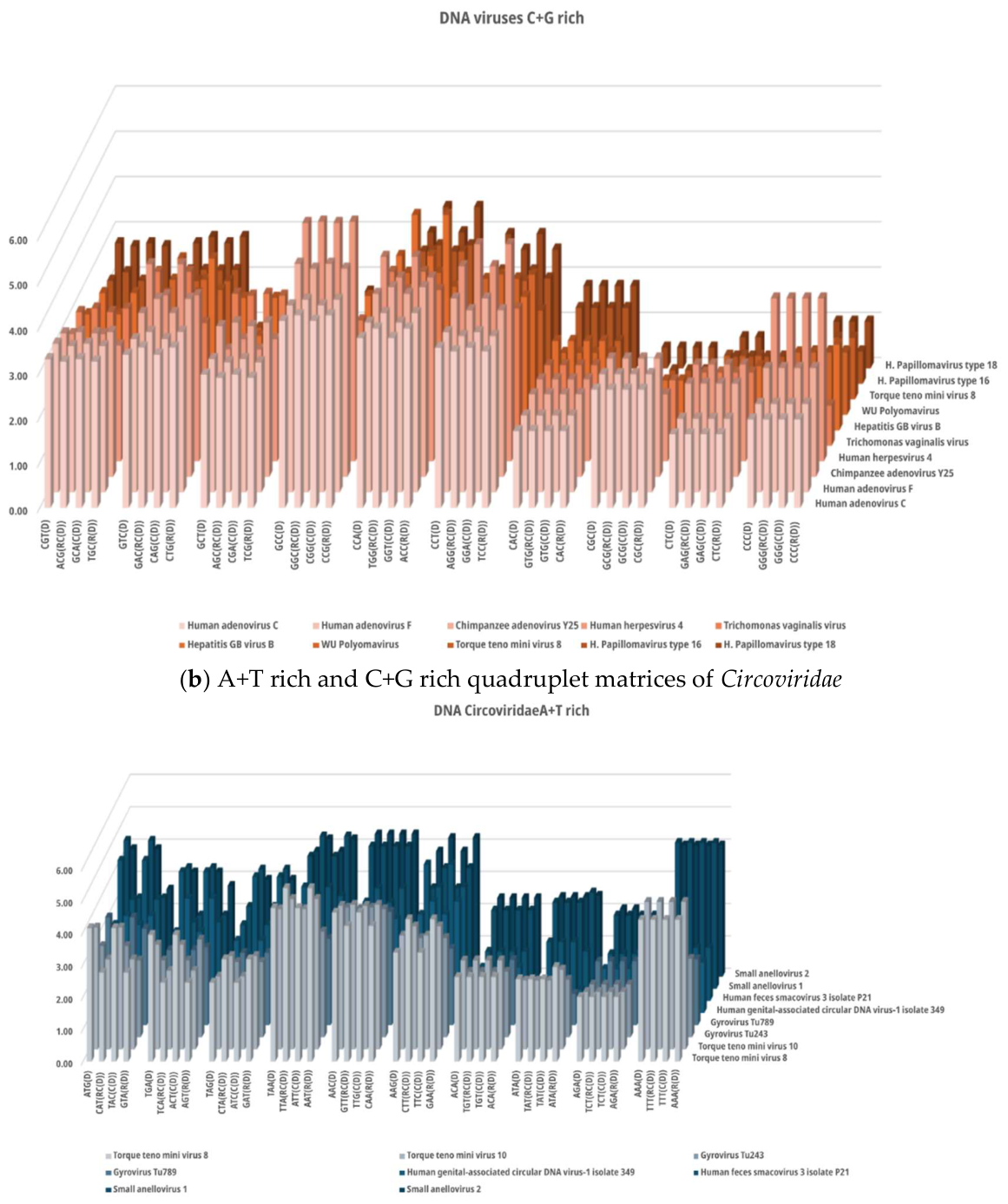
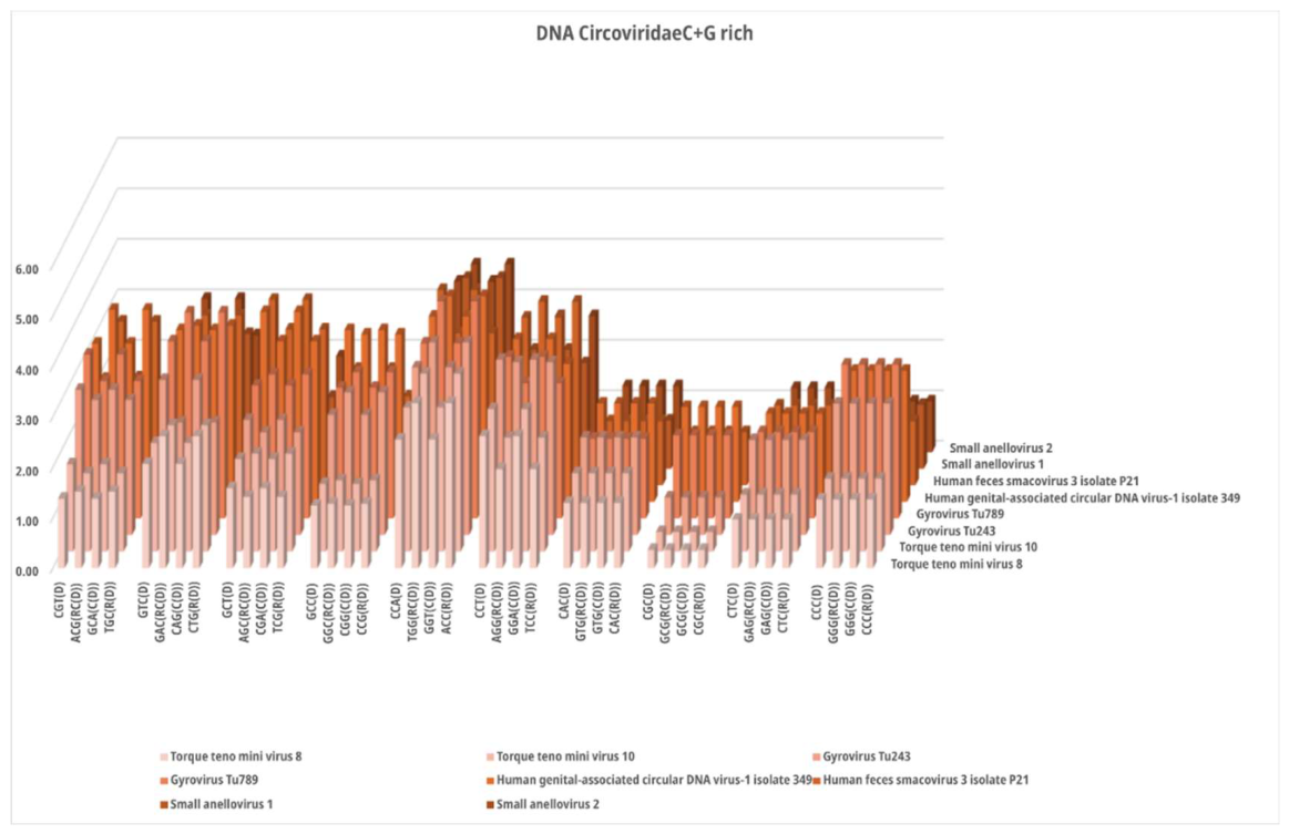
The double stranded DNA viruses and DNA Circoviridae do not have complete quadruplet symmetries because their genomes are too short (˂100kb). However, all DNA viruses have Watson-Crick pairing of A↔T and C↔G bases between both strands and have quadruplets as seen in A+T rich and C+G rich quadruplet matrices with quadruplet boxes (QboxD-RC) and (QboxC-R) (Figure 4). Because of Watson-Crick pairing, they have equal relative frequencies (f) between both strands of direct (D) and complement (C), as well as reverse complement (RC) and reverse (R) trinucleotides. Certainly, they do not have equal relative frequencies in one strand between direct (D) and reverse complement (RC) as Chargaffʼs second parity rule (strand symmetry), except symmetrical trinucleotides (Figure 4).
3.3.3. Symmetry in Prokaryotes Bacteria and Eukaryotes
Bacteria with all four trinucleotides in each quadruplet structure have the same sum of relative frequencies in each quadruplet box [(QboxD-RC) and (QboxC-R)], and the same sum in both strands of the complete quadruplet (f direct = f reverse complement = f complement = f reverse) (Rosandić et al., 2016, 2022, Rosandić and Paar, 2023B). Each quadruplet includes both bidirectional 5ʼ3ʼ↔3ʼ5ʼstrands of the DNA molecule with Watson-Crick pairing and the mirror symmetry between strands which automatically leads to Chargaffʼs first and Chargaffʼs second parity rule (Figure 2) and complete DNA quadruplet symmetry is created. The result is 10 A+T rich and 10 C+G rich symmetrical quadruplet matrices in all DNA species, such as bacteria and eukaryotes (Figure 5). At the same time, quadruplet symmetries in bacteria and eukaryotes have purine-pyrimidine mirror symmetry between bases and trinucleotides in each strand as well as between both strands of Q-boxes. They also have the mirror symmetry between hydrogen-bonds and free energy symmetry (Figure 2b).
After statistical analysis, we concluded:
- -
- Single strand RNA viruses do have not Watson-Crick A↔T and C↔G nucleotide pairing (Figure 3).
- -
- They cannot create 10 A+T rich and 10 C+G rich quadruplet symmetries because they have only one strand and a complete genome <100 bp.
- -
- However, they have quadruplets in form direct (D) – reverse complement (RC) – complement (C) – reverse (R) (Figure 3).
- -
- Double strand DNA viruses also do have not the quadruplet symmetries because their genomes are shorter than 100 kb (Figure 4).
- -
- All DNA genomes of bacteria and eukaryotes have much more than 100 kb and can create 10 A+T rich and 10 C+G rich quadruplet symmetries according to our classification of trinucleotides/codons with fD = fRC = fC = fR (Figure 5).
- -
- The DNA quadruplet symmetries show that the structure and stability of DNA are influenced by Watson-Crick pairing and the Natural law of DNA creation and conservation, according to which the same mono-oligonucleotide insertion must be inserted simultaneously into both strands of DNA (Figure 2c).
- -
- We also showed that 98% of non-coding human chromosomes have DNA quadruplet symmetries as well as strand symmetry (Chargaffʼs second parity rule). However, 2% of coding human chromosomes do not have quadruplet and strand symmetries [Rosandić et al., 2022].
In conclusion, we can say that fundamental purine-pyrimidine symmetry development originated initially from RNA single-strand viruses and DNA viruses up to complete symmetry of DNA protist bacteria, present bacteria and all eukaryotes. This evolution followed hydrogen-bond symmetry and free energy symmetry (Table 2).
3.4. Has the Genetic Code Evolved over Time?
The genetic code, with its 61 codons and 3 stop signals, is the code for 20 natural amino acids and is responsible for the protein synthesis in the process of the origin of life. The structure of our SSyGC table is strictly defined on the principle of physicochemical laws and with mathematically determined symmetries between bases, codons and amino acids. In this way, each codon as a basic unit is strictly and iqually positioned within the genetic code, and it is not possible to replace it with another codon without breaking the symmetries of the genetic code (Figure 6) [Rosandić and Paar 2022, 2024].
Figure 6.
Relationship between the SSyGC table, alternative genetic code and H-bonds symmetry between purines and pyrimidines symmetry. The SSyGC table incorporates 2 × 8 boxes with four codons in each box and starts with an AUG initiation start signal. Only the SSyGC table has in continuity the codons of three amino acids with six codons each: serine, arginine and leucine. The SSyGC table has the same distribution of the purine/pyrimidine profile in both columns, and simultaneously the same profile distribution pairs of codon rows within each box. In the left column, there are weak second bases (A and U) in all codons. In the right column, there are in all codons strong second bases (C and G). With a horizontal and vertical central symmetry axis (arrow), it creates the purine–pyrimidine symmetry net which is “the golden rule” for all RNA and DNA living species. Between both columns in the same row, there are alternate transformations of A+U-rich and C+G-rich codons. There is a translational symmetry between weak, strong and mix boxes of codons with respect to horizontal symmetry axis (see colors). For the first time, we show the hydrogen bonds of weak bases A and T with 2 H-bonds, and strong bases C and G with 3 H-bonds, which are identical according to direct and complement Watson-Crick pair of boxes. Sum of free energy of all codons according to horizontal mirror symmetry axis of the SSyGC is almost identical above (127.5 kcal/mol) and below (129.5 kcal/mol) the horizontal mirror symmetry axis [Rosandić and Paar, 2023A]. 0 pu, purine (A,G); 1 py, pyrimidine (U, C); dark yellow, two pairs of split boxes with direct–complement Watson-Crick symmetry between codons; dark blue, two pairs of non-split boxes with direct–complement Watson-Crick symmetry between codons; light yellow, two pairs of split boxes with purine ↔ purine, pyrimidine ↔ pyrimidine transformation between codons; light blue, two pairs of non-split boxes with purine ↔ purine, pyrimidine ↔ pyrimidine transformation between codons. The mitochondrial trematode (liver-fluke) code (red codons of its amino accids) incorporated in the Supersymmetry Genetic Code table: Methionine (Met) expands to the neighbouring isoleucine (Ile) codon AUA; tryptophan (Trp) expands to the neighbouring stop UGA codon; arginine (Arg) neighbouring AGA and AGG codons become the 7th and 8th codons for serine (Ser); asparagine (Asn) expands to the neighbouring AAA codon from lysine (Lys). In alternative genetic codes, individual amino acids usually capture a codon from the SSyGC table neighbouring amino acid. But the purine–pyrimidine symmetry net always remains unchanged, no codons change their position into the SSyGC table, only some codons change their amino acid. All more than thirty nuclear or mitochondrial genetic codes different from the Standard Genetic Code have also been incorporated into the SSyGC table without interrupting its symmetries.
Figure 6.
Relationship between the SSyGC table, alternative genetic code and H-bonds symmetry between purines and pyrimidines symmetry. The SSyGC table incorporates 2 × 8 boxes with four codons in each box and starts with an AUG initiation start signal. Only the SSyGC table has in continuity the codons of three amino acids with six codons each: serine, arginine and leucine. The SSyGC table has the same distribution of the purine/pyrimidine profile in both columns, and simultaneously the same profile distribution pairs of codon rows within each box. In the left column, there are weak second bases (A and U) in all codons. In the right column, there are in all codons strong second bases (C and G). With a horizontal and vertical central symmetry axis (arrow), it creates the purine–pyrimidine symmetry net which is “the golden rule” for all RNA and DNA living species. Between both columns in the same row, there are alternate transformations of A+U-rich and C+G-rich codons. There is a translational symmetry between weak, strong and mix boxes of codons with respect to horizontal symmetry axis (see colors). For the first time, we show the hydrogen bonds of weak bases A and T with 2 H-bonds, and strong bases C and G with 3 H-bonds, which are identical according to direct and complement Watson-Crick pair of boxes. Sum of free energy of all codons according to horizontal mirror symmetry axis of the SSyGC is almost identical above (127.5 kcal/mol) and below (129.5 kcal/mol) the horizontal mirror symmetry axis [Rosandić and Paar, 2023A]. 0 pu, purine (A,G); 1 py, pyrimidine (U, C); dark yellow, two pairs of split boxes with direct–complement Watson-Crick symmetry between codons; dark blue, two pairs of non-split boxes with direct–complement Watson-Crick symmetry between codons; light yellow, two pairs of split boxes with purine ↔ purine, pyrimidine ↔ pyrimidine transformation between codons; light blue, two pairs of non-split boxes with purine ↔ purine, pyrimidine ↔ pyrimidine transformation between codons. The mitochondrial trematode (liver-fluke) code (red codons of its amino accids) incorporated in the Supersymmetry Genetic Code table: Methionine (Met) expands to the neighbouring isoleucine (Ile) codon AUA; tryptophan (Trp) expands to the neighbouring stop UGA codon; arginine (Arg) neighbouring AGA and AGG codons become the 7th and 8th codons for serine (Ser); asparagine (Asn) expands to the neighbouring AAA codon from lysine (Lys). In alternative genetic codes, individual amino acids usually capture a codon from the SSyGC table neighbouring amino acid. But the purine–pyrimidine symmetry net always remains unchanged, no codons change their position into the SSyGC table, only some codons change their amino acid. All more than thirty nuclear or mitochondrial genetic codes different from the Standard Genetic Code have also been incorporated into the SSyGC table without interrupting its symmetries.
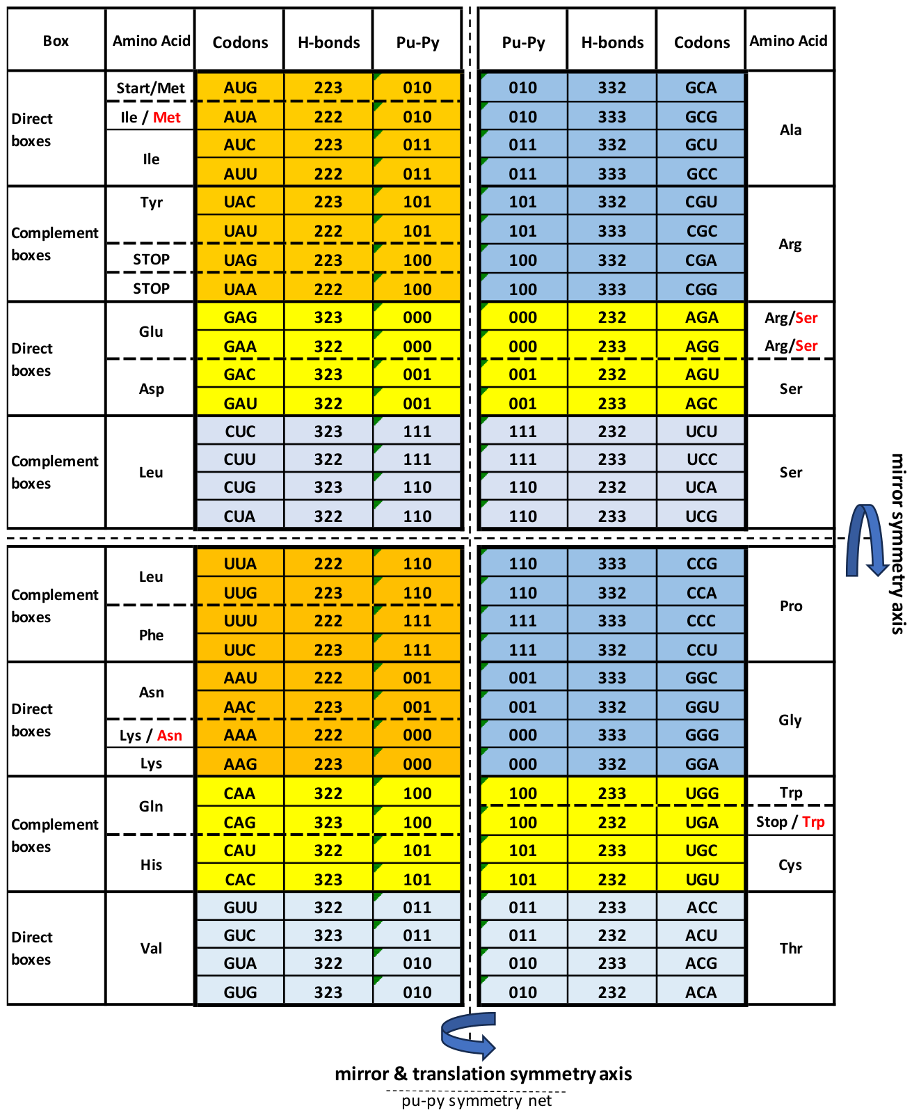
Watson-Crick codon-anticodon purine-pyrimidine pairing with dominant mirror symmetry in the genetic code as the code of life related to hydrogen-bonds mirror symmetry and the energy code is common to all RNA and DNA species and has remained unchanged throughout evolution.
3.4.1. Why Must Codons Have Trinucleotide Configuration?
Mononucleotides as codons satisfy only four combinations (41), dinucleotide only sixteen (42), which is an insufficient number of codons for 20 amino acids and the complete genetic code which is necessary for proteinogenesis in order to satisfy all metabolic processes. Only the trinucleotide form with 61 different codons and 3 stop signals (43) enables this important biological role. But trinucleotide codons have many functions.
The important role of the first and second bases in the codons of the SSyGC table reveals which codons belong to an amino acid with the whole non-split box of four codons, and which belong to amino acids with two codons from each split box [Rosandić and Paar, 2024]. The result is that the translation process circulates without misreading because:
- -
- four non-split boxes have strong bases (GG, CC, GC, CG);
- -
- four split boxes have weak bases (AA, UU, AU, UA);
- -
- mixed boxes have one strong and one weak base, which are not split because the second base is always pyrimidine (AC, UC, GU, CU);
- -
- mixed boxes which are split because the second base is always purine (GA, CA, AG, UG).
In the split box the third base of both codons from one amino acid are purines A and G, and for the other amino acid are pyrimidines U and C. In the Standard Genetic Code table, the third bases in all boxes are identical in the U-C-A-G array and as a result of this were ignored. In the SSyGC table, third bases Watson-Crick pairing G-A-C-U ↔ C-U-G-A / A-G-U-C ↔ U-C-A-G into the vertical pair of boxes was discovered (Figure 6).
There are some differences in the total numbers of codons and tRNAs: for different species, there are on average 40 tRNAs as opposed to the necessary 61 codons and stop signals UAG, UAA and UGA. We show that wobble pairing corrects this disagreement and minimises damage that can be caused by a misreading of the code also according to the special role of the second base of the codon, which detects whether the codon from mRNA belongs an amino acid with two or four codons. Namely, wobble tolerates pairing between 5’ G anticodon ↔ 3’ U codon or 5’ U anticodon ↔ 3’ G codon in amino acids with two codons, and inosine A, C, U as the anticodon first base (5’) in amino acid with four codons [Rosandić and Paar, 2024].
As a result of the bidirectional position of codons 5ʼ3ʼ →← 3ʼ5ʼ anticodons canonical second base with Watson-Crick pairing or wobble pairing permanently stay in the same position (1-2-3 codon ↔ 3-2-1anticodon), preventing misreading in the translation process due to only having one chance to translate information from mRNA to tRNA.
The hypothesis of evolution before the LUCA period, that initial codons have four bases (Tessera) [Gonzales et al., 2019], shows that the central positions of each codon and anticodon would have two bases and because of the bidirectionality codon 5ʼ3ʼ →← 3ʼ5ʼ anticodons, they exchange positions (codon 1-2-3-4 ↔ 4-3-2-1 or 4-2-3-1 anticodon). In this case, there are two random chances to translate information from mRNA. In such a case, translation did not have an unambiguous solution resulting with misreading during proteinogenesis.
3.4.2. The Specific Function of the Codon’s Second Base in the SSyGC Table
Our supersymmetry genetic code as 2x8 boxes table with four codons in each box has two basic symmetries: between bases as well as between codons in the form of Watson-Crick pairing in the vertical direction, and purine-purine and pyrimidine – pyrimidine pairing in the horizontal direction. All other symmetries follow from these two basic symmetries (Figure 6).
By careful analysis, we see the difference between symmetries according to the horizontal and vertical symmetry axis. The horizontal symmetry axis divides the purine-pyrimidine symmetry net into two identical halves with mirror symmetry.
With respect to vertical symmetry axis of the SSyGC table, there are purine-pyrimidine compositions reflected in each pair of horizontal boxes with mirror symmetry in two rows of codons (e.g., 010 ↔ 010), and translation symmetry (e.g., 011 ↔ 011) in other two rows of codons. With respect to all symmetries and Watson-Crick pairing, it is unexpected that the second base in all 32 codons in the left column of the SSyGC is weak (A or U), and in all other 32 codons in the right column is strong (C or G) (Figure 6). This configuration of codon symmetry in the genetic code is important in the translation process without misreading in proteinogenesis detecting whether a codon from mRNA belongs to an amino acid with two or four codons (Rosandić and Paar, 2024). Based on symmetries, the SSyGC table enables the identification of codon localisation as unique and unchangeable during evolution and identical for all RNA and DNA species. The alternative genetic codes have equal purine-pyrimidine symmetries and Watson-Crick pairing as in the SSyGC table, but some codons code a different, often neighbouring amino acid (Figure 6).
The key symmetry differences between non-free-living organisms and free-living organisms.
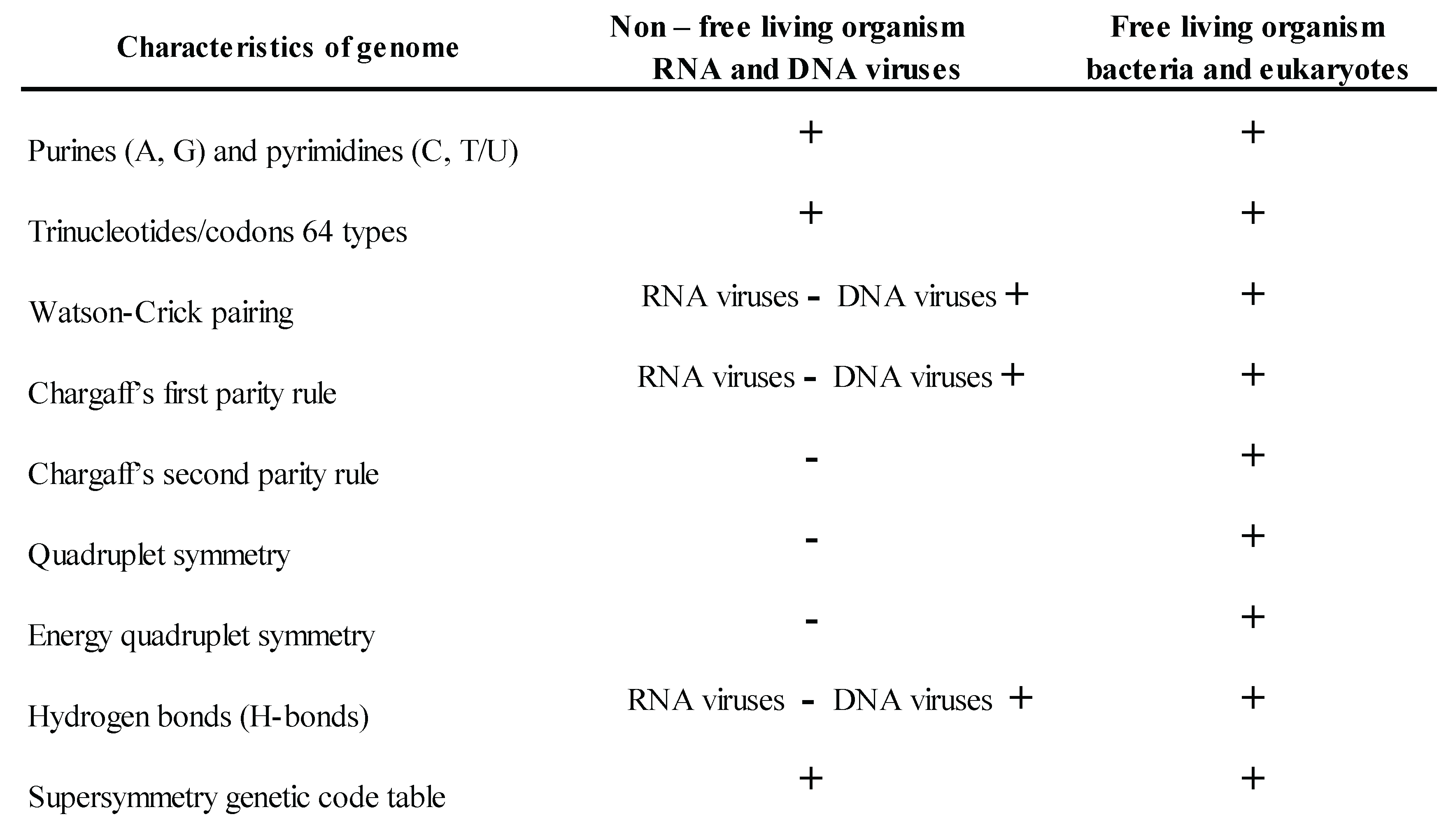
DNA quadruplet symmetries (Chargaffʼs second parity rule is part of quadruplet symmetry) and energy symmetries present a fundamental difference between free-living organisms which have autonomous replication, and non-free-living organisms which are parasites and need a host for replication: free-living organisms have DNA and energy symmetries while non-free-living organisms do not. On the other hand, the supersymmetry genetic code is common for all RNA and DNA species and unchangeable during evolution according to their important symmetry role in the translation process without misreading (Table 1).

Table 1.
Differences in physicochemical purines and pyrimidines symmetries between non-free and free-living organisms.
Table 1.
Differences in physicochemical purines and pyrimidines symmetries between non-free and free-living organisms.
 |
4. Discussion
We are still feeling the devastating effects of the COVID-19 pandemic. The coding virus mutated right before our eyes and as such, reduced the effectiveness of vaccines. Why did it mutate so rapidly? The coronavirus is a single-strand RNA virus, the simplest form of living organisms on Earth. (Figure 3). Viruses cannot replicate themselves rather, they reproduce in co-evolution of virus and DNA hosts as obligate intracellular parasites. Outside the host cell, they are inert and are like living crystals due to the symmetrical capsid which has a crystal protein structure often in form of icosahedral. The virus genome has on average approximately 2-3000 bases. The smallest genome known today, the hepatitis D virus, has only 1700 bases. The coronavirus belongs to large single-strand RNA viruses and has approximately 30000 bases (Table S1). An exception is the giant virus as a gyrus with the largest viral genomes of 1.9 to 2.5 mega-base pair. The human respiratory syncytial virus has a single-stranded non-segmented RNA genome with a length of approximately 2 kb.
RNA viruses have the same SSyGC, which in their structure contains fundamental Watson-Crick base pairing and represents the evolutionary instructions for the creation of highly sophisticated DNA molecules. However, because the coronavirus has a short single-strand genome smaller than 100000 bases (100kb), it cannot create quadruplet symmetry like the two-strands genomes of DNA organisms such as bacteria, which on average have mostly 5000000 bases (Figure 5a, Table S3). We proved that quadruplet symmetry preserves the integrity of DNA molecules larger than 100kb, which means that each trinucleotide must appear in the DNA genome at least 1500 times [Rosandić et al., 2022]. In general, all single-strand RNA viruses, of which there are 70% compared to DNA viruses of which there are 30%, are prone to rapid mutations not only because of the short length of their genome, but also because of their inability to create quadruplet symmetries. However, quadruplet analysis of relative frequencies of their trinucleotides of single-strand RNA coronaviruses showed the beginnings of symmetry between some symmetrical trinucleotides such as ATA↔TAT, CGC↔GCG, or CCC↔GGG, but not, for example AAA↔TTT (Figure 3) .
Double-strand DNA viruses can have over a million bases because they contain genes from their host. They have a significantly lower rate of mutations compared to single-strand viruses since they can create initial symmetries. Namely, viruses cannot independently create complex symmetries in their genome. As such, they carry out the replication necessary for life solely using their host on which they parasitise. One could say that viruses, because of their pure structure and inability to replicate, are “a blind alley” in evolution, without their own ability to evolve into more sophisticated organisms.
4.1. Are There Identical Symmetries in the DNA Molecule and the Genetic Code?
Since the discovery of DNA structure in 1953 [Watson and Crick, 1953] and the Standard genetic code table in 1968 [Crick, 1968], the role of symmetries in the origin of life and evolution has been examined extensively. Ignoring the importance of the second strand of DNA molecule and the third base of codons in the genetic code caused a significant delay in the discovery of their symmetries.
With our discovery of the SSyGC table in 2022 (Figure 6), we show that this code is common to all RNA and DNA species, from RNA viruses to Homo sapiens sapiens. We considered that from LUCA, when codons with a trinucleotide form were formed, the SSyGC table was structured at the same time [Rosandić and Paar, 2022, 2024]. The genetic code table is not an organ as is the DNA molecule an organ in the genome of living species. The genetic code is a table which shows us that codons are mutually and functionally connected with physicochemical symmetries and simultaneously connected with identical symmetries of the DNA molecule. The genetic code which has 61 codons and 3 stop signals is the code for 20 natural amino acids, responsible for the protein synthesis in the process of the origin of life. The structure of the SSyGC table is strictly defined on the principle of physicochemical laws and with mathematically determined symmetries between bases, codons and amino acids. In this way, each codon as a basic unit is strictly and uniquely positioned within the genetic code, and it is not possible to replace it with another codon. According to these symmetries, the direct transformation of the SSyGC table into a DNA molecule is possible [Rosandić et al., 2023B, Rosandić and Paar, 2024].
Our discovery of DNA and genetic code symmetries shows that they attract attention in broader scientific studies [Negadi, 2023, 2024; Henn et al., 2024; Han et al., 2024; Warr and Hatlon, 2025]. We point out that Negadi combining mathematical tools with the chemical composition of amino acids describing the hydrogen atom content showed deeper mathematical principles revealed in our ideal and supersymmetry genetic code. Warr and Hatlon with mathematical analysis of the inverse symmetry (Chargaffʼs second parity rule or strand symmetry) use as an example our existence of a conservation principle in the form of the Natural symmetry law of DNA creation and conservation.
We discovered that, using mathematical and statistical methods, less than 2% of the coding human DNA does not have quadruplet symmetry and Chargaffʼs parity rule, but 98% of non-coding DNA consists of quadruplet symmetries [Rosandić et al., 2022].
4.2. How Are Mutations and Symmetries Simultaneously Connected in the Genome During Evolution?
Symmetries have a fundamental function in the structure of DNA genomes and the genetic code, as well as in the transfer of information from mRNA to tRNA to synthesise the identical protein without misreading in the translation process of proteinogenesis [Rosandić and Paar, 2024; Rosandić 2025]. In 2012, Eugen W. Koonin made the following comment on the origin of life: “The origin of life - or to be more precise, the origin of the first replicator system and the origin of translation – remains a huge enigma, and progress in solving these problems has been very modest – in the case of translation, nearly negligible” [Koonin, 2012]. We hope that after our discovery the role of symmetries in vital living processes such as translation in proteinogenesis [Rosandić and Paar, 2024] and systems such as DNA and the genetic code, we broaden our horizon of knowledge.
Recalling again the mathematical theorem of Emmy Nöther for energy conservation [Nöther, 2018], according to which we propose that the genetic code, which has remained unchanged during all of evolution, is an excellent example of the power of natural law. We can imagine what kind of consequences mutations such as random processes have on the genetic code. Otherwise, the genetic code would not be a stable code for proteinogenesis as the basic biological process for the origin of life. Life would not be able to survive, as the integrity of each species would be under constant threat because of endless confusion due to mutations.
Attempts at creating “synthetic species” by removing only two (2/6) serine codons and one stop signal from the entire genome of Escherichia coli, as a simple-structure species, brought about the mutation of the bacterial phenotype. However, they did not survive any difficult procedures [Freedens, 2019]. This provides strong evidence that the SSyGC remaining unchanged during all of evolution with its stable number of codons and the physicochemical symmetries between them, preserves the integrity of every living species. We concluded that life was born when all members of the genetic code had been generated [Rosandić and Paar, 2022]. Our hypothesis supported that presence of the prebiotic molecules such twelve amino acids on asteroid Ryugu [Yokoyama et al., 2022; Nakamura et al., 2022] and fourteen amino acids with all nucleic bases (A, T/U, C, G) on asteroid Benu [Glavan et al., 2025], both old 4.5 billion years as our solar system, shaw asteroid crucial role in delivering the components for life, such as amino acids to Earth in the distant past.
Contrary to the stability of the genetic code, mutations attack the DNA molecule of every genome of all species. Thereby, if they attack the non-coding part of the DNA molecule, which 98% of the human genome is comprised of, they can alter the functions of the regulators within them and not the genes. This is currently the focus of research [Glunčić et al., 2022]. Simultaneously, the large non-coding part protects genes from dangerous mutations of the coding part of the genome. Namely, the statistical probability of the genes’ random mutations is significantly reduced, because less than 2% of the coding part is scattered into 98% of the non-coding DNA molecule.
Mutations influence coding DNA and mRNA the most. We showed that, when identical mutations enter both strands of the DNA molecule, regardless of their location, they do not disrupt quadruplet symmetries of the genome, and this is characteristic for every species. In this way, the entry of one mutation into one strand of DNA as direct, simultaneously enters as reverse due to the bidirectional orientation of the other strand. Both mutations create complementary pairs in the opposite strand (direct ↔ complement and reverse ↔ reverse complement). It can be concluded that one mutation enters fourfold as a quadruplet while at the same time increases the genome (Figure 2a) [Rosandić and Paar, 2024]. This explains the huge increase of 98% of the non-coding part of its genome during the evolution of Homo sapiens sapiens. As such, only “good mutations” that do not disrupt quadruplet symmetry of the genome create new characteristics during evolution. These new characteristics usually improve the survival of some species and, ultimately, create a new species. In addition, it is not necessarily the case that only the fittest and strongest species prevail during evolution. This kind of fortunate combination of mutations, that does not disrupt symmetry, can lead to more intelligent or shrewd species prevailing, especially regarding cognitive abilities, and not just the fittest and strongest [Glunčić et al., 2019].
Contrary to this, when a mutation enters only one strand of the DNA molecule and cannot repair itself naturally, they create “bad mutations”, especially if it enters a coding DNA or mRNA by entering the translation process directly, because this leads to the creation of pathological proteins in proteinogenesis. The most striking example is malignant diseases. If immature forms of cells in a tissue are affected by mutations, the malignant process is more potent. And if these mutations affect vital organs, they lead to death of the species. A current example is Alzheimer’s disease, explaining mutations which render a primary soluble glycoprotein insoluble and lead to the accumulation of amyloid fibres and plaques in the central nervous system.
The earliest form of life on our planet came about around 3.8 billion years ago. Among the first “free living organisms” with the ability to replicate were protists Cyanobacteria (Figure 5a) which evolved around 2.7 billion years ago and their metabolism would have been anaerobic. Namely, about 4.6 billion years ago when the Earth was formed, it had a reducing atmosphere without oxygen, consisting of carbon dioxide, methane and water. Cyanobacteria (Blue-green Algae) had the ability to perform photosynthesis and utilise water as fuel for oxygen generators. For 200-300 million years, during Great Oxidation Event, oxygen oxygenated the sea water and accumulated into the atmosphere [Schirrmeister et al., 2015; Kartik, 2022]. Stromatolites, such as fossilised cyanobacteria in oceans which are approximately 2.7 billion years old, also produce oxygen. As such, the Earth’s atmosphere transformed into an aerobic one suitable for aerobic metabolism and rich in complex life with multicellular species. Cyanobacteria as a protist already had completely symmetrical A+T rich and C+G rich quadruplet symmetries (Figure 5a).
Before the discovery of physicochemical symmetries in the SSyGC table, the concept of broken symmetries in the genetic code referred to algebraic models to explain how 64 codons code only 20 amino acids evolved from a more symmetric simpler form into the current degeneration [Findly et al., 1982; Hornos and Hornos,1993; Forger et al., 1997; Kent et al., 1998; Hornos et al., 1999; Antoneli and Forger, 2011; Lenstra, 2014; Gonzales et al., 2019]. The degeneracy of the genetic code is a result of a symmetry-breaking process, similar to the breaking of symmetry in physics. However, I. Stewart [Stewart, 1994] made the following comment: “It is worth bearing in mind that symmetry breaking is a mathematical technique for organising structure and need not correspond to temporal evolution. Hornosʼs result may indicate potential patterns inherent in the molecular for but not actually adopted by nature-clues to the “geography” of the space from which the genetic code was selected rather than relicts of the actual selection process”. The similarities of codon and degeneracy pattern correspond to the symmetry group, preserving the differences between codons known as Hamming distances [Lenstra, 2015]. Stepwise breaking of the group into subgroups divides the 64 codons into progressively smaller subsets with each block able to encode a different message.
This acting pattern is observed in the Standard genetic code table [Crick, 1968] in which each block (box) has a U-C-A-G array of bases. The broken symmetry method in the genetic code cannot recognise individual codons and their physicochemical qualities, as opposed to the SSyGC table with its purine-pyrimidine symmetries is common for all RNA and DNA species and unchangeable during evolution.
5. Conclusions
During twenty years research of DNA and genetic code symmetries discovery of quadruplet symmetries in DNA genomes, the Natural law of DNA creation and conservation, the purine-pyrimidine symmetry classification of trinucleotides/codons, the Supersymmetry Genetic Code table and the role of symmetries in the translation process of proteinogenesis [Rosandić and Paar, 2022, 2024; Rosandić et al., 2016, 2022, 2023B;] showed that position of symmetries in the biological processes as origin of life and evolution are fundamental. If natural selection relied solely on mutations, life would be extremely chaotic with a huge increase in entropy, and the integrity of each living species would be highly unstable. The evolution of more sophisticated living species would be doubtful, even impossible.
The physicochemical purine-pyrimidine symmetries of the DNA molecule and genetic code, energy code and H-bonds symmetry make a fundamental difference between free-living organisms with autonomous replication and non-free-living organisms which need hosts for replication (Table 1).
In addition to mutations, natural selection relies on symmetries. They create order in every physical and even biological process, strive to use as little energy as possible and decrease information entropy. The genetic code is a prime example of perfect symmetries, right down to the minutest details. These symmetries are transferred simultaneously to the DNA molecule and show a holistic model of biopoiesis, and not the random process. This is why mutations that lead to positive natural selection can only be those that do not disrupt the symmetry of the DNA molecule. Thanks to the role of symmetries in the development of all species during evolution leading up to Homo sapiens sapiens, we can witness today to the incredible abundance of living species on our planet.
Concluding, the all-encompassing view of the evolution of life has three fundamental principles: symmetries, mutations and natural selection - in addition to mutation there is a balanced DNA and the genetic code relationship between symmetries and mutations that do not violate them, and the role of natural selection under the influence of natural physicochemical laws.
References
- Albrecht-Buehler, G., 2007. Inversions and inverted transpositions as the basis for an almost universal “format” of genome sequences. Genomics 90, 297–305. [CrossRef]
- Antoneli, F., Forger, N., 2011. Symmetry breaking in the genetic code: Finite groups. Math. Comput. Model. 53, 1469-1488.
- Breslauer, K. J., Frank, R., Blocker, H., Marky, L. A., 1986. Predicting DNA duplex stability from the base sequence. Proc. Natl. Acad. SCI. USA 83, 3746-3750. [CrossRef]
- Chargaff, E., 1951. Structure and function nucleic acids as cell constituents. Fed. Proc. 10, 654-659.
- Crick, F. H., 1968. Evolution of the genetic code. J. Mol. Biol. [CrossRef]
- Findly., G. L., Findly, A. M., McGlynn, S. P., 1982. Symmetry characteristic of genetic code. Proc. Natl. Acad. Sci. USA 79, 7061-7065. [CrossRef]
- Forger, M., Hornos, Y. M. M., Hornos, J. E. M., 1997. Global aspects in the algebraic approach to the genetic code. Re. E. Phys.56, 7078-7082. [CrossRef]
- Gonzales, G. L, Giannerini, S., Rosa, R., 2019. On the origin of degeneracy in the genetic code. Interface Focus 9, 20190038. [CrossRef]
- Hornos, Y. M. M., Hornos, J. E. M., 1993. Algebraic model for the evolution of the genetic code. Phys. Rev. Lett, 71, 4401. [CrossRef]
- Hornos, Y. M. M., Hornos, J. E. M., Forger, M., 1999. Symmetry and symmetry breaking: An algebraic approach to the genetic code. Int. J. Mod. Phys. 13, 2795-2885. [CrossRef]
- Kent, R. D., Schlesinger, M., Wybourne, B., G., 1998. On algebraic approaches to the genetic code. Canad. J. Phys. 445-452. [CrossRef]
- Lenstra, R., 2014. Evolution of the genetic code through progressive symmetry breaking. J. Theor. Biol. 347, 95-108. [CrossRef]
- Freeland, S. J., Hurst, I. D., 1998. The genetic code is one in a million. J. Mol. Evol. 47, 238-248. [CrossRef]
- Fredens, J., Wang, K., de la Torre, D., Funke, L.F.H., Robertson, W.E., Christova, Y., Chia, T., Schmied, W.H., Dunkelmann, D.L., Beranek, V., al., 2019. Total synthesis of Escherichia coli with a recoded genome. Nature 569, 514–518. [CrossRef]
- Gajzago E. and Marx G, Atomki Közl 16, Suppl. 177-184 (1974).
- Glavan, D. P., Dworkin, J. P., Alexander, C. M., Aponte, J. C., Baczynsky, A. A., Barnes, J. J., Bechtel, H. A., Berger, E. L., Burton, A. S., Caselli, P.; et al., 2025. Abundant ammonia and nitrogen-rich soluble organic matter in samples from asteroid (101955) Bennu. Nature Astronomy 9, 199-2010. [CrossRef]
- Glunčić, M., Vlahović, I., Rosandić, M., Paar, V., 2019. Tandemly repeated NBPF HOR copies (Olduvai triplets): Possible impact on human brain evolution. Life Sci. Alliance 2022, e202101306. [CrossRef]
- Gross, D. J., 1996. The role of symmetry in fundamental physics. Proceedings of the National Academy of Sciences, 93, 14256-14259. [CrossRef]
- Han Chen, Guo-Xin Zhang, Xiao-Ying Zhou., 2024. Identification of hub genes associated with Helicobacter pylori infection and type2 diabetes mellitus. World Journal of Diabetes15 (2), 170-185. [CrossRef]
- Henn, L., Sievers, A., Hausmann, M., Hildebrand. G., 2024. Specific patterns in correlations of super-short tandem repeats (SSTRs)with G+C content, genetic and intergenic regions, and retrotransposons on all human chromosomes. Genes 15 (1), 33, . [CrossRef]
- Kartik Aier, 2022. The great oxidation event: How Cyanobacteria changed life. American Society for Microbiology. https://asm.org2022, February.
- Klump, H. H., Volker, J., Breslauer, K., J, 2020. Energy Mapping of the Genetic Code and Genomic Domains: Implications for Code Evolution and Molecular Darwinism. Cambridge University Press: Cambridge, UK.
- Koonin, E., Novozhilov, A. S., 2009. Origin and evolution of the genetic code: the universal enigma. IUBMB Life 61, 99-111, 101002/lub146. [CrossRef]
- Koonin, E.V., 2012. The logic of chance: the nature and origin of biological evolution. Library of Congress Cataloguing-Publication Data 2012.
- Lenstra, R., 2015. Geometry and Symmetries of the Genetic Code with Hamming metric. Symmetry 7(3), 1211-1260. [CrossRef]
- Mendel, G., 1865. Versuche űber Pflanzen-Verhandungen des naturforschenden Vereines zu Brűnn. German 4, 3-47.
- Monod, J., 1978. On symmetry and function in biological systems. In Selected Papers in Molecular Biology by Jacques Monod; Ullmann, A., Ed.; Academic Press: New York, USA 701-713.
- Nakamura, E, Kobayashi, K., Tanaka, R., Hunihiro, T., Kitagawa, H, Potiszil, C., Ota, T., Sakaguchi, C, Yamanaka, M., Ratnayake, D.M., et al., 2022. On the origin and evolution of the asteroid Ryugu: A comprehensive geochemical perspective. Proc. Jpn. Acad. 98, 227-282. [CrossRef]
- Négadi, T., 2023. Revealing the Genetic Code Symmetries through Computations Involving Fibonacci-like Sequences and their Properties. Computation 11 (8) 154. [CrossRef]
- Négadi, T., 2024. Fibonacci-like Sequences Reveal the Genetic Code Symmetries, Also When the Amino Acids Are in a Physiological Environment. Symmetry16 (3), 293. [CrossRef]
- Nirenberg, M. W., Matthaei, J. H., 1961. The dependence of cell-free protein synthesis in E. coli upon naturally polyribonucleotides. Proceed. Nat. Acad. Sci. USA 47.101588. [CrossRef]
- Nöther, E., 1918. Invariante Variantionsprobleme. Nachr. d. König. Gesellsch. d. Wiss. zu Göttingen, Math-2-phys. Klasse, Gesellsch. d. Wiss. (Zu Göttingen, Göttingen, Germany 235–257).
- Rosandić, M., Paar, V., Glunčić, M., 2013. Fundamental role of start/stop regulator in whole DNA and new trinucleotide classification. Gene531,184-190. [CrossRef]
- Rosandić, M., Vlahović, I., Glunčić, M., Paar, V., 2016. Trinucleotide’s quadruplet symmetries and natural symmetry law of DNA creation ensuing Chargaff’s second parity rule. J. Biomol. Struc. Dyn. 34, 1383–1394. [CrossRef]
- Rosandić, M. Vlahović, I., Pilaš, I., Glunčić, M., Paar, V., 2022. An Explanation of Exceptions from Chargaff’s Second Parity Rule/Strand Symmetry of DNA Molecules. Genes 1950223. [CrossRef]
- Rosandić, M., Paar, V., 2022. Standard Genetic code vs. Supersymmetry Genetic Code–Alphabetical table vs. physicochemical table. BioSystems 14, 110748.
- Rosandić, M., Paar, V., 2024. Maximal Genetic Code Symmetry Is a Physicochemical Purine-Pyrimidine Symmetry Language for Transcription and Translation in the Flow of Genetic Information from DNA to Protein. Int. J. Mol. Sci. 25, 9543. [CrossRef]
- Rosandić, M., 2025. Translation in Proteinogenesis Based on Genetic Code Symmetry. Scholarly Community Encyclopedia. [CrossRef]
- Rosandić, M., Paar, V., Glunčić, M., 2013. Fundamental role of start/stop regulators in whole DNA and new trinucleotide classification. Gene 531, 184–190. [CrossRef]
- Rosandić, M., Paar, V., 2023A. The Supersymmetry Genetic Code Table and Quadruplet Symmetries of DNA Molecules Are Unchangeable and Synchronized with Codon-Free Energy Mapping during Evolution. Genes 14, 2200. [CrossRef]
- Rosandić, M., Paar, V., 2023B. The Evolution of Life Is a Road Paved with the DNA Quadruplet Symmetry and the Supersymmetry Genetic Code. Int. J. Mol. Sci. .24, 12029. [CrossRef]
- Schrödinger, E.,1944. What Is Life? Cambridge Universal Press: Cambridge, UK.
- Schirrmeister, B. E., Gugger, M., Donoghue, P. C. J., 2015. Cyanobacteria and the Great Oxidation: evidence from genes and fossils. Palaeontology, . [CrossRef]
- Stefano, G. B., Kream, R. M., 2022. Viruses broaden the definition of life by genomic incorporation of artificial intelligence and machine learning processes. Current Neuropharmacology 20, 1888-1893. [CrossRef]
- Ulbricht, T.L.V., 1962. Attempts to induce optical activity with polarized ß-radiation. In: Optical Asymmetry of Metabolites in Comparative Biochemistry, Vol. 4 (Edited by M., Florkin and H., Mason) eds. New York: Academic, 1-25.
- Warr, G., Hatlon, L., 2025. The architecture of the genome integrates scale independence with inverse symmetry. Aca. Mol. Bio. Gen. 7650.
- Watson, J. D., Crick, F. H., 1953. Molecular structure of nucleic acids: A structure for deoxyribose nucleic acid. Nature 171, 737–738. [CrossRef]
- Yokoyama, T, Nagashima, K, Nakai, L, Young, E.D., Abe, Y, Aleon, J, O’D Alexander, C.M., Amari, A. S, Amelin, Y., Bajo, K.I., et al., 2022. Samples returned from the asteroid Ryugu are similar to Ivuna-type carbonaceous meteorites. Science 379, eabn78. [CrossRef]
Figure 2.
DNA quadruplet symmetry and the DNA Natural law of creation and conservation. a) Chargaffʼs second parity rule, also called strand symmetry, showed a marked similarity of frequencies of oligonucleotides such as trinucleotides (direct D, black) to those of their respective reverse complements (RC, red) within each DNA strand in long enough segments (˃ 100kb for trinucleotides). Here we show an example of DNA strand symmetry of all 64 possible combinations of trinucleotides (32 A+T rich trinucleotides and 32 C+G rich trinucleotides). This does not show neither Watson-Crick pairing or mirror symmetry. The DNA is reduced to a binary system and symmetries among trinucleotides are not evident. b) Example of the DNA quadruplet symmetry of trinucleotides ACG (direct), CGT (reverse complement), TGC (complement) and GCA (reverse). There are three quadruplet symmetries: purine – pyrimidine symmetry, direct – complement symmetry, both on the principle of Watson – Crick pairing between DNA strands, and mirror symmetry (arrow) between both DNA strands in Qbox D-RC (quadruplet box direct-reverse complement) as well as Qbox c-R (quadruplet box complement-reverse), and between both quadruplet boxes. Each quadruplet consists of structural symmetries, and the DNA is reduced to a quartic system. Their mirror symmetry with complementary base pairing leads directly to Chargaffʼs second parity rule. Quadruplet mirror symmetry is present in the purine A, G (0) and pyrimidine T, C (1) relationship. All members of each box have the same frequencies (fD = fRC = fC = fR), but the frequencies between both boxes mutually differ, except symmetrical trinucleotides. Namely, for quadruplets with symmetrical trinucleotides, such as GTG or AAA there is no difference in frequencies between boxes (e.g., fGTG direct = fGTG reverse, and fCAC complement = fCAC reverse complement). However, frequencies from both strands of DNA for each individual quadruplet are identical regardless of whether the trinucleotides are symmetrical or asymmetrical. The free energy value follows quadruplet symmetries in a bidirectional form (top strand 5ʼ3ʼ, bottom strand 3ʼ5ʼ) [Rosandić and Paar, 2023A]. (The input measured free energy values are from [Breslauer et al., 1986] and [Clump et al., 2020]. Quadruplet mirror symmetry between both DNA strands is also shown in the hydrogen bonds (H-bonds) of bases: A and T (weak bases) with two hydrogen bonds (2), and C and G (strong bases) with three hydrogen bonds (3). (Figure 2b) c) According to the Natural law of DNA creation and conservation, all mono/oligonucleotides which enter one strand (top strand) of DNA must enter the second strand (bottom strand) regardless of their location, ordering, as well as the grouping of bases. Binding with a complementary pair, the quadruplet structures with mirror symmetry between both strands created fundamental quadruplet symmetry in a bidirectional 5′3′↔3′5′ manner. The total number of bases in both strands is identical. The DNA quadruplet symmetry structure includes simultaneously Chargaffʼs first and Chargaffʼs second parity rule in all DNA sequences longer than 100 kb [Rosandić et al., 2016, 2022]. The structure and stability of DNA are influenced by the quadruplet symmetry structure and cannot be influenced solely by Watson-Crick pairing (Chargaffʼs first parity rule): fA (top strand) = fT (bottom strand) and opposite), and fC = fG and opposite.
Figure 2.
DNA quadruplet symmetry and the DNA Natural law of creation and conservation. a) Chargaffʼs second parity rule, also called strand symmetry, showed a marked similarity of frequencies of oligonucleotides such as trinucleotides (direct D, black) to those of their respective reverse complements (RC, red) within each DNA strand in long enough segments (˃ 100kb for trinucleotides). Here we show an example of DNA strand symmetry of all 64 possible combinations of trinucleotides (32 A+T rich trinucleotides and 32 C+G rich trinucleotides). This does not show neither Watson-Crick pairing or mirror symmetry. The DNA is reduced to a binary system and symmetries among trinucleotides are not evident. b) Example of the DNA quadruplet symmetry of trinucleotides ACG (direct), CGT (reverse complement), TGC (complement) and GCA (reverse). There are three quadruplet symmetries: purine – pyrimidine symmetry, direct – complement symmetry, both on the principle of Watson – Crick pairing between DNA strands, and mirror symmetry (arrow) between both DNA strands in Qbox D-RC (quadruplet box direct-reverse complement) as well as Qbox c-R (quadruplet box complement-reverse), and between both quadruplet boxes. Each quadruplet consists of structural symmetries, and the DNA is reduced to a quartic system. Their mirror symmetry with complementary base pairing leads directly to Chargaffʼs second parity rule. Quadruplet mirror symmetry is present in the purine A, G (0) and pyrimidine T, C (1) relationship. All members of each box have the same frequencies (fD = fRC = fC = fR), but the frequencies between both boxes mutually differ, except symmetrical trinucleotides. Namely, for quadruplets with symmetrical trinucleotides, such as GTG or AAA there is no difference in frequencies between boxes (e.g., fGTG direct = fGTG reverse, and fCAC complement = fCAC reverse complement). However, frequencies from both strands of DNA for each individual quadruplet are identical regardless of whether the trinucleotides are symmetrical or asymmetrical. The free energy value follows quadruplet symmetries in a bidirectional form (top strand 5ʼ3ʼ, bottom strand 3ʼ5ʼ) [Rosandić and Paar, 2023A]. (The input measured free energy values are from [Breslauer et al., 1986] and [Clump et al., 2020]. Quadruplet mirror symmetry between both DNA strands is also shown in the hydrogen bonds (H-bonds) of bases: A and T (weak bases) with two hydrogen bonds (2), and C and G (strong bases) with three hydrogen bonds (3). (Figure 2b) c) According to the Natural law of DNA creation and conservation, all mono/oligonucleotides which enter one strand (top strand) of DNA must enter the second strand (bottom strand) regardless of their location, ordering, as well as the grouping of bases. Binding with a complementary pair, the quadruplet structures with mirror symmetry between both strands created fundamental quadruplet symmetry in a bidirectional 5′3′↔3′5′ manner. The total number of bases in both strands is identical. The DNA quadruplet symmetry structure includes simultaneously Chargaffʼs first and Chargaffʼs second parity rule in all DNA sequences longer than 100 kb [Rosandić et al., 2016, 2022]. The structure and stability of DNA are influenced by the quadruplet symmetry structure and cannot be influenced solely by Watson-Crick pairing (Chargaffʼs first parity rule): fA (top strand) = fT (bottom strand) and opposite), and fC = fG and opposite.

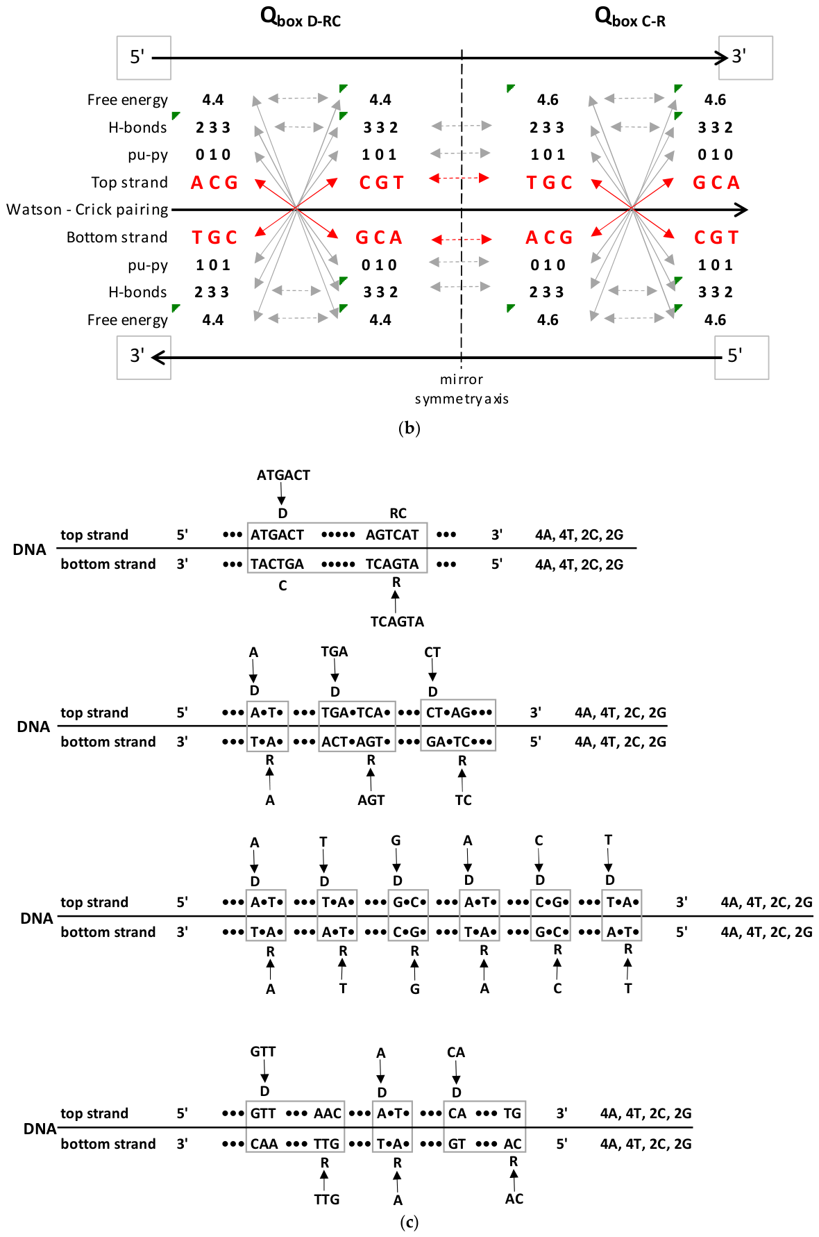
Figure 3.
Trinucleotide relative frequencies (f) of 10 A+T rich and 10 C+G rich quadruplets from 56 single-strand RNA coronaviruses. 1-7 human coronaviruses, 8-20 bat coronaviruses, 21-56 mix coronaviruses (S1). As single-strand viruses, they do not have Watson-Crick pairing. However, Chargaffʼs second parity rule (fD=fRC) and mirror symmetry between purines and pyrimidines, as well as between hydrogen bonds of trinucleotides, are present in 10/20 quadruplets (7 C+G rich and 3 A+T rich) (see Figure 3C). In A+T rich and C+G rich groups left column 1s t- 5th quadruplets, right column 6th – 10th quadruplets. a) RNA viruses: Trinucleotide relative frequencies from 56 single-strand coronaviruses in 10 A+T rich quadruplets. b) RNA viruses: Trinucleotide relative frequencies from 56 single-strand coronaviruses in 10 C+G rich quadruplets. c) Some group according mirror symmetry, Chargaffʼs second parity rule and H-bonds as initial symmetry or groups of relative frequencies from trinucleotides of single-strand RNA coronaviruses. Three different groups of some purine–pyrimidine quadruplet symmetries are identified: 1) identical symmetry in C+G rich quadruplets 7 and incomplete in 4; 2) approximate symmetry in C+G rich quadruplets 1, 2, 3, 7, 10, and in A+T rich quadruplets only in quadruplet 6; 3) approximately identical high (↑) and low (↓) frequencies in C+G rich quadruplet 5, and in A+T rich quadruplets 4 and 7. Dominant symmetry is between direct (D) and reverse complement (RC) or complement (C) and reverse (R) in the form of purine (0) - pyrimidine (1) mirror symmetry and Chargaffʼs second parity rule (strand symmetry) (←---→ arrow). There is no quadruplet mirror symmetry between D ≠ C and RC ≠ R. Parallel to some purine-pyrimidine symmetries, there is, in all examples, mirror symmetry between weak bases with 2 hydrogen bonds (H-bonds) and strong bases with 3 H-bonds (←---→ arrow). In all symmetrical or partial symmetrical examples participate symmetrical trinucleotides (e.g., ATA (D, R), TAT (RC, C), and trinucleotides with only two different bases (e.g., CCG (D), CGG (RC), GGC (C), and GCC (R). In the same examples there are relative frequency equality and free energy value equality of Chargaffʼs second parity rule between D and RC (see Figure 2).
Figure 3.
Trinucleotide relative frequencies (f) of 10 A+T rich and 10 C+G rich quadruplets from 56 single-strand RNA coronaviruses. 1-7 human coronaviruses, 8-20 bat coronaviruses, 21-56 mix coronaviruses (S1). As single-strand viruses, they do not have Watson-Crick pairing. However, Chargaffʼs second parity rule (fD=fRC) and mirror symmetry between purines and pyrimidines, as well as between hydrogen bonds of trinucleotides, are present in 10/20 quadruplets (7 C+G rich and 3 A+T rich) (see Figure 3C). In A+T rich and C+G rich groups left column 1s t- 5th quadruplets, right column 6th – 10th quadruplets. a) RNA viruses: Trinucleotide relative frequencies from 56 single-strand coronaviruses in 10 A+T rich quadruplets. b) RNA viruses: Trinucleotide relative frequencies from 56 single-strand coronaviruses in 10 C+G rich quadruplets. c) Some group according mirror symmetry, Chargaffʼs second parity rule and H-bonds as initial symmetry or groups of relative frequencies from trinucleotides of single-strand RNA coronaviruses. Three different groups of some purine–pyrimidine quadruplet symmetries are identified: 1) identical symmetry in C+G rich quadruplets 7 and incomplete in 4; 2) approximate symmetry in C+G rich quadruplets 1, 2, 3, 7, 10, and in A+T rich quadruplets only in quadruplet 6; 3) approximately identical high (↑) and low (↓) frequencies in C+G rich quadruplet 5, and in A+T rich quadruplets 4 and 7. Dominant symmetry is between direct (D) and reverse complement (RC) or complement (C) and reverse (R) in the form of purine (0) - pyrimidine (1) mirror symmetry and Chargaffʼs second parity rule (strand symmetry) (←---→ arrow). There is no quadruplet mirror symmetry between D ≠ C and RC ≠ R. Parallel to some purine-pyrimidine symmetries, there is, in all examples, mirror symmetry between weak bases with 2 hydrogen bonds (H-bonds) and strong bases with 3 H-bonds (←---→ arrow). In all symmetrical or partial symmetrical examples participate symmetrical trinucleotides (e.g., ATA (D, R), TAT (RC, C), and trinucleotides with only two different bases (e.g., CCG (D), CGG (RC), GGC (C), and GCC (R). In the same examples there are relative frequency equality and free energy value equality of Chargaffʼs second parity rule between D and RC (see Figure 2).
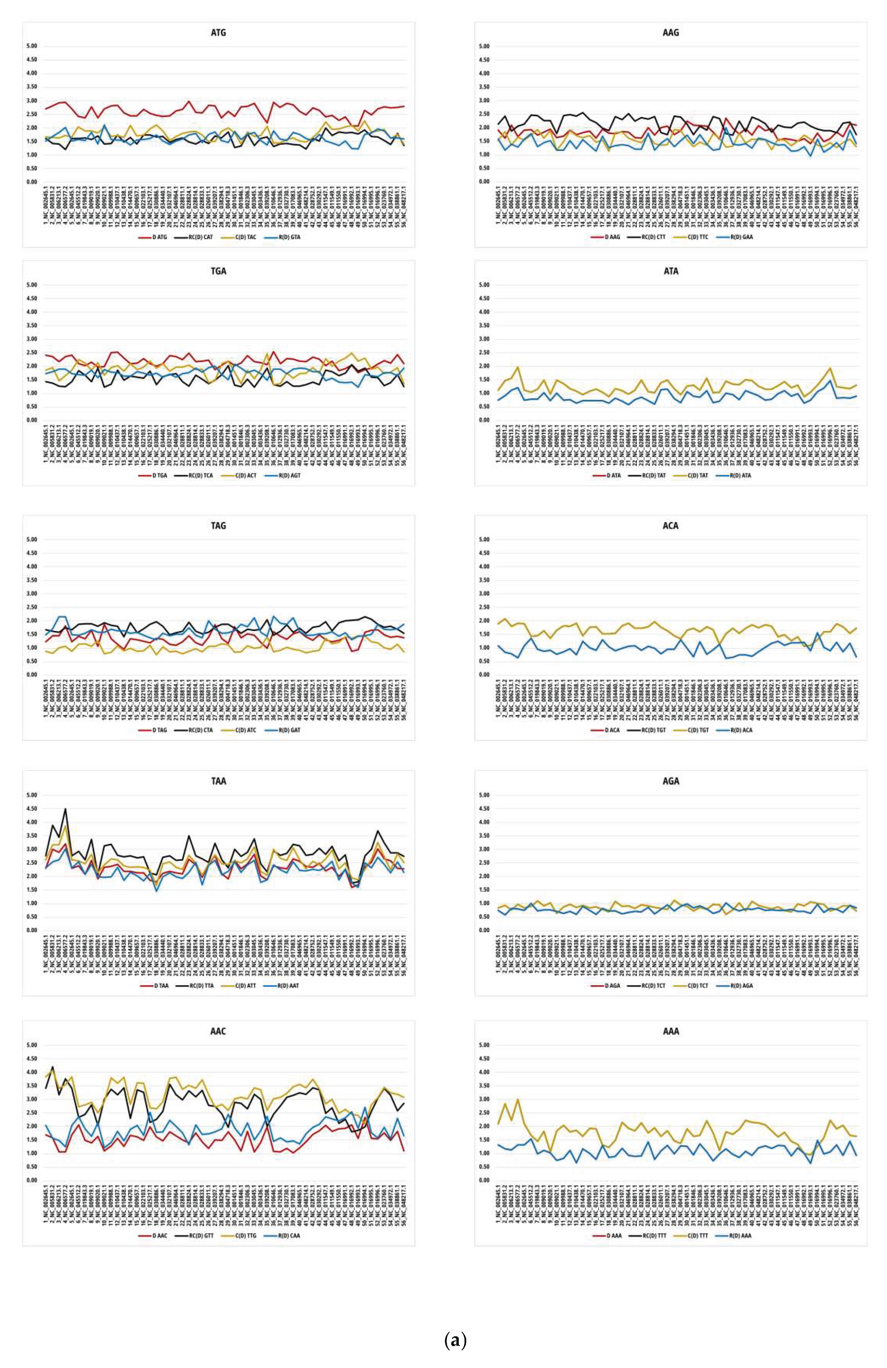
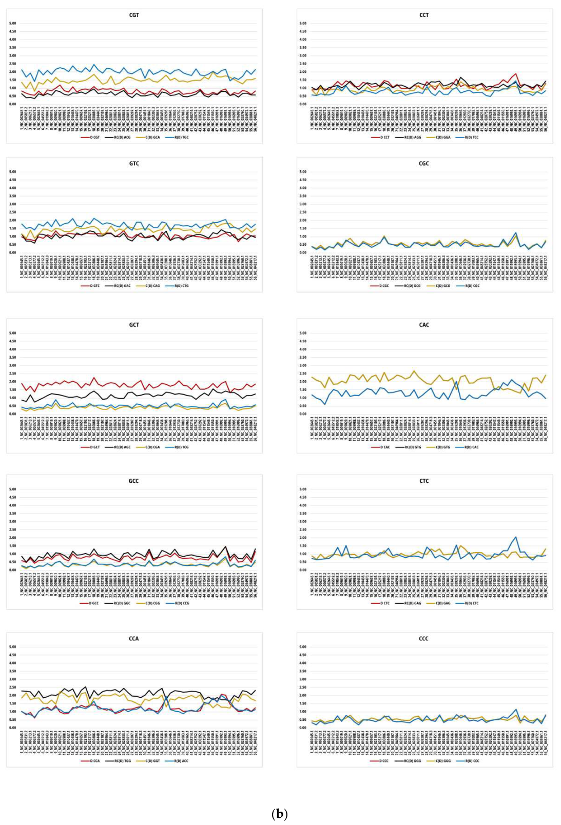
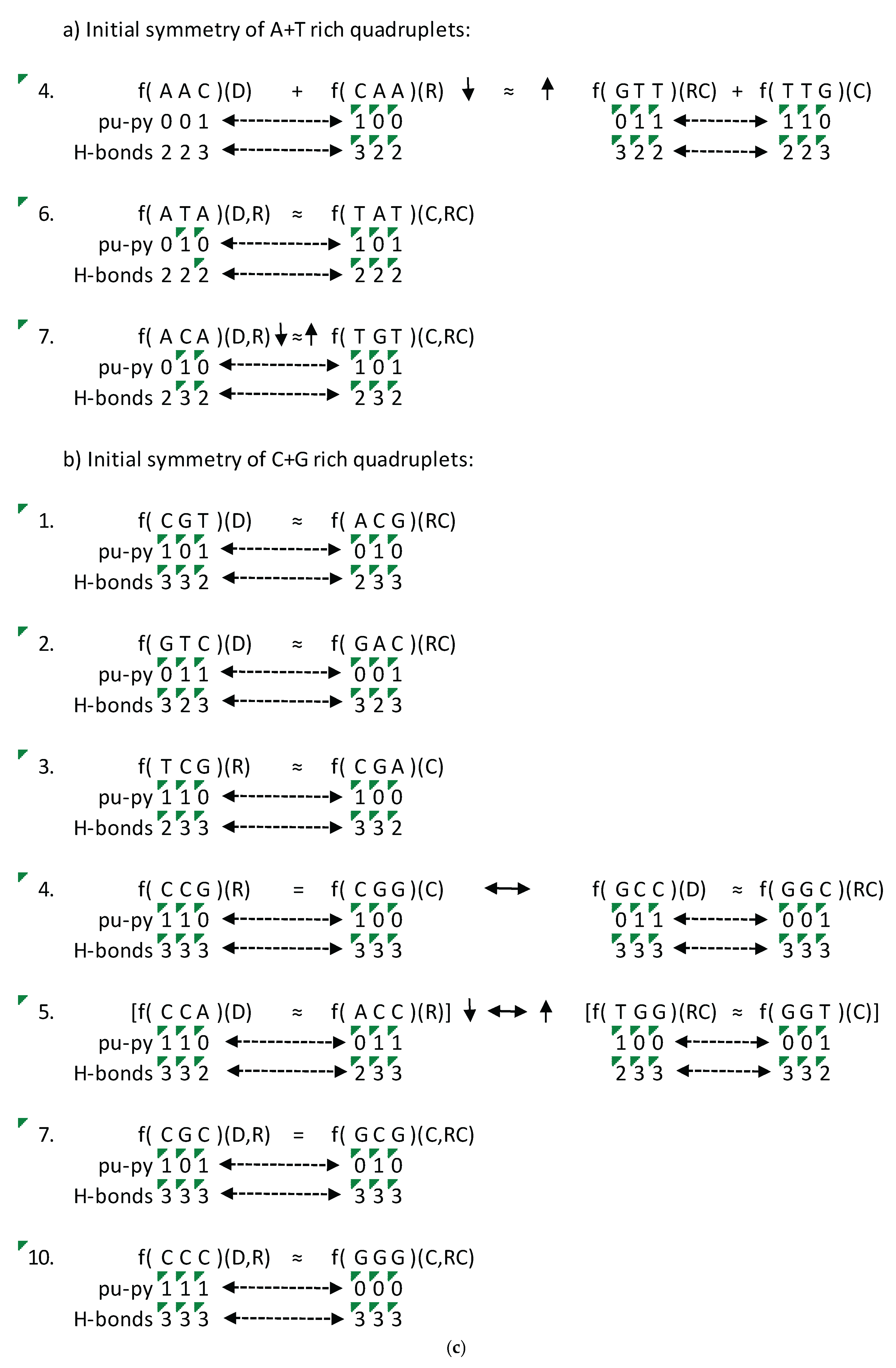
Figure 4.
The complete genomes of all double-strand DNA viruses (a) and Circoviridae (b). They are formed on the principle of quadruplets and have A+T rich and C+G rich quadruplet matrices. All eight symmetrical A+T rich and C+G rich trinucleotides such as ACA or GCG have complete quadruplet symmetries with Watson-Crick pairing and dominant purine-pyrimidine mirror symmetry with the plateau on the upper edge of the quadruplet box direct-reverse complement (QboxD-RC) and the quadruplet box complement-reverse (QboxC-R). The other six A+T rich as well as six C+G rich quadruplets with non-symmetrical trinucleotides have only Watson-Crick pairing. The numerical values can be seen in Supplementary Table S2.
Figure 4.
The complete genomes of all double-strand DNA viruses (a) and Circoviridae (b). They are formed on the principle of quadruplets and have A+T rich and C+G rich quadruplet matrices. All eight symmetrical A+T rich and C+G rich trinucleotides such as ACA or GCG have complete quadruplet symmetries with Watson-Crick pairing and dominant purine-pyrimidine mirror symmetry with the plateau on the upper edge of the quadruplet box direct-reverse complement (QboxD-RC) and the quadruplet box complement-reverse (QboxC-R). The other six A+T rich as well as six C+G rich quadruplets with non-symmetrical trinucleotides have only Watson-Crick pairing. The numerical values can be seen in Supplementary Table S2.
Disclaimer/Publisher’s Note: The statements, opinions and data contained in all publications are solely those of the individual author(s) and contributor(s) and not of MDPI and/or the editor(s). MDPI and/or the editor(s) disclaim responsibility for any injury to people or property resulting from any ideas, methods, instructions or products referred to in the content. |
© 2026 by the authors. Licensee MDPI, Basel, Switzerland. This article is an open access article distributed under the terms and conditions of the Creative Commons Attribution (CC BY) license (http://creativecommons.org/licenses/by/4.0/).
Copyright: This open access article is published under a Creative Commons CC BY 4.0 license, which permit the free download, distribution, and reuse, provided that the author and preprint are cited in any reuse.


