

Submitted:
03 July 2026
Posted:
06 July 2026
You are already at the latest version
Preprints on COVID-19 and SARS-CoV-2
Abstract
Six years into the COVID-19 pandemic, understanding the complex interplay between SARS-CoV-2 infection and the host innate immune response remains pivotal. This update review synthesizes current knowledge on the activation of innate immunity by SARS-CoV-2, emphasizing type I and III IFN responses, key antiviral proteins, and critical immune cells such as plasmacytoid dendritic cells, alveolar macrophages, and neutrophils. We explore the phenomenon of hyperinflammation or "cytokine storm," characterized by excessive production of pro-inflammatory cytokines mediated by pathways including NF-κB, JAK-STAT, and MAPK, and highlight its contributions to severe COVID-19 pathogenesis. Following this, we discuss the unique immunothrombotic landscape observed in COVID-19-associated coagulopathy, underlining its significance in disease severity and progression. Lastly, we detail SARS-CoV-2’s sophisticated immune evasion strategies, illustrating how viral adaptations further exacerbate disease severity by undermining host antiviral defenses.
Keywords:
COVID-19
; SARS-CoV-2
; immune response
; cytokine storm
; hyperinflammation
; innate immunity
; immunothrombosis
; COVID-19-associated coagulopathy
; viral immune evasion
Introduction
Since its emergence in 2019, SARS-CoV-2 has persistently challenged global public health, revealing intricate mechanisms by which it interacts with and disrupts the host immune system. Early antiviral defenses mediated by the innate immune system are critical determinants of disease trajectory, and their timely activation is pivotal for controlling viral replication. Type I and III IFNs orchestrate a primary antiviral response through the activation of interferon-stimulated genes (ISGs), forming an early line of defense to restrict viral spread. However, SARS-CoV-2 frequently circumvents these initial defenses, exploiting delayed IFN responses to facilitate rapid viral dissemination. Subsequent dysregulated immune responses, characterized by a pronounced hyperinflammatory state termed "cytokine storm", exacerbate tissue damage and amplify disease severity. Simultaneously, aberrant immune activation intersects profoundly with coagulation pathways, resulting in distinct thrombotic complications recognized as COVID-19-associated coagulopathy (CAC). This review systematically dissects the complex relationship between SARS-CoV-2 infection and innate immune activation, hyperinflammation, immunothrombosis, and immune evasion mechanisms, providing a comprehensive understanding of their collective impact on COVID-19 pathophysiology.
1. SARS-CoV-2 Innate Immune Evasion
SARS-CoV-2 has evolved multiple immune evasion strategies that suppress early IFN signalling, predisposing patients to critical disease courses. 23 out of the 29 viral proteins are implicated in innate immune interference, and their interactions with host mechanisms can be seen in Figure 1.
These include: host PRR interference (13 proteins); viral RNA concealment (9 proteins); JAK-STAT signalling antagonization (8 proteins); antiviral proteins translation suppression (6 proteins); and inhibiting nuclear transport of immune signalling molecules (5 proteins) [1].
Specifically, NSP1 binds to the host 40S ribosomal subunit, inhibiting translation and antagonizing IFN signalling [2]. It also epigenetically represses antiviral gene expression through demethylation of histone H3K9 at key antiviral gene promoters, highlighting a previously unrecognized mechanism of host suppression [3]. NSP3, via its PLpro domain, not only processes viral polyproteins but also removes ubiquitin and ISG15 from host proteins, blunting antiviral signalling [4,5]. NSP13 suppresses IFN activation by targeting TBK1 for degradation, whereas NSP15 functions as an endoribonuclease that degrades viral dsRNA intermediates to evade PRR detection [6,7]. NSP8 interferes with IRF3 activation and nuclear translocation to supress IFN I and III; NSP8 also further suppress IFN I through inhibition of MDA5, a critical cytosolic sensor of viral dsRNA, by impairing TRIM4-mediated MDA5 K63-linked polyubiquitination [8,9]. NSP9 also suppresses IFN and inflammatory signalling by reducing RIG-I expression, NF-κB signalling, and TBK1 activity [10]. Finally, NSP14 reduces surface IFNAR expression, weakening IFN signal transduction [11].
Accessory proteins also contribute significantly. ORF6 inhibits nuclear translocation of STAT1/2 and suppresses IRF3 and NF-κB activation[12,13]. ORF7b blocks upstream IFN production and ORF8 disrupts IRF3 function and IFN signalling, robustly suppressing ISG transcription[14,15]. ORF3a inhibits STAT1 phosphorylation, reduces IFN signalling, and promotes inflammasome activation; ORF3b potently suppresses IFN induction through IRF3 suppression; while ORF3c localizes to mitochondria, promoting MAVS cleavage and reducing IFN-β production[16,17,18]. IFN signalling is further blocked by ORF9b disrupting MAVS signalling through TOM70 targeting, and by ORF9c, which impairs antigen presentation and inflammatory pathways[19,20]. ORF10s role currently remains unclear, however, it may modulate immune responses through STRING interaction[21]. Finally, structural M protein disrupts RIG-I/MDA5 signalling, reducing IFN-β promoter activation[22].
In addition to IFN evasion, SARS-CoV-2 modulates other innate immune pathways. The nucleocapsid protein dysregulates the NLRP3 inflammasome, promoting IL-1β and IL-18 secretion and pyroptosis, leading to inflammation rather than viral clearance[23]. NSP6 and ORF3a disrupt autophagy by inhibiting autophagosome maturation and lysosomal fusion, impairing antigen presentation[24,25]. NSP1 and ORF6 also appear to impair NK cell cytotoxicity by downregulating the NKG2D activating ligands on infected cells, reducing early viral clearance[26,27].
Host factors further influence the IFN response to SARS-CoV-2 infection. Genetic mutations in critical components of IFN signalling pathways, such as TLR7, IRF7, and IRF9, compromise IFN production[28,29]. Additionally, neutralizing autoantibodies against type I IFNs (found in a subset of severe COVID-19 cases) reduce functional IFN-α/ω levels, permitting uncontrolled viral replication and severe inflammation[30].
2. Key Immune Cells in COVID-19 Pathogenesis
Airway epithelial cells are the main initial targets for SARS-CoV-2 and among the first to produce IFNs. However, often in severe disease, this response is notably delayed and insufficient to immediately control viral replication, preventing a robust antiviral state from establishing. During disease course, additional immune cells are activated through multiple complementary mechanisms. Below, the most relevant to pathogenesis are discussed.
Plasmacytoid Dendritic Cells (pDCs), often called “professional interferon-producing cells”, rapidly secrete large quantities of IFN-α/β upon viral detection, bridging innate and adaptive immunity by activating NK cells and T cells for virus clearance[31]. Notably, studies indicate pDCs detect SARS-CoV-2 more effectively by sensing infected cells, rather than free virus particles, through CD54-CD11a engagement between pDCs and infected cells[32]. Selective pressures have promoted some variants to limit this sensing, through reduced CD11a induction for Alpha, or perturbation of IFN production in the case of Delta and Omicron[32]. As pDCs are typically the largest producers of type I and III IFN during infection by a factor of 10–100, this lack of early pDC-driven IFN is a hallmark of severe COVID-19, and in such patients, a pronounced lack of functional pDCs in both blood and lung tissues is evident from disease onset[33,34,35,36,37,38]. This “pDC desert” may result from excessive recruitment into infected tissues alongside rapid, chronic activation, causing functional impairment, exhaustion and apoptosis. Resultingly, pDCs in the lungs of severe patients produce little to no IFN, contributing to a deficient antiviral response. Conversely, some studies caution that if pDCs persistently produce IFN, it might contribute late stage tissue damage, so balanced pDC response is key[39,40,41].
Alveolar macrophages (AMs) are tissue-resident cells that quietly maintain lung homeostasis by clearing debris and pathogens while preventing excessive inflammation. During early SARS-CoV-2 infection they engulf viral particles and secrete cytokines to recruit additional immune cells. In severe patients, viral propagation diminishes functional AMs and the remaining infected AMs upregulate IL1B, CCL4, CCL20, CXCL10 and CXCL11, which recruit massive numbers of blood monocytes (and T cells) to the lung for reinforcements[42]. Normally, these monocytes then differentiate into monocyte-derived macrophages (MDMs) with pro-inflammatory (M1) or anti-inflammatory (M2) signatures, dependent on cytokine environment, to balance the immune response[43]. SARS-CoV-2 however, induces a molecular shift in MDMs by promoting the uptake of viral RNA, triggering proinflammatory cytokine release and driving MDMs toward an M1-like phenotype whilst preventing reversion to an anti-inflammatory state[44]. In severe cases, this becomes a positive inflammatory feedback loop as incoming monocytes flood the alveolar space, differentiating into M1-like MDMs and secreting abundant IL-6, IL-8, TNF-α and other cytokines[45]. These cytokines majorly contribute to the cytokine storm, whilst the chemokines sustain lung inflammation through monocyte and neutrophil recruitment. Further, MDMs in the lungs of severe COVID-19 patients often express tissue-factor and contribute to immunothrombosis and fibrosis, linking the inflammatory response to some COVID-19 vascular complications[46,47].
Neutrophils are rapid responders that migrate to infection sites within minutes of trauma. However, in the lungs and alveolar space, efficient AM pathogen clearance restricts neutrophil recruitment to only periods of significant infection, preventing constant inflammation[48]. When recruited neutrophils clear pathogens through phagocytosis, inactivating them within phagolysosomes to minimize bystander damage. In contrast, excessive stimulation by PAMPs and DAMPs triggers neutrophils to unleash their full arsenal without restraint - NETs, granules, oxidants, and proteases – leading to lung injury and thrombosis. Specifically, uncontrolled degranulation and NET release damage the alveolar–capillary barrier via enzymes such as elastase and MPO, and cytokines like IL-8[49,50,51]. Furthermore, studies have found excessive NETs in the blood of severe COVID-19 patients forming thromboinflammatory clusters that contribute to microthrombi and organ damage[52,53]. Importantly, SARS-CoV-2 induces emergency granulopoiesis in severe disease, prematurely releasing immature neutrophils and granulocytic myeloid-derived suppressor cells (G-MDSCs)[54]. These left-shifted neutrophils exhibit enhanced degranulation and ROS production, and uncontrolled NET formation, whilst G-MDSCs are defined most notably by supressing T-cell proliferation and cytokine production, through secretion of molecules like arginase-1, ROS, and IL-10[54,55]. This creates a situation in which neutrophils exacerbate tissue-damaging inflammation while concurrently dampening adaptive immunity.
3. The “Cytokine Storm”
In patients where the immune response and IFN production are not sufficiently calibrated - such as with COVID-19 clinical risk factors like advanced age, obesity, lung disease, etc. - SARS-CoV-2 gains an early foothold that predisposes to life-threatening outcomes. During the initial “stealth” phase, infected cells produce minimal IFN while secreting proinflammatory chemokines, allowing unchecked viral replication[56].
By the time a substantial IFN response is mounted, the infection is often widespread. A late “compensatory” surge of IFNs may occur as accumulating viral RNA and cell damage eventually trigger PRRs. Paradoxically, IFNs that are protective when produced early can be detrimental if produced too late or excessively, with elevated systemic IFN during later stages of COVID-19 disease correlated with worsening disease outcomes[57]. Prolonged or high-dose IFN exposure can amplify inflammation and impede lung tissue-repair processes[58,59]. This weak initial IFN activity followed by an uncoordinated surge, leads to an overzealous innate response that spirals towards immunopathology. The innate immune system, now fully alarmed, releases torrents of cytokines into the lungs and circulation, creating a vicious cycle that recruits additional immune cells which then secrete more cytokines and increase the number of target cells for infection[60,61,62]. The lung becomes a battleground of uncontrolled inflammation, leading to diffuse alveolar damage, pneumonia, and ARDS[63]. In critical cases, inflammation spills over systemically, causing vasculature dysfunction, coagulopathy, and multi-organ exhaustion and injury.
NF-κB is a master transcription factor, orchestrating the expression of numerous inflammatory cytokines and mediators. It is maintained in an inactive state by IκB binding to its RHD, preventing its nuclear localization. During SARS-CoV-2 infection, however, NF-κB is markedly activated through multiple mechanisms, including TLR-mediated sensing of S protein (TLR2/4) and viral RNA (TLR3/7/8), cytosolic detection via RIG-I/MDA5, and TNF-α-driven amplification[64,65,66]. NSP3, NSP14, ORF3a, ORF6, and Nucleocapsid also show direct or indirect activation of NF-κB[17,67,68,69,70]. NF-κB is also additionally activated through cellular stress and ROS, a scenario especially pertinent in patients with pre-existing conditions where baseline oxidative stress and NF-κB activity are already elevated and thus one of the reasons such conditions are risk factors for severe disease[71,72]. Once within the nucleus, NF-κB drives the transcription of proinflammatory cytokines and chemokines (e.g. IL-1β, IL-6, TNF-α) that further launch their own signaling cascades. Moreover, its interplay with inflammatory pathways such as JAK-STAT and MAPK amplifies cytokine production. For example, NF-κB-driven IL-6 expression activates STAT3, further promoting additional proinflammatory mediator transcription, a key event in the progression toward cytokine storm[73]. High levels of NF-κB activity have been directly correlated with the severity of disease in COVID-19[74]. Concurrently, NF-κB also regulates the expression of cell survival, proliferation, and apoptosis inhibiting genes. While these functions are beneficial in mounting an effective immune response, their persistent activation can result in pathological cellular survival and prolonged inflammation. In COVID-19 patients, particularly those with pre-existing conditions or advanced age, the regulatory mechanisms that normally dampen NF-κB activity (re-synthesis of IκB proteins) may be compromised[75]. This imbalance predisposes to the uncontrolled and sustained inflammatory response observed in severe cases.
IL-1β and TNF-α are among the first cytokines released following NF-κB activation and play pivotal roles in inflammatory responses. Monocytes and macrophages predominantly produce IL-1β in a two-step process: NF-κB induced transcription of pro-IL-1β and NLRP3 as a first step, if cellular stress is then detected, NLRP3 inflammasome assembles and activates caspase-1 which cleaves pro-IL-1β into its mature, secreted form[76]. Caspase-1 is also responsible for plasma membrane rupture during pyroptosis, via GSDMD and NINJ1, allowing cytokine secretion, and contributing to pathological inflammation[76]. Once released, IL-1β binds to IL-1R1 and culminates in further NF-κB and MAPK pathway activation. In COVID-19, excessive IL-1β signaling triggers endothelial activation and dysfunction, inducing tissue factor expression and initiating the extrinsic coagulation cascade with subsequent thrombin generation and fibrin deposition[77]. Concurrent suppression of anticoagulant pathways and increased PAI-1 production shift the balance toward a prothrombotic state, driving hypercoagulation and, in severe cases, disseminated intravascular coagulation (DIC), increasing morbidity and mortality[78]. In a complementary manner, TNF-α, rapidly produced by macrophages, T cells, B cells, and dendritic cells, signals to also activate both NF-κB and MAPK pathways[76]. In particular, TNFR1 recruits TRADD and TRAF2, leading to the activation of TAK1 and downstream inflammatory cascades. TNF-α also enhances IL-1β signaling and vice versa, contributing to cytokine cross-talk that fuels the storm[78]. In the pulmonary system, TNF-α drives tissue injury by promoting bronchial constriction, increasing endothelial permeability, and stimulating epithelial cells to release inflammatory mediators such as GM-CSF and IL-8[79]. TNF-α neutrophil activation results in degranulation and protease release, further degrading lung architecture and function.
IL-6 has emerged as another critical cytokine in COVID-19. Studies consistently demonstrate markedly elevated levels in critically ill patients that strongly predict respiratory failure and mechanical ventilation requirements[78]. Following NF-κB activation, monocytes, macrophages, and dendritic cells rapidly secrete IL-6, which when bound to its receptor, recruits gp130, initiating the JAK/STAT3 pathway[78]. Activated STAT3 translocates to the nucleus to regulate proinflammatory and prothrombotic gene expression. Through STAT3 activation, IL-6 drives Th17 differentiation, promotes B cell maturation, enhances cytotoxic T cell responses, and increases neutrophil recruitment, whilst suppressing regT cell generation, skewing the immune balance toward uncontrolled inflammation[78]. STAT3 also upregulates acute-phase proteins (CRP, fibrinogen) in hepatocytes, increasing blood viscosity, enhancing platelet activation and increasing expression of adhesion molecules and tissue factor, further tipping the hemostatic balance toward a prothrombotic state[80]. Such mechanisms link IL-6 to the COVID-19 hypercoagulable state. Moreover, IL-6 trans-signaling in endothelial cells is particularly pathogenic, as it induces the release of IL-8 and MCP-1, which recruits proinflammatory neutrophils and macrophages, and stimulates VEGF secretion, which disrupts tight junction proteins to increase endothelial permeability[78].
These processes in severe COVID-19 are highly interconnected, unfolding concurrently as delayed IFN responses enable unchecked viral replication, subsequently triggering dysregulated cytokine production via pathways such as NF-κB, MAPK, and JAK-STAT. A self-amplifying cycle is created, driving systemic inflammation, coagulopathy, and multi-organ injury. Importantly, this highlights just a fraction of the many molecules and pathways involved in the complex pathophysiology of cytokine storm. This hyperinflammatory state is tightly linked with the next topic, coagulopathy via the thromboinflammatory cycle.
4. Immunothrombosis: Intersection of Inflammation and Coagulation
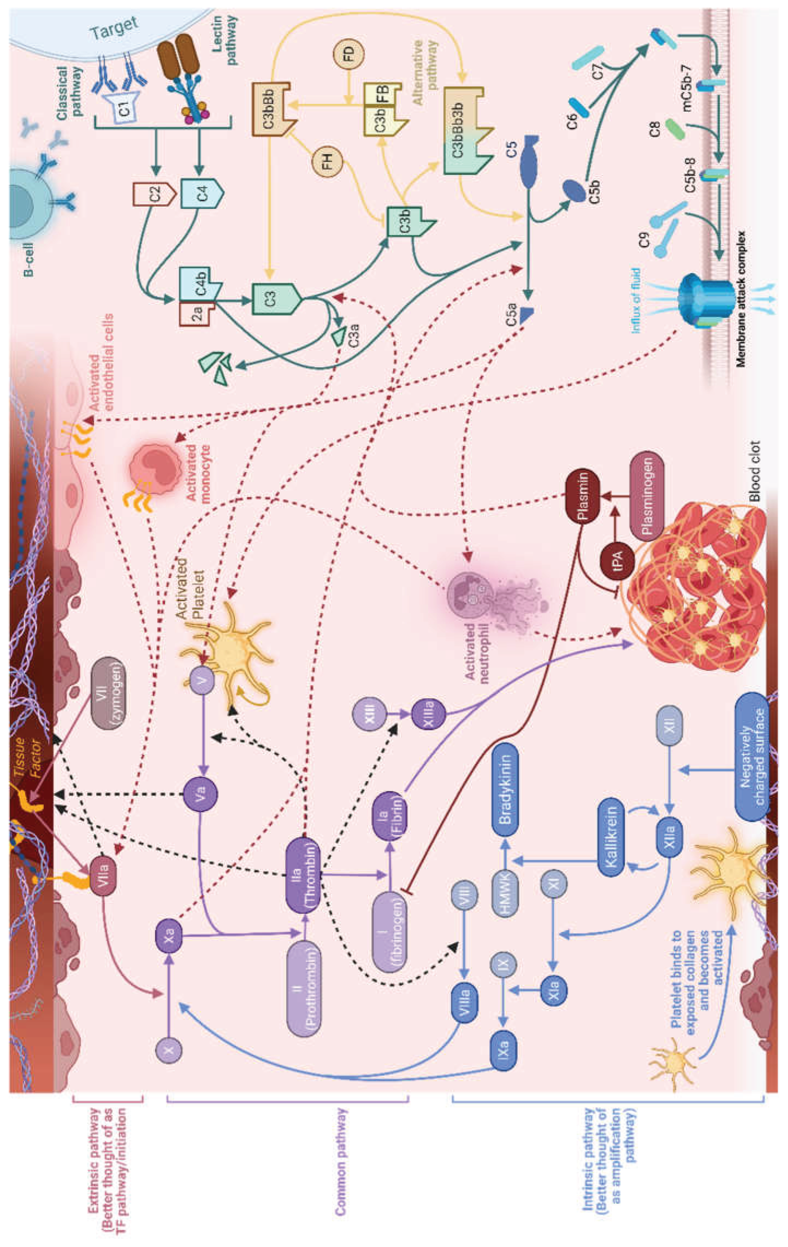
A striking feature of COVID-19 is coagulopathy with microthrombosis in the microvasculature of various organs, particularly lungs. Figure 2 represents and discusses a simplified normal interaction of complement and coagulation pathways in heathy individuals, whilst the below text discusses in detail how these pathways are perturbed in COVID-19 infection.
Although coagulopathy occurs in various severe infections such as bacterial sepsis, viral hemorrhagic fevers, and influenza, CAC predominantly exhibits a thrombotic rather than hemorrhagic profile, though bleeding complications may still occur[81]. CAC is notably identifiable through elevations in D-dimer and fibrinogen, mild thrombocytopenia, and generally normal clotting times[82]. Elevated D-dimer (>0.5–1.0 μg/mL) correlate with poorer outcomes, with rising levels during hospitalization linked to increased mortality. Beyond microvascular thrombosis, CAC also predisposes patients to macrovascular complications such as deep vein thrombosis, pulmonary embolism, and stroke[83]. This distinct profile likely reflects a multifactorial pathogenesis involving direct viral injury, heightened inflammation, complement activation, platelet hyperreactivity, and NETosis.
Endothelial viral infection leads to endotheliitis and a loss of its inherent antithrombotic functions[84]. Endothelial cell damage reduces protective molecules like thrombomodulin and heparin sulfate, which inhibit clotting, while increasing prothrombotic factors such as vWF and P-selectin[85,86]. Although autopsy studies demonstrate widespread endothelial inflammation in COVID-19, direct evidence of active viral replication within endothelial cells remains limited and controversial[87,88,89,90]. Thus, vascular injury likely results from indirect immune-mediated responses or initial viral entry without full replication.
Such endothelial dysfunction creates a localized environment favouring thrombosis over bleeding, contributing to the overall hypercoagulable state observed in COVID-19.
Immunologically, CAC is driven by several mechanisms. Pro-inflammatory cytokines (IL-6, TNF-α) stimulate hepatic fibrinogen synthesis and upregulate TF expression on endothelial cells and monocytes[91]. This activity triggers the extrinsic coagulation cascade and subsequent thrombin generation. Elevated IL-6 correlates with increased fibrinogen, vWF, factor VIII, and D-dimer, whilst TNF-α and IL-1β suppress natural anticoagulants like thrombomodulin and the EPCR[92,93,94]. Neutrophils also contribute through NET release, trapping platelets and clotting factors to form thrombi; their presence in lung tissues and blood of COVID-19 patients strongly correlates with thrombotic events[52,95].
Complement activation also plays a critical role, detectable in early or mild disease and correlating with severity[96]. Mechanistically, SARS-CoV-2 triggers aberrant activation of the lectin and alternative pathways, generating potent anaphylatoxins that amplify cytokine release and promote leukocyte recruitment, intensifying pulmonary and systemic inflammation[97]. This dysregulated response also promotes the formation of the membrane attack complex, contributing to the endothelial injury and microvascular thrombosis in severe COVID-19 cases[97]. Evidence further suggests that SARS-CoV-2 may directly interact with complement components, leading to the formation of persistent immune complexes and sustained complement activation during infection and up to a year following[98,99]. Additionally, impaired regulation of complement-mediated clearance mechanisms may contribute to autoinflammatory responses and has been implicated in the pathogenesis of Post-Acute Sequelae of SARS-CoV-2 infection (PASC) and multisystem inflammatory syndrome in children (MIS-C) [100,101,102].
Immunothrombosis significantly contributes to organ damage in severe COVID-19. Autopsy studies revealed widespread microvascular thrombi in pulmonary alveolar capillaries, which are strongly implicated in ARDS development and impaired gas exchange[103]. Similarly, microthrombi in coronary microcirculation have been linked to myocardial injury and dysfunction, presenting as arrhythmias or heart failure[104]. Renal involvement often involves thrombotic obstruction of small vessels and proximale tubale dysfunction, resulting kidney damage[105,106]. In the cerebral circulation, immunothrombosis is associated with an increased stroke risk in critical patients[107]. Collectively, these findings illustrate the crucial interplay between immune responses and coagulation pathways in driving both localized thrombosis and multi-organ dysfunction during severe COVID-19.
Conclusion
The interaction between SARS-CoV-2 and the innate immune system is characterized by an intricate balance between antiviral defence and immune-mediated pathology. Effective early activation of type I and III interferon responses is crucial for viral containment, yet SARS-CoV-2 possesses sophisticated strategies to evade these defences, leading to delayed and dysregulated immune responses. The subsequent "cytokine storm" mediated by NF-κB, MAPK, and JAK-STAT pathways not only exacerbates pulmonary and systemic inflammation but also promotes extensive immunothrombosis, substantially contributing to organ dysfunction and severe disease outcomes. Understanding these mechanisms highlights potential therapeutic targets for mitigating hyperinflammation and thrombotic complications. Continued research into SARS-CoV-2’s evolving immune evasion strategies and host immune responses remains essential to developing effective interventions to manage COVID-19 and future pandemics.
Acknowledgements
Conceptualization, L.B. and L.E.; resources, J.C.Y., L.B., and L.E.; writing—original draft. preparation, B.W; writing—review and editing, J.C.Y., L.B., and L.E.; supervision, L.B. and L.E.; funding acquisition, J.C.Y., L.B., and L.E. All authors have read and agreed to the published version of the manuscript. This work was supported by the Sofina COVID Solidarity Fund, administered by the King Baudouin Foundation, initiated by the Fondation Saint-Luc (grant number 2021-I4201010−221801), and the FNRS Urgent Research Credit (CUR: HC01020F). B.W. is the recipient of an FRC starting grant (Promotor Leïla Belkhir) and an FSR grant (Promotor Laure Elens).
Conflicts of Interest
No conflicts of interest to declare.
References
- Minkoff, J. M.; tenOever, B. Innate immune evasion strategies of SARS-CoV-2. Nat. Rev. Microbiol. 2023. [Google Scholar] [CrossRef]
- Schubert, K.; et al. SARS-CoV-2 Nsp1 binds the ribosomal mRNA channel to inhibit translation. Nat. Struct. Mol. Biol. 2020, 27, 959–966. [Google Scholar] [PubMed]
- Anastasakis, D. G.; Benhalevy, D.; Çuburu, N.; Altan-Bonnet, N.; Hafner, M. Epigenetic repression of antiviral genes by SARS-CoV-2 NSP1. PLoS ONE 2024, 19, e0297262. [Google Scholar] [PubMed]
- Shin, D.; et al. Papain-like protease regulates SARS-CoV-2 viral spread and innate immunity. Nature 2020, 587, 657–662. [Google Scholar] [PubMed]
- Cao, D.; et al. The SARS-CoV-2 papain-like protease suppresses type I interferon responses by deubiquitinating STING. Sci. Signal. 2023, 16, eadd0082. [Google Scholar] [PubMed]
- Vazquez, C.; et al. SARS-CoV-2 viral proteins NSP1 and NSP13 inhibit interferon activation through distinct mechanisms. PLoS ONE 2021, 16, e0253089. [Google Scholar] [PubMed]
- Otter, C. J.; et al. SARS-CoV-2 nsp15 endoribonuclease antagonizes dsRNA-induced antiviral signaling. Proc. Natl. Acad. Sci. 2024, 121, e2320194121. [Google Scholar] [PubMed]
- Deng, J.; et al. SARS-CoV-2 NSP8 suppresses type I and III IFN responses by modulating the RIG-I/MDA5, TRIF, and STING signaling pathways. J. Med. Virol. 2023, 95, e28680. [Google Scholar] [PubMed]
- Zhang, X.; et al. SARS-CoV-2 Nsp8 suppresses MDA5 antiviral immune responses by impairing TRIM4-mediated K63-linked polyubiquitination. PLoS Pathog. 2023, 19, e1011792. [Google Scholar] [PubMed]
- Lundrigan, E.; Toudic, C.; Pennock, E.; Pezacki, J. P. SARS-CoV-2 Protein Nsp9 Is Involved in Viral Evasion through Interactions with Innate Immune Pathways. ACS Omega 2024, 9, 26428–26438. [Google Scholar] [PubMed]
- Maddaloni, L.; et al. Differential expression of Type I interferon and inflammatory genes in SARS-CoV-2-infected patients treated with monoclonal antibodies. Immun. Inflamm. Dis. 2023, 11, e968. [Google Scholar] [PubMed]
- Miorin, L.; et al. SARS-CoV-2 Orf6 hijacks Nup98 to block STAT nuclear import and antagonize interferon signaling. Proc. Natl. Acad. Sci. 2020, 117, 28344–28354. [Google Scholar] [PubMed]
- Lu, Y.; Michel, H. A.; Wang, P.-H.; Smith, G. L. Manipulation of innate immune signaling pathways by SARS-CoV-2 non-structural proteins. Front. Microbiol. 2022, 13, 1027015. [Google Scholar] [PubMed]
- Rashid, F.; Dzakah, E. E.; Wang, H.; Tang, S. The ORF8 protein of SARS-CoV-2 induced endoplasmic reticulum stress and mediated immune evasion by antagonizing production of interferon beta. Virus Res. 2021, 296, 198350. [Google Scholar] [PubMed]
- Shemesh, M.; et al. SARS-CoV-2 suppresses IFNβ production mediated by NSP1, 5, 6, 15, ORF6 and ORF7b but does not suppress the effects of added interferon. PLoS Pathog. 2021, 17, e1009800. [Google Scholar] [PubMed]
- Wang, R.; et al. ORF3a Protein of Severe Acute Respiratory Syndrome Coronavirus 2 Inhibits Interferon-Activated Janus Kinase/Signal Transducer and Activator of Transcription Signaling via Elevating Suppressor of Cytokine Signaling 1. Front. Microbiol. 2021, 12, 752597. [Google Scholar] [PubMed]
- Rashid, F.; et al. Roles and functions of SARS-CoV-2 proteins in host immune evasion. Front. Immunol. 2022, 13, 940756. [Google Scholar] [PubMed]
- Stewart, H.; et al. The SARS-CoV-2 protein ORF3c is a mitochondrial modulator of innate immunity. iScience 2023, 26, 108080. [Google Scholar] [PubMed]
- Dominguez Andres, A.; et al. SARS-CoV-2 ORF9c Is a Membrane-Associated Protein that Suppresses Antiviral Responses in Cells. 2020. [Google Scholar] [CrossRef] [PubMed]
- Ayinde, K. S.; Pinheiro, G. M. S.; Ramos, C. H. I. Binding of SARS-CoV-2 protein ORF9b to mitochondrial translocase TOM70 prevents its interaction with chaperone HSP90. Biochimie 2022, 200, 99–106. [Google Scholar] [PubMed]
- Han, L.; et al. SARS-CoV-2 ORF10 antagonizes STING-dependent interferon activation and autophagy. J. Med. Virol. 2022, 94, 5174–5188. [Google Scholar] [PubMed]
- Zheng, Y.; et al. Severe acute respiratory syndrome coronavirus 2 (SARS-CoV-2) membrane (M) protein inhibits type I and III interferon production by targeting RIG-I/MDA-5 signaling. Signal Transduct. Target. Ther. 2020, 5, 299. [Google Scholar] [PubMed]
- Pan, P.; et al. SARS-CoV-2 N protein promotes NLRP3 inflammasome activation to induce hyperinflammation. Nat. Commun. 2021, 12, 4664. [Google Scholar] [PubMed]
- Zhang, Y.; et al. The SARS-CoV-2 protein ORF3a inhibits fusion of autophagosomes with lysosomes. Cell Discov. 2021, 7, 31. [Google Scholar] [PubMed]
- Zhang, C.; et al. SARS-CoV-2 NSP6 reduces autophagosome size and affects viral replication via sigma-1 receptor. J. Virol. 2024, 98, e00754-24. [Google Scholar] [PubMed]
- Lee, M. J.; et al. SARS-CoV-2 escapes direct NK cell killing through Nsp1-mediated downregulation of ligands for NKG2D. Cell Rep. 2022, 41, 111892. [Google Scholar] [PubMed]
- Hartmann, J. A.; et al. Evasion of NKG2D-mediated cytotoxic immunity by sarbecoviruses. Cell 2024, 187, 2393–2410.e14. [Google Scholar] [PubMed]
- Van Der Made, C. I.; et al. Presence of Genetic Variants Among Young Men With Severe COVID-19. JAMA 2020, 324, 663. [Google Scholar] [CrossRef] [PubMed]
- Zhang, Q.; et al. Inborn errors of type I IFN immunity in patients with life-threatening COVID-19. Science 2020, 370, eabd4570. [Google Scholar] [CrossRef] [PubMed]
- Bastard, P.; et al. Autoantibodies against type I IFNs in patients with life-threatening COVID-19. Science 2020, 370, eabd4585. [Google Scholar] [CrossRef] [PubMed]
- McKenna, K.; Beignon, A.-S.; Bhardwaj, N. Plasmacytoid Dendritic Cells: Linking Innate and Adaptive Immunity. J. Virol. 2005, 79, 17–27. [Google Scholar] [CrossRef] [PubMed]
- Cai, C.; Pham, T. N. Q.; Adam, D.; Brochiero, E.; Cohen, É. A. Sensing of SARS-CoV-2-infected cells by plasmacytoid dendritic cells is modulated via an interplay between CD54/ICAM-1 and CD11a/LFA-1 αL integrin. J. Virol. 2025, 99, e0123524. [Google Scholar] [PubMed]
- Fitzgeraldbocarsly, P.; Dai, J.; Singh, S. Plasmacytoid dendritic cells and type I IFN: 50 years of convergent history. Cytokine Growth Factor Rev. 2008, 19, 3–19. [Google Scholar] [CrossRef] [PubMed]
- Bénard, A.; et al. Interleukin-3 is a predictive marker for severity and outcome during SARS-CoV-2 infections. Nat. Commun. 2021, 12, 1112. [Google Scholar] [PubMed]
- Kreutmair, S.; et al. Distinct immunological signatures discriminate severe COVID-19 from non-SARS-CoV-2-driven critical pneumonia. Immunity 2021, 54, 1578–1593.e5. [Google Scholar] [PubMed]
- Severa, M.; et al. Differential plasmacytoid dendritic cell phenotype and type I Interferon response in asymptomatic and severe COVID-19 infection. PLoS Pathog. 2021, 17, e1009878. [Google Scholar] [CrossRef] [PubMed]
- Van Der Sluis, R. M.; et al. TLR2 and TLR7 mediate distinct immunopathological and antiviral plasmacytoid dendritic cell responses to SARS-CoV-2 infection. EMBO J. 2022, 41, e109622. [Google Scholar] [PubMed]
- Van der Sluis, R. M.; Holm, C. K.; Jakobsen, M. R. Plasmacytoid dendritic cells during COVID-19: Ally or adversary? Cell Rep. 2022, 40, 111148. [Google Scholar] [CrossRef] [PubMed]
- Carvalho, T.; Krammer, F.; Iwasaki, A. The first 12 months of COVID-19: a timeline of immunological insights. Nat. Rev. Immunol. 2021, 21, 245–256. [Google Scholar] [CrossRef] [PubMed]
- Sposito, B.; et al. The interferon landscape along the respiratory tract impacts the severity of COVID-19. Cell 2021, 184, 4953–4968.e16. [Google Scholar] [CrossRef] [PubMed]
- Wack, A. Monocyte and dendritic cell defects in COVID-19. Nat. Cell Biol. 2021, 23, 445–447. [Google Scholar] [CrossRef] [PubMed]
- Grant, R. A.; et al. Circuits between infected macrophages and T cells in SARS-CoV-2 pneumonia. Nature 2021, 590, 635–641. [Google Scholar] [PubMed]
- Chaintreuil, P.; et al. The generation, activation, and polarization of monocyte-derived macrophages in human malignancies. Front. Immunol. 2023, 14, 1178337. [Google Scholar] [CrossRef] [PubMed]
- Simón-Fuentes, M.; et al. MAFB shapes human monocyte–derived macrophage response to SARS-CoV-2 and controls severe COVID-19 biomarker expression. JCI Insight 2023, 8, e172862. [Google Scholar] [PubMed]
- Knoll, R.; Schultze, J. L.; Schulte-Schrepping, J. Monocytes and Macrophages in COVID-19. Front. Immunol. 2021, 12, 720109. [Google Scholar] [CrossRef] [PubMed]
- Meisel, S. R.; et al. Differentiation of adherent human monocytes into macrophages markedly enhances tissue factor protein expression and procoagulant activity. Atherosclerosis 2002, 161, 35–43. [Google Scholar] [CrossRef] [PubMed]
- Sachetto, A. T. A.; Mackman, N. Tissue Factor and COVID-19: An Update. Curr. Drug Targets 2022, 23, 1573–1577. [Google Scholar] [CrossRef] [PubMed]
- Neupane, A. S.; et al. Patrolling Alveolar Macrophages Conceal Bacteria from the Immune System to Maintain Homeostasis. Cell 2020, 183, 110–125.e11. [Google Scholar] [CrossRef] [PubMed]
- Castanheira, F. V. S.; Kubes, P. Neutrophils and NETs in modulating acute and chronic inflammation. Blood 2019, 133, 2178–2185. [Google Scholar] [CrossRef] [PubMed]
- Peñaloza, H. F.; Lee, J. S.; Ray, P. Neutrophils and lymphopenia, an unknown axis in severe COVID-19 disease. PLoS Pathog. 2021, 17, e1009850. [Google Scholar] [CrossRef] [PubMed]
- Yang, S.-C.; Tsai, Y.-F.; Pan, Y.-L.; Hwang, T.-L. Understanding the role of neutrophils in acute respiratory distress syndrome. Biomed. J. 2021, 44, 439–446. [Google Scholar] [CrossRef] [PubMed]
- Zuo, Y.; et al. Neutrophil extracellular traps in COVID-19. JCI Insight 2020. [Google Scholar] [CrossRef]
- Ackermann, M.; et al. Patients with COVID-19: in the dark-NETs of neutrophils. Cell Death Differ. 2021, 28, 3125–3139. [Google Scholar] [CrossRef] [PubMed]
- Schulte-Schrepping, J.; et al. Severe COVID-19 Is Marked by a Dysregulated Myeloid Cell Compartment. Cell 2020, 182, 1419–1440.e23. [Google Scholar] [CrossRef] [PubMed]
- Youn, J.-I.; Gabrilovich, D. I. The biology of myeloid-derived suppressor cells: the blessing and the curse of morphological and functional heterogeneity. Eur. J. Immunol. 2010, 40, 2969–2975. [Google Scholar] [PubMed]
- Blanco-Melo, D.; et al. Imbalanced Host Response to SARS-CoV-2 Drives Development of COVID-19. Cell 2020, 181, 1036–1045.e9. [Google Scholar] [PubMed]
- Da Silva, R. P.; Gonçalves, J. I. B.; Zanin, R. F.; Schuch, F. B.; De Souza, A. P. D. Circulating Type I Interferon Levels and COVID-19 Severity: A Systematic Review and Meta-Analysis. Front. Immunol. 2021, 12, 657363. [Google Scholar] [CrossRef] [PubMed]
- Park, A.; Iwasaki, A. Type I and Type III Interferons – Induction, Signaling, Evasion, and Application to Combat COVID-19. Cell Host Microbe 2020, 27, 870–878. [Google Scholar] [CrossRef] [PubMed]
- Schultze, J. L.; Aschenbrenner, A. C. COVID-19 and the human innate immune system. Cell 2021, 184, 1671–1692. [Google Scholar] [CrossRef] [PubMed]
- Acharya, D.; Liu, G.; Gack, M. U. Dysregulation of type I interferon responses in COVID-19. Nat. Rev. Immunol. 2020, 20, 397–398. [Google Scholar] [CrossRef] [PubMed]
- Brewer, R. C.; Robinson, W. H.; Lanz, T. V. SARS-CoV-2 infection of monocytes: balancing acts of antibodies and inflammasomes. Signal Transduct. Target. Ther. 2022, 7, 250. [Google Scholar] [PubMed]
- Zanza, C.; et al. Cytokine Storm in COVID-19: Immunopathogenesis and Therapy. Medicina (Mex.) 2022, 58, 144. [Google Scholar] [CrossRef]
- Domingo, P.; et al. The four horsemen of a viral Apocalypse: The pathogenesis of SARS-CoV-2 infection (COVID-19). EBioMedicine 2020, 58, 102887. [Google Scholar] [PubMed]
- Khan, S.; et al. SARS-CoV-2 spike protein induces inflammation via TLR2-dependent activation of the NF-κB pathway. eLife 2021, 10, e68563. [Google Scholar] [PubMed]
- Milani, D.; et al. p53/NF-kB Balance in SARS-CoV-2 Infection: From OMICs, Genomics and Pharmacogenomics Insights to Tailored Therapeutic Perspectives (COVIDomics). Front. Pharmacol. 2022, 13, 871583. [Google Scholar] [PubMed]
- Niu, C.; et al. SARS-CoV-2 spike protein induces the cytokine release syndrome by stimulating T cells to produce more IL-2. Front. Immunol. 2024, 15, 1444643. [Google Scholar] [CrossRef] [PubMed]
- Negron, S.; Kessinger, C.; Lin, Z. Expressing a truncated SARS-COV-2 spike protein in mouse heart induces cardiac hypertrophy. Eur. Heart J. 2021, 42, ehab724.3287. [Google Scholar] [CrossRef]
- Gori Savellini, G.; Anichini, G.; Gandolfo, C.; Cusi, M. G. Nucleopore Traffic Is Hindered by SARS-CoV-2 ORF6 Protein to Efficiently Suppress IFN-β and IL-6 Secretion. Viruses 2022, 14, 1273. [Google Scholar] [PubMed]
- Li, T.-W.; et al. SARS-CoV-2 Nsp14 protein associates with IMPDH2 and activates NF-κB signaling. Front. Immunol. 2022, 13, 1007089. [Google Scholar] [PubMed]
- Xie, S.; et al. The SARS-unique domain (SUD) of SARS-CoV-2 nsp3 protein inhibits the antiviral immune responses through the NF-κB pathway. J. Med. Virol. 2024, 96, e70007. [Google Scholar] [PubMed]
- Dhiman, M.; Thakur, S.; Upadhyay, S.; Kaur, A.; Mantha, A. K. Oxidative Stress and Inflammation in Cardiovascular Diseases: Two Sides of the Same Coin. In Free Radicals in Human Health and Disease; Rani, V., Yadav, U. C. S., Eds.; Springer India: New Delhi, 2015; pp. 259–278. [Google Scholar] [CrossRef]
- Newsholme, P.; Cruzat, V. F.; Keane, K. N.; Carlessi, R.; De Bittencourt, P. I. H. Molecular mechanisms of ROS production and oxidative stress in diabetes. Biochem. J. 2016, 473, 4527–4550. [Google Scholar] [CrossRef] [PubMed]
- Nan, J.; Wang, Y.; Yang, J.; Stark, G. R. IRF9 and unphosphorylated STAT2 cooperate with NF-κB to drive IL6 expression. Proc. Natl. Acad. Sci. 2018, 115, 3906–3911. [Google Scholar] [PubMed]
- Prezioso, C.; et al. Role of miR-9 in Modulating NF-κB Signaling and Cytokine Expression in COVID-19 Patients. Int. J. Mol. Sci. 2024, 25, 8930. [Google Scholar] [PubMed]
- Yan, J.; McCombe, P. A.; Pender, M. P.; Greer, J. M. Reduced IκB-α Protein Levels in Peripheral Blood Cells of Patients with Multiple Sclerosis—A Possible Cause of Constitutive NF-κB Activation. J. Clin. Med. 2020, 9, 2534. [Google Scholar] [PubMed]
- Paik, S.; Kim, J. K.; Silwal, P.; Sasakawa, C.; Jo, E.-K. An update on the regulatory mechanisms of NLRP3 inflammasome activation. Cell. Mol. Immunol. 2021, 18, 1141–1160. [Google Scholar] [CrossRef] [PubMed]
- Subramaniam, S.; Kothari, H.; Bosmann, M. Tissue factor in COVID-19-associated coagulopathy. Thromb. Res. 2022, 220, 35–47. [Google Scholar] [CrossRef] [PubMed]
- Kang, S.; Kishimoto, T. Interplay between interleukin-6 signaling and the vascular endothelium in cytokine storms. Exp. Mol. Med. 2021, 53, 1116–1123. [Google Scholar] [CrossRef] [PubMed]
- Thomson, E. M.; Williams, A.; Yauk, C. L.; Vincent, R. Overexpression of Tumor Necrosis Factor-α in the Lungs Alters Immune Response, Matrix Remodeling, and Repair and Maintenance Pathways. Am. J. Pathol. 2012, 180, 1413–1430. [Google Scholar] [CrossRef] [PubMed]
- Zhou, Z.; Xu, M.-J.; Gao, B. Hepatocytes: a key cell type for innate immunity. Cell. Mol. Immunol. 2016, 13, 301–315. [Google Scholar] [PubMed]
- Thomas, M. R.; Scully, M. Clinical features of thrombosis and bleeding in COVID-19. Blood 2022, 140, 184–195. [Google Scholar] [CrossRef] [PubMed]
- Gerber, G. F.; Chaturvedi, S. How to recognize and manage COVID-19-associated coagulopathy. Hematol. Am. Soc. Hematol. Educ. Program 2021, 2021, 614–620. [Google Scholar] [CrossRef]
- Manolis, A. S.; Manolis, T. A.; Manolis, A. A.; Papatheou, D.; Melita, H. COVID-19 Infection: Viral Macro- and Micro-Vascular Coagulopathy and Thromboembolism/Prophylactic and Therapeutic Management. J. Cardiovasc. Pharmacol. Ther. 2021, 26, 12–24. [Google Scholar] [PubMed]
- Flaumenhaft, R.; Enjyoji, K.; Schmaier, A. A. Vasculopathy in COVID-19. Blood 2022, 140, 222–235. [Google Scholar] [CrossRef] [PubMed]
- Zhang, J.; Tecson, K. M.; McCullough, P. A. Endothelial dysfunction contributes to COVID-19-associated vascular inflammation and coagulopathy. Rev. Cardiovasc. Med. 2020, 21, 315. [Google Scholar] [CrossRef] [PubMed]
- Plášek, J.; et al. COVID-19 associated coagulopathy: Mechanisms and host-directed treatment. Am. J. Med. Sci. 2022, 363, 465–475. [Google Scholar] [PubMed]
- Bhatnagar, J.; et al. Evidence of Severe Acute Respiratory Syndrome Coronavirus 2 Replication and Tropism in the Lungs, Airways, and Vascular Endothelium of Patients With Fatal Coronavirus Disease 2019: An Autopsy Case Series. J. Infect. Dis. 2021, 223, 752–764. [Google Scholar] [CrossRef] [PubMed]
- Bryce, C.; et al. Pathophysiology of SARS-CoV-2: the Mount Sinai COVID-19 autopsy experience. Mod. Pathol. 2021, 34, 1456–1467. [Google Scholar] [PubMed]
- Bgatova, N.; et al. Intracellular organelles remodeling in myocardial endotheliocytes in COVID-19: an autopsy-based study. Ultrastruct. Pathol. 2024, 48, 66–74. [Google Scholar] [PubMed]
- Bussani, R.; et al. Lung damage in SARS - CoV -2 patients: An autopsy study in the era of vaccination. Eur. J. Clin. Invest. 2025, 55, e14325. [Google Scholar] [PubMed]
- Esmon, C. T. Possible involvement of cytokines in diffuse intravascular coagulation and thrombosis. Best Pract. Res. Clin. Haematol. 1999, 12, 343–359. [Google Scholar] [CrossRef]
- Nan, B.; Lin, P.; Lumsden, A. B.; Yao, Q.; Chen, C. Effects of TNF-α and curcumin on the expression of thrombomodulin and endothelial protein C receptor in human endothelial cells. Thromb. Res. 2005, 115, 417–426. [Google Scholar] [CrossRef] [PubMed]
- Menschikowski, M.; Hagelgans, A.; Eisenhofer, G.; Siegert, G. Regulation of endothelial protein C receptor shedding by cytokines is mediated through differential activation of MAP kinase signaling pathways. Exp. Cell Res. 2009, 315, 2673–2682. [Google Scholar] [CrossRef] [PubMed]
- McConnell, M. J.; et al. Liver injury in COVID-19 and IL-6 trans-signaling-induced endotheliopathy. J. Hepatol. 2021, 75, 647–658. [Google Scholar] [PubMed]
- Obermayer, A.; et al. Neutrophil Extracellular Traps in Fatal COVID-19-Associated Lung Injury. Dis. Markers 2021, 2021, 1–10. [Google Scholar] [CrossRef]
- Meroni, P. L.; et al. Complement activation predicts negative outcomes in COVID-19: The experience from Northen Italian patients. Autoimmun. Rev. 2023, 22, 103232. [Google Scholar] [PubMed]
- Skendros, P.; et al. Complement and tissue factor–enriched neutrophil extracellular traps are key drivers in COVID-19 immunothrombosis. J. Clin. Invest. 2020, 130, 6151–6157. [Google Scholar] [CrossRef] [PubMed]
- Barratt-Due, A.; et al. Escalated complement activation during hospitalization is associated with higher risk of 60-day mortality in SARS-CoV-2-infected patients. J. Intern. Med. 2024, 296, 80–92. [Google Scholar] [PubMed]
- Gebetsberger, L.; et al. SARS-CoV-2 hijacks host CD55, CD59 and factor H to impair antibody-dependent complement-mediated lysis. Emerg. Microbes Infect. 2024, 13, 2417868. [Google Scholar] [PubMed]
- Baillie, K.; et al. Complement dysregulation is a predictive and therapeutically amenable feature of long COVID. Prepr. At. 2023. [Google Scholar] [CrossRef]
- Cervia-Hasler, C.; et al. Persistent complement dysregulation with signs of thromboinflammation in active Long Covid. Science 2024, 383, eadg7942. [Google Scholar] [CrossRef] [PubMed]
- Rajamanickam, A.; et al. Levels of Complement Components in Children With Acute COVID-19 or Multisystem Inflammatory Syndrome. JAMA Netw. Open 2023, 6, e231713. [Google Scholar] [CrossRef] [PubMed]
- Ackermann, M.; et al. Pulmonary Vascular Endothelialitis, Thrombosis, and Angiogenesis in Covid-19. N. Engl. J. Med. 2020, 383, 120–128. [Google Scholar] [CrossRef] [PubMed]
- Pellegrini, D.; et al. Microthrombi as a Major Cause of Cardiac Injury in COVID-19: A Pathologic Study. Circulation 2021, 143, 1031–1042. [Google Scholar] [PubMed]
- Henry, B. M.; et al. Complement Levels at Admission Reflecting Progression to Severe Acute Kidney Injury (AKI) in Coronavirus Disease 2019 (COVID-19): A Multicenter Prospective Cohort Study. Front. Med. 2022, 9, 796109. [Google Scholar] [CrossRef]
- Werion, A.; et al. SARS-CoV-2 causes a specific dysfunction of the kidney proximal tubule. Kidney Int. 2020, 98, 1296–1307. [Google Scholar] [PubMed]
- 107; Meaney, J. F. M.; O’Donnell, J. S.; Bridgewood, C.; Harbison, J.; McGonagle, D. Perspective: The Case for Acute Large Vessel Ischemic Stroke in COVID-19 Originating Within Thrombosed Pulmonary Venules. Stroke 2022, 53, 2411–2419. [Google Scholar] [CrossRef] [PubMed]
Figure 1.
Mechanisms of SARS-CoV-2 immune evasion. 1: SARS-CoV-2 attaches to and enters cell through ACE2, releasing its genome into the cell. 2: Viral polymerase is translated using host machinery, followed by viral transcription and translation inside replication organelles, generating dsRNA intermediates and releasing viral proteins. NSP15 degrades viral dsRNA to evade detection and reduce IFN induction. 3: Endosomal TLR3/7 detect viral RNA and activate antiviral response through TBK1. 4: RIG-I and MDA5 recognise cytosolic viral dsRNA ligands and recruit and activate the adapter protein MAVS. NSP9 suppresses RIG-I expression, whilst NSP8 binds to MDA5 and prevents polyubiquitination, reducing antiviral responsiveness. 5: Activated RIG-I and MDA5 translocate to the mitochondria and activate MAVS signalling. ORF3c additionally localises to the mitochondria to promote MAVS cleavage, halting this process. ORF9b also disrupts this signalling by targeting TOM70, a crucial receptor that binds MAVS and recruits downstream signal components. 6: The MAVS pathway goes onto recruit and subsequently activate TBK1; cGAS-STING can also be indirectly triggered via organelle damage. Structural M protein and NSP13 disrupt RIG-I/MDA5 signalling by promoting TBK1 degradation. Additionally, ORF10 binds to STING, blocking TBK1 association and IRF activation, while NSP3 inhibits signalling through STING deubiquitylation. 7: Activated TBK1 phosphorylates IRFs, allowing nuclear entry; NF-κB also enters following activation via TAK1-IKKβ. These molecules upregulated viral response genes. NSP3, NSP8, ORF3b, and ORF8 all disrupt either activation or nuclear translocation of IRF3, impairing its viral response pathway. 8: Viral response genes are upregulated. NSP1, ORF8, and ORF9c all suppress the transcription and/or translation of ISG/NF-κB transcriptional products. ORF9c further impairs antigen presentation to the endoplasmic reticulum, impairing recognition by CD8+ T-cells. 9: As part of the antiviral response, the NLRP3 inflammasome is activated. Nucleocapsid protein dysregulates this process, promoting inflammatory action rather than viral clearance actions in its downstream products. 10: Released IFNs alert cells to initiate antiviral defences. In infected cells, NSP14 reduces surface IFNAR expression, weakening IFN signal transduction. ORF6 also inhibits cell signalling by inducing shedding of NKG2D activating ligands (MIC-A/B) on infected cells, preventing recognition by NK cells. 11: IFNAR activation signalling uses the JAK/STAT pathway to induce ISGs. This is disrupted by ORF6 inhibiting nuclear translocation of STAT1/2, and by ORF7b and ORF3a inhibiting their phosphorylation. 12: Autophagy delivers proteins to lysosomes for antigen processing. NSP6 limits autophagosome growth and ORF3a blocks autophagosome-lysosome fusion, reducing antigen presentation. Created in BioRender. Huaux, F. (2025) https://BioRender.com/nznylu5.
Figure 1.
Mechanisms of SARS-CoV-2 immune evasion. 1: SARS-CoV-2 attaches to and enters cell through ACE2, releasing its genome into the cell. 2: Viral polymerase is translated using host machinery, followed by viral transcription and translation inside replication organelles, generating dsRNA intermediates and releasing viral proteins. NSP15 degrades viral dsRNA to evade detection and reduce IFN induction. 3: Endosomal TLR3/7 detect viral RNA and activate antiviral response through TBK1. 4: RIG-I and MDA5 recognise cytosolic viral dsRNA ligands and recruit and activate the adapter protein MAVS. NSP9 suppresses RIG-I expression, whilst NSP8 binds to MDA5 and prevents polyubiquitination, reducing antiviral responsiveness. 5: Activated RIG-I and MDA5 translocate to the mitochondria and activate MAVS signalling. ORF3c additionally localises to the mitochondria to promote MAVS cleavage, halting this process. ORF9b also disrupts this signalling by targeting TOM70, a crucial receptor that binds MAVS and recruits downstream signal components. 6: The MAVS pathway goes onto recruit and subsequently activate TBK1; cGAS-STING can also be indirectly triggered via organelle damage. Structural M protein and NSP13 disrupt RIG-I/MDA5 signalling by promoting TBK1 degradation. Additionally, ORF10 binds to STING, blocking TBK1 association and IRF activation, while NSP3 inhibits signalling through STING deubiquitylation. 7: Activated TBK1 phosphorylates IRFs, allowing nuclear entry; NF-κB also enters following activation via TAK1-IKKβ. These molecules upregulated viral response genes. NSP3, NSP8, ORF3b, and ORF8 all disrupt either activation or nuclear translocation of IRF3, impairing its viral response pathway. 8: Viral response genes are upregulated. NSP1, ORF8, and ORF9c all suppress the transcription and/or translation of ISG/NF-κB transcriptional products. ORF9c further impairs antigen presentation to the endoplasmic reticulum, impairing recognition by CD8+ T-cells. 9: As part of the antiviral response, the NLRP3 inflammasome is activated. Nucleocapsid protein dysregulates this process, promoting inflammatory action rather than viral clearance actions in its downstream products. 10: Released IFNs alert cells to initiate antiviral defences. In infected cells, NSP14 reduces surface IFNAR expression, weakening IFN signal transduction. ORF6 also inhibits cell signalling by inducing shedding of NKG2D activating ligands (MIC-A/B) on infected cells, preventing recognition by NK cells. 11: IFNAR activation signalling uses the JAK/STAT pathway to induce ISGs. This is disrupted by ORF6 inhibiting nuclear translocation of STAT1/2, and by ORF7b and ORF3a inhibiting their phosphorylation. 12: Autophagy delivers proteins to lysosomes for antigen processing. NSP6 limits autophagosome growth and ORF3a blocks autophagosome-lysosome fusion, reducing antigen presentation. Created in BioRender. Huaux, F. (2025) https://BioRender.com/nznylu5.
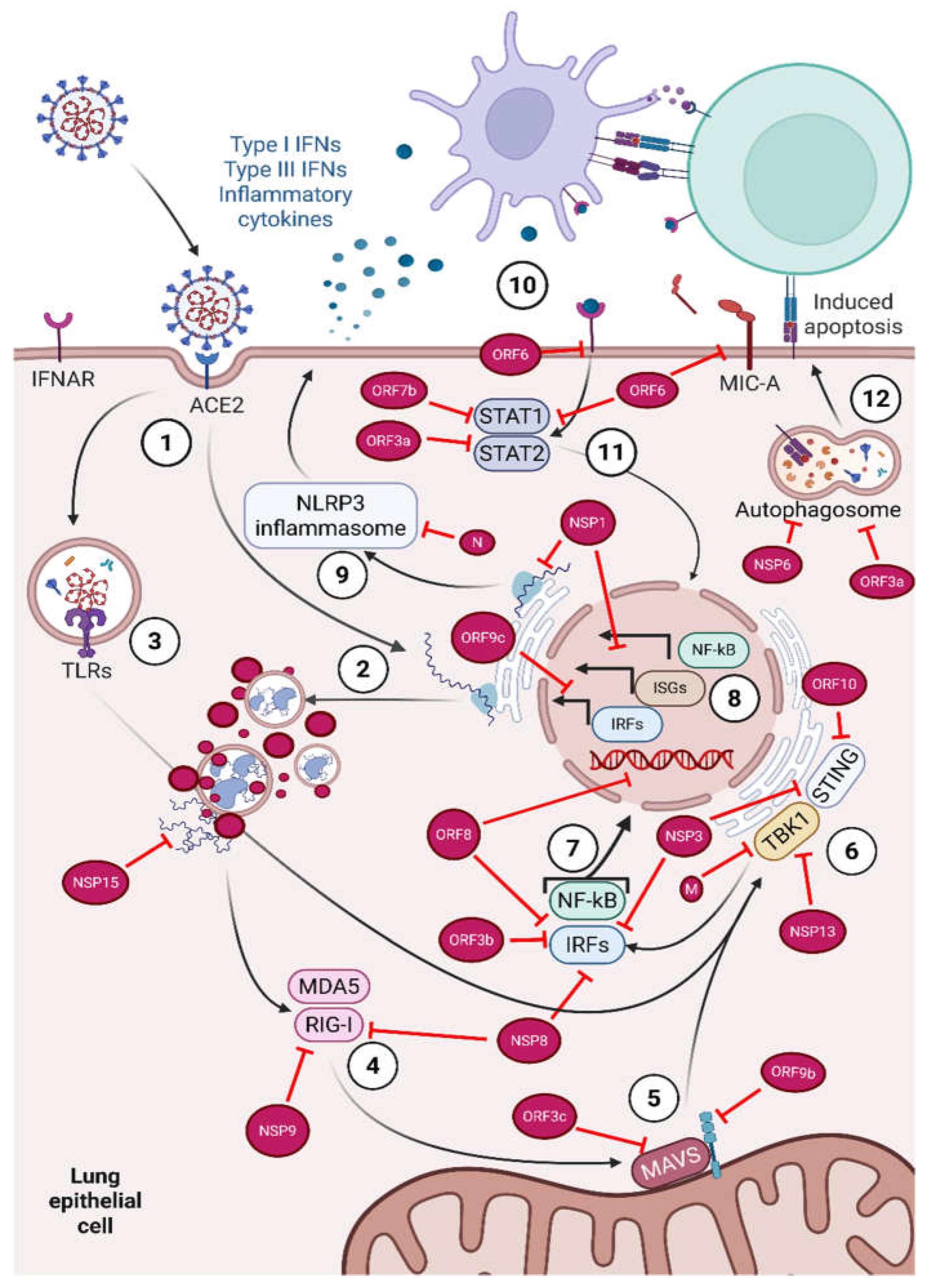
Figure 2.
Complement and coagulation pathways interactions (simplified). Coagulation: Vascular injury exposes tissue factor (TF) on subendothelial cells (extrinsic pathway). Factor VII moves to this TF surface and is activated by trace amounts of already activated VII (VIIa), activated factor X (Xa), or thrombin (IIa). TF binds VIIa and the resulting complex activates factor X. In conjunction, factor V is recruited to activated platelets and activated mainly by thrombin. Xa and Va assemble on a phospholipid membrane and convert prothrombin (factor II) to thrombin (factor IIa), which converts fibrinogen (factor I) to fibrin (factor Ia). In parallel, exposed collagen/vWF capture and activate platelets, providing the negatively charged surface where most thrombin is generated in vivo (intrinsic pathway). Highly anionic surfaces (notably platelet polyphosphate, NET DNA/RNA, extracellular RNA from damaged cells, exposed basement membrane, and foreign materials) promote factor XII activation (XIIa). XIIa converts prekallikrein to kallikrein, which feeds back to activate more XII; XIIa also activates factor XI (Xia), which then activates factor IX (IXa). IXa binds to FVIIIa to form intrinsic tenase, which greatly amplifies thrombin and fibrin generation. Further, kallikrein cleaves High-molecular-weight kininogen (HMWK) to bradykinin, increasing vascular permeability and vasodilation. Complement: The classical (IgM/IgG–C1qrs binding) and lectin (MBL/ficolins–MASP binding) pathways both lead to C2 and C4 cleavage to form the same classical C3 convertase (C4b2a). This then cleaves large amounts of C3 to C3b and C3a (alongside other fragments). Once a large amount of C3b forms on a surface, it binds with C4b2a to form the C5 convertase of the classical and lectin pathways. In conjunction, the alternative pathway binds C3b to factor B and promotes its cleavage via Factor D, forming C3bBb, the alternative C3 convertase. This is an amplification loop to dump large amounts of C3b onto whichever surface is not protected by complement regulators (factor H, MCP/CD46, etc.). C3bBb then binds another C3b to form the alternative C5 convertase. Factor H (FH) regulates this by serving as a cofactor for C3b cleavage and accelerates the decay of C3bBb. Both C5 convertases cleave C5 into C5a and C5b, which recruits C6, C7, C8, and polymerizes C9 to form a pore (MAC). On host cells at sublytic levels, MAC activates endothelium and platelets, increasing TF, vWF release, permeability, and leukocyte adhesion, making them procoagulant and pro-inflammatory. Crosstalk: C5a (and, to a lesser extent, C3a) activates monocytes, endothelium, and platelets, driving TF expression (promoting extrinsic pathway activation) and platelet aggregation. NETs bind platelets and provide a scaffold for factor XII activation. Conversely, thrombin, factor Xa, and plasmin can directly cleave C3/C5 to C3a/C5a-like fragments, sustaining complement activity. Endothelium releases tPA, which converts plasminogen to plasmin on fibrin; plasmin clears crosslinked fibrin (and can degrade fibrinogen) while also contributing to complement cleavage. Created in BioRender. Elens, L. (2025) https://BioRender.com/w1ylbg9.
Figure 2.
Complement and coagulation pathways interactions (simplified). Coagulation: Vascular injury exposes tissue factor (TF) on subendothelial cells (extrinsic pathway). Factor VII moves to this TF surface and is activated by trace amounts of already activated VII (VIIa), activated factor X (Xa), or thrombin (IIa). TF binds VIIa and the resulting complex activates factor X. In conjunction, factor V is recruited to activated platelets and activated mainly by thrombin. Xa and Va assemble on a phospholipid membrane and convert prothrombin (factor II) to thrombin (factor IIa), which converts fibrinogen (factor I) to fibrin (factor Ia). In parallel, exposed collagen/vWF capture and activate platelets, providing the negatively charged surface where most thrombin is generated in vivo (intrinsic pathway). Highly anionic surfaces (notably platelet polyphosphate, NET DNA/RNA, extracellular RNA from damaged cells, exposed basement membrane, and foreign materials) promote factor XII activation (XIIa). XIIa converts prekallikrein to kallikrein, which feeds back to activate more XII; XIIa also activates factor XI (Xia), which then activates factor IX (IXa). IXa binds to FVIIIa to form intrinsic tenase, which greatly amplifies thrombin and fibrin generation. Further, kallikrein cleaves High-molecular-weight kininogen (HMWK) to bradykinin, increasing vascular permeability and vasodilation. Complement: The classical (IgM/IgG–C1qrs binding) and lectin (MBL/ficolins–MASP binding) pathways both lead to C2 and C4 cleavage to form the same classical C3 convertase (C4b2a). This then cleaves large amounts of C3 to C3b and C3a (alongside other fragments). Once a large amount of C3b forms on a surface, it binds with C4b2a to form the C5 convertase of the classical and lectin pathways. In conjunction, the alternative pathway binds C3b to factor B and promotes its cleavage via Factor D, forming C3bBb, the alternative C3 convertase. This is an amplification loop to dump large amounts of C3b onto whichever surface is not protected by complement regulators (factor H, MCP/CD46, etc.). C3bBb then binds another C3b to form the alternative C5 convertase. Factor H (FH) regulates this by serving as a cofactor for C3b cleavage and accelerates the decay of C3bBb. Both C5 convertases cleave C5 into C5a and C5b, which recruits C6, C7, C8, and polymerizes C9 to form a pore (MAC). On host cells at sublytic levels, MAC activates endothelium and platelets, increasing TF, vWF release, permeability, and leukocyte adhesion, making them procoagulant and pro-inflammatory. Crosstalk: C5a (and, to a lesser extent, C3a) activates monocytes, endothelium, and platelets, driving TF expression (promoting extrinsic pathway activation) and platelet aggregation. NETs bind platelets and provide a scaffold for factor XII activation. Conversely, thrombin, factor Xa, and plasmin can directly cleave C3/C5 to C3a/C5a-like fragments, sustaining complement activity. Endothelium releases tPA, which converts plasminogen to plasmin on fibrin; plasmin clears crosslinked fibrin (and can degrade fibrinogen) while also contributing to complement cleavage. Created in BioRender. Elens, L. (2025) https://BioRender.com/w1ylbg9.
Disclaimer/Publisher’s Note: The statements, opinions and data contained in all publications are solely those of the individual author(s) and contributor(s) and not of MDPI and/or the editor(s). MDPI and/or the editor(s) disclaim responsibility for any injury to people or property resulting from any ideas, methods, instructions or products referred to in the content. |
© 2026 by the authors. Licensee MDPI, Basel, Switzerland. This article is an open access article distributed under the terms and conditions of the Creative Commons Attribution (CC BY) license (http://creativecommons.org/licenses/by/4.0/).
Copyright: This open access article is published under a Creative Commons CC BY 4.0 license, which permit the free download, distribution, and reuse, provided that the author and preprint are cited in any reuse.


