

Submitted:
03 July 2026
Posted:
03 July 2026
You are already at the latest version
Abstract
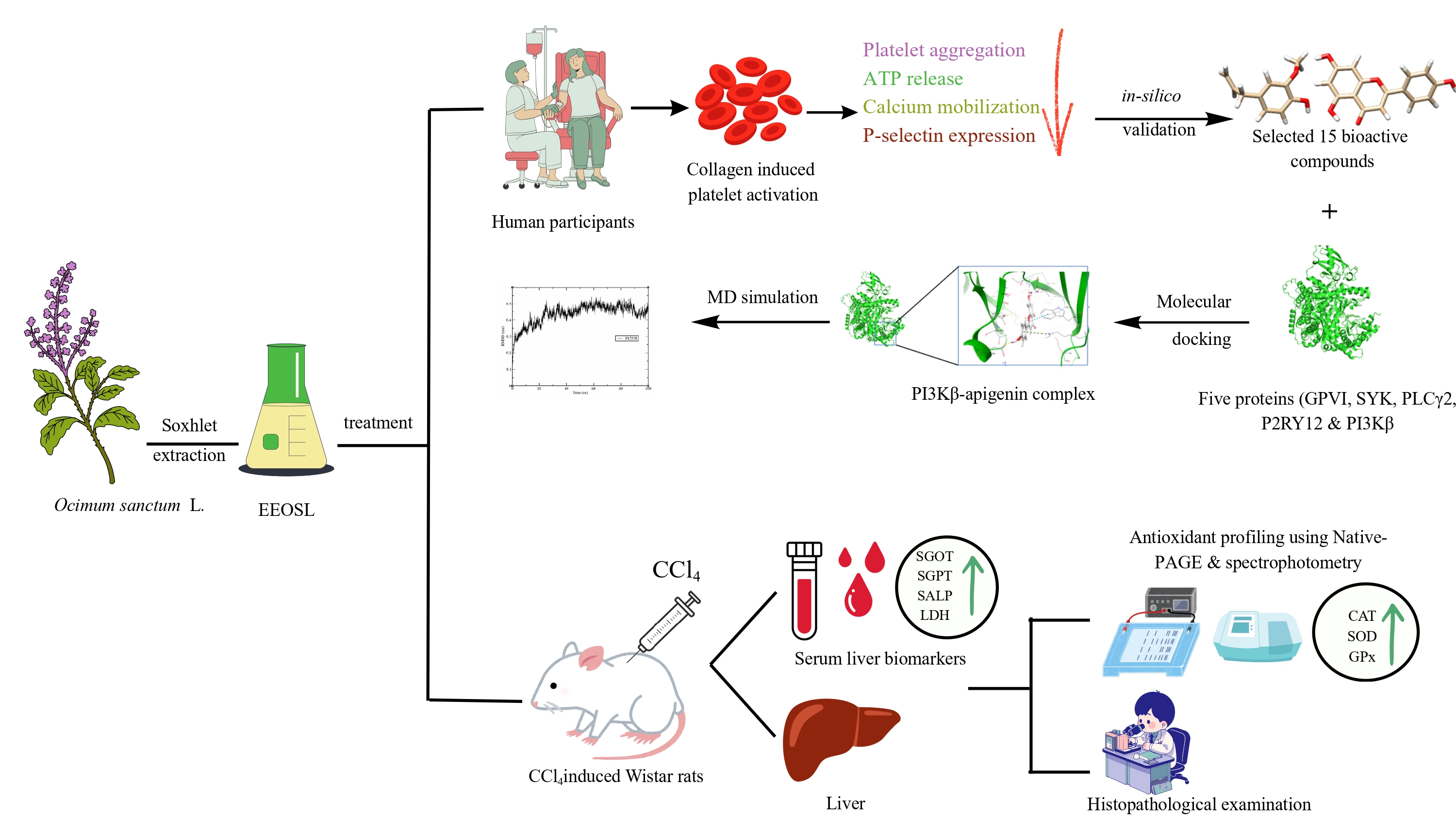
Natural products represent an important source of bioactive compounds with therapeutic potential in the fields of thrombotic and oxidative stress related disorders. The antiplatelet and hepatoprotective activity of ethanolic leaf extract of Ocimum sanctum L. was studied. Collagen-induced human platelet activation (CIHPA) and CCl4-induced hepatotoxicity were used in rats as models to assess the (EEOSL) activity. EEOSL (1-10 mg/mL) showed to significantly and dose-dependently inhibit collagen-induced platelet aggregation. The extract also inhibited P-selectin expression, ATP release and intracellular Ca2+ mobilization, suggesting inhibition of major pathways in platelet activation. To understand the mechanisms, molecular docking was carried out with signaling molecules of platelets such as GPVI, SYK, PLCγ2, P2RY12, and PI3Kβ. Some phytoconstituents have good binding affinities, apigenin having the highest binding affinity to PI3Kβ (−8.01 kcal/mol), and binding was further validated by molecular dynamics simulations showing the formation of stable complexes. Intravenous injection of CCl4 significantly increased the serum hepatic markers (SGOT, SGPT, LDH and SALP); the EEOSL treatment significantly reduced these increases in vivo. The extract normalized antioxidant enzyme activities (catalase, SOD and glutathione peroxidase) and antioxidant isozyme patterns. Histopathological results revealed a significant level of protection against liver damage. Overall, the results showed that EEOSL has strong antiplatelet and hepatoprotective properties, likely due to its ability to modulate platelet signaling pathways and increase antioxidant defense mechanisms.
Keywords:
Ocimum sanctum L.
; antiplatelet activity
; CCl4-induced hepatotoxicity
; oxidative stress
; molecular docking
; histopathology
; native PAGE
1. Introduction
Platelets are small anucleated fragments of megakaryocytes which normally circulate in a dormant form. Upon the exposure of subendothelial collagen by vascular injury, platelet glycoprotein VI (GPVI) and integrin α2β1 receptors are activated, triggering intracellular signaling pathways that include the activation of phospholipase Cγ2 and the production of inositol 1,4,5 trisphosphate (IP3) [1]. Signaling through IP3 quickly releases intracellular Ca2+ stores, which is replaced by prolonged extracellular intracellular Ca2+ influx which is another important platelet activation step [2]. There is an increase in cytosolic Ca2+, which leads to conformational activation of integrin α2β1, cytoskeleton rearrangement, and dense granule exocytosis, intensifying platelet aggregation [3]. The content of dense granules is released leading to extracellular release of ATP and ADP which stimulate platelet recruitment by purinergic (P2) receptor signaling pathways [4]. At the same time, the α-granule secretion causes the platelet activation marker of P-selectin (CD62P) surface expression, which promotes platelet-leukocyte adhesion and includes thromboinflammatory mechanisms [5].
Therefore, intracellular Ca2+ mobilization, ATP release, and P-selectin expression are reputable biochemical evidence of platelet activation. Uncontrolled amplification of these pathways is tightly connected with thrombotic diabolical cardiovascular diseases, such as coronary artery disease and ischemic stroke [6]. However, even though the use of antiplatelet drugs like aspirin and P2Y 12 receptor antagonist has proved effective in preventing thrombotic events, their long-run use has been linked to bleeding issues and hypersensitivity in some patients [7]. These restrictions indicate that safer, multitarget agents which have the capacity to mediate platelet signaling pathways are necessary. There is mounting evidence that platelet hyperreactivity is an important effect of oxidative stress. Reactive oxygen species (ROS) enhance Ca2+ signaling, granulocyte secretion, and P-selectin expression by activating redox-sensitive kinases and transcription [8,9]. Thus, antioxidant agents could reduce platelet activation through intracellular calcium homeostasis stabilization and secretory amplification.
The liver is the key metabolite and detoxifier of xenobiotics, and it is very prone to oxidative stress. Carbon tetrachloride (CCl4) is a popular hepatotoxin used in experimental studies as its metabolic activation by cytochrome P450 enzymes leads to the formation of reactive trichloromethyl radicals, which cause lipid peroxidation and membrane damage [10,11]. This oxidative damage interferes with the cell integrity and reduces the activity of the endogenous antioxidant defenses such as superoxide dismutase (SOD), catalase (CAT), and glutathione peroxidase (GPx), which promotes hepatocellular necrosis and inflammation [12]. Oxidative stress is a common mechanistically related process in platelet activation and hepatic injury, and thus, agents that can restore redox balance can apply protective effects in either of these systems.
Ocimum sanctum L. is a medicinal plant that contains a lot of phenolic compounds, flavonoids, and essential oil constituents like eugenol and rosmarinic acid that have antioxidant and anti-inflammatory properties [13,14]. Such bioactive components have been said to counteract free radicals, suppress lipid peroxidation and control redox-sensitive signaling pathways such as NF-κB and MAPK cascades [15,16]. Though the hepatoprotective effect of O. sanctum on chemically induced liver injury has been previously reported [17], its direct effect on specified platelet activation parameters of intracellular Ca2+ mobilization, ATP release by dense granules, and P-selectin surface expression has not been thoroughly examined. Moreover, no study has so far given a simultaneous test on its impact on platelet activation by collagen and on oxidative hepatotoxicity by CCl4 under one mechanistic framework. Taking into consideration that oxidative stress is one of the most frequent pathological denominations between thrombotic diseases and liver injury, measurement of agents that influence redox-dependent pathways in both settings is of great clinical interest. Thus, the current paper has explored the influence of the ethanolic extract of Ocimum sanctum leaves (EEOSL) on human platelet stimulation on collagen stimulated platelets through the measurement of intracellular Ca2+ mobilization, ATP release, and the expression of the P-selectin. To further validate the experimental data, molecular docking was performed to evaluate the binding affinity of the EEOSL major phytoconstituents against protein involved in platelet activation signaling. Following this, molecular dynamics (MD) simulation was conducted for the top-ranked docked complex to evaluate its stability during the interaction. Moreover, the extracts’ hepatoprotective effects against CCl4 induced liver damage in the rats with focus on antioxidant enzyme status and the histopathological changes involved.
2. Results
2.1. EEOSL Inhibits Collagen-Induced Human Platelet Aggregation
In the current study, EEOSL was found to greatly block platelet aggregation induced by collagen in human platelets. In the control group, a high response of platelets to stimulation was seen when collagen was used to stimulate the platelets. Nevertheless, pre-incubation of platelets with the presence of either 1 to 10 mg/mL EEOSL led to a significant and concentration dependent decrease in aggregation (Figure 1a). EEOSL showed fair inhibitory effect at lower concentrations (1 mg/mL), which reveals a partial block of collagen-mediated signaling pathways. The more the concentration of the EEOSL (10 mg/mL) exhibited significant inhibitory effect of platelet aggregation. This dose dependent effect demonstrates that the anti-platelet action of EEOSL increases with the concentration.
2.2. EEOSL Inhibits ATP Release, Intracellular [Ca2+]i Levels and Surface P-Selectin Expression
To further clarify the antiplatelet effect of EEOSL, its influence on major indicators of platelet activation was also determined.
2.2.1. Inhibition of ATP Release
An early step in platelet activation is dense granule secretion which is an important step in platelet activation. Freed ATP and ADP serve as secondary mediators that enhance platelet activation through purinergic receptors (P2Y1 and P2Y12), which increase their aggregation. EEOSL inhibited dose-dependently the collagen-induced ATP release (Figure 1b). As the EEOSL concentration increased, the ATP secretion was inhibited, which revealed that the dense granule exocytosis was inhibited. This implies that EEOSL disrupts the amplification of pathways required in maintaining the platelet aggregation.
2.2.2. Suppression of Intracellular [Ca2+] Mobilization
Calcium is an intracellular molecule that controls platelet activation through the granule secretion, cytoskeleton restructuring, activation of phospholipase, and the activation of integrin αIIbβ3. Stimulation of collagen is usually followed by a rapid increase in the level of calcium in the cell cytoplasm, which occurs due to the release of the intracellular stores and the entry of calcium into the cells. EEOSL at both concentrations tested had a significant effect on the elevation of intracellular [Ca2+] caused by collagen (Figure 1c). This observation suggests that EEOSL can disrupt the upstream signaling events (including activation of phospholipase C, production of IP3, activation of calcium channels, etc.), thus impairing the calcium-sensitive platelets.
2.2.3. Suppressing Surface P-Selectin Expression
The p-selectin is held in the α-granules, and during activation translocate to the platelet surface. Surface P-selectin expression is a well-known hallmark marker of platelet activation and has a significant contribution in platelet-leukocyte interaction and thrombus stabilization. The P-selectin surface expression was significantly enhanced by collagen stimulation. EEOSL however, strongly inhibited this upregulation (Figure 1d), which implies that it inhibited α-granule secretion. This observation also supports the fact that EEOSL inhibits platelet activation at the secretory responses level.
2.3. Molecular Modeling Studies
Initial screening used molecular docking to estimate how well different plant-based compounds might bind to enzymes that control platelet activation. By comparing the Glide SP scores for all the targets, we identified clear preferences. Lower (more negative) scores suggest a tighter fit.
Among the ligands studied, apigenin stood out with the strongest binding affinity in the entire study, scoring -8.00 kcal/mol against PI3Kβ (UniProt: P42338). When we looked at the pose, apigenin fit snugly inside the active site by forming three distinct hydrogen bonds with Glu852, Val854, and Asp862. This shows stable accommodation of the ligand within the active site (Figure 2A & B). The other proteins also had their best matches, but with weaker scores: 3-O-acetylpadmatin docked to GPVI (PDB: 2GI7) at -4.60 kcal/mol, methyl salicylate to SYK (PDB: 4FL2) at -3.52 kcal/mol, isoferulic acid to P2RY12 (PDB: 4NTJ) at -6.59 kcal/mol and kaempferol-3-glucuronide bound to PLCγ2 (PDB: 8T7C) at -5.84 kcal/mol (as summarized in Table 1). The two-dimensional (2D) protein-ligand interactions is presented in the Supplementary Figure S1. These computational insights suggest that several compounds in the extract likely interfere with distinct points in the platelet signalling network. However, since the PI3Kβ-apigenin pairing produced such a notably strong score, we prioritized it as our main candidate for MD simulations.
2.4. Molecular Dynamics Simulation
To ensure that the docking pose of the PI3Kβ-apigenin complex was not just a static artifact, we conducted a 100 ns molecular dynamics simulation. We measured RMSD, RMSF, H-bond, and Rg to assess the structural stability of the complex in a simulated aqueous environment.
Tracking the backbone RMSD showed an initial rise from 0.20 nm to about 0.35 nm as the protein adjusted to the solvent, indicating early structural changes after solvation and equilibration. However, by the 25 ns mark, the curve flattened out between 0.45 and 0.50 nm. It remained stable in that range for the rest of the run, demonstrating that the overall structure of the PI3Kβ-apigenin complex reached a stable state (Figure 3a).
The local flexibility analysis through RMSF showed that most of the protein backbone barely moved, fluctuating by less than 0.2 nm. A few isolated peaks reached 0.5–0.7 nm, but mapping these onto the 3D structure confirmed they were just loose terminal ends and surface loops, leaving the core binding pocket intact (Figure 3b).
The H-bond analysis revealed that apigenin maintained between 1 and 4 hydrogen bonds with PI3Kβ during the simulation, briefly spiking to 5 at times. This consistent bonding pattern shows the stability of the complex, as the ligand did not drift out of the pocket; it remained firmly docked throughout the entire 100 ns period (Figure 3c).
Finally, the Rg barely shifted, staying steady between 3.24 and 3.32 nm from start to finish. Since the Rg did not suddenly jump or drop, it indicates that the protein did not unfold or swell abnormally when apigenin bound to it (Figure 3d). Overall, these metrics strongly support the docking results, confirming that apigenin forms a dynamically stable complex with PI3Kβ.
2.5. Effect of EEOSL on Serum Enzyme Parameters
In this experiment, rats of Group II who were fed carbon tetrachloride (CCl4) showed a high level of serum SGOT, SGPT, SALP, and LDH in comparison to normal control group (Group I). CCl4 is a proven hepatotoxicant which causes hepatotoxicity by the creation of reactive metabolites like trichloromethyl (•CCl3) and trichloromethyl peroxy (•OOCCl3). This radical species trigger lipid peroxidation, membrane disruption of hepatocytes and eventually lead to release of intracellular enzymes to the blood.
Treatment of Group III (CCl4 + EEOSL) however significantly reduced the enzyme levels back to normal levels (Figure 4a-d). The high decrease in the SGOT and the SGPT levels indicates the reduction of hepatocellular injury, and the restoration of normal SALP levels are indicators of better biliary functions and membrane stabilization. On the same note, a drop in LDH activity indicates a drop in cellular necrosis and an increase in tissue integrity. It has been shown that serum enzyme levels in EEOSL-treated rats are restored, which is an indication of its hepatoprotective property, which could be due to antioxidant activity, free radical scavenging activity, and stabilization of hepatocyte membranes. The results indicate that EEOSL can be used to prevent the impact of CCl4 on the hepatotoxicity and liver functions.
2.6. EEOSL Enhances CCl4- Mediated Reduction of Antioxidant Enzymes
In the current experiment, the hepatic SOD, CAT, and GPx activities were significantly lowered in rats of Group II (CCl4 -treated) relative to the normal control group (Group I) (Table 2). The fact that these antioxidant enzyme activities were reduced shows that the oxidative stress was acute, caused by the administration of CCl4. Nonetheless, EEOSL treatment in Group III (CCl4 + EEOSL) raised the SOD, CAT, and GPx activities near to normal levels. The increase in the activity of SOD indicates better degradation of superoxide radicals into hydrogen peroxide. The increase of CAT and GPx activity demonstrates that there are better processes of hydrogen peroxide and lipid peroxide detoxification, and the oxidative harm to the hepatic tissue is decreased.
2.7. EEOSL Regulates the Isoenzyme Profile of Antioxidant Enzyme in CCl4 Induced Liver in Rats
2.7.1. Effects on CAT Isozyme
Considering the liver homogenates of all three experimental groups, the results of non-denaturing electrophoretic analysis showed one CAT isozyme band that was detectable (Figure 5a), which implies that no other isozyme variants were activated under the experimental conditions. Nevertheless, the intensity of the bands was found to differ significantly in the groups, revealing the difference in the activity and level of expression of CAT enzyme. Group I, the normal control, had a high-intensity CAT band with a band area of 62107.2, which is a good defense against physiological stress conditions. Oppositely, Group II rats that received only CCl4 had a significantly smaller band intensity (band area: 36,597.48), indicating great depletion or inactivation of CAT at the hands of CCl4-induced oxidative stress. The decreased intensity of the staining is an indication of inhibited ability to deactivate hydrogen peroxide and this increases oxidative injury in hepatic tissue. Interestingly, the Group III rats (CCl4 + EEOSL treatment) showed a significant recovery of CAT band intensity (band area: 39,198.91) of the CCl4 group (Figure 5a). The partial recovery was found although the intensity was not as high as in the normal control group, showing that EEOSL is effective in reducing oxidative stress and improving CAT enzyme stability or expression.
2.7.2. Effects on SOD Isozyme
In this study, all the experimental groups showed only one protein band of SOD isozyme (Figure 5b), which showed that no other isoforms were induced by the administration of CCl4 and EEOSL treatment. It was however found that there were significant differences in band intensity in the groups resulting in differences in enzyme activity and expression. Group I had a strong stained SOD band with a band area of 38,968.13 which was a sign of normal antioxidant capacity. On the same note, Group III rats (CCl4 + EEOSL) had one of the highest band intensities with a band area of 31,177.95. Though a little lower than the control group, the intensity was still significantly higher than the CCl4 only group.
Conversely, the intensity of the band of Group II rats that were exposed to CCl4 alone was significantly lower and the band area was 13,431.11 (Figure 5b). Such tremendous decrease is an indication of severe depletion or inactivation of SOD by overwhelming oxidative stress caused by the CCl4 metabolites. The reduced SOD would increase the superoxide radicals, which would further promote lipid peroxidation and injury of the hepatocellular injury.
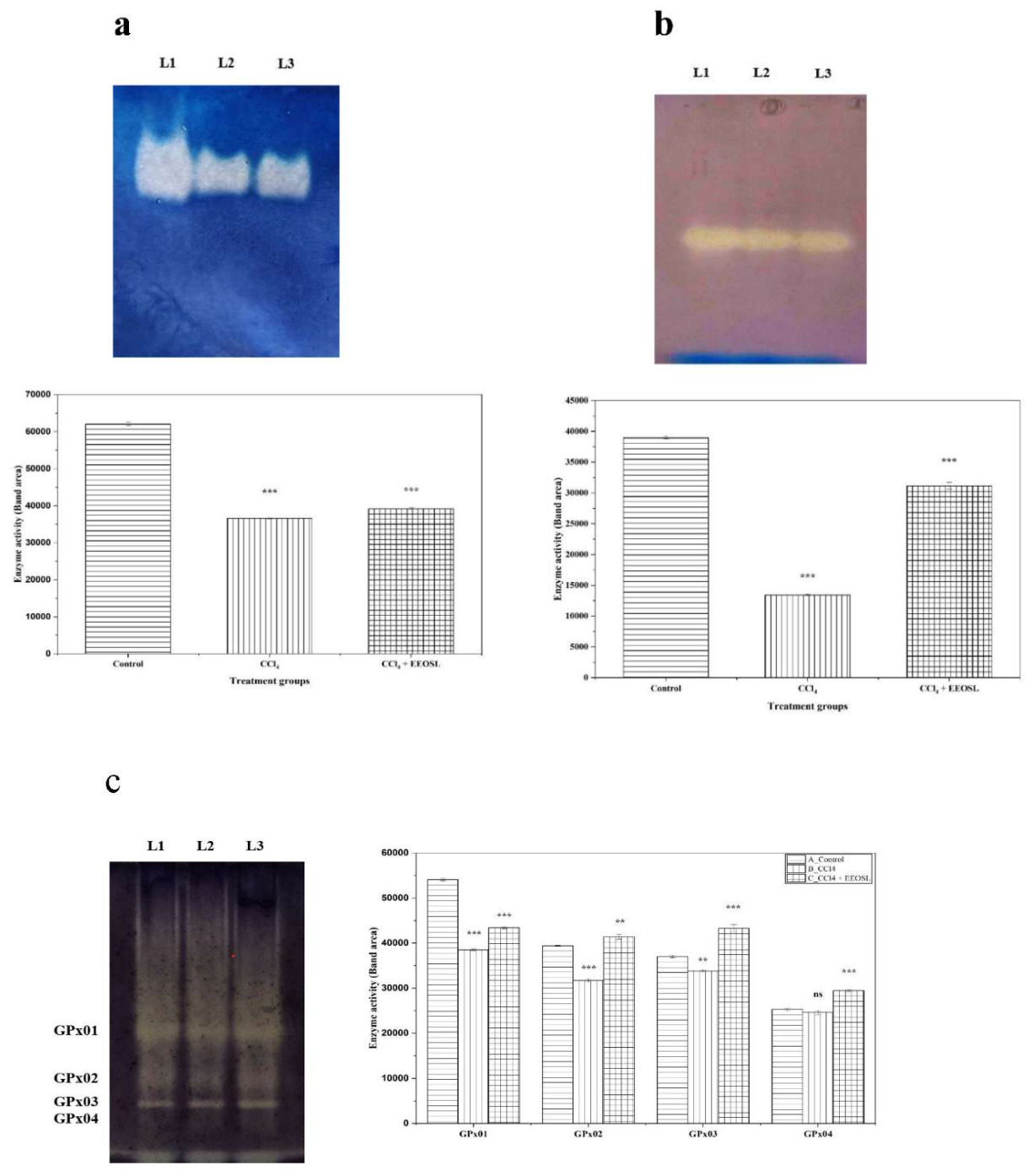
Figure 5.
EEOSL influences the antioxidant enzymes catalase (a), superoxide dismutase (b) and glutathione peroxidase (c) isozyme in liver tissue of rats induced with CCl4. Native-PAGE was used to measure catalase (CAT), superoxide dismutase (SOD) and glutathione peroxidase (GPx) isozyme in liver tissue. (top panel) Representation gel picture of L1 (normal), L2 (treated with CCl4) and L3 (treated with CCl4+ EEOSL). (bottom panel) Densitometric pattern of isozyme activity in liver tissue. The results are in the form of mean, standard error of the mean (n=5). **p < 0.01, *** p < 0.001, a non-significant difference compared to the control group is denoted by ns.
Figure 5.
EEOSL influences the antioxidant enzymes catalase (a), superoxide dismutase (b) and glutathione peroxidase (c) isozyme in liver tissue of rats induced with CCl4. Native-PAGE was used to measure catalase (CAT), superoxide dismutase (SOD) and glutathione peroxidase (GPx) isozyme in liver tissue. (top panel) Representation gel picture of L1 (normal), L2 (treated with CCl4) and L3 (treated with CCl4+ EEOSL). (bottom panel) Densitometric pattern of isozyme activity in liver tissue. The results are in the form of mean, standard error of the mean (n=5). **p < 0.01, *** p < 0.001, a non-significant difference compared to the control group is denoted by ns.
2.7.3. Effects on Gpx Isozyme
All three experimental groups showed that there were four different GPx isozymes (Figure 5c), which showed that there were a variety of GPx forms expressed in the hepatic tissue. A major difference in the intensity of the band staining was however found between the groups. The four GPx isozymes were easily observed in the normal control group (Group I) and their staining was moderate, indicating normal antioxidant endowment. Conversely, the staining intensity of the four GPx isozymes of Group II rats which had only CCl4 treatment showed significant reduction (Figure 5c). This reduction implies that there is oxidative inactivation or down-regulation of the GPx enzymes caused by the overproduction of free radicals and lipid peroxidation by CCl4. This reduced GPx activity would hinder the process of peroxide detoxification and thus increase the level of hepatic oxidative injury.
Interestingly, the Group III rats (CCl4 + EEOSL treatment) in comparison to the Group I rats (control group) had an increase in the intensity of all GPx isozymes except GPx01. The higher activity of the GPx isozymes in EEOSL-treated rats shows the stimulatory or protective action on the expression and stability of antioxidant enzymes. This increase could indicate the mobilization of endogenous antioxidant defense mechanisms and an increase in redox homeostasis of hepatic tissue.
2.8. Effects of EEOSL on CCl4-Induced Histological Alterations
To determine the structural changes in the liver tissues of the experimental groups, histopathological examination of liver tissues was conducted under the hematoxylin and eosin (H&E) staining according to experimental groups.
Sections of the liver of the normal control (Group I) displayed the intact hepatic architecture with properly organized hepatocytes having typical polygonal form, central position of prominent nuclei, and distinct plasma membranes. The hepatic lobular structure remained undisturbed with central veins being normal and sinusoidal spaces well defined. The portal vein, hepatic artery, and the bile duct made the so-called periportal triad were also found to be structurally normal, which speaks of the preservation of the vascular and biliary integrity (Figure 6a and b). In comparison, liver sections of Group II rats that received CCl4 exhibited severe pathological changes. Diffuse cytoplasmic vacuolation, hepatocellular ballooning, inflammatory infiltration of the peripheral areas, vascular congestion and necrosis foci were severe (Figure 6c and d). Such structural defects are typical of CCl4-induced hepatotoxicity and are indicative of extensive oxidative injury, membrane disturbance, and inflammatory reactions in hepatic tissue.
Nonetheless, liver segments of Group III (CCl4 + EEOSL treatment) showed significant enhancement of histological architecture when compared to CCl4 alone group. Despite the fact that moderate cytoplasmic vacuolation, mild perivascular inflammation and congestion was still evident, the hepatic architecture as a whole did not seem to be significantly affected. Hepatocytes maintained almost normal morphology and the structural composition of hepatic cords and sinusoids was significantly reinstated (Figure 6e and f). These histological results support the biochemical and enzymatic results that EEOSL has a protective effect on CCl4-induced hepatic injury.
3. Discussion
In recent years, plants have been extensively explored for their therapeutic and pharmacological activities. Owing to the presence of diverse bioactive constituents, medicinal plants serve as valuable sources for commercial drug development [18]. The current study aimed to investigate the combined effect of EEOSL as both an antiplatelet and hepatoprotective agent. Overall, the findings indicate a significant modulatory role of the extract in thrombosis and hepatic injury.
The antiplatelet study demonstrated that treatment with EEOSL reduced collagen-induced platelet aggregation. Platelet activation can be triggered by various agonists [19]. Collagen, the agonist used in this study, plays a pivotal role in platelet activation by binding to the glycoprotein VI (GPVI) receptor on platelets. During vascular injury, platelets adhere to the exposed subendothelial matrix component, collagen, forming a complex that acts both as an adhesive surface and a signaling stimulus, thereby initiating platelet activation. Treatment with EEOSL showed significant, dose-dependent inhibition of collagen-induced platelet aggregation, suggesting its effect on platelet–platelet interaction and the phospholipase C-mediated signaling pathway [20].
Platelet activation initiates a cascade involving tyrosine kinase signaling, leading to increased intracellular calcium concentration and exocytosis of granules such as P-selectin and ADP/ATP. ATP functions as a secondary agonist, recruiting additional platelets, while elevated intracellular calcium levels trigger morphological changes, granule secretion, and aggregation, thereby promoting thrombus growth. P-selectin, stored in α-granules, is translocated to the platelet surface upon activation. The release of α-granules during exocytosis is a hallmark of platelet activation and is commonly assessed by measuring P-selectin expression [21]. In the present study, EEOSL dose-dependently inhibited ATP release and impaired calcium mobilization. Furthermore, a reduction in P-selectin expression was observed. Additionally, molecular docking performed validated the role of EEOSL phytoconstituents in platelet activation process. The bioactive phytocompounds docked showed promising ability against the collagen induced GPVI/SYK/PLCγ2 and ADP signaling P2RY12/ PI3Kβ proteins with the PI3Kβ-apigenin complex being the most favorable binding with the binding score of -8.00 kcal/mol. Further, evaluation of PI3Kβ-apigenin complex through MD simulation confirmed its stability. Collectively, these findings indicate that EEOSL modulates both the early and late phases of platelet activation. These observations are consistent with previous reports [20,21] demonstrating that eugenol and methyl eugenol two important constituents of Ocimum species suppress collagen-induced platelet activation.
The liver is a vital organ responsible for numerous biological functions, including metabolism, detoxification, and digestion [22]. Liver diseases account for approximately 4% of global deaths annually (1 in 25 deaths worldwide) [23]. Hepatic disorders can arise through multiple mechanisms, including exposure to environmental pollutants. Acute liver injury is closely associated with oxidative stress, which leads to the generation of reactive oxygen species (ROS). These free radicals impair liver function by creating an imbalance between oxidants and the antioxidant defense system. Consequently, oxidative stress contributes to hepatic damage characterized by inflammation, vasodilation, cellular hypertrophy, bile duct proliferation, fibrosis, and necrosis [24]. Various experimental models have been employed to study drug-induced liver injury [25]. In the present study, CCl4-induced hepatotoxicity was used as the experimental model. CCl4 is a well-known hepatotoxin that induces liver damage primarily through oxidative stress. The resulting free radical formation leads to leukocyte infiltration, vascular occlusion, and excessive collagen deposition, ultimately causing severe hepatic injury [24].
The natural products have been one of the most reliable sources of bioactive compounds that have varied pharmacological effects and in particular antioxidant potential. Many clinically useful medications such as quinine, morphine and paclitaxel have their source in plants [26]. Lamiaceae is a cosmopolitan family which contains a large variety of medicinally valuable plants. Ocimum sanctum (also referred to as Holy Basil or Tulsi) is one of them and has been used conventionally in a number of therapeutic systems due to its antioxidant, anti-inflammatory, anticancer, anti-ulcer, and antimicrobial properties [14]. Considering these pharmacological characteristics, the current study was aimed at determining the protection of EEOSL on liver damage caused by CCl4.
Biochemical indicators expressed in serum are sensitive indicators of liver dysfunction. Liver damage causes hyper permeability of membrane and injury to hepatocellular causing leakage of intracellular enzymes including SGPT (ALT), SGOT (AST), SALP (ALP), and LDH in the bloodstream [27]. This serum enzymes increased significantly in rats treated with CCl4 in the current study and this proves the damage to hepatocells. Nevertheless, EEOSL treatment was also associated with the significant decrease in the serum levels of these enzymes, which demonstrated the stabilization of hepatocyte membranes and liver functioning recovery. The results are in line with previous literature that has shown the hepatoprotective property of plant-based antioxidants [28,29]. The CCl4 induced hepatotoxicity is mainly administered via oxidative stress. When activated in the liver metabolically, CCl4 produces extremely reactive free radicals, which cause lipid peroxidation, protein oxidation, and membrane damage of cells. Thus, interventions which are rich in antioxidants are a potential method to reverse oxidative stress induced liver damage. The liver has a strong enzyme antioxidant defense mechanism such as catalase (CAT), superoxide dismutase (SOD), and glutathione peroxidase (GPx). The enzymes act in synergy to counter the reactive oxygen species (ROS). SOD catalyzes the breakdown of the superoxide radicals into hydrogen peroxide that is later neutralized into water and oxygen by CAT and GPx. Abnormality in this antioxidant defense mechanism causes excess accumulation of ROS, which eventually causes hepatic dysfunction [22].
In the present study, CCl4 treatment had a great impact on lessening the activity of CAT, SOD, and GPx, which supports the research results from earlier studies [22,28,30]. The reduction of the antioxidant enzyme activity is the sign of excessive oxidative stress and the weakening of the cell defenses. It is worth noting that EEOSL treatment regained the activities of these enzymes up to the normal levels indicating that the extract has a strong antioxidant effect. This restoration suggests that EEOSL can directly scavenge the free radicals or increase the expression of endogenous antioxidant enzymes, which in turn prevents hepatocytes against oxidative damage caused by CCl4. Biochemical results were further supported by histopathological analysis. In Group II (CCl4 treated) rats there was severe disruption of the architecture of the liver tissues with cell degeneration and structural disorganization. Group III (CCl4-exposed rats treated with EEOSL) on the other hand showed significantly better hepatic architecture with little structural changes and is very close to the control group (Group I). These results confirm the protective effect of EEOSL on the tissue level. Similar hepatoprotective effects of natural products in a CCl4 model have been shown by previous studies. Normal hepatocyte architecture was restored in Pleurospermum candollei methanolic extract with clear nuclei [22]. There was a dose-dependent reduction of the inflammatory cell inflammation and hepatocyte destruction in mice treated with CCl4 by Shibi tea extract [28]. Aqueous extract of Carica papaya seeds enhanced the hepatocellular degeneration and fatty acids changes [31]. Similarly, Pleurotus ostreatus (oyster mushroom) with antioxidant properties resorted to the hepatocyte architecture and sinuosity of liver damage due to CCl4 in Wistar albino rats [30]. This pattern of protection, as detected in the current study, is in line with these results, which serve to support the therapeutic potential of antioxidant-rich natural products. Thus, the biochemical and histopathological changes in CCl4-treated rats were well reduced by the use of EEOSL, which indicates a high degree of hepatoprotective properties. Its antioxidant properties may explain the protective effect, which is counteracted by oxidative stress, cellular integrity, and normal restoration of hepatic architecture. All these results underscore the therapeutic potential of EEOSL as a natural antioxidant agent in the treatment of oxidative stress-induced liver diseases.
4. Materials and Methods
4.1. General Experimental Procedure
All the reagents and chemicals in the experiment were of analytical grade. Fresh leaves of Ocimum sanctum Linn. were gathered in the area of Pondicherry University, India and were verified by Dr. N. Parthasarathy, Professor, Pondicherry University. The leaves collected were dried in shade during one week and roughly powdered. The extraction was done on the powdered material by use of a Soxhlet apparatus using ethanol as the solvent. A rotary evaporator was used to concentrate the resulting crude extract under reduced pressure and then stored at 4ºC until it is used further.
4.2. Platelet Preparation
Blood samples of healthy volunteers that had not taken any non-steroidal anti-inflammatory drugs (NSAIDs) within two weeks before the study were obtained. As anticoagulant, whole blood was gathered in polypropylene tubes with acid citrate/dextrose solution (ACD; 9:1, v/v). The blood that was collected was centrifuged at 120xg and the platelet-rich plasma (PRP) was obtained after 10 min centrifugation. The PRP layer was thoroughly aspirated and incubated with heparin and prostaglandin E1 to avoid platelet activity. Then, PRP was centrifuged (500 x g) and collected after 10 min to get platelet pellet. The pellets were washed and resuspended twice in Tyrode solution containing bovine serum albumin (BSA, 3.5 mg/ mL). Calcium chloride was introduced to obtain the final Ca2+ of 1 mM. This was brought up to the final platelet concentration of 3.6 x 10⁸ cells/mL. The study protocol for human platelet experiments was approved by the Institutional Review Board of Taipei Medical University (TMU-JIRB-N202112047).
4.3. Platelet Aggregation Assay
The turbidimetric technique in a luminometric aggregometer was used to measure platelet aggregation. Pre-incubation of EEOSL (1-10 mg/mL) and vehicle (0.1% DMSO) were incubated with washed platelet suspensions (3.6×10⁸ cells/mL) for 3 min. The aggregation of platelets was triggered by the addition of collagen (1 μg/mL). The test was further subjected to aggregation which was recorded in response to an extra 6 min which was expressed as the percentage aggregation in comparison with control.
4.4. ATP Release and Calcium Mobilization Assay
A luciferase/luciferin bioluminescence measurement was used to determine the ATP release. The calcium mobilization of intracellular calcium was established by fluorescent calcium indicator Fura-2 AM. Fluorescence signals (Ca2+ mobilization), luminescence (ATP release) was measured on a Hitachi F-7000 Fluorescence Spectrophotometer, as per the manufacturer instructions. The intracellular calcium concentration was determined by ratio intensity of fluorescence excitation at 340 /380 nm.
4.5. P-Selectin Expression
Flow cytometry was used to analyze the surface expression of P-selectin. The platelets that were washed with FITC-conjugated with anti-P-selectin monoclonal antibody (2μg/mL) were incubated with EEOSL (5 and 10 mg/mL) for 3 min. After preincubation, collagen (1 μg/mL) was used to induce P-selectin expression. Platelets with the FAM-labeled fluorescein were identified by BD FAC Scan (Becton Dickson, San Jose, CA, USA). Each sample was analyzed on 10000 platelets. Gating and identification of platelets was done according to the characteristic light profiles of forward scatter (FSC) and side scatter (SSC). The findings were expressed as surface P-selectin expression measured as mean fluorescence intensity (MFI).
4.6. Molecular Docking
The target proteins selected for molecular docking were glycoprotein VI (GPVI; PDB ID: 2GI7), spleen tyrosine kinase (SYK; PDB ID: 4FL2), phospholipase Cγ2 (PLCγ2; PDB ID: 8T7C), and the purinergic receptor (P2RY12; PDB ID: 4NTJ), and the respective three-dimension (3D) structures were retrieved from the RCSB Protein Data Bank (RCSB PDB: https://www.rcsb.org/), along with phosphoinositide 3-kinase β (PI3Kβ; UniProt ID: P42338), obtained from the UniProt database (https://www.uniprot.org/). These proteins were selected because they represent key components of platelet activation pathways involved in collagen and ADP-mediated signaling.
Molecular docking was performed using the Schrödinger suite (Schrödinger Release 2025-1: Glide, Schrödinger, LLC, New York, NY, 2025) [32,33,34]. Protein structures were prepared using the Protein Preparation Wizard by removing non-essential water molecules, adding missing side-chain atoms, assigning appropriate bond orders and protonation states, and minimizing the structures to relieve steric clashes [35]. Ligands were prepared using the LigPrep module of Maestro (Schrödinger Release 2025-1: LigPrep, Schrödinger, LLC, New York, NY, 2025.), which generated low-energy three-dimensional conformations with appropriate stereochemical, protonation, and ionization states.
Receptor grids were generated based on the known ligand-binding region of each target protein, and binding-site prediction was further supported using the COACH-D 2.0 server [36]. The active-site residues used for docking are listed in Table 3, and the structures of the selected ligands are shown in Figure 7. Flexible ligand docking was then carried out using the standard-precision (SP) Glide algorithm [37], and docking scores together with protein-ligand interaction patterns were used to identify the most promising complexes for further molecular dynamics analysis.
4.7. Molecular Dynamics Simulation
Molecular dynamics (MD) simulation of the selected protein-ligand complex was performed using GROMACS version 2023.4 [38,39] with the CHARMM27 force field (CHARMM22 plus CMAP correction for proteins) [40]. The topology and parameters for the apigenin ligand, compatible with the CHARMM force field, were generated using the SwissParam web server [41]. The complex was dissolved in a triclinic simulation box using the TIP3P water model. A minimum distance of 1.0 nm maintained between the solute and the box boundaries using the GROMACS editconf module. The solvated system was neutralized by replacing solvent molecules with Na⁺ and Cl⁻ ions using the gmx genion module.
Energy minimization was carried out using the steepest descent algorithm to remove steric clashes and unfavorable contacts. The system was then equilibrated under NVT and NPT ensembles with positional restraints of 1000 kJ mol⁻¹ nm⁻² applied to the complex. We removed the restraints and let the production simulation run for 100 nanoseconds (ns). We examined the structural stability and dynamic behavior of the complex by analyzing the backbone root mean square deviation (RMSD), root mean square fluctuation (RMSF), radius of gyration (Rg), and intermolecular hydrogen bonds (H-bonds) using standard GROMACS analysis tools.
4.8. Animal Experiments
Male healthy Wistar rats (180-250g) purchased at Biogen Laboratory Animal Facility, Bangalore, India. The animals were housed and kept in Central Animal House Facility, Pondicherry University, in a controlled environment (25 ± 2ºC; 12 h light/ darkness cycle). Animals were permitted to acclimatize over a period of 14 days, followed by experiments, which were done on normal pellet food and water ad libitum. Three groups of (n = 5) rats were randomly selected: Group I (Normal Control): Feasted on vehicle (olive oil, 1 mL/kg body weight) on four days. Group II (Toxin Control): Perceived vehicle on day one and four; on day two and three, CCl4 (50 percent v/v in olive oil, 2 mL/kg b.w.) was used on the rats. Group III (Test Group): Obtained EEOSL (500 mg/kg b.w.) on 1st and 4th days; on 2- and 3-days rats were treated with both EEOSL and CCl4. All the treatments were delivered through the intraperitoneal route. Animals were sacrificed on day 5. Blood samples were obtained through retro-orbital plexus and liver tissues were cut down instantaneously. Samples of serum and liver were frozen at -80ºC to be subjected to biochemical tests later. The experimental procedures involving rats were approved by The Institutional Animal Ethics Committee (IAEC), Pondicherry University (Approval No. PU/CAHF/30th IAEC/2025/04) approved the experimental protocol and were carried out strictly in accordance with the CPCSEA (Committee for the Purpose of Control and Supervision of Experiments on Animals) guidelines.
4.9. Determination of Serum Liver Marker Enzymes
Enzymes that occur in the liver are present in the serum and are detected by the test. Serum biochemical indicators (SGPT (ALT), SGOT (AST), SALP (ALP), and LDH) were measured as per the standard laboratory measures. The enzyme activities were reported in U/L (SGPT, SGOT, SALP) and IU/L (LDH).
4.10. Preparation of Rat Tissue Homogenate
Liver tissue (100 mg) was homogenized in 1 mL of 50 mM phosphate buffer (pH 7.0) in the state of ice-cold. The homogenate was centrifuged at a speed of 10,000 rpm with a duration of 15 min at 4ºC. The supernatant that appeared was taken in order to run biochemical tests. The amount of total proteins was determined based on the standard procedure [42].
4.11. Quantitative Analysis of Antioxidant Enzymes
4.12. Native-PAGE Isozyme Detection
Antioxidant isozymes are detected by Native-PAGE. Native-PAGE was done as per the procedure of [46]. Liver homogenates were made without any form of denaturing agent like SDS and samples were not heat-treated before electrophoresis. The proteins (20µg) were loaded equally on the gels. The separation of SOD and GPx isozymes was done in 10% polyacrylamide gels and CAT isozyme separation was done on 8% gels. Electrophoresis was performed at 4ºC using a fixed voltage of 50V (stacking gel) and 80V (separating gel). Isozymes specific staining was performed according to the procedures [47,48,49] of the SOD, GPx, and CAT respectively.
4.13. Histopathological Examination
Histopathological examination of liver tissues was done with the conventional paraffin-embedding and hematoxylin & eosin (H&E) staining with slight modifications [50]. At the end of the experimental duration, the rats were euthanized under CO2 euthanasia, and necropsy was performed. The livers were carefully excised out and fixed into 10% neutral buffered formalin for tissue preservation. The fixed tissues were cut into small sections (4mm) and kept overnight under running tap water for removing formalin. Following this process, tissue processing was done using grades of alcohol (50%, 70% and 90%; absolute alcohol 1 and 2) for dehydration and xylene for clearance and blocks prepared with 60°C melted paraffin. Tissue sections were taken using a rotary microtome at 4-micron thickness and dried slides were stained with H & E staining, where Haematoxylin is the basophilic stain gives blue colour to the nucleus and Eosin is the eosinophilic or acidophilic stain gives pink colour to the cytoplasm. The stained slides were mounted with DPX mount. Finally mounted dried slides examined under the microscope from low power to high power.
4.14. Statistical Analysis
The experimental results were used to represent the mean and the standard error of mean (SEM). One-way analysis of variance (ANOVA) with Tukey honestly significant difference (HSD) post-hoc test was conducted in OriginPro2026 to measure the statistical significance of the differences. A preset value p<0.05 was regarded as statistically significant.
5. Conclusion
The current research shows the protective effect of EEOSL on platelet activation triggered by collagen as indicated by a significant inhibitory effect on platelet aggregation, the decrease in ATP release, subdued intracellular calcium mobilization and decreased P-selectin. The docking and MD simulation studies further supported EEOSL anti-platelet potential. In particular, apigenin with the binding score of -8.00574 kcal/mol with PI3Kβ. The overall trend of these results is the evidence of the antiplatelet effect of EEOSL by regulating early and late activities of the platelet activation cascade. Parallel to this, EEOSL demonstrated strong hepatoprotective effect against CCl4 -induced liver damage. The extract was effective in the restoration of altered biochemical markers of serum, improved endogenous antioxidant enzyme activities and normal hepatic histoarchitecture. The above findings suggest that the EEOSL protective action is mainly mediated by the antioxidant properties of the compound and its capability to counteract the detrimental effects of oxidative stress on cells.
Together, the results highlight the multimodal therapeutic value of Ocimum sanctum. Its high repertoire of phytoconstituents could be attributed to the bioactivity. EEOSL thus comes up as a potent natural remedy of liver diseases and platelet-associated complication such as cardiovascular diseases. More mechanistic and clinical studies are justified to confirm its translational opportunities.
Supplementary Materials
The following supporting information can be downloaded at the website of this paper posted on Preprints.org, Figure S1. Two-dimensional (2D) protein–ligand interaction diagrams generated using "Ligand Preparation" option of the Maestro, illustrating the binding interactions of (A) apigenin with PI3Kβ (UniProt: P42338), (B) 3-O-acetylpadmatin with GPVI (PDB ID: 2GI7), (C) methyl salicylate with SYK (PDB ID: 4FL2), (D) isoferulic acid with the P2RY12 receptor (PDB ID: 4NTJ), and (E) kaempferol-3-glucuronide with PLCγ2 (PDB ID: 8T7C). Hydrogen bonds, hydrophobic interactions, π-interactions, and other non-covalent contacts stabilizing the ligand–protein complexes are depicted, with interacting amino acid residues identified by their residue names and sequence numbers with the default settings.
Author Contributions
M. M: Writing- review and editing, Writing- original draft, Methodology, Formal analysis, Conceptualization, Investigation, Data curation, Software. J. -R. S., A. B. and S. P.: Writing- review and editing, and Visualization. J. P. and K. M. K.: Writing- review and editing, Data curation, Methodology, Investigation, Software, Formal analysis. J. T.: Writing- review and editing, Writing- original draft, Methodology, Resources, Investigation, Formal analysis, Data curation, Validation, Supervision, Funding acquisition.
Funding
This work was supported by grants from the Department of Science and Technology-Science and Engineering Research Board (DST-SERB) of India under the CRG project (CRG/2023/001796), the National Science and Technology Council of Taiwan (NSTC 112-2320-B-038-037-MY3 and NSTC 114-2320-B-038-033-MY3) and the University Grant Commission (UGC) of India under the JRF fellowship scheme (NTA Ref. No.: 220510291550).
Institutional Review Board Statement
The study was approved by the Institutional Animal Ethics Committee (IAEC) of Pondicherry University (protocol code PU/CAHF/30th IAEC/2025/04, 08.04.2025).
Informed Consent Statement
Not applicable.
Data availability
The original contributions presented in this study are included in the article/supplementary material. Further inquiries can be directed to the corresponding author(s).
Conflicts of Interest
The authors declare no conflict of interest.
Abbreviations
ADP- adenosine diphosphate; ATP- adenosine triphosphate; BSA- bovine serum albumin; b.w.- body weight; CAT- catalase; CCl4- carbon tetrachloride; EEOSL- ethanolic leaf extract of Ocimum sanctum; FSC- forward scatter; GPVI- glycoprotein VI; GPx- glutathione peroxidase; h- hour; H & E- hematoxylin & eosin; IP3- inositol 1,4,5-trisphosphate; LDH- lactate dehydrogenase; MAPK- mitogen-activated protein kinase; mg/mL- milligram/milliliter; mL/kg- milliliter/kilogram; MD-molecular dynamics; mM- millimolar; NBF- neutral buffered formalin; NF-κB- nuclear factor kappa B; NSAIDs- non-steroidal anti-inflammatory drugs; PAGE- polyacrylamide gel electrophoresis; PRP- platelet-rich plasma; Rg- radius of gyration; ROS- reactive oxygen species; rpm- revolution per minute; SALP- serum glutamate alkaline phosphate; SGOT- serum glutamate oxaloacetate transaminase; SGPT- serum glutamate pyruvate transaminase; SOD- superoxide dismutase; SSC- side scatter; V- voltage; μg/mL- microgram/milliliter.
References
- Gibbins, J.M. Platelet adhesion signalling and the regulation of thrombus formation. J. Cell Sci. 2004, 117, 3415–3425. [Google Scholar] [CrossRef] [PubMed]
- Varga-Szabo, D.; Braun, A.; Nieswandt, B. Calcium signaling in platelets. J. Thromb. Haemost. 2009, 7, 1057–1066. [Google Scholar] [CrossRef] [PubMed]
- Li, Z.; Delaney, M.K.; O'Brien, K.A.; Du, X. Signaling during platelet adhesion and activation. Arterioscler. Thromb. Vasc. Biol. 2010, 30, 2341–2349. Available online: https://www.ahajournals.org/doi/10.1161/ATVBAHA.110.207522. [CrossRef] [PubMed]
- Golebiewska, E.M.; Poole, A.W. Platelet secretion: From haemostasis to wound healing and beyond. Blood Rev. 2015, 29, 153–162. [Google Scholar] [CrossRef] [PubMed]
- Michelson, A.D.; Cattaneo, M.; Frelinger, A.L.; Newman, P.J. Platelets, 4th ed.; Academic Press: London, UK, 2019. [Google Scholar]
- Libby, P.; Ridker, P.M.; Maseri, A. Inflammation and atherosclerosis. Circulation 2002, 105, 1135–1143. [Google Scholar] [CrossRef] [PubMed]
- Patrono, C.; García Rodríguez, L.A.; Landolfi, R.; Baigent, C. Low-dose aspirin for the prevention of atherothrombosis. N. Engl. J. Med. 2005, 353, 2373–2383. [Google Scholar] [CrossRef] [PubMed]
- Begonja, A.J.; Gambaryan, S.; Geiger, J.; Aktas, B.; Pozgajova, M.; Nieswandt, B.; Walter, U. Platelet NAD(P)H oxidase-generated ROS production regulates αIIbβ3-integrin activation independent of the NO/cGMP pathway. Blood 2005, 106, 2757–2760. [Google Scholar] [CrossRef] [PubMed]
- Son, Y.; Kim, S.; Chung, H.T.; Pae, H.O. Reactive oxygen species in the activation of MAP kinases. Methods Enzymol. 2013, 528, 27–48. [Google Scholar] [CrossRef] [PubMed]
- Recknagel, R.O.; Glende, E.A., Jr.; Dolak, J.A.; Waller, R.L. Mechanisms of carbon tetrachloride toxicity. Pharmacol. Ther. 1989, 43, 139–154. [Google Scholar] [CrossRef] [PubMed]
- Weber, L.W.D.; Boll, M.; Stampfl, A. Hepatotoxicity and mechanism of action of haloalkanes: Carbon tetrachloride as a toxicological model. Crit. Rev. Toxicol. 2003, 33, 105–136. [Google Scholar] [CrossRef] [PubMed]
- Jaeschke, H. Reactive oxygen and mechanisms of inflammatory liver injury. J. Gastroenterol. Hepatol. 2011, 26 (Suppl. 1), 173–179. [Google Scholar] [CrossRef] [PubMed]
- Prakash, P.; Gupta, N. Therapeutic uses of Ocimum sanctum Linn. (Tulsi) with a note on eugenol and its pharmacological actions: A short review. Indian J. Physiol. Pharmacol. 2005, 49, 125–131. [Google Scholar] [PubMed]
- Pattanayak, P.; Behera, P.; Das, D.; Panda, S.K. Ocimum sanctum Linn. A reservoir plant for therapeutic applications: An overview. Pharmacogn. Rev. 2010, 4, 95–105. [Google Scholar] [CrossRef] [PubMed]
- Kelm, M.A.; Nair, M.G.; Strasburg, G.M.; DeWitt, D.L. Antioxidant and cyclooxygenase inhibitory phenolic compounds from Ocimum sanctum Linn. Phytomedicine 2000, 7, 7–13. [Google Scholar] [CrossRef] [PubMed]
- Morgan, M.J.; Liu, Z.G. Crosstalk of reactive oxygen species and NF-κB signaling. Cell Res. 2011, 21, 103–115. [Google Scholar] [CrossRef] [PubMed]
- Ubaid, R.S.; Anantrao, K.M.; Jaju, J.B.; Mateenuddin, M. Effect of Ocimum sanctum (OS) leaf extract on hepatotoxicity induced by antitubercular drugs in rats. Indian J. Physiol. Pharmacol. 2003, 47, 465–470. [Google Scholar] [PubMed]
- Nasim, N.; Sandeep, I.S.; Mohanty, S. Plant-derived natural products for drug discovery: Current approaches and prospects. Nucleus 2022, 65, 399–411. [Google Scholar] [CrossRef] [PubMed]
- Chung, C.L.; Chen, J.H.; Huang, W.C.; Sheu, J.R.; Hsia, C.W.; Jayakumar, T.; Hsia, C.H.; Chiou, K.R.; Hou, S.M. Glabridin, a bioactive flavonoid from licorice, effectively inhibits platelet activation in humans and mice. Int. J. Mol. Sci. 2022, 23, 11372. [Google Scholar] [CrossRef] [PubMed]
- Huang, W.C.; Shu, L.H.; Kuo, Y.J.; Lai, K.S.; Hsia, C.W.; Yen, T.L.; Hsia, C.H.; Jayakumar, T.; Yang, C.H.; Sheu, J.R. Eugenol suppresses platelet activation and mitigates pulmonary thromboembolism in humans and murine models. Int. J. Mol. Sci. 2024, 25, 2098. [Google Scholar] [CrossRef] [PubMed]
- Chang, Y.; Sheu, J.R.; Hsia, C.W. Plant-derived methyl-eugenol as a selective modulator of platelet function and a potential cardiovascular protective agent. Food Sci. Nutr. Res. 2025, 8, 1–5. [Google Scholar]
- Fatima, M.; Khan, M.R.; Al-Keridis, L.A.; Alshammari, N.; Patel, M.; Adnan, M.; Sahreen, S. Pleurospermum candollei methanolic extract ameliorates CCl4-induced liver injury by modulating oxidative stress, inflammatory, and apoptotic markers in rats. ACS Omega 2023, 8, 25999–26011. [Google Scholar] [CrossRef] [PubMed]
- Devarbhavi, H.; Asrani, S.K.; Arab, J.P.; Nartey, Y.A.; Pose, E.; Kamath, P.S. Global burden of liver disease: 2023 update. J. Hepatol. 2023, 79, 516–537. [Google Scholar] [CrossRef] [PubMed]
- Ullah, H.; Khan, A.; Baig, M.W.; Ullah, N.; Ahmed, N.; Tipu, M.K.; Ali, H.; Khan, S. Poncirin attenuates CCl4-induced liver injury through inhibition of oxidative stress and inflammatory cytokines in mice. BMC Complement. Med. Ther. 2020, 20, 115. [Google Scholar] [CrossRef] [PubMed]
- McGill, M.R.; Jaeschke, H. Animal models of drug-induced liver injury. Biochim. Biophys. Acta Mol. Basis Dis. 2019, 1865, 1031–1039. [Google Scholar] [CrossRef] [PubMed]
- Cheuka, P.M.; Mayoka, G.; Mutai, P.; Chibale, K. The role of natural products in drug discovery and development against neglected tropical diseases. Molecules 2017, 22, 58. [Google Scholar] [CrossRef] [PubMed]
- Kew, M.C. Serum aminotransferase concentration as evidence of hepatocellular damage. Lancet 2000, 355, 591–592. [Google Scholar] [CrossRef] [PubMed]
- Chen, R.; Lian, Y.; Wen, S.; Li, Q.; Sun, L.; Lai, X.; Zhang, Z.; Zhu, J.; Tang, L.; Xuan, J.; Yuan, E. Shibi tea (Adinandra nitida) and Camellianin A alleviate CCl4-induced liver injury in C57BL/6J mice by attenuation of oxidative stress, inflammation, and apoptosis. Nutrients 2022, 14, 3037. [Google Scholar] [CrossRef] [PubMed]
- Salim, N.S.; El-Maati, M.F.; Abdelnour, S.A.; Abdel-Alim, M.E. Hepatoprotective activity of Taraxacum officinale extract against CCl4-induced liver injury in rats. Food Biosci. 2025, 68, 106708. [Google Scholar] [CrossRef]
- Jayakumar, T.; Ramesh, E.; Geraldine, P. Antioxidant activity of the oyster mushroom, Pleurotus ostreatus, on CCl4-induced liver injury in rats. Food Chem. Toxicol. 2006, 44, 1989–1996. [Google Scholar] [CrossRef] [PubMed]
- Shaban, N.Z.; El-Kot, S.M.; Awad, O.M.; Hafez, A.M.; Fouad, G.M. The antioxidant and anti-inflammatory effects of Carica papaya Linn. seeds extract on CCl4-induced liver injury in male rats. BMC Complement. Med. Ther. 2021, 21, 302. [Google Scholar] [CrossRef] [PubMed]
- Friesner, R.A.; Murphy, R.B.; Repasky, M.P.; Frye, L.L.; Greenwood, J.R.; Halgren, T.A.; Sanschagrin, P.C.; Mainz, D.T. Extra precision Glide: Docking and scoring incorporating a model of hydrophobic enclosure for protein–ligand complexes. J. Med. Chem. 2006, 49, 6177–6196. [Google Scholar] [CrossRef] [PubMed]
- Halgren, T.A.; Murphy, R.B.; Friesner, R.A.; Beard, H.S.; Frye, L.L.; Pollard, W.T.; Banks, J.L. Glide: A new approach for rapid, accurate docking and scoring. 2. Enrichment factors in database screening. J. Med. Chem. 2004, 47, 1750–1759. [Google Scholar] [CrossRef] [PubMed]
- Friesner, R.A.; Banks, J.L.; Murphy, R.B.; Halgren, T.A.; Klicic, J.J.; Mainz, D.T.; Repasky, M.P.; Knoll, E.H.; Shelley, M.; Perry, J.K.; Shaw, D.E. Glide: A new approach for rapid, accurate docking and scoring. 1. Method and assessment of docking accuracy. J. Med. Chem. 2004, 47, 1739–1749. [Google Scholar] [CrossRef] [PubMed]
- Samantaray, M.; Pattabiraman, R.; Murthy, T.K.; Ramaswamy, A.; Murahari, M.; Krishna, S.; Kumar, S.B. Structure-based virtual screening of natural compounds against wild and mutant (R1155K, A1156T and D1168A) NS3-4A protease of hepatitis C virus. J. Biomol. Struct. Dyn. 2024, 42, 8505–8522. [Google Scholar] [CrossRef] [PubMed]
- Wu, Q.; Peng, Z.; Zhang, Y.; Yang, J. COACH-D: Improved protein–ligand binding sites prediction with refined ligand-binding poses through molecular docking. Nucleic Acids Res. 2018, 46, W438–W442. [Google Scholar] [CrossRef] [PubMed]
- Friesner, R.A.; Murphy, R.B.; Zhang, Y.; Xiong, Y.; Devlaminck, P.A.; Tubert-Brohman, I.; Jerome, S.V. Glide WS: Methodology and initial assessment of performance for docking accuracy and virtual screening. J. Chem. Theory Comput. 2025, 21, 12696–12708. [Google Scholar] [CrossRef] [PubMed]
- Abraham, M.J.; Murtola, T.; Schulz, R.; Páll, S.; Smith, J.C.; Hess, B.; Lindahl, E. GROMACS: High performance molecular simulations through multi-level parallelism from laptops to supercomputers. SoftwareX 2015, 1–2, 19–25. [Google Scholar] [CrossRef]
- Van Der Spoel, D.; Lindahl, E.; Hess, B.; Groenhof, G.; Mark, A.E.; Berendsen, H.J.C. GROMACS: Fast, flexible, and free. J. Comput. Chem. 2005, 26, 1701–1718. [Google Scholar] [CrossRef] [PubMed]
- Bjelkmar, P.; Larsson, P.; Cuendet, M.A.; Hess, B.; Lindahl, E. Implementation of the CHARMM force field in GROMACS: Analysis of protein stability effects from correction maps, virtual interaction sites, and water models. J. Chem. Theory Comput. 2010, 6, 459–466. [Google Scholar] [CrossRef] [PubMed]
- Bugnon, M.; Goullieux, M.; Röhrig, U.F.; Perez, M.A.; Daina, A.; Michielin, O.; Zoete, V. SwissParam 2023: A modern web-based tool for efficient small molecule parametrization. J. Chem. Inf. Model. 2023, 63, 6469–6475. [Google Scholar] [CrossRef] [PubMed]
- Bradford, M.M. A rapid and sensitive method for the quantitation of microgram quantities of protein utilizing the principle of protein-dye binding. Anal. Biochem. 1976, 72, 248–254. [Google Scholar] [CrossRef] [PubMed]
- Marklund, S.; Marklund, G. Involvement of the superoxide anion radical in the autoxidation of pyrogallol and a convenient assay for superoxide dismutase. Eur. J. Biochem. 1974, 47, 469–474. [Google Scholar] [CrossRef] [PubMed]
- Rotruck, J.T.; Pope, A.L.; Ganther, H.E.; Swanson, A.B.; Hafeman, D.G.; Hoekstra, W.G. Selenium: Biochemical role as a component of glutathione peroxidase. Science 1973, 179, 588–590. [Google Scholar] [CrossRef] [PubMed]
- Sinha, A.K. Colorimetric assay of catalase. Anal. Biochem. 1972, 47, 389–394. [Google Scholar] [CrossRef] [PubMed]
- Laemmli, U.K. Cleavage of structural proteins during the assembly of the head of bacteriophage T4. Nature 1970, 227, 680–685. [Google Scholar] [CrossRef] [PubMed]
- Beauchamp, C.; Fridovich, I. Superoxide dismutase: Improved assays and an assay applicable to acrylamide gels. Anal. Biochem. 1971, 44, 276–287. [Google Scholar] [CrossRef] [PubMed]
- Lin, C.L.; Chen, H.J.; Hou, W.C. Activity staining of glutathione peroxidase after electrophoresis on native and sodium dodecyl sulfate polyacrylamide gels. Electrophoresis 2002, 23, 513–516. [Google Scholar] [CrossRef]
- Woodbury, W.; Spencer, A.K.; Stahmann, M.A. An improved procedure using ferricyanide for detecting catalase isozymes. Anal. Biochem. 1971, 44, 301–305. [Google Scholar] [CrossRef] [PubMed]
- Drury, R.A.B.; Wallington, E.A. Carleton's Histological Technique, 5th ed.; Churchill Livingstone: New York, NY, USA, 1980. [Google Scholar]
Figure 1.
Effects of EEOSL on collagen-induced platelet activation. Washed human platelets (3.6 × 10⁸ cells/mL) were preincubated with a solvent control (0.1% dimethyl sulfoxide (DMSO) or EESOL (1–10 mg/mL) and subsequently treated with 1μg/ml collagen. The effects that ensued were quantified in terms of (a) platelet aggregation, (b) ATP release, (c) relative [Ca2+]i mobilization, and (d) surface P-selectin expression. The data is presented in terms of the mean plus standard error of the mean (SEM) (n=4). (A–C) ⁎p < 0.05, ⁎⁎p < 0.01, and ⁎⁎⁎p < 0.001 compared with the 0.1 % DMSO + collagen group. (D) ⁎p < 0.05 compared with the resting group; #p < 0.05 compared with the 0.1 % DMSO + collagen group.
Figure 1.
Effects of EEOSL on collagen-induced platelet activation. Washed human platelets (3.6 × 10⁸ cells/mL) were preincubated with a solvent control (0.1% dimethyl sulfoxide (DMSO) or EESOL (1–10 mg/mL) and subsequently treated with 1μg/ml collagen. The effects that ensued were quantified in terms of (a) platelet aggregation, (b) ATP release, (c) relative [Ca2+]i mobilization, and (d) surface P-selectin expression. The data is presented in terms of the mean plus standard error of the mean (SEM) (n=4). (A–C) ⁎p < 0.05, ⁎⁎p < 0.01, and ⁎⁎⁎p < 0.001 compared with the 0.1 % DMSO + collagen group. (D) ⁎p < 0.05 compared with the resting group; #p < 0.05 compared with the 0.1 % DMSO + collagen group.
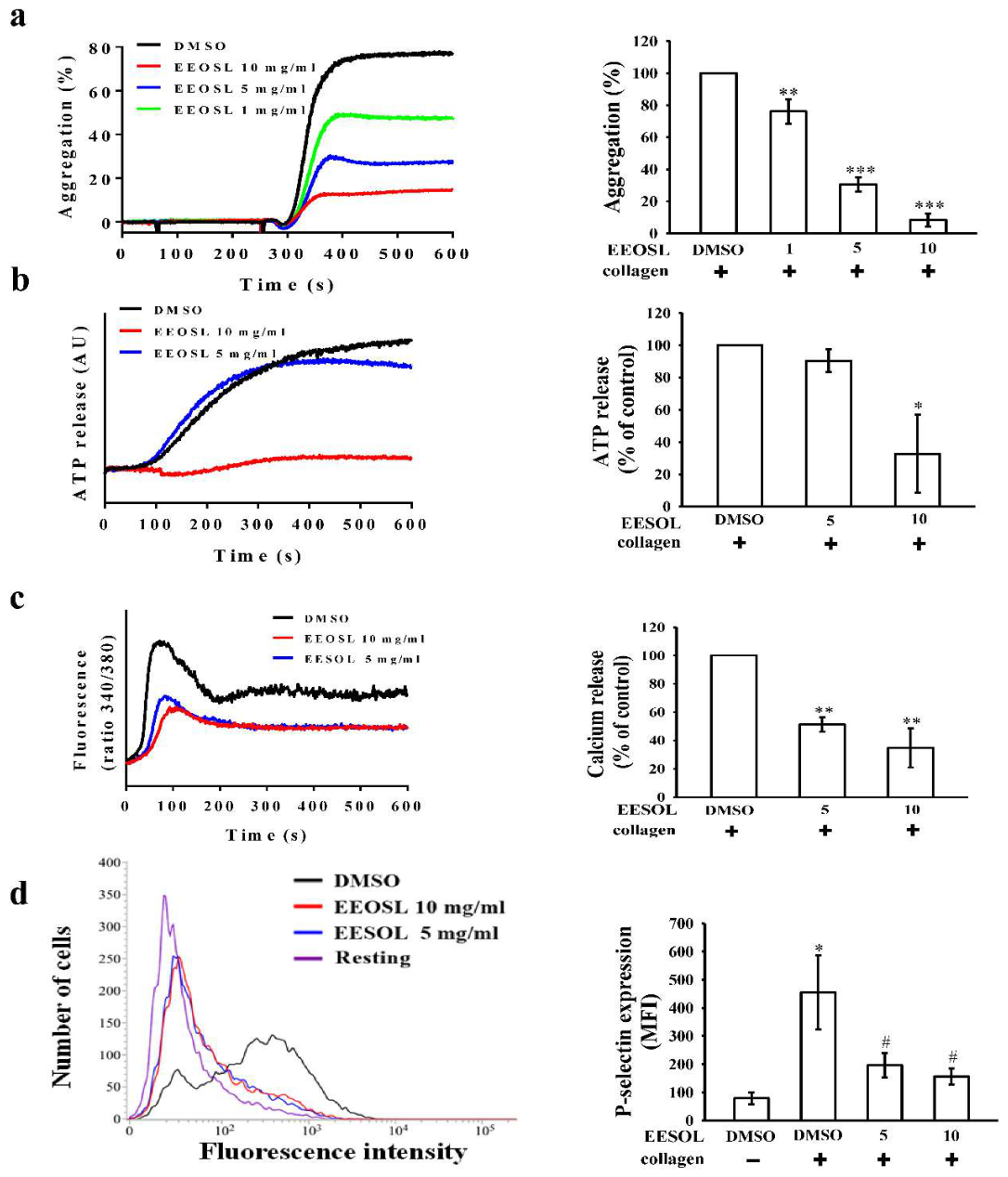
Figure 2.
A. The 3D map shows how apigenin interacts inside the catalytic pocket of PI3Kβ. The image highlights important non-covalent contacts and outlines both the hydrogen-bonding network and significant hydrophobic interactions with nearly residues; B. Detailed 3D binding orientation of apigenin captured within the PI3Kβ receptor cavity. Apigenin is shown in a stick model, clearly highlighting the spatial arrangement of the nearby amino acids that helps with the binding event.
Figure 2.
A. The 3D map shows how apigenin interacts inside the catalytic pocket of PI3Kβ. The image highlights important non-covalent contacts and outlines both the hydrogen-bonding network and significant hydrophobic interactions with nearly residues; B. Detailed 3D binding orientation of apigenin captured within the PI3Kβ receptor cavity. Apigenin is shown in a stick model, clearly highlighting the spatial arrangement of the nearby amino acids that helps with the binding event.
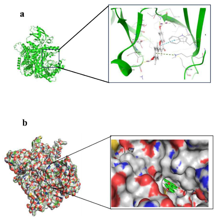
Figure 3.
Molecular dynamics simulation analyses showing the physical behaviour of the PI3Kβ-apigenin complex during a 100 ns simulation. (a) The backbone RMSD curve demonstrates the system's structural equilibration and stabilization over time. (b) The RMSF scatter plot highlights the localized flexibility of individual protein residues. (c) The H-bond tracking indicates consistent, uninterrupted molecular interactions between the ligand and the active site. (d) The Rg plot measures the protein's overall compactness, confirming that the complex kept its folded shape without expanding or collapsing during the simulation.
Figure 3.
Molecular dynamics simulation analyses showing the physical behaviour of the PI3Kβ-apigenin complex during a 100 ns simulation. (a) The backbone RMSD curve demonstrates the system's structural equilibration and stabilization over time. (b) The RMSF scatter plot highlights the localized flexibility of individual protein residues. (c) The H-bond tracking indicates consistent, uninterrupted molecular interactions between the ligand and the active site. (d) The Rg plot measures the protein's overall compactness, confirming that the complex kept its folded shape without expanding or collapsing during the simulation.
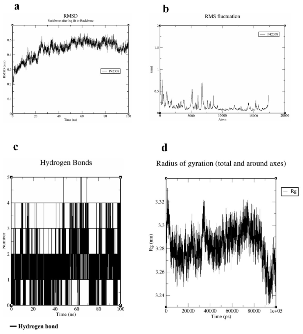
Figure 4.
EEOSL effect on serum liver biomarker enzymes in CCl4-induced liver injury in Wistar rats. The levels of (a) serum glutamic oxaloacetic transaminase (SGOT), (b) serum glutamic pyruvate transaminase (SGPT), (c) serum alkaline phosphatase (SALP), and (d) lactate dehydrogenase (LDH) were determined in Wistar rats with CCl4 induced liver damage. The results are in the form of mean, standard error of the mean (n=5). *p < 0.05, *** p < 0.001, a non-significant difference compared to the control group is denoted by ns.
Figure 4.
EEOSL effect on serum liver biomarker enzymes in CCl4-induced liver injury in Wistar rats. The levels of (a) serum glutamic oxaloacetic transaminase (SGOT), (b) serum glutamic pyruvate transaminase (SGPT), (c) serum alkaline phosphatase (SALP), and (d) lactate dehydrogenase (LDH) were determined in Wistar rats with CCl4 induced liver damage. The results are in the form of mean, standard error of the mean (n=5). *p < 0.05, *** p < 0.001, a non-significant difference compared to the control group is denoted by ns.
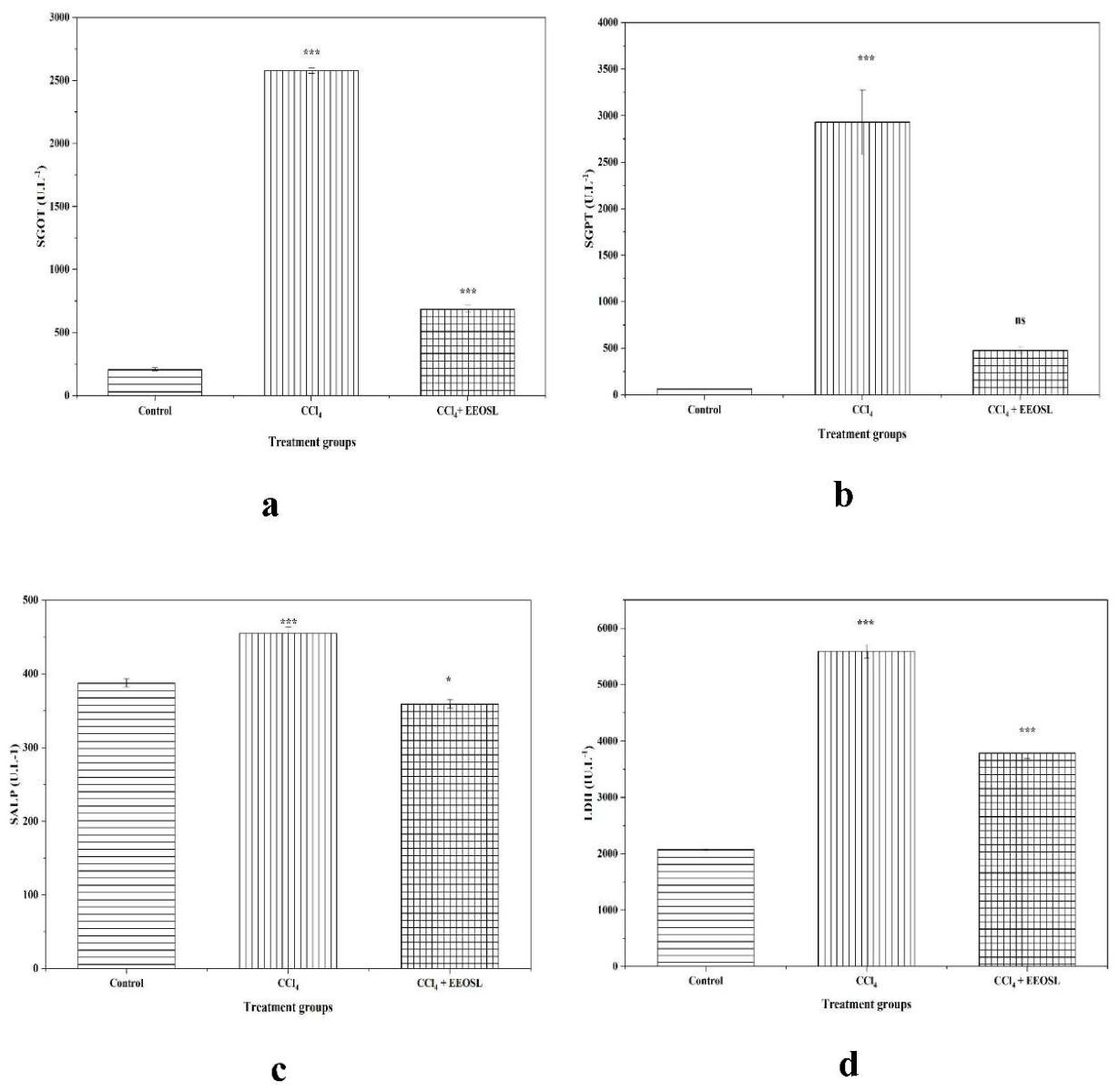
Figure 6.
Photomicrographs of liver tissue sections of the control, CCl4 -treated and EEOSL -treated rats. Photomicrographs of H& E staining liver tissue sections depicting (a, b) the control, (c, d) the carbon tetrachloride (CCl4)-treated, and (e, f) the CCl4 + EEOSL (500 mg/kg b.w.)-treated are given at a magnification of 100x (scale bar = 100μm). In panel (a, b), blue arrows mark normal hepatocytes, white arrows mark sinusoids, black arrows mark the central vein, and blue pointed arrows mark the portal triad. The presence of severe diffuse vacuolation in the hepatic cytoplasm is indicated by yellow arrows in panel (c, d), perivascular inflammation by black pointed arrows, congestion by green pointed arrows, and multifocal necrosis by green arrows. In panel (e, f) the yellow arrows represent moderate diffuse cytoplasmic vacuolation of the hepatic cytoplasm, the black pointed arrows represent perivascular inflammation, and the green arrows represent congestion.
Figure 6.
Photomicrographs of liver tissue sections of the control, CCl4 -treated and EEOSL -treated rats. Photomicrographs of H& E staining liver tissue sections depicting (a, b) the control, (c, d) the carbon tetrachloride (CCl4)-treated, and (e, f) the CCl4 + EEOSL (500 mg/kg b.w.)-treated are given at a magnification of 100x (scale bar = 100μm). In panel (a, b), blue arrows mark normal hepatocytes, white arrows mark sinusoids, black arrows mark the central vein, and blue pointed arrows mark the portal triad. The presence of severe diffuse vacuolation in the hepatic cytoplasm is indicated by yellow arrows in panel (c, d), perivascular inflammation by black pointed arrows, congestion by green pointed arrows, and multifocal necrosis by green arrows. In panel (e, f) the yellow arrows represent moderate diffuse cytoplasmic vacuolation of the hepatic cytoplasm, the black pointed arrows represent perivascular inflammation, and the green arrows represent congestion.
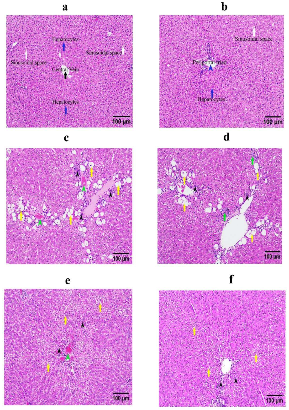
Figure 7.
Spatial 3D chemical structures of the fifteen unique phytoconstituents evaluated in the molecular docking study.
Figure 7.
Spatial 3D chemical structures of the fifteen unique phytoconstituents evaluated in the molecular docking study.
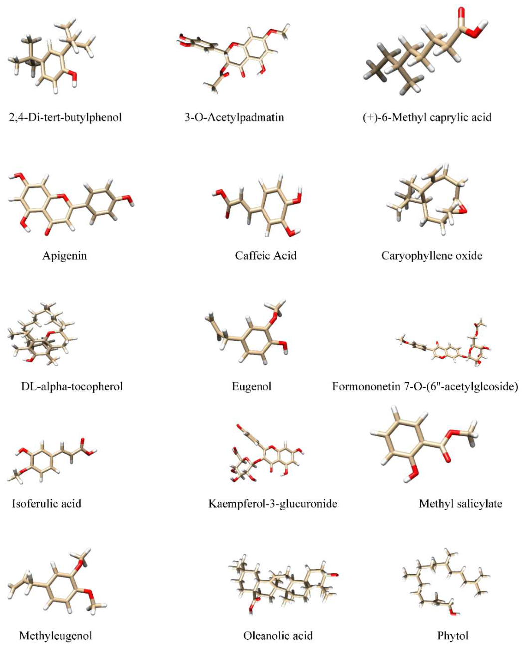

Table 1.
Molecular docking scores (kcal/mol) of the top-ranked phytoconstituents against key platelet activation proteins.
Table 1.
Molecular docking scores (kcal/mol) of the top-ranked phytoconstituents against key platelet activation proteins.
| Sl. No. | Protein | Protein ID | Ligand | Compound ID (CID) | Docking Score (kcal/mol) |
| 1 | GPVI | PDB: 2GI7 | 3-O-Acetylpadmatin | 10406203 | -4.60632 |
| 2 | SYK | PDB: 4FL2 | Methyl salicylate | 4133 | -3.52672 |
| 3 | PLCγ2 | PDB: 8T7C | Kaempferol-3-glucuronide | 22846027 | -5.84275 |
| 4 | P2RY12 | PDB: 4NTJ | Isoferulic acid | 736186 | -6.59831 |
| 5 | PI3Kβ | UniProt: P42338 | Apigenin | 5280443 | -8.00574 |
Table 2.
Effects of EEOSL on the activities of catalase (CAT), superoxide dismutase (SOD) and glutathione peroxidase (GPx) in the liver tissue of rats with CCl4-induced injury.
Table 2.
Effects of EEOSL on the activities of catalase (CAT), superoxide dismutase (SOD) and glutathione peroxidase (GPx) in the liver tissue of rats with CCl4-induced injury.
| Antioxidant Enzymes | Group of animals | ||
|
Group-I (Control) |
Group-II (CCl4-Induced) |
Group-III (CCl4 + EEOSL) |
|
| CAT | 705.06 ± 9.722 | 224.12 ± 13.253*** | 615.02 ± 8.055*** |
| SOD | 1.46 ± 0.005 | 0.71 ± 0.011*** | 0.92 ± 0.007*** |
| GPx | 1.48 ± 0.005 | 0.69 ± 0.005*** | 1.03 ± 0.002*** |
Values are expressed as mean ± standard error of mean (n=5); ***p < 0.001 compared with control. CAT- µmoles of H2O2 utilized/ min/ mg protein. SOD- unit/mg protein. GPx- µmoles of GSH oxidized/min/mg protein.
Table 3.
Target proteins and their predicted active-site residues used for molecular docking.
| Protein | Active site |
| GPVI (PDB ID: 2GI7) | L35, R37, E39, Y46, R66, Q70, W75 |
| SYK (PDB ID:4FL2) | S299, G300, N301, F302, V305, A320, K322, V353, M368, E369, M370, A371, P375, R418, N419, L421, D432, Y445, K453 |
| PLCγ2 (PDB ID: 8T7C) | N328, E357, D359, E406 |
| P2RY12 (PDB ID: 4NTJ) | F85, Y86, M89, Y90, I155, F160, F206, F210, F213, H214, L245, S249 |
| PI3Kβ (Uniprot ID: P42338) | M779, P785, W787, I803, K805, D813, Y839, I851, V853, V854, S857, D923, M926, I936, D937 |
Disclaimer/Publisher’s Note: The statements, opinions and data contained in all publications are solely those of the individual author(s) and contributor(s) and not of MDPI and/or the editor(s). MDPI and/or the editor(s) disclaim responsibility for any injury to people or property resulting from any ideas, methods, instructions or products referred to in the content. |
© 2026 by the authors. Licensee MDPI, Basel, Switzerland. This article is an open access article distributed under the terms and conditions of the Creative Commons Attribution (CC BY) license (http://creativecommons.org/licenses/by/4.0/).
Copyright: This open access article is published under a Creative Commons CC BY 4.0 license, which permit the free download, distribution, and reuse, provided that the author and preprint are cited in any reuse.