
Submitted:
02 July 2026
Posted:
03 July 2026
You are already at the latest version
Abstract
Cervical cytology and Human Papilloma virus (HPV) testing are the two main pillars of cervical cancer screening. The cytology-based Papanicolaou test is the conventional screening and triage method for identifying cervical cancer lesions; however studies indicate that it can be subjective and, similar to HPV testing, can exhibit limited sensitivity as these cannot distinguish transient and transforming infections. The p16/Ki67 dual stain (DS) cytology test identifies oncogenic transformation in cervical cells and can be performed utilising the same clinician-collected sample obtained in preliminary HPV screening. A considerable number of studies have highlighted the superior clinical performance of the p16/Ki67 DS test. This comprehensive review outlines the clinical application and accuracy of DS in identifying high-risk HPV-positive women at risk of invasive cervical cancer. We also review its utility in various clinical scenarios where DS testing may improve patient management, and propose potential DS applications of future research.
Keywords:
cervical cancer
; cervical dysplasia
; CIN
; dual stain
; p16/Ki67
; cytology
; human papillomavirus
; PAP smear
; cervical cytology
; cervical screening
1. Introduction
Cervical cancer is the fourth most common cancer in women in terms of incidence and mortality, with global estimates of 660,000 new cases and 350,000 deaths in 2022 [1]. It is the leading cause of cancer deaths in 37 countries, primarily sub-Saharan Africa, South America and South-Eastern Asia. Incidence and mortality rates vary at least 10-fold, with the highest rates found in sub-Saharan Africa [2]. The World Health Organization (WHO) launched the Global Cervical Cancer Elimination Initiative involving all countries, aiming to increase intervention targets to 90% coverage of human papilloma virus (HPV) vaccination, 70% coverage of twice-lifetime screening with HPV testing, 90% access to cervical precancer treatment and invasive cancer treatment services, by 2030 [3]. Although broad access to HPV screening, diagnostic, precancer and cancer treatment programs in high income countries has led to declining rates of cervical cancer, the uptake of cervical screening has been low or inconsistent in low-medium income countries [4].
The prevalence of HPV in cervical carcinomas is over 99%, and is considered a “necessary” cause of cervical cancer [5]. Over 200 HPV strains have been identified, 15-18 of which have been classified as high-risk (HR) genotypes according to their oncogenic potential [6]. HPV16 and HPV18 genotypes are associated with a high incidence of lesions, and worldwide, approximately 70% of all cervical cancers are attributable to those two genotypes [7]. HPV infection is a common sexually transmitted infection, most of which are asymptomatic, and more than 90% of new infections will clear or become undetectable within 1 to 2 years [8]. However, long-lasting, persistent infections can progress to cervical dysplasia and invasion. Traditionally, it was thought that long-lasting HPV infection caused cervical intraepithelial neoplasia (CIN) to develop in a progressive linear fashion, beginning with CIN1 (low grade), a “molecular switch” suggests that CIN3 could evolve directly from normal epithelium infected by HPV [9].
While the sensitivity of HPV testing may be as high as 95%, its specificity is lower than cytology [10]. Guidelines from the WHO and most countries for HPV screening recommend that women who are positive for HPV16 or HPV18 should undergo immediate colposcopy [11,12,13,14,15,16,17]. Women who are HR-HPV-positive, regardless of genotype, may require repeated cytology follow-ups, imposing additional burden and anxiety to patients. Thus, a more effective triage method may better identify women at risk of progressing to precancerous lesions, and provide a more streamlined clinical management approach as well as reassurance to patients.
2. Cervical Cancer Cytology and HPV Testing
Cervical Cytology and Screening Algorithms
Global elimination of cervical cancer is dependent on effective screening strategies that rely on early detection of prevalent cervical precancerous lesions to prevent their development into invasive cancer. Currently, in many settings, there are three main screening methods for cervical cancer: cervical cytology, HPV testing, and a combination of cervical cytology with HPV testing. The Papanicolaou test, once the gold standard of cervical cancer screening, involves the collection of cervical cells that are either smeared onto a slide (conventional cytology), or placed into a liquid medium (liquid based cytology) and examined for cellular abnormalities that may signify the onset of oncogenesis. Since its introduction, cervical cytology has significantly reduced the rates of cervical cancer incidence and mortality, saving millions of lives [18]; however incidence rates have plateaued in some highly-screened countries, indicating that improvements to the screening program may be beneficial [19]. Both conventional and liquid based cytology (LBC) can detect low-grade or high-grade cellular abnormalities; however they are unable to distinguish those with the potential for oncogenic transformation, thus requiring repeated cytology testing [20]. Cytology methodologies rely on the skill and experience of the observer, which has the potential for discrepancies among evaluators, leading to variation in interpretation that can result in inaccuracies and false negative tests [21]. Systematic reviews of published studies comparing the accuracy of LBC with conventional cytology have produced variable results depending on the quality of studies analysed [22,23]. False negative cytology is primarily due to misclassification of an abnormal cytology test as normal, which has been shown to be as high as 61% in some studies [24]. Evaluation of data from the ATHENA trial on the impact of inter-laboratory variations on the performance of LBC showed considerable differences among laboratories in both overall cytological abnormal rates (3.8 to 9.9%) and in sensitivity to detect CIN2 or worse (CIN2+) (42.0% to 73%), compared to HPV positivity rate which varied only from 10.9% to 13.4% [25]. The combination of HPV testing, particularly HPV16/18 genotyping, and cytology has been shown to lower rates of colposcopy referral and improve outcomes; however repeated testing of a high number of lesions including those that might not progress is required [26,27]. In light of these variations, a more effective alternative biomarker may be beneficial to ensure accurate classification of precancer lesions.
3. Overview of the p16/Ki67 Dual Stain Biomarker
Molecular Mechanism
The p16/Ki67 dual stain (DS) assay is an immunocytochemical test that detects co-expression of the biomarkers p16INK4a and Ki67 in the same cervical epithelial cells and can be performed on the same clinician-collected specimen used for HPV testing or LBC. p16INK4A (p16), is a cyclin-dependent kinase inhibitor that functions as a tumor suppressor through regulation of the retinoblastoma (Rb) pathway. In HPV-driven cervical neoplasia, the viral E7 oncoprotein inactivates Rb, leading to compensatory overexpression of p16. While p16 expression is minimal in normal cervical epithelium, diffuse overexpression is characteristic of transforming high-risk HPV infections and is observed in the majority of cervical precancers and cancers [28]. The human Ki67 protein is present during all active phases of the cell cycle, and is a definitive marker of cell proliferation and hence the malignant potential of tumors [29]. The overexpression of p16 in cells where Ki67 is expressed is rare, and their co-expression within a single cervical epithelial cell is a robust indicator of cell cycle deregulation and oncogenic transformation mediated by HR-HPV infection. Using DS immunocytochemistry, p16 is found in the cytoplasm and nucleus of cells and is detected as a brown signal against antibodies specific to p16, while Ki67 is localized only within the nucleus, and is displayed as a red nuclear signal. A positive DS test shows co-expression of both proteins in the same cell, indicating oncogenesis regardless of the morphological appearance of the cell [30].
McMenamin and colleagues evaluated the intra- and inter-observer reproducibility of the CINtec® PLUS dual stain Cytology (Roche Diagnostics) in ThinPrep® specimens and reported a high level of agreement across all evaluators ranging from 82.8% to 94.9% (kappa 0.65 to 0.91), and 89.2% to 93% (kappa 0.83-0.88), for intra- and inter-observer agreement respectively [31]. Similarly, Wentzensen and colleagues reported good to excellent reproducibility for DS cytology [83% to 91% (kappa 0.65 to 0.81)], and that the clinical performance among newly trained evaluators demonstrated that the technique can be routinely implemented in cytology practices, may be used in LBC as well as conventional smears, with minimal training [32].
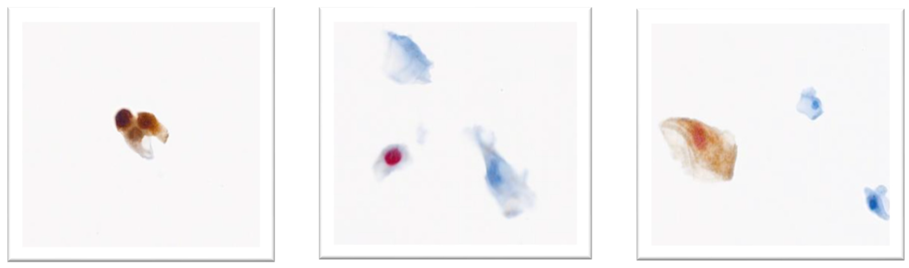
Figure 1.
Examples of DS staining. (a) Brown staining by p16. Reported as Dual Stain negative. (b) Red nuclear staining by Ki67. Reported as Dual Stain negative. (c) Co-expression of red nuclear staining of Ki67 and brown cytoplasmic staining of p16 within the same epithelial cell, indicates oncogenic transformation mediated by HPV. Reported as Dual Stain positive. (Images are of patient samples from citizens outside the U.S. and are the property of Roche Diagnostics).
Figure 1.
Examples of DS staining. (a) Brown staining by p16. Reported as Dual Stain negative. (b) Red nuclear staining by Ki67. Reported as Dual Stain negative. (c) Co-expression of red nuclear staining of Ki67 and brown cytoplasmic staining of p16 within the same epithelial cell, indicates oncogenic transformation mediated by HPV. Reported as Dual Stain positive. (Images are of patient samples from citizens outside the U.S. and are the property of Roche Diagnostics).
4. Clinical Application of the p16/Ki67 Dual Stain Test
4.1. Preliminary Evaluation of p16/Ki67
Preliminary evaluation of the p16/Ki67 DS test in cervical samples obtained immediately before colposcopy found that positive DS was significantly associated with HR-HPV infection, particularly HPV16 and HPV18, and was strongly related to the presence of a CIN2+ or HSIL [33]. Multiple studies conducted over the past 15 years have demonstrated the advantages of DS compared to LBC and conventional cytology for HR-HPV-positive women. Given that the majority of new HPV infections are transient and carry a low risk of progressing to CIN3+, DS staining as an optimal triage test exhibits high sensitivity and high negative predictive value (NPV) to help rule out individuals whose HPV infections are likely to resolve spontaneously, thus eliminating the need for colposcopy. Across screening cohorts (Table 1) as well as referral and post-treatment populations (Table 2), DS has consistently demonstrated higher sensitivity for detecting CIN2+ and CIN3+ lesions, with specificity that is generally comparable to LBC.
4.2. Clinical Trial Data
The IMproved Primary screening And Colposcopy Triage (IMPACT) trial evaluated the performance of p16/Ki67 DS compared to LBC with or without HPV16/18 genotyping for the triage of HR-HPV-positive women aged 25 to 65 years [47]. The sensitivity of DS triage in HPV16/18-positive women for CIN2+ and CIN3+ was 91.2% and 91.9%, respectively, versus LBC which was 75.3% and 77.9%, respectively. The sensitivity of DS triage of HPV (non-16/18) positive women was 83.0% for CIN2+ and 86% for CIN3+ (compared to LBC which was 58.8% and 66.7% respectively) [38]. Overall, DS provided superior risk stratification, simplified management pathways irrespective of HPV genotype, and represents a safe and clinically effective triage strategy within HPV-based primary screening programs [38].
The COMPASS trial is an ongoing Australian randomised controlled trial comparing primary HPV testing to cytology for screening of a highly vaccinated population [48]. The study includes an arm of women testing positive for HPV (non-16/18) who were further randomised to either LBC or DS triage [48]. Rates for detecting CIN2+ and CIN3+ for the LBC triage group were 1.0% and 0.7% respectively, and for the DS triage group were 1.2% and 0.8% respectively. DS triage brought forward the detection of CIN2+ as evidenced by the subsequent rates of colposcopy and biopsy diagnosis in the LBC triage group. [49].
A prospective observational study conducted at the Kaiser Permanente Northern California (KPNC) from 2015 to 2018 evaluated the performance of DS triage on 3,225 HPV-positive women undergoing HPV and LBC testing [37]. DS outperformed LBC for detecting CIN3+ for measures of sensitivity (88.6% vs 84.3%), specificity (53.1% vs 42.9%), positive predictive value (13% vs 10.4%), and negative predictive value (98.3% vs 97.2%; see Table 1). DS triage also led to a 32% reduction in colposcopies per detection of CIN3+ compared to LBC testing, with similar results observed for CIN2+. Importantly, these findings showed that DS can safely replace Pap cytology triage strategies for primary HPV screening and may safely extend the retesting intervals in HPV (non-16/18) positive women with negative DS results to 3 years [37]. Longitudinal evaluation of women participating in the KPNC study showed that those testing negative with DS had a significantly lower risk of CIN2+ compared to those with normal LBC, both at baseline (4.0% vs 6.2%) and at 5-year follow-up (8.5% vs 12.3%) [50]. Similar risk estimates were observed for CIN3+, highlighting the value of DS triage in the management of HPV-positive women who are DS-negative in avoiding unnecessary colposcopy referrals, and safely extending the re-testing intervals for 3 years [50].
5. Current Guidelines That Incorporate DS as Triage Tool
5.1. WHO
The WHO global strategy to eliminate cervical cancer, launched in 2020, includes guidelines for cervical cancer screening and treatment [3]. The 2024 WHO publication included guidance for the use of DS cytology in the screen-triage-and-treat approach [11]. The guidelines suggest using DS cytology as a triage method for women who test positive for HR-HPV, and a positive DS result, or a positive HPV 16/18 result, leads to a recommendation for colposcopy [11]. For DS negative results after HPV positivity, follow-up strategies include repeat HPV testing or co-testing in two years [11]. They found that DS had higher accuracy and provided better immediate and long-term risk stratification compared to cervical cytology. Long-term reassurance against precancer was better for DS-negative patients compared to cervical cytology-negative patients. Furthermore, proficiency in interpreting DS cytology could be attained more swiftly than for cervical cytology, and the reproducibility of DS results surpassed that of cervical cytology [11].
In a 2026 WHO update, in order to reduce loss to follow-up, recommends visual inspection with acetic acid (VIA) or colposcopic impression (without histological confirmation) as the preferred triage method for HPV-positive women, over cytology or dual stain cytology. It is important to note that this is a conditional recommendation and that the effectiveness of a selected triage method depends on clinical resources, infrastructure, technical capacity, workforce training, and quality assurance [51].
5.2. US-ASCCP Guideline Update 2024
The US Enduring Consensus Cervical Cancer Screening and Management Guidelines Committee developed recommendations for DS testing with CINtec® PLUS Cytology (Roche Diagnostics) to triage HR-HPV-positive women [13]. To ensure broad clinical applicability, risk estimates from two studies were included: the KPNC study [37] and the STudying Risk to Improve DisparitiES in Cervical Cancer (STRIDES) study, which evaluated cervical precancer risk in a cohort of 24,796 racially diverse individuals [52].
For each clinical scenario, authors estimated the immediate and 3-year risks of developing CIN3+. In triaging HR-HPV-positive individuals, the colposcopy risk threshold was crucial: colposcopy is recommended when the immediate risk of CIN3+ is between 4% and 24%, while follow-up after one year is indicated when the 5-year CIN3+ risk is 0.55% to 4%. If the CIN3+ risk is ≥60%, the guidelines advise expedited treatment [13]. For triaging HPV (non-16/18)-positive women, colposcopy is recommended for DS-positive individuals. DS-negative individuals are recommended to undergo yearly follow-up with HPV testing. For HPV16/18-positive women or those with high-grade cytology, immediate colposcopy was recommended. Interestingly, the immediate risk for those who were HPV16/18-positive and DS-negative was 2.6% and 1.1%, respectively in the KNPC study [37], and 1.8% and 0.0% (respectively) immediate risk in the STRIDES study [52], which are all below the 4% threshold for colposcopy referral. Given this, a “Special situation” was created for HPV16/18-positive DS-negative as the authors felt that despite the low immediate risk of CIN3+, more follow-up data was required to change guidelines, and until then, those HPV16/18-positive women would be referred to colposcopy [13]. They concluded that DS is a strong predictor of CIN3+ risk and can be effectively integrated into clinical management. DS offered improved risk differentiation compared to cervical cytology, with patient history having a lesser effect on risk estimates derived from DS results compared to those from cervical cytology [13]. Details of these guidelines and DS testing algorithms are outlined in Table 3.
5.3. Other Country Guidelines
Since the FDA approval for the use of DS testing, several countries have recently updated their guidelines to incorporate DS testing, some of which are summarized in Table 3.
5.4. Discordant HPV-Cytology-Colposcopy and Persistent HPV Results
Women who present with recurrent HR-HPV-positive results, accompanied by discordance between cytological and colposcopic or histopathological reports pose a significant challenge for clinicians and are frequently discussed at multidisciplinary meetings. Pearce and colleagues conducted a retrospective chart review of women who had discordant co-testing results, and found a significant risk of CIN3+ in this cohort, particularly women with persistent HPV(non-16/18) infections [54]. DS has been shown to be of value in women with persistent HPV-positive results. Abrue and colleagues examined whether the CINtec® PLUS Cytology Kit could be used as a complementary diagnostic method in women with persistent HPV-positive results but equivocal cytology, and found remarkable sensitivity of 100% and specificity of 71% in detecting CIN2+ lesions [40]. Consensus practice guidelines for such scenarios offer a conservative and safe approach.
The American Society for Colposcopy and Cervical Pathology (ASCCP) provides guidance for co-testing of HPV, cervical cytology and DS [13]. For those with possible HSIL or higher, immediate colposcopy is recommended, regardless of DS results. For individuals with LSIL or lower cervical cytology and DS-positive results, immediate colposcopy is recommended due to the immediate CIN3+ risk being above the 4% colposcopy threshold. For individuals with LSIL or lower cytology and DS-negative, 1 year follow up is advised (except for HPV16/18-positive cases) [13]. Given the benefits of the addition of DS co-testing, it could be considered to support the management of discordant results.
It is possible that in co-testing scenarios, the HPV test is negative regardless of cervical cytology whereas DS staining is positive. Such cases were reported in a Canada study, where 55 of 600 (9%) HPV negative women had DS+ results [41,55]. Of the 55, only 19 had biopsy results showing CIN2+ in 2 and <CIN1 in 17 cases [41,55]. In a retrospective Portugal study, 5 of 68 women (7%) who DS+ had a previous negative HR-HPV molecular test, regardless of cytology result [40]. The authors emphasized the importance of the methodology selected to detect HPV, as molecular methods may vary in sensitivities, and how DS can be maximally effective when another method such as cervical cytology is used as a guide together with HPV testing, since a high percentage of disease would be missed if this HR-HPV test was used alone [40].
5.5. Unsatisfactory Colposcopy with Type 3 Transformation Zone in Older Women
Post-menopausal women have a higher incidence and mortality rate from cervical cancer, and are more likely to be diagnosed at advanced stages of disease [56,57,58]. A Danish study reported that the highest incidence of cervical cancer was observed in women aged 75-79 years, and that estimates uncorrected for hysterectomy tended to underestimate cervical cancer incidence rates in older women [56]. The sensitivity of LBC for triaging HR-HPV-positive older women is notably lower than younger populations [59,60,61]. Additionally, colposcopy in this demographic is potentially compromised due to age-related changes to the cervix and retraction of the squamocolumnar junction into the cervical canal, rendering sampling and microscopic evaluation challenging. This increases the risk of incomplete removal of the transformation zone and leading to underdiagnosis and treatment failure in older women [62]. Concerns have been raised about the lack of correlation between LBC and histology findings for diagnosing CIN2+ in postmenopausal women, suggesting that cervical cytology could be less effective in this cohort [63,64].
The clinical utility of DS compared to cytology for triaging women over 45 years of age with transformation zone type 3 was evaluated by Gustafson and colleagues [65]. Women in the study were primarily postmenopausal with a median age of 68 years and were referred for colposcopy based on a positive HPV test. DS had higher sensitivity (96.7% vs 70%) and better negative predictive value (97.6% vs 86.4%) compared to LBC for CIN2+ detection. Although the specificity of DS was lower than LBC (63.5% vs 90.5%), the authors concluded that the superior negative predictive value of DS may safely avoid diagnostic excision in older women who were DS-negative [65].
DS has also been evaluated in postmenopausal women with positive ASC-US cytology. DS detected CIN2+ lesions with a sensitivity of 94.5%, a specificity of 86.6% and a negative predictive value of 95.9% [66]. Several other studies have reported estimates of the sensitivity, specificity, and negative predictive value that are consistent with these findings for DS compared to LBC in postmenopausal women [67,68,69].
5.6. Management of CIN2 in Young Women
Management of young women diagnosed with CIN2 presents challenges and concerns regarding overtreatment that may impact future reproductive capabilities [70]. There is considerable evidence that women under the age of 25 years diagnosed with CIN2 lesions will spontaneously regress without treatment at rates ranging from 42% to 74% [71,72,73,74,75,76]. Sykes and colleagues reported histologically confirmed regression of untreated CIN2 lesions in 64% of women under the age of 25 years within 2 years of diagnosis, highlighting the varied natural history of CIN2 lesions and questioning the need for costly treatment approaches that may negatively impact the quality of life in this cohort [77]. Evaluation of the ASCCP recommendations for management and follow-up of young women with CIN2+ found that colposcopy and LBC have limited capacity to exclude persistent high-grade abnormalities in this demographic undergoing observational management for CIN2 [78]. Consensus-based clinical practice guidelines for cervical screening disseminated by the Cancer Council Australia recommend ablation of abnormal tissue or diagnostic excision of the transformation zone for all women with histologically confirmed CIN2+ lesions [79]. Additionally, women who test positive for HR-HPV with a LBC report of HSIL, and who have undergone colposcopy and have histologically confirmed LSIL (≤CIN1), are considered for diagnostic excision of the transformation zone [80]. These recommendations are potentially problematic for younger women, as excision procedures such as Large Loop Excision of the Transformation Zone (LLETZ) are associated with increased risks of preterm birth in subsequent pregnancies [81].
The clinical performance of DS in younger women from reports of screening and referrals for colposcopy demonstrated high sensitivity for CIN2+ and CIN3+ in this cohort, although specificity was somewhat variable across studies (Table 4).
The likelihood of regression is high in young women with CIN2, and managing these cases can therefore be challenging. DS could be integrated into a co-testing strategy with LBC to help clinicians decide whether to keep younger women under surveillance or to treat them.
5.7. Surveillance Post-Treatment
Women who have been treated for HSIL (CIN2/3) have elevated recurrence risk, and up to four times the risk of invasive disease than those with normal test results, for 10-25 years, highlighting the importance of continued surveillance post-treatment [85,86,87,88]. The Australian National Cervical Screening guidelines recommend that women treated for HSIL should have an HPV test after 12 months; if HPV16/18-positive or cytology is possible HSIL+, colposcopy is recommended [89]. However, if HR-HPV(non-16/18)-positive, LBC should be performed. If LBC is LSIL or lower, HPV and LBC testing should be repeated 12-monthly unless HPV is not detected or three consecutive HR-HPV(non-16/18) are seen [89]. The ASCCP (US) guidelines include the use of DS for monitoring patients following colposcopy and treatment [13]. This recommendation is based on the finding that the risk of CIN3+ in HPV-positive, DS-positive women was 7.9% post-colposcopy, escalating to 18% post-treatment, which are well above the 4% colposcopy referral threshold [13]. Conversely, HPV-positive, DS-negative women have a low risk of CIN3+ of 0.39% post-colposcopy and 0.0% post-treatment, which are well below the referral threshold for colposcopy. Packet and colleagues explored the DS positivity rates for recurrence among patients referred for the LLETZ procedure for abnormal screening results [90]. The DS positivity rate prior to treatment and at 6 months was 70.9% and 30.1% respectively, with cervical cytology showing a similar significant reduction in positivity rates [90]. They also reported that the highest proportion of positive DS results was seen in HPV16 patients at the time of follow-up [90]. In a subsequent analysis that included 3 years of follow-up, they showed that the need for intense post-treatment follow-up was significantly associated with a positive DS result at baseline and follow-up, and a positive DS result at baseline was independently associated with the need for more intensive post-treatment surveillance many years after treatment [91]. Polman and colleagues evaluated the performance of DS for post-treatment in women referred for LLETZ [45]. When considering co-testing, DS and HPV co-testing had comparable sensitivity to LBC and HPV co-testing, but significantly better specificity for recurrent CIN2+ and CIN3+ (90.4% and 86.9% respectively) compared to LBC (70.8% and 67.4% respectively), suggesting that the best co-testing strategy for post-treatment surveillance for high-grade CIN was DS and HPV co-testing [45]. Additional evidence to support DS post-surveillance was shown by Liu and colleagues who examined 312 women post-treatment for cervical or vaginal lesions, and 284 cases with suspected malignancies [46]. The CIN2+ sensitivity and specificity of DS compared to LBC for the cervical lesions was 91% vs 42.8% and 95.5% vs 95.2%, respectively, although 47% were suspected malignancies, rather than all post-treatment cases [46].
6. Potential Future Areas of Research (Limited Evidence)
6.1. Cervical Management in Pregnant Women
Pregnancy-related hormones tend to influence cell proliferation of the cervical epithelium, potentially increasing susceptibility to HPV infection and the development of precancerous lesions, highlighting the importance of ongoing monitoring during pregnancy and postpartum [92]. Most cases of LSIL tend to remain stable, but HSILs have been shown to persist and negatively impact pregnancy outcomes, including preterm birth, miscarriage, intrauterine growth restriction leading to low birth weight and fetal death [93,94]. Routine screening during pregnancy is challenging because of physiological changes to the cervix, and guidelines for this cohort are based on limited data. Colposcopy with biopsy during pregnancy appears to be reliable with fewer risks of bleeding if conducted by a skilled gynaecologist during the first two trimesters or ≤20 weeks of pregnancy [95]. However, the Cancer Council Australia National Cervical Screening Guideline clarifies that biopsy is not usually necessary in pregnancy, unless invasive disease is suspected [96].
Stuebs and colleagues evaluated colposcopic findings in 655 pregnant women with abnormal cytology and found that the overall rate of accuracy for major colposcopic findings was relatively high (89.2%), and included findings of invasive carcinoma (42.9%), and CIN3 (57.1%) [97]. Available evidence suggests that these findings are due to the frequency with which pregnant women are seen by experienced examiners, highlighting the benefit of additional screening during pregnancy [97]. A recent retrospective case series including pregnant women with histologically confirmed HSIL showed that conservative approaches with close surveillance during pregnancy appear to be safe and effective with favourable postpartum outcomes [98]. A small retrospective study of women diagnosed with CIN lesions found less intense and more variable staining for p16 and Ki67 in pregnant women compared to nonpregnant women, suggesting that pregnancy status may affect the expression of the DS biomarkers [99]. It is likely that the prevalence of cervical lesions in pregnant women will rise particularly in countries where women tend to delay childbearing, and robust data from larger patient cohorts are needed to direct treatment approaches that ensure optimal maternal and fetal outcomes.
6.2. Immune Deficiencies
The immune clearance of HPV necessitates effective cell-mediated immunity and optimal T-cell functioning; thus, in conditions where these mechanisms are compromised, a heightened incidence of HPV-related diseases can be observed [100]. Individuals with HIV/AIDS have been linked to an increased prevalence of precancerous cervical lesions, with the rates of HR-HPV observed in HIV-positive women surpassing those found in the general population. Elevated risk of cervical cancer among women living with HIV/AIDS may be due to inadequate coverage by cervical cancer screening programs despite regular contact with medical services [101,102]. A large retrospective cohort study conducted in Australia involving women with a wide spectrum of rare auto-immune deficiencies and HIV revealed that cohorts with systemic lupus erythematosus and/or mixed connective tissue disease (SLE/MCTD) and HIV had significantly higher rates of both high-grade histological and cytological abnormalities compared to controls [103]. The Cancer Council Australia National Cervical Screening Guidelines advocate for screening every three years using HPV testing for women who have not previously tested positive for HR-HPV. Consensus-based recommendations are that women who are immune-deficient and are positive for HR-HPV should be referred for colposcopy [104]. To date there are no studies specifically focusing on DS for triage of HPV-positive women in immune-deficient populations. Given the limitations of cervical cytology and recent WHO inclusion of DS in testing algorithms, this remains an important area for future research.
6.3. Psychological Impact of Repeated Cervical Screening and Colposcopies for Uncertain Diagnoses
Women with abnormal screening tests who undergo colposcopy often experience psychological distress, especially concerning a cervical cancer diagnosis, future fertility, and their general health outcomes [105]. In Australia, although 47% of high-risk women receive colposcopy within three months, only 69.4% have attended within one year [106], indicating a longer than expected wait for some women. Clinical guidelines from the Cancer Council Australia recommend repeat LBC at the time of colposcopy if there is a delay in colposcopy of more than three months since referral for LBC [107]. Given the evidence that DS may identify additional cases of CIN3+, it could be an option for assessing residual LBC samples, which will help progress the triage of patients who require urgent evaluation and provide greater reassurance for those whose wait times exceed 12 months. The uncertainty of lengthy wait times for colposcopy or HPV re-testing, particularly for women who are HPV-positive for HPV(non-16/18) genotypes, can lead to anxiety, depression and isolation. Recent studies summarizing current knowledge on psychological outcomes of HPV diagnosis and subsequent treatment suggest a negative impact on depressive and anxiety symptoms, sexual functioning, and poorer quality of life [108,109]. It is foreseeable that DS testing’s greater prognostic certainty may reduce psychological stress, particularly in women whose abnormal cervical cytology is not a definitive diagnosis for treatment.
6.4. Medical Tourism Scenarios for Cancer Screening
Medical tourism, whereby an individual seeks medical procedures in a foreign country, is a rapidly growing market, estimated to reach $186 Billion USD globally by 2035 [110]. Asia Pacific is leading the market; Thailand had 2.5 million medical tourists in 2012 alone, the highest globally [111]. Most medical tourists are seeking advanced technologies or improved healthcare, with some seeking faster or cheaper services [111].
A study of 999 immigrant women aged 21-65 showed that women who engaged in medical tourism were twice as likely to have had cervical cancer screening compared to those who did not [112]. While medical tourism may be beneficial in the short term, testing methodologies based on LBC or cervical cytology may not correctly identify individuals who need further follow-up, and the lack of interaction with the local healthcare system would limit the value of such testing. The rise in medical tourism where women travel to another country or their home country for a quick and reassuring clinical decision warrants further investigation into whether DS augments clinical decisions as a triage tool together with HPV testing with or without cervical cytology.
6.5. Remote, Rural and Culturally Diverse Populations
Cancer and other neoplasms account for 23% of all deaths among Aboriginal and Torres Strait Islanders (referred to as Indigenous Australians), with the highest rates seen in outer regional areas. Cancer rates vary according to the cancer type, with cervical cancer rates 2.2 times higher in Indigenous women compared to non-Indigenous women [113]. There is evidence to suggest that women from culturally diverse backgrounds contribute to these lower rates. A study of immigrant women in Australia found that barriers to participation included low-risk perception, insufficient knowledge and engagement with health professionals [114].
Whilst the self-collect HPV strategy has made significant progress with underscreened populations, a return visit for a clinician-collected sample would be required for both LBC and DS. Although DS cannot currently overcome the need for a clinician-collected sample, it could offer the benefit of ensuring that only those with a high-risk of CIN3+ are referred to colposcopy. This is particularly important for populations with barriers such as geographical remoteness. The Australian States of South Australia, Queensland and Northern Territory all encompass large regions that are up to 11 hours’ drive from a hospital [115]. Moreover, only 33% of very remote women referred to colposcopy attend within 3 months and 57% by 12 months [106]. Recently the Australian National Aboriginal Community Controlled Health Organisation (NACCHO) has highlighted the significant need for Indigenous people to have greater equity in access to screening, early diagnosis and treatment in all Communities [116]. Given the challenges faced by underserved and remote populations, further investigation is warranted into the potential benefits that DS testing in regional centers may offer, including limiting unnecessary travel, given distance and costs involved.
6.6. Potential Clinical Implications of Earlier Diagnosis of CIN2+/CIN3+
Future data will provide comprehensive insights into attrition rates among women undergoing 12-month surveillance following LBC triage. These findings may reveal important limitations in LBC’s comparative effectiveness, as the accelerated identification of CIN2+/CIN3+ through DS could demonstrate superior clinical outcomes. Specifically, since more disease was identified after LBC referral, the performance of LBC may diminish when accounting for cases lost to follow-up [49].
Beyond diagnostic accuracy, expedited CIN2+/CIN3+ identification may reduce psychological burden (as outlined in Section VIII c above). Women undergoing repeated screening and colposcopy amid diagnostic uncertainty experience substantial anxiety [105]. Efficient triage methods like DS shorten uncertain waiting periods and minimize distress from recurrent procedures. Optimizing triage strategies can therefore improve both diagnostic accuracy and patient well-being.
6.7. Potential Impact on Large-Scale HPV Screening Programs of DS Cytology Automation
From a healthcare systems perspective, DS presents interesting workforce considerations, particularly given opportunities to streamline the process through automation. Automation of DS cytology could help address the widespread cytologist workforce shortage that has resulted in high levels of stressed staff and increased job turnover that may be compromising patient care [117]. As screening programs shift toward extended HPV testing intervals, organizations should anticipate substantial variability in demand for both triage and initial screening procedures. Consequently, implementing automated systems for both the initial screening phase and subsequent triage steps will be critical for scaling HPV-based screening initiatives effectively across large populations [49].
7. Cost-Effectiveness of DS
Recent cost-effectiveness studies have demonstrated the potential benefits of incorporating DS into cervical cancer screening protocols in the USA, Belgium, Germany and Thailand, which have been summarised by Harper and colleagues [118]. Moreover, the 2024 update to the WHO guidelines evaluated the cost-effectiveness of incorporating DS into cervical cancer screening. [11]. Incorporating DS into cervical cancer screening has demonstrated promising cost-effectiveness. While the addition of DS may increase upfront screening and medical costs, it results in improved detection of precancerous lesions, reduces the need for follow-up visits, and ultimately decreases the prevalence of cervical cancer [119,120]. Furthermore, the cost-effectiveness models suggest that these benefits may lead to long-term savings, both in terms of healthcare costs and improved patient outcomes, making DS a valuable tool for enhancing cervical cancer screening programs.
8. Limitations and Ongoing Considerations
Although p16/Ki67 dual stain (DS) cytology has demonstrated strong and consistent performance across multiple screening and triage settings, several considerations remain as implementation expands.
While DS generally offers higher sensitivity for CIN2+ and CIN3+ compared with cytology, gains in sensitivity may be accompanied by modest reductions in specificity in some populations. The net impact on colposcopy referral rates is therefore context-dependent and influenced by local screening algorithms, HPV prevalence, and risk thresholds. Importantly, large real-world programs have shown that DS can improve risk stratification while maintaining manageable referral volumes.
Long-term outcome data continue to evolve. Longitudinal cohort analyses support the safety of extended follow-up intervals in HPV-positive, DS-negative women. However, reductions in cervical cancer incidence attributable specifically to DS-based triage are currently inferred from risk modelling rather than cancer endpoints. Ongoing surveillance and longer follow-up will further clarify its long-term impact.
Interpretation of CIN2 outcomes warrants caution, as CIN2 represents a biologically heterogeneous category with variable regression potential, particularly in younger women. As with other screening biomarkers, clinical management decisions should consider age, reproductive plans, and overall risk context.
The evidence base includes diverse study designs and populations, enhancing generalizability but introducing heterogeneity. As HPV vaccination reduces disease prevalence, the predictive values of all screening and triage tools—including DS—will shift. As disease prevalence declines in highly vaccinated populations, recalibration of risk thresholds and triage strategies may be required for all screening tools, including DS. Continued evaluation in vaccinated and low-prevalence populations will therefore be important to refine risk-based management approaches.
Implementation considerations vary by setting. DS currently requires clinician-collected samples and laboratory infrastructure, which may limit scalability in some low-resource environments. Nonetheless, integration within organized HPV-based programs has been feasible in multiple healthcare systems, and inclusion in WHO and national guidelines reflects growing confidence in its clinical utility.
Biologically, DS detects co-expression of p16 and Ki67 as a marker of HPV-driven cell-cycle deregulation. Although no biomarker is entirely free from false-positive or false-negative results, the dual-marker approach provides greater specificity than single-marker assays and has shown reproducible performance across laboratories.
Overall, while continued long-term and implementation data will strengthen the evidence base, current findings support DS as a valuable and increasingly integrated component of modern, risk-based cervical cancer screening strategies.
9. Conclusions
The WHO’s ambitious target for cervical cancer elimination highlights the ongoing need to optimise screening and management strategies worldwide. Although substantial progress has been achieved, opportunities remain to improve risk stratification, case identification, and patient-centred management. p16/Ki67 DS cytology has demonstrated higher sensitivity for detecting high-grade CIN compared with LBC in multiple clinical settings and provides enhanced risk differentiation among HPV-positive women. The growing body of evidence supporting its use as a triage tool suggests that integrating DS into screening and follow-up algorithms may improve the precision of clinical decision-making while reducing unnecessary interventions. Strengthening screening accuracy and management pathways is central to achieving cervical cancer elimination targets, particularly in underserved and rural communities where access barriers and healthcare inequities continue to influence screening participation and outcomes.
Author Contributions
Original draft, LTH, ARJ-L; Editing and review, LEH, ARJ-L, MKO. All authors have read and agreed to the published version of this manuscript.
Funding
This manuscript was supported by Roche Diagnostics Australia.
Institutional Review Board Statement
Not applicable.
Informed Consent Statement
Not applicable.
Data Availability Statement
No new data were created or analyzed in this study. Images presented are the property of Roche Diagnostics.
Acknowledgments
The authors acknowledge Dr Sharon Johnatty of SugarApple Communications for providing medical writing support, which was funded by Roche Diagnostics Australia in accordance with Good Publication Practice (GPP3) guidelines (https://www.ismpp.org/gpp3).
Conflicts of Interest
(forms attached).
References
- Bray, F.; Laversanne, M.; Sung, H.; Ferlay, J.; Siegel, R.L.; Soerjomataram, I.; Jemal, A. Global cancer statistics 2022: GLOBOCAN estimates of incidence and mortality worldwide for 36 cancers in 185 countries. CA Cancer J. Clin. 2024, 74, 229–263. [Google Scholar] [CrossRef] [PubMed]
- Guida, F.; Kidman, R.; Ferlay, J.; Schuz, J.; Soerjomataram, I.; Kithaka, B.; Ginsburg, O.; Mailhot Vega, R.B.; Galukande, M.; Parham, G.; et al. Global and regional estimates of orphans attributed to maternal cancer mortality in 2020. Nat. Med. 2022, 28, 2563–2572. [Google Scholar] [CrossRef] [PubMed]
- World Health Organization. Global strategy to accelerate the elimination of cervical cancer as a public health problem. Available online: https://www.who.int/publications/i/item/9789240014107 (accessed on 13 May 2026).
- Canfell, K.; Kim, J.J.; Brisson, M.; Keane, A.; Simms, K.T.; Caruana, M.; Burger, E.A.; Martin, D.; Nguyen, D.T.N.; Benard, E.; et al. Mortality impact of achieving WHO cervical cancer elimination targets: A comparative modelling analysis in 78 low-income and lower-middle-income countries. Lancet 2020, 395, 591–603. [Google Scholar] [CrossRef] [PubMed]
- Walboomers, J.M.; Jacobs, M.V.; Manos, M.M.; Bosch, F.X.; Kummer, J.A.; Shah, K.V.; Snijders, P.J.; Peto, J.; Meijer, C.J.; Munoz, N. Human papillomavirus is a necessary cause of invasive cervical cancer worldwide. J. Pathol. 1999, 189, 12–19. [Google Scholar] [CrossRef]
- Bedell, S.L.; Goldstein, L.S.; Goldstein, A.R.; Goldstein, A.T. Cervical Cancer Screening: Past, Present, and Future. Sex. Med. Rev. 2020, 8, 28–37. [Google Scholar] [CrossRef] [PubMed]
- National Cancer Institute. Cervical Cancer Causes, Risk Factors, and Prevention. Available online: https://www.cancer.gov/types/cervical/causes-risk-prevention (accessed on 13 May 2026).
- Markowitz, L.E.; Unger, E.R. Human Papillomavirus Vaccination. N Engl. J. Med. 2023, 388, 1790–1798. [Google Scholar] [CrossRef] [PubMed]
- Massad, L.S.; Einstein, M.H.; Huh, W.K.; Katki, H.A.; Kinney, W.K.; Schiffman, M.; Solomon, D.; Wentzensen, N.; Lawson, H.W.; Conference, A.C.G. 2012 updated consensus guidelines for the management of abnormal cervical cancer screening tests and cancer precursors. J. Low. Genit. Tract. Dis. 2013, 17, S1–S27. [Google Scholar] [CrossRef] [PubMed]
- Cuzick, J.; Clavel, C.; Petry, K.U.; Meijer, C.J.; Hoyer, H.; Ratnam, S.; Szarewski, A.; Birembaut, P.; Kulasingam, S.; Sasieni, P.; et al. Overview of the European and North American studies on HPV testing in primary cervical cancer screening. Int. J. Cancer 2006, 119, 1095–1101. [Google Scholar] [CrossRef] [PubMed]
- World Health Organization. WHO guideline for screening and treatment of cervical pre-cancer lesions for cervical cancer prevention. Use of dual-stain cytology to triage women after a positive test for human papillomavirus (HPV) 2024. Available online: https://iris.who.int/bitstream/handle/10665/376492/9789240091658-eng.pdf (accessed on 13 May 2026).
- The Hong Kong College of Obstetricians and Gynecologists. HKCOG Guidelines for Cervical Cancer Prevention and Screening. Available online: https://www.hkcog.org.hk/hkcog/Download/Guidelines_for_Cervical_Cancer_Prevention_and_Screening.pdf (accessed on 12 May 2026).
- Clarke, M.A.; Wentzensen, N.; Perkins, R.B.; Garcia, F.; Arrindell, D.; Chelmow, D.; Cheung, L.C.; Darragh, T.M.; Egemen, D.; Guido, R.; et al. Recommendations for Use of p16/Ki67 Dual Stain for Management of Individuals Testing Positive for Human Papillomavirus. J. Low. Genit. Tract. Dis. 2024, 28, 124–130. [Google Scholar] [CrossRef] [PubMed]
- Sepodes, B.; Rebelo, T.; Santos, F.; Oliveira, D.; Catalao, C.; Aguas, F.; Fernandes, G. Optimization of HPV-positive women triage with p16/Ki67 dual staining cytology in an organized cervical cancer screening program in the center region of Portugal. Eur. J. Obstet. Gynecol. Reprod. Biol. 2024, 302, 111–115. [Google Scholar] [CrossRef] [PubMed]
- Ministry of Health Socialist Republic of Vietnam. GUIDELINE FOR PREVENTION AND MANAGEMENT OF CERVICAL CANCER. Available online: https://thuvienphapluat.vn/van-ban/The-thao-Y-te/Quyet-dinh-3792-QD-BYT-2024-tai-lieu-chuyen-mon-Huong-dan-du-phong-ung-thu-co-tu-cung-635871.aspx (accessed on 13 May 2026).
- Li, M.; Li, J.; Li, X.; Wang, X.; Kong, L.; Chen, F.; Cong, Q.; Wu, P.; Huang, X.; Zhao, F.; et al. Guidelines for cervical cancer screening in China II. Gynecol. Obstet. Clin. Med. 2025, 5, e000205. [Google Scholar] [CrossRef]
- Country Office for Thailand; World Health Organization. Eradicating cervical cancer in Thailand: A journey of hope and progress. Available online: https://www.who.int/publications/i/item/9789290221074 (accessed on 12 May 2026).
- Michalas, S.P. The Pap test: George N. Papanicolaou (1883-1962). A screening test for the prevention of cancer of uterine cervix. Eur. J. Obstet. Gynecol. Reprod. Biol. 2000, 90, 135–138. [Google Scholar] [CrossRef] [PubMed]
- Singh, D.; Vignat, J.; Lorenzoni, V.; Eslahi, M.; Ginsburg, O.; Lauby-Secretan, B.; Arbyn, M.; Basu, P.; Bray, F.; Vaccarella, S. Global estimates of incidence and mortality of cervical cancer in 2020: A baseline analysis of the WHO Global Cervical Cancer Elimination Initiative. Lancet Glob. Health 2023, 11, e197–e206. [Google Scholar] [CrossRef] [PubMed]
- Eun, T.J.; Perkins, R.B. Screening for Cervical Cancer. Med. Clin. North Am. 2020, 104, 1063–1078. [Google Scholar] [CrossRef] [PubMed]
- Koliopoulos, G.; Nyaga, V.N.; Santesso, N.; Bryant, A.; Martin-Hirsch, P.P.; Mustafa, R.A.; Schunemann, H.; Paraskevaidis, E.; Arbyn, M. Cytology versus HPV testing for cervical cancer screening in the general population. Cochrane Database Syst. Rev. 2017, 8, CD008587. [Google Scholar] [CrossRef] [PubMed]
- Davey, E.; Barratt, A.; Irwig, L.; Chan, S.F.; Macaskill, P.; Mannes, P.; Saville, A.M. Effect of study design and quality on unsatisfactory rates, cytology classifications, and accuracy in liquid-based versus conventional cervical cytology: A systematic review. Lancet 2006, 367, 122–132. [Google Scholar] [CrossRef] [PubMed]
- Arbyn, M.; Bergeron, C.; Klinkhamer, P.; Martin-Hirsch, P.; Siebers, A.G.; Bulten, J. Liquid Compared With Conventional Cervical Cytology: A Systematic Review and Meta-analysis. Obstet. Gynecol. 2008, 111, 167–177. [Google Scholar] [CrossRef] [PubMed]
- Macios, A.; Nowakowski, A. False Negative Results in Cervical Cancer Screening-Risks, Reasons and Implications for Clinical Practice and Public Health. Diagnostics 2022, 12. [Google Scholar] [CrossRef] [PubMed]
- Wright, T.C.; Stoler, M.H.; Behrens, C.M.; Sharma, A.; Sharma, K.; Apple, R. Interlaboratory variation in the performance of liquid-based cytology: Insights from the ATHENA trial: Performance of liquid-based cytology. Int. J. Cancer 2014, 134, 1835–1843. [Google Scholar] [CrossRef] [PubMed]
- Dijkstra, M.G.; van Niekerk, D.; Rijkaart, D.C.; van Kemenade, F.J.; Heideman, D.A.; Snijders, P.J.; Meijer, C.J.; Berkhof, J. Primary hrHPV DNA testing in cervical cancer screening: How to manage screen-positive women? A POBASCAM trial substudy. Cancer Epidemiol. Biomark. Prev. 2014, 23, 55–63. [Google Scholar] [CrossRef] [PubMed]
- Rijkaart, D.C.; Berkhof, J.; van Kemenade, F.J.; Coupe, V.M.; Hesselink, A.T.; Rozendaal, L.; Heideman, D.A.; Verheijen, R.H.; Bulk, S.; Verweij, W.M.; et al. Evaluation of 14 triage strategies for HPV DNA-positive women in population-based cervical screening. Int. J. Cancer 2012, 130, 602–610. [Google Scholar] [CrossRef] [PubMed]
- Klaes, R.; Friedrich, T.; Spitkovsky, D.; Ridder, R.; Rudy, W.; Petry, U.; Dallenbach-Hellweg, G.; Schmidt, D.; von Knebel Doeberitz, M. Overexpression of p16(INK4A) as a specific marker for dysplastic and neoplastic epithelial cells of the cervix uteri. Int. J. Cancer 2001, 92, 276–284. [Google Scholar] [CrossRef] [PubMed]
- Scholzen, T.; Gerdes, J. The Ki-67 protein: From the known and the unknown. J. Cell. Physiol. 2000, 182, 311–322. [Google Scholar] [CrossRef]
- Yu, L.; Fei, L.; Liu, X.; Pi, X.; Wang, L.; Chen, S. Application of p16/Ki-67 dual-staining cytology in cervical cancers. J. Cancer 2019, 10, 2654–2660. [Google Scholar] [CrossRef] [PubMed]
- McMenamin, M.; McKenna, M.; McDowell, A.; Dawson, C.; McKenna, R. Intra- and inter-observer reproducibility of CIN tec® PLUS in ThinPrep® cytology preparations. Cytopathology 2017, 28, 284–290. [Google Scholar] [CrossRef] [PubMed]
- Wentzensen, N.; Fetterman, B.; Tokugawa, D.; Schiffman, M.; Castle, P.E.; Wood, S.N.; Stiemerling, E.; Poitras, N.; Lorey, T.; Kinney, W. Interobserver reproducibility and accuracy of p16/ K i-67 dual-stain cytology in cervical cancer screening. Cancer Cytopathol. 2014, 122, 914–920. [Google Scholar] [CrossRef] [PubMed]
- Dona, M.G.; Vocaturo, A.; Giuliani, M.; Ronchetti, L.; Rollo, F.; Pescarmona, E.; Carosi, M.; Vocaturo, G.; Benevolo, M. p16/Ki-67 dual staining in cervico-vaginal cytology: Correlation with histology, Human Papillomavirus detection and genotyping in women undergoing colposcopy. Gynecol. Oncol. 2012, 126, 198–202. [Google Scholar] [CrossRef] [PubMed]
- Petry, K.U.; Schmidt, D.; Scherbring, S.; Luyten, A.; Reinecke-Lüthge, A.; Bergeron, C.; Kommoss, F.; Löning, T.; Ordi, J.; Regauer, S.; et al. Triaging Pap cytology negative, HPV positive cervical cancer screening results with p16/Ki-67 Dual-stained cytology. Gynecol. Oncol. 2011, 121, 505–509. [Google Scholar] [CrossRef] [PubMed]
- Ikenberg, H.; Bergeron, C.; Schmidt, D.; Griesser, H.; Alameda, F.; Angeloni, C.; Bogers, J.; Dachez, R.; Denton, K.; Hariri, J.; et al. Screening for cervical cancer precursors with p16/Ki-67 dual-stained cytology: Results of the PALMS study. J. Natl. Cancer Inst. 2013, 105, 1550–1557. [Google Scholar] [CrossRef] [PubMed]
- Wentzensen, N.; Fetterman, B.; Castle, P.E.; Schiffman, M.; Wood, S.N.; Stiemerling, E.; Tokugawa, D.; Bodelon, C.; Poitras, N.; Lorey, T.; et al. p16/Ki-67 Dual Stain Cytology for Detection of Cervical Precancer in HPV-Positive Women. J. Natl. Cancer Inst. 2015, 107, djv257. [Google Scholar] [CrossRef] [PubMed]
- Wentzensen, N.; Clarke, M.A.; Bremer, R.; Poitras, N.; Tokugawa, D.; Goldhoff, P.E.; Castle, P.E.; Schiffman, M.; Kingery, J.D.; Grewal, K.K.; et al. Clinical Evaluation of Human Papillomavirus Screening With p16/Ki-67 Dual Stain Triage in a Large Organized Cervical Cancer Screening Program. JAMA Intern. Med. 2019, 179, 881. [Google Scholar] [CrossRef] [PubMed]
- Wright, T.C.; Stoler, M.H.; Ranger-Moore, J.; Fang, Q.; Volkir, P.; Safaeian, M.; Ridder, R. Clinical validation of p16/Ki-67 dual-stained cytology triage of HPV -positive women: Results from the IMPACT trial. Int. J. Cancer 2022, 150, 461–471. [Google Scholar] [CrossRef] [PubMed]
- Ovestad, I.T.; Dalen, I.; Andersland, M.S.; Vintermyr, O.K.; Moltu, P.; Berland, J.M.; Janssen, E.A.M.; Haugland, H.K. Triaging HPV-Positive Cervical Samples with p16 and Ki-67 Dual Stained Cytology within an Organized Screening Program-A Prospective Observational Study from Western Norway. Int. J. Mol. Sci. 2023, 24. [Google Scholar] [CrossRef] [PubMed]
- Abreu, Â.L.; Silva, R.A.; Fernandes, S. Validation of CINtec® PLUS Cytology Kit in the Diagnosis of Persistent HPV Infections - Cohort Study in the Portuguese Population. J. Cytol. 2021, 38, 94–100. [Google Scholar] [CrossRef] [PubMed]
- Ratnam, S.; Jang, D.; Gilbert, L.; Alaghehbandan, R.; Schell, M.; Needle, R.; Ecobichon-Morris, A.; Wang, P.P.; Rahman, M.; Costescu, D.; et al. CINtec PLUS and cobas HPV testing for triaging Canadian women referred to colposcopy with a history of low-grade squamous intraepithelial lesion: Baseline findings. Papillomavirus Res. 2020, 10, 100206. [Google Scholar] [CrossRef] [PubMed]
- Waldstrøm, M.; Christensen, R.K.; Ørnskov, D. Evaluation of p16INK4a /Ki-67 dual stain in comparison with an mRNA human papillomavirus test on liquid-based cytology samples with low-grade squamous intraepithelial lesion. Cancer Cytopathol. 2013, 121, 136–145. [Google Scholar] [CrossRef] [PubMed]
- Zhang, R.; Ge, X.; You, K.; Guo, Y.; Guo, H.; Wang, Y.; Geng, L. p16/Ki67 dual staining improves the detection specificity of high-grade cervical lesions. J. Obstet. Gynaecol. Res. 2018, 44, 2077–2084. [Google Scholar] [CrossRef] [PubMed]
- Zhu, Y.; Ren, C.; Yang, L.; Zhang, X.; Liu, L.; Wang, Z. Performance of p16/Ki67 immunostaining, HPV E6/E7 mRNA testing, and HPV DNA assay to detect high-grade cervical dysplasia in women with ASCUS. BMC Cancer 2019, 19, 271. [Google Scholar] [CrossRef] [PubMed]
- Polman, N.J.; Uijterwaal, M.H.; Witte, B.I.; Berkhof, J.; Van Kemenade, F.J.; Spruijt, J.W.M.; Van Baal, W.M.; Graziosi, P.G.C.M.; Van Dijken, D.K.E.; Verheijen, R.H.M.; et al. Good performance of p16/ki-67 dual-stained cytology for surveillance of women treated for high-grade CIN. Int. J. Cancer 2017, 140, 423–430. [Google Scholar] [CrossRef] [PubMed]
- Liu, W.; Gong, J.; Xu, H.; Zhang, D.; Xia, N.; Li, H.; Song, K.; Lv, T.; Chen, Y.; Diao, Y.; et al. Good performance of p16/Ki-67 dual-stain cytology for detection and post-treatment surveillance of high-grade CIN / VAIN in a prospective, cross-sectional study. Diagn. Cytopathol. 2020, 48, 635–644. [Google Scholar] [CrossRef] [PubMed]
- Safaeian, M.; Wright, T.C.; Stoler, M.H.; Ranger-Moore, J.; Rehm, S.; Aslam, S.; Fang, Q.; Volkir, P.; Ridder, R. The IMproving Primary Screening And Colposcopy Triage trial: Human papillomavirus, cervical cytology, and histopathologic results from the baseline and 1-year follow-up phase. Am. J. Obstet. Gynecol. 2021, 225, e271–e278-278.e216. [Google Scholar] [CrossRef] [PubMed]
- Canfell, K.; Saville, M.; Caruana, M.; Gebski, V.; Darlington-Brown, J.; Brotherton, J.; Heley, S.; Castle, P.E. Protocol for Compass: A randomised controlled trial of primary HPV testing versus cytology screening for cervical cancer in HPV-unvaccinated and vaccinated women aged 25-69 years living in Australia. BMJ Open 2018, 8, e016700. [Google Scholar] [CrossRef] [PubMed]
- Canfell, K.; Caruana, M.; Gebski, V.; Darlington-Brown, J.; Heley, S.; Brotherton, J.; Gertig, D.; Jennett, C.J.; Farnsworth, A.; Tan, J.; et al. Cervical screening with primary HPV testing or cytology in a population of women in which those aged 33 years or younger had previously been offered HPV vaccination: Results of the Compass pilot randomised trial. PLoS Med. 2017, 14, e1002388. [Google Scholar] [CrossRef] [PubMed]
- Clarke, M.A.; Cheung, L.C.; Castle, P.E.; Schiffman, M.; Tokugawa, D.; Poitras, N.; Lorey, T.; Kinney, W.; Wentzensen, N. Five-Year Risk of Cervical Precancer Following p16/Ki-67 Dual-Stain Triage of HPV-Positive Women. JAMA Oncol. 2019, 5, 181–186. [Google Scholar] [CrossRef] [PubMed]
- World Health Organization. WHO guideline for screening and treatment of cervical pre-cancer lesions for cervical cancer prevention: Use of human papillomavirus (HPV) DNA genotyping. Available online: https://www.who.int/publications/i/item/9789240121744 (accessed on 28 June 2026).
- Risley, C.; Stewart, M.W.; Geisinger, K.R.; Hiser, L.M.; Morgan, J.C.; Owens, K.J.; Ayyalasomayajula, K.; Rives, R.M.; Jannela, A.; Grunes, D.E.; et al. STRIDES - STudying Risk to Improve DisparitiES in Cervical Cancer in Mississippi - Design and baseline results of a Statewide Cohort Study. Prev. Med. 2021, 153, 106740. [Google Scholar] [CrossRef] [PubMed]
- Salta, S.; Sequeira, J.P.; Lobo, J.; Sousa, A.; Sousa, H.; Baldaque, I.; Monteiro, P.; Tavares, F.; Henrique, R.; Jeronimo, C. Preliminary outcomes of the Cervical Cancer Screening Program of Northern Portugal: A snapshot. J. Infect. Public Health 2024, 17, 1057–1064. [Google Scholar] [CrossRef] [PubMed]
- Peace, S.M.; Jennings, A.J. Prevalence of high-grade dysplasia in cytology-negative, HPV-positive cervical cancer screening. Arch. Gynecol. Obstet. 2022, 305, 87–93. [Google Scholar] [CrossRef] [PubMed]
- Ratnam, S.; Jang, D.; Gilbert, L.; Alaghehbandan, R.; Schell, M.; Needle, R.; Ecobichon-Morris, A.; Wang, P.P.; Rahman, M.; Costescu, D.; et al. Corrigendum to CINtec PLUS and cobas HPV testing for triaging Canadian women referred to colposcopy with a history of low-grade squamous intraepithelial lesion: Baseline findings [Papillomavirus Res. 10 (2020) 100206]. Tumour Virus Res. 2021, 11, 200215. [Google Scholar] [CrossRef] [PubMed]
- Hammer, A.; Kahlert, J.; Rositch, A.; Pedersen, L.; Gravitt, P.; Blaakaer, J.; Soegaard, M. The temporal and age-dependent patterns of hysterectomy-corrected cervical cancer incidence rates in Denmark: A population-based cohort study. Acta Obstet. Et. Gynecol. Scand. 2017, 96, 150–157. [Google Scholar] [CrossRef] [PubMed]
- Rustagi, A.S.; Kamineni, A.; Weinmann, S.; Reed, S.D.; Newcomb, P.; Weiss, N.S. Cervical screening and cervical cancer death among older women: A population-based, case-control study. Am. J. Epidemiol. 2014, 179, 1107–1114. [Google Scholar] [CrossRef] [PubMed]
- Cancer Council Australia. Cervical Cancer Screening: Do I need the Cervical Screening Test if I’m over 50. Available online: https://www.cancer.org.au/cervicalscreening/i-am-over-25/do-i-need-the-test/what-if-i-am-over-50 (accessed on 13 May 2026).
- Wikström, I.; Stenvall, H.; Wilander, E. Low Prevalence of High-risk HPV in Older Women Not Attending Organized Cytological Screening: A Pilot Study. Acta Derm.-Venereol. 2007, 87, 554–555. [Google Scholar] [CrossRef] [PubMed]
- Miki, M.; Imaeda, Y.; Takahashi, H.; Iwata, A.; Tsukamoto, T.; Nomura, H.; Kotani, K.; Mitani, T.; Ichikawa, R.; Fujii, T. Lower accuracy of cytological screening for high-grade squamous intraepithelial neoplasia in women over 50 years of age in Japan. Int. J. Clin. Oncol. 2022, 27, 427–433. [Google Scholar] [CrossRef] [PubMed]
- Yan, P.; Du, H.; Wang, C.; Song, F.; Huang, X.; Luo, Y.; Wu, R. Differential diagnosis of high-grade squamous intraepithelial lesions and benign atrophy in older women using p16 immunocytochemistry. Gynecol. Obstet. Clin. Med. 2021, 1, 14–18. [Google Scholar] [CrossRef]
- Gustafson, L.W.; Petersen, L.K.; Bor, P.; Andersen, B.; Hammer, A. Cervical cancer prevention among older women – challenges in screening, diagnostic workup and treatment. Acta Obstet. Et. Gynecol. Scand. 2021, 100, 1364–1368. [Google Scholar] [CrossRef] [PubMed]
- Hermansson, R.S.; Olovsson, M.; Hoxell, E.; Lindstrom, A.K. HPV prevalence and HPV-related dysplasia in elderly women. PLoS ONE 2018, 13, e0189300. [Google Scholar] [CrossRef] [PubMed]
- Gilani, S.M.; Mazzara, P.F. Cytohistologic correlation in premenopausal and postmenopausal women. Acta Cytol. 2013, 57, 575–580. [Google Scholar] [CrossRef] [PubMed]
- Gustafson, L.W.; Tranberg, M.; Christensen, P.N.; Brondum, R.; Wentzensen, N.; Clarke, M.A.; Andersen, B.; Petersen, L.K.; Bor, P.; Hammer, A. Clinical utility of p16/Ki67 dual-stain cytology for detection of cervical intraepithelial neoplasia grade two or worse in women with a transformation zone type 3: A cross-sectional study. BJOG 2023, 130, 202–209. [Google Scholar] [CrossRef] [PubMed]
- Bruno, M.T.; Guaita, A.; Boemi, S.; Mazza, G.; Sudano, M.C.; Palumbo, M. Performance of p16/Ki67 Immunostaining for Triage of Elderly Women with Atypical Squamous Cells of Undetermined Significance. J. Clin. Med. 2023, 12, 3400. [Google Scholar] [CrossRef] [PubMed]
- Dovnik, A.; Repse Fokter, A. P16/Ki-67 Immunostaining in the Triage of Postmenopausal Women With Low-Grade Cytology Results. J. Low. Genit. Tract. Dis. 2020, 24, 235–237. [Google Scholar] [CrossRef] [PubMed]
- Dai, Y.; Chen, T.; Li, X.; Zhang, C.; Li, T.; Zhao, Y.; Wang, Y.; Chen, S.; Yu, L.; Jiang, M.; et al. Evaluation of the clinical performance of p16/Ki-67 dual-staining cytology for cervical lesion detection in premenopausal and postmenopausal Chinese women. J. Cancer Res. Clin. Oncol. 2023, 149, 10645–10658. [Google Scholar] [CrossRef] [PubMed]
- Manley, K.; Patel, A.; Pawade, J.; Glew, S.; Hunt, K.; Villeneuve, N.; Mukonoweshuro, P.; Thompson, S.; Hoskins, H.; López-Bernal, A.; et al. The use of biomarkers and HPV genotyping to improve diagnostic accuracy in women with a transformation zone type 3. Br. J. Cancer 2022, 126, 91–99. [Google Scholar] [CrossRef] [PubMed]
- Sykes, P.; Innes, C.; Harker, D.; Whitehead, M.; Van Der Griend, R.; Lawton, B.; Hibma, M.; Fitzgerald, P.; Dudley, N.; Petrich, S.; et al. Observational Management of CIN 2 in Young Women: A Prospective Multicenter Trial. J. Low. Genit. Tract. Dis. 2016, 20, 343–347. [Google Scholar] [CrossRef] [PubMed]
- Munro, A.; Powell, R.G.; Cohen, P.A.; Bowen, S.; Spilsbury, K.; O’Leary, P.; Semmens, J.B.; Codde, J.; Williams, V.; Steel, N.; et al. Spontaneous regression of CIN 2 in women aged 18–24 years: A retrospective study of a state-wide population in Western Australia. Acta Obstet. Et. Gynecol. Scand. 2016, 95, 291–298. [Google Scholar] [CrossRef] [PubMed]
- McAllum, B.; Sykes, P.H.H.; Sadler, L.; Macnab, H.; Simcock, B.J.; Mekhail, A.K. Is the treatment of CIN 2 always necessary in women under 25 years old? Am. J. Obstet. Gynecol. 2011, 205, e471–e478-478.e477. [Google Scholar] [CrossRef] [PubMed]
- Moscicki, A.-B.; Ma, Y.; Wibbelsman, C.; Darragh, T.M.; Powers, A.; Farhat, S.; Shiboski, S. Rate of and Risks for Regression of Cervical Intraepithelial Neoplasia 2 in Adolescents and Young Women. Obstet. Gynecol. 2010, 116, 1373–1380. [Google Scholar] [CrossRef] [PubMed]
- Guedes, A.C.; Brenna, S.M.F.; Coelho, S.A.S.; Martinez, E.Z.; Syrjanen, K.J.; Zeferino, L.C. p16INK4a Expression does not predict the outcome of cervical intraepithelial neoplasia grade 2. Int. J. Gynecol. Cancer 2007, 17, 1099–1103. [Google Scholar] [CrossRef] [PubMed]
- Discacciati, M.G.; De Souza, C.A.S.; d’Otavianno, M.G.; Ângelo-Andrade, L.A.L.; Westin, M.C.A.; Rabelo-Santos, S.H.; Zeferino, L.C. Outcome of expectant management of cervical intraepithelial neoplasia grade 2 in women followed for 12 months. Eur. J. Obstet. Gynecol. Reprod. Biol. 2011, 155, 204–208. [Google Scholar] [CrossRef] [PubMed]
- Fuchs, K.; Weitzen, S.; Wu, L.; Phipps, M.G.; Boardman, L.A. Management of Cervical Intraepithelial Neoplasia 2 in Adolescent and Young Women. J. Pediatr. Adolesc. Gynecol. 2007, 20, 269–274. [Google Scholar] [CrossRef] [PubMed]
- Sykes, P.H.; Simcock, B.J.; Innes, C.R.; Harker, D.; Williman, J.A.; Whitehead, M.; Van Der Griend, R.A.; Lawton, B.A.; Hibma, M.; Fitzgerald, P.; et al. Predicting regression of cervical intraepithelial neoplasia grade 2 in women under 25 years. Am. J. Obstet. Gynecol. 2022, 226, e221–e222-222.e213. [Google Scholar] [CrossRef] [PubMed]
- Dempster-Rivett, K.; Innes, C.R.; Simcock, B.J.; Harker, D.; Williman, J.A.; Van Der Griend, R.A.; Whitehead, M.; Hibma, M.; Lawton, B.A.; Fitzgerald, P.; et al. Evaluation of guidelines for observational management of cervical intraepithelial neoplasia 2 in young women. Am. J. Obstet. Gynecol. 2020, 223, e401–e408-408.e411. [Google Scholar] [CrossRef] [PubMed]
- Cancer Council Australia. Clinical Guidelines: Cervical Cancer Colposcopy and Treatment. Available online: https://app.magicapp.org/#/guideline/Eez2Kj/section/j7AJD3 (accessed on 13 May 2026).
- Cancer Council Australia. Clinical Guidelines: Cervical Cancer Management of histologically confirmed low-grade squamous abnormalities. Available online: https://app.magicapp.org/#/guideline/Eez2Kj/section/jxQw2q (accessed on 13 May 2026).
- Sasieni, P.; Castanon, A.; Landy, R.; Kyrgiou, M.; Kitchener, H.; Quigley, M.; Poon, L.; Shennan, A.; Hollingworth, A.; Soutter, W.; et al. Risk of preterm birth following surgical treatment for cervical disease: Executive summary of a recent symposium. BJOG An. Int. J. Obstet. Gynaecol. 2016, 123, 1426–1429. [Google Scholar] [CrossRef] [PubMed]
- Wentzensen, N.; Schwartz, L.; Zuna, R.E.; Smith, K.; Mathews, C.; Gold, M.A.; Allen, R.A.; Zhang, R.; Dunn, S.T.; Walker, J.L.; et al. Performance of p16/Ki-67 Immunostaining to Detect Cervical Cancer Precursors in a Colposcopy Referral Population. Clin. Cancer Res. 2012, 18, 4154–4162. [Google Scholar] [CrossRef] [PubMed]
- Frega, A.; Pavone, M.; Sesti, F.; Leone, C.; Bianchi, P.; Cozza, G.; Colombrino, C.; Lukic, A.; Marziani, R.; De Sanctis, L.; et al. Sensitivity and specificity values of high-risk HPV DNA, p16/ki-67 and HPV mRNA in young women with atypical squamous cells of undetermined significance (ASCUS) or low-grade squamous intraepithelial lesion (LSIL). Eur. Rev. Med. Pharmacol. Sci. 2019, 23, 10672–10677. [Google Scholar] [CrossRef] [PubMed]
- Pirtea, L.; Secosan, C.; Margan, M.; Moleriu, L.; Balint, O.; Grigoras, D.; Sas, I.; Horhat, F.; Jianu, A.; Ilina, R. p16/Ki-67 dual staining has a better accuracy than human papillomavirus (HPV) testing in women with abnormal cytology under 30 years old. Bosn. J. Basic Med. Sci. 2019, 19, 336–341. [Google Scholar] [CrossRef] [PubMed]
- Rebolj, M.; Helmerhorst, T.; Habbema, D.; Looman, C.; Boer, R.; van Rosmalen, J.; van Ballegooijen, M. Risk of cervical cancer after completed post-treatment follow-up of cervical intraepithelial neoplasia: Population based cohort study. BMJ 2012, 345, e6855. [Google Scholar] [CrossRef] [PubMed]
- Kalliala, I.; Anttila, A.; Pukkala, E.; Nieminen, P. Risk of cervical and other cancers after treatment of cervical intraepithelial neoplasia: Retrospective cohort study. BMJ 2005, 331, 1183–1185. [Google Scholar] [CrossRef] [PubMed]
- Soutter, W.P.; Sasieni, P.; Panoskaltsis, T. Long-term risk of invasive cervical cancer after treatment of squamous cervical intraepithelial neoplasia. Int. J. Cancer 2006, 118, 2048–2055. [Google Scholar] [CrossRef] [PubMed]
- Melnikow, J.; McGahan, C.; Sawaya, G.F.; Ehlen, T.; Coldman, A. Cervical intraepithelial neoplasia outcomes after treatment: Long-term follow-up from the British Columbia Cohort Study. J. Natl. Cancer Inst. 2009, 101, 721–728. [Google Scholar] [CrossRef] [PubMed]
- Cancer Council Australia. Clinical Guidelines: Cervical Cancer Test of Cure after treatment for HSIL (CIN2/3). Available online: https://app.magicapp.org/#/guideline/Eez2Kj/section/nJBbwg (accessed on 13 May 2026).
- Packet, B.; Poppe, W.; Vanherck, M.; Weynand, B. p16/Ki-67 dual stain, PAP cytology and HR-HPV test results prior to and 6 months after a LLETZ procedure: A prospective observational cohort study. Arch. Gynecol. Obstet. 2022, 307, 519–524. [Google Scholar] [CrossRef] [PubMed]
- Packet, B.; Goyens, J.; Weynand, B.; Poppe, W.; Dewilde, K. Association between p16/Ki-67 dual stain cytology results prior to and 6 months after LLETZ treatment for CIN and the follow-up regimen three years after treatment: A retrospective cohort study. Arch. Gynecol. Obstet. 2024, 310, 493–499. [Google Scholar] [CrossRef] [PubMed]
- Balan, T.A.; Balan, R.A.; Socolov, D.; Gheorghita, V.R.; Butureanu, T.A.; Pavaleanu, I.; Cosovanu, E.T.; Caruntu, I.D. Pregnancy-Related Precancerous Cervical Lesions: Pathogenesis, Diagnosis, Evolution, and Impact upon Gestation and Fertility. J. Clin. Med. 2024, 13. [Google Scholar] [CrossRef] [PubMed]
- Condrat, C.E.; Filip, L.; Gherghe, M.; Cretoiu, D.; Suciu, N. Maternal HPV Infection: Effects on Pregnancy Outcome. Viruses 2021, 13. [Google Scholar] [CrossRef] [PubMed]
- Kaplan, K.J.; Dainty, L.A.; Dolinsky, B.; Rose, G.S.; Carlson, J.; McHale, M.; Elkas, J.C. Prognosis and recurrence risk for patients with cervical squamous intraepithelial lesions diagnosed during pregnancy. Cancer 2004, 102, 228–232. [Google Scholar] [CrossRef] [PubMed]
- Ciavattini, A.; Serri, M.; Di Giuseppe, J.; Liverani, C.A.; Fallani, M.G.; Tsiroglou, D.; Papiccio, M.; Delli Carpini, G.; Pieralli, A.; Clemente, N.; et al. Reliability of colposcopy during pregnancy. Eur. J. Obstet. Gynecol. Reprod. Biol. 2018, 229, 76–81. [Google Scholar] [CrossRef] [PubMed]
- Cancer Council Australia. Routine Cervical Screening (Ages 25-69 Years). Available online: https://www.cancer.org.au/assets/pdf/routine-cervical-screening (accessed on 11 May 2026).
- Stuebs, F.A.; Koch, M.C.; Dietl, A.K.; Schulmeyer, C.E.; Behrens, A.S.; Seibold, A.; Adler, W.; Geppert, C.; Hartman, A.; Knoll, A.; et al. Management of Cervical Intraepithelial Neoplasia in Pregnant Women. Anticancer Res. 2023, 43, 3153–3158. [Google Scholar] [CrossRef] [PubMed]
- Mihai, M.; Maya, P.; Roy, K.; Sharon, D. Intraepithelial lesions of the uterine cervix during pregnancy: Reaffirming the safety of conservative management. Gynecol. Oncol. Rep. 2025, 60, 101792. [Google Scholar] [CrossRef] [PubMed]
- Ciavattini, A.; Sopracordevole, F.; Di Giuseppe, J.; Moriconi, L.; Lucarini, G.; Mancioli, F.; Zizzi, A.; Goteri, G. Cervical intraepithelial neoplasia in pregnancy: Interference of pregnancy status with p16 and Ki-67 protein expression. Oncol. Lett. 2017, 13, 301–306. [Google Scholar] [CrossRef] [PubMed]
- Liu, G.; Sharma, M.; Tan, N.; Barnabas, R.V. HIV-positive women have higher risk of human papilloma virus infection, precancerous lesions, and cervical cancer. AIDS 2018, 32, 795–808. [Google Scholar] [CrossRef] [PubMed]
- Clifford, G.M.; Gonçalves, M.A.G.; Franceschi, S. Human papillomavirus types among women infected with HIV: A meta-analysis. AIDS 2006, 20, 2337–2344. [Google Scholar] [CrossRef] [PubMed]
- Dal Maso, L.; Polesel, J.; Serraino, D.; Lise, M.; Piselli, P.; Falcini, F.; Russo, A.; Intrieri, T.; Vercelli, M.; Zambon, P.; et al. Pattern of cancer risk in persons with AIDS in Italy in the HAART era. Br. J. Cancer 2009, 100, 840–847. [Google Scholar] [CrossRef] [PubMed]
- Foster, E.; Malloy, M.J.; Jokubaitis, V.G.; Wrede, C.D.H.; Butzkueven, H.; Sasadeusz, J.; Van Doornum, S.; Macrae, F.; Unglik, G.; Brotherton, J.M.L.; et al. Increased risk of cervical dysplasia in females with autoimmune conditions—Results from an Australia database linkage study. PLoS ONE 2020, 15, e0234813. [Google Scholar] [CrossRef] [PubMed]
- Cancer Council Australia. Clinical Guidelines: Cervical Cancer Screening in immune-deficient women. Available online: https://app.magicapp.org/#/guideline/Eez2Kj/section/nyqraP (accessed on 11 May 2026).
- Sharp, L.; Cotton, S.C.; Cruickshank, M.E.; Gray, N.M.; Neal, K.; Rothnie, K.; Thornton, A.J.; Walker, L.G.; Little, J.; Group, T. Long-Term Worries after Colposcopy: Which Women Are at Increased Risk? Womens Health Issues 2015, 25, 517–527. [Google Scholar] [CrossRef] [PubMed]
- National Health and Medical Research Council Center of Research Excellence in Cervical Cancer. Treatment Uptake Indicators: Colposcopy Attendance. Available online: https://report.cervicalcancercontrol.org.au/treatment-uptake-indicators/ (accessed on 11 May 2026).
- Cancer Council Australia. Cervical Cancer Colposcopy: History, examination and investigation. Available online: https://app.magicapp.org/#/guideline/Eez2Kj/section/jxQw4z (accessed on 13 May 2026).
- Sikorska, M.; Pawlowska, A.; Antosik-Wojcinska, A.; Zygula, A.; Suchonska, B.; Dominiak, M. The Impact of HPV Diagnosis and the Electrosurgical Excision Procedure (LEEP) on Mental Health and Sexual Functioning: A Systematic Review. Cancers 2023, 15. [Google Scholar] [CrossRef] [PubMed]
- Li, C.C.; Chang, T.C.; Huang, C.H.; Chang, C.W.; Tsai, Y.F.; Chen, L. Impact of HPV test results and emotional responses on psychosocial burden among Taiwanese women: A cross-sectional study. BMC Womens Health 2025, 25, 142. [Google Scholar] [CrossRef] [PubMed]
- Towards Healthcare. Medical Tourism Market Performance, Trends and Strategic Recommendations. Available online: https://www.towardshealthcare.com/insights/medical-tourism-market-sizing (accessed on 11 May 2026).
- Baker, D.M. Medical Tourism Development, Challenges and Opportunities for Asia. Almatourism-J. Tour. Cult. Territ. Dev. 2015, 6, 193–210. [Google Scholar] [CrossRef] [PubMed]
- Jang, S.H.; Meischke, H.; Ko, L.K. The impact of medical tourism on cervical cancer screening among immigrant women in the U.S. BMC Womens Health 2021, 21, 414. [Google Scholar] [CrossRef] [PubMed]
- Australian Government Aboriginal and Torres Strait Islander Health Performance Framework. Health Status and Outcome: Cancer. Available online: https://www.indigenoushpf.gov.au/measures/1-08-cancer (accessed on 13 May 2026).
- Alam, Z.; Shafiee Hanjani, L.; Dean, J.; Janda, M. Cervical Cancer Screening Among Immigrant Women Residing in Australia: A Systematic Review. Asia Pac. J. Public Health 2021, 33, 816–827. [Google Scholar] [CrossRef] [PubMed]
- Barbieri, S.; Jorm, L. Travel times to hospitals in Australia. Sci. Data 2019, 6, 248. [Google Scholar] [CrossRef] [PubMed]
- National Aboriginal Community Controlled Health Organisation. Public Consultation for Australian National Cervical Screening Program Guidelines, Submission to Cancer Council Australia November 2023. Available online: https://www.naccho.org.au/app/uploads/2024/04/NACCHO-response-to-Australian-National-Cervical-Screening-Program-Guidelines.pdf (accessed on 13 May 2026).
- Satturwar, S.; Compton, M.; Miller, D.; Goldberg, A.; McGrath, C.; Friedlander, M.; Sharma, A.; Hegde, P.; Filomena, C.A.; Mehrotra, S. American Society of Cytopathology’s cytopathology workforce survey in the United States. J. Am. Soc. Cytopathol. 2025, 14, 65–77. [Google Scholar] [CrossRef] [PubMed]
- Harper, D.M.; Paczos, T.; Ridder, R.; Huh, W.K. p16/ki-67 dual stain triage of individuals positive for HPV to detect cervical precancerous lesions. Int. J. Cancer 2025, 156, 2257–2264. [Google Scholar] [CrossRef] [PubMed]
- Tjalma, W.A.A.; Kim, E.; Vandeweyer, K. The impact on women’s health and the cervical cancer screening budget of primary HPV screening with dual-stain cytology triage in Belgium. Eur. J. Obstet. Gynecol. Reprod. Biol. 2017, 212, 171–181. [Google Scholar] [CrossRef] [PubMed]
- Termrungruanglert, W.; Khemapech, N.; Tantitamit, T.; Havanond, P. Cost effectiveness analysis of HPV primary screening and dual stain cytology triage compared with cervical cytology. J. Gynecol. Oncol. 2019, 30, e17. [Google Scholar] [CrossRef] [PubMed]

Table 1.
DS clinical performance of p16/Ki67 dual stain (DS) compared with liquid-based cytology (LBC) for triage of HPV-positive women in screening cohorts.
Table 1.
DS clinical performance of p16/Ki67 dual stain (DS) compared with liquid-based cytology (LBC) for triage of HPV-positive women in screening cohorts.
| Study (Year, Country) | Age (years) | Population | CIN2+ Sensitivity (%) DS vs LBC | CIN2+ Specificity (%) DS vs LBC | CIN3+ Sensitivity (%) DS vs LBC | CIN3+ Specificity (%) DS vs LBC |
|---|---|---|---|---|---|---|
| Petry 2011 (Germany) [34] | ≥30 | HPV+ screening | 91.9 vs 68.5 | 82.1 vs 95.4 | 96.4 vs 73.6 | 76.9 vs 95.1 |
| Ikenberg 2013 (PALMS, Europe) [35] | ≥18 | Screening | 86.7 vs 68.5 | 95.2 vs 95.4 | 87.4 vs 73.6 | 94.8 vs 95.1 |
| Wentzensen 2015 (USA) [36] | ≥30 | HPV+ screening | 83 vs 77 | 59 vs 50 | 87 vs 84 | 57 vs 49 |
| Wright 2014 (ATHENA USA) [25] | ≥21 | HPV+ screening | 70 vs 52 | 76 vs 76 | 75 vs 52 | 74 vs 75 |
| Wentzensen 2019 (KPNC, USA) [37] | ≥25 | Organized screening | — | — | 88.6 vs 84.3 | 53.1 vs 42.9 |
| Wright 2022 (IMPACT, USA) [38] | 26-65 | HPV+ screening | 90 vs 76 | 41 vs 48 | 94 vs 87 | 39 vs 46 |
| Ovestad 2023 (Norway) [39] | 33-69 | HPV+ screening | 83 vs 62 | 66 vs 75 | 84 vs 65 | 64 vs 73 |
Abbreviations: HPV+; high-risk HPV positive.
Table 2.
Clinical performance of p16/Ki67 dual stain (DS) in referral and post-treatment cohorts.
| Study (Year, Country) | Age (years) | Population | CIN2+ Sensitivity (%) DS vs LBC | CIN2+ Specificity (%) DS vs LBC | CIN3+ Sensitivity (%) DS vs LBC | CIN3+ Specificity (%) DS vs LBC |
|---|---|---|---|---|---|---|
| Abrue 2021 (Spain) [40] | Persistent HPV+, equivocal cytology | 18–72 | 100.0 | 71.0 | — | — |
| Ratnam 2020 (Canada) [41] | LSIL referral | ≥30 | 96.0 | 55.0 | — | — |
| Waldstrøm 2013 (Denmark) [42] | LSIL referral | 16–65 | 89.0 | 51.0 | 96.0 | 48.0 |
| Zhang 2018 (China) [43] | ASC-US/LSIL referral | 20–73 | 90.0 | 70.0 | — | — |
| Zhu 2019 (China) [44] | ASC-US referral | 18–70 | 98.0 | 83.0 | 100.0 | 74.0 |
| Polman 2017 (Netherlands) [45] | Post-LLETZ surveillance | — | — | 90.4 | — | 86.9 |
| Liu 2020 (China) [46] | Post-treatment cervical lesions | — | 91.0 | 95.5 | — | — |
Abbreviations: ASC-US, atypical squamous cells of undetermined significance; LSIL, low-grade squamous intraepithelial lesion; LLETZ, large loop excision of the transformation zone.
Table 3.
Cervical Cancer Screening Guidelines with recent updates incorporating DS in testing algorithms.
Table 3.
Cervical Cancer Screening Guidelines with recent updates incorporating DS in testing algorithms.
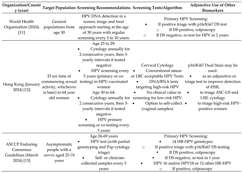 |
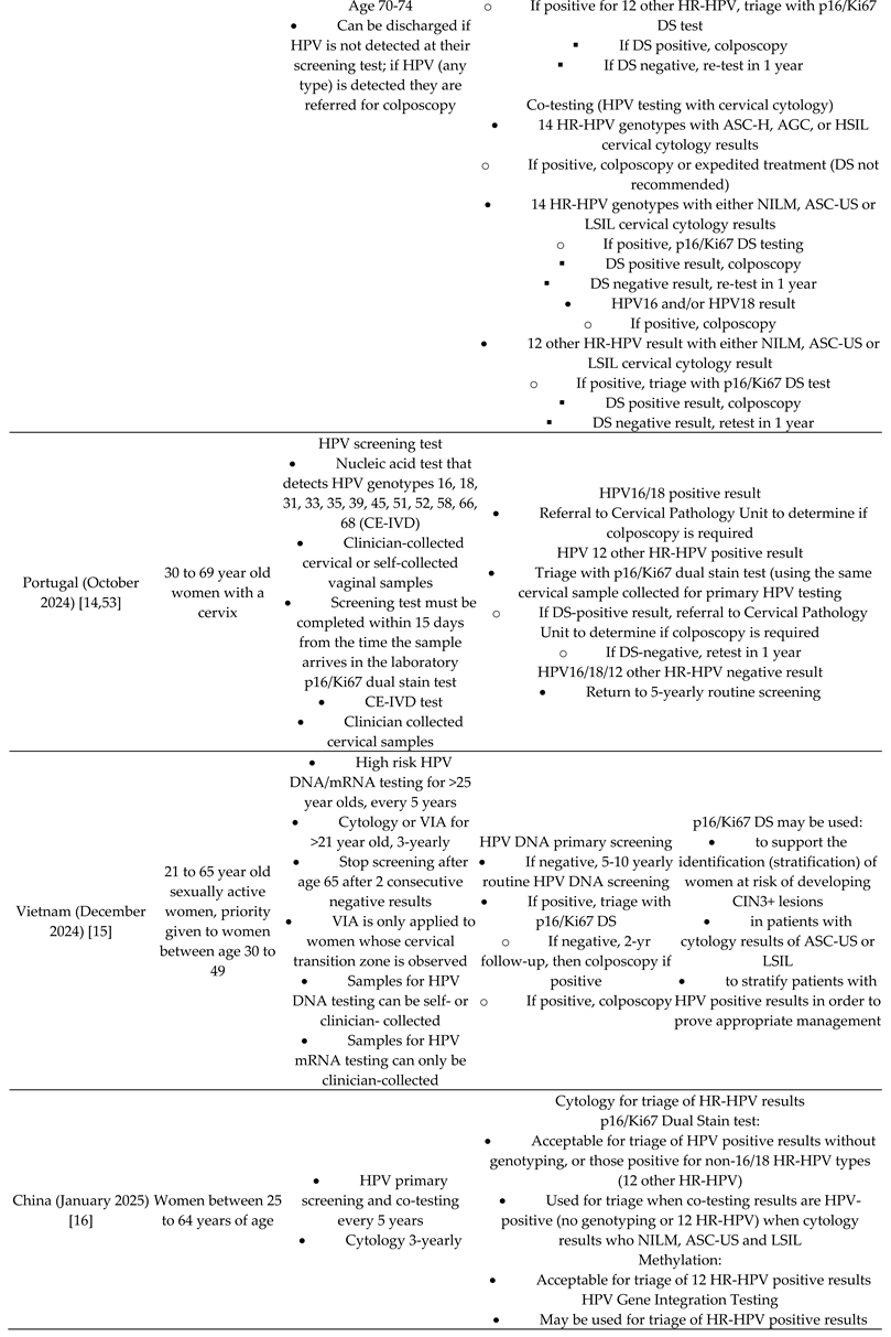 |
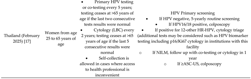 |
HPV=human papillomavirus; LBC=Liquid-based cytology; HSIL= High-grade Squamous Intraepithelial Lesion; ASC-US=Atypical Squamous Cells of Undetermined Significance, ASC-H=Atypical Squamous Cells but cannot rule out high-grade; AGC=atypical glandular cells but cannot rule out high-grade; LSIL= low-grade squamous intraepithelial lesion; HR-HPV=high risk HPV; NILM= Negative for Intraepithelial Lesion or Malignancy; DS=Dual Stain; CE-IVD= Conformité Européenne In Vitro Diagnostic (in vitro diagnostic test conforming to European Union standardsPotential usefulness of DS staining in various clinical settings and impact on patient management.
Table 4.
Clinical performance of p16/Ki67 dual stain (DS) in women under 30 years of age.
| Study (Year, Country) | Age (years) | Population | CIN2+ Sensitivity (%) DS vs LBC | CIN2+ Specificity (%) DS vs LBC | CIN3+ Sensitivity (%) DS vs LBC | CIN3+ Specificity (%) DS vs LBC |
|---|---|---|---|---|---|---|
| Ikenberg 2013 (PALMS, Europe) [35] | Screening | 18–29 | 89.4 | 92.0 | 87.3 | 91.3 |
| Waldstrøm 2013 (Denmark) [42] | Screening | <30 | 89.4 | 46.5 | 100.0 | 42.9 |
| Wentzensen 2012 (USA) [82] | Colposcopy referral | <30 | 86.8 | 55.4 | 90.4 | 40.5 |
| Frega 2019 (Italy) [83] | ASC-US/LSIL referral | 21–24 | 90.9 | 81.8 | 99.9 | 73.7 |
| Pirtea 2019 (France) [84] | ASC-US/LSIL referral | <30 | 66.0 | 93.0 | 59.0 | 79.0 |
| Ratnam 2020 (Canada) [41] | LSIL referral | <30 | 70.0 | 47.5 | — | — |
| Abrue 2021 (Spain) [40] | Persistent HPV+, referral | <30 | 100.0 | 70.0 | — | — |
Abbreviations: ASC-US, atypical squamous cells of undetermined significance; LSIL, low-grade squamous intraepithelial lesion; CIN2+/CIN3+, cervical intraepithelial neoplasia grade 2 or 3 or worse.
Disclaimer/Publisher’s Note: The statements, opinions and data contained in all publications are solely those of the individual author(s) and contributor(s) and not of MDPI and/or the editor(s). MDPI and/or the editor(s) disclaim responsibility for any injury to people or property resulting from any ideas, methods, instructions or products referred to in the content. |
© 2026 by the authors. Licensee MDPI, Basel, Switzerland. This article is an open access article distributed under the terms and conditions of the Creative Commons Attribution (CC BY) license (http://creativecommons.org/licenses/by/4.0/).
Copyright: This open access article is published under a Creative Commons CC BY 4.0 license, which permit the free download, distribution, and reuse, provided that the author and preprint are cited in any reuse.