

Submitted:
01 July 2026
Posted:
02 July 2026
You are already at the latest version
Abstract
Extracellular vesicles (EVs), including exosomes and microvesicles, have emerged as promising therapeutic vectors and diagnostic biomarkers across various branches of biomedicine. However, the clinical translation of EV-based technologies remains constrained by persistent challenges in manufacturing: insufficient yield from primary cell sources, limited control over cargo composition, and the absence of scalable, standardized production platforms. Nanosecond pulsed electric fields (nsPEF) represent an emerging biophysical approach that can address several of these limitations. Unlike conventional electroporation, which targets the plasma membrane using microsecond-to-millisecond pulses, nsPEF delivers ultrashort (1-300 ns), high-amplitude (10-300 kV/cm) pulses that penetrate intracellularly to directly perturb endosomal membranes, the endoplasmic reticulum, and the multivesicular body (MVB) compartment, the very organelles where exosome biogenesis and cargo sorting happen. Through coordinated effects on intracellular calcium mobilization, cytoskeletal remodeling, SNARE-mediated membrane fusion, and phospholipid redistribution, nsPEF can stimulate rapid, non-lethal vesicle release, a process labeled as "electro-exocytosis." Emerging evidence suggests that nsPEF does not merely increase EV yield but actively modulates the proteomic, lipidomic, and nucleic acid composition of released vesicles, offering a potential route to cargo engineering. In addition, the same biophysical principles that drive electro-exocytosis can be exploited in reverse: nsPEF-mediated transient permeabilization of EVs membranes allows for post-isolation loading of exogenous therapeutic cargo, small molecules, nucleic acids, or proteins into pre-formed vesicles without destroying their structural integrity. This review discusses current knowledge on EVs biogenesis and release mechanisms, introduces the biophysical foundations of nsPEF-cell and nsPEF-membrane interactions, and, by evaluating the experimental evidence supporting nsPEF-driven EVs engineering, outlines a translational roadmap for the application and development of this technology toward clinical-grade EVs manufacturing.
Keywords:
extracellular vesicles
; exosomes
; nanosecond pulsed electric fields
; electroporation
; electro-exocytosis
; EV engineering
; cargo loading
; biomanufacturing
1. Introduction
In accordance with MISEV2023 guidelines [1], we use the umbrella term ‘extracellular vesicles (EVs)’ throughout this review. Where size-based distinctions are relevant, we employ ‘small EVs’ and ‘medium/large EVs’. Traditional terms such as ‘exosomes’ and ‘microvesicles’ are employed within text or paragraphs that clarify their biogenesis-specific markers. Extracellular vesicles have transitioned, over the past two decades, from biological curiosities to one of the most actively investigated therapeutic platforms in modern biomedicine. Their natural capacity to transport bioactive cargo, proteins, lipids, mRNAs, microRNAs, and metabolites across biological barriers, combined with their inherent biocompatibility, low immunogenicity, and ability to home to specific tissues, makes them attractive candidates for drug delivery, immunotherapy, and regenerative medicine [1,2,3].
The clinical promise, however, has consistently outpaced clinical reality. As of early 2026, no standalone EV-based therapeutic has yet received regulatory approval for routine clinical use [4]. The reasons for this translational gap are multiple, but they converge on a central challenge: manufacturing. Producing EVs at the scale, purity, reproducibility, and functional consistency required for clinical application remains extraordinarily difficult [5,6]. Three specific manufacturing limitations deserve emphasis. The first is related to production rates. Primary cells, the most clinically relevant EVs source for many applications, produce EVs in quantities that are orders of magnitude below therapeutic requirements. A typical T75 flask of cultured cells yields nanogram-to-low-microgram quantities of EV protein over 48-72 hours, whereas a single therapeutic dose may require tens to hundreds of micrograms [7]. Scaling up through larger culture vessels, bioreactors, or cell expansion is possible but introduces additional variables, costs, and quality control challenges.
The second challenge is cargo heterogeneity. EVs released under standard culture conditions carry a cargo profile dictated by the constitutive sorting machinery of the producer cell. For many therapeutic applications such as cancer immunotherapy, anti-inflammatory treatment, or regenerative medicine, it would be desirable to enrich specific molecules (tumor antigens, anti-inflammatory cytokines, pro-angiogenic factors) in the EV cargo while depleting undesirable components. Current approaches to cargo modulation rely primarily on genetic engineering of producer cells (overexpression of desired cargo, fusion proteins targeting the EVs lumen) or post-isolation loading techniques (electroporation, sonication, freeze-thaw cycling, extrusion) [8,9,10]. Each approach has significant limitations: genetic modification is time-consuming, costly, and raises regulatory concerns for autologous applications; post-isolation loading methods often damage the EVs’ integrity and offer limited control over loading efficiency. Finally, the production of standardized products. The Minimal Information for Studies of Extracellular Vesicles (MISEV) guidelines, most recently updated in 2023, provide a framework for EVs characterization but explicitly acknowledge that standardized production protocols remain elusive [1]. The field lacks consensus on optimal cell culture conditions, EVs isolation methods, quality control metrics, and functional potency assays, complicating regulatory approval and cross-study comparability.
It is within this context that we introduce nanosecond pulsed electric fields as a biophysical tool that may address multiple manufacturing bottlenecks simultaneously [11,12]. nsPEF operates through physical mechanisms fundamentally different from those of conventional electroporation, offering unique access to the intracellular compartments where EV biogenesis occurs. The following sections build the case for nsPEF-driven EV engineering from first principles: beginning with EVs biogenesis and release mechanisms, proceeding through the biophysics of nsPEF-membrane interactions, reviewing the experimental evidence, and finally concluding with a critical assessment of opportunities and challenges.
2. EV Biogenesis and Release
A rational approach to engineering EVs production requires understanding the molecular machinery that governs vesicle formation, cargo loading, and release. Each step in this pathway represents a potential point of biophysical intervention.
2.1. Exosome Biogenesis: The Endosomal Pathway
Exosomes originate within the endosomal system. Early endosomes, formed by internalization of plasma membrane and extracellular material, mature into late endosomes through progressive acidification and molecular remodeling. During this maturation, the limiting membrane of late endosomes buds inward, generating intraluminal vesicles (ILVs) within structures termed multivesicular bodies (MVBs) [13,14]. ILV formation is orchestrated by the ESCRT machinery, a modular system comprising four subcomplexes (ESCRT-0, -I, -II, -III) and accessory proteins including ALIX, TSG101, VPS4, and HD-PTP [15]. ESCRT-0 (HRS, STAM) recognizes and clusters ubiquitinated cargo on the endosomal membrane. ESCRT-I (TSG101, VPS28, VPS37, MVB12) and ESCRT-II (EAP20, EAP30, EAP45) drive membrane deformation. ESCRT-III (CHMP proteins) mediates the final membrane scission event that pinches off the ILV into the MVB lumen. VPS4, an AAA-ATPase, disassembles the ESCRT-III polymer for recycling [16]. Parallel ESCRT-independent pathways also contribute to ILV formation. Neutral sphingomyelinase 2 (nSMase2) generates ceramide from sphingomyelin in the endosomal membrane; ceramide’s cone-shaped geometry promotes spontaneous inward curvature and budding [17]. Tetraspanin-enriched microdomains such as clusters of CD63, CD81, CD9, and associated proteins, serve as cargo-sorting platforms that can drive ILV formation independently of ESCRT components [18]. Syndecan-syntenin-ALIX interactions provide yet another route, coupling heparan sulfate proteoglycan clustering to ESCRT-III-mediated budding [19]. The fate of mature MVBs is governed by a competition between two trafficking pathways: fusion with lysosomes (leading to cargo degradation) or fusion with the plasma membrane (leading to exosome release). Rab GTPases are central regulators of this decision. Rab27a and Rab27b mediate MVB docking at the plasma membrane; Rab11 and Rab35 regulate recycling endosome-to-plasma membrane trafficking; Rab7 directs MVBs toward lysosomal degradation [20,21]. The balance among these Rab proteins determines the fraction of MVBs that release their ILV content as exosomes versus the fraction that is degraded. The final fusion event is mediated by SNARE (Soluble NSF Attachment protein REceptor) complexes. On immune and epithelial cells, the relevant SNAREs include VAMP7 (v-SNARE on the MVB membrane), syntaxin-4, and SNAP-23 (t-SNAREs on the plasma membrane) [22]. SNARE-mediated fusion is calcium-dependent: synaptotagmin family members (Syt7 in non-neuronal cells) serve as calcium sensors that trigger SNARE complex zippering upon calcium binding [23]. This calcium dependence is a critical point of intervention for nsPEF.
2.2. Microvesicle Biogenesis
Microvesicles (MVs, also termed ectosomes or microparticles) are generated by outward budding and fission of the plasma membrane, a process that requires coordinated changes in membrane lipid asymmetry, cytoskeletal architecture, and membrane curvature [24]. In resting cells, aminophospholipids, particularly phosphatidylserine (PS) and phosphatidylethanolamine (PE), are confined to the inner leaflet of the plasma membrane by the activity of flippases (ATP-dependent aminophospholipid translocases). Microvesicle formation requires the disruption of this asymmetry: calcium-activated scramblases randomize phospholipid distribution, while calpain-mediated cleavage of cytoskeletal anchoring proteins (spectrin, talin, filamin) releases the membrane from cortical actin constraints [25]. Small GTPases such as ARF6, RhoA, and Rac1 regulate actin dynamics and contractile ring formation at the budding neck, driving membrane fission [26]. MV biogenesis is dependent on many variables that can be influenced by nsPEF, such as calcium influx, phosphatidylserine (PS) localization, and cortical actin organization. nsPEF is among the most potent known inducers of rapid intracellular calcium transients, which is essential for virtually every step of MV formation: scramblase activation, calpain activation, actin remodeling, and contractile ring assembly all require elevated cytoplasmic calcium. Additionally, nsPEF modulates and destabilizes cortical actin, a mechanism required for membrane budding and EV release.
2.3. Cargo Sorting
The functional identity of EVs is determined not by its membrane alone but by its cargo. Cargo sorting operates through several parallel mechanisms. Protein sorting involves ubiquitin-dependent recognition by ESCRT-0 (for exosomes), post-translational modifications (sumoylation, palmitoylation, myristoylation) that target proteins to lipid raft domains, and direct protein-protein interactions with tetraspanin scaffolds [27]. RNA sorting is mediated by RNA-binding proteins that recognize specific sequence motifs. hnRNPA2B1 binds EXO-motifs (GGAG) in microRNAs; SYNCRIP recognizes GGCU motifs; YBX1 mediates miR-223 sorting [28,29,30]. The sumoylation status of hnRNPA2B1 regulates its affinity for target microRNAs, providing a regulatory switch that can be modulated by cellular stress, including, potentially, nsPEF-induced stress. Lipid sorting involves enrichment of sphingomyelin, ceramide, cholesterol, and glycosphingolipids in the EV membranes relative to the parent cell [31]. The lipid composition influences membrane rigidity, curvature, and fusogenicity, directly affecting EVs’ stability and uptake efficiency. Understanding these sorting mechanisms is essential for interpreting nsPEF effects on EV cargo. If nsPEF modulates ESCRT dynamics, hnRNPA2B1 sumoylation, or sphingolipid metabolism, all plausible given its intracellular effects, then the resulting changes in EV cargo composition reflect genuine reprogramming of the sorting machinery, not merely nonspecific membrane disruption. The canonical pathways of EV biogenesis and the molecular checkpoints amenable to nsPEF modulation are summarized in Figure 1.
3. nsPEF-Membrane Interactions
3.1. Conventional Electroporation
To appreciate the novelty of nsPEF, it is helpful to contrast it with conventional electroporation. In conventional electroporation, electric pulses of microsecond to millisecond duration (typically 100 μs–10 ms) and moderate field strength (0.5–5 kV/cm) are applied to cells. These pulse durations exceed the plasma membrane charging time constant (τ ≈ RC ≈ 100 ns–1 μs for mammalian cells, depending on cell radius), allowing complete capacitive charging of the outer membrane. The resulting transmembrane voltage (typically 0.5–1.5 V) exceeds the electroporation threshold, creating transient aqueous pores in the lipid bilayer [32]. Conventional electroporation has been widely used for EV loading: isolated EVs are mixed with cargo molecules (siRNA, miRNA, drugs) and subjected to electrical pulses that create transient pores in the EV membrane, allowing cargo entry [33]. However, this approach has well-documented limitations: EV aggregation during electroporation, RNA degradation, inconsistent loading efficiency, and loss of EV surface markers [34]. These problems arise partly because conventional electroporation parameters optimized for cells (micron-scale) are poorly suited for EVs (nanoscale), which have much shorter membrane charging times and require fundamentally different field conditions. The contrasting biophysical mechanisms of conventional electroporation and nsPEF are illustrated in Figure 2.
3.2. nsPEF Stimulation
Nanosecond pulsed electric fields occupy a qualitatively different biophysical regime. When pulse durations (1-300 ns) are shorter than the plasma membrane charging time constant, the outer membrane cannot fully charge during the pulse. Instead, the electric field penetrates the cell interior, inducing transmembrane voltages across intracellular membrane-bound organelles such as the endoplasmic reticulum, mitochondria, Golgi apparatus, and critically, endosomes and MVBs [35,36]. The magnitude of the voltage induced across an organelle’s membrane depends on the organelle’s radius, the applied field strength, and the pulse duration, according to the Schwan equation modified for intracellular compartments. Because organelles are smaller than the cell, they require higher field strengths to achieve electroporation-relevant voltages, thus the high amplitudes (10-300 kV/cm) characteristic of nsPEF [36]. Several distinct biophysical effects result from nsPEF exposure.
nsPEF is believed to be able to generate nanopores in the plasma membrane. nsPEF creates numerous small-diameter (< 2 nm) pores, in contrast to the fewer, larger pores produced by conventional electroporation. These nanopores are permeable to ions and small molecules but not to macromolecules, producing characteristic electrical conductance changes without the large-scale membrane disruption associated with conventional electroporation [37,38]. The most distinctive feature of nsPEF is its ability to permeabilize intracellular organellar membranes. This has been demonstrated directly for the endoplasmic reticulum (calcium release), mitochondria (cytochrome c release, membrane depolarization), and the nuclear envelope (propidium iodide access to the nucleus) [39,40,41]. For EV engineering purposes, the permeabilization of endosomal and MVB membranes is of particular interest, as it may alter ILV formation dynamics, cargo sorting fidelity, and the probability of MVB-plasma membrane fusion.
As mentioned earlier, nsPEF induces calcium release from intracellular stores (primarily the ER) within milliseconds of pulse delivery, producing cytoplasmic calcium transients that peak within 1-5 seconds and decay over 30-120 seconds [39,42]. This calcium transient is independent of extracellular calcium (it persists in calcium-free media) and is not blocked by plasma membrane calcium channel inhibitors, confirming its intracellular origin. The functional significance for EV biology is bound to the role of calcium in triggering both SNARE-mediated exosome release and scramblase/calpain-dependent microvesicle shedding.
Furthermore, nsPEF induces rapid, dose-dependent PS externalization that is detectable within seconds of pulse delivery [43]. This effect is mechanistically distinct from apoptotic PS exposure: it occurs at sublethal pulse parameters, is rapidly reversible, and does not require caspase activation. PS externalization by nsPEF reflects direct electric field effects on lipid translocation, either through field-driven migration of charged lipid headgroups or through activation of calcium-dependent scramblases by the nsPEF-induced calcium transient. Finally, nsPEF exerts pronounced effects on cytoskeletal organization. nsPEF transiently disrupts the actin cytoskeleton, causing cell rounding, bleb formation, and loss of cortical actin integrity [40,44]. This effect is dose-dependent and reversible at moderate pulse parameters. For microvesicle biology, cortical actin disruption removes the physical barrier to membrane budding; for exosome biology, actin remodeling may alter MVB trafficking and docking at the plasma membrane.
4. nsPEF-Stimulated Electro-Exocytosis of EVs
Nanosecond pulsed electric field (nsPEF)-mediated electro-exocytosis refers to the voltage-dependent release of intracellular cargo, including microvesicles (MVs), antigens, and metabolic intermediates, induced by ultrashort electric pulses, a process initially characterized in excitable cell lines where calcium mobilization couples membrane depolarization to vesicle fusion. Excitable cells such as neurons and chromaffin cells undergo regulated exocytosis through voltage-gated ion channels and SNARE-mediated machinery, whereas non-excitable cells lack regenerative electrical signaling but respond to imposed fields through electroporation, passive ion redistribution, and activation of calcium-permeable channels. Zaklit et al. [45] demonstrated that nsPEF evokes heterogeneous calcium release from the endoplasmic reticulum of adrenal chromaffin cells, establishing organelle-level membrane permeabilization as a primary trigger, while earlier work confirmed that nsPEF generates rapid intracellular calcium transients even when extracellular calcium is absent [39,42], indicating that internal store mobilization is sufficient to initiate downstream exocytotic cascades. The mechanistic framework connecting these calcium transients to EV release operates through at least two partially distinct but converging pathways: first, calcium activates synaptotagmin family members and drives SNARE complex assembly, comprising VAMP2, VAMP3, and VAMP7 [22], to mediate multivesicular body (MVB)-plasma membrane fusion and exosome release, with calcium-triggered conformational rearrangements in the synaptotagmin-1/SNARE primary interface serving as the proximal fusion trigger [46]; second, the same calcium transient activates scramblases and calpains, disrupting phospholipid asymmetry and cortical actin integrity to promote microvesicle budding and shedding directly from the plasma membrane. Phosphatidylserine (PS) externalization, which Vernier et al. [43] demonstrated occurs within nanoseconds of pulse delivery, creates microvesicle-competent membrane domains that further enhance MV budding, while intracellular membrane permeabilization [41] alters endosomal and MVB membrane dynamics in ways that potentially affect intraluminal vesicle formation, cargo sorting, and the balance between lysosomal degradation and exosomal secretion. Cytoskeletal reorganization represents a critical convergence point for both pathways, as nsPEF-induced disruption of the cortical actin barrier [40] simultaneously facilitates MVB docking and fusion for exosome release and removes the physical constraint on microvesicle shedding, a dual role consistent with the observation that F-actin remodeling participates in electrotransfer and transmembrane potential modulation in non-excitable human endothelial cell lines [47]. Applying this mechanistic framework to cancer models, Szlasa et al. [48] showed that nsPEF at 8 kV/cm (200 ns, 100 pulses, 10 kHz) induced electro-exocytosis of MAGE protein-containing MVs from melanoma cells, with upregulation of MAGE-A1 and MAGE-A2 at both mRNA and protein levels, a proposed mechanism of compensatory antigen overexpression following MV release, and improved cytolytic T cell activation; lipidomic profiling in the same study revealed phosphoethanolamine at 36% of negative lipid composition attributed to PCYT2 downregulation [49], a finding of immunological significance given PEtn-mediated T cell suppression in the tumor microenvironment [50], alongside hexosylceramide increases that bind VDAC1/2 during Bax/Bak-mediated apoptotic pore formation [51,52]. In a complementary pancreatic cancer study, Szlasa et al. [53] found that daunorubicin-resistant EPP85-181RDB cells underwent MV release detectable at five minutes with maximal release at thirteen minutes, driven by surface tension reduction from 0 to −30 mN/m producing membrane invaginations in low-cholesterol regions, with confocal microscopy confirming reversible F-actin disorganization twenty-four hours post-treatment; when combined with 50 nM paclitaxel, the RDB line uniquely became more susceptible up to forty-eight hours despite absence of electro-transport, while N-cadherin was nearly undetectable in treated lines, suggesting decreased adhesion and reduced metastatic potential. Collectively, these findings predict that nsPEF simultaneously stimulates both exosome and microvesicle release through partially distinct calcium-dependent, cytoskeleton-dependent, and membrane asymmetry-dependent pathways, establishing nsPEF not merely as an ablation modality but as a tunable biophysical tool for membrane sensitization, immunologically relevant MV release, tumor-associated antigen upregulation, metabolic reprogramming toward apoptotic phenotypes, and chemotherapy sensitization. The current evidence landscape for nsPEF-driven EV engineering, stratified by level of experimental support, is presented in Figure 3.
5. Limitations of Conventional Electroporation for EV Loading
While not identical to nsPEF, the extensive literature on conventional electroporation for EV cargo loading provides relevant context and highlights limitations that nsPEF might address. Alvarez-Erviti and colleagues demonstrated the feasibility of loading exogenous siRNA into small EVs by electroporation, achieving functional gene silencing in target cells in vitro and in vivo [33]. However, subsequent studies revealed significant technical challenges: RNA aggregation on the EV surface (rather than true luminal loading), EV aggregation and loss during the process, and highly variable loading efficiency depending on EV source and electroporation parameters [34]. These problems arise at least partly because conventional electroporation parameters (millisecond pulses, kV/cm fields) are optimized for micron-scale cells, not nanoscale vesicles. The membrane charging time for a 100 nm small EV is approximately 0.1 ns, four to five orders of magnitude shorter than for a typical cell. Consequently, conventional millisecond pulses vastly overcharge EV membranes, causing irreversible damage rather than controlled, transient pore formation. nsPEF, with pulse durations closer to the EV membrane charging time, may offer a more appropriate biophysical match for direct EV manipulation. This represents a testable hypothesis with immediate practical implications for EV-based drug delivery.
6. Prospective Clinical Applications of nsPEF-Derived EVs
The convergence of nsPEF-mediated EV engineering with the broader expansion of EV therapeutics opens several near-term and medium-term clinical applications that leverage the unique cargo-modulation capabilities described above. We believe that there are three prospective application domains such as cancer vaccines, mesenchymal-stromal-cell (MSC) EV therapies, and EV-integrated biomaterial scaffolds that are more likely to exploit the nsPEF-EV benefits. The first and most immediately translatable application is the use of nsPEF-derived tumor EVs as cancer vaccines . While nsPEF has been hypothesized to protect host from future tumor growth after treatment [54,55], a further rationale builds directly on the melanoma data of Szlasa and colleagues [56]. EVs shed from nsPEF-treated tumor cells carry upregulated MAGE-A1 and MAGE-A2 antigens and externalized PEtn, creating particles that are simultaneously enriched in tumor-specific epitopes and tagged for efficient phagocytic uptake by antigen-presenting cells. This dual-signal architecture recapitulates the design principles of synthetic nanoparticle vaccines, antigen plus adjuvant in a single particle, but in a fully biological format that preserves native protein folding, glycosylation, and membrane context. Early-phase clinical trials of dendritic-cell-derived small EVs vaccines (Dex) demonstrated safety and immunogenicity in melanoma and non-small-cell lung cancer patients, but efficacy was limited by low antigen density and inconsistent manufacturing [57,58]. nsPEF stimulation could address both limitations by increasing the per-vesicle antigen load through transcriptional upregulation (MAGE) and by providing a reproducible, physically defined stimulus that replaces the variable cytokine-cocktail protocols used for Dex generation. The second application domain is regenerative medicine using MSC-derived EVs. MSC-EVs are among the most advanced EV therapeutics, with multiple clinical trials investigating their efficacy in acute kidney injury, graft-versus-host disease, chronic wound healing, and osteoarthritis [2,3,59]. The therapeutic cargo of MSC-EVs, anti-inflammatory cytokines (IL-10, TGF-β), pro-angiogenic factors (VEGF, FGF-2), and tissue-remodeling microRNAs (miR-21, miR-146a), is strongly influenced by the culture conditions and stimulation protocols used during production [2,3]. nsPEF stimulation of MSCs could enhance EV yield through electro-exocytosis while simultaneously modulating cargo composition through calcium-dependent transcriptional programs and cytoskeletal remodeling, potentially generating EVs with superior anti-inflammatory or regenerative potency compared to those produced by serum starvation or hypoxic preconditioning. The last application integrates nsPEF-derived EVs into biomaterial scaffolds for tissue engineering. Brennan and colleagues [60] demonstrated that EVs incorporated into hydrogels, electrospun fibers, and three-dimensional-printed scaffolds retain their biological activity and can be released in a sustained manner to promote angiogenesis, osteogenesis, and immunomodulation at the implant site. Born and colleagues [61] showed that EV-functionalized scaffolds enhance mesenchymal stem cell recruitment and differentiation in bone-defect models. nsPEF stimulation could provide the high EV yields needed to functionalize clinically relevant scaffold volumes while also enriching the EVs in pro-regenerative cargo through the parameter-dependent modulation mechanisms described in this review. The integration of nsPEF-derived EVs into biomaterial scaffolds is particularly attractive for orthopedic and dental applications, where local delivery of anti-inflammatory and osteogenic signals is critical and systemic EV administration would be inefficient due to rapid hepatic clearance [60,61]. Across all application domains, the clinical case for nsPEF-derived EVs rests on the platform’s unique combination of three features that no single conventional method provides: enhanced yield through electro-exocytosis, programmable cargo modulation through parameter-dependent biophysical mechanisms, and scalability through the flow-through pulse-chamber architectures discussed below.
7. Challenges in nsPEF-Driven EV Manufacturing
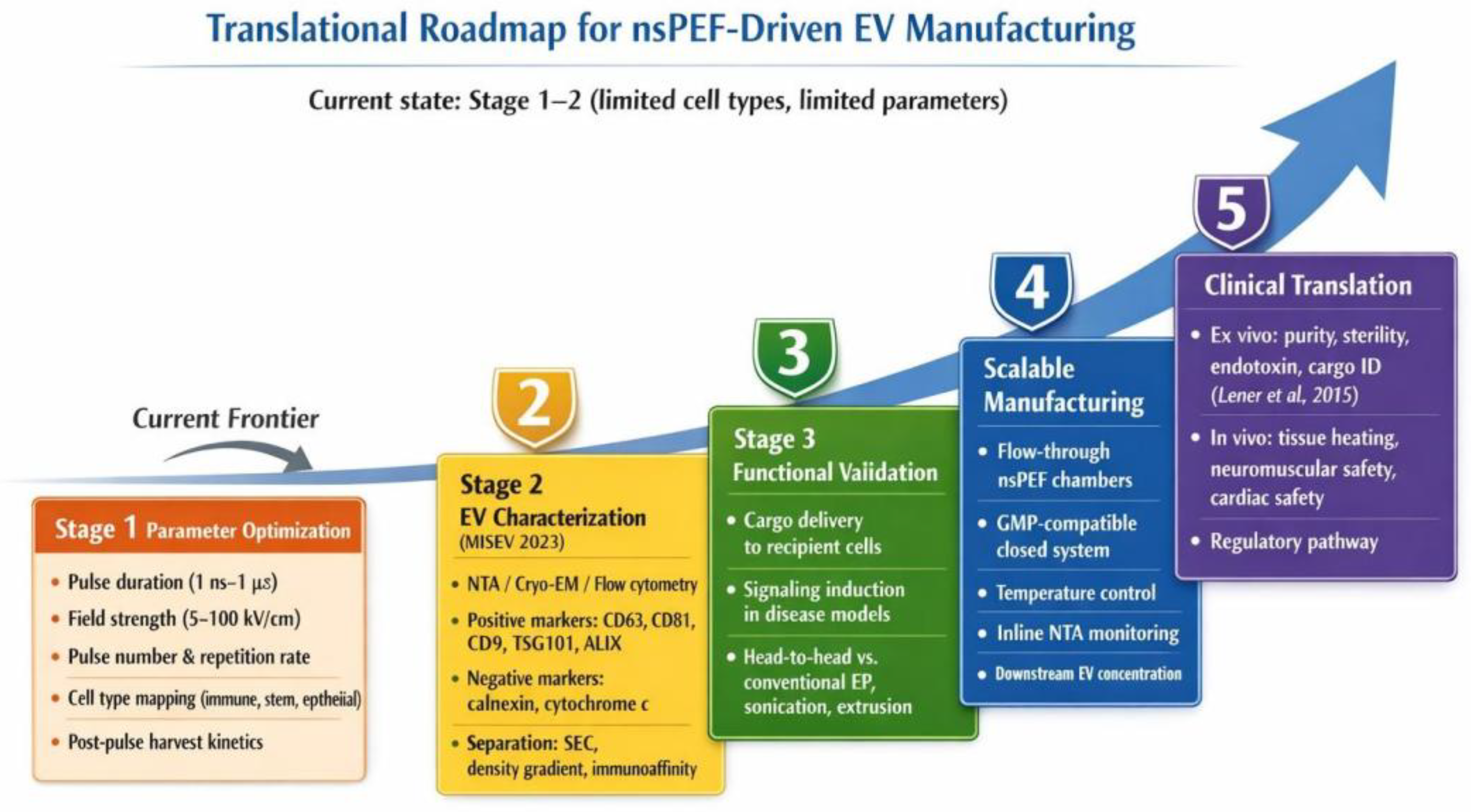
The translation of nsPEF-mediated electro-exocytosis from a laboratory observation to a reproducible manufacturing platform faces several interconnected challenges that must be addressed before clinical-grade extracellular vesicle production becomes feasible. Current nsPEF data span a narrow parameter space, predominantly 10-30 ns pulses at 15-100 kV/cm with limited pulse repetitions, obtained in only a handful of cell types including fibroblasts, pancreatic cancer cells, and melanoma lines, meaning that systematic exploration across pulse duration (1 ns to 1 μs), field strength (5-100 kV/cm), pulse number and repetition rate, and post-pulse recovery kinetics remains necessary to define optimal conditions for each EV subtype, since shorter pulses favor intracellular effects while longer pulses preferentially permeabilize the plasma membrane and the threshold for EV stimulation will vary by cell type, membrane composition, and baseline secretory activity, with primary immune cells and stem cells representing therapeutically critical but entirely unstudied targets. A coordinated multi-laboratory parameter mapping effort, analogous to the systematic electroporation studies that established standard transfection protocols in the 1990s, would accelerate this process considerably. Any resulting manufacturing workflow must comply with the MISEV 2023 guidelines [1], which require physical characterization of size distribution and morphology, molecular confirmation of EV identity through transmembrane markers such as CD63, CD81, and CD9 alongside cytosolic markers including TSG101, ALIX, and syntenin-1, and exclusion of non-EV contaminants such as calnexin and cytochrome c to rule out apoptotic bodies, organellar fragments, and protein aggregates, a concern of particular relevance given that nsPEF-induced cellular stress may trigger release of debris that co-isolates with bona fide EVs and confounds functional assessments, necessitating rigorous separation through size exclusion chromatography, density gradient ultracentrifugation, or immunoaffinity capture. Functional validation demonstrating that nsPEF-generated EVs retain biologically relevant activity (cargo delivery, receptor-mediated signaling in recipient cells, or therapeutic efficacy in disease models) constitutes an additional MISEV requirement that no nsPEF study has yet systematically fulfilled. Beyond characterization, scalability presents a fundamental engineering bottleneck, as current nsPEF devices are designed either for cuvette-based research treating microliter volumes or for clinical tumor ablation via needle electrodes, neither of which is suitable for cell-based EV manufacturing at therapeutic scale; flow-through treatment chambers in which cell suspensions pass continuously through a pulsing zone [62] represent the most promising path forward but must deliver uniform electric fields, maintain temperature control to prevent thermal artifacts at high repetition rates, and operate under sterile closed-system conditions compatible with good manufacturing practice, while integrating downstream EV collection, concentration, and potentially real-time nanoparticle monitoring into the flow path. Safety considerations bifurcate according to application context: for ex vivo approaches in which nsPEF is applied to donor cells and only the resulting EVs are administered to patients, standard EV therapeutic requirements (purity, sterility, endotoxin levels, absence of residual cellular material, and cargo identity) apply under existing regulatory frameworks [4]; however, for potential in vivo applications in which nsPEF would be delivered directly to tissues to stimulate therapeutic EV release in situ, additional concerns including tissue heating, neuromuscular stimulation, cardiac rhythm disturbance near the thorax, and DNA damage from high-field exposures require dedicated evaluation, since clinical safety data from nsPEF tumor ablation in dermatological settings [63] cannot be extrapolated directly to lower-dose EV-stimulatory protocols applied to non-tumor tissues. A proposed translational roadmap for nsPEF-driven EV manufacturing, highlighting current progress and remaining milestones, is depicted in Figure 4.
8. Conclusion
Extracellular vesicle engineering stands at the cusp of clinical maturity, yet its progress continues to be constrained by the same manufacturing challenges that have plagued the field for over a decade: insufficient yield, limited cargo control, and absence of scalable production platforms. Nanosecond pulsed electric fields offer a biophysical approach that is fundamentally different from existing methods, not merely as a faster or more intense version of conventional electroporation, but a qualitatively distinct technology that accesses the intracellular compartments where EV biogenesis actually occurs. The concept of nsPEF-mediated electro-exocytosis of vesicular release through coordinated calcium mobilization, cytoskeletal remodeling, and SNARE activation is supported by a coherent biophysical framework and a growing, if still modest, body of experimental evidence. The additional possibility of using nsPEF for direct, post-isolation cargo loading and surface modification of EVs further broadens the potential toolkit. Despite the high promise, the most critical experiments on nsPEF parameter optimization for primary immune and stem cells, comprehensive cargo characterization, functional validation in disease-relevant models, and head-to-head comparison with alternative approaches, remain to be performed. Further investigations on nsPEF-mediated exocytosis are needed to further clarify the potential of applied nsPEF in the EV manufacturing era.
Author Contributions
Conceptualization, R.M.L.C.B.; resources, R.M.L.C.B.; writing—original draft preparation, A.N., and T.H.; writing—review and editing, M.S., A.G., A.D., S.J.B., & R.M.L.C.B.; supervision, R.M.L.C.B.; project administration, R.M.L.C.B.; funding acquisition, R.M.L.C.B. All authors have read and agreed to the published version of the manuscript.
Funding
The current work was funded by the “Pam and Rich Nuccitelli Foundation”, Old Dominion University SEED Grant, PI : Colunga-Biancatelli.
Institutional Review Board Statement
Not applicable.
Informed Consent Statement
Not applicable.
Data Availability Statement
Not Applicable.
Acknowledgments
We thank the Pam and Rich Nuccitelli Foundation for their generous offer to support our research. During the preparation of this manuscript, the authors used Napkin AI for the generation of Figure 4. The authors have reviewed and edited the output and take full responsibility for the content of this publication.”.
Conflicts of Interest
The authors declare no conflicts of interest.
Abbreviations
The following abbreviations are used in this manuscript:
| Abbreviation | Definition |
| nsPEF | Nanosecond pulsed electric field(s) |
| sEVs | Small extracellular vesicles |
| EVs | Extracellular vesicles |
| MVB | Multivesicular body |
| SNARE | Soluble N-ethylmaleimide-sensitive factor attachment protein receptor |
| PS | Phosphatidylserine |
| ER | Endoplasmic reticulum |
| ILV | Intraluminal vesicle |
| Ca2+ | Calcium ion |
| siRNA | Small interfering RNA |
| miRNA | MicroRNA |
| MVs | Microvesicles |
| VAMP2/3/7 | Vesicle-associated membrane protein 2/3/7 |
| PEtn | Phosphoethanolamine |
| PCYT2 | Phosphate cytidylyltransferase 2, ethanolamine |
| VDAC1/2 | Voltage-dependent anion channel 1/2 |
| MAGE | Melanoma-associated antigen |
| F-actin | Filamentous actin |
| ESCRT | Endosomal sorting complexes required for transport |
| MSC | Mesenchymal stromal cell |
| MSC-EVs | Mesenchymal stromal cell–derived extracellular vesicles |
| IL-10 | Interleukin-10 |
| TGF-β | Transforming growth factor-beta |
| VEGF | Vascular endothelial growth factor |
| FGF-2 | Fibroblast growth factor-2 |
| Dex | Dendritic cell–derived exosomes |
| MISEV | Minimal information for studies of extracellular vesicles |
| CD63/CD81/CD9 | Cluster of differentiation 63/81/9 |
| TSG101 | Tumor susceptibility gene 101 |
| ALIX | ALG-2-interacting protein X |
| SEC | Size exclusion chromatography |
| NTA | Nanoparticle tracking analysis |
| cryo-EM | Cryogenic electron microscopy |
| GMP | Good manufacturing practice |
| DNA | Deoxyribonucleic acid |
| DC | Dendritic cell |
| ISEV | International Society for Extracellular Vesicles |
References
- Welsh, J.A.; Goberdhan, D.C.I.; O'Driscoll, L.; Buzas, E.I.; Blenkiron, C.; Bussolati, B.; Cai, H.; Di Vizio, D.; Driedonks, T.A.P.; Erdbrügger, U.; et al. Minimal information for studies of extracellular vesicles (MISEV2023): From basic to advanced approaches. J. Extracell. Vesicles 2024, 13, e12404. [Google Scholar] [CrossRef] [PubMed]
- Vader, P.; Mol, E.A.; Pasterkamp, G.; Schiffelers, R.M. Extracellular vesicles for drug delivery. Adv. Drug Deliv. Rev. 2016, 106, 148–156. [Google Scholar] [CrossRef] [PubMed]
- Elsharkasy, O.M.; Nordin, J.Z.; Hagey, D.W.; de Jong, O.G.; Schiffelers, R.M.; Andaloussi, S.E.; Vader, P. Extracellular vesicles as drug delivery systems: Why and how? Adv. Drug Deliv. Rev. 2020, 159, 332–343. [Google Scholar] [CrossRef] [PubMed]
- Lener, T.; Gimona, M.; Aigner, L.; Börger, V.; Buzas, E.; Camussi, G.; Chaput, N.; Chatterjee, D.; Court, F.A.; Del Portillo, H.A.; et al. Applying extracellular vesicles based therapeutics in clinical trials - an ISEV position paper. J. Extracell. Vesicles 2015, 4, 30087. [Google Scholar] [CrossRef] [PubMed]
- Gudbergsson, J.M.; Johnsen, K.B.; Skov, M.N.; Duroux, M. Systematic review of factors influencing extracellular vesicle yield from cell cultures. Cytotechnology 2016, 68, 579–592. [Google Scholar] [CrossRef] [PubMed]
- Herrmann, I.K.; Wood, M.J.A.; Fuhrmann, G. Extracellular vesicles as a next-generation drug delivery platform. Nat. Nanotechnol. 2021, 16, 748–759. [Google Scholar] [CrossRef] [PubMed]
- Ng, C.Y.; Kee, L.T.; Al-Masawa, M.E.; Lee, Q.H.; Subramaniam, T.; Kok, D.; Ng, M.H.; Law, J.X. Scalable Production of Extracellular Vesicles and Its Therapeutic Values: A Review. Int. J. Mol. Sci. 2022, 23. [Google Scholar] [CrossRef] [PubMed]
- René, C.A.; Parks, R.J. Bioengineering extracellular vesicle cargo for optimal therapeutic efficiency. Mol. Ther. Methods Clin. Dev. 2024, 32, 101259. [Google Scholar] [CrossRef] [PubMed]
- Peruzzi, J.A.; Gunnels, T.F.; Edelstein, H.I.; Lu, P.; Baker, D.; Leonard, J.N.; Kamat, N.P. Enhancing extracellular vesicle cargo loading and functional delivery by engineering protein-lipid interactions. Nat. Commun. 2024, 15, 5618. [Google Scholar] [CrossRef] [PubMed]
- Mediratta, K.; Diab, M.D.; Han, P.; Hu, H.; Wang, L. Emerging Strategies for Cargo Loading and Engineering of Extracellular Vesicles for Breast Cancer Treatment. Nanomaterials 2025, Vol. 15, 1418. [Google Scholar]
- Schoenbach, K.H.; Joshi, R.; Kolb, J.; Buescher, S.; Beebe, S. Subcellular effects of nanosecond electrical pulses. Conf. Proc. IEEE Eng. Med. Biol. Soc. 2004, 2004, 5447–5450. [Google Scholar] [CrossRef] [PubMed]
- Beebe, S.J.; Fox, P.M.; Rec, L.J.; Willis, E.L.; Schoenbach, K.H. Nanosecond, high-intensity pulsed electric fields induce apoptosis in human cells. Faseb J. 2003, 17, 1493–1495. [Google Scholar] [CrossRef] [PubMed]
- Colombo, M.; Raposo, G.; Théry, C. Biogenesis, secretion, and intercellular interactions of exosomes and other extracellular vesicles. Annu Rev. Cell Dev. Biol. 2014, 30, 255–289. [Google Scholar] [CrossRef] [PubMed]
- Hessvik, N.P.; Llorente, A. Current knowledge on exosome biogenesis and release. Cell Mol. Life Sci. 2018, 75, 193–208. [Google Scholar] [CrossRef] [PubMed]
- Hurley, J.H. ESCRTs are everywhere. Embo J. 2015, 34, 2398–2407. [Google Scholar] [CrossRef] [PubMed]
- Schöneberg, J.; Lee, I.H.; Iwasa, J.H.; Hurley, J.H. Reverse-topology membrane scission by the ESCRT proteins. Nat. Rev. Mol. Cell Biol. 2017, 18, 5–17. [Google Scholar] [CrossRef] [PubMed]
- Trajkovic, K.; Hsu, C.; Chiantia, S.; Rajendran, L.; Wenzel, D.; Wieland, F.; Schwille, P.; Brügger, B.; Simons, M. Ceramide triggers budding of exosome vesicles into multivesicular endosomes. Science 2008, 319, 1244–1247. [Google Scholar] [CrossRef] [PubMed]
- van Niel, G.; Charrin, S.; Simoes, S.; Romao, M.; Rochin, L.; Saftig, P.; Marks, M.S.; Rubinstein, E.; Raposo, G. The tetraspanin CD63 regulates ESCRT-independent and -dependent endosomal sorting during melanogenesis. Dev. Cell 2011, 21, 708–721. [Google Scholar] [CrossRef] [PubMed]
- Baietti, M.F.; Zhang, Z.; Mortier, E.; Melchior, A.; Degeest, G.; Geeraerts, A.; Ivarsson, Y.; Depoortere, F.; Coomans, C.; Vermeiren, E.; et al. Syndecan-syntenin-ALIX regulates the biogenesis of exosomes. Nat. Cell Biol. 2012, 14, 677–685. [Google Scholar] [CrossRef] [PubMed]
- Ostrowski, M.; Carmo, N.B.; Krumeich, S.; Fanget, I.; Raposo, G.; Savina, A.; Moita, C.F.; Schauer, K.; Hume, A.N.; Freitas, R.P.; et al. Rab27a and Rab27b control different steps of the exosome secretion pathway. Nat. Cell Biol. 2010, 12, 19-30; sup pp 11-13. [Google Scholar] [CrossRef] [PubMed]
- Hsu, C.; Morohashi, Y.; Yoshimura, S.; Manrique-Hoyos, N.; Jung, S.; Lauterbach, M.A.; Bakhti, M.; Grønborg, M.; Möbius, W.; Rhee, J.; et al. Regulation of exosome secretion by Rab35 and its GTPase-activating proteins TBC1D10A-C. J. Cell Biol. 2010, 189, 223–232. [Google Scholar] [CrossRef] [PubMed]
- Fader, C.M.; Sánchez, D.G.; Mestre, M.B.; Colombo, M.I. TI-VAMP/VAMP7 and VAMP3/cellubrevin: two v-SNARE proteins involved in specific steps of the autophagy/multivesicular body pathways. Biochim Biophys. Acta 2009, 1793, 1901–1916. [Google Scholar] [CrossRef] [PubMed]
- Südhof, T.C. The molecular machinery of neurotransmitter release (Nobel lecture). Angew. Chem. Int. Ed. Engl. 2014, 53, 12696–12717. [Google Scholar] [CrossRef] [PubMed]
- Clancy, J.W.; Schmidtmann, M.; D'Souza-Schorey, C. The ins and outs of microvesicles. FASEB Bioadv 2021, 3, 399–406. [Google Scholar] [CrossRef] [PubMed]
- Barteneva, N.S.; Fasler-Kan, E.; Bernimoulin, M.; Stern, J.N.H.; Ponomarev, E.D.; Duckett, L.; Vorobjev, I.A. Circulating microparticles: square the circle. BMC Cell Biol. 2013, 14, 23. [Google Scholar] [CrossRef] [PubMed]
- Muralidharan-Chari, V.; Clancy, J.; Plou, C.; Romao, M.; Chavrier, P.; Raposo, G.; D'Souza-Schorey, C. ARF6-regulated shedding of tumor cell-derived plasma membrane microvesicles. Curr. Biol. 2009, 19, 1875–1885. [Google Scholar] [CrossRef] [PubMed]
- Villarroya-Beltri, C.; Baixauli, F.; Gutiérrez-Vázquez, C.; Sánchez-Madrid, F.; Mittelbrunn, M. Sorting it out: regulation of exosome loading. Semin Cancer Biol. 2014, 28, 3–13. [Google Scholar] [CrossRef] [PubMed]
- Villarroya-Beltri, C.; Gutiérrez-Vázquez, C.; Sánchez-Cabo, F.; Pérez-Hernández, D.; Vázquez, J.; Martin-Cofreces, N.; Martinez-Herrera, D.J.; Pascual-Montano, A.; Mittelbrunn, M.; Sánchez-Madrid, F. Sumoylated hnRNPA2B1 controls the sorting of miRNAs into exosomes through binding to specific motifs. Nat. Commun. 2013, 4, 2980. [Google Scholar] [CrossRef] [PubMed]
- Santangelo, L.; Giurato, G.; Cicchini, C.; Montaldo, C.; Mancone, C.; Tarallo, R.; Battistelli, C.; Alonzi, T.; Weisz, A.; Tripodi, M. The RNA-Binding Protein SYNCRIP Is a Component of the Hepatocyte Exosomal Machinery Controlling MicroRNA Sorting. Cell Rep. 2016, 17, 799–808. [Google Scholar] [CrossRef] [PubMed]
- Shurtleff, M.J.; Temoche-Diaz, M.M.; Karfilis, K.V.; Ri, S.; Schekman, R. Y-box protein 1 is required to sort microRNAs into exosomes in cells and in a cell-free reaction. Elife 2016, 5. [Google Scholar] [CrossRef] [PubMed]
- Skotland, T.; Sandvig, K.; Llorente, A. Lipids in exosomes: Current knowledge and the way forward. Prog. Lipid Res. 2017, 66, 30–41. [Google Scholar] [CrossRef] [PubMed]
- Kotnik, T.; Kramar, P.; Pucihar, G.; Miklavčič, D.; Tarek, M. Cell membrane electroporation- Part 1: The phenomenon. IEEE Electr. Insul. Mag. 2012, 28. [Google Scholar] [CrossRef]
- Alvarez-Erviti, L.; Seow, Y.; Yin, H.; Betts, C.; Lakhal, S.; Wood, M.J. Delivery of siRNA to the mouse brain by systemic injection of targeted exosomes. Nat. Biotechnol. 2011, 29, 341–345. [Google Scholar] [CrossRef] [PubMed]
- Kooijmans, S.A.A.; Stremersch, S.; Braeckmans, K.; de Smedt, S.C.; Hendrix, A.; Wood, M.J.A.; Schiffelers, R.M.; Raemdonck, K.; Vader, P. Electroporation-induced siRNA precipitation obscures the efficiency of siRNA loading into extracellular vesicles. J. Control Release 2013, 172, 229–238. [Google Scholar] [CrossRef] [PubMed]
- Vernier, P.T.; Sun, Y.; Marcu, L.; Salemi, S.; Craft, C.M.; Gundersen, M.A. Calcium bursts induced by nanosecond electric pulses. Biochem Biophys. Res. Commun. 2003, 310, 286–295. [Google Scholar] [CrossRef] [PubMed]
- Kotnik, T.; Miklavcic, D. Theoretical evaluation of voltage inducement on internal membranes of biological cells exposed to electric fields. Biophys. J. 2006, 90, 480–491. [Google Scholar] [CrossRef] [PubMed]
- Pakhomov, A.G.; Shevin, R.; White, J.A.; Kolb, J.F.; Pakhomova, O.N.; Joshi, R.P.; Schoenbach, K.H. Membrane permeabilization and cell damage by ultrashort electric field shocks. Arch. Biochem Biophys. 2007, 465, 109–118. [Google Scholar] [CrossRef] [PubMed]
- Pakhomov, A.G.; Bowman, A.M.; Ibey, B.L.; Andre, F.M.; Pakhomova, O.N.; Schoenbach, K.H. Lipid nanopores can form a stable, ion channel-like conduction pathway in cell membrane. Biochem Biophys. Res. Commun. 2009, 385, 181–186. [Google Scholar] [CrossRef] [PubMed]
- White, J.A.; Blackmore, P.F.; Schoenbach, K.H.; Beebe, S.J. Stimulation of capacitative calcium entry in HL-60 cells by nanosecond pulsed electric fields. J. Biol. Chem. 2004, 279, 22964–22972. [Google Scholar] [CrossRef] [PubMed]
- Pakhomov, A.G.; Xiao, S.; Pakhomova, O.N.; Semenov, I.; Kuipers, M.A.; Ibey, B.L. Disassembly of actin structures by nanosecond pulsed electric field is a downstream effect of cell swelling. Bioelectrochemistry 2014, 100, 88–95. [Google Scholar] [CrossRef] [PubMed]
- Tekle, E.; Oubrahim, H.; Dzekunov, S.M.; Kolb, J.F.; Schoenbach, K.H.; Chock, P.B. Selective field effects on intracellular vacuoles and vesicle membranes with nanosecond electric pulses. Biophys. J. 2005, 89, 274–284. [Google Scholar] [CrossRef] [PubMed]
- Semenov, I.; Xiao, S.; Pakhomov, A.G. Primary pathways of intracellular Ca(2+) mobilization by nanosecond pulsed electric field. Biochim Biophys. Acta 2013, 1828, 981–989. [Google Scholar] [CrossRef] [PubMed]
- Vernier, P.T.; Sun, Y.; Marcu, L.; Craft, C.M.; Gundersen, M.A. Nanosecond pulsed electric fields perturb membrane phospholipids in T lymphoblasts. FEBS Lett. 2004, 572, 103–108. [Google Scholar] [CrossRef] [PubMed]
- Stacey, M.; Fox, P.; Buescher, S.; Kolb, J. Nanosecond pulsed electric field induced cytoskeleton, nuclear membrane and telomere damage adversely impact cell survival. Bioelectrochemistry 2011, 82, 131–134. [Google Scholar] [CrossRef] [PubMed]
- Zaklit, J.; Craviso, G.L.; Leblanc, N.; Yang, L.; Vernier, P.T.; Chatterjee, I. Adrenal Chromaffin Cells Exposed to 5-ns Pulses Require Higher Electric Fields to Porate Intracellular Membranes than the Plasma Membrane: An Experimental and Modeling Study. J. Membr. Biol. 2017, 250, 535–552. [Google Scholar] [CrossRef] [PubMed]
- Toulmé, E.; Salazar Lázaro, A.; Trimbuch, T.; Rizo, J.; Rosenmund, C. Neurotransmitter release is triggered by a calcium-induced rearrangement in the Synaptotagmin-1/SNARE complex primary interface. Proc. Natl. Acad. Sci. U S A 2024, 121, e2409636121. [Google Scholar] [CrossRef] [PubMed]
- Kanthou, C.; Kranjc, S.; Sersa, G.; Tozer, G.; Zupanic, A.; Cemazar, M. The endothelial cytoskeleton as a target of electroporation-based therapies. Mol. Cancer Ther. 2006, 5, 3145–3152. [Google Scholar] [CrossRef] [PubMed]
- Szlasa, W.; Sauer, N.; Baczyńska, D.; Ziętek, M.; Haczkiewicz-Leśniak, K.; Karpiński, P.; Fleszar, M.; Fortuna, P.; Kulus, M.J.; Piotrowska, A.; et al. Pulsed electric field induces exocytosis and overexpression of MAGE antigens in melanoma. Sci. Rep. 2024, 14, 12546. [Google Scholar] [CrossRef] [PubMed]
- Zhou, L.; Zhang, S.; Wang, L.; Liu, X.; Yang, X.; Qiu, L.; Zhou, Y.; Huang, Q.; Meng, Y.; Lei, X.; et al. PCYT2 inhibits epithelial-mesenchymal transition in colorectal cancer by elevating YAP1 phosphorylation. JCI Insight 2024, 9. [Google Scholar] [CrossRef] [PubMed]
- Osawa, T.; Shimamura, T.; Saito, K.; Hasegawa, Y.; Ishii, N.; Nishida, M.; Ando, R.; Kondo, A.; Anwar, M.; Tsuchida, R.; et al. Phosphoethanolamine Accumulation Protects Cancer Cells under Glutamine Starvation through Downregulation of PCYT2. Cell Rep. 2019, 29, 89–103.e107. [Google Scholar] [CrossRef] [PubMed]
- Chin, H.S.; Li, M.X.; Tan, I.K.L.; Ninnis, R.L.; Reljic, B.; Scicluna, K.; Dagley, L.F.; Sandow, J.J.; Kelly, G.L.; Samson, A.L.; et al. VDAC2 enables BAX to mediate apoptosis and limit tumor development. Nat. Commun. 2018, 9, 4976. [Google Scholar] [CrossRef] [PubMed]
- Dadsena, S.; Bockelmann, S.; Mina, J.G.M.; Hassan, D.G.; Korneev, S.; Razzera, G.; Jahn, H.; Niekamp, P.; Müller, D.; Schneider, M.; et al. Ceramides bind VDAC2 to trigger mitochondrial apoptosis. Nat. Commun. 2019, 10, 1832. [Google Scholar] [CrossRef] [PubMed]
- Szlasa, W.; Michel, O.; Sauer, N.; Novickij, V.; Lewandowski, D.; Kasperkiewicz, P.; Tarek, M.; Saczko, J.; Kulbacka, J. Nanosecond pulsed electric field suppresses growth and reduces multi-drug resistance effect in pancreatic cancer. Sci. Rep. 2023, 13, 351. [Google Scholar] [CrossRef] [PubMed]
- Ruiz-Fernández, A.R.; Rosemblatt, M.; Perez-Acle, T. Nanosecond pulsed electric field (nsPEF) and vaccines: a novel technique for the inactivation of SARS-CoV-2 and other viruses? Ann. Med. 2022, 54, 1749–1756. [Google Scholar] [CrossRef] [PubMed]
- Rossi, A.; Pakhomova, O.N.; Pakhomov, A.G.; Weygandt, S.; Bulysheva, A.A.; Murray, L.E.; Mollica, P.A.; Muratori, C. Mechanisms and immunogenicity of nsPEF-induced cell death in B16F10 melanoma tumors. Sci. Rep. 2019, 9, 431. [Google Scholar] [CrossRef] [PubMed]
- Szlasa, W.; Sauer, N.; Baczyńska, D.; Ziętek, M.; Haczkiewicz-Leśniak, K.; Karpiński, P.; Fleszar, M.; Fortuna, P.; Kulus, M.J.; Piotrowska, A.; et al. Pulsed electric field induces exocytosis and overexpression of MAGE antigens in melanoma. Sci. Rep. 2024, 14, 12546. [Google Scholar] [CrossRef] [PubMed]
- Escudier, B.; Dorval, T.; Chaput, N.; André, F.; Caby, M.P.; Novault, S.; Flament, C.; Leboulaire, C.; Borg, C.; Amigorena, S.; et al. Vaccination of metastatic melanoma patients with autologous dendritic cell (DC) derived-exosomes: results of thefirst phase I clinical trial. J. Transl. Med. 2005, 3, 10. [Google Scholar] [CrossRef] [PubMed]
- Morse, M.A.; Garst, J.; Osada, T.; Khan, S.; Hobeika, A.; Clay, T.M.; Valente, N.; Shreeniwas, R.; Sutton, M.A.; Delcayre, A.; et al. A phase I study of dexosome immunotherapy in patients with advanced non-small cell lung cancer. J. Transl. Med. 2005, 3, 9. [Google Scholar] [CrossRef] [PubMed]
- Syromiatnikova, V.; Prokopeva, A.; Gomzikova, M. Methods of the Large-Scale Production of Extracellular Vesicles. Int. J. Mol. Sci. 2022, 23. [Google Scholar] [CrossRef] [PubMed]
- Brennan, M.; Layrolle, P.; Mooney, D.J. Biomaterials functionalized with MSC secreted extracellular vesicles and soluble factors for tissue regeneration. Adv. Funct. Mater. 2020, 30. [Google Scholar] [CrossRef] [PubMed]
- Born, L.J.; Harmon, J.W.; Jay, S.M. Therapeutic potential of extracellular vesicle-associated long noncoding RNA. Bioeng. Transl. Med. 2020, 5, e10172. [Google Scholar] [CrossRef] [PubMed]
- Guo, S.; Burcus, N.I.; Hornef, J.; Jing, Y.; Jiang, C.; Heller, R.; Beebe, S.J. Nano-Pulse Stimulation for the Treatment of Pancreatic Cancer and the Changes in Immune Profile. Cancers 2018, 10. [Google Scholar] [CrossRef] [PubMed]
- Nuccitelli, R.; Wood, R.; Kreis, M.; Athos, B.; Huynh, J.; Lui, K.; Nuccitelli, P.; Epstein, E.H., Jr. First-in-human trial of nanoelectroablation therapy for basal cell carcinoma: proof of method. Exp. Dermatol. 2014, 23, 135–137. [Google Scholar] [CrossRef] [PubMed]
Figure 1.
EV biogenesis pathways and nsPEF intervention points. Schematic depicting exosome formation via the endosomal pathway (left) and microvesicle budding from the plasma membrane (right). Key molecular machinery is shown, including ESCRT complexes, nSMase2/ceramide, Rab GTPases, SNARE proteins, scramblases, and calpain. Yellow stars indicate calcium-dependent steps directly activated by nsPEF-induced intracellular calcium mobilization, including SNARE-mediated MVB-plasma membrane fusion (exosomes) and PS externalization with cortical actin disruption (microvesicles).
Figure 1.
EV biogenesis pathways and nsPEF intervention points. Schematic depicting exosome formation via the endosomal pathway (left) and microvesicle budding from the plasma membrane (right). Key molecular machinery is shown, including ESCRT complexes, nSMase2/ceramide, Rab GTPases, SNARE proteins, scramblases, and calpain. Yellow stars indicate calcium-dependent steps directly activated by nsPEF-induced intracellular calcium mobilization, including SNARE-mediated MVB-plasma membrane fusion (exosomes) and PS externalization with cortical actin disruption (microvesicles).
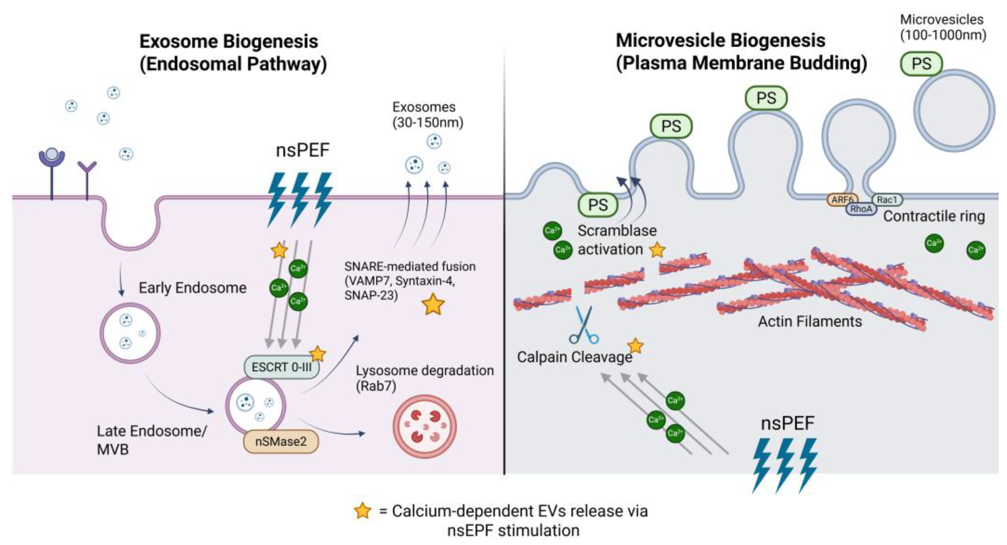
Figure 2.
Biophysical comparison of conventional electroporation and nanosecond pulsed electric fields. Conventional electroporation (top) uses long pulses (100 μs-10 ms, 0.5-5 kV/cm) that fully charge the plasma membrane, generating large macromolecule-permeable pores while shielding intracellular organelles. nsPEF (bottom) delivers ultrashort pulses (1-300 ns, 10-300 kV/cm) that incompletely charge the plasma membrane, allowing field penetration to intracellular compartments including the ER, mitochondria, and MVBs, producing nanoscale ion-permeable pores at both plasma and organelle membranes.
Figure 2.
Biophysical comparison of conventional electroporation and nanosecond pulsed electric fields. Conventional electroporation (top) uses long pulses (100 μs-10 ms, 0.5-5 kV/cm) that fully charge the plasma membrane, generating large macromolecule-permeable pores while shielding intracellular organelles. nsPEF (bottom) delivers ultrashort pulses (1-300 ns, 10-300 kV/cm) that incompletely charge the plasma membrane, allowing field penetration to intracellular compartments including the ER, mitochondria, and MVBs, producing nanoscale ion-permeable pores at both plasma and organelle membranes.
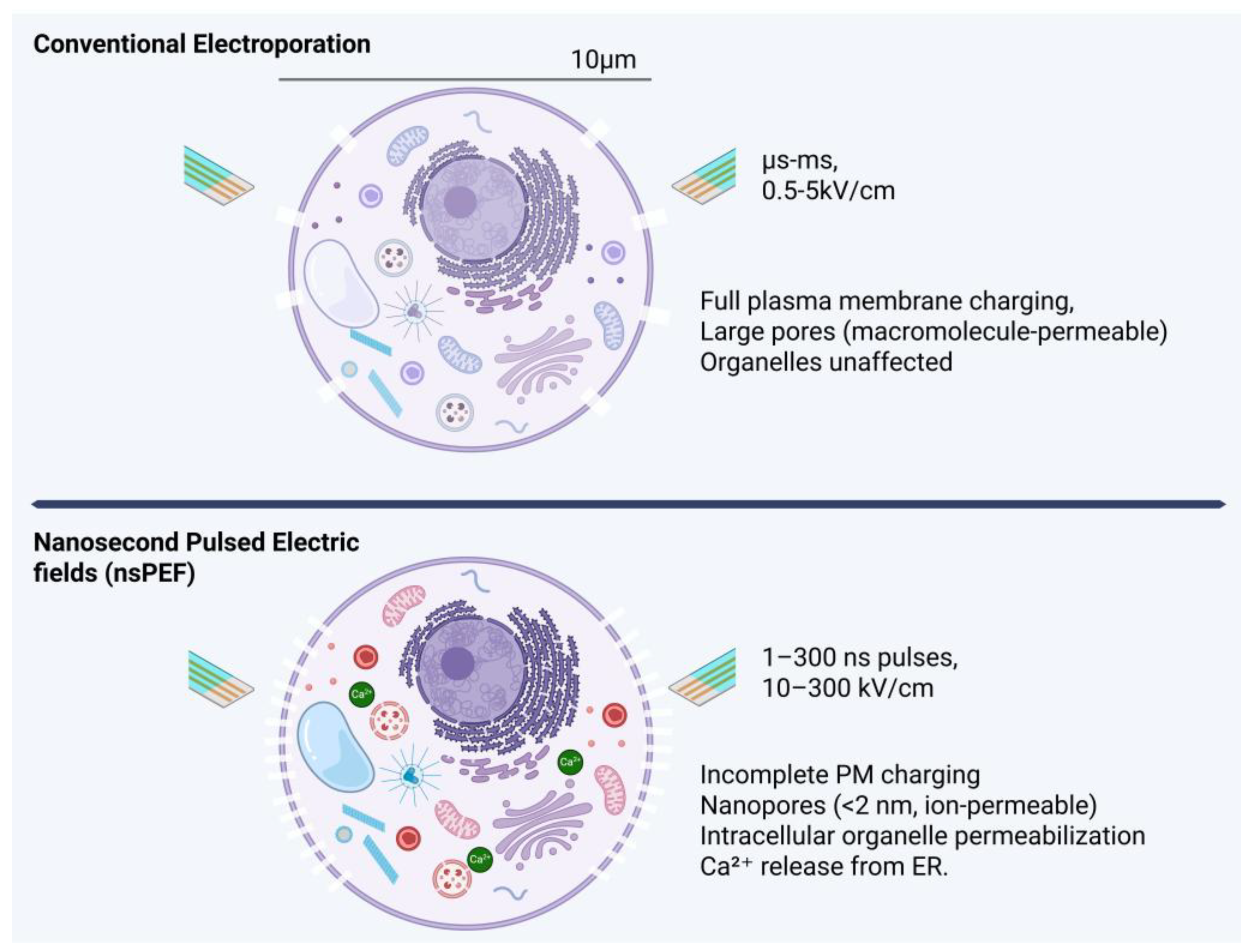
Figure 3.
Evidence landscape for nsPEF-driven EV engineering. Pyramid stratifying current evidence into three tiers. Well-established findings (bottom, green): intracellular Ca2+ mobilization, PS externalization, enhanced EV release, and actin disruption. Supported but requiring replication (middle, yellow): altered EV proteomes, MAGE antigen upregulation, lipidomic reprogramming, chemosensitization, and immunogenic EV release. Hypothesized but untested (top, red): direct cargo loading, surface engineering, primary cell applications, ESCRT modulation, and clinical-grade manufacturing. Colored circles indicate studied cell types (orange = cancer lines; blue = fibroblasts; gray = unstudied).
Figure 3.
Evidence landscape for nsPEF-driven EV engineering. Pyramid stratifying current evidence into three tiers. Well-established findings (bottom, green): intracellular Ca2+ mobilization, PS externalization, enhanced EV release, and actin disruption. Supported but requiring replication (middle, yellow): altered EV proteomes, MAGE antigen upregulation, lipidomic reprogramming, chemosensitization, and immunogenic EV release. Hypothesized but untested (top, red): direct cargo loading, surface engineering, primary cell applications, ESCRT modulation, and clinical-grade manufacturing. Colored circles indicate studied cell types (orange = cancer lines; blue = fibroblasts; gray = unstudied).
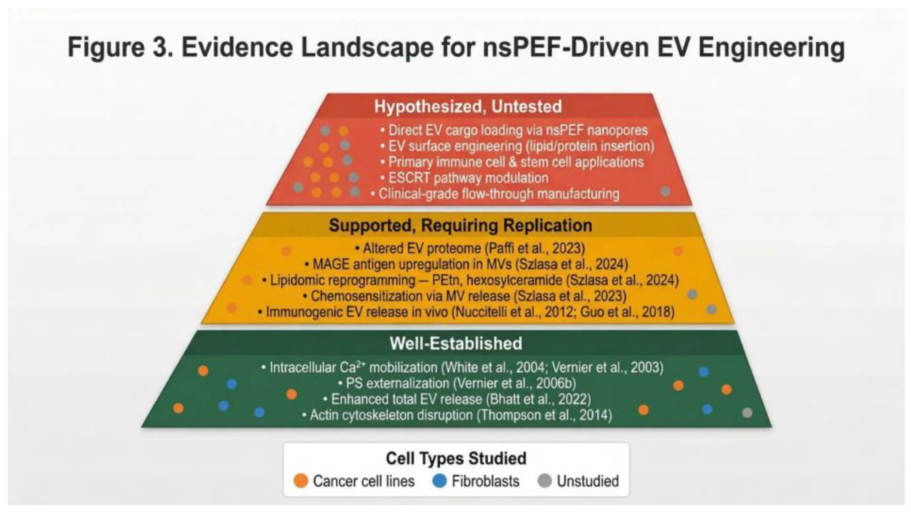
Figure 4.
Translational roadmap for nsPEF-driven EV manufacturing. Five-stage horizontal pipeline: (1) Parameter Optimization: pulse duration, field strength, pulse number, cell-type mapping, harvest kinetics; (2) EV Characterization: MISEV 2023 markers (CD63, CD81, CD9, TSG101, ALIX, syntenin-1; negative: calnexin, cytochrome c), NTA, cryo-EM, SEC/density gradient; (3) Functional Validation: cargo delivery, signaling assays, head-to-head comparison with conventional methods; (4) Scalable Manufacturing: flow-through chambers, GMP-compatible closed systems, inline monitoring; (5) Clinical Translation: purity/sterility standards, safety evaluation, regulatory pathway.
Figure 4.
Translational roadmap for nsPEF-driven EV manufacturing. Five-stage horizontal pipeline: (1) Parameter Optimization: pulse duration, field strength, pulse number, cell-type mapping, harvest kinetics; (2) EV Characterization: MISEV 2023 markers (CD63, CD81, CD9, TSG101, ALIX, syntenin-1; negative: calnexin, cytochrome c), NTA, cryo-EM, SEC/density gradient; (3) Functional Validation: cargo delivery, signaling assays, head-to-head comparison with conventional methods; (4) Scalable Manufacturing: flow-through chambers, GMP-compatible closed systems, inline monitoring; (5) Clinical Translation: purity/sterility standards, safety evaluation, regulatory pathway.
Disclaimer/Publisher’s Note: The statements, opinions and data contained in all publications are solely those of the individual author(s) and contributor(s) and not of MDPI and/or the editor(s). MDPI and/or the editor(s) disclaim responsibility for any injury to people or property resulting from any ideas, methods, instructions or products referred to in the content. |
© 2026 by the authors. Licensee MDPI, Basel, Switzerland. This article is an open access article distributed under the terms and conditions of the Creative Commons Attribution (CC BY) license (http://creativecommons.org/licenses/by/4.0/).
Copyright: This open access article is published under a Creative Commons CC BY 4.0 license, which permit the free download, distribution, and reuse, provided that the author and preprint are cited in any reuse.


