

Submitted:
02 July 2026
Posted:
02 July 2026
You are already at the latest version
Abstract
The tumor suppressor gene p53 plays a central role in tumor suppression and is the most frequently mutated gene in human cancers; p53 mutation is most often associated with reduction or loss of function of the protein. BALL-1 and Panc-1 cells harbor p53 mutations and overexpress cyclin D1, making them resistant to drugs that induce p53-dependent apoptosis. 3EE, 20Ac-ingenol activates p53 in these cells via ATR/ATM activation, which induces the expression of p21 and PTEN, thereby inducing apoptosis in p53-mutant cancer cells. It is well known that the genetic basis of pancreatic pathway, which strongly induces p53-dependent apoptosis, is expected to be activated by the mutant Ras gene and genotoxic stress. It has also been reported that DNA damage responses induced by genotoxic stress triggers apoptosis specifically in pancreatic cancer cells expressing ARF in a p53-independent manner. In this study, we investigated whether the DNA damage response induced by 3EE, 20Ac-ingenol can restore the reduced or lost function of p53 in p53-mutant Panc-1 cells or induce a p53-independent apoptosis via ARF expression. Based on previous and current results, unlike in MCF-7 cells in which ARF expression was not detected, in p53-mutant Panc-1 cells, 3EE, 20Ac-ingenol induced the ARF-MDM2-p53 pathway via coactivation of the replication stress enhancement pathway induced by the excessive cyclin D1 expression and the activating Ras/β-catenin signaling pathway and thereby ARF, suggesting that 3EE, 20Ac-ingenol can restore the function of p53 in p53-mutant pancreatic cancer cells, and induces p53-independent apoptosis.
Keywords:
p53-dependent and-independent apoptosis
; MDM2 inhibition
; Cdk2
; E2F1
; ARF and PTEN expression
1. Introduction
In DNA damage response signaling, ATR (ataxia telangiectasis ang Rad3-rerated) is activated via recruitment to tracts of single-stranded DNA in the S phase and ATM (ataxia telangiectasia mutated) acts downstream of ATR [1]. ATM is potently activated in response to DSBs (double-strand breaks), and although less so, in response to extrinsic DNA replication stress caused by exposure to chemicals or other adverse conditions, both of which affect p53 phosphorylation [1,2,3,4]. E2F1 (E2 transcription factor 1), which regulates cell cycle progression and apoptosis, is activated by the Rb (retinoblastoma) /E2F pathway in response to growth stimulation and stabilizes p53 through ATM/ATR activation induced by DNA damage [2]. E2F1 is also activated via Cdk2 (cyclin/cyclin-dependent kinase), leading to the expression of ARF (alternative reading frame), which is involved in tumor suppression [5,6,7,8]. On the other hand, E2F1 has also been reported to be induced by Ras (rat sarcoma viral oncogene)-activating mutations via ATR activation, as well as by DNA damage in the cells [3,9,10]. While AZT (azidothymidine) and cisplatin have been demonstrated to increase ARF protein expression in exponentially proliferating OVCAR-3 cells [11]. OVCAR-3 cancer cells exhibit mutations in c-Ki-Ras and β-catenin activation, and transformation of the BJ fibroblasts by this combination of Ha-Ras and β-catenin activation induces p14ARF [11,12,13,14]. However, ARF expression, which is only and mildly induced by oncogenic stress alone, can be strongly induced by induction of the E2F1 gene [15]. Cisplatin induces stalling replication forks [16], activating Cdk2 and E2F1 [7]. Simultaneous activation of the ARF expression pathway by oncogenes and the DNA damage signaling pathway triggered by proliferation stress, hypoxia or genotoxic drugs constitutes the ARF-MDM2-p53 pathway and plays a crucial role in determining the outcomes of genotoxic stress [17]. On the other hand, Panc-1 cells and PaTu8988t cells express PRMT1, and PaTu8988t cells that express ARF in particular stabilize ARF through methylation. It has been reported that methylated ARF functions as a p53-independent tumor suppressor [18].
It has been reported that in cells overexpressing cyclin D1, chromatin licensing and DNA replication factor 1 (Cdt1) are not degraded, re-replication occurs during the S phase, the effects of DNA damage are amplified, and the number of apoptotic cells (sub-G1 phase population) increases [19]. Enhanced DNA damage response strongly induced by 3EE, 20Ac-ingenol progresses in an ATM/ATR-dependent fashion via γH2AX (Phosphorylated H2AX) formation in BALL-1 cells overexpressing cyclin D1, as well as DNA fragmentation and caspase 3 activation, which are known to lead to the formation of γH2AX [20]. Flow-cytometric analysis of these BALL-1 cells revealed cell-cycle arrest in the S phase and an increase in the number of apoptotic cells in the sub-G1 phase [21,22]. 3EE, 20Ac-ingenol induces a DNA damage response that results in the formation of γH2AX in response induced by replication stress, leading to p53-dependent apoptosis of BALL-1 cells. In BALL-1 cells harboring p53 mutations, there are reports that DNA damage signals do not trigger p53 activation [23,24]. Similar to BALL-1 cells, Panc-1 cells also harbor p53 mutations [25]. 3EE, 20Ac-ingenol stabilized p53 in BALL-1 and Panc-1 cells by enhancing the DNA damage response, as compared with that in cancer cells that do not overexpress cyclin D1, and further specifically induced apoptosis through activation of p21 [21,22]. In p53-dependent apoptosis induction, in which ARF expression is involved, two normal p53 responses are involved. One response is the stabilization of p53 through ATM/ATR activation by the DNA damage itself [1], and the other is stabilization of p53 by inhibition of MDM2 activity induced by the ARF expression caused by DNA damage [26]. Both stabilizations contribute to the induction of potent p53-dependent apoptosis by ARF [26]. p53-mutated Panc-1 cells exhibit resistance to DNA damage drugs such as irinotecan and camptothecin, which induce p53-dependent apoptosis through ATM/ATR activation [29,2728]. In wild-type p53 Capan-2 cells, a pancreatic cancer cell line, treatment with an MDM2 (mouse double minute 2 homolog) inhibitor stabilized p53 and induced apoptosis [30]. Treatment with MDM2 inhibitors not only failed to stabilize p53 levels, but also affected apoptosis induction in the p53-mutated Capan-1 cells, which exhibit resistance to DNA damage [30]. 3EE, 20Ac-ingenol induced apoptosis in p53-mutant Panc-1 cells through p53-dependent p21 activation via ATR/ATR activation, not involving γH2AX formation, unlike in BALL-1 cells (Figure 1) [29]. To understand the recovery mechanism from the mutation, that is, the mechanism underlying 3EE, 20Ac-ingenol-induced p53-dependent apoptosis in mutant Panc-1 cells in the absence of γH2AX formation, we investigated the inhibition of MDM2 activity by ARF expression induced by Cdk2 (cyclin/cyclin-dependent kinase2 )/E2F1 activation.
2. Materials and Methods
2.1. Cell Lines, Compound and Cellular Proliferation
MCF-7 and Panc-1 cells was provided by the RIKEN BRC through the National Bio-Resource Project of the MEXT, Japan. The diterpene compound, 3EE, 20Ac-ingenol (3-O-(2′E,4′E-decadienoyl)-20-O- acetylingenol) was purified from the roots of Euphorbia kausui [31] and dissolved in dimethyl sulfoxide. The cancer cells were incubated in RPMI 1640 supplemented with 10% fetal calf serum at 37 °C. Cell growth was determined by an MTT assay using the Cell Proliferation Kit I (Roche Applied Science) as described previously [21].
2.2. Immunoblotting
MCF-7 and Panc-1 cells were cultured for various time points in the presence of 2μM 3EE, 20Ac-ingenol, respectively and washed with PBS. The protein concentrations were determined using the Bradford reagent for protein assays (Bio-Rad Laboratories). A total of 20 μg of the cell lysates was resolved on 5%-10%, 15%-20% (FUJIFILM) and 10% SDS-polyacrylamide gels and transferred onto a polyvinylidene difluoride membrane. The blots were prepared using anti-p53, p21 and Cdt1 (Cell Signaling Technology), anti-ATR, ATM, MDM2, PARP-1 (Poly [ADP-ribose] polymerase 1), E2F1, Cdk2, cyclinD1 and p14ARF (Santa Cruz, Cell Signaling Technology and Abcam), anti-caspase-3 (MBL) and anti-actin (Sigma) antibodies, followed by detection using an enhanced chemiluminescence system. For quantification of proteins, the proteins were subjected to western blotting using specific antibodies and the blot density was measured using an Image Lab software (Bio-Rad Laboratories).
3. Results
3.1. Effects of 3EE, 20Ac-Ingenol on p53, p21 and MDM2 Expressions and γH2AX Formation
We investigated whether the DDR (DNA damage response) induced by 3EE, 20Ac-ingenol induces ATM/ATR-dependent p53 accumulation and γH2AX formation, which result in apoptosis of the cell. Because MDM2 inhibition stabilizes p53, we also examined the effects of 3EE, 20Ac-ingenol treatment. In MCF-7 cells, the control cells showed only slight p53 expression, and the expression did not change significantly after the treatment (Figure 1). In Panc-1 cells, p53 expression increased 24 h after 3EE, 20Ac-ingenol treatment, which became even stronger after 48 h and was maintained for up to 72 h, resulting in approximately 3.2-fold activation compared to the control (Figure 1). Strong p21 expression was already observed in control MCF-7 cells, and even stronger expression was observed 24 and 72 h after treatment with 3EE, 20Ac-ingenol (Figure 1). In Panc-1 cells, no expression was observed in the control cells, but p21 was expressed 24 h after the addition of 3EE, 20Ac-ingenol, and this expression level continued for up to 72 h. (Figure 1). MCF-7 cells treated with 3EE, 20Ac-ingenol showed slight γH2AX formation after 48 h and strong γH2AX formation after 72 h (Figure 1). However, γH2AX was not always completely detectable in all the Panc-1 cell samples (Figure 1)
MDM2 is overexpressed in pancreatic cancer cell lines in a K-Ras dependent manner [32]. The p53 dependence of two MDM2 bands expressed in pancreatic cancer cell lines has been reported [32]. We also detected a major band and a faint band in the control Panc-1 cells (Figure 1). Two bands were observed with molecular weights similar to the previously reported MDM2 band. After treatment with 3EE, 20Ac-ingenol, the MDM2 band around 90 kDa temporarily increased after 24 h, then began to decrease sharply after 48 h, and by 72 h the high-expression band had decreased to a negligible level. The MDM2 band around 76 kDa began to decrease sharply from 24 h after treatment, and this rapid decrease continued until 72 h. (Figure 1). In MCF-7 cells, two bands are detected [33]. In this study, one strong band and one faint band were detected (Figure 1). After treatment with 3EE 20Ac-ingenol, the two bands were slightly decreased (Figure 1).
3.2. Effects of 3EE, 20Ac-Ingenol on ATM/ATR Activation
Although ATM activity was weakly expressed in control Panc-1 cells, strong ATM expression was observed 24 h after the addition of 3EE, 20Ac-ingenol, and the activity persisted for up to 72 h. (Figure 2). In the Panc-1 cells, ATR activity was observed at 24 h after the addition of 3EE, 20Ac-ingenol, with the activity continuing to increase until 72 h (Figure 2). In the MCF-7 cells, only slight ATM activity was observed in the control cells, with no significant change observed after the addition of 3EE, 20Ac-ingenol (Figure 2), and ATR was not detected.
3.3. Effects on Cyclin D1, Cyclin E, Cyclin A and Cdt1 Expressions Induced by 3EE, 20Ac-Ingenol
Cyclin D1 plays an important role in regulating the cell cycle through formation of a complex with Cdk4/6 and S phase progression [34]. Cyclin D1 was expressed in both control MCF-7 and Panc-1 cells, but was more abundantly expressed in the MCF-7 cells than in the Panc-1 cells (Figure 3). A faint band of cyclin E was observed in the control MCF-7 cells, while strong expression was observed from 24 h after the addition of 3EE, 20Ac-ingenol, with the strong expression persisting until 72 h (Figure 3). Cyclin E expression was barely detectable in the control Panc-1 cells. However, after the addition of 3EE, 20Ac-ingenol, expression was observed from 24 to 48 hours, but then decreased at 72 hours (Figure 3). Slight increase in cyclin A expression was observed in both the MCF-7 and Panc-1 cells at 24 hours after addition of 3EE, 20Ac-ingenol, with further increase observed at 48 and 72 hours (Figure 3). In Panc-1 control cells, only a small amount of Cdt1 was observed, that was not degraded by cyclin D1. The Cdt1 levels increased slightly at 24 h after 3EE, 20Ac-ingenol treatment, then decreased at 48 h, and increased again at 72 h, indicating repeated initiation of replication (Figure 3). In the MCF-7 cells, Cdt1 was strongly expressed in the control cells, but the expression decreased transiently at 24 h after the addition. It increased at 48 h, but decreased again after 72 h (Figure 3).
3.4. Effects of 3EE, 20Ac-Ingenol Treatment on Cdk2, E2F1, ARF and PARP1 Expressions
In the Panc-1 cells, Cdk2 expression was upregulated 24 h after the addition of 3EE, 20Ac-ingenol, the expression level increasing by approximately 2.2-fold (P <0.02) as compared with that in the control cells; the increased expression persisted for up to 72 h (Figure 4). In the MCF-7 cells, slight Cdk2 activity was detected in the control cells, with only a slight increase in expression at 48 h and 72 h after the addition of 3EE, 20Ac-ingenol (Figure 4). Faint expression of E2F1 was observed in the control MCF-7 cells and persisted from 24 until 72 h after the addition of 3EE, 20Ac-ingenol (Figure 4). In the Panc-1 cells, slight E2F1 activation was detected in the control cells, which gradually increased by 24 h after the addition of 3EE, 20Ac-ingenol (Figure 4). Furthermore, at 48 h, E2F1 expression increased by approximately 4-fold (P <0.02) as compared with that in the control cells, the increased expression persisting for up to 72 h (Figure 4). In the Panc-1 cells, increase in ARF expression was observed at 24 h after the addition of 3EE, 20Ac-ingenol, while the control cells showed only minimal expression. At 48 h after the addition of 3EE, 20Ac-ingenol, an approximately 2.2-fold increase in expression was observed (P <0.002) as compared with the level in the control cells, this increased expression persisting for up to 72 h (Figure 4). The MCF-7 cells showed no ARF expression.
Poly [ADP-ribose] polymerase 1 (PARP1) plays several possible roles in the response to replication stress [35]. In the MCF-7 cells, strong expression of PARP1 was observed in the control cells, but at 24 h after the addition of 3EE, 20Ac-ingenol, the expression decreased, the level continuing to decrease for up to 72 h (Figure 4). In the Panc-1 cells, slight expression was observed in the control cells, with the expression increasing between 24 and 48 h after the addition of 3EE, 20Ac-ingenol; the expression level decreased sharply after 72 h (Figure 4).
3.5. Effects of 3EE, 20Ac-Ingenol and Irinotecan on the Proliferative Activities and Caspase 3 Activity of the MCF-7 and Panc-1 Cells
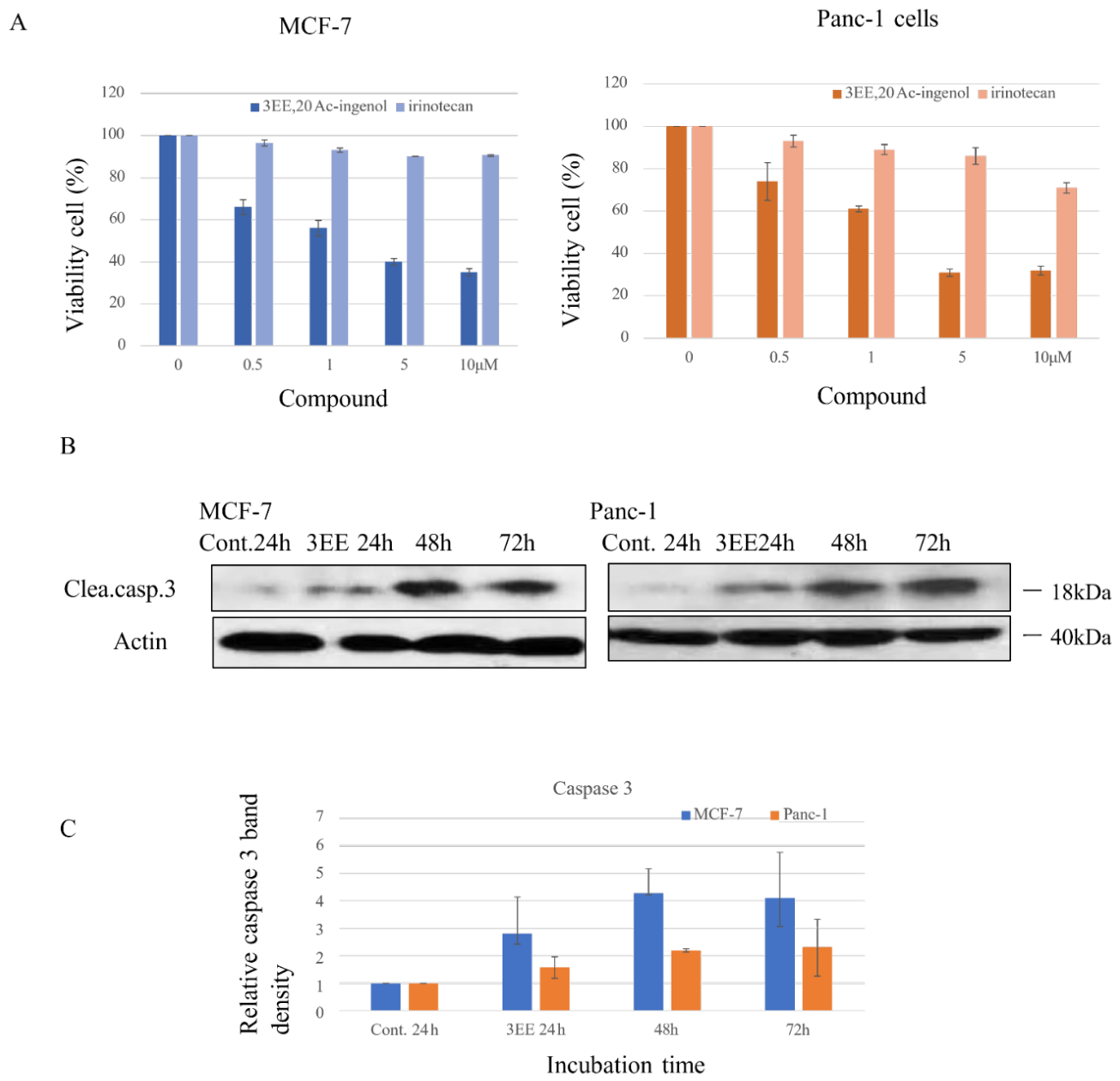
The inhibition of cell proliferation in MCF-7 cells gradually increased with increasing concentrations of 3EE, 20Ac-ingenol, with the maximal inhibition of approximately 65% reached at 10 μM; the IC50 of the drug for the MCF-7 cells was about 2 μM (Figure 5A). In the Panc-1 cell line, a maximal inhibition of approximately 70% was observed at a concentration of 10 μM; the IC50 for the Panc-1 cells was about 1.5 μM (Figure 5A). The effect of the irinotecan (CT-11), on the proliferation of the MCF-7 and Panc-1 cells was examined using the MTT assay (Figure 5A). CT-11 induced only 10% inhibition of the cell proliferative activity of the MCF-7 cells at a concentration of 10 μM. In the Panc-1 cells also, only 10% inhibition was observed at concentrations ranging from 0.5 μM to 5 μM, while 30% inhibition was observed at 10 μM (Figure 5A); both MCF-7 and Panc-1 cells showed poor sensitivity
We investigated whether 3EE, 20Ac-ingenol induces apoptosis in MCF-7 and Panc-1 cells. Activation of caspase-3 activity was observed in the Panc-1 cells at 24 h after addition of 3EE, 20Ac-ingenol, which continued to increase until 72 h after the addition (Figure 5B). In MCF-7 cells, caspase 3 activation began 24 h after the addition of 3EE, 20Ac-ingenol, showed strong activity 48 h later, and continued for up to 72 h (Figure 5B).
4. Discussion
The widely accepted view is that ARFs are not directly induced by acute DNA damage caused by hypoxia or genotoxic drugs, but rather that ARF tumor suppressors are produced by activated oncogenes [17]. In regard to ARF expression induced by exposure to genotoxic drugs, the coactivation of ARF by oncogenes and DNA-damage signaling pathways is caused by the DNA damage leading to replication fork collapse [17]. Cisplatin exposure has been shown to induce the coactivation of ARF by c-Ki-Ras (K-Ras)/β-catenin-mediated oncogenic signaling and a DNA damage by genotoxic drug accompanied by Cdk2/E2F1 expression in p53-mutant OVCAR-3 cells [7,11]. Cancer cells expressing detectable levels of endogenous p14ARF show abnormal or complete absence of p53 protein function [36]. Panc-1 cells, like the OVCAR-3 cells, harbor mutations in p53 and K-Ras and also overexpress cyclin D1 and β-catenin [9,37]. Therefore, in response to genotoxic drug induced by 3EE, 20Ac-ingenol exposure, activating Cdk2/E2F1 expression signaling may induce ARF expression in Panc-1 cancer cells through coactivation with oncogene signaling. P14ARF is activated by hyperproliferative signals from oncogenes such as K-Ras, E1A (early region 1A) and E2F1 [11,38]. Panc-1 cells undergo repeated replication initiation due to inhibition of Cdt1 degradation because cyclin D1 is not degraded (Figure 3). Cyclin D1 activates E2F1 expression, promoting cell cycle progression and apoptosis [2,34]. In addition to hyperproliferative signals from oncogenes, the proliferation in response to the activation of Cyclin D1/Cdk4/6 and Cdk2/E2F1 induced by 3EE,20Ac-ingenol further strongly induces ARF expression. (Figure 3 and Figure 4).
The PARP1 protein was originally thought to be a DNA damage detection molecule, as it initiates transcription of the ARF gene by finding and interacting with DNA strand break sites [35]. Poly(ADP-ribose) synthesis by PARP1 at the sites activates E2F1-dependent ARF transcription via DNA damage signaling [35]. In Panc-1 cells, PARP1 was present in the control cells, with its level increasing after 24 and 48 hours of exposure to 3EE and 20Ac-ingenol, and an increase in ARF was induced by both E2F1 and PARP1 activation. However, PARP1 activation was transient and decreased by 72 hours of exposure (Figure 4). It is well known that ARF is induced by E2F1 through the Rb/E2F pathway, and further that activation of PARP1 may activate E2F1-dependent ARF transcription via poly (ADP-ribose) synthesis. Thus, in the DNA damage response induced by 3EE, 20Ac-ingenol, ARF expression is induced via two pathways.
Induction of apoptosis by DNA damage depends on the stabilization of p53 activity through inhibition of MDM2, a major negative regulator of p53, by ARF activation [39]. It has been reported that in p53-mutant cells, inhibition of MDM2 does not result in p53 activation [30], but in p53-mutant Panc-1 cells, 3EE, 20Ac-ingenol promoted activation of p53 by inhibiting MDM2 (Figure1). Inhibition of MDM2 leads to the stabilization of p53 and p53-dependent expression of p21, which reduces replication stress through inhibition of DNA replication and attenuates the formation of γH2AX [40]. The reduction in γH2AX formation by 3EE, 20Ac-ingenol may be due to the reduced replication stress caused by inhibition of DNA replication, as MDM2 inhibition by ARF activation induces p53 stabilization and consequent p21 expression (Figure 1). Inhibition of γH2AX formation by stabilization of p53 (Figure 1) indicates that the 3EE, 20Ac-ingenol-induced DNA damage response restores the lost function of p53 and allows it to pass through the p53 response site that activates the two p21 pathways necessary for DNA replication repression [1,26]. On the other hand, it has been reported that BALL-1 cells are deficient in ARF [41]. In BALL-1 cells treated with 3EE, 20Ac-ingenol, mutant p53 was activated, albeit at a low level, leading to p53 stabilization. As a result, γH2AX formation was observed via p53-dependent p21 expression. This suggests that BALL-1 cells were unable to properly stabilize p53 due to the ARF gene deficiency, but γ-H2AX formation was still observed via p21 activation following the restoration of the p53 deficiency [21]. However, while MCF-7 cells also showed no ARF expression, these cells showed high p53-independent p21 activation, resulting in inhibition of replication and induction of γH2AX formation, observed after 48 hours of exposure (Figure 1). DNA damage induced by 3EE, 20Ac-ingenol has been shown to activate PTEN (phosphatase and tensin homolog delated on chromosome 10) and to decrease phosphorylation of GSK-3β (glycogen synthase kinase -3β) (Ser9) in BALL-1 and Panc-1 cells (21, 22, 29, 42). Inhibition of MDM2 by PTEN not only protects p53 through suppression of pAkt, but also inhibits MDM2 expression and stabilizes p53 (43, 44). A series of experiments using Panc-1 cells harboring the K-Ras-β-catenin system suggested that 3EE, 20Ac-ingenol not only induces ARF expression, but also activates the AKT-PTEN-MDM2-p53 pathway via PTEN activation, thereby comprehensively inducing the ARF-MDM2-p53 pathway and restoring the activity of mutant p53 (Figure 1 and Figure 4) [21,22,29,42]. These results demonstrate that in p53-mutant Panc-1 cells, which show resistance to the p53-dependent anticancer drug irinotecan, 3EE, 20Ac-ingenol treatment can restore the lost function of p53 and thereby suppress proliferation and induce apoptosis of the cells (Figure 5A, B).
Hypoxia-induced replication stress through ATM/ATR activation has been reported to induce the expression of Cdk2 and E2F1 [45,46,47]. DNA damage induced by 3EE,20Ac-ingenol and hypoxia exhibits a replication stress mechanism accompanied by similar Cdk2 and E2F1 expression (Figure 4). Panc-1 cells express the KRas and β-catenin [9,37], and Cdk2 and E2F1 which are expressed under hypoxic conditions are also expressed under the replication stress induced by 3EE, 20Ac-ingenol (Figure 4). Therefore, in Panc-1 cells, ARF expression may occur in response to both E2F1 activation caused by 3EE, 20Ac-ingenol-induced replication stress [2,5,6] and hypoxic stress [45,46,47]. Panc-1 cells respond to replicative stress induced by mild hypoxia and 3EE, 20Ac-ingenol initially by accumulation of HIF-1α (hypoxia-inducible factor 1, alpha subunit) [42]. Consequently, according to previous studies, under mild hypoxic conditions, p53 stabilization does not occur and therefore degradation of HIF-1α and PD-L1 does not occur, accelerating cancer progression [4,48]. Our results suggest that even under mild hypoxic conditions (0.5% O2), replication stress induced by 3EE, 20Ac-ingenol in Panc-1 cells, activates p53 via ARF activation, leading to degradation of HIF-1α and PD-L1 (programmed death-ligand 1), just like under exposure to severe hypoxia (<0.1% O2) (5).
p16INK4a (p16 inhibition of cyclin-dependent kinase 4a) and p14 ARF are encoded in the INK4a/ARF locus [49]. While p16INK4a mRNA is highly expressed in various human tissues, p14ARF mRNA expression varies across tissues and its expression is not detected in the pancreas. This suggests that there are tissue-specific regulatory mechanisms and that p14ARF transcription is suppressed by some unknown mechanism in the pancreas [49]. In Panc-1 cells, the entire INK4/ARF gene locus is deleted and no expression of p15INK4B/p14ARF/p16INK4A mRNA is detected [50]. ARF expression analysis using promoter assays revealed that the response to oncogenic stress alone was minimal, indicating that intracellular expression of Ras and E2F1 is necessary and that this is due to their synergistic effect [15]. Although the unknown ARF transcription mechanism in pancreatic cancer remains unclear [49,50], there are reports that human cancer ARF transcription directly or indirectly binds E2F1 and other factors to promoters, thereby regulating specific activations [15,51] In ovarian carcinoma cells with activated oncogenic signaling, it has been suggested that coactivation of p14ARF expression is regulated by the activating oncogenic signal and E2F1 activation by replication stress induced by DNA damage [7,11,17]. In Panc-1 cells, only minimal ARF expression was observed in the control group. However, replication stress induced by 3EE,20Ac-ingenol increased ARF expression through the activation of the E2F1 signaling pathway, as well as the coactivation of ARF by that pathway and K-Ras signaling. (Figure 4).
The human ARF promoter is a CpG island containing the Sp1-binding site, and its expression is suppressed [51]. ARF mRNA is specifically altered and expressed in various human tissues, but its expression has not been detected in the pancreas [49,50]. In control ovarian tumor OVCAR-3 cells, ARF mRNA expression was detected, and regarding pl4ARF mRNA expression in response to cisplatin-induced DNA damage, transcriptional changes were minimal, and at least a partial correlation was observed. Nevertheless, in exponentially proliferating these cancer cells with activated Ras/β-catenin, a clear increase in ARF protein expression was observed by Cdk2/E2F1 expression via stalling replication forks, which inhibits cell proliferation without p53 activation [11,52]. In Panc-1 cells, 3EE,20Ac-ingenol induces apoptosis via DNA damage associated with replication fork collapse (22, 29, 42). The induction of ARF expression in Panc-1 cells appears to involve ARF coactivation through a pathway similar to the ARF activation observed in cisplatin-treated OVCAR-3 cells, which is mediated by activated Ras/β-catenin signaling and Cdk2/E2F1 expression (Figure 4). This suggests that the growth-inhibitory effect of ARF protein in Panc-1 cells may be mediated not only by a p53-dependent pathway but also by a pathway that does not require p53 activation. Furthermore, unique responses to genotoxic stress have been reported in pancreatic cancer. In PaTu8988t cells expressing ARF, genotoxic stress triggers an interaction between p14ARF and PRMT1, which leads to p14ARF methylation, resulting in release from the nucleolus and induction of stress-induced tumor suppressor function of p14ARF [18]. Activation of p14ARF by this reaction did not lead to p53 stabilization, and p53-independent apoptosis was detected by annexin V/propidium iodide staining, which indicates late apoptosis. Since Panc-1 cells express PRMT1 [18] and ARF expression through genotoxic stress by 3EE, 20Ac-ingenol is induced (Figure 4), it is thought that in this experiment as well, Panc-1 cells exhibit p53-independent ARF activation by methylation mediated by the interaction between ARF and PRMT1. ARF has been shown to inhibit transcriptional activity of E2F1 and inhibit of D1/S transition phase as a p53-independent effect [52,53]. 3EE,20Ac-ingenol activates the ARF-MDM2-p53 pathway via ARF activation, restoring activity lost due to mutations and inducing p53-dependent apoptosis (Figure 1 and Figure 4). Furthermore, in the Panc-1 cells, ARF and methylated ARF suppress p53-indepenrently cell proliferation and induce apoptosis by inhibiting the transcriptional activity of genes involved in the G1/S phase. It has been reported that treatment with benzofuran, which causes DNA damage in MCF-7 cells, leads to p53-independent p21 activation, resulting in growth inhibition and apoptosis [54,55]. Our results were consistent with that report; while 3EE, 20Ac-ingenol induced p53-independent p21 overexpression and apoptosis in MCF-7 cells (Figure 1), this was distinct from the effect observed in the Panc-1 cells. While MCF-7 cells showed resistance to irinotecan (Figure 5) [56], 3EE, 20Ac-ingenol suppressed cell proliferation and induced apoptosis in these cells, although the precise mechanism remains unclear (Figure 5).
5. Conclusions
3EE,20Ac-ingenol and its stereoisomer, 3EZ,20Ac-ingenol, have been identified as compounds that specifically inhibit cell proliferation and induce p53-dependent apoptosis, with particularly pronounced effects in pancreatic cancer cells where cyclin D1 is excessively present in the nucleus. In this study, these cells were cancerous due to KRas/Wnt (wingless &int-1) mutations and exhibited active cell proliferation. 3EE 20Ac-ingenol promoted ARF expression via the KRas/β-catenin/cyclin D1 signaling pathway, in cooperation with Cdk2/E2F1 pathway induced by genotoxic stress response signals. The characteristics of these reaction systems suggest that apoptosis is induced from proliferation inhibition via both p53-dependent and p53-independent pathways
Funding Declaration: No financial support.
Author Contributions
S.M. performed the experiments for biochemistry, and M.O., K. I. and S. K. performed the experibments for isolation of compounds. T.I. performed protein quantification after electrophoresis. All authors contributed to the writing of the paper.
Acknowledgments
Part of this research was conducted by S. Miyata and S. Kitanaka while they were employed at Nihon University. We would like to express our gratitude to Nihon University.:
Conflictive of Interest: The authors declare no conflict of interest.
References
- Smith, J.; Tho, L.M.; Xu, N.; Gillespie, D.A. The ATM-Chk2 and ATR-Chk1 pathways in DNA damage signaling and cancer. Adv. Cancer Res. 2010, 108, 73–112. [Google Scholar] [PubMed]
- Carcagno, A.L.; Ogara, M.F.; Sonzogni, S.V.; Marazita, M.C.; Sirkin, P.F.; Ceruti, J.M.; Cánepa, E.T. E2F1 transcription is induced by genotoxic stress through ATM/ATR activation. IUBMB Life 2009, 61, 537–543. [Google Scholar] [PubMed]
- Carlos, A.R.; Escandell, J.M.; Kotsantis, P.; Suwaki, N.; Bouwman, P.; Badie, S.; Folio, C.; Benitez, J.; Gomez-Lopez, G.; Pisano, D.G.; Jonkers, J.; Tarsounas, M. ARF triggers senescence in Brca2-deficient cells by altering the spectrum of p53 transcriptional targets. Nat. Commun. 2013, 4, 2697. [Google Scholar] [CrossRef] [PubMed]
- Olcina, M.; Lecane, P.S.; Hammond, E.M. Targeting hypoxic cells through the DNA damage response. Clin. Cancer Res. 2010, 16, 5624–5629. [Google Scholar] [CrossRef] [PubMed]
- Daigh, L.H.; Liu, C.; Chung, M.; Cimprich, K.A.; Meyer, T. Stochastic Endogenous Replication Stress Causes ATR-Triggered Fluctuations in CDK2 Activity that Dynamically Adjust Global DNA Synthesis Rates. Cell Syst. 2018, 7, 17–27.e3. [Google Scholar] [CrossRef] [PubMed]
- Wang, W.H.; Hullinger, R.L.; Andrisani, O.M. Hepatitis B virus X protein via the p38MAPK pathway induces E2F1 release and ATR kinase activation mediating p53 apoptosis. J. Biol. Chem. 2008, 283, 28729–28740. [Google Scholar]
- Yu, F.; Megyesi, J.; Safirstein, R.L.; Price, P.M. Involvement of the CDK2-E2F1 pathway in cisplatin cytotoxicity in vitro and in vivo. Am. J. Physiol. Renal. Physiol. 2007, 293, F52–F59. [Google Scholar] [PubMed]
- Sun, J.; Wang, X.; Shen, Q.; Wang, M.; Chen, S.; Zhang, X.; Huang, Y.; Zhang, Z.; Li, W.; Yuan, Y.; Huang, Z. DNASE1L3 inhibits hepatocellular carcinoma by delaying cell cycle progression through CDK2. Cell. Oncol. 2022, 45, 1187–1202. [Google Scholar] [CrossRef]
- Chu, P.C.; Yang, M.C.; Kulp, S.K.; Salunke, S.B.; Himmel, L.E.; Fang, C.S.; Jadhav, A.M.; Shan, Y.S.; Lee, C.T.; Lai, M.D.; Shirley, L.A.; Bekaii-Saab, T.; Chen, C.S. Regulation of oncogenic KRAS signaling via a novel KRAS-integrin-linked kinase-hnRNPA1 regulatory loop in human pancreatic cancer cells. [PubMed]
- Oncogene 2016, 35, 3897–3908. [PubMed]
- Fouad, S.; Hauton, D.; D’Angiolella, V. E2F1: Cause and Consequence of DNA Replication Stress. Front. Mol. Biosci. 2021, 7, 599332. [Google Scholar] [CrossRef] [PubMed]
- Vaskivuo, L.; Rysä, J.; Koivuperä, J.; Myllynen, P.; Vaskivuo, T.; Chvalova, K.; Serpi, R.; Savolainen, E.R.; Puistola, U.; Vähäkangas, K. Azidothymidine and cisplatin increase p14ARF expression in OVCAR-3 ovarian cancer cell line. [CrossRef] [PubMed]
- Toxicol. Appl. Pharmacol. 2006, 216, 89–97. [PubMed]
- Evangelou, K.; Bartkova, J.; Kotsinas, A.; Pateras, I.S.; Liontos, M.; Velimezi, G.; Kosar, M.; Liloglou, T.; Trougakos, I.P.; Dyrskjot, L.; Andersen, C.L.; Papaioannou, M.; Drosos, Y.; Papafotiou, G.; Hodny, Z.; Sosa-Pineda, B.; Wu, X.R.; Klinakis, A.; Ørntoft, T.; Lukas, J.; Bartek, J.; Gorgoulis, V.G. The DNA damage checkpoint precedes activation of ARF in response to escalating oncogenic stress during tumorigenesis. Cell Death Differ. 2013, 20, 1485–1497. [Google Scholar] [CrossRef] [PubMed]
- Hung, W.C.; Chai, C.Y.; Huang, J.S.; Chuang, L.Y. Expression of cyclin D1 and c-Ki-ras gene product in human epithelial ovarian tumors. Hum. Pathol. 1996, 27, 1324–1328. [Google Scholar] [PubMed]
- Zhang, Y.; Chen, S.; Wei, C.; Rankin, G.O.; Ye, X.; Chen, Y.C. Dietary compound proanthocyanidins from Chinese bayberry (Myrica rubra Sieb. et Zucc.) leaves attenuate chemotherapy-resistant ovarian cancer stem cell traits via targeting the Wnt/β-catenin signaling pathway and inducing G1 cell cycle arrest. Food Funct. 2018, 9, 525–533. [Google Scholar] [PubMed]
- Berkovich, E.; Lamed, Y.; Ginsberg, D. E2F and Ras synergize in transcriptionally activating p14ARF expression. Cell Cycle 2003, 2, 127–133. [Google Scholar] [PubMed]
- Wu, C.K.; Shiu, J.L.; Wu, C.L.; Hung, C.F.; Ho, Y.C.; Chen, Y.T.; Tung, S.Y.; Yeh, C.F.; Shen, C.H.; Liaw, H.; Su, W.P. APLF facilitates interstrand DNA crosslink repair and replication fork protection to confer cisplatin resistance. Nucleic Acids Res. 2024, 52, 5676–5697. [Google Scholar] [CrossRef] [PubMed]
- Sherr, C.J. Divorcing ARF and p53: an unsettled case. Nat. Rev. Cancer. 2006, 6, 663–673. [Google Scholar] [CrossRef] [PubMed]
- Repenning, A.; Happel, D.; Bouchard, C.; Meixner, M.; Verel-Yilmaz, Y.; Raifer, H.; Holembowski, L.; Krause, E.; Kremmer, E.; Feederle, R.; Keber, C.U.; Lohoff, M.; Slater, E.P.; Bartsch, D.K.; Bauer, U.M. PRMT1 promotes the tumor suppressor function of p14ARF and is indicative for pancreatic cancer prognosis. EMBO J. 2021, 40, e106777. [Google Scholar] [CrossRef] [PubMed]
- Kim, J.K.; Diehl, J.A. Nuclear cyclin D1: an oncogenic driver in human cancer. [CrossRef] [PubMed]
- J. Cell Physiol. 2009, 220, 292–296. [PubMed]
- Darzynkiewicz, Z.; Zhao, H.; Halicka, H.D.; Rybak, P.; Dobrucki, J.; Wlodkowic, D. DNA damage signaling assessed in individual cells in relation to the cell cycle phase and induction of apoptosis. Crit.Rev. Clin. Lab. Sci. 2012, 49, 199–217. [Google Scholar] [CrossRef] [PubMed]
- Miyata, S.; Fukuda, Y.; Tojima, H.; Matsuzaki, K.; Kitanaka, S.; Sawada. [CrossRef] [PubMed]
- Mechanism of the inhibition of leukemia cell growth and induction of apoptosis through the activation of ATR and PTEN by the topoisomerase inhibitor 3EZ, 20Ac-ingenol. Leuk. Res. 2015, 39, 927–932. [CrossRef] [PubMed]
- Miyata, S.; Wang, L.Y.; Kitanaka, S. 3EZ, 20Ac-ingenol induces cell-specific apoptosis in cyclin D1 over-expression through the activation of ATR and downregulation of p-Akt. Leuk. Res. 2018, 64, 46–51. [Google Scholar] [PubMed]
- Jia, L.Q.; Osada, M.; Ishioka, C.; Gamo, M.; Ikawa, S.; Suzuki, T.; Shimodaira, H.; Niitani, T.; Kudo, T.; Akiyama, M.; Kimura, N.; Matsuo, M.; Mizusawa, H.; Tanaka, N.; Koyama, H.; Namba, M.; Kanamaru, R.; Kuroki, T. Screening the p53 status of human cell lines using a yeast functional assay. Mol. Carcinog. 1997, 19, 243–253. [Google Scholar] [PubMed]
- Esaka, Y.; Nagahara, Y.; Hasome, Y.; Nishio, R.; Ikekita, M. Coenzyme Q2 induced p53-dependent apoptosis. Biochim. Biophys. Acta 2005, 1724, 49–58. [Google Scholar] [CrossRef] [PubMed]
- Ozaki, T.; Nakamura, M.; Ogata, T.; Sang, M.; Yoda, H.; Hiraoka, K.; Sang, M.; Shimozato, O. Depletion of pro-oncogenic RUNX2 enhances gemcitabine (GEM) sensitivity of p53-mutated pancreatic cancer Panc-1 cells through the induction of pro-apoptotic TAp63. Oncotarget 2016, 7, 71937–71950. [Google Scholar] [PubMed]
- Fu, X.; Yucer, N.; Liu, S.; Li, M.; Yi, P.; Mu, J.J.; Yang, T.; Chu, J.; Jung, S.Y.; O’Malley, B.W.; Gu, W.; Qin, J.; Wang, Y. RFWD3-Mdm2 ubiquitin ligase complex positively regulates p53 stability in response to DNA damage. Proc. Natl. Acad. Sci. U S A 2010, 107, 4579–4584. [Google Scholar] [PubMed]
- Hofmann, C.; Buttenschoen, K.; Straeter, J.; Henne-Bruns, D.; Kornmann, M. Pre-clinical evaluation of the activity of irinotecan as a basis for regional chemotherapy. Anticancer Res. 2005, 25, 795–804. [Google Scholar] [PubMed]
- Jeansonne, D.P.; Koh, G.Y.; Zhang, F.; Kirk-Ballard, H.; Wolff, L.; Liu, D.; Eilertsen, K.; Liu, Z. Paclitaxel-induced apoptosis is blocked by camptothecin in human breast and pancreatic cancer cells. Oncol. Rep. 2011, 25, 1473–1480. [Google Scholar] [CrossRef] [PubMed]
- Miyata, S.; Nakamura, T.; Kitanaka, S. 3EZ, 20Ac-ingenol-induced Apoptosis in Chemoresistant Cancers With Cyclin D1 Accumulation. Anticancer Res. 2020, 40, 6237–6246. [Google Scholar] [PubMed]
- Azmi, A.S.; Philip, P.A.; Aboukameel, A.; Wang, Z.; Banerjee, S.; Zafar, S.F.; Goustin, A.S.; Almhanna, K.; Yang, D.; Sarkar, F.H.; Mohammad, R.M. Reactivation of p53 by novel MDM2 inhibitors: implications for pancreatic cancer therapy. Curr. Cancer Drug Targets 2010, 10, 319–331. [Google Scholar] [CrossRef] [PubMed]
- Wang, L.Y.; Wang, N.L.; Yao, X.S.; Miyata, S.; Kitanaka, S. Diterpenes from the roots of Euphorbia kansui and their in vitro effects on the cell division of Xenopus. J. Nat. Prod. 2002, 65, 1246–1251. [Google Scholar] [CrossRef] [PubMed]
- Sui, X.; Shin, S.; Zhang, R.; Firozi, P.F.; Yang, L.; Abbruzzese, J.L.; Reddy, S.A. Hdm2 is regulated by K-Ras and mediates p53-independent functions in pancreatic cancer cells. Oncogene 2009, 28, 709–720. [Google Scholar] [PubMed]
- Alkhalaf, M.; El-Mowafy, A.M.; Abou-Zeid, L.A. Progesterone inhibition of MDM2 p90 protein in MCF-7 human breast cancer cell line is dependent on p53 levels. J. Mol. Genet. Med. 2005, 1, 33–37. [Google Scholar] [PubMed]
- Zhou, Y.; Nakajima, R.; Shirasawa, M.; Fikriyanti, M.; Zhao, L.; Iwanaga, R.; Bradford, A.P.; Kurayoshi, K.; Araki, K.; Ohtani, K. Expanding Roles of the E2F-RB-p53 Pathway in Tumor Suppression. Biology 2023, 12, 1511. [Google Scholar] [PubMed]
- Orlando, G.; Khoronenkova, S.V.; Dianova, I.I.; Parsons, J.L.; Dianov, G.L. ARF induction in response to DNA strand breaks is regulated by PARP1. [CrossRef] [PubMed]
- Nucleic Acids Res. 2014, 42, 2320–2329. [CrossRef] [PubMed]
- Stott, F.J.; Bates, S.; James, M.C.; McConnell, B.B.; Starborg, M.; Brookes, S.; Palmero, I.; Ryan, K.; Hara, E.; Vousden, K.H.; Peters, G. The alternative product from the human CDKN2A locus, p14(ARF), participates in a regulatory feedback loop with p53 and MDM2. EMBO J. 1998, 17, 5001–5014. [Google Scholar] [PubMed]
- Farhana, L.; Dawson, M.I.; Das, J.K.; Murshed, F.; Xia, Z.; Hadden, T.J.; Hatfield, J.; Fontana, J.A. Adamantyl Retinoid-Related Molecules Induce Apoptosis in Pancreatic Cancer Cells by Inhibiting IGF-1R and Wnt/β-Catenin Pathways. [PubMed]
- J. Oncol. 2012, 2012, 796729. [PubMed]
- Li, Z.; Hou, J.; Sun, L.; Wen, T.; Wang, L.; Zhao, X.; Xie, Q.; Zhang, S.Q. NMI mediates transcription-independent ARF regulation in response to cellular stresses. Mol. Biol. Cell. 2012, 23, 4635–4646. [Google Scholar] [CrossRef] [PubMed]
- Hernández Borrero, L.J.; El-Deiry, W.S. Tumor suppressor p53: Biology, signaling pathways, and therapeutic targeting. Biochim. Biophys. Acta Rev. Cancer. 2021, 1876, 188556. [Google Scholar] [CrossRef] [PubMed]
- Vilgelm, A.E.; Cobb, P.; Malikayil, K.; Flaherty, D.; Andrew Johnson, C.; Raman, D.; Saleh, N.; Higgins, B.; Vara, B.A.; Johnston, J.N.; Johnson, D.B.; Kelley, M.C.; Chen, S.C.; Ayers, G.D.; Richmond, A. MDM2 Antagonists Counteract Drug-Induced DNA Damage. EBioMedicine 2017, 24, 43–55. [Google Scholar] [CrossRef] [PubMed]
- Sugihara, E.; Shimizu, T.; Kojima, K.; Onishi, N.; Kai, K.; Ishizawa, J.; Nagata, K.; Hashimoto, N.; Honda, H.; Kanno, M.; Miwa, M.; Okada, S.; Andreeff, M.; Saya, H. Ink4a a;nd Arf are crucial factors in the determination of the cell of origin and the therapeutic sensitivity of Myc-induced mouse lymphoid tumor. Oncogene 2012, 31, 2849–2861. [Google Scholar] [PubMed]
- Miyata, S.; Ishii, T.; Kitanaka, S. Reduction of HIF-1α/PD-L1 by Catalytic Topoisomerase Inhibitor Induces Cell Death Through Caspase Activation in Cancer Cells Under Hypoxia. Anticancer Res. 2024, 44, 49–59. [Google Scholar] [PubMed]
- Mayo, L.D.; Dixon, J.E.; Durden, D.L.; Tonks, N.K.; Donner, D.B. PTEN protects p53 from Mdm2 and sensitizes cancer cells to chemotherapy. J. Biol. Chem. 2002, 277, 5484–5489. [Google Scholar] [CrossRef] [PubMed]
- Chang, C.J.; Freeman, D.J.; Wu, H. PTEN regulates Mdm2 expression through the P1 promoter. J. Biol. Chem. 2004, 279, 29841–29848. [Google Scholar] [CrossRef] [PubMed]
- Wu, M.; Wang, X.; McGregor, N.; Pienta, K.J.; Zhang, J. Dynamic regulation of Rad51 by E2F1 and p53 in prostate cancer cells upon drug-induced DNA damage under hypoxia. Mol. Pharmacol. 2014, 85, 866–876. [Google Scholar] [PubMed]
- Nuñez-Hernandez, D.M.; Camacho-Jiménez, L.; González-Ruiz, R.; Mata-Haro, V.; Ezquerra-Brauer, J.M.; Yepiz-Plascencia, G. Cyclin-dependent kinase 2 (Cdk-2) from the White shrimp Litopenaeus vannamei: Molecular characterization and tissue-specific expression during hypoxia and reoxygenation. [PubMed]
- Comp. Biochem. Physiol. Part A 2019, 230, 56–63.
- Yang, Y.; Sun, F.; Zhang, C.; Wang, H.; Wu, G.; Wu. [CrossRef] [PubMed]
- Hypoxia promotes cell proliferation by modulating E2F1 in chicken pulmonary arterial smooth muscle cells. J. Anim. Sci. Biotechnol. 2013, 4, 28. [CrossRef] [PubMed]
- Schmid, T.; Zhou, J.; Brüne, B. HIF-1 and p53: communication of transcription factors under hypoxia. J. Cell. Mol. Med. 2004, 8, 423–431. [Google Scholar] [PubMed]
- Robertson, K.D.; Jones, P.A. Tissue-specific alternative splicing in the human INK4a/ARF cell cycle regulatory locus. Oncogene 1999, 18, 3810–3820. [Google Scholar] [PubMed]
- Li, J.; Knobloch, T.J.; Poi, M.J.; Zhang, Z.; Davis, A.T.; Muscarella, P.; Weghorst, C.M. Genetic alterations of RD(INK4/ARF) enhancer in human cancer cells. [PubMed]
- Mol. Carcinog. 2014, 53, 211–218. [CrossRef] [PubMed]
- Parisi, T.; Pollice, A.; Cristofano, A.D.; Calabrò, V.; Mantia, G.L. Transcriptional regulation of the human tumor suppressor p14(ARF) by E2F1, E2F2, E2F3, and Sp1-like factors. Biochem. Biophys. Res. Commun. 2002, 291, 1138–1145. [Google Scholar] [PubMed]
- Eymin, B.; Karayan, L.; Séité, P.; Brambilla, C.; Brambilla, E.; Larsen, C.J.; Gazzéri, S. Human ARF binds E2F1 and inhibits its transcriptional activity. [CrossRef] [PubMed]
- Oncogene 2001, 20, 1033–1041. [PubMed]
- Inoshita, S.; Terada, Y.; Nakashima, O.; Kuwahara, M.; Sasaki, S.; Marumo, F. Regulation of the G1/S transition phase in mesangial cells by E2F1. [CrossRef] [PubMed]
- Kidney Int. 1999, 56, 1238–1241. [PubMed]
- Giordano, C.; Rovito, D.; Barone, I.; Mancuso, R.; Bonofiglio, D.; Giordano, F.; Catalano, S.; Gabriele, B.; Andò, S. Benzofuran-2-acetic ester derivatives induce apoptosis in breast cancer cells by upregulating p21Cip/WAF1 gene expression in p53-independent manner. DNA Repair. 2017, 51, 20–30. [Google Scholar] [PubMed]
- Princiotto, S.; Pinna, C.; Mattio, L.M.; Annunziata, F.; Beretta, G.L.; Pinto, A.; Dallavalle, S. Cytotoxicity of Benzofuran-Containing Simplified Viniferin Analogues. Pharmaceuticals 2024, 17, 1012. [Google Scholar] [CrossRef] [PubMed]
- Keyvani-Ghamsari, S.; Rabbani-Chadegani, A.; Sargolzaei, J.; Shahhoseini, M. Effect of irinotecan on HMGB1, MMP9 expression, cell cycle, and cell growth in breast cancer (MCF-7) cells. Tumor Biol. 2017, 39, 1–10. [Google Scholar] [CrossRef]
Figure 1.
Effects of 3EE, 20Ac-ingenol on p53 accumulation and p21 activation, and MDM2 expression and γH2AX formation. (A) MCF-7 and Panc-1 cells were cultured in the absence (control): for 24h or presence of 2μM 3EE, 20Ac-ingenol (3EE) for 24,48 or 72h. Expression of p53, p21, MDM2 and γH2AX were analyzed by western blotting. (B) Comparison of the results of western blotting is shown. Data from independent experiments are shown as the means ± standard deviation.
Figure 1.
Effects of 3EE, 20Ac-ingenol on p53 accumulation and p21 activation, and MDM2 expression and γH2AX formation. (A) MCF-7 and Panc-1 cells were cultured in the absence (control): for 24h or presence of 2μM 3EE, 20Ac-ingenol (3EE) for 24,48 or 72h. Expression of p53, p21, MDM2 and γH2AX were analyzed by western blotting. (B) Comparison of the results of western blotting is shown. Data from independent experiments are shown as the means ± standard deviation.
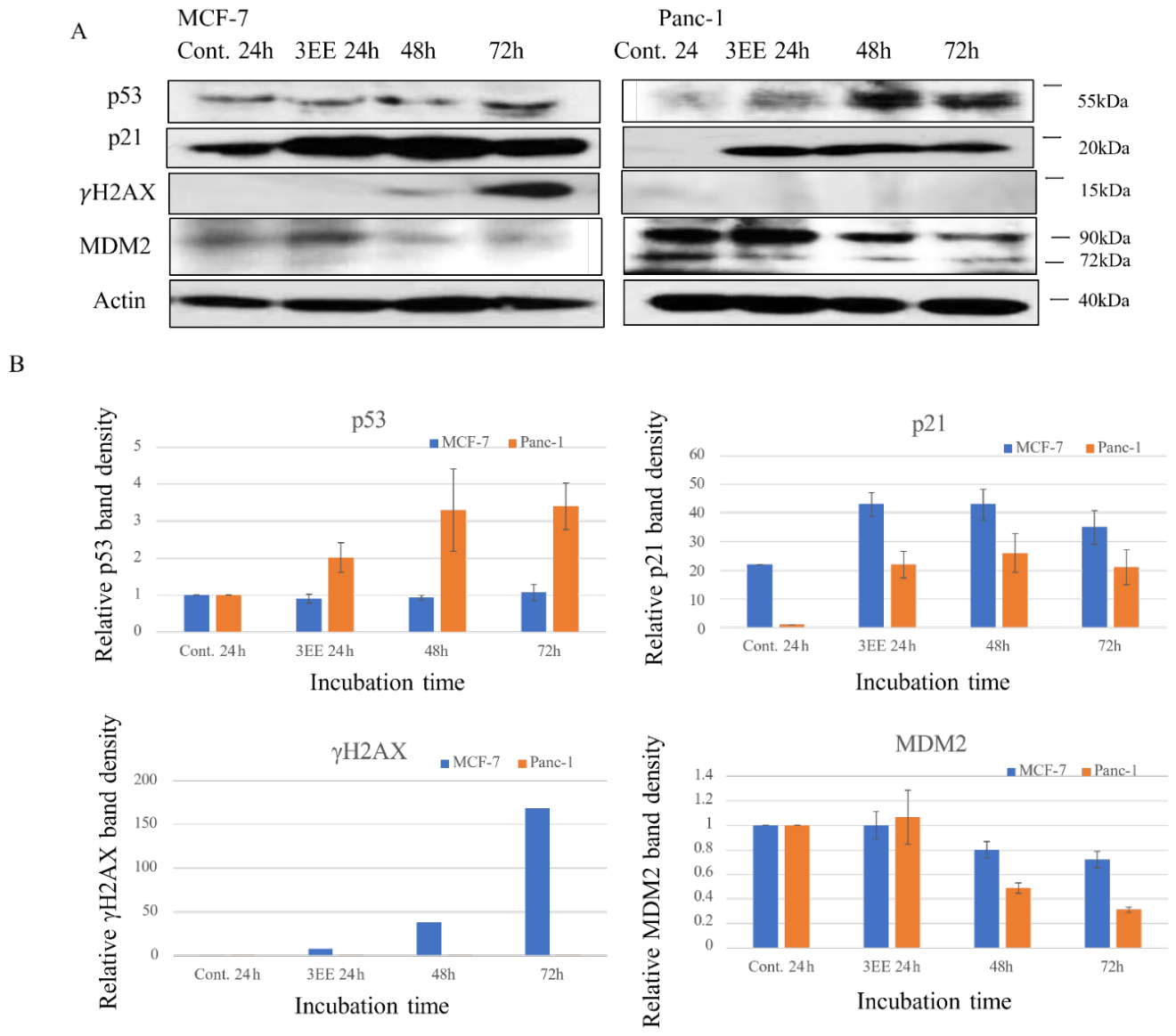
Figure 2.
Effect of 3EE, 20Ac-ingenol on ATM and ATR activation. (A) MCF-7 and Panc-1 cells were cultured in the absence (control): for 24h or presence of 2 μM 3EE for 24, 48 or 72 h. ATM and ATR analyzed by western blotting. (B) Comparison of the results of western blotting is shown.
Figure 2.
Effect of 3EE, 20Ac-ingenol on ATM and ATR activation. (A) MCF-7 and Panc-1 cells were cultured in the absence (control): for 24h or presence of 2 μM 3EE for 24, 48 or 72 h. ATM and ATR analyzed by western blotting. (B) Comparison of the results of western blotting is shown.
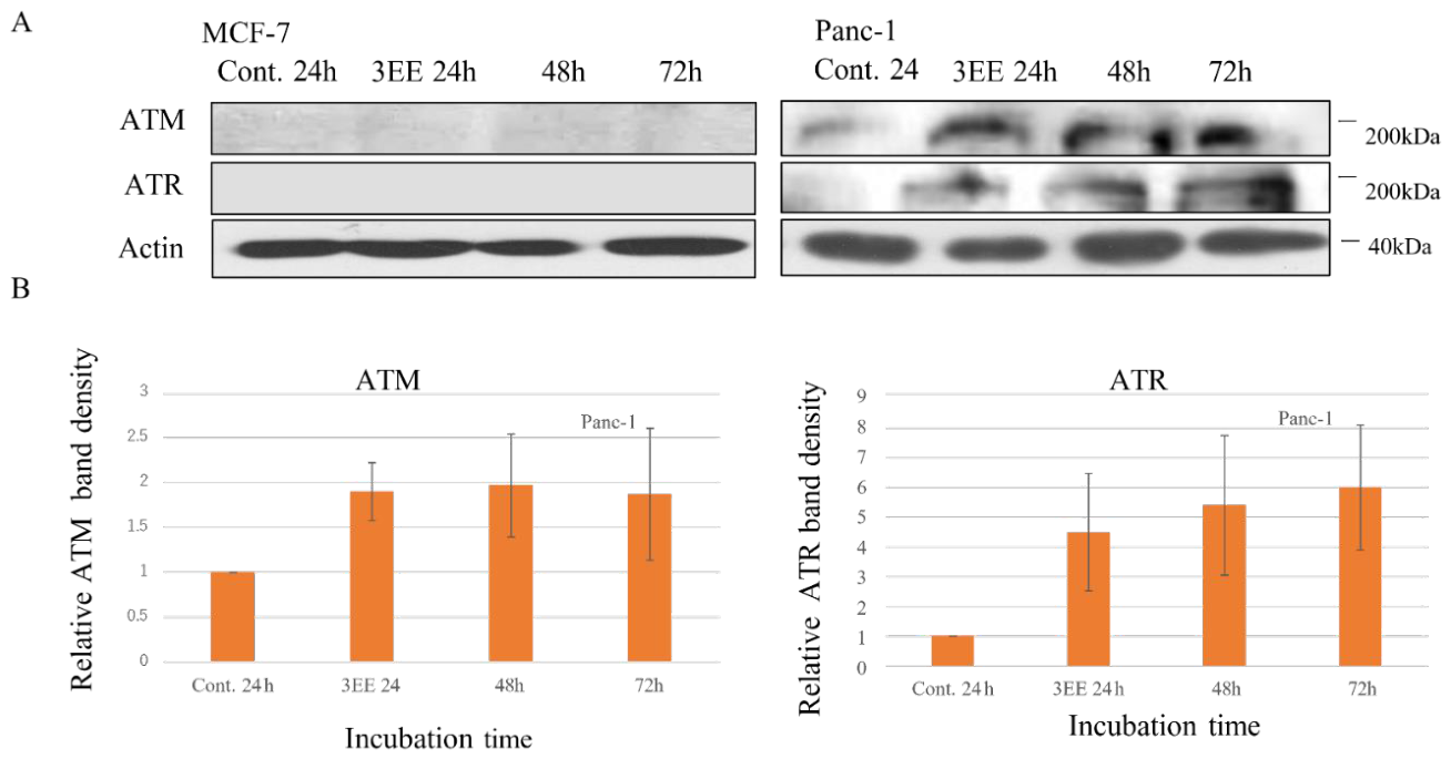
Figure 3.
Effects on cyclin D1, cyclin E, cyclin A and Cdt1 expressions induced by 3EE, 20Ac-ingenol. (A) MCF-7 and Panc-1 cells were cultured in the absence (control): for 24h or presence of 2μM 3EE for 24,48 or 72h. Expression of cyclin D1, cyclin E, cyclin A and Cdt1 were analyzed by western blotting. (B) Comparison of the results of western blotting is shown. (C) Comparison of the results of Western blotting of Panc-1 is shown in enlarged form.
Figure 3.
Effects on cyclin D1, cyclin E, cyclin A and Cdt1 expressions induced by 3EE, 20Ac-ingenol. (A) MCF-7 and Panc-1 cells were cultured in the absence (control): for 24h or presence of 2μM 3EE for 24,48 or 72h. Expression of cyclin D1, cyclin E, cyclin A and Cdt1 were analyzed by western blotting. (B) Comparison of the results of western blotting is shown. (C) Comparison of the results of Western blotting of Panc-1 is shown in enlarged form.
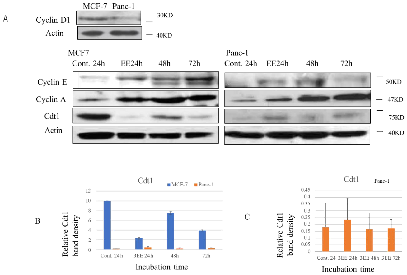
Figure 4.
Effects of 3EE, 20Ac-ingenol on Cdk2, E2F1, ARF and PARP1 expression. (A) MCF-7 and Panc-1 cells were treated in the absence (control): for 24 h or presence of 2 μM 3EE for 24, 48 or 72 h. Cyclin D1, Cdt1, Cdk2, E2F1, ARF and PARP1 analyzed by western blotting. (B) Comparison of the results of western blotting is shown.
Figure 4.
Effects of 3EE, 20Ac-ingenol on Cdk2, E2F1, ARF and PARP1 expression. (A) MCF-7 and Panc-1 cells were treated in the absence (control): for 24 h or presence of 2 μM 3EE for 24, 48 or 72 h. Cyclin D1, Cdt1, Cdk2, E2F1, ARF and PARP1 analyzed by western blotting. (B) Comparison of the results of western blotting is shown.
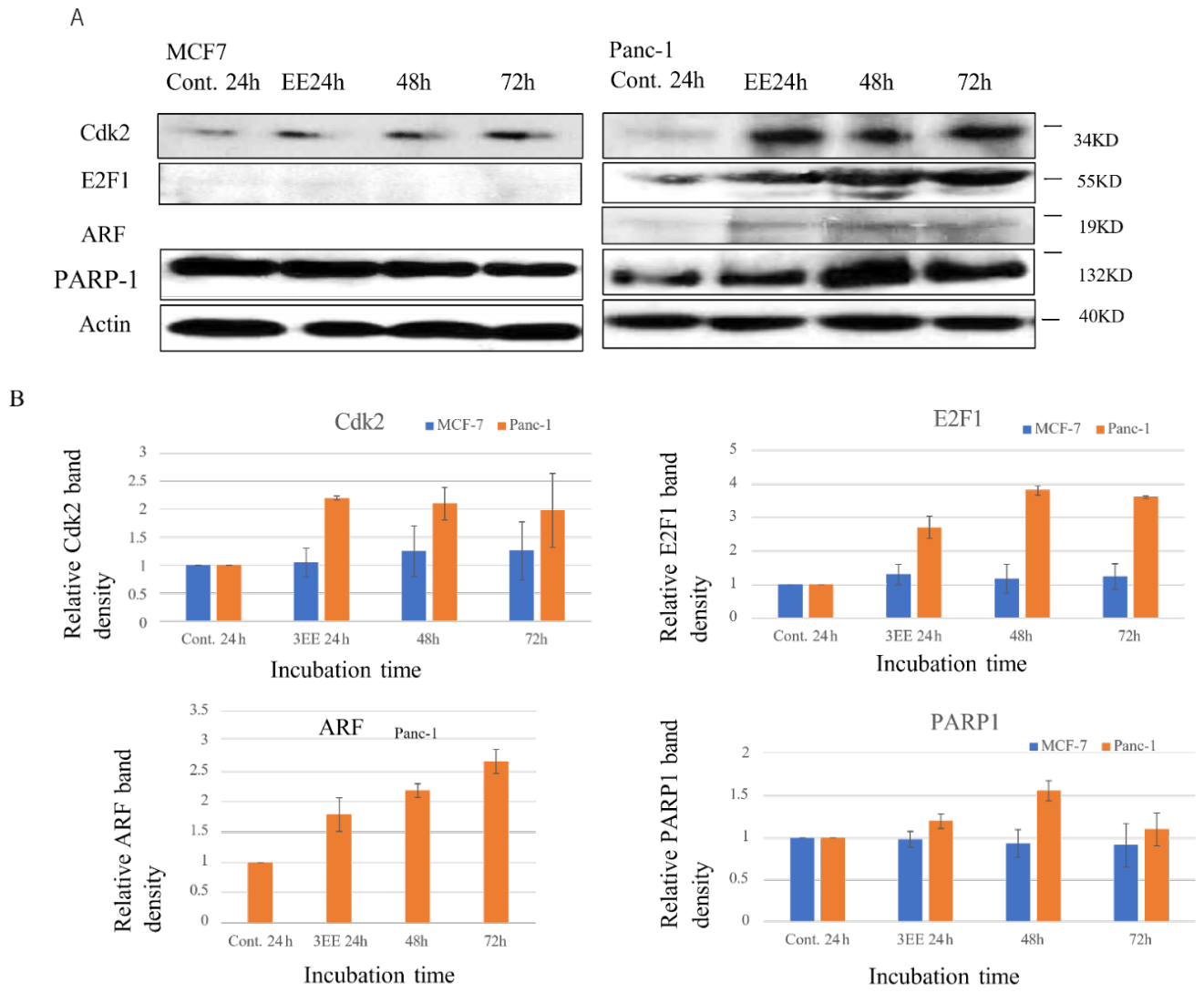
Figure 5.
Effects of 3EZ, 20Ac-ingenol or irinotecan on the proliferative activities and caspase 3 activity. (A) The cells were seeded on to 96-well plates (1x10⁴ MCF-7 cells or 3x10³ Panc-1 cells per well in 100 μL) and treated with 0 (control), 0.5, 1, 5, or 10 μM 3EE at 37 °C for 72 h. The relative cell growth was determined via an MTT assay. The growth of untreated MCF-7 and Panc-1 cells was set as 100%, and the growth of treated MCF-7 and Panc-1 cells was expressed relative to the growth of the untreated cells. All experiments were performed in triplicate, and the data are presented as the mean ± standard deviation. (B) MCF-7 and Panc-1 cells were cultured in the absence (control): for 24h or presence of 2 μM 3EE for 24, 48 or 72 h. Cleaved caspase 3 analyzed by western blotting. (C) Comparison of the results of western blotting is shown.
Figure 5.
Effects of 3EZ, 20Ac-ingenol or irinotecan on the proliferative activities and caspase 3 activity. (A) The cells were seeded on to 96-well plates (1x10⁴ MCF-7 cells or 3x10³ Panc-1 cells per well in 100 μL) and treated with 0 (control), 0.5, 1, 5, or 10 μM 3EE at 37 °C for 72 h. The relative cell growth was determined via an MTT assay. The growth of untreated MCF-7 and Panc-1 cells was set as 100%, and the growth of treated MCF-7 and Panc-1 cells was expressed relative to the growth of the untreated cells. All experiments were performed in triplicate, and the data are presented as the mean ± standard deviation. (B) MCF-7 and Panc-1 cells were cultured in the absence (control): for 24h or presence of 2 μM 3EE for 24, 48 or 72 h. Cleaved caspase 3 analyzed by western blotting. (C) Comparison of the results of western blotting is shown.
Disclaimer/Publisher’s Note: The statements, opinions and data contained in all publications are solely those of the individual author(s) and contributor(s) and not of MDPI and/or the editor(s). MDPI and/or the editor(s) disclaim responsibility for any injury to people or property resulting from any ideas, methods, instructions or products referred to in the content. |
© 2026 by the authors. Licensee MDPI, Basel, Switzerland. This article is an open access article distributed under the terms and conditions of the Creative Commons Attribution (CC BY) license (http://creativecommons.org/licenses/by/4.0/).
Copyright: This open access article is published under a Creative Commons CC BY 4.0 license, which permit the free download, distribution, and reuse, provided that the author and preprint are cited in any reuse.


