

Submitted:
01 July 2026
Posted:
02 July 2026
You are already at the latest version
Abstract
The default mode network (DMN)—positioned on the medial prefrontal cortex (mPFC), the posterior cingulate cortex (PCC)/precuneus, and the angular gyrus, beside medial temporal and lateral temporal aids—was originally distinguished by its high resting activity and its deactivation during externally focused tasks. Although emotion has traditionally been associated with subcortical and limbic structures, growing evidence associates the DMN in the construction, appraisal, and regulation of affect. This narrative review synthesizes two complementary streams of evidence: functional and resting-state neuroimaging, which links DMN nodes to emotional processing correlationally; and lesion studies, which provide causal evidence that damage to DMN hubs—particularly the ventromedial prefrontal cortex (vmPFC)—disrupts emotional experience and value-guided decision-making. Our organizing argument is that the strongest conclusions arise from the convergence of these methods: the regions where focal lesions disrupt affect, the lesion-derived circuits associated with post-lesion depression, and the nodes implicated by neuroimaging overlap substantially, and lesion-, stimulation-, and imaging-derived depression maps converge on common circuitry. We review the network’s functional-anatomic architecture, the theoretical frameworks linking it to affect, and its clinical relevance to depression, anxiety, and trauma, treating lesion network mapping as a methodological bridge while presenting the active debate over its validity. We conclude that the DMN provides the self-relevant, conceptual scaffolding of emotion, operating within interacting large-scale networks.
Keywords:
default mode network
; emotion
; emotion regulation
; ventromedial prefrontal cortex
; lesion network mapping
; resting-state functional connectivity
; self-referential processing
; depression
1. Introduction
For most of the twentieth century, the neuroscience of emotion was organized around subcortical and paralimbic structures: the amygdala, hypothalamus, periaqueductal gray, insula, and anterior cingulate. Cortical association areas, when implicated, were assigned a regulatory or interpretive role layered atop more “primitive” affective machinery. The discovery and characterization of the default mode network (DMN) have complicated this picture. First named by Raichle and colleagues [1], who detected that a coherent set of regions reduced their activity below baseline during externally oriented tasks, the DMN was initially defined by what it did not do. Subsequent network analyses [2] established it as a coherent, intrinsically connected large-scale system, and a decade of task-based work elucidated that its regions actively increase their engagement during internally focused cognition, such as autobiographical memory, prospection, mentalizing, self-referential thought, and mind-wandering [3,4,5].
Several of these internally instructed operations are devoted to emotions. Simultaneously, cognitive and affective acts include remembering a loss, predicting a feared outcome, evaluating one’s worth, or reckoning another person’s feelings. Therefore, it is predictable that the DMN has arisen as a candidate substrate for emotional processing—not as an initiator of raw affect, but as a system that establishes affect within self-relevant, conceptual, and temporally continued representations. This proposal carries clinical weight: disorders defined partly by maladaptive self-focused affect—major depressive disorder (MDD), generalized anxiety, and post-traumatic stress, show some of the most reliable DMN abnormalities in the literature [6,7,8].
This review brings together two methodologically distinct but complementary lines of evidence to support this hypothesis. Neuroimaging—task-based functional MRI (fMRI), positron emission tomography, and resting-state functional connectivity—reveals where and when DMN activity covaries with emotional states, but is fundamentally correlational. Lesion studies, in which focal brain damage is related to changes in emotional behavior, provide the causal inference that imaging cannot, at the cost of anatomical precision and sample size. The central thesis we develop is that these approaches are most informative in combination, where causal lesion evidence, lesion-derived disease circuits, and correlational imaging independently converge on the same nodes, confidence in a genuine functional role is warranted in a way that no single method licenses. Our aims are to (1) summarize the functional-anatomic organization of the DMN relevant to emotion; (2) review the theoretical frameworks linking the network to affect; (3) evaluate the neuroimaging and lesion evidence; (4) integrate these findings into a convergence-based, network-level account that includes interactions with the salience and executive-control systems; and (5) discuss the clinical relevance, methodological limitations, and future directions. Although prior reviews have separately examined the DMN’s role in self-reference [14], social cognition [15], or its dysfunction in depression [8], the present review offers a distinct contribution by symmetrically triangulating causal and correlational methodologies. Specifically, we focus on the convergence between (i) focal lesion evidence for necessity, (ii) lesion network mapping-derived circuits, (iii) therapeutic stimulation convergence, and (iv) task and resting-state functional imaging. Unlike previous narrative accounts, we explicitly treat the methodological validity of lesion network mapping as a live debate (Section 9) and differentiate DMN-intrinsic effects from those involving adjacent salience and executive control networks. Our central argument-that the strongest inference comes from cross-method convergence rather than any single technique-provides a framework for evaluating causal claims that have not been systematically applied to the DMN-emotion literature.
2. Scope and Approach
This is a narrative rather than a systematic review; it does not aim for exhaustive, pre-registered coverage, and does not include a quantitative synthesis. To improve reproducibility, we explicitly defined our search procedure. We searched PubMed, Scopus, and Google Scholar for English-language primary studies, meta-analyses, and reviews published between January 2001 and March 2026 (final search: March 2026), combining terms for the network (“default mode network,” “default network,” “medial prefrontal cortex,” “posterior cingulate,” “precuneus,” “ventromedial prefrontal cortex”) with terms for affect (“emotion,” “affect,” “emotion regulation,” “self-referential,” “rumination”) and for method (“lesion,” “lesion network mapping,” “neuroimaging,” “fMRI,” “resting-state”). Reference lists of key reviews were screened for additional sources.
The inclusion criteria were as follows: (i) original empirical studies in human adults, (ii) meta-analyses with quantitative pooled estimates, and (iii) methodological or theoretical reviews directly addressing the DMN-affect relationship. The exclusion criteria were as follows: (i) animal models, (ii) purely computational or simulation studies without empirical human data, and (iii) studies focused exclusively on non-affective cognitive processes (e.g., purely attentional or motor tasks).
Crucially, we did not select only the confirmatory evidence. Conflicting, null, and non-convergent findings are explicitly discussed in Section 4.3 (competing theoretical frameworks), Section 6.2 and Section 6.3 (the limitation that lesion-derived circuits include extra-DMN regions), and Section 9 (the active debate over the validity of lesion network mapping, including critiques that LNM may reflect nonspecific connectome properties). Readers seeking quantitative pooled estimates are directed to the meta-analyses cited throughout the text.
3. The Default Mode Network: Functional-Anatomic Architecture
3.1. Core Hubs and Constituent Regions
The DMN comprises a set of midline and lateral association cortices: the anterior medial prefrontal cortex (amPFC) and adjacent ventromedial prefrontal cortex (vmPFC); the posterior cingulate cortex (PCC) and precuneus; the inferior parietal lobule, particularly the angular gyrus; the lateral temporal cortex; and medial temporal lobe (MTL) structures, including the hippocampal formation and parahippocampal cortex [1,3]. The PCC and amPFC are consistently identified as connector hubs, which are regions with dense and distributed connectivity that anchor the network [3,9]. Intrinsic functional connectivity among these regions was first demonstrated through low-frequency BOLD fluctuations at rest [10], and the functional connectivity of the network tracks its underlying structural connectivity [11].
3.2. Subsystems
Influential work by Andrews-Hanna et al. [4,9] fractionated the DMN into a midline core and two interacting subsystems (summarized in Figure 1). The core (amPFC and PCC) is preferentially engaged in self-relevant, affectively significant decisions. A dorsomedial prefrontal cortex (dmPFC) subsystem, comprising the dmPFC, temporoparietal junction (TPJ), lateral temporal cortex, and temporal poles, supports mentalizing, social inference, and introspection regarding mental states. An MTL subsystem, comprising the hippocampal formation, parahippocampal and retrosplenial cortex, posterior inferior parietal lobule, and vmPFC, supports memory-based mental scene construction, including episodic recall and future simulation. This tripartite scheme has been broadly reproduced by unbiased whole-brain parcellation [12]. This is important because the regions most strongly implicated in emotion—the vmPFC and amPFC—straddle the core and MTL subsystem, while the social-affective dmPFC subsystem maps onto mentalizing.
3.3. Functional Signature
Two features make the DMN relevant to affective states. First, its constituent processes—autobiographical memory, prospection, self-evaluation, and social inference—are routinely so emotionally charged. Second, the defining dynamic of the network is competition with externally oriented systems: DMN activity typically falls when attention is captured by demanding external tasks and rises during internally generated thought. As discussed below, the failure to suppress the DMN during external or goal-directed processing is a recurring theme in affective psychopathology [13].
4. Theoretical Frameworks Linking the DMN to Emotion
4.1. Self-Referential Processing
A meta-analysis of self-related processing identified cortical midline structures overlapping substantially with DMN core regions as a common substrate for representing the self [14]. Because emotions are frequently about the self (“I am threatened,” “I have failed,” “I am loved”), a network specialized for self-reference is well positioned to bind affective signals to self-relevant meanings. From this perspective, the DMN does not produce arousal or valence per se but supplies the self-referential frame within which affect acquires personal significance.
4.2. Mentalizing and the Social Brain
The dmPFC subsystem overlaps closely with the “social brain” recruited during theory-of-mind tasks and inference about others’ mental states [15]. Social emotions—guilt, embarrassment, compassion, and social anxiety—depend on representing how one is regarded by others, an operation that the dmPFC subsystem appears to support.
4.3. Constructionist Accounts—and Their Critics
Psychological construction theories hold that discrete emotions are not delivered by dedicated subcortical circuits but are constructed from more basic ingredients—core affect (valence and arousal), interoceptive signals, and conceptual knowledge—assembled in the service of meaning-making [16]. Within this framework, Satpute and Lindquist [17] explicitly argue that the DMN is essential for constructing discrete emotional experiences (e.g., anger, fear, and disgust) by applying conceptual and self-relevant knowledge to bodily and affective states; importantly, they marshal convergent support from neuroimaging, invasive electrical stimulation, and lesion studies, the same triangulation this review pursues. A large meta-analysis found that the neural correlates of emotion categories are distributed and overlapping rather than each mapping onto a single dedicated region, consistent with a conceptual-construction role for association cortex [16].
This framework is not consensual, and we flagged disagreements rather than adjudicating them. Basic-emotion theories hold that at least some emotions have evolutionarily conserved, partly dedicated neural signatures, and appraisal theories locate emotion in patterned evaluations of events; both can accommodate DMN involvement without granting it the constitutive role that construction theories propose. The same imaging data are therefore open to more than one reading, and the lesion evidence reviewed in Section 6—showing that focal vmPFC damage disrupts affect-guided behavior while sparing general intellect—is interpreted differently across these camps. We adopt the constructionist framing as an organizing device because it most directly motivates a network account, but the review’s central empirical claims (Section 7) do not depend on it.
4.4. A Dual-Process View of the Medial Prefrontal Cortex
A recent synthesis emphasizes the mPFC as both a DMN hub and a critical substrate for emotional experience and regulation, proposing functional differentiation along the dorsal–ventral axis: the dmPFC, with frontoparietal control regions, supports effortful, controlled regulation, such as reappraisal, whereas the vmPFC supports more automatic, low-effort affective processes, such as extinction and value-based appraisal [18]. This dorsal/ventral distinction recurs throughout the lesion and imaging literature and provides the organizing axis used in Figure 1 and below; it is further supported by primary meta-analytic and review evidence on regulation [19,20].
5. Evidence from Neuroimaging Studies
5.1. Emotional Experience and Discrete Emotions
Task-based imaging consistently recruits DMN regions during the elaboration of emotional experiences, particularly when stimuli are self-relevant or require conceptual appraisal. The vmPFC/amPFC is implicated in representing the affective value of stimuli and in self-referential appraisal, while PCC/precuneus engagement is associated with the personal salience and autobiographical depth of emotional material, consistent with these nodes’ established roles in autobiographical memory and self-reference [5,14]. Meta-analytic evidence indicates that DMN regions are engaged across a range of discrete emotion categories, supporting the proposal that they contribute conceptual and self-relevant content to emotional experiences rather than coding single emotions in isolation [16,17].
5.2. Emotion Regulation
The neuroimaging literature on emotion regulation maps onto the dorsal/ventral mPFC distinction. A large meta-analysis of cognitive reappraisal found that effortful, deliberate reappraisal recruits a frontoparietal control system together with the dorsomedial and dorsolateral prefrontal regions, which in turn modulate amygdala responses [19]. This controlled form of regulation aligns with dmPFC and executive control contributions. In contrast, more automatic forms of regulation, such as extinction learning, placebo effects, and implicit downregulation, preferentially engage the vmPFC, a core DMN node [18,20]. Resting-state studies have linked individual differences in habitual regulation strategies to DMN intrinsic connectivity, with cognitive reappraisal and expressive suppression associated with distinct fronto-parietal/DMN efficiency profiles [21]. Together, these findings position the DMN at the interface of automatic affect appraisal (ventral) and the conceptual scaffolding on which controlled regulation operates.
5.3. Resting-State Connectivity, Affective Traits, and Dynamics
Because the DMN is most readily measured at rest, much evidence concerning its emotional relevance comes from resting-state functional connectivity (rsFC). The most robust and clinically replicated finding is the increased coupling between the DMN and the subgenual prefrontal/anterior cingulate cortex in depression, which tracks rumination severity [8,13] (see Section 8). Beyond depression, altered within-DMN connectivity has been reported in association with trait rumination and vulnerability to negative affect, although effect sizes and directionality vary across studies and samples, and several such associations rest on individual reports rather than meta-analytic consensus [7]. The literature also frequently describes reciprocal dynamics between amygdala reactivity and DMN engagement during emotional processing, which we note as a recurring observation that nonetheless requires confirmation in adequately powered, pre-registered designs.
6. Evidence from Lesion Studies
Neuroimaging cannot establish that a region is necessary for a function, but lesion studies can. The lesion literature relevant to the DMN has historically centered on the vmPFC, but recent network-based lesion methods have substantially broadened this scope.
6.1. Ventromedial Prefrontal Cortex Lesions
The foundational observations come from Damasio and colleagues’ studies of patients with focal vmPFC damage [22]. Such patients typically retain normal intelligence, memory, language, and logical reasoning, yet show severe impairments in personal and social decision-making, blunted or inappropriate emotional responses, and difficulty in using affective signals to guide behavior [23,24]. The somatic marker hypothesis was formulated to explain this dissociation: the vmPFC integrates bodily/affective signals (“somatic markers”) with situational knowledge so that anticipated emotional outcomes can bias choice, particularly under uncertainty [22,25]. Experimental support comes from the Iowa Gambling Task, in which vmPFC-lesioned patients fail to develop anticipatory skin-conductance responses to risky choices and continue selecting disadvantageous options even after they can verbalize which decks are bad [26]. A controlled comparison dissociated the contributions of the vmPFC and amygdala to affect-guided decisions, indicating partly distinct roles within an interacting affective system [27]. Cases of early onset vmPFC damage produce a syndrome resembling the adult-onset profile but with additionally impaired acquisition of social and moral knowledge, underscoring the region’s developmental importance for affective social competence [24].
The somatic marker hypothesis is not without challenge; reviewers have questioned aspects of the supporting evidence and noted the involvement of additional regions, such as the dorsolateral prefrontal cortex and insula [25]. Nonetheless, the central lesion finding—that vmPFC damage disrupts the use of emotion in valuation and decision-making while sparing general intellect—is robust and widely replicated. Because the vmPFC is a core DMN node, these data constitute some of the strongest causal evidence that a DMN region is necessary for normal emotional processing.
6.2. From Focal Lesions to Lesion-Derived Affective Circuits
A persistent puzzle in lesion neuroscience is that anatomically heterogeneous lesions can produce the same neuropsychiatric symptoms, while symptoms frequently fail to localize to a single region. Lesion network mapping (LNM), introduced by Boes et al. [28] and elaborated by Fox [29], addresses this by computing the normative functional connectivity of each lesion location using a large healthy connectome and identifying the connected network common to lesions that produce a given symptom. Applied to post-lesion depression, this approach is directly relevant to the DMN–emotion. Padmanabhan et al. [30] pooled five independent lesion datasets (N = 461) with heterogeneous etiologies and found that lesions associated with depression—though scattered anatomically—were connected to a common circuit, with lesion locations linked to depression showing connectivity to the left dorsolateral prefrontal cortex and a broader fronto-cingulate network. Critically, this lesion-derived depression circuit is not identical to the DMN; its peaks include the dorsolateral prefrontal cortex and subgenual cingulate, regions adjacent to and interacting with the DMN rather than contained within it. This nuance is important for the integrative argument below. It is critical to emphasize that this lesion-derived depression circuit is not isomorphic to the DMN. Its peaks include the dorsolateral prefrontal cortex (DLPFC) and subgenual cingulate (sgACC)regions, which are traditionally assigned to the executive-control and salience networks, respectively. Thus, when we speak of ‘DMN involvement’ in depression, we refer to the DMN’s interactions with these adjacent systems rather than purely DMN-intrinsic dysfunction. This distinction is maintained throughout the integrative account in Section 7.
6.3. Convergence of Lesion and Stimulation Evidence
The strongest causal claim available comes from the triangulation of lesions with therapeutic neuromodulation. Siddiqi and colleagues [31] combined depression-associated brain lesions (n = 461), transcranial magnetic stimulation sites (n = 151), and deep brain stimulation sites (n = 101) across 14 datasets and reported that all three converged on a common brain circuit, with connectivity to this circuit predicting out-of-sample antidepressant efficacy of stimulation sites. Because lesions remove tissue while stimulation modulates it, agreement between the two is difficult to explain, except by a genuine causal role for the shared circuitry. The convergence of causal (lesion, stimulation) and correlational (resting-state) evidence on overlapping fronto-cingulate and DMN-adjacent circuitry is, in our view, the single most compelling argument for the DMN’s involvement in affective function and the organizing point of this review (Section 7). For a broader conceptual framework of mapping symptoms to connectome-defined circuits, see [29,32].
6.4. Posterior Medial and Medial Temporal Contributions
Direct lesion evidence for posterior DMN hubs is sparser, in part because isolated focal damage to the PCC/precuneus is anatomically uncommon and, when extensive, often accompanied by profound disturbances of consciousness. Indirect evidence comes from connectivity studies in patients with bilateral MTL damage: in chronic amnesia, cortical DMN connectivity is largely preserved, but the pattern is altered—reduced PCC–MTL coupling alongside increased PCC–cortical coupling—consistent with the reorganization of the MTL subsystem within the broader network and with its role in the memory-based construction of emotionally laden scenes [33]. These observations reinforce the view that the posterior-medial and medial temporal nodes contribute to the mnemonic and scene-construction machinery on which much emotional elaboration depends, even where direct lesion–emotion data remain limited.
7. Integration: A Convergence-Based Network Account
The contribution we wish to advance is not another box-and-arrow model but an argument for evidentiary convergence. However, the strength of the causal inference varies across methods. Focal vmPFC lesions provide the strongest direct causal evidence for the necessity of a DMN hub in affect-guided behavior [22,26]. Lesion network mapping provides suggestive circuit-level localization but remains correlated with respect to the normative connectome and is subject to ongoing methodological debate (Section 9). Stimulation convergence [31] offers complementary causal modulation evidence but is spatially coarse. Resting-state fMRI provides only correlational and node-level associations. Bearing these gradients of inference in mind, three independent lines converge on overlapping neuroanatomy. First, focal lesions in the vmPFC, a core DMN node, causally and selectively disrupt affect-guided behavior [22,26]. Second, lesion network mapping shows that anatomically diverse lesions causing depression connect to a common fronto-cingulate circuit that overlaps and borders the DMN, and that lesion-, TMS-, and DBS-derived depression maps converge [30,31]. Third, task and resting-state imaging independently implicate the same medial prefrontal and posterior-medial nodes in self-referential appraisal, emotion regulation, and rumination [8,13,19]. Table 1 summarizes representative studies supporting each of these converging lines. Because these methods have non-overlapping weaknesses—imaging is correlational, lesions are anatomically imprecise, and stimulation is spatially coarse—their agreement is more informative than any one of them. A reasonable inference is that the DMN and DMN-adjacent circuitry are genuinely and partly causally involved in affective function.
The nature of this involvement can now be stated more precisely. The DMN is not an emotion-generating module; it is the substrate through which affect is rendered self-relevant, conceptually categorized, mnemonically contextualized, and projected across time and social spaces. Within it, a ventral stream centered on the vmPFC/amPFC binds bodily and affective signals to value and self-relevant meaning and supports automatic regulation; a dorsal stream centered on the dmPFC supports mentalizing and, with frontoparietal regions, controlled reappraisal; and posterior-medial and MTL nodes supply the memory and scene-construction processes on which emotional elaboration draws (Figure 1).
Crucially, the DMN does not act alone. The triple network model [34] situates it alongside the salience network (anterior insula, dorsal anterior cingulate) and the central executive/frontoparietal control networks. The salience network detects behaviorally relevant events and toggles engagement between internally oriented (DMN) and externally oriented (executive) processing [35]. Healthy emotional functioning depends on appropriately timed transitions: salience signals recruit executive resources to regulate or act, suppressing self-referential DMN activity when external demands require it and releasing it when they do not. The convergence evidence above is consistent with this view, since the lesion-derived depression circuit straddles the DMN, salience-adjacent cingulate, and executive (DLPFC) territory rather than sitting within any one network—exactly what a dysregulated inter-network account predicts.
8. Clinical Implications
8.1. Depression and Rumination
The most developed clinical application concerns major depressive disorder (MDD). Sheline et al. [13] reported that depressed patients exhibit both stimulus-induced hyperactivity and a failure to normally downregulate DMN activity, providing a network framework for the disordered self-referential thought of depression. Hamilton and colleagues [8] then presented meta-analytic evidence for increased functional connectivity between the DMN and subgenual prefrontal cortex (sgPFC) in MDD, connectivity that often tracks depressive rumination severity. Their model proposes that this DMN–sgPFC coupling represents a pathological integration of self-referential processing (DMN) with affectively laden behavioral-withdrawal signals (sgPFC), instantiating the recursive negative self-focus that defines rumination. The lesion and stimulation evidence reviewed in Section 6 dovetails with this account: causal manipulation of overlapping circuitry alters depression severity [30,31], and connectivity to the convergent circuit predicts the treatment response. Critically, this DMN-sgPFC coupling model does not imply that depression is a ‘DMN disorder.’ Rather, it suggests a pathological inter-network dynamic: excessive self-referential processing (DMN) coupled with heightened salience/withdrawal signals (sgPFC/salience network) and insufficient executive control (DLPFC). This aligns with the triple-network perspective introduced below and with the lesion-derived circuit findings in Section 6.2, which also span multiple networks.
8.2. Anxiety and Trauma-Related Disorders
DMN abnormalities have been reported in anxiety and post-traumatic stress disorders, generally framed in terms of impaired switching between internally and externally oriented processing and altered coupling with the salience network [7,34]. Because the vmPFC supports extinction and automatic regulation, its dysfunction is plausibly relevant to the persistence of conditioned fear and intrusive affective memory, although direct causal lesion evidence in these specific disorders remains limited, and the disorder-specificity of reported DMN findings is not yet established.
8.3. Treatment Targets
Network framing has practical consequences. If affective symptoms reflect circuit-level dysfunction, then circuit-level interventions, such as neuromodulation guided by connectivity, neurofeedback targeting DMN regulation, and treatments that normalize DMN dynamics, become rational targets. The convergence between lesion- and stimulation-derived maps [31] suggests that the circuits revealed by naturally occurring damage may also be effective targets for therapeutic stimulation, a promising direction for treatment-resistant mood disorders, provided the methodological caveats in Section 9 are respected.
9. Limitations and Methodological Considerations
Several caveats constrain the conclusions drawn from this study. First, most imaging evidence is correlational; covariation between DMN activity and emotional states does not establish necessity or direction. Second, reverse inference—inferring a mental process from regional activation—is hazardous for DMN regions, which participate in many functions; mPFC activity during an emotional task does not, by itself, demonstrate an emotion-specific role. Third, network definitions are inconsistent: boundaries, the inclusion of the vmPFC versus the amPFC, and subsystem partitions vary with the method and atlas, complicating cross-study comparisons. Fourth, lesion studies have their own limitations: lesions rarely respect functional boundaries, samples are small and heterogeneous, damage often extends beyond the region of interest, and functional reorganization may occur over time.
Fifth, and most pressing for the convergence argument, the validity of the lesion network mapping is the subject of active debate. van den Heuvel and colleagues [36] argued that, because LNM repeatedly samples the same normative functional-connectivity matrix, it tends to map heterogeneous inputs onto the same low-dimensional, nonspecific connectome properties, producing strikingly similar networks across disorders as different as addiction, depression, psychosis, and epilepsy—cautioning against interpreting LNM maps as disorder-specific biology. Proponents have responded that the procedures critiqued do not reflect those used in most LNM studies, that the similarity between maps does not preclude meaningful, statistically demonstrable differences, and that specificity testing across many prior datasets supports lesion–deficit specificity [37]. A measured commentary has proposed that robust null models (e.g., label permutation) can recover symptom-specific signals and that recapitulation of connectome structure by LNM is itself biologically informative [38]. Therefore, we treat lesion-network results as suggestive of network-level localization rather than as definitive disorder-specific maps, and we note that the lesion–stimulation convergence finding [31] rests partly on connectome-based methods subject to this same debate. Finally, the constructionist framing adopted in Section 4 is contested (Section 4.3). Alternative theories interpret the same data differently, and the field has not reached a consensus on whether discrete emotions have dedicated neural signatures.
10. Future Directions
Progress will come from methodological convergence rather than from any single technique. Priorities include: (1) combining lesion, stimulation, and connectivity data within the same individuals to triangulate causal network claims while controlling for the connectome-sampling bias that is currently under debate; (2) precision, individualized parcellation of the DMN and its subsystems to reduce noise from group-average definitions; (3) dynamic, state-dependent analyses characterizing how DMN–salience–executive transitions unfold in real time during emotional episodes, rather than static resting-state snapshots; (4) computational models that specify what the DMN contributes to emotion construction (e.g., conceptual priors, self-relevant value) in formal terms; and (5) prospective, longitudinal designs testing whether DMN signatures predict onset, course, and treatment response. Resolving the LNM debate through pre-registration, rigorous null models, and out-of-sample validation is a particularly urgent prerequisite for confident causal inference.
11. Conclusions
Converging neuroimaging and lesion evidence support the reconceptualization of the default mode network as a central contributor to emotional processing. Far from being merely a “task-negative” or introspective system, the DMN supplies the self-referential, conceptual, mnemonic, and social-inferential operations through which affect becomes meaningful, personally relevant, and temporally extended. The review’s organizing claim is evidentiary: causal lesion findings, lesion-derived disease circuits, lesion–stimulation convergence, and correlational imaging independently implicate overlapping medial prefrontal and posterior-medial circuitry, and their agreement across methods with non-overlapping weaknesses warrants confidence that no single method could achieve this. The clinical corollary, best developed in depression, is that affective disorders may reflect disturbances in network dynamics rather than focal abnormalities. Important caveats remain the correlational nature of imaging, the hazards of reverse inference, inconsistent network definitions, and the unsettled validity of lesion network mapping, which temper but do not overturn the central conclusion that emotion is, in substantial part, a network phenomenon in which the default mode network plays an organizing role.
Author Contributions
Conceptualization: Nidhi Parihar; methodology: Nidhi Parihar; investigation (literature search and screening): Nidhi Parihar and Manish Kumar Verma; writing—original draft preparation: Nidhi Parihar; writing—review and editing: Nidhi Parihar and Manish Kumar Verma; visualization: Nidhi Parihar; supervision: Manish Kumar Verma. All authors have read and agreed to the published version of the manuscript.
Funding
This research received no external funding.
Institutional Review Board Statement
Not applicable. This is a narrative review of previously published literature and did not involve human participants or animals.
Informed Consent Statement
Not applicable.
Data Availability Statement
No new data were created or analyzed in this study. Data sharing is not applicable to this article.
Acknowledgments
None.
Conflicts of Interest
The authors declare no conflicts of interest.
Abbreviations
The following abbreviations are used in this manuscript:
| DMN | default mode network |
| mPFC | medial prefrontal cortex |
| amPFC | anterior medial prefrontal cortex |
| vmPFC | ventromedial prefrontal cortex |
| dmPFC | dorsomedial prefrontal cortex |
| DLPFC | dorsolateral prefrontal cortex |
| PCC | posterior cingulate cortex |
| MTL | medial temporal lobe |
| TPJ | temporoparietal junction |
| LTC | lateral temporal cortex |
| RSC | retrosplenial cortex |
| HF | hippocampal formation |
| fMRI | functional magnetic resonance imaging |
| LNM | lesion network mapping |
| TMS | transcranial magnetic stimulation |
| DBS | deep-brain stimulation |
| MDD | Major Depressive Disorder |
| sgPFC | subgenual prefrontal cortex |
References
- Raichle, M.E.; MacLeod, A.M.; Snyder, A.Z.; Powers, W.J.; Gusnard, D.A.; Shulman, G.L. A default mode of brain function. Proc. Natl. Acad. Sci. USA 2001, *98*, 676–682. [Google Scholar] [CrossRef] [PubMed]
- Greicius, M.D.; Krasnow, B.; Reiss, A.L.; Menon, V. Functional connectivity in the resting brain: A network analysis of the default mode hypothesis. Proc. Natl. Acad. Sci. USA 2003, *100*, 253–258. [Google Scholar] [CrossRef] [PubMed]
- Buckner, R.L.; Andrews-Hanna, J.R.; Schacter, D.L. The brain’s default network: Anatomy, function, and relevance to disease. Ann. N. Y. Acad. Sci. 2008, *1124*, 1–38. [Google Scholar] [CrossRef] [PubMed]
- Andrews-Hanna, J.R.; Smallwood, J.; Spreng, R.N. The default network and self-generated thought: Component processes, dynamic control, and clinical relevance. Ann. N. Y. Acad. Sci. 2014, *1316*, 29–52. [Google Scholar] [CrossRef] [PubMed]
- Spreng, R.N.; Mar, R.A.; Kim, A.S.N. The common neural basis of autobiographical memory, prospection, navigation, theory of mind, and the default mode: A quantitative meta-analysis. J. Cogn. Neurosci. 2009, *21*, 489–510. [Google Scholar] [CrossRef] [PubMed]
- Broyd, S.J.; Demanuele, C.; Debener, S.; Helps, S.K.; James, C.J.; Sonuga-Barke, E.J.S. Default-mode brain dysfunction in mental disorders: A systematic review. Neurosci. Biobehav. Rev. 2009, *33*, 279–296. [Google Scholar] [CrossRef] [PubMed]
- Whitfield-Gabrieli, S.; Ford, J.M. Default mode network activity and connectivity in psychopathology. Annu. Rev. Clin. Psychol. 2012, *8*, 49–76. [Google Scholar] [CrossRef] [PubMed]
- Hamilton, J.P.; Farmer, M.; Fogelman, P.; Gotlib, I.H. Depressive rumination, the default-mode network, and the dark matter of clinical neuroscience. Biol. Psychiatry 2015, *78*, 224–230. [Google Scholar] [CrossRef] [PubMed]
- Andrews-Hanna, J.R.; Reidler, J.S.; Sepulcre, J.; Poulin, R.; Buckner, R.L. Functional-anatomic fractionation of the brain’s default network. Neuron 2010, *65*, 550–562. [Google Scholar] [CrossRef] [PubMed]
- Biswal, B.; Yetkin, F.Z.; Haughton, V.M.; Hyde, J.S. Functional connectivity in the motor cortex of resting human brain using echo-planar MRI. Magn. Reson. Med. 1995, *34*, 537–541. [Google Scholar] [CrossRef] [PubMed]
- Greicius, M.D.; Supekar, K.; Menon, V.; Dougherty, R.F. Resting-state functional connectivity reflects structural connectivity in the default mode network. Cereb. Cortex 2009, *19*, 72–78. [Google Scholar] [CrossRef] [PubMed]
- Yeo, B.T.T.; Krienen, F.M.; Sepulcre, J.; Sabuncu, M.R.; Lashkari, D.; Hollinshead, M.; Roffman, J.L.; Smoller, J.W.; Zöllei, L.; Polimeni, J.R.; et al. The organization of the human cerebral cortex estimated by intrinsic functional connectivity. J. Neurophysiol. 2011, *106*, 1125–1165. [Google Scholar] [CrossRef] [PubMed]
- Sheline, Y.I.; Barch, D.M.; Price, J.L.; Rundle, M.M.; Vaishnavi, S.N.; Snyder, A.Z.; Mintun, M.A.; Wang, S.; Coalson, R.S.; Raichle, M.E. The default mode network and self-referential processes in depression. Proc. Natl. Acad. Sci. USA 2009, *106*, 1942–1947. [Google Scholar] [CrossRef] [PubMed]
- Northoff, G.; Heinzel, A.; de Greck, M.; Bermpohl, F.; Dobrowolny, H.; Panksepp, J. Self-referential processing in our brain—A meta-analysis of imaging studies on the self. NeuroImage 2006, *31*, 440–457. [Google Scholar] [CrossRef] [PubMed]
- Mars, R.B.; Neubert, F.X.; Noonan, M.P.; Sallet, J.; Toni, I.; Rushworth, M.F.S. On the relationship between the “default mode network” and the “social brain”. Front. Hum. Neurosci. 2012, *6*, 189. [Google Scholar] [CrossRef] [PubMed]
- Lindquist, K.A.; Wager, T.D.; Kober, H.; Bliss-Moreau, E.; Barrett, L.F. The brain basis of emotion: A meta-analytic review. Behav. Brain Sci. 2012, *35*, 121–143. [Google Scholar] [CrossRef] [PubMed]
- Satpute, A.B.; Lindquist, K.A. The default mode network’s role in discrete emotion. Trends Cogn. Sci. 2019, *23*, 851–864. [Google Scholar] [CrossRef] [PubMed]
- Zhou, F.; Becker, B. The default mode network and emotion—Dual roles of the medial prefrontal cortex in emotional experience and regulation. Curr. Opin. Behav. Sci. 2025, *58*, 101456. [Google Scholar] [CrossRef]
- Buhle, J.T.; Silvers, J.A.; Wager, T.D.; Lopez, R.; Onyemekwu, C.; Kober, H.; Weber, J.; Ochsner, K.N. Cognitive reappraisal of emotion: A meta-analysis of human neuroimaging studies. Cereb. Cortex 2014, *24*, 2981–2990. [Google Scholar] [CrossRef] [PubMed]
- Etkin, A.; Büchel, C.; Gross, J.J. The neural bases of emotion regulation. Nat. Rev. Neurosci. 2015, *16*, 693–700. [Google Scholar] [CrossRef] [PubMed]
- Pan, J.; Zhan, L.; Hu, C.; Yang, J.; Wang, C.; Gu, L.; Zhong, S.; Huang, Y.; Wu, Q.; Xie, X.; et al. Emotion regulation and complex brain networks: Association between expressive suppression and efficiency in the fronto-parietal network and default-mode network. Front. Hum. Neurosci. 2018, *12*, 70. [Google Scholar] [CrossRef] [PubMed]
- Damasio, A.R. Descartes’ Error: Emotion, Reason, and the Human Brain; Putnam: New York, NY, USA, 1994. [Google Scholar]
- Bechara, A.; Damasio, A.R.; Damasio, H.; Anderson, S.W. Insensitivity to future consequences following damage to human prefrontal cortex. Cognition 1994, *50*, 7–15. [Google Scholar] [CrossRef] [PubMed]
- Anderson, S.W.; Bechara, A.; Damasio, H.; Tranel, D.; Damasio, A.R. Impairment of social and moral behavior related to early damage in human prefrontal cortex. Nat. Neurosci. 1999, *2*, 1032–1037. [Google Scholar] [CrossRef] [PubMed]
- Dunn, B.D.; Dalgleish, T.; Lawrence, A.D. The somatic marker hypothesis: A critical evaluation. Neurosci. Biobehav. Rev. 2006, *30*, 239–271. [Google Scholar] [CrossRef] [PubMed]
- Bechara, A.; Damasio, H.; Tranel, D.; Damasio, A.R. Deciding advantageously before knowing the advantageous strategy. Science 1997, *275*, 1293–1295. [Google Scholar] [CrossRef] [PubMed]
- Bechara, A.; Damasio, H.; Damasio, A.R.; Lee, G.P. Different contributions of the human amygdala and ventromedial prefrontal cortex to decision-making. J. Neurosci. 1999, *19*, 5473–5481. [Google Scholar] [CrossRef] [PubMed]
- Boes, A.D.; Prasad, S.; Liu, H.; Liu, Q.; Pascual-Leone, A.; Caviness, V.S., Jr.; Fox, M.D. Network localization of neurological symptoms from focal brain lesions. Brain 2015, *138*, 3061–3075. [Google Scholar] [CrossRef] [PubMed]
- Fox, M.D. Mapping symptoms to brain networks with the human connectome. N. Engl. J. Med. 2018, *379*, 2237–2245. [Google Scholar] [CrossRef] [PubMed]
- Padmanabhan, J.L.; Cooke, D.; Joutsa, J.; Siddiqi, S.H.; Ferguson, M.; Darby, R.R.; Soussand, L.; Horn, A.; Kim, N.Y.; Voss, J.L.; et al. A human depression circuit derived from focal brain lesions. Biol. Psychiatry 2019, *86*, 749–758. [Google Scholar] [CrossRef] [PubMed]
- Siddiqi, S.H.; Schaper, F.L.W.V.J.; Horn, A.; Hsu, J.; Padmanabhan, J.L.; Brodtmann, A.; Cash, R.F.H.; Corbetta, M.; Choi, K.S.; Dougherty, D.D.; et al. Brain stimulation and brain lesions converge on common causal circuits in neuropsychiatric disease. Nat. Hum. Behav. 2021, *5*, 1707–1716. [Google Scholar] [CrossRef] [PubMed]
- Siddiqi, S.H.; Kording, K.P.; Parvizi, J.; Fox, M.D. Causal mapping of human brain function. Nat. Rev. Neurosci. 2022, *23*, 361–375. [Google Scholar] [CrossRef] [PubMed]
- Hayes, S.M.; Salat, D.H.; Verfaellie, M. Default network connectivity in medial temporal lobe amnesia. J. Neurosci. 2012, *32*, 14622–14629. [Google Scholar] [CrossRef] [PubMed]
- Menon, V. Large-scale brain networks and psychopathology: A unifying triple network model. Trends Cogn. Sci. 2011, *15*, 483–506. [Google Scholar] [CrossRef] [PubMed]
- Seeley, W.W.; Menon, V.; Schatzberg, A.F.; Keller, J.; Glover, G.H.; Kenna, H.; Reiss, A.L.; Greicius, M.D. Dissociable intrinsic connectivity networks for salience processing and executive control. J. Neurosci. 2007, *27*, 2349–2356. [Google Scholar] [CrossRef] [PubMed]
- van den Heuvel, M.P.; Libedinsky, I.; Quiroz Monnens, S.; Cocchi, L.; et al. Investigating the methodological foundation of lesion network mapping. Nat. Neurosci. 2026, *29*, 1015–1024. [Google Scholar] [CrossRef] [PubMed]
- Siddiqi, S.; Horn, A.; Schaper, F. L.; Khosravani, S.; Cohen, A. L.; Joutsa, J.; Rolston, J. D.; Ferguson, M. A.; Snider, S. B.; Winkler, A. M.; Akram, H.; Smith, S. M.; Nichols, T. E.; Friston, K.; Boes, A. D.; Fox, M. D. The methodological foundations of lesion network mapping remain sound. In bioRxiv (Cold Spring Harbor Laboratory); 2026. [Google Scholar] [CrossRef] [PubMed]
- Zalesky, A.; Cash, R. F. H. Null models for lesion network mapping. Nat. Neurosci. 2026, 29(5), 1025–1028. [Google Scholar] [CrossRef] [PubMed]
Figure 1.
Schematic of the DMN core and its dorsomedial and medial temporal subsystems on a simplified medial view (anterior to the left; approximate node positions). Annotations indicate the proposed functional differentiation along the dorsal–ventral mPFC axis—ventral mPFC supporting automatic, low-effort affective processes and value-based appraisal; dorsal mPFC supporting controlled regulation and mentalizing; posterior-medial/MTL nodes supplying self-relevance and memory-based scene construction—together with toggling interactions with salience (anterior insula/dorsal anterior cingulate) and executive-control (dorsolateral prefrontal) networks. amPFC, anterior medial PFC; dmPFC, dorsomedial PFC; vmPFC, ventromedial PFC; PCC, posterior cingulate cortex; PCun, precuneus; TPJ, temporoparietal junction; LTC, lateral temporal cortex; RSC, retrosplenial cortex; HF, hippocampal formation; AI, anterior insula; dACC, dorsal anterior cingulate; DLPFC, dorsolateral PFC.
Figure 1.
Schematic of the DMN core and its dorsomedial and medial temporal subsystems on a simplified medial view (anterior to the left; approximate node positions). Annotations indicate the proposed functional differentiation along the dorsal–ventral mPFC axis—ventral mPFC supporting automatic, low-effort affective processes and value-based appraisal; dorsal mPFC supporting controlled regulation and mentalizing; posterior-medial/MTL nodes supplying self-relevance and memory-based scene construction—together with toggling interactions with salience (anterior insula/dorsal anterior cingulate) and executive-control (dorsolateral prefrontal) networks. amPFC, anterior medial PFC; dmPFC, dorsomedial PFC; vmPFC, ventromedial PFC; PCC, posterior cingulate cortex; PCun, precuneus; TPJ, temporoparietal junction; LTC, lateral temporal cortex; RSC, retrosplenial cortex; HF, hippocampal formation; AI, anterior insula; dACC, dorsal anterior cingulate; DLPFC, dorsolateral PFC.
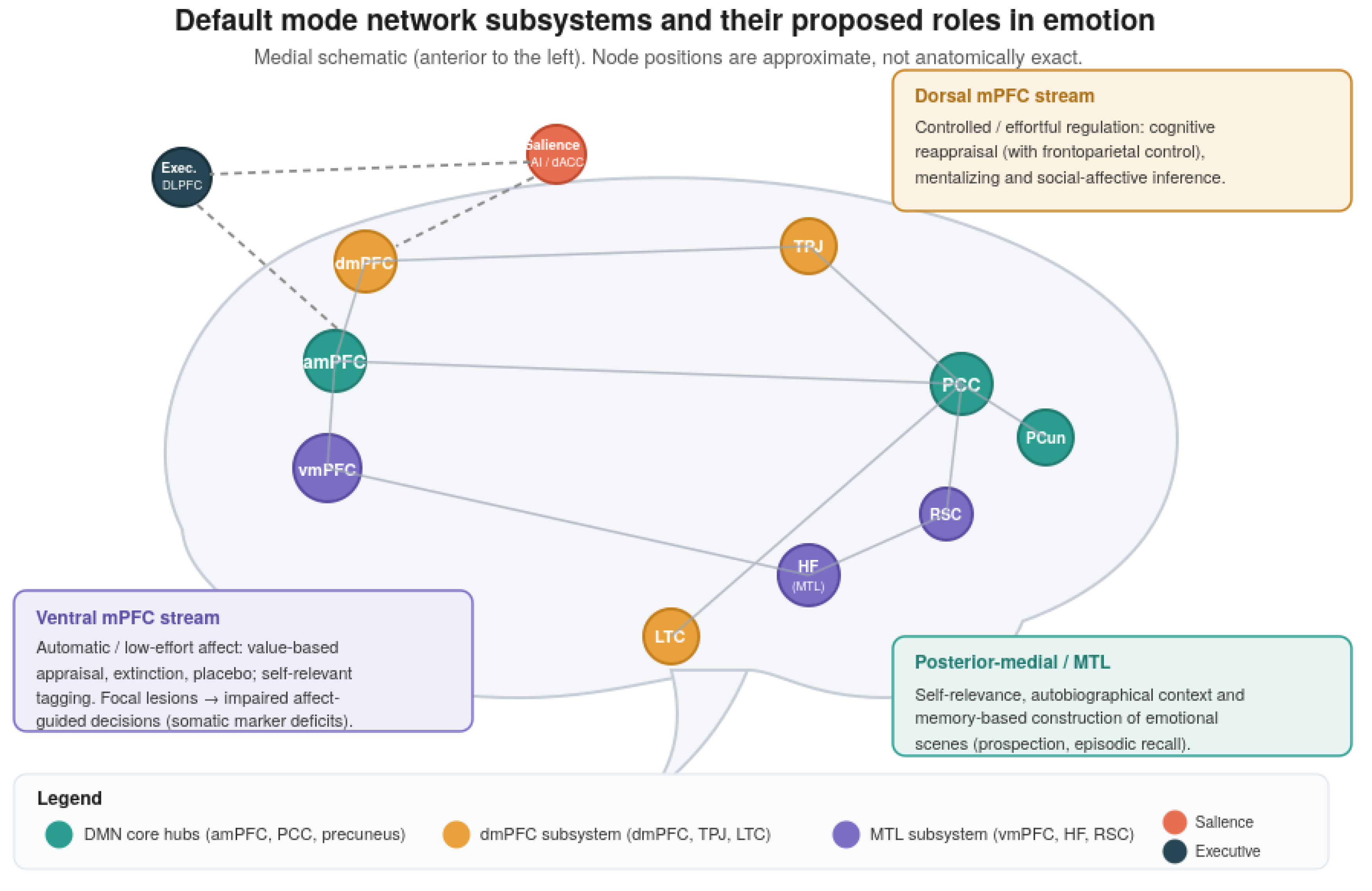

Table 1.
Representative lesions and neuroimaging evidence linking DMN nodes to emotional processing. Abbreviations are as in Figure 1.
Table 1.
Representative lesions and neuroimaging evidence linking DMN nodes to emotional processing. Abbreviations are as in Figure 1.
| DMN Node/Circuit | Subsystem | Neuroimaging Evidence | Lesion / Causal Evidence | Ref. |
| Ventromedial PFC (vmPFC) | Core / MTL | Value-based appraisal; automatic regulation (extinction, placebo); self-referential tagging | Focal damage → impaired affect-guided decisions, altered emotional responses, abnormal Iowa Gambling Task | [20,22,23,24,25,26,27] |
| Dorsomedial PFC (dmPFC) | Dorsomedial | Controlled cognitive reappraisal (with frontoparietal control); mentalizing | Inferred from social-affective deficits; limited isolated-lesion data | [15,18,19] |
| Anterior medial PFC (amPFC) | Core | Self-referential appraisal; affectively significant decisions | Overlaps vmPFC lesion territory | [9,14] |
| PCC / precuneus | Core | Personal salience, autobiographical depth of emotional material | Isolated focal lesions rare; often disrupt consciousness when extensive | [3,5] |
| Medial temporal lobe (HF, RSC) | MTL | Memory-based construction of emotional scenes; prospection | Bilateral MTL damage alters resting DMN connectivity pattern | [4,33] |
| DMN–subgenual cingulate coupling | Core + adjacent | Hyperconnectivity tracks rumination severity in MDD | — | [8,13] |
| Lesion-derived depression circuit | DMN + DLPFC/sgACC | Resting connectivity to the circuit predicts antidepressant response | Heterogeneous depression-causing lesions connect to a common circuit; lesion/TMS/DBS maps converge | [30,31] |
Disclaimer/Publisher’s Note: The statements, opinions and data contained in all publications are solely those of the individual author(s) and contributor(s) and not of MDPI and/or the editor(s). MDPI and/or the editor(s) disclaim responsibility for any injury to people or property resulting from any ideas, methods, instructions or products referred to in the content. |
© 2026 by the authors. Licensee MDPI, Basel, Switzerland. This article is an open access article distributed under the terms and conditions of the Creative Commons Attribution (CC BY) license (http://creativecommons.org/licenses/by/4.0/).
Copyright: This open access article is published under a Creative Commons CC BY 4.0 license, which permit the free download, distribution, and reuse, provided that the author and preprint are cited in any reuse.


