

Submitted:
30 June 2026
Posted:
02 July 2026
You are already at the latest version
Abstract
Endometrial cancer is one of the most rapidly increasing gynaecological malignancies worldwide. The clinically adapted molecular classification of endometrial carcinoma, derived from The Cancer Genome Atlas, comprises four major subtypes: POLE-mutated, mismatch repair deficiency, p53-abnormal expression, and no specific molecular profile. Its clinical implementation has improved prognostic stratification, risk assessment, and treatment decision-making in patients with endometrial carcinoma. However, current workflows rely on immunohistochemistry and targeted sequencing, which increase costs, turnaround times, and infrastructure requirements, thereby limiting their universal adoption in routine practice. In this context, recent advances in artificial intelligence (AI), particularly deep learning models capable of predicting molecular features directly from H&E-stained whole-slide images, have emerged as promising tools for precision oncology. In addition to reproducing established molecular classification, these approaches may reveal previously unrecognised biomarker-defined histologic patterns that are difficult to detect using conventional methods. This article synthesises the current evidence on AI-based molecular classification in endometrial carcinoma from a pathologist-centred perspective, with an emphasis on the biological rationale, methodological limitations, and future directions for clinical translation.
Keywords:
endometrial cancer
; molecular classification
; biomarker
; digital pathology
; whole-slide image
; artificial intelligence
; deep learning
; computational pathology
; precision oncology
1. Introduction
Endometrial cancer (EC) is the sixth most frequently diagnosed malignancy in women, accounting for 420,242 new cases and 97,704 deaths worldwide in 2022 [1]. The incidence of EC is particularly high in developed countries and is closely associated with established risk factors, including obesity, metabolic syndrome, ageing, and prolonged exposure to unopposed oestrogen [2,3].
In 2013, The Cancer Genome Atlas (TCGA) Research Network established a molecular classification for EC by integrating genomic, transcriptomic, and proteomic data [4]. This landmark study stratified EC into four molecular subgroups: POLE-ultramutated, microsatellite instability (MSI), copy number (CN)-low, and CN-high subgroups. Although this framework has transformed the biological understanding of EC, the sequencing-based and multi-omics approaches used in the original TCGA study are difficult to implement directly in routine diagnostic practice. Accordingly, subsequent translational efforts have focused on clinically feasible surrogate algorithms that preserve the biological and prognostic relevance of TCGA classification [5,6]. In this context, the Proactive Molecular Risk Classifier for Endometrial Cancer (ProMisE) and TransPORTEC groups proposed more practical approaches based on next-generation sequencing (NGS) to detect pathogenic POLE exonuclease domain mutations (POLEmut), immunohistochemistry (IHC) to identify mismatch repair deficiency (MMRd) and aberrant p53 expression (p53abn), and the classification of tumours lacking these alterations as no specific molecular profile (NSMP) [7,8].
This molecular classification has substantially improved EC management by refining prognostic assessment, resolving diagnostically ambiguous high-grade cases, guiding adjuvant treatment decisions, enabling biomarker-driven clinical trials, and improving diagnostic reproducibility by reducing the reliance on histology alone [9,10]. However, current workflows still rely on IHC, polymerase chain reaction (PCR)-based assays, and NGS. These methods require additional tissue, cost, turnaround time, laboratory infrastructure, and technical expertise, which may limit the scalable implementation of molecular classification, particularly in resource-limited settings.
Digital pathology provides a potential translational bridge between routine histopathology and molecular cancer care. The widespread adoption of slide scanners and whole-slide images (WSIs) has created a technical foundation for the application of artificial intelligence (AI) in routine pathology practice [11]. In particular, deep learning (DL) models have emerged as promising tools for inferring molecular features directly from haematoxylin and eosin (H&E)-stained WSIs [12,13]. These approaches may complement established molecular assays by identifying histomorphology patterns associated with molecular subtypes, prioritising cases for confirmatory testing, and supporting more timely and accessible molecular risk stratification.
Several recent reviews have addressed the expanding role of AI and digital pathology in gynaecological oncology; however, many have taken a broad perspective across tumour types, imaging modalities, and computational tasks [14,15]. In contrast, our study aims to present how AI can learn relevant histologic features with specific molecular biomarkers from WSIs and its interpretability and adoptability in real-world clinical workflows from the perspective of pathologists, rather than focusing on AI architecture technologies. In addition, we investigated how these approaches can be integrated into clinical pathology and oncology workflows to improve the prognosis of patients with EC (Figure 1).
We synthesised current evidence on AI-based molecular inference in EC using digital pathology, with particular emphasis on four-class molecular subtype and tumour mutational burden (TMB) prediction. The relevant studies are summarised in Table 1.
| N/A | |
2. Biological Basis of Molecular Inference from Histopathology
The rationale for AI-based molecular inference from H&E-stained WSIs rests on the premise that genomic alterations may influence cellular phenotype, tumour architecture, and tumour microenvironment (TME), thereby generating specific morphological patterns that may be learnable by DL models [31,32,33,34]. Genomic alterations, including DNA sequence-level mutations, copy number alterations (CNA), chromosomal structural rearrangements, and epigenetic modifications, can perturb cellular signalling networks involved in proliferation, differentiation, chromatin regulation, DNA repair, epithelial adhesion, immune recognition, stromal remodelling, and vascular responses. These downstream effects may be reflected across multiple histological scales, including nuclear atypia, mitotic activity, tumour growth patterns, tumour–stromal interactions, lymphocytic infiltration, and spatial heterogeneity [35,36,37,38]. This concept has motivated a series of studies evaluating whether DL models can predict molecular subtypes and clinically relevant biomarkers directly from histopathology.
The NSMP group illustrates the potential and limitations of morphology-based molecular inference. Although many NSMP tumours exhibit apparently low-risk endometrioid morphology, this molecular subgroup is biologically heterogeneous and encompasses a range of clinically relevant genomic alterations that may not be readily apparent on routine histopathological assessment. CTNNB1 mutations, which predominantly occur in NSMP tumours, activate Wnt/β-catenin signalling and may influence proliferation, differentiation, epithelial adhesion, polarity, and epithelial–mesenchymal transition. Clinically, CTNNB1-mutated NSMP tumours are important because they are associated with an increased risk of recurrence despite otherwise low-risk histological features [39]. This discrepancy between favourable morphology and adverse molecularly driven behaviour suggests that the NSMP classification alone may be insufficient for accurate risk assessment in this patient population. Therefore, in NSMP tumours, AI may be more useful for risk refinement and the identification of subtle differences in molecular-histological patterns than for simple NSMP subtype confirmation.
TME further expands the information content of WSIs. Tumour-infiltrating lymphocytes, desmoplastic reaction, extracellular matrix deposition, tumour–stroma ratio, vascular patterns, and invasive-front features of tumours are not merely background findings; they reflect the dynamic interactions between tumour cells and host tissue. These features may be shaped by tumour immunogenicity, epithelial–mesenchymal transition, stromal activation, angiogenic signalling, and other molecular programs [36]. Accordingly, DL models may improve molecular inference by integrating epithelial morphology with the TME context rather than relying solely on tumour cell-intrinsic features.
Although previous studies have attempted to link molecular alterations with histological phenotypes [40,41,42,43,44], the precise relationship between specific genomic changes and morphological features is unclear. In AI inference studies, most current interpretations are based on recurrent phenotypes of molecular-histological alterations rather than definitive biological relevance. In this context, AI could be used as a complementary approach by detecting subtle, spatially distributed, or sub-visual histological patterns that are difficult to recognize using conventional assessment. Furthermore, by integrating large-scale image analysis with molecular and clinical data, AI may help identify novel molecular-histological features associated with clinically relevant biomarkers, therapeutic responses, and prognosis.
3. MMRd Subtype
MMRd tumours account for approximately 25–30% of ECs [45]. The DNA mismatch repair (MMR) system repairs single-base mismatches and short insertion–deletion loops that arise during DNA replication. Loss of MMR function leads to the accumulation of insertion–deletion mutations, particularly in repetitive microsatellite sequences, resulting in MSI. In routine practice, MMRd is assessed by IHC for MLH1, PMS2, MSH2, and MSH6 proteins, whereas MSI is detected using PCR- or NGS-based assays. In this review, we use the term “MMRd” to collectively refer to the MMRd and MSI statuses. Hypermutated MMRd tumours have an increased neoantigen load and frequently exhibit immune-rich histological features, providing a biological rationale for their sensitivity to immune checkpoint inhibitors. Accordingly, MMRd status is clinically important for both immunotherapy eligibility and Lynch syndrome screening [44,46,47].
Among molecular targets, MMRd has emerged as the most extensively studied molecular subtype for AI-based prediction of H&E-stained WSIs in EC [16,19,25]. This is largely attributable to the availability of annotated ground-truth datasets generated by routine clinical assessment of MMRd status and the immunotherapeutic relevance of MMRd [40,44,48]. Moreover, MMRd inference in EC benefits from a mature research ecosystem that has already been established for colorectal and gastric cancers [49,50].
Fremond et al. [17] developed a clinically oriented interpretability framework for four EC molecular subtypes. Through an expert review of attention heatmaps and top-attended tiles, the study identified subtype-associated morphologies, including lymphocyte-rich and predominantly solid growth patterns in POLEmut and MMRd tumours; smooth luminal borders with mild nuclear atypia, low lymphocyte density, focal squamous differentiation, and low tumour-to-stroma ratio in NSMP tumours; and marked nuclear atypia, low lymphocyte density, high tumour-to-stroma ratio, and ragged glandular luminal surfaces in p53abn tumours. This overlapping lymphocyte-rich morphology may contribute to the misclassification of POLEmut and MMRd tumours, posing a challenge for the clinical deployment of H&E-based AI models.
Subsequent studies have incorporated multi-resolution or hierarchical modelling strategies to analyse morphology across multiple histological scales. Whangbo et al. [20] proposed a multi-resolution ensemble model that integrates features from 2.5×, 5×, and 10× magnifications, thereby capturing both the broad architectural context and the fine cytological details. Wang et al. [26] further extended this hierarchical approach by developing an IMAN that represents histology across the slide, bag, patch, and cell levels. This framework predicted histological subtypes, MMRd, and p53mut in EC. Together, these studies suggest that multi-scale and multi-level attention modelling may enhance molecular inference from histopathology by integrating information distributed across the tissue architecture, local image patches, and cellular-level features.
Recent studies have shifted attention from accuracy-centred model development toward practical requirements for clinical deployment, including inference speed, workflow scalability, and human–AI collaboration. Wang et al. [19] developed a weakly supervised pipeline designed for efficient clinical use, achieving rapid inference at 1.03 s per slide, and demonstrating its potential for high-throughput screening. In addition, in a pathologist reader study, Liu et al. [25] showed that MMRNet outperformed junior and senior pathologists and achieved a performance comparable to that of expert pathologists, while consistently maintaining higher sensitivity across experience levels. Notably, the authors proposed a human–machine fusion strategy that integrated MMRNet predictions with pathologist’s assessments. The improved performance of this combined approach supports the use of AI-assisted screening as a decision-support tool in routine pathological workflows.
4. p53abn Subtype
The p53abn subtype is characterised by TP53 alterations and CN-high, representing the most aggressive molecular category of EC. It accounts for approximately 5–15% of ECs and is consistently associated with poor clinical outcomes, supporting the need for intensified adjuvant treatment in appropriate settings [45,51]. Although p53abn EC is classically associated with uterine serous carcinoma, it can occur across multiple histological subtypes and may exhibit high-grade cytology, serous-like or ambiguous morphology, and aggressive growth patterns [42,52]. In routine practice, p53 IHC is widely used as a practical surrogate for p53abn status assessment. Aberrant p53 expression patterns include diffuse, strong nuclear overexpression, complete absence of nuclear staining, and cytoplasmic expression patterns [53,54].
Recent methodological advances have attempted to improve p53abn prediction by enhancing the detection of subtle cytological and architectural features of lesions. For example, frameworks that combine super-resolution enhancement with transformer-based lesion segmentation may better capture high-grade nuclear atypia, complex growth patterns, and other diagnostically relevant features, thereby supporting more stable performance across multicentre cohorts [27].
5. POLEmut Subtype
The POLEmut subtype represents the most favourable prognostic category of EC, accounting for approximately 5–15% of cases [45]. Pathogenic POLE exonuclease domain mutations impair DNA replication proofreading and generate an ultramutated genomic phenotype characterised by an exceptionally high single-nucleotide variant burden [43]. Despite their favourable clinical behaviour, POLEmut ECs often exhibit aggressive histological features [45]. Morphologically, these tumours frequently retain endometrioid differentiation but may show FIGO grade 3 histology, intratumor heterogeneity, prominent lymphocytic infiltration, eosinophilic cytoplasmic changes, bizarre nuclei, and occasional serous-like or ambiguous features [41].
Accurate identification of POLEmut status is clinically important because current guidelines support adjuvant therapy de-escalation in patients with POLEmut EC [10,51]. However, definitive confirmation requires targeted sequencing, which may not be universally accessible in all laboratories. Accordingly, AI-based prediction of POLEmut status from WSIs has been explored as a promising strategy to address the challenges of sequencing. Hong et al. [16] introduced Panoptes, a multi-resolution DL architecture that integrates spatially aligned image tiles at 2.5×, 5×, and 10× magnifications, mimicking the multi-scale diagnostic approach used by pathologists in routine practice. However, the direct prediction of POLEmut status achieved only moderate performance, with an area under the receiver operating characteristic curve (AUROC) of 0.681, highlighting the difficulty in inferring POLE mutations from histology alone.
More recent approaches have leveraged large-scale pathology foundation models and hierarchical representations to mitigate the data scarcity associated with the relatively low prevalence of POLEmut tumours. The hi-UNI pipeline adapted the UNI pathology foundation model and incorporated task-specific fine-tuning with a three-scale hierarchical representation to capture both macroscopic tissue architecture and microscopic cytological details. This approach achieved superior performance compared to single-scale UNI-based models, with an AUROC of 0.886. These findings suggest that foundation model-based feature extraction and hierarchical representation learning may improve POLEmut inference, although further external validation is required [28].
Interpretability analyses provide insights into the challenges associated with POLEmut prediction. Guo et al. [30] combined Grad-CAM-based patch visualisation with Hover-Net-based nuclear segmentation and single-cell feature extraction to investigate morpho-molecular correlates of EC molecular subtypes. In their analyses, POLEmut tumours were associated with solid growth, cytological atypia, dispersed high-activation regions, and greater morphological and spatial heterogeneity. MMRd tumours also showed lymphocyte-rich morphology, whereas p53abn tumours demonstrated high-grade cytological features and serous-like architecture. In addition, NSMP tumours have increased stromal cellularity and small volumes of inflammatory cells. These findings suggest that AI-based POLEmut inference may rely on a combination of patch-level morphology, immune contexture, and cell-level spatial features rather than on a single, distinctive histological pattern.
Despite these advances, POLEmut remains an intrinsically challenging target for H&E-based molecular inference in EC. Its low prevalence limits the availability of training cases, and its morphology may substantially overlap with that of immune-rich MMRd tumours. Consequently, the sensitivity and positive predictive value may remain unstable for POLEmut prediction, even when the AUROC values are acceptable.
6. NSMP Subtype
The NSMP subtype represents the largest category, accounting for approximately 30–40% of ECs [45]. Tumours are classified as NSMP when POLEmut, MMRd, and p53abn features are absent; therefore, this category encompasses biologically heterogeneous tumours without a single dominant molecular driver. Most NSMP tumours show low-grade endometrioid histology and have an overall intermediate prognosis; however, clinical outcomes vary substantially within this subgroup [10]. Molecularly, NSMP tumours often harbour PI3K–AKT pathway alterations, show high oestrogen receptor (ER) or progesterone receptor (PR) expression, and contain CTNNB1 mutations, whereas a subset lacks detectable alterations in routinely assessed markers [4,5]. ER IHC is clinically relevant in this context, as ER status provides prognostic information within the NSMP subgroup and predicts response to endocrine therapy in advanced or recurrent disease [51]. From an AI perspective, the lack of a single defining molecular or morphological signature may make NSMP more difficult to infer from H&E-stained WSIs.
Despite these challenges, AI can provide clinical value by refining risk stratification in this heterogeneous subgroup. Darbandsari et al. [24] identified a p53abn-like subset of NSMP ECs associated with poorer survival than that of other NSMP cases. This subgroup exhibited marked nuclear atypia and increased CNA despite a wild-type p53 IHC pattern, and some cases harboured TP53 mutations on sequencing. These findings suggest that H&E-based AI may identify adverse morphological and molecular programs within NSMP tumours that are not fully captured by conventional assays in EC.
Multimodal integrative approaches may further enhance risk prediction in heterogeneous tumours. HECTOR, developed by Volinsky-Fremond et al. [23], integrates H&E WSI-derived histology, im4MEC-derived molecular class predictions, and anatomical stage to predict the postoperative distant recurrence risk and identify patients who may benefit from adjuvant therapy for EC. Its superior performance over H&E-only DL models suggests that combining image-derived molecular information and clinical staging provides additive prognostic value.
7. Prediction of TMB
TMB has been investigated as a biomarker for predicting the response to immune checkpoint inhibitor therapy in patients with cancer. As a quantitative measure of the somatic mutation load within a tumour, TMB reflects the potential capacity of cancer cells to generate neoantigens that can be recognised by the host immune system. Accordingly, highly mutated tumours may be more immunogenic and more susceptible to immune checkpoint blockade, particularly in molecular contexts such as MMRd or POLEmut EC [55].
In EC, TMB varies substantially across molecular subtypes: POLEmut tumours typically exhibit extremely high mutation burdens (>100 mutations/Mb), MMRd tumours generally show high TMB levels (10–100 mutations/Mb), whereas NSMP and p53abn tumours usually have low TMB (<10 mutations/Mb) [10]. However, despite its clinical relevance, TMB assessment requires comprehensive sequencing-based genomic profiling, which is costly, technically demanding, and not universally accessible in all laboratories. These limitations have motivated recent studies to explore whether DL models can directly infer TMB from routine H&E-stained WSIs.
Wang et al. [22] proposed a multilayer attention-based multiple-instance learning framework, termed TR-MAMIL, which used truncated ResNet feature encoders to analyse H&E-stained WSIs. The model was designed to classify ECs into histologically aggressive and non-aggressive risk groups and to predict TMB directly from routine histopathology images. In the TCGA cohort, TR-MAMIL demonstrated a strong performance in distinguishing aggressive from non-aggressive histological groups, achieving an AUROC of 0.88. In contrast, TMB prediction showed substantial subgroup-dependent variability: the model achieved an AUROC of 0.82 in aggressive tumours but only 0.56 in non-aggressive tumours. These findings are informative because they demonstrate clear task-dependent differences in the feasibility of H&E-based prediction.
The inference of aggressive versus non-aggressive EC was relatively robust, whereas TMB prediction was substantially more challenging, particularly in histologically non-aggressive tumours. This difference is biologically plausible because TMB is a sequencing-derived quantitative biomarker rather than a directly observable histological phenotype. Its relationship with morphology is likely indirect and mediated through associated features such as tumour grade, immune infiltration, genomic instability, and molecular subtype composition. The subgroup-dependent performance of TR-MAMIL suggests that TMB-associated morphological signals may be more detectable in aggressive EC, whereas in non-aggressive tumours, such cues may be weaker, spatially dispersed, or less tightly coupled to visible morphology. Therefore, poor performance in non-aggressive EC should not be interpreted solely as a modelling limitation but may also reflect inherent biological and morphological constraints on H&E stained WSI-based TMB inference.
Similar patterns have been reported using an ETMIL-SSLViT, which achieved strong performance for aggressive and non-aggressive subtype classification but encountered comparable difficulties in predicting TMB within non-aggressive tumours [29].
8. Discussion
8.1. Methodological Pitfalls
Several methodological limitations should be considered when interpreting AI-based molecular inferences from H&E-stained WSIs in EC (Figure 2).
First, the dataset composition can substantially influence the model performance [56,57]. Many studies rely on public datasets or single-institutional cohorts that may not fully reflect the diversity of real-world practices [58]. Selection bias related to disease stage, specimen type, molecular testing availability, histological subtype distribution, and molecular subtype prevalence may lead to an overestimation of performance [59,60]. Small cohort sizes and class imbalances are particularly problematic for rare molecular subtypes, such as POLEmut EC [56,58]. In addition, institutional bias and domain shift may impair generalisability because models may inadvertently learn site-specific staining patterns, tissue processing artefacts, scanner characteristics, or other preanalytical variations rather than biologically meaningful morphological features [59,61].
Second, the definition of ground truth remains a major challenge [57,62]. Labels derived from IHC, PCR, and sequencing-based assays are not always interchangeable. For example, p53 IHC results may not completely concord with TP53 sequencing results [63]. Discordance can arise from subclonal TP53 alterations, resulting in heterogeneous p53 expression, technical variations, interpretative differences between observers, and sampling bias effects. Similarly, imperfect agreement may occur among MMR IHC, PCR-based MSI testing, and NGS-based MSI assessment, particularly in cases with subclonal or heterogeneous MMR alterations that result in focal or heterogeneous loss of MMR protein expression [44]. These discrepancies introduce label noise into slide-level annotations and may constrain both the model performance and interpretability.
Consequently, WSI-based molecular inference is vulnerable to intratumoural heterogeneity and sampling effects [11,36]. Molecularly relevant histological signals may be spatially distributed, focal, or unevenly represented across tumour regions. A single slide from an excision specimen or biopsy sample may not capture the full spectrum of tumour morphology or molecular heterogeneity. This is particularly relevant for ECs with mixed histological patterns and subclonal cahnges. Consequently, the case-level molecular labels assigned to WSIs may not accurately represent all tumour regions. Patch-level model attention may be difficult to interpret when the reference assay is performed on a different block or region [11,64].
These limitations are not only applicable to one molecular subtype of EC. Therefore, future studies should prioritise large, representative, multicentre cohorts, balanced sampling of rare subtypes, standardised preanalytical and scanning workflows, and rigorous definitions of molecular ground truths [11,57,64]. Where possible, the model performance should be evaluated separately according to the IHC-, PCR-, and sequencing-defined labels of the training data. In addition, interpretability may be improved when molecular testing used for ground truth and model inference is performed on the same tumour regions.
8.2. Interpretability and Biological Validation
Interpretability is essential for assessing the biological validity and clinical credibility of AI-based molecular inferences [59,64]. Attention heatmaps and other visualisation methods can provide partial insights into the model behaviour by highlighting the image regions that contribute to the prediction. However, such visual explanations do not automatically establish the biological plausibility of models. Model-highlighted regions should be systematically reviewed by expert pathologists to determine whether they correspond to diagnostically or biologically meaningful features, such as tumour morphology, immune infiltration, stromal remodelling, necrosis, solid growth, or nuclear atypia, rather than irrelevant artefacts, including tissue folds, staining variation, out-of-focus areas, pen marks, debris, or slide preparation effects [65].
Therefore, more biologically grounded interpretability frameworks are required. These may include the integration of attention-based visualisation with nuclear segmentation, cell-level morphometric analysis, spatial immune profiling, stromal quantification, and correlation with genomic, transcriptomic, or immunophenotypic data. Such approaches can help determine whether AI models learn biologically meaningful morphology–genome relationships, rather than exploit confounding technical or cohort-specific features [64,66].
8.3. Clinical Translation as a Triage Tool
Current evidence suggests that the clinical utility of H&E-stained WSI-based AI in EC should be framed primarily as calibrated triage and decision support rather than as a direct replacement for established molecular assays [67,68,69]. Because AI models infer molecular status from probabilistic histological signals, their outputs can be affected by intratumoural heterogeneity, sampling variation, domain shifts, and imperfect concordance among reference assays.
For NSMP tumours, AI may be more useful for risk refinement and the discovery of high-risk morpho-molecular patterns than for simple subtype confirmation [24]. Similarly, AI-based TMB prediction remains an adjunctive strategy with potential value for risk enrichment or prioritisation of genomic testing in selected high-risk contexts [22,29].
Within a human-in-the-loop workflow, slide-level probabilities and patch-level heatmaps may help pathologists prioritise confirmatory assays, particularly when tissue is limited or molecular testing resources are constrained [30]. Accordingly, clinical validation should extend beyond model-level performance metrics to evaluate whether AI-assisted workflows improve diagnostic efficiency, reduce unnecessary testing, shorten turnaround time, support treatment selection, and maintain patient safety. Overall, H&E-based AI is best positioned as a calibrated triage tool that complements, rather than replaces, guideline-recommended molecular testing.
8.4. Future Directions
Future research should move beyond single-task molecular subtype classification toward clinically integrated decision-support systems. Multimodal models that combine H&E histology with clinical variables, molecular data, radiologic features, and treatment information may better reflect real-world decision-making in EC management (Figure 3). Such approaches could support not only molecular subtype prediction but also recurrence risk assessment, treatment stratification, and prediction of therapeutic response [23,70].
Foundation models pretrained on large-scale histopathology datasets represent another important approach. By learning generalisable tissue representations, these models may improve data efficiency, reduce the need for task-specific training from scratch, and support diverse downstream applications, including molecular subtype prediction, biomarker discovery, recurrence risk prediction, and treatment response modelling [65,70,71]. However, their clinical deployment requires rigorous calibration, interpretability analysis, and external and prospective validation across different scanners, staining protocols, institutions, and patient cohorts.
Emerging spatial and single-cell technologies can further enhance the biological interpretability of AI-based molecular inferences. Spatial transcriptomics, single-cell profiling, and multiplex immunofluorescence can help link image-derived features with the underlying molecular programs, immune contexture, and tumour–stroma interactions. These approaches may be particularly valuable for heterogeneous molecular categories and morphologically overlapping tumours, such as immune-rich MMRd and POLEmut tumours [72,73].
Finally, standardised reporting and risk-of-bias frameworks, including TRIPOD+AI and PROBAST+AI, should be adopted to improve methodological rigor, transparency, reproducibility, and comparability across studies [74,75]. Prospective studies should define the intended clinical use, pre-specified decision thresholds, calibration strategy, and confirmatory testing pathways, and evaluate whether AI-assisted workflows improve diagnostic efficiency, treatment selection, and patient outcomes [12].
9. Conclusions
The application of AI to digital pathology in EC has the potential to complement conventional assays for molecular classification and broaden access to molecularly informed care. Its greatest near-term value is likely to lie in calibrated triage, risk stratification, and the discovery of clinically meaningful image-based biomarkers rather than replacing established molecular assays. Safe clinical translation will require robust external validation and prospective evaluation of the impact of AI-assisted workflows. Ultimately, AI-driven digital pathology may serve as a bridge between routine pathology workflows and precision oncology, supporting more accessible, patient-centred, and molecularly informed cancer care.
Author Contributions
Conceptualization, Y.J. and S.H.L.; methodology, S.A.; software, Y.J. and S.H.; validation, Y.J., S.A. and S.H.L.; formal analysis, Y.J. and S.A.; investigation, Y.J. and S.H.L.; resources, Y.J.; data curation, Y.J.; writing—original draft preparation, Y.J.; writing—review and editing, Y.J., S.H., S.A. and S.H.L.; visualization, Y.J. and S.H.; supervision, S.A. and S.H.L.; project administration, S.A. and S.H.L.; funding acquisition, S.H.L. All authors have read and agreed to the published version of the manuscript.
Funding
This research received no external funding.
Institutional Review Board Statement
Ethics approval and consent to participate were not applicable.
Informed Consent Statement
Not applicable.
Data Availability Statement
No new data were created or analyzed in this study. Data sharing is not applicable to this article.
Acknowledgments
The authors thank Da Som Hwang of EDesign for assistance with figure illustration and preparation.
Conflicts of Interest
The authors declare no conflict of interest.
Abbreviations
| AI | Artificial intelligence |
| AUROC | Area under the receiver operating characteristic curve |
| CN | Copy number |
| CNA | Copy number alteration |
| DL | Deep learning |
| EC | Endometrial cancer |
| ER | Oestrogen receptor |
| H&E | Haematoxylin and eosin |
| IHC | Immunohistochemistry |
| MMR | Mismatch repair |
| MMRd | Mismatch repair deficiency |
| MSI | Microsatellite instability |
| NGS | Next-generation sequencing |
| NSMP | No specific molecular profile |
| p53abn | Aberrant p53 expression |
| POLEmut | Pathogenic POLE exonuclease domain mutation |
| PR | Progesterone receptor |
| ProMisE | Proactive Molecular Risk Classifier for Endometrial Cancer |
| TCGA | The Cancer Genome Atlas |
| TMB | Tumour mutational burden |
| TME | Tumour microenvironment |
| WSIs | Whole-slide images |
References
- Bray, F.; Laversanne, M.; Sung, H.; et al. Global cancer statistics 2022: GLOBOCAN estimates of incidence and mortality worldwide for 36 cancers in 185 countries. CA Cancer J. Clin. 2024, 74, 229–263. [Google Scholar] [CrossRef] [PubMed]
- Bruggmann, D.; Ouassou, K.; Klingelhofer, D.; et al. Endometrial cancer: mapping the global landscape of research. J. Transl. Med. 2020, 18, 386. [Google Scholar] [CrossRef] [PubMed]
- Gordhandas, S.; Zammarrelli, W.A.; Rios-Doria, E.V.; et al. Current Evidence-Based Systemic Therapy for Advanced and Recurrent Endometrial Cancer. J. Natl. Compr. Canc Netw. 2023, 21, 217–226. [Google Scholar] [CrossRef] [PubMed]
- Cancer Genome Atlas Research N; Kandoth, C.; Schultz, N.; et al. Integrated genomic characterization of endometrial carcinoma. Nature 2013, 497, 67–73. [Google Scholar] [CrossRef] [PubMed]
- Stelloo, E.; Bosse, T.; Nout, R.A.; et al. Refining prognosis and identifying targetable pathways for high-risk endometrial cancer; a TransPORTEC initiative. Mod. Pathol. 2015, 28, 836–844. [Google Scholar] [PubMed]
- Talhouk, A.; McConechy, M.K.; Leung, S.; et al. A clinically applicable molecular-based classification for endometrial cancers. Br. J. Cancer 2015, 113, 299–310. [Google Scholar] [CrossRef] [PubMed]
- Stelloo, E.; Nout, R.A.; Osse, E.M.; et al. Improved Risk Assessment by Integrating Molecular and Clinicopathological Factors in Early-stage Endometrial Cancer-Combined Analysis of the PORTEC Cohorts. Clin. Cancer Res. 2016, 22, 4215–4224. [Google Scholar] [PubMed]
- Talhouk, A.; McConechy, M.K.; Leung, S.; et al. Confirmation of ProMisE: A simple, genomics-based clinical classifier for endometrial cancer. Cancer 2017, 123, 802–813. [Google Scholar] [CrossRef] [PubMed]
- Jamieson, A.; Barroilhet, L.M.; McAlpine, J.N. Molecular classification in endometrial cancer: Opportunities for precision oncology in a changing landscape. Cancer 2022, 128, 2853–2857. [Google Scholar] [CrossRef] [PubMed]
- Jamieson, A.; McAlpine, J.N. Molecular Profiling of Endometrial Cancer From TCGA to Clinical Practice. J. Natl. Compr. Canc Netw. 2023, 21, 210–216. [Google Scholar] [CrossRef] [PubMed]
- Bera, K.; Schalper, K.A.; Rimm, D.L.; et al. Artificial intelligence in digital pathology - new tools for diagnosis and precision oncology. Nat. Rev. Clin. Oncol. 2019, 16, 703–715. [Google Scholar] [PubMed]
- Aggarwal, A.; Bharadwaj, S.; Corredor, G.; et al. Artificial intelligence in digital pathology - time for a reality check. Nat. Rev. Clin. Oncol. 2025, 22, 283–291. [Google Scholar] [PubMed]
- Wang, C.-W.; Lai, P.-J.; Muzakky, H.; et al. Deep learning to identify aggressive endometrial cancer and predict tumor mutational burden from histopathology slide. Biomed. Signal Process. Control 2025, 104. [Google Scholar] [CrossRef]
- Paiboonborirak, C.; Abu-Rustum, N.R.; Wilailak, S. Artificial intelligence in the diagnosis and management of gynecologic cancer. Int. J. Gynaecol. Obstet. 2025, 171 1 Suppl, 199–209. [Google Scholar] [CrossRef]
- Rockall, A.G.; Chiu, S.M.; Aboagye, E.O.; et al. Artificial intelligence in women’s cancers: innovation and challenges in clinical translation. Lancet Digit Health 2025, 7, 100940. [Google Scholar] [CrossRef] [PubMed]
- Hong, R.; Liu, W.; DeLair, D.; et al. Predicting endometrial cancer subtypes and molecular features from histopathology images using multi-resolution deep learning models. Cell Rep. Med. 2021, 2, 100400. [Google Scholar] [CrossRef] [PubMed]
- Fremond, S.; Andani, S.; Barkey Wolf, J.; et al. Interpretable deep learning model to predict the molecular classification of endometrial cancer from haematoxylin and eosin-stained whole-slide images: a combined analysis of the PORTEC randomised trials and clinical cohorts. Lancet Digit Health 2023, 5, e71–e82. [Google Scholar] [PubMed]
- Zhang, Y.; Chen, S.; Wang, Y.; et al. Deep learning-based methods for classification of microsatellite instability in endometrial cancer from HE-stained pathological images. J. Cancer Res. Clin. Oncol. 2023, 149, 8877–8888. [Google Scholar] [PubMed]
- Wang, C.W.; Muzakky, H.; Firdi, N.P.; et al. Deep learning to assess microsatellite instability directly from histopathological whole slide images in endometrial cancer. npj Digit Med. 2024, 7, 143. [Google Scholar] [CrossRef] [PubMed]
- Whangbo, J.; Lee, Y.S.; Kim, Y.J.; et al. Predicting Mismatch Repair Deficiency Status in Endometrial Cancer through Multi-Resolution Ensemble Learning in Digital Pathology. J. Imaging Inf. Med. 2024, 37, 1674–1682. [Google Scholar] [CrossRef]
- Umemoto, M.; Mariya, T.; Nambu, Y.; et al. Prediction of Mismatch Repair Status in Endometrial Cancer from Histological Slide Images Using Various Deep Learning-Based Algorithms. Cancers (Basel) 2024, 16. [Google Scholar]
- Wang, C.W.; Firdi, N.P.; Lee, Y.C.; et al. Deep learning for endometrial cancer subtyping and predicting tumor mutational burden from histopathological slides. npj Precis Oncol. 2024, 8, 287. [Google Scholar] [CrossRef] [PubMed]
- Volinsky-Fremond, S.; Horeweg, N.; Andani, S.; et al. Prediction of recurrence risk in endometrial cancer with multimodal deep learning. Nat. Med. 2024, 30, 1962–1973. [Google Scholar] [CrossRef] [PubMed]
- Darbandsari, A.; Farahani, H.; Asadi, M.; et al. AI-based histopathology image analysis reveals a distinct subset of endometrial cancers. Nat. Commun. 2024, 15, 4973. [Google Scholar] [CrossRef] [PubMed]
- Liu, L.L.; Jing, B.Z.; Liu, X.; et al. MMRNet: Ensemble deep learning models for predicting mismatch repair deficiency in endometrial cancer from histopathological images. Cell Rep. Med. 2025, 6, 102099. [Google Scholar] [CrossRef] [PubMed]
- Wang, C.W.; Muzakky, H.; Lee, Y.C.; et al. Interpretable multi-stage attention network to predict cancer subtype, microsatellite instability, TP53 mutation and TMB of endometrial and colorectal cancer. Comput Med. Imaging Graph 2025, 121, 102499. [Google Scholar] [CrossRef] [PubMed]
- Qi, P.; Yao, T.; Li, H.; et al. Clinical-grade AI model for molecular subtyping of endometrial cancer: a multi-center cohort study in China. Mol. BioMed 2025, 6, 102. [Google Scholar] [PubMed]
- Cui, H.; Guo, Q.; Xu, J.; et al. Prediction of molecular subtypes for endometrial cancer based on hierarchical foundation model. Bioinformatics 2025, 41. [Google Scholar] [CrossRef] [PubMed]
- Wang, C.W.; Liu, T.C.; Lai, P.J.; et al. Ensemble transformer-based multiple instance learning to predict pathological subtypes and tumor mutational burden from histopathological whole slide images of endometrial and colorectal cancer. Med. Image Anal. 2025, 99, 103372. [Google Scholar] [PubMed]
- Guo, Q.; Cui, H.; Zhang, Y.; et al. An interpretable deep learning model for predicting endometrial cancer molecular subtypes from H&E-stained slides. npj Precis Oncol. 2026, 10, 71. [Google Scholar] [PubMed]
- Coudray, N.; Tsirigos, A. Deep learning links histology, molecular signatures and prognosis in cancer. Nat. Cancer 2020, 1, 755–757. [Google Scholar] [CrossRef] [PubMed]
- Hanahan, D.; Coussens, L.M. Accessories to the crime: functions of cells recruited to the tumor microenvironment. Cancer Cell 2012, 21, 309–322. [Google Scholar] [CrossRef] [PubMed]
- Nassiri, I.; McCall, M.N. Systematic exploration of cell morphological phenotypes associated with a transcriptomic query. Nucleic Acids Res. 2018, 46, e116. [Google Scholar] [CrossRef] [PubMed]
- Tsai, P.C.; Lee, T.H.; Kuo, K.C.; et al. Histopathology images predict multi-omics aberrations and prognoses in colorectal cancer patients. Nat. Commun. 2023, 14, 2102. [Google Scholar] [CrossRef] [PubMed]
- Sakuragi, N.; Nishiya, M.; Ikeda, K.; et al. Decreased E-Cadherin Expression in Endometrial Carcinoma Is Associated with Tumor Dedifferentiation and Deep Myometrial Invasion. Gynecol. Oncol. 1994, 53, 183–189. [Google Scholar] [CrossRef] [PubMed]
- Stanta, G.; Bonin, S. Overview on Clinical Relevance of Intra-Tumor Heterogeneity. Front Med. 2018, 5, 85. [Google Scholar] [CrossRef]
- Taki, M.; Abiko, K.; Ukita, M.; et al. Tumor Immune Microenvironment during Epithelial-Mesenchymal Transition. Clin. Cancer Res. 2021, 27, 4669–4679. [Google Scholar] [PubMed]
- Tan, Z.; Sheng, B.; Chen, L.; et al. Inflammation-driven mechanisms in endometrial cancer: pathways from inflammatory microenvironment remodeling to immune escape. Front Immunol. 2025, 16, 1689114. [Google Scholar] [CrossRef] [PubMed]
- Ledinek, Z.; Sobocan, M.; Knez, J. The Role of CTNNB1 in Endometrial Cancer. Dis. Markers 2022, 2022, 1442441. [Google Scholar] [CrossRef] [PubMed]
- De Smedt, L.; Lemahieu, J.; Palmans, S.; et al. Microsatellite instable vs stable colon carcinomas: analysis of tumour heterogeneity, inflammation and angiogenesis. Br. J. Cancer 2015, 113, 500–509. [Google Scholar] [CrossRef] [PubMed]
- Hussein, Y.R.; Weigelt, B.; Levine, D.A.; et al. Clinicopathological analysis of endometrial carcinomas harboring somatic POLE exonuclease domain mutations. Mod. Pathol. 2015, 28, 505–514. [Google Scholar] [CrossRef] [PubMed]
- Momeni-Boroujeni, A.; Dahoud, W.; Vanderbilt, C.M.; et al. Clinicopathologic and Genomic Analysis of TP53-Mutated Endometrial Carcinomas. Clin. Cancer Res. 2021, 27, 2613–2623. [Google Scholar] [CrossRef] [PubMed]
- Rosa, R.C.A.; Yurchenko, A.A.; Chahud, F.; et al. First description of ultramutated endometrial cancer caused by germline loss-of-function and somatic exonuclease domain mutations in POLE gene. Genet Mol. Biol. 2020, 43, e20200100. [Google Scholar] [PubMed]
- Shia, J.; Black, D.; Hummer, A.J.; et al. Routinely assessed morphological features correlate with microsatellite instability status in endometrial cancer. Hum. Pathol. 2008, 39, 116–125. [Google Scholar] [CrossRef] [PubMed]
- Oaknin, A.; Bosse, T.J.; Creutzberg, C.L.; et al. Endometrial cancer: ESMO Clinical Practice Guideline for diagnosis, treatment and follow-up. Ann. Oncol. 2022, 33, 860–877. [Google Scholar] [CrossRef] [PubMed]
- Gordhandas, S.; Kahn, R.M.; Gamble, C.; et al. Clinicopathologic features of endometrial cancer with mismatch repair deficiency. Ecancermedicalscience 2020, 14, 1061. [Google Scholar] [CrossRef] [PubMed]
- Pakish, J.B.; Jazaeri, A.A. Immunotherapy in Gynecologic Cancers: Are We There Yet? Curr. Treat. Options Oncol. 2017, 18, 59. [Google Scholar] [CrossRef] [PubMed]
- Le, D.T.; Uram, J.N.; Wang, H.; et al. PD-1 Blockade in Tumors with Mismatch-Repair Deficiency. N Engl. J. Med. 2015, 372, 2509–2520. [Google Scholar] [CrossRef] [PubMed]
- Lee, S.H.; Jang, H.J. Deep learning-based prediction of molecular cancer biomarkers from tissue slides: A new tool for precision oncology. Clin. Mol. Hepatol. 2022, 28, 754–772. [Google Scholar] [CrossRef] [PubMed]
- Wagner, S.J.; Reisenbuchler, D.; West, N.P.; et al. Transformer-based biomarker prediction from colorectal cancer histology: A large-scale multicentric study. Cancer Cell 2023, 41, 1650–1661 e1654. [Google Scholar] [PubMed]
- Concin, N.; Matias-Guiu, X.; Cibula, D.; et al. ESGO-ESTRO-ESP guidelines for the management of patients with endometrial carcinoma: update 2025. Lancet Oncol. 2025, 26, e423–e435. [Google Scholar] [PubMed]
- Gatius, S.; Matias-Guiu, X. Practical issues in the diagnosis of serous carcinoma of the endometrium. Mod. Pathol. 2016, 29 1 Suppl, S45–58. [Google Scholar] [CrossRef]
- WHO Classification of Tumours Editorial Board. Female Genital Tumours; International Agency for Research on Cancer: Lyon (France), 2020; p. 254. [Google Scholar]
- Soslow, R.A.; Tornos, C.; Park, K.J.; et al. Endometrial Carcinoma Diagnosis: Use of FIGO Grading and Genomic Subcategories in Clinical Practice: Recommendations of the International Society of Gynecological Pathologists. Int. J. Gynecol. Pathol. 2019, 38 1 Suppl, S64–S74. [Google Scholar] [CrossRef]
- Chalmers, Z.R.; Connelly, C.F.; Fabrizio, D.; et al. Analysis of 100,000 human cancer genomes reveals the landscape of tumor mutational burden. Genome Med. 2017, 9, 34. [Google Scholar] [CrossRef] [PubMed]
- Chen, I.Y.; Pierson, E.; Rose, S.; et al. Ethical Machine Learning in Healthcare. Annu Rev. BioMed Data Sci. 2021, 4, 123–144. [Google Scholar] [CrossRef] [PubMed]
- van der Laak, J.; Litjens, G.; Ciompi, F. Deep learning in histopathology: the path to the clinic. Nat. Med. 2021, 27, 775–784. [Google Scholar] [CrossRef] [PubMed]
- Alam, M.R.; Seo, K.J.; Abdul-Ghafar, J.; et al. Recent application of artificial intelligence on histopathologic image-based prediction of gene mutation in solid cancers. Brief. Bioinform. 2023, 24. [Google Scholar] [CrossRef] [PubMed]
- Cifci, D.; Veldhuizen, G.P.; Foersch, S.; et al. AI in Computational Pathology of Cancer: Improving Diagnostic Workflows and Clinical Outcomes? Annu. Rev. Cancer Biol. 2023, 7, 57–71. [Google Scholar] [CrossRef]
- Franchet, C.; Schwob, R.; Bataillon, G.; et al. Bias reduction using combined stain normalization and augmentation for AI-based classification of histological images. Comput Biol. Med. 2024, 171, 108130. [Google Scholar] [CrossRef] [PubMed]
- Vahadane, A.; Peng, T.; Sethi, A.; et al. Structure-Preserving Color Normalization and Sparse Stain Separation for Histological Images. IEEE Trans. Med. Imaging 2016, 35, 1962–1971. [Google Scholar] [CrossRef] [PubMed]
- Aldea, M.; Salto-Tellez, M.; Marra, A.; et al. ESMO basic requirements for AI-based biomarkers in oncology (EBAI). Ann. Oncol. 2026, 37, 414–430. [Google Scholar] [PubMed]
- Vermij, L.; Leon-Castillo, A.; Singh, N.; et al. p53 immunohistochemistry in endometrial cancer: clinical and molecular correlates in the PORTEC-3 trial. Mod. Pathol. 2022, 35, 1475–1483. [Google Scholar] [CrossRef] [PubMed]
- Diao, J.A.; Wang, J.K.; Chui, W.F.; et al. Human-interpretable image features derived from densely mapped cancer pathology slides predict diverse molecular phenotypes. Nat. Commun. 2021, 12, 1613. [Google Scholar] [CrossRef] [PubMed]
- Vorontsov, E.; Bozkurt, A.; Casson, A.; et al. A foundation model for clinical-grade computational pathology and rare cancers detection. Nat. Med. 2024, 30, 2924–2935. [Google Scholar] [CrossRef] [PubMed]
- Zhao, W.; Guo, Z.; Fan, Y.; et al. Aligning knowledge concepts to whole slide images for precise histopathology image analysis. npj Digit Med. 2024, 7, 383. [Google Scholar] [CrossRef] [PubMed]
- Cifci, D.; Foersch, S.; Kather, J.N. Artificial intelligence to identify genetic alterations in conventional histopathology. J. Pathol. 2022, 257, 430–444. [Google Scholar] [CrossRef] [PubMed]
- Fremond, S.; Koelzer, V.H.; Horeweg, N.; et al. The evolving role of morphology in endometrial cancer diagnostics: From histopathology and molecular testing towards integrative data analysis by deep learning. Front Oncol. 2022, 12, 928977. [Google Scholar] [CrossRef] [PubMed]
- Lu, M.Y.; Williamson, D.F.K.; Chen, T.Y.; et al. Data-efficient and weakly supervised computational pathology on whole-slide images. Nat. BioMed Eng. 2021, 5, 555–570. [Google Scholar] [PubMed]
- Ding, T.; Wagner, S.J.; Song, A.H.; et al. A multimodal whole-slide foundation model for pathology. Nat. Med. 2025, 31, 3749–3761. [Google Scholar] [CrossRef] [PubMed]
- Xu, H.; Usuyama, N.; Bagga, J.; et al. A whole-slide foundation model for digital pathology from real-world data. Nature 2024, 630, 181–188. [Google Scholar] [PubMed]
- He, B.; Bergenstrahle, L.; Stenbeck, L.; et al. Integrating spatial gene expression and breast tumour morphology via deep learning. Nat. BioMed Eng. 2020, 4, 827–834. [Google Scholar] [CrossRef] [PubMed]
- Huang, T.; Liu, T.; Babadi, M.; et al. STPath: a generative foundation model for integrating spatial transcriptomics and whole-slide images. npj Digit Med. 2025, 8, 659. [Google Scholar] [CrossRef] [PubMed]
- Collins, G.S.; Moons, K.G.M.; Dhiman, P.; et al. TRIPOD+AI statement: updated guidance for reporting clinical prediction models that use regression or machine learning methods. BMJ 2024, 385, e078378. [Google Scholar] [CrossRef] [PubMed]
- Moons, K.G.M.; Damen, J.A.A.; Kaul, T.; et al. PROBAST+AI: an updated quality, risk of bias, and applicability assessment tool for prediction models using regression or artificial intelligence methods. BMJ 2025, 388, e082505. [Google Scholar] [CrossRef] [PubMed]
Figure 1.
Workflow of AI-based molecular inference from H&E-stained WSIs for clinical decision support in endometrial cancer. Artificial intelligence-based analysis of routine H&E-stained whole-slide images can infer the molecular features of endometrial cancer and support clinical decision-making. By linking histomorphology with molecular subtype prediction, testing triage, risk stratification, personalised therapy, and biomarker discovery, digital pathology may serve as a translational bridge toward more accessible molecularly informed care. Abbreviations: MMRd, mismatch repair-deficiency; NSMP, no specific molecular profile; p53abn, aberrant p53 expression; POLEmut, pathogenic POLE exonuclease domain mutations.
Figure 1.
Workflow of AI-based molecular inference from H&E-stained WSIs for clinical decision support in endometrial cancer. Artificial intelligence-based analysis of routine H&E-stained whole-slide images can infer the molecular features of endometrial cancer and support clinical decision-making. By linking histomorphology with molecular subtype prediction, testing triage, risk stratification, personalised therapy, and biomarker discovery, digital pathology may serve as a translational bridge toward more accessible molecularly informed care. Abbreviations: MMRd, mismatch repair-deficiency; NSMP, no specific molecular profile; p53abn, aberrant p53 expression; POLEmut, pathogenic POLE exonuclease domain mutations.
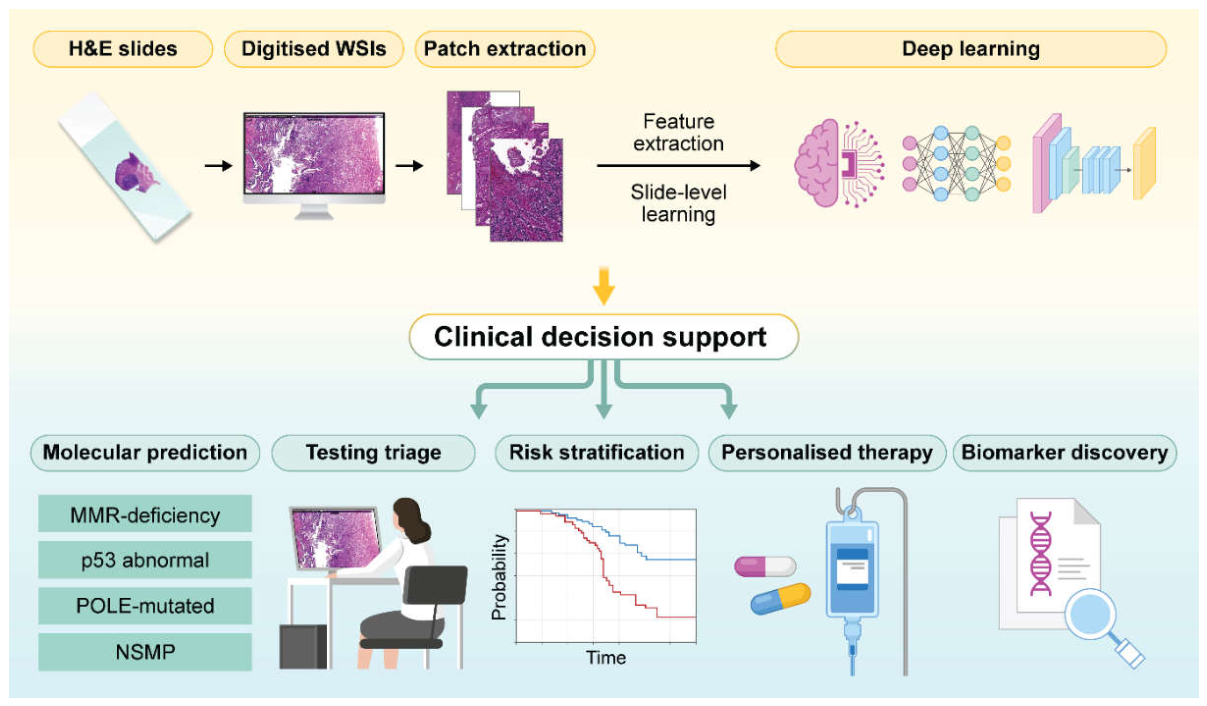
Figure 2.
Methodological pitfalls, interpretability, and validation requirements for the clinical translation of WSI-based AI molecular classification in endometrial cancer. AI-based molecular inference from histopathology is affected by several methodological challenges, including dataset composition bias, domain shifts, and label noise. Interpretability strategies, including attention heatmaps of WSIs or cell-level morphometric analysis of high-attention patch visualisation, may help determine whether model predictions correspond to biologically meaningful tumour features or non-biological confounders. Rigorous study design, transparent reporting, external and prospective validation, and clinical trials are required for clinical implementation. Abbreviations: AI, artificial intelligence; IHC, immunohistochemistry; MMRd, mismatch repair deficiency; WSI, whole-slide image.
Figure 2.
Methodological pitfalls, interpretability, and validation requirements for the clinical translation of WSI-based AI molecular classification in endometrial cancer. AI-based molecular inference from histopathology is affected by several methodological challenges, including dataset composition bias, domain shifts, and label noise. Interpretability strategies, including attention heatmaps of WSIs or cell-level morphometric analysis of high-attention patch visualisation, may help determine whether model predictions correspond to biologically meaningful tumour features or non-biological confounders. Rigorous study design, transparent reporting, external and prospective validation, and clinical trials are required for clinical implementation. Abbreviations: AI, artificial intelligence; IHC, immunohistochemistry; MMRd, mismatch repair deficiency; WSI, whole-slide image.
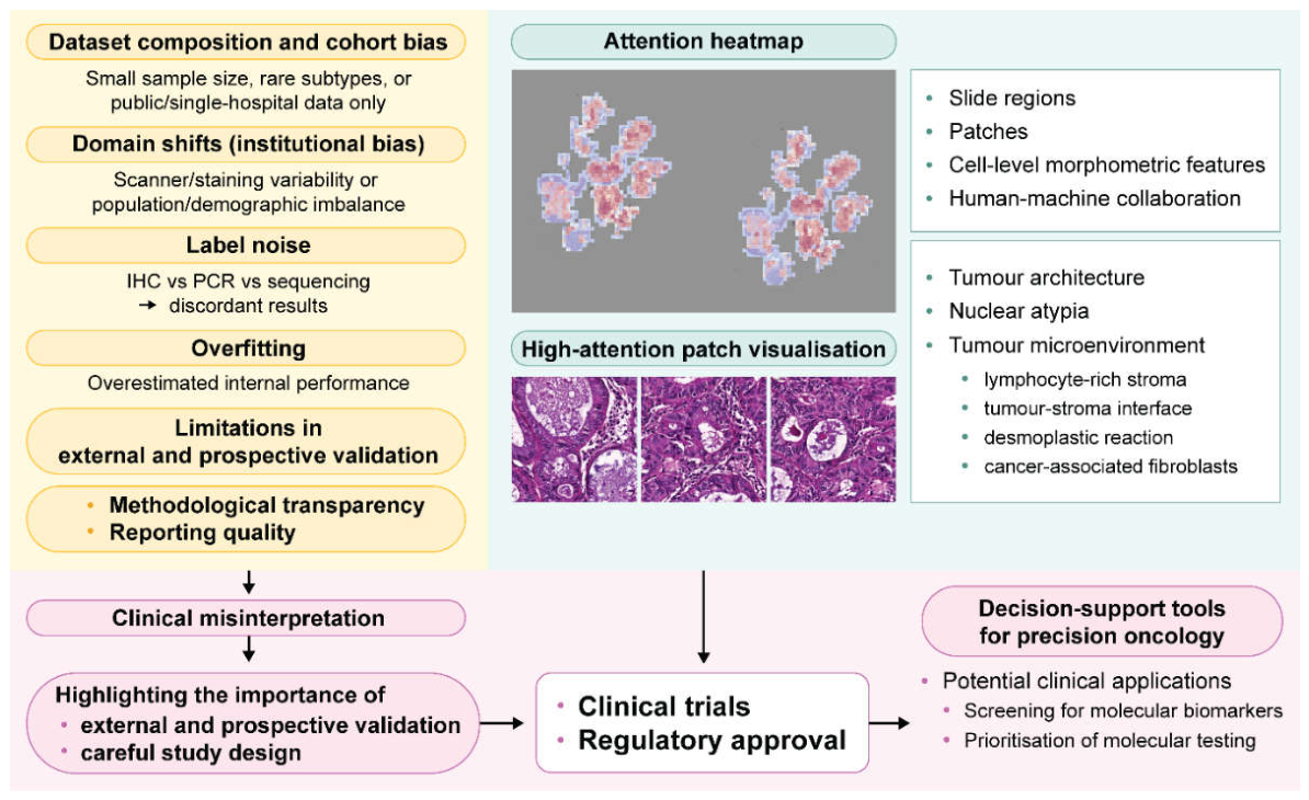
Figure 3.
Multimodal integration of AI for precision oncology in endometrial cancer. Multimodal integration of AI and computational pathology for clinical translation. H&E-stained WSIs, radiomic and genomic data, and clinical information can be integrated within foundation-model-based frameworks to support personalised prognosis, risk stratification, treatment optimisation, targeted therapy planning, and improved patient outcomes. Abbreviations: AI, artificial intelligence; H&E, haematoxylin and eosin; WSI, whole-slide image.
Figure 3.
Multimodal integration of AI for precision oncology in endometrial cancer. Multimodal integration of AI and computational pathology for clinical translation. H&E-stained WSIs, radiomic and genomic data, and clinical information can be integrated within foundation-model-based frameworks to support personalised prognosis, risk stratification, treatment optimisation, targeted therapy planning, and improved patient outcomes. Abbreviations: AI, artificial intelligence; H&E, haematoxylin and eosin; WSI, whole-slide image.
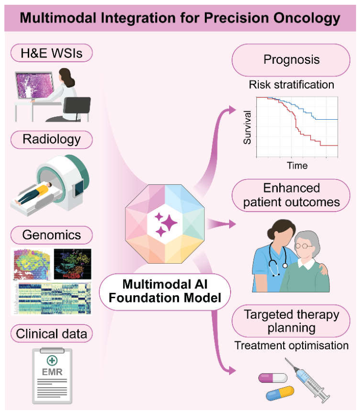

Table 1.
Overview of research on AI-based molecular classification using WSIs of endometrial cancer.
Table 1.
Overview of research on AI-based molecular classification using WSIs of endometrial cancer.
| Author (year) | Model/architecture | Internal cohort/dataset | External cohort/dataset | Task | Internal performance | External performance | |||
| Hong et al. (2021) [16] |
“Panoptes”, custom multi-resolution, InceptionResNet-based CNN (2.5×, 5×, 10×) |
TCGA and CPTAC, 496 WSIs of 456 pts, (train/val/test 8:1:1) |
NYU, 137 WSIs of 41 pts | MSI | AUC 0.827 | AUC 0.667 | |||
| CNV-H, CNV-L, POLE, TP53 (+17 genes) |
CNV-H/TP53/POLE/CNV-L, AUC 0.934/0.873/0.681/0.889, POLE (multi-model), 0.89 |
CNV-H/TP53/POLE/CNV-L, AUC 0.795/0.920/NA/0.850 |
|||||||
| Fremond et al. (2023) [17] |
“im4MEC”, SSL-MoCo-v2+ResNet50, attention, HoVer-Net |
PORTEC and multiple clinical cohorts, total 2028 pts, 4-fold CV |
PORTEC-3, 393 pts | MSI | N/A | AUC 0.844 | |||
| 4-class ProMisE | Macro-average AUC 0.874 | Macro-average AUC 0.876, TP53/POLE/NSMP, AUC 0.928/0.849/0.883 |
|||||||
| Zhang et al. (2023) [18] |
ResNet34, GAM-VGG16 |
TCGA, 95 WSIs of 95 pts, (train/test 70:25) |
N/A | MSI | AUC/Accu/Sens/F1-score, 0.799/0.80/0.857/0.826 |
N/A | |||
| Wang et al. [npjDM]a (2024) [19] |
Weakly supervised DL with FPS+MFCN+IPS +InceptionV3+WSID |
TCGA, 529 pts, (train/test ⅔:⅓) |
N/A | MSI (GT: NGS-based) |
Accu/Prec/Sens/F-measure, G1G2, 0.94/0.93/1.00/0.96; G3, 0.84/0.81/0.94/0.87, Inference time (1.03s/WSI) |
N/A | |||
| Whangbo et al. (2024) [20] |
Multi-resolution ensemble, ImageNet, EfficientNetB2 (2.5×, 5×, 10×) | GUGMC, 1168 WSIs of 325 pts, (train/test 8:2) | N/A | MMRd (GT: 4 IHC-based) |
AUC/Accu/Sens/Spec, 0.821/0.778/0.827/0.764 |
N/A | |||
| Umemoto et al. (2024) [21] |
ResNet50, API-Net-based |
SMUH, 114 pts, (train/val/test 70:15:15) |
N/A | MMRd (GT: PMS2, MSH6 IHC-based) |
AUC/Accu/Prec/Recall/F-score (per-tile level), ResNet50: 0.91/0.79/0.89/0.65/0.75; API: 0.85/0.85/0.75/0.69/0.72 |
N/A | |||
| Wang et al. [npjPO]a (2024) [22] |
Truncated ResNet50 (for subtype), Truncated ResNet152 (for TMB), ImageNet-pretrained TR-MAMIL |
TCGA, 918 WSIs of 529 pts for TMB; TSGH, TMA, 242 cores for MMR and TP53, [train/val/test (⅔ⅹ0.9: ⅔ⅹ0.1): ⅓] |
N/A |
N/A | MSI biomarkers (GT: IHC-based) |
MLH1/MSH2/MSH6/PMS2, MeanSS: 0.92/0.83/0.83/0.84 | N/A | ||
| N/A | |||||||||
| TMB-high/low by 10 mut/Mb (GT: NGS-based), p53, TP53 |
AUC/MeanSS, TMB in aggressive type 0.82/0.73; TMB in non-aggressive 0.56/0.68, p53 (TMA) 0.78/0.78, TP53 0.68/0.73 |
N/A | |||||||
| Volinsky -Fremond et al. (2024) [23] |
“HECTOR” (multimodal), 3-arm multimodal architecture (WSI+im4MEC+stage) |
Total 2751 pts, 5-fold CV, (train/test 1408:353 pts) |
UMCG, 160 pts; LUMC, 151 pts |
Recurrence risk associated with molecular type |
Unimodal; 2arm; HECTOR(3arm), Mean C-index: 0.775; 0.782; 0.795, held-out test set: 0.789 |
UMCG; LUMC, C-index: 0.828; 0.802-0.815 |
|||
| Darbandsari et al. (2024) [24] |
VarMIL, ResNet34 |
TCGA, 155 WSIs of 146 pts; TU, 431 WSIs of 222 pts, 10-fold CV, (train/val/test per fold 60:20:20) |
BC, TMA, 290 pts; CC 640 WSIs of 614 pts from 26 hospitals |
NSMP, p53abn, p53abn-like NSMP |
p53abn vs. NSMP, AUC/Accu, 0.95/0.894 |
p53abn vs. NSMP, AUC/Accu, BC 0.88/0.798; CC 0.88-0.95/0.663-0.885 |
|||
| Liu et al. (2025) [25] |
“MMRNet”, ensemble, EfficientNet, ResNet18, reader study, human-machine fusion |
Internal-UCEC, 1027 WSIs of 1026 pts, 5-fold CV |
TCGA, 401 WSIs of 369 pts; MultiCenter, 230 WSIs of 230 pts; GWCH, 421 WSIs of 421 pts |
MMRd | AUC/Sens/Spec/NPV, 0.897/0.628/0.949/0.892 |
MMRNet (3 cohorts) AUC 0.790;0.807;0.863; H-M fusion (3 cohorts) AUC 0.802;0.913;0.932 |
|||
| Wang et al. [CMIG]a (2025) [26] |
“IMAN”, SwAV-SSL-ResNet50 |
TSGH, TMA, 242 cores, (train/test ⅔:⅓) |
N/A | MSI biomarkers (GT: IHC-based) |
MLH1/MSH2/MSH6/PMS2, MeanSS 0.85/0.92/0.91/0.92, Inference time (18.71s/WSI) |
N/A | |||
| TP53 (GT: IHC-based) | MeanSS 0.81 | N/A | |||||||
| Qi et al. (2025) [27] |
SRResGAN, MedSAM, ResNet101, Grad-CAM |
393 pts, (train/test 8:2) |
OGHFU, 83 pts; PMCHH, 35 pts |
MMRd | AUC 0.92 | AUC, O: 0.96/ P: 0.93 | |||
| 4-class ProMisE (GT: POLE testing +IHC-based) |
average AUC/Sens /Spec/overall Accu , 0.980/0.907/0.924/0.914 , p53abn/POLE/NSMP, AUC 0.91/0.92/0.90 |
p53abn/POLE/NSMP, AUC O: 0.97/0.98/0.94; P: 0.94/0.97/0.91 |
|||||||
| Cui et al. (2025) [28] |
“hi-UNI”, DeepLab v3 |
FUSCC, 364 WSIs of 324 pts, 5-fold CV |
N/A | MSI | Macro-average AUC 0.829 |
N/A | |||
| 4-class ProMisE (GT: NGS-based) |
Macro-average AUC 0.879, TP53/POLE/NSMP, AUC 0.899/0.886/0.899 |
N/A | |||||||
| Wang et al. [MIA]a (2025) [29] |
ETMIL-SSLViT | TCGA 918 WSIs of 529 pts, (train/test ⅔:⅓) |
N/A | histologic subtype, TMB |
AUC/MeanSS, TMB in aggressive type 0.82/0.77; TMB in non-aggressive 0.61/0.64, Inference time (26.8s/WSI) |
N/A | |||
| Wang et al. [BSPC]a (2025) [13] |
InceptionV3 | TCGA 529 pts; TSGH, TMA, 242 cores for MMR biomarkers, (train/test ⅔:⅓) |
N/A | MSI biomarkers (GT: IHC-based) |
MLH1/MSH2/MSH6/PMS2, AUC 0.947/0.889/0.905/0.894, MeanSS 0.89/0.84/0.81/0.81 |
N/A | |||
| TMB-high/low, by 10 mut/Mb (GT: NGS-based) |
AUC/MeanSS, TMB in aggressive type 0.734/0.76; TMB in non-aggressive 0.555/0.61, Inference time (0.37s/WSI) |
N/A | |||||||
| Guo et al. (2026) [30] |
DeepLab-v3, EfficientNetV2, Grad-CAM, human-in-the-loop triage tool |
Fudan 364 WSIs of 324 pts, 5-fold CV |
TCGA, 296 WSIs of 274 pts; Suzhou, 36 WSIs of 33 pts |
MSI | AUC 0.846 | AUC, T: 0.775; S: 0.761 | |||
| 4-class ProMisE (GT: NGS-based) |
Macro-average AUC 0.867, TP53/POLE/NSMP, AUC 0.910/0.835/0.876 |
Macro-average AUC, T: 0.844; S: 0.847, TP53/POLE/NSMP, AUC, T: 0.950/0.798/0.844; S: 0.862/NA/0.873 |
a, all marked studies were first authored by Wang et al.; journal abbreviations are provided to distinguish these studies. BSPC, Biomedical Signal Processing and Control; CMIG, Computerized Medical Imaging and Graphics; MIA, Medical Image Analysis; npjDM, npj Digital Medicine; npjPO, npj Precision Oncology. Abbreviations: Accu, accuracy; AI, artificial intelligence; AUC, area under the receiver operating characteristic curve; C-index, concordance index; CNN, convolutional neural network; CNV-H, copy number high; CNV-L, copy number low; CV, cross-validation; DL, deep learning; GT, ground truth; H-M fusion, human-machine fusion; IHC, immunohistochemistry; MeanSS, mean sensitivity and specificity; MIL, multiple instance learning; MMR, mismatch repair; MMRd, mismatch repair deficiency; MSI, microsatellite instability; NGS, next-generation sequencing; NPV, negative predictive value; NSMP, no specific molecular profile; p53abn, aberrant p53 expression; POLE, pathogenic POLE exonuclease domain mutations; Prec, precision; ProMisE, Proactive Molecular Risk Classifier for Endometrial Cancer; pts, patients; Sens, sensitivity; Spec, specificity; test, test dataset; TMB, tumour mutational burden; train, training dataset; val, validation dataset; WSI, whole slide image. Model- and method-related abbreviations: ETMIL-SSLViT, ensemble transformer-based multiple instance learning with self-supervised learning vision transformer feature encoder; FPS, foreground patch selection; GAM, GRU-based attention module; GRU, gated recurrent unit; HECTOR, histopathology-based endometrial cancer tailored outcome risk; hi-UNI, hierarchical UNI; im4MEC, image-based four molecular classes in endometrial cancer; IMAN, weakly supervised interpretable multi-stage attention deep learning network; IPS, iterative patch sampling; MFCN, modified fully convolutional network; MoCoV2, momentum contrast V2; SSL, self-supervised learning; SwAV, swapping assignments between multiple views; TR-MAMIL, truncated ResNet-based multilayer attention multiple instance deep learning framework; WSID, weighted softmax integrated decision. Cohort abbreviations: BC, British Columbia cohort; CC, Cross Canada cohort; CPTAC, Clinical Proteomic Tumor Analysis Consortium cohort; FUSCC, Fudan University Shanghai Cancer Center cohort; GUGMC, Gachon University Gil Medical Center cohort; GWCH, Guangdong Women and Children Hospital cohort; LUMC, Leiden University Medical Center cohort; NYU, New York University cohort; OGHFU, Obstetrics and Gynecology Hospital of Fudan University cohort; PMCHH, Pingdingshan Maternal and Child Health Hospital cohort; SMUH, Sapporo Medical University Hospital cohort; TCGA, The Cancer Genome Atlas; TSGH, Tri-Service General Hospital cohort; TU, Tübingen University cohort; UCEC, uterine corpus endometrial carcinoma cohort; UMCG, University Medical Center Groningen cohort.
Disclaimer/Publisher’s Note: The statements, opinions and data contained in all publications are solely those of the individual author(s) and contributor(s) and not of MDPI and/or the editor(s). MDPI and/or the editor(s) disclaim responsibility for any injury to people or property resulting from any ideas, methods, instructions or products referred to in the content. |
© 2026 by the authors. Licensee MDPI, Basel, Switzerland. This article is an open access article distributed under the terms and conditions of the Creative Commons Attribution (CC BY) license (http://creativecommons.org/licenses/by/4.0/).
Copyright: This open access article is published under a Creative Commons CC BY 4.0 license, which permit the free download, distribution, and reuse, provided that the author and preprint are cited in any reuse.


