

Submitted:
30 June 2026
Posted:
01 July 2026
You are already at the latest version
Abstract
Background/Objectives: Gastric cancer (GC) remains a major health burden in Northern Thailand. Elevated expression of interleukin-8 (IL-8) and vascular endothelial growth factor A (VEGF-A) is often associated with poor prognosis and tumor progression in gastric cancer. However, the concurrent expression of VEGF-A and IL-8 regarding chemotherapy response and survival outcomes in Thai patients remains poorly understood. This study characterizes VEGF-A and IL-8 expression profiles in Northern Thai gastric cancer patients to fill these knowledge gaps. Methods: mRNA expression of VEGF-A and IL-8 was analyzed in 83 advanced GC patients using RT-qPCR. Outcomes were evaluated by using Kaplan-Meier curves and multivariable Cox proportional-hazard models to identify independent prognostic factors. Results: VEGF-A was upregulated in 31.51% of patients, while IL-8 was elevated in 71.15%. No significant correlation was found between VEGF-A and IL-8; 53.19% of cases showed discordant patterns (high IL-8 level with low VEGF-A level). Post-treatment VEGF-A induction was observed in a subset of patients. Multivariable analysis identified VEGF-A overexpression as an independent predictor of poor prognosis (Hazard Ratio: 2.09; P<0.05). Neoadjuvant chemotherapy was associated with superior survival compared to adjuvant regimens. Conclusions: VEGF-A and IL-8 display unique expression patterns and differential responses to chemotherapy. The independent prognostic value of VEGF-A and its compensatory upregulation highlight its potential as a clinically actionable biomarker. These findings support personalized molecular profiling to guide anti-angiogenic and combinatorial therapies in Northern Thai GC patients.
Keywords:
VEGF-A gene expression
; IL-8 gene expression
; gastric cancer
; northern Thai gastric cancer profiling
; chemotherapy
1. Introduction
Gastric cancer (GC) remains a formidable global health burden. According to GLOBOCAN, GC is the fifth most frequently diagnosed malignancy and the fourth leading cause of cancer-related death worldwide [1]. Despite declining incidence in certain Western nations, prevalence remains high across East and Southeast Asia, including China, Japan, South Korea, and Thailand [2,3]. Within Thailand, GC continues to pose significant challenges, particularly in the northern region, where socioeconomic disparities, dietary habits, environmental exposures, and the prevalence of Helicobacter pylori infection may contribute to unique epidemiological and molecular profiles [4,5,6,7]. Although national cancer screening initiatives have improved early detection for several cancers, GC in Thailand is still often diagnosed at advanced stages when curative strategies are limited. Indeed, the five-year survival rate for advanced GC remains under 30%, underscoring the urgent need for more effective therapeutic approaches and reliable predictive biomarkers [8,9].
Current standard-of-care for GC involves a multimodal approach, combining surgical resection when feasible with neoadjuvant or adjuvant chemotherapy and radiotherapy. In patients with unresectable or metastatic disease, systemic chemotherapy remains the mainstay of therapy, although resistance inevitably develops and median survival remains poor. In recent years, molecular-targeted therapies and immunotherapy have emerged as promising adjuncts, with anti-angiogenic strategies drawing particular interest due to the central role of tumor vascularization in malignant growth and dissemination [10,11,12].
Angiogenesis—the formation of new blood vessels from existing vasculature—is widely recognized as a hallmark of cancer [13]. Tumors rely on neovascularization to secure oxygen, nutrients, and routes for metastasis [14]. Among the regulators of angiogenesis, the vascular endothelial growth factor (VEGF) family, and particularly VEGF-A, has been intensively studied and therapeutically targeted. VEGF-A binds to VEGFR-1 (Flt-1) and VEGFR-2 (KDR / Flk-1) on endothelial cells, launching downstream signaling cascades that support endothelial proliferation, migration, and survival. Overexpression of VEGF-A is common across many solid tumors, including gastric, colorectal, breast, and lung cancers, it is frequently associated with increased micro-vessel density, lymph node metastasis, and poorer survival outcomes [15,16]. In gastric cancer, elevated VEGF-A expression has been correlated with advanced disease stage and has led its consideration as both a prognostic biomarker and a potential therapeutic target [17,18].
Multiple anti-VEGF agents have been evaluated in gastric cancer. Bevacizumab—a monoclonal antibody against VEGF-A—has shown efficacy in cancers such as colorectal and breast, but results in GC trials have been inconsistent [19]. In contrast, ramucirumab, a monoclonal antibody targeting VEGFR-2, has demonstrated more promising results [20]. In the REGARD and RAINBOW phase III trials, ramucirumab (alone or in combination with paclitaxel) conferred significant improvements in progression-free survival and overall survival in patients with advanced gastric or gastroesophageal junction adenocarcinoma who had failed first-line chemotherapy [21,22,23]. Other agents under investigation include VEGF-trap fusion proteins (e.g., aflibercept) and multi-kinase inhibitors such as lapatinib and regorafenib, though their benefits appear restricted to selected patient subsets [24,25]. The key challenge remains that not all patients respond to anti-angiogenic therapy, highlighting the critical need for biomarkers to stratify likely responders.
In Asian populations, several studies and meta-analyses have shown that VEGF overexpression is associated with worse prognostic features in GC. For example, a meta-analysis of Asian GC cohorts reported that VEGF-positive tumors had significantly poorer overall survival (relative risk ~2.45) compared to VEGF-negative counterparts [26]. However, data specific to Thai patients are scarce, and studies specifically addressing northern Thailand are almost nonexistent [27]. This paucity is problematic because regional genetic, environmental, and lifestyle factors may influence the biology of angiogenic signaling and therapeutic responsiveness.
Beyond angiogenesis, mounting evidence implicates inflammation as a driver of gastric tumorigenesis and resistance to therapy. Interleukin-8 (IL-8, also known as CXCL8) is a pro-inflammatory chemokine secreted by tumor cells, macrophages, and stromal elements in response to stress, cytokine stimulation, and exposure to chemotherapy [28]. IL-8 activates its receptors CXCR1 and CXCR2, promoting cancer cell proliferation, survival, epithelial-to-mesenchymal transition (EMT), angiogenesis, and modulation of the immune microenvironment [29]. Elevated IL-8 gene expression has been linked to deeper tumor invasion, lymph node metastasis, and poor survival in GC [30,31]. Preclinical studies further suggest that IL-8 may synergize with VEGF-A: IL-8 has been shown to induce VEGF-A production in endothelial cells, thereby promoting angiogenesis in a feed-forward manner [32]. Some clinical studies have demonstrated elevated VEGF-A and IL-8 in gastric cancers relative to adjacent non-neoplastic mucosa, although direct correlations between the two were not always consistent [15,33].
Notably, chemotherapy itself can sometimes induce IL-8 expression in tumor cells, potentially creating a pro-inflammatory and pro-angiogenic microenvironment that promotes tumor regrowth, resistance, or metastasis [34]. Despite these insights, little is known about concurrent expression dynamics of VEGF-A and IL-8 in Thai GC patients, particularly in relation to chemotherapy and treatment outcome.
This study seeks to fill these knowledge gaps by characterizing the expression profiles of VEGF-A and IL-8 in gastric cancer tissues from patients in Northern Thailand. We aim to compare mRNA expression levels of VEGF-A and IL-8 between tumor tissues and matched adjacent normal tissues, evaluate how expression changes following chemotherapy, and examine co-expression patterns to explore the interplay of angiogenic and inflammatory pathways. By integrating molecular data with clinicopathological factors, we intend to develop a more nuanced understanding of the angiogenic–inflammatory landscape in Northern Thai gastric cancer patients. Our goal is to inform biomarker-guided strategies that may improve selection for anti-VEGF therapies and potentially support combined targeting of IL-8 pathways in this specific population.
2. Materials and Methods
2.1. Patients
A total of 83 patients diagnosed with advanced-stage gastric cancer were enrolled from Maharaj Nakorn Chiang Mai Hospital in Thailand between 2006 and 2018. Informed consent was obtained from all participants prior to sample collection. Tissue specimens were collected in accordance with ethical standards and approved by the Research Ethics Committee of the Faculty of Medicine, Chiang Mai University (Approval No. SUR-2558-03015). All the patients were observed until the completion of the study or their deaths. All specimens were processed within a day of sample collection. The clinical features of all patients, including age at diagnosis, gender, site of disease, surgery type and histological type were obtained from clinical and pathological reports. All patients were diagnosed according to the criteria of the World Health Organization (WHO) and Lauren Classification.
2.2. Biopsy VEGF-A mRNA Expression by Relative Quantification Real Time Reverse Transcription-Polymerase Chain Reaction
Gastric mucosal biopsies were collected from three distinct anatomical sites in all Thai and Japanese study participants. Total mRNA was extracted from each tissue sample and subsequently reverse-transcribed into cDNA. Quantitative analysis was performed using both raw relative quantitation (RQ) values and log10-transformed data to ensure normalization and statistical accuracy. Gene expression was assessed using Human TaqMan probe primers specific to VEGF-A (Assay ID: Hs00900055_m1) and GAPDH (Assay ID: Hs02758991_g1), both designed and supplied by Applied Biosystems (USA). GAPDH served as internal control, matched to the template copy number. Real-time quantitation values were normalized against baseline expression levels obtained from AGS cell line controls prior to analysis.
2.3. Total mRNA Extraction with Reverse Transcriptase Reaction for cDNA Synthesis
GS cells were cultured until reaching a density of 2 × 10⁶ to 4 × 10⁶ cells prior to harvesting for mRNA extraction. Total mRNA was isolated using a standard extraction protocol, followed by reverse transcription with the High-Capacity RNA-to-cDNA Kit (Applied Biosystems) [35].
2.4. Total RNA Isolation and Expression of IL-8 and VEGF-A Genes by Reverse Transcription-Polymerase Chain Reaction (RT-qPCR) Analysis
The patient biopsy tissues and AGS cells were extracted using NucleoZOL reagent (Macherey-Nagel™, Düren, Germany) according to the manufacturer’s instructions. The concentration and purity of the extracted RNA were determined using NanoDrop 2000/2000 c Spectrophotometers (Thermo Fisher Scientific, Waltham, MA, USA), ensuring that the RNA quality was suitable for further analysis. Next, we performed reverse transcription to convert the RNA into complementary DNA (cDNA) using a Mastercycler® nexus gradient machine (Eppendorf, GA, Hamburg, Germany). Quantitative real-time PCR technique was determined using a qRT-PCR ABITM 7500 Fast & 7500 Real-Time PCR machine (Thermo Fisher Scientific, Waltham, MA, USA). Gene expressions were analyzed using QuantStudio 6 Flex real-time PCR system software v1.0 (Applied Biosystems, Waltham, MA, USA). Quantitative gene expression analysis was performed using the 2−ΔΔCT method. Glyceraldehyde-3-phosphate dehydrogenase (GAPDH) served as the internal control reference gene. All expression values were normalized to those of AGS cells. To assess differential expression, values were further compared to adjacent non-tumorous tissue samples.
2.5. Statistics
Statistical analysis was performed using Jamovi (Version 2.6). For the clinical data, the endpoint was the overall survival (OS), which was calculated from the date of first chemotherapy treatment to the date of death. Survival curves were plotted using the Kaplan-Meier method, and group differences in the survival curves were investigated by the log-rank test. Cox regression analysis was used for univariate and multivariate analyses. A Cox proportional-hazard model was used to identify variables that were independently associated with OS. P value <0.05 was considered statistically significant.
3. Results
3.1. Basal Expression Level of IL-8 and VEGF-A in Northern Thai Gastric Cancer Cohort
To characterize the molecular landscape of angiogenic and inflammatory signaling in gastric cancer, we analyzed the expression profiles of vascular endothelial growth factor A (VEGF-A) and interleukin-8 (IL-8) in tissue samples obtained from Northern Thai gastric cancer patients. Samples were collected prior to and after chemotherapy. Quantitative gene expression analysis was conducted using the 2−ΔΔCT method, with GAPDH serving as the internal control. All values were normalized to AGS gastric cancer cells and compared to adjacent non-tumorous tissue to assess differential expressions. Clinical characteristics of the cohort revealed that most patients presented with poorly differentiated tumors and were diagnosed at an advanced pathological stage, as summarized in Table 1.
IL-8 expression was markedly increased in 71.15% of 52 patient samples, showing a consistent overexpression pattern compared to adjacent normal tissues (Figure 1A). This suggests that IL-8 plays a key role and is consistently expressed within the inflammatory tumor microenvironment in this patient population. In contrast, in the pre-chemotherapy setting, VEGF-A mRNA expression was upregulated in only 31.51% of 73 patient tumor samples (Figure 1B). This result highlights a high heterogeneity in VEGF-A signaling, suggesting that only a specific group of patients might be suitable for therapies targeting VEGF-A.
IL-8 gene expression is a well-established poor prognostic marker in Thai gastric cancer [35,36] and has been implicated in promoting angiogenesis [37], epithelial-to-mesenchymal transition (EMT) [38], and resistance to chemotherapy [39,40]. Therefore, its high basal level of IL-8 gene expression in this cohort may contribute to the observed aggressive tumor phenotypes. In summary, these findings suggest that IL-8 could serve as a more universal molecular driver of disease progression in Northern Thai gastric cancer patients, while VEGF-A may function as a stratification biomarker for anti-angiogenic therapy in a specific subset of patients.
3.2. Basal Co-Expression of IL-8 and VEGF-A in Northern Thai Gastric Cancer Cohort
To examine the basal co-expression patterns of IL-8 and VEGF-A, a subgroup of 47 patients—out of the total 83 enrolled—had matched expression data available for both genes in the pre-chemotherapy condition. Analysis of this subset revealed marked discrepancies in gene expression profiles, suggesting divergence in the regulation of angiogenic and inflammatory pathways.
Notably, 53.19% of patients in this subgroup showed a discordant expression pattern, characterized by upregulation of IL-8 and simultaneous downregulation or no expression of VEGF-A (Figure 2). This finding shows that, in a substantial proportion of patients, shows inflammation-mediated signaling. This population has a more dominant role of VEGF-A–driven angiogenesis in tumor progression. Such an imbalance of IL-8 expression level may mimic the effectiveness of VEGF-A–targeted therapies in these cases. Therefore, authors highlight the potential important role of IL-8 gene expression of as a co-driver of tumor aggressiveness and therapeutic resistance.
The observed heterogeneity in VEGF-A and IL-8 co-expression emphasized the complexity of the tumor microenvironment in gastric cancer. These findings support the necessity of personalized molecular profiling to guide treatment decisions—particularly in region-specific populations such as the Northern Thai cohort, where unique genetic and environmental factors may shape tumor biology.
3.3. Impact of Chemotherapy on IL-8 and VEGF-A Expression
To investigate the effect of chemotherapy on IL-8 and VEGF-A gene expression, we analyzed a subset of gastric cancer patients from the cohort database which matched gene expression data before and after chemotherapy. Specifically, six patients had IL-8 expression data in both pre- and post-chemotherapy conditions, while nine patients had complete VEGF-A expression profiles across both time points.
Among the six patients with IL-8 data, 33.3% (2 out of 6) showed a notable suppression of IL-8 expression following chemotherapy (Figure 3A). Importantly, in these cases, IL-8 expression remained suppressed and did not rebound post-treatment, suggesting a potentially durable inhibitory effect of chemotherapy on this inflammatory marker. This observation may have clinical implications, as IL-8 is associated with poor prognosis and therapy resistance.
In contrast, the VEGF-A expression response to chemotherapy was more variable. Of the nine patients analyzed, 11.11% (1 out of 9) demonstrated an inverse expression pattern—VEGF-A was downregulated in the pre-chemotherapy sample but became upregulated after treatment (Figure 3B). This result suggests that, in some cases, VEGF-A expression may be induced or restored during or after chemotherapy, potentially reactivating angiogenic signaling pathways.
These findings highlight the dynamic nature of molecular responses to chemotherapy and underscore the importance of monitoring VEGF-A expression throughout the treatment course. The post-treatment upregulation of VEGF-A in some patients suggests that anti-angiogenic co-treatment strategies may be warranted. Such cases may benefit from the combined use of chemotherapy and targeted anti-VEGF therapies to prevent tumor revascularization and improve therapeutic outcomes.
3.4. Cox Regression Analysis and Survival Rate of VEGF-A Expression in Pre-Chemotherapy Treatment
To identify independent predictors of survival in gastric cancer patients, both univariate and multivariable Cox proportional hazards analyses were performed. Based on the characteristics shown in Table 1, Stage IV patients represented the largest subgroup among those with elevated IL-8 and VEGF-A levels, with the majority exhibiting poorly differentiated histology.
The multivariable analysis, which simultaneously accounted for multiple clinical and molecular factors, was performed to determine their prognostic significance. As shown in Table 2,36 patients with complete data across all variables were included in this survival analysis, which evaluated VEGF-A expression alongside other clinical parameters. Importantly, the results revealed that VEGF-A overexpression was a significant predictor of poor prognosis (p = 0.049). Patients with high VEGF-A expression had a hazard ratio (HR) of 2.09 (95% CI: 1.0–4.35) compared to those with normal expression, indicating a significantly increased risk of mortality.
Similarly, a multivariable Cox proportional hazards analysis was utilized to assess the prognostic value of IL-8 expression within a subgroup of the 36 patients from the VEGF-A analysis who also had available IL-8 data. As shown in Table 3, a total of 19 patients with complete data across all variables were included in this analysis. The results indicated that IL-8 overexpression was not a significant independent predictor of survival in this patient subgroup (p = 0.482). Although patients with IL-8 overexpression exhibited a hazard ratio (HR) of 1.92 (95% CI: 0.31–11.77), the association did not reach statistical significance.
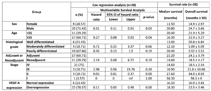
Additionally, an independent multivariable Cox proportional hazards analysis was performed for IL-8 expression in a separate cohort. As shown in Table 4, a total of 28 patients with complete data were included in this analysis. In this group, IL-8 overexpression was found to be a significant independent predictor of survival (p = 0.003), with a hazard ratio (HR) of 0.11 (95% CI: 0.03–0.48). Furthermore, other clinical factors also showed significant prognostic value, including Sex (HR: 0.31, p = 0.032), Age (HR: 0.27, p = 0.036), and Stage II disease (HR: 0.05, p = 0.004). The median survival for patients with IL-8 overexpression was 18.30 months, compared to 16.10 months for those with normal expression.
These findings highlight the clinical relevance of VEGF-A expression in predicting survival outcomes in gastric cancer patients and support the potential utility of VEGF-A as a prognostic biomarker for risk stratification and treatment planning.
4. Discussion
Gastric cancer remains a major global health challenge, ranking as the fifth most common malignancy and the fourth leading cause of cancer-related mortality worldwide, with more than one million new cases annually [41]. In endemic regions such as Northern Thailand, diagnosis often occurs at advanced stages, and the lack of robust prognostic biomarkers hampers timely clinical decision-making. Angiogenesis and inflammation are two central processes in gastric tumorigenesis, and vascular endothelial growth factor A (VEGF-A) and interleukin-8 (IL-8) are among the most extensively studied mediators of these pathways [42,43,44].
Our findings demonstrated that VEGF-A was upregulated in only 31.51% of gastric cancer patients in the pre-chemotherapy setting, reflecting considerable inter-patient heterogeneity. This aligns with previous studies showing that VEGF-A overexpression is present in approximately 30–50% of gastric cancer cases and is strongly correlated with advanced disease stage, lymph node involvement, and reduced survival [15,45]. In contrast, IL-8 expression was elevated in over 70% of cases, consistent with its well-established role as a pro-inflammatory cytokine and promoter of angiogenesis, invasion, and metastasis [28,37,46]. Notably, the expression patterns of VEGF-A and IL-8 were not correlated, suggesting independent regulatory mechanisms within gastric tumors. This reinforces the concept of molecular heterogeneity in gastric cancer and highlights the need for individualized biomarker profiling.
In terms of therapeutic implications, our data indicated that chemotherapy did not consistently suppress VEGF-A expression. In some patients, VEGF-A levels were even found to increase following chemotherapy, possibly reflecting an adaptive angiogenic escape mechanism—a phenomenon also reported in colorectal and gastric cancers treated with cytotoxic therapy [37]. Such compensatory VEGF-A upregulation may contribute to chemoresistance and tumor progression. By contrast, IL-8 expression decreased in a subset of patients after chemotherapy and remained suppressed in post-treatment samples, suggesting a differential responsiveness of inflammatory pathways compared to angiogenic signaling.
Multivariable survival analysis confirmed that VEGF-A overexpression was an independent predictor of poor prognosis, with a hazard ratio of 2.09, consistent with prior meta-analyses reporting hazard ratios between 1.5–2.0 [47,48].
The lack of correlation between IL-8 and VEGF-A expression, combined with IL-8’s consistent upregulation in aggressive tumors, suggests that these molecules play distinct but potentially complementary roles in gastric cancer progression. IL-8 may act as a general marker of tumor-promoting inflammation, while VEGF-A functions as a specific driver of angiogenesis in a subset of patients. These findings support the rationale for combinatorial therapeutic strategies—such as anti-VEGF agents alongside IL-8 or CXCR2 inhibitors—to target both angiogenic and inflammatory pathways simultaneously [49,50].
Despite these promising insights, the study has several limitations. Due to patient mortality and logistical constraints, matched pre- and post-chemotherapy samples were not obtained in all cases, limiting the power of longitudinal comparisons. Additionally, none of the patients in this cohort received VEGF-targeted agents, preventing direct evaluation of predictive biomarker utility. Future studies should integrate VEGF/IL-8 profiling with anti-angiogenic or immunomodulatory therapies to assess treatment response. Moreover, molecular features such as microsatellite instability, EBV status, and HER2 amplification were not assessed in this study; these could provide more clinical data and additional biological profile of VEGF-A and IL-8 regulation in advanced gastric cancer patients
Nonetheless, this study contributes novel data from a previously underrepresented population and underscores the importance of molecular profiling in gastric cancer. Our findings reinforce VEGF-A expression as a potential prognostic biomarker and provide rationale for its integration into clinical decision-making, particularly for identifying patients who may be advantage from anti-angiogenic therapy.
5. Conclusions
In conclusion, this study highlights substantial heterogeneity in VEGF-A and IL-8 gene expression among gastric cancer patients in Northern Thailand. While VEGF-A was upregulated in a subset of tumors and was not consistently affected by chemotherapy, its overexpression was significantly associated with poorer survival outcomes, indicating its potential role as a prognostic biomarker. In contrast, IL-8 was more frequently upregulated and appeared partially responsive to chemotherapy, suggesting differential regulation of inflammatory and angiogenic pathways in gastric cancer. These findings support the importance of molecular profiling to identify patients who may benefit from anti-angiogenic therapy. Future prospective studies with larger cohorts and incorporation of VEGF-targeted treatments are warranted to validate VEGF-A as a clinically actionable biomarker and to further clarify its role in guiding personalized therapeutic strategies for gastric cancer.
Funding
This research was partially supported by the Faculty of Medicine, Chiang Mai University, for the clinical and basic research of the Gastric Cancer group, research funding number SUR-2558-03015, and partially supported by the Clinical Surgical Research Center, Chiang Mai University (CSRC), Chiang Mai University.
Institutional Review Board Statement
This research was approved by the Research Ethics Committee of the Faculty of Medicine, Chiang Mai University (Reference No. SUR-2558-03015). Informed consent was obtained from all participants prior to their inclusion in the study.
Informed Consent Statement
Informed consent was obtained from all subjects involved in the study.
Data Availability Statement
The data presented in this study are available on request from the corresponding author due to ethical reasons.
Acknowledgments
This research work was partially supported by Clinical Surgical Research Center, Chiang Mai University (CSRC), Chiang Mai University.
Conflicts of Interest
The authors declare no conflicts of interest.
Abbreviations
The following abbreviations are used in this manuscript:
| GC | Gastric cancer |
| IL-8 | Interleukin-8 |
| VEGF-A | Vascular endothelial growth factor A |
References
- Sung, H.; Ferlay, J.; Siegel, R.L.; Laversanne, M.; Soerjomataram, I.; Jemal, A.; Bray, F. Global Cancer Statistics 2020: GLOBOCAN Estimates of Incidence and Mortality Worldwide for 36 Cancers in 185 Countries. CA Cancer J. Clin. 2021, 71, 209–249. [Google Scholar] [CrossRef] [PubMed]
- Shin, W.S.; Xie, F.; Chen, B.; Yu, P.; Yu, J.; To, K.F.; Kang, W. Updated Epidemiology of Gastric Cancer in Asia: Decreased Incidence but Still a Big Challenge. Cancers 2023, 15. [Google Scholar] [CrossRef] [PubMed]
- Ilic, M.; Ilic, I. Epidemiology of stomach cancer. World J. Gastroenterol. 2022, 28, 1187–1203. [Google Scholar] [CrossRef] [PubMed]
- Zaidi, S.F. Helicobacter pylori associated Asian enigma: Does diet deserve distinction? World J. Gastrointest. Oncol. 2016, 8, 341–350. [Google Scholar] [CrossRef] [PubMed]
- Subsomwong, P.; Miftahussurur, M.; Uchida, T.; Vilaichone, R.-k.; Ratanachu-ek, T.; Mahachai, V.; Yamaoka, Y. Prevalence, risk factors, and virulence genes of Helicobacter pylori among dyspeptic patients in two different gastric cancer risk regions of Thailand. PLoS ONE 2017, 12, e0187113. [Google Scholar] [CrossRef] [PubMed]
- Suwanrungruang, K.; Sriamporn, S.; Wiangnon, S.; Rangsrikajee, D.; Sookprasert, A.; Thipsuntornsak, N.; Satitvipawee, P.; Poomphakwaen, K.; Tokudome, S. Lifestyle-related risk factors for stomach cancer in northeast Thailand. Asian Pac. J. Cancer Prev. 2008, 9, 71–75. [Google Scholar] [PubMed]
- Rocco, A.; Nardone, G. Diet, H pylori infection and gastric cancer: evidence and controversies. World J. Gastroenterol. 2007, 13, 2901–2912. [Google Scholar] [CrossRef] [PubMed]
- Guan, W.-L.; He, Y.; Xu, R.-H. Gastric cancer treatment: recent progress and future perspectives. J. Hematol. Oncol. 2023, 16, 57. [Google Scholar] [CrossRef] [PubMed]
- Zhang, H.; Yang, W.; Tan, X.; He, W.; Zhao, L.; Liu, H.; Li, G. Long-term relative survival of patients with gastric cancer from a large-scale cohort: a period-analysis. BMC Cancer 2024, 24, 1420. [Google Scholar] [CrossRef] [PubMed]
- Yang, S.; Fang, Y.; Ma, Y.; Wang, F.; Wang, Y.; Jia, J.; Yang, Y.; Sun, W.; Zhou, Q.; Li, Z. Angiogenesis and targeted therapy in the tumour microenvironment: From basic to clinical practice. Clin. Transl. Med. 2025, 15, e70313. [Google Scholar] [CrossRef] [PubMed]
- Ramjiawan, R.R.; Griffioen, A.W.; Duda, D.G. Anti-angiogenesis for cancer revisited: Is there a role for combinations with immunotherapy? Angiogenesis 2017, 20, 185–204. [Google Scholar] [CrossRef] [PubMed]
- Dianat-Moghadam, H.; Nedaeinia, R.; Keshavarz, M.; Azizi, M.; Kazemi, M.; Salehi, R. Immunotherapies targeting tumor vasculature: challenges and opportunities. Front Immunol. 2023, 14, 1226360. [Google Scholar] [CrossRef] [PubMed]
- Saman, H.; Raza, S.S.; Uddin, S.; Rasul, K. Inducing Angiogenesis, a Key Step in Cancer Vascularization, and Treatment Approaches. Cancers 2020, 12. [Google Scholar] [CrossRef] [PubMed]
- Nishida, N.; Yano, H.; Nishida, T.; Kamura, T.; Kojiro, M. Angiogenesis in cancer. Vasc. Health Risk Manag 2006, 2, 213–219. [Google Scholar] [CrossRef] [PubMed]
- Wei, B.; Tai, Y.; Tong, H.; Wen, S.L.; Tang, S.H.; Huan, H.; Huang, Z.Y.; Liu, R.; Tang, Y.M.; Yang, J.H.; et al. Correlations between VEGF-A expression and prognosis in patients with gastric adenocarcinoma. Int. J. Clin. Exp. Pathol. 2017, 10, 8461–8469. [Google Scholar] [PubMed]
- do Espírito Santo, G.F.; Galera, B.B.; Duarte, E.C.; Chen, E.S.; Azis, L.; Damazo, A.S.; Saba, G.T.; de Sousa Gehrke, F.; Guerreiro da Silva, I.D.; Waisberg, J. Prognostic significance of vascular endothelial growth factor polymorphisms in colorectal cancer patients. World J. Gastrointest. Oncol. 2017, 9, 78–86. [Google Scholar] [CrossRef] [PubMed]
- Macedo, F.; Ladeira, K.; Longatto-Filho, A.; Martins, S.F. Gastric Cancer and Angiogenesis: Is VEGF a Useful Biomarker to Assess Progression and Remission? J. Gastric Cancer 2017, 17, 1–10. [Google Scholar] [CrossRef] [PubMed]
- Karayiannakis, A.J.; Syrigos, K.N.; Polychronidis, A.; Zbar, A.; Kouraklis, G.; Simopoulos, C.; Karatzas, G. Circulating VEGF levels in the serum of gastric cancer patients: correlation with pathological variables, patient survival, and tumor surgery. Ann. Surg. 2002, 236, 37–42. [Google Scholar] [CrossRef] [PubMed]
- Garcia, J.; Hurwitz, H.I.; Sandler, A.B.; Miles, D.; Coleman, R.L.; Deurloo, R.; Chinot, O.L. Bevacizumab (Avastin®) in cancer treatment: A review of 15 years of clinical experience and future outlook. Cancer Treat. Rev. 2020, 86, 102017. [Google Scholar] [CrossRef] [PubMed]
- Smyth, E.C.; Tarazona, N.; Chau, I. Ramucirumab: targeting angiogenesis in the treatment of gastric cancer. Immunotherapy 2014, 6, 1177–1186. [Google Scholar] [CrossRef] [PubMed]
- Ren, R.; Zhang, Z.; Zhai, S.; Yang, J.; Tusong, B.; Wang, J. Efficacy and safety of ramucirumab for gastric or gastro-esophageal junction adenocarcinoma: a systematic review and meta-analysis. Eur. J. Clin. Pharmacol. 2024, 80, 1697–1714. [Google Scholar] [CrossRef] [PubMed]
- Young, K.; Smyth, E.; Chau, I. Ramucirumab for advanced gastric cancer or gastro-oesophageal junction adenocarcinoma. Ther. Adv. Gastroenterol. 2015, 8, 373–383. [Google Scholar] [CrossRef] [PubMed]
- Yamaguchi, K.; Shimada, Y.; Hironaka, S.; Sugimoto, N.; Komatsu, Y.; Nishina, T.; Omuro, Y.; Tamura, T.; Piao, Y.; Homma, G.; et al. Quality of Life Associated with Ramucirumab Treatment in Patients with Advanced Gastric Cancer in Japan: Exploratory Analysis from the Phase III RAINBOW Trial. Clin. Drug Investig. 2021, 41, 53–64. [Google Scholar] [CrossRef] [PubMed]
- Jitawatanarat, P.; Wee, W. Update on antiangiogenic therapy in colorectal cancer: aflibercept and regorafenib. J. Gastrointest. Oncol. 2013, 4, 231–238. [Google Scholar] [CrossRef] [PubMed]
- Stewart, M.W. Aflibercept (VEGF-TRAP): the next anti-VEGF drug. Inflamm. Allergy Drug Targets 2011, 10, 497–508. [Google Scholar] [CrossRef] [PubMed]
- Chen, J.; Li, T.; Wu, Y.; He, L.; Zhang, L.; Shi, T.; Yi, Z.; Liu, M.; Pang, X. Prognostic significance of vascular endothelial growth factor expression in gastric carcinoma: a meta-analysis. J. Cancer Res. Clin. Oncol. 2011, 137, 1799–1812. [Google Scholar] [CrossRef] [PubMed]
- Meemon, N.; Paek, S.C.; Pradubmook Sherer, P.; Keetawattananon, W.; Marohabutr, T. Transnational Mobility and Utilization of Health Services in Northern Thailand: Implications and Challenges for Border Public Health Facilities. J. Prim. Care Community Health 2021, 12, 21501327211053740. [Google Scholar] [CrossRef] [PubMed]
- Meier, C.; Brieger, A. The role of IL-8 in cancer development and its impact on immunotherapy resistance. Eur. J. Cancer 2025, 218, 115267. [Google Scholar] [CrossRef] [PubMed]
- Zhao, Z.; Wang, S.; Lin, Y.; Miao, Y.; Zeng, Y.; Nie, Y.; Guo, P.; Jiang, G.; Wu, J. Epithelial-mesenchymal transition in cancer: Role of the IL-8/IL-8R axis. Oncol. Lett. 2017, 13, 4577–4584. [Google Scholar] [PubMed]
- Li, X.; Zhai, J.; Shen, Y.; Zhang, T.; Wang, Y.; He, Y.; You, Q.; Shen, L. Tumor-derived IL-8 facilitates lymph node metastasis of gastric cancer via PD-1 up-regulation in CD8(+) T cells. Cancer Immunol. Immunother. 2022, 71, 3057–3070. [Google Scholar] [CrossRef] [PubMed]
- Shi, J.; Li, Y.J.; Yan, B.; Wei, P.K. Interleukin-8: A potent promoter of human lymphatic endothelial cell growth in gastric cancer. Oncol. Rep. 2015, 33, 2703–2710. [Google Scholar] [CrossRef] [PubMed]
- Petreaca, M.L.; Yao, M.; Liu, Y.; Defea, K.; Martins-Green, M. Transactivation of vascular endothelial growth factor receptor-2 by interleukin-8 (IL-8/CXCL8) is required for IL-8/CXCL8-induced endothelial permeability. Mol. Biol. Cell 2007, 18, 5014–5023. [Google Scholar] [CrossRef] [PubMed]
- Kido, S.; Kitadai, Y.; Hattori, N.; Haruma, K.; Kido, T.; Ohta, M.; Tanaka, S.; Yoshihara, M.; Sumii, K.; Ohmoto, Y.; et al. Interleukin 8 and vascular endothelial growth factor -- prognostic factors in human gastric carcinomas? Eur. J. Cancer 2001, 37, 1482–1487. [Google Scholar] [CrossRef] [PubMed]
- Han, L.; Yuan, Y.; Feng, Y.; Li, X. Association of Serum Interleukin-6 and Interleukin-8 Levels with Clinical Benefit from Immune Checkpoint Inhibitors in Patients with Advanced Gastric Cancer. EJMO 2023, 7. [Google Scholar] [CrossRef]
- Yamada, S.; Kato, S.; Matsuhisa, T.; Makonkawkeyoon, L.; Yoshida, M.; Chakrabandhu, T.; Lertprasertsuk, N.; Suttharat, P.; Chakrabandhu, B.; Nishiumi, S.; et al. Predominant mucosal IL-8 mRNA expression in non-cagA Thais is risk for gastric cancer. World J. Gastroenterol. 2013, 19, 2941–2949. [Google Scholar] [CrossRef] [PubMed]
- Chongruksut, W.; Limpakan Yamada, S.; Chakrabandhu, B.; Ruengorn, C.; Nanta, S. Correlation of Helicobacter pylori and interleukin-8 mRNA expression in high risk gastric cancer population prediction. World J. Gastrointest. Oncol. 2016, 8, 215–221. [Google Scholar] [CrossRef] [PubMed]
- Shi, J.; Wei, P.K. Interleukin-8: A potent promoter of angiogenesis in gastric cancer. Oncol. Lett. 2016, 11, 1043–1050. [Google Scholar] [CrossRef] [PubMed]
- Ma, Y.; Fu, Y.; Fan, X.; Ji, Q.; Duan, X.; Wang, Y.; Zhang, Y.; Wang, Z.; Hao, H. FAK/IL-8 axis promotes the proliferation and migration of gastric cancer cells. Gastric Cancer 2023, 26, 528–541. [Google Scholar] [CrossRef] [PubMed]
- Limpakan, S.; Wongsirisin, P.; Yodkeeree, S.; Chakrabandhu, B.; Chongruksut, W.; Limtrakul, P. Interleukin-8 associated with chemosensitivity and poor chemotherapeutic response to gastric cancer. J. Gastrointest. Oncol. 2019, 10, 1120–1132. [Google Scholar] [CrossRef]
- Jiang, H.; Cui, J.; Chu, H.; Xu, T.; Xie, M.; Jing, X.; Xu, J.; Zhou, J.; Shu, Y. Targeting IL8 as a sequential therapy strategy to overcome chemotherapy resistance in advanced gastric cancer. Cell Death Discov. 2022, 8, 235. [Google Scholar] [CrossRef] [PubMed]
- Mamun, T.I.; Younus, S.; Rahman, M.H. Gastric cancer—Epidemiology, modifiable and non-modifiable risk factors, challenges and opportunities: An updated review. Cancer Treat. Res. Commun. 2024, 41, 100845. [Google Scholar] [CrossRef] [PubMed]
- Kitadai, Y. Angiogenesis and lymphangiogenesis of gastric cancer. J. Oncol. 2010, 2010, 468725. [Google Scholar] [CrossRef] [PubMed]
- Ghalehbandi, S.; Yuzugulen, J.; Pranjol, M.Z.I.; Pourgholami, M.H. The role of VEGF in cancer-induced angiogenesis and research progress of drugs targeting VEGF. Eur. J. Pharmacol. 2023, 949, 175586. [Google Scholar] [CrossRef] [PubMed]
- Song, J.H.; Kim, S.G.; Jung, S.A.; Lee, M.K.; Jung, H.C.; Song, I.S. The interleukin-8-251 AA genotype is associated with angiogenesis in gastric carcinogenesis in Helicobacter pylori-infected Koreans. Cytokine 2010, 51, 158–165. [Google Scholar] [CrossRef] [PubMed]
- Yang, Y.; Cao, Y. The impact of VEGF on cancer metastasis and systemic disease. Semin. Cancer Biol. 2022, 86, 251–261. [Google Scholar] [CrossRef] [PubMed]
- Kim, S.J.; Uehara, H.; Karashima, T.; McCarty, M.; Shih, N.; Fidler, I.J. Expression of interleukin-8 correlates with angiogenesis, tumorigenicity, and metastasis of human prostate cancer cells implanted orthotopically in nude mice. Neoplasia 2001, 3, 33–42. [Google Scholar] [CrossRef] [PubMed]
- Peng, L.; Zhan, P.; Zhou, Y.; Fang, W.; Zhao, P.; Zheng, Y.; Xu, N. Prognostic significance of vascular endothelial growth factor immunohistochemical expression in gastric cancer: a meta-analysis. Mol. Biol. Rep. 2012, 39, 9473–9484. [Google Scholar] [CrossRef] [PubMed]
- Zhan, P.; Ji, Y.N.; Yu, L.K. VEGF is associated with the poor survival of patients with prostate cancer: a meta-analysis. Transl. Androl. Urol. 2013, 2, 99–105. [Google Scholar] [CrossRef] [PubMed]
- Lugano, R.; Ramachandran, M.; Dimberg, A. Tumor angiogenesis: causes, consequences, challenges and opportunities. Cell Mol. Life Sci. 2020, 77, 1745–1770. [Google Scholar] [CrossRef] [PubMed]
- Martin, D.; Galisteo, R.; Gutkind, J.S. CXCL8/IL8 stimulates vascular endothelial growth factor (VEGF) expression and the autocrine activation of VEGFR2 in endothelial cells by activating NFκB through the CBM (Carma3/Bcl10/Malt1) complex. J. Biol. Chem. 2009, 284, 6038–6042. [Google Scholar] [PubMed]
Figure 1.
The expression of (A) IL-8 (N=52) and (B) VEGF-A (N=73) in the pre-chemotherapy group. “Up” means upregulation, “ down “ means downregulation, and “no” means no expression, which was log2 less than 1 in both normal adjacent tissue and tumor tissue.
Figure 1.
The expression of (A) IL-8 (N=52) and (B) VEGF-A (N=73) in the pre-chemotherapy group. “Up” means upregulation, “ down “ means downregulation, and “no” means no expression, which was log2 less than 1 in both normal adjacent tissue and tumor tissue.
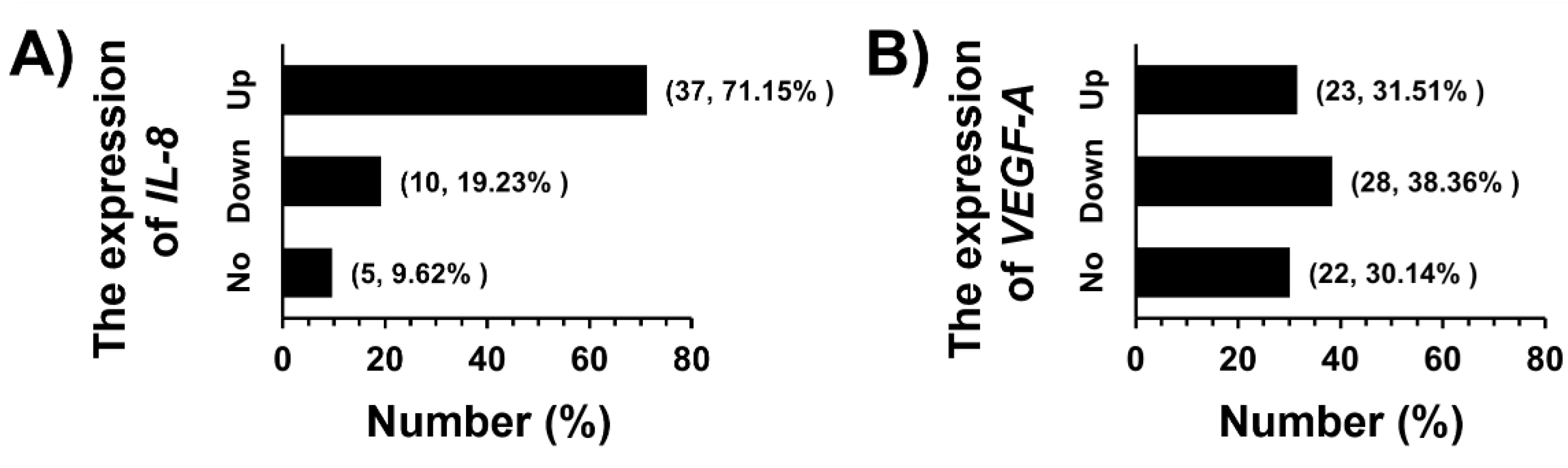
Figure 2.
The relationship between IL-8 and VEGF-A mRNA expression in the pre-chemotherapy group (N=48) “Up” means upregulation, “ down “ means downregulation, and “no” means no expression, which was log2 less than 1 in both normal adjacent tissue and tumor tissue.
Figure 2.
The relationship between IL-8 and VEGF-A mRNA expression in the pre-chemotherapy group (N=48) “Up” means upregulation, “ down “ means downregulation, and “no” means no expression, which was log2 less than 1 in both normal adjacent tissue and tumor tissue.
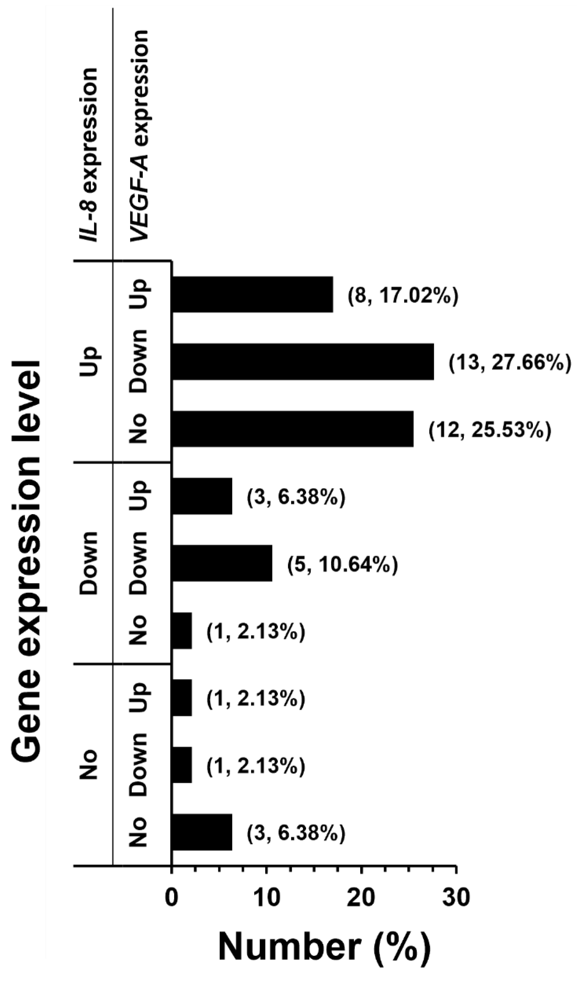
Figure 3.
The expression of (A) IL-8 (N=6) and (B) VEGF-A (N=9) compare between pre- and post-chemotherapy conditions. “Up” means upregulation, “ down “ means downregulation, and “no” means no ex-pression, which was log2 less than 1 in both normal adjacent tissue and tumor tissue.
Figure 3.
The expression of (A) IL-8 (N=6) and (B) VEGF-A (N=9) compare between pre- and post-chemotherapy conditions. “Up” means upregulation, “ down “ means downregulation, and “no” means no ex-pression, which was log2 less than 1 in both normal adjacent tissue and tumor tissue.
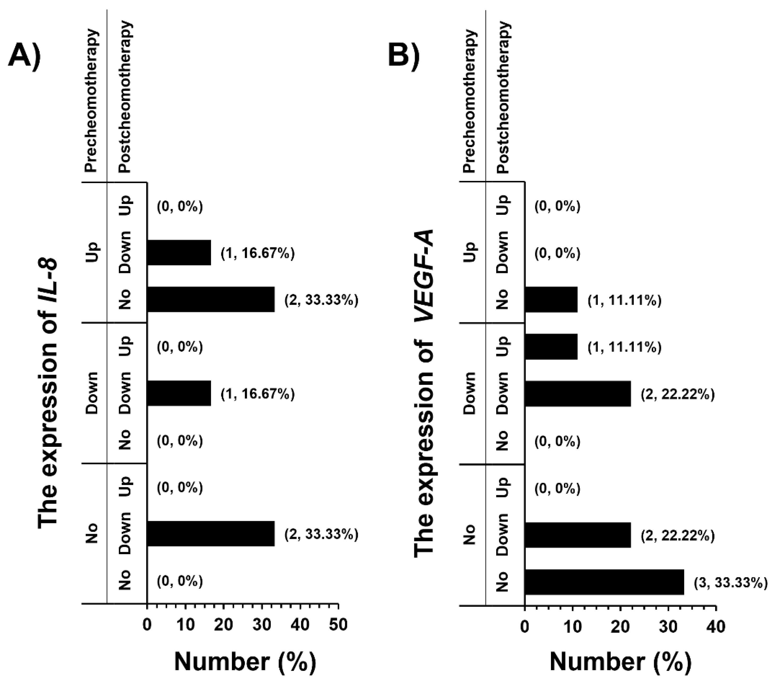

Table 1.
Clinical characteristics of gastric cancer patients.
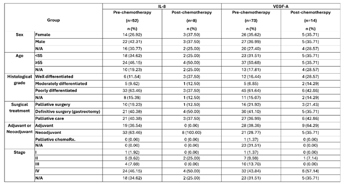 |
Table 2.
Multivariate cox regression analysis and survival rate of VEGF-A expression profile (N=36).
Table 2.
Multivariate cox regression analysis and survival rate of VEGF-A expression profile (N=36).
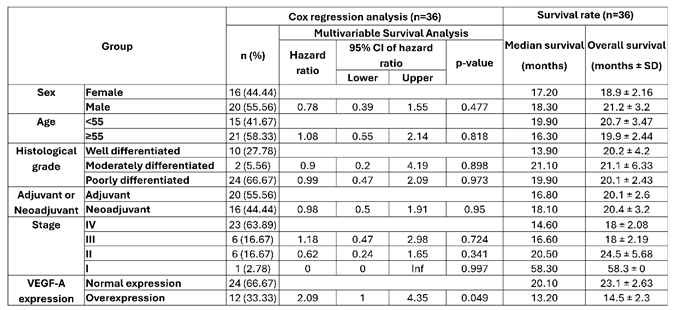 |
Table 3.
Multivariate cox regression analysis and survival rate of IL-8 expression profile subgroup from 36 VEGF-A expression samples (N=19).
Table 3.
Multivariate cox regression analysis and survival rate of IL-8 expression profile subgroup from 36 VEGF-A expression samples (N=19).
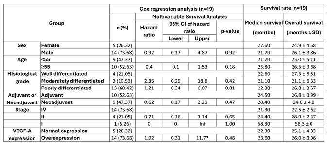 |
Table 4.
Multivariate cox regression analysis and survival rate of IL-8 expression profile (N=28).
 |
Disclaimer/Publisher’s Note: The statements, opinions and data contained in all publications are solely those of the individual author(s) and contributor(s) and not of MDPI and/or the editor(s). MDPI and/or the editor(s) disclaim responsibility for any injury to people or property resulting from any ideas, methods, instructions or products referred to in the content. |
© 2026 by the authors. Licensee MDPI, Basel, Switzerland. This article is an open access article distributed under the terms and conditions of the Creative Commons Attribution (CC BY) license.
Copyright: This open access article is published under a Creative Commons CC BY 4.0 license, which permit the free download, distribution, and reuse, provided that the author and preprint are cited in any reuse.


