

Submitted:
27 June 2026
Posted:
02 July 2026
You are already at the latest version
Abstract
Introduction: Children are particularly vulnerable to foot burns due to slower reactions to danger, which increase the likelihood of deep burns. Early surgical intervention is frequently hindered by significant blood loss during excision. Material and methods: This retrospective study analyzed outcomes for 59 pediatric patients treated for deep burns between 2015 and 2022 at Samarkand Centre of Emergency Medical Care, Uzbekistan. Patients were divided into two groups based on the timing of surgical intervention. Group 1 (21 patients) received early excision and skin grafting under a tourniquet within 3–4 days of injury. Group 2 (38 patients) underwent traditional treatment, including delayed excision and grafting. Results: Early intervention reduced blood loss, shortened operative time, minimized postoperative complications, and improved cosmetic outcomes. Hospital stays averaged 10 days for Group 1, compared to 25 days for Group 2. Early surgical treatment under a tourniquet is effective in enhancing recovery and reducing complications with deep foot burns. Conclusions: The procedures helped to improve the general condition of patients, shortened their stay in hospital and also reduced expenses and lessened joint deformities.
Keywords:
burns
; foot
; tourniquet
; treatment
Introduction
Both extensive burns and those of functionally important structures are difficult to treat in the pediatric age group.[1] Children are an at-risk group for burns to the feet. The thinner skin of children, along with their inability to react appropriately to remove themselves from danger, makes them more susceptible to deeper burns.[2] Deep burns of the feet are the most common cause of skin deformities that affect the functions of the entire lower extremity and may cause spinal column and pelvis distortion and other structural abnormalities.[3] Infants rolling into unprotected fires or explosions of poorly made stoves are the most common causes of these burns in developing countries. It is also common for neurotrophic patients to suffer from hot water immersion burns and direct thermal injury from heating pads.[4,5] In Uzbekistan, sandal burns are of special interest. Sandal burns are common, particularly in children aged 6 months to 3 years. In Central Asia, and particularly in Uzbekistan, many episodes of burns take place at homes because of using sandal heaters. Despite improvements in living conditions and public education efforts, the traditional device, a sandal, containing live coals, is still used for heating the lower part of the human body. Cases of sandal burns are more frequent during winter. Most of the patients with sandal burns have upper- or lower-limb injuries. Most of the patients with sandal burns have upper- or lower-limb injuries.[6]
Sandal burns are a pervasive pediatric public health problem and a leading cause of deep burn injuries to the foot in Uzbekistan.[7,8,9] In most cases, operative treatment of deep burns involves two main types of operations. The first involves the removal of necrotic tissue (escharectomy). The second type involves the restoration of the lost skin. Significant blood loss during escharectomy is one of the factors limiting the opportunity for early operative treatment. [10,11] The anatomical and functional specifics of the foot and ankle joint, marked by the complexity of its structure and the absence of protective adipose and muscular tissue, result in contracture with injury to the deep foot structure after burn trauma in children.[12]
The tourniquet was introduced by Jean Louis Petit in 1718 [13]. Post-operative blood loss in lower extremities can reach about 1000-1500 ml, so this device became irreplaceable for stopping the flow of blood in extremity surgery. Applying a tourniquet decreases blood loss during the operation and provides better visualization of anatomic structures. [14]
In deep burns of the feet, during the excision of necrotic tissue, blood loss of 1-3 ml per square centimeter of the injured surface is possible, and often blood loss reaches 400-500 ml [15]. The following methods of blood loss reduction are known: hemodilution, electrocoagulation, and physical factors. Decrease of intraoperative blood loss can be achieved through previous infiltration of tissues with adrenaline solutions. Treatments for patients with deep foot burns are done through measures which are directed at correcting the general state, addressing joint inflammation and edema, providing adequate wound healing in the burn zone, selective amputation, and preventing contractures. The objective of our work was to improve early escharectomy, decrease intraoperative blood loss performed under the tourniquet, shorten the operative time, reduce complications during surgery, and improve cosmetic results in deep foot burns.
Material and Methods
A retrospective evaluation of patients with severe foot burns who underwent treatment between 2015 and 2022 was conducted. This study received approval from the Samarkand Centre of Emergency Medical Care (RCSUMA) and the Inter-Regional Burn Centre, Samarkand, Uzbekistan. These centers admit patients from the Samarkand region, with a population of more than 2 million. When the cases of burn injuries were examined, the most frequent were sandal burns in 28 (47.5%); scalds in eight (13.6%); flame burns in seven (11.9%); hot ash in seven (11.9%); chemicals in three (5.1%); and others in six (10.2%) (Table 1).
Of these, there were 14 patients younger than 1 year of age, 29 patients aged 1-3 years, eight patients aged 3-5 years, and eight patients over 5 years of age (Table 2).
Most burns were classified as sandal burns, i.e., 28 (47.5%). The patients had severe foot burns caused by sandal heaters. In these cases, the sandal burns caused especially deep and severe injuries to tissue because of the immediate contact with the burning agent. Most of the sandal burns were circular foot burns.
In accordance with the aim of the investigation, 59 patients with isolated deep burns of the foot were divided into two groups, depending on the timing of surgery. The first group (tourniquet group) underwent operation after initial stabilization. The group included 21 patients, admitted during the first 3 days postburn and before burn wound suppuration. After burn resuscitation, early excision under a tourniquet was performed on no more than 4–5% of the body surface area. A tourniquet was applied under general anesthesia on the lower third of the ankle proximal to the burn wounds and was removed before grafting (Figure 1).
The depth of necrotic tissue excised depended on the burn degree: for IIIB, within hypodermic-adipose tissue, and for IV degree, to fascia or by excision of deeper layers of soft tissue.
We determined intraoperative blood loss by the mass of blood-soaked dry sponges. The resulting value was increased by 50% and summed with the volume of blood in the electric suction bank.
After early excision, we immediately placed meshed or non-meshed skin grafts. The thickness of skin grafts ranged from 0.2 to 0.3 mm. Grafts from the external and internal surfaces of the thigh were normally used. After that, the wound was tightly bandaged in 2-3 stages with the aid of coarse gauze impregnated with polyhexamethylene biguanide (Kerlix AMD). Then, a negative-pressure apparatus was applied (Suprasorb Easy Dress, Lohmann & Rauscher, UK) suitable to the size of the extremity and its location. A constant negative pressure of 90-110 mmHg was applied. On the second or third day, the vacuum system was removed, and water-drying aseptic dressings were applied, with dressing changes every other day. Engraftment, as a rule, was noted on the fifth to seventh day (Figure 2).
The second group (control group) included 38 patients who were treated traditionally at the Samarkand Inter-Regional Burn Centre. They underwent daily dressing with topical antiseptics (povidone-iodine) and, in severe cases, silver sulfadiazine and net dressings containing silver. The following surgical method of treatment was carried out: escharotomy, to prevent secondary necrosis and accelerate the rejection of necrotic tissue. This surgical procedure was performed in a longitudinal direction down to the healthy tissue. Then the wound was covered with chemotherapeutic material (trypsin and chymotrypsin) that promoted quicker removal of necrotic tissue.
The patients underwent dressing changes every other day. After the first removal of necrotic tissues, another escharectomy was performed, taking in the whole depth of the necrosis. Full escharectomy was usually performed 10-14 days postburn. Excision was accompanied by significant blood loss, approximately 1 ml per 1 cm2, totaling about 450-500 ml. When the wound was completely ready for closure, skin grafting was performed. Skin grafting was conducted 18-22 days postburn. Graft thickness ranged from 0.2-0.3 mm. The external and internal surfaces of the thigh were usually used as grafts (Figure 3).
Results
The first group underwent early excision and skin grafting under a tourniquet, involving 21 patients, which made it possible to accelerate the operation. Bleeding from the wound was observed at 1-3 ml per 1 cm2, requiring hemostasis by local methods. This approach prevented the development of early infectious complications such as suppuration, hematoma, and sequestration, and covered a short recovery period of 8–10 days for healing
A second operation was not required in these patients. As a rule, healing coincided with the patients’ discharge from the hospital at 10.1 ± 3.2 days.
The second group, consisting of 38 patients treated according to traditional methods, was expected to recover within 20–22 days. In our observations, blood loss of 400–450 ml was noted before the preparation of the wound for skin grafting. The second operation was performed on seven patients. The causes of transplant non-closure were suppuration of injuries, hypoproteinemia, exhaustion, and the use of widely meshed skin grafts.
Patients stayed in the hospital on average for 25±30 days. Complete healing in the control group provided ‘closure’ of cells because meshed skin grafts were used for these patients. After the restoration of the skin and discharge from the burn department, patients were recommended to wear a special shoe for 3-4 months with periodic removal during the day for therapeutic gymnastics and massage.
Analysis of the near-term results of the first group showed that 3 out of 21 patients returned to the hospital for repeat surgery due to loss of mobility in their extremities. These patients did not follow the prescribed recommendations in the postoperative period (physical therapy, physiotherapy, massage, restriction of physical activity on the foot, etc.). The second group showed that 11 out of 38 patients returned for repeat surgery because of the growth and retraction of skin grafts and foot contractures of I-II-III degree. Analysis of the follow-up results in the group showed that in children, foot contractures of I-II-III degree developed due to the growth and retraction of transplants that required planned operative treatment; in five children, long courses (8-12 months) of conservative treatment were carried out. Comparative analysis of the present and future treatment results of patients with different kinds of burns is presented in Table 3.
Discussion
Children’s burn injuries are considered a serious problem worldwide. They most often occur in children under the age of 16, particularly under the age of 5. [16] In cases in which burn injuries are not severe, admission to the hospital is not necessary. However, in severe cases, patients need treatment in burn centers under the care of specialists who have experience in managing patients with burns. Lethal outcomes are less than 3% in this highly specialized centers.[17]
Children with foot burns are an at-risk group. The skin of children is more vulnerable, and in addition, they are not able to assess situations that result in deeper burns.[18] It has been reported that isolated foot burns are caused by scalds in the UK and Netherlands.[19] In Uzbekistan, sandal burns are common, particularly children aged 6 months to 3 years. Cases of sandal burns are more frequent during winter when people in distant mountain regions use inappropriate heating systems such as the sandal. Most patients with sandal burns have upper- or lower-limb injuries.[20]
There is need to improve early escharectomy, decrease intraoperative blood loss performed under the tourniquet and shorten the operative time.
The first attempt to excise the wound in deep burns was carried out by Reverden in 1869.[21] In addition to excision, some authors began to perform primary skin grafting with free skin grafts in the early stages. [22] Along with that, engraftment takes place in aseptic conditions in contrast to staged surgical management; this leads to better functional results of treatment. Restoration of skin integrity by means of primary excision with simultaneous skin grafting in deep burns decreases the frequency of development of hypertrophic and keloid scars.[23] Patients with deep foot burns are treated with immediate hospital admission, special treatment, and timely preparation of the injured site for skin grafting. Most of our patients had sandal burns. In all cases, we tried to use one graft to cover the foot. Therapeutic gymnastics begins after the assessment of the patient’s condition by the physician or the day after surgery. The main goal of physical exercises is the rehabilitation of foot function. Treatment begins after a thorough examination of the patient’s body by the rehabilitation doctor, the day after admission to the hospital. [24] The main purpose of physical exercises is to rehabilitate foot function, prevent contracture formation, and lower the risk of complications. Physical training must take place 2 or 3 times a day for 30 or 40 minutes, and also after discharge from the hospital up to scar maturation.
Credit Author Statement
BMS developed the study conception. Surgeries, data collection and analysis were performed by BMS who wrote the manuscript. KRT supervised the study and participated in discussion. All authors read and approved the final manuscript.
Ethical Approval
This study received approval from Samarkand Centre of Emergency Medical Care (RCSUMA) and Inter-Regional Burn Centre, Samarkand, Uzbekistan.
Patient Consent Statement
The parents of children provided their written informed consent to participate in this study.
Conflicts of Interest Statement
No conflict of interest to declare.
Funding
Internal resources of the Centre of Emergency Medical Care and Samarkand State Medical University supported the study which had no external funding.
Acknowledgments
We thank Vladimir Tsibulsky, Bobby Shakirov and Nargiza Mavlyanova for their help with the project and the manuscript preparation.
References
- Florence, N.; Bhaveshkumar, P.; Craig, M. Epidemiology of isolated foot burns in children presenting to a Queensland pediatric burns centre— a two-year study in warmer climate. Burn. Trauma 2017, 5, 6. [Google Scholar]
- Krishnamoorthy, V.; Ramaiah, R.; Bhananker, S.M. Pediatric burn injuries. Int. J. Crit. Illn. Inj. Sci. 2012, 2(3), 128–34. [Google Scholar] [CrossRef] [PubMed]
- Asquith, C.; Kimble, R.; Stockton, K. Too hot to trot (barefoot)A study of burns in children caused by sun heated surfaces in Queensland, Australia. Burns 2015, 41, 177–80. [Google Scholar] [PubMed]
- Hennington-Gorse, S.; Pellard, S.; Wilson-Jones, N.; Potokar, T. Foot burns: epidemiology and management. Burns 2007, 33, 1041–5. [Google Scholar] [CrossRef]
- Zachary, L.S.; Heggers, J.P.; Robson, M.C.; Smith, D.J.; Maniker, A.A.; Sachs, R.J. Burns of the feet. J. Burn Care Rehabil. 1987, 8, 192–4. [Google Scholar] [CrossRef] [PubMed]
- Cahill, T.J.; Rode, H.; Millar, A.J. Ashes to ashes: thermal contact burns in children caused by recreational fires. Burns 2008, 34, 1153–7. [Google Scholar] [CrossRef] [PubMed]
- Shakirov, B.M. Deep foot burns: effects of early excision and grafting. Burns 2011, 37, 1435–1438. [Google Scholar] [CrossRef] [PubMed]
- Shakirov, B.M. Sandal Burns and their Treatment in Children. J. Burn Care Rehabil. 2004, 25, 501–505. [Google Scholar] [CrossRef] [PubMed]
- Shakirov, B.M.; Tursunov, B.S.; Tagaev, K.R. Treatment of sandal burns in children (abstract). Presented at the British Trauma Society annual clinical meeting, 12-13 October 2006; p. 0145. [Google Scholar]
- Shakirov, B.M. Treatment of sandal burns of the feet in children in a moist environment. Burns 2014, 40, 520–4. [Google Scholar] [CrossRef] [PubMed]
- Janzekovic, Z. Excision of burns. Burns Burns 1977; 4:61-66. 1977, 4(1), 61–6. [Google Scholar] [CrossRef]
- Cartotto, R.; Musgrave, M.A.; Beveridge, M.; Fish, J.; Gomez, M. Minimizing blood loss in burn surgery. J. Trauma Acute Care Surg. 2000, 49(6), 1034–9. [Google Scholar] [CrossRef]
- Shakirov, B.M. Surgical Techniques used for correction of post burn contractures and deformities of the foot. J. Clin. Res. Foot Ankle. 2021, 9(3), 307–312. [Google Scholar]
- Noordin, S.; McEwen, J.A.; Kragh, C.J., Jr.; Eisen, A.; Masri, B.A. Surgical Tourniquets in Orthopaedics. JBJS 2009, 91(12), 2958–67. [Google Scholar] [CrossRef]
- Tai, T.W.; Chang, C.W.; Lai, K.A.; Lin, C.J.; Yang, C.Y. Effects of Tourniquet use on blood loss and soft-tissue damage in total knee arthroplasty: A randomized controlled trial. JBJS 2012, 94(24), 2209–15. [Google Scholar]
- Bogdanov, S.B. Early surgical treatment of patients with deep burns of the extremities. 16th European Burns Association Congress, Hannover, 2015; p. 213. [Google Scholar]
- Carlsson, A.; Udén, G.; Håkansson, A.; Karlsson, E.D. Burn injuries in small children, a population-based study in Sweden. J. Clin.-Nurs. 2006, 15, 129–34. [Google Scholar] [PubMed]
- Sheridan, R.L.; Remensnyder, J.P.; Schnitzer, J.J.; Schulz, J.T.; Ryan, C.M.; Tompkins, R.G. Current expectations for survival in pediatric burns. Arch. Pediatr. Adolesc. Med. 2000, 154, 245–9. [Google Scholar] [CrossRef] [PubMed]
- Kilic, T.; Krijnen, P.; Tuinebreijer, W.E.; Breederveld, R.S. Epidemiology of foot burns in a Dutch burn centre. Burn. Trauma. 2015, 3, 5. [Google Scholar] [CrossRef]
- Shakirov, B.M. Treatment of severe foot burns in children. Burns 2005, 31(7), 901–5. [Google Scholar] [CrossRef] [PubMed]
- Martin, L.; Rea, S.; McWilliams, T.; Wood, F. Hot ash burns in the children of Western Australia: how and why they happen. Burns 2014, 40, 1030–2. [Google Scholar] [CrossRef] [PubMed]
- Toledo-Pereyra, L.H. Claudius Galenus of Pergamum: surgeon of gladiators. Father of experimental physiology. J. Invest. Surg. 2002, 15(6), 299–301. [Google Scholar] [CrossRef] [PubMed]
- Hendon, D.N. Total burn care, 2nd edition; W.B. Saunders, 2001; p. 670. [Google Scholar]
- Barret, J.P. Burns reconstruction. BMJ 2004, 329(7460), 274–6. [Google Scholar] [CrossRef] [PubMed]
Figure 1.
Tourniquet was applied on the lower third of the ankle.
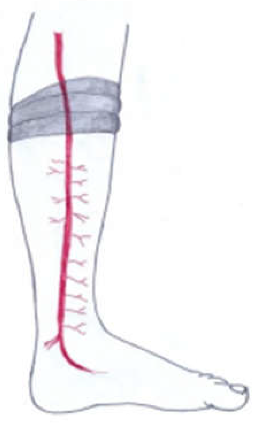
Figure 2.
Patient A., 3 years old, with a IIIB degree thermal burn on the dorsum of the left foot.
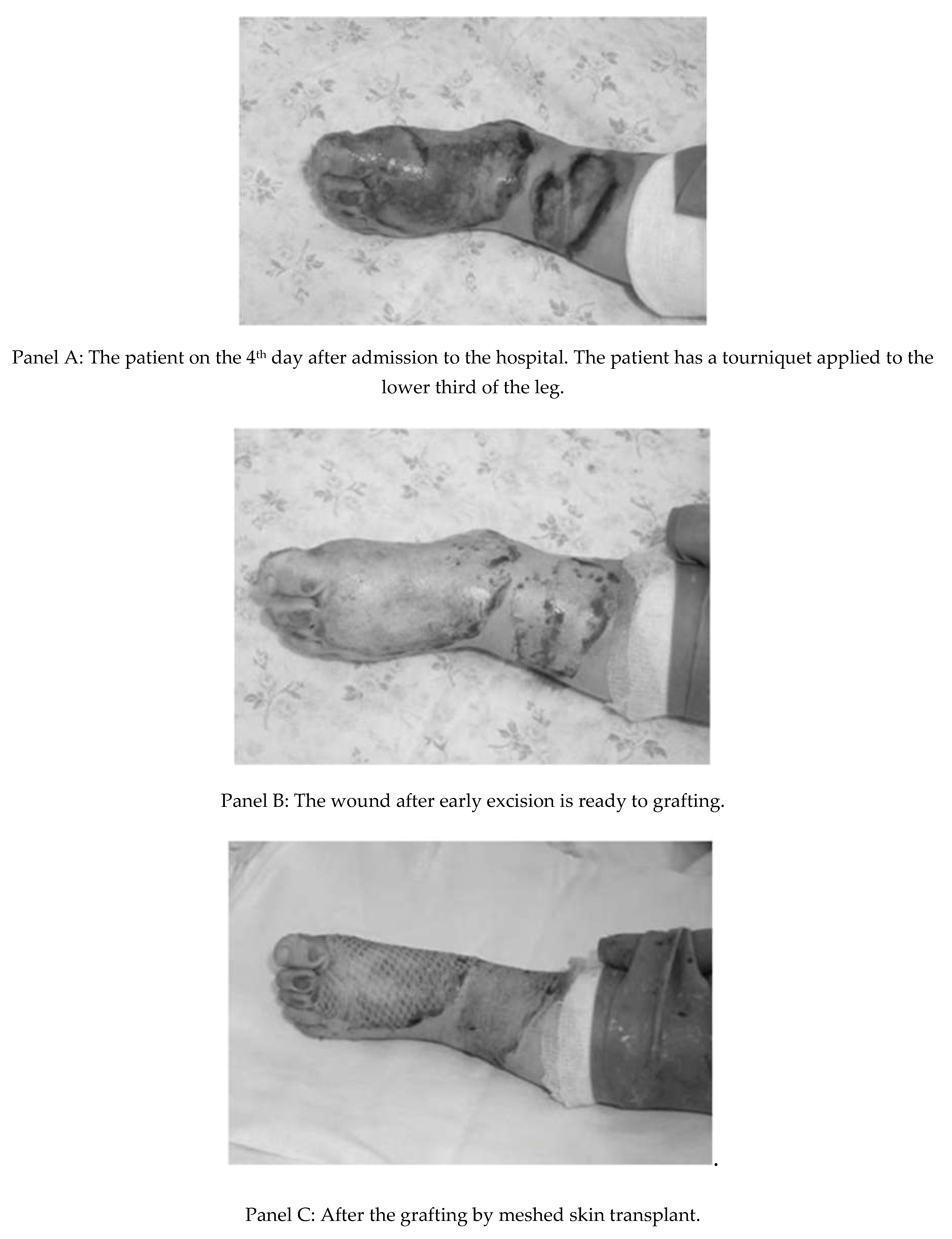
Figure 3.
Patient B., 1.5 years old, with IIIB-IV degree burns of both feet on the 18th day after admission to hospital.
Figure 3.
Patient B., 1.5 years old, with IIIB-IV degree burns of both feet on the 18th day after admission to hospital.
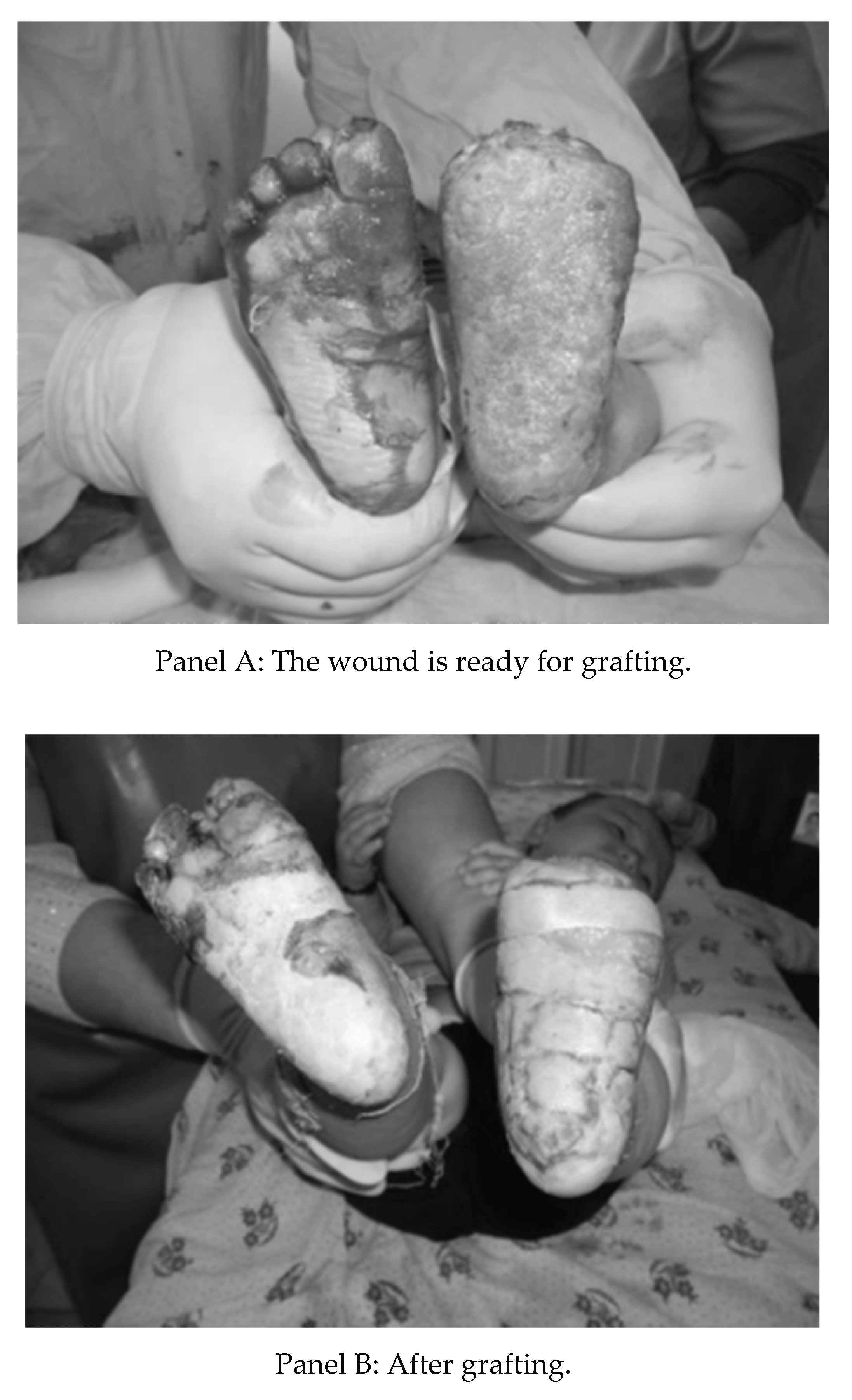

Table 1.
The distribution of patients by different kinds of burns. Fisher Exact test did not show any statistically significant difference between groups.
Table 1.
The distribution of patients by different kinds of burns. Fisher Exact test did not show any statistically significant difference between groups.
| Kind of burns | Main group (n=21) | Second group (n=38) | Total (%) |
|---|---|---|---|
| Sandal burns | 11 | 17 | 28 (47.5) |
| Scalds | 3 | 5 | 8 (13.6) |
| Flame | 2 | 5 | 7 (11.9) |
| Hot ash | 4 | 3 | 7 (11.9) |
| Chemical burns | 0 | 3 | 3 (5.1) |
| Other burns | 1 | 5 | 6 (10.2) |
Table 2.
Distribution of patients by age group. Fisher Exact test did not show any statistically significant difference between groups.
Table 2.
Distribution of patients by age group. Fisher Exact test did not show any statistically significant difference between groups.
| Patients | Main group (n=21) | Second Group (n=38) | Total (%) |
|---|---|---|---|
| Under 1 year | 3 | 11 | 14 (23.7) |
| 1-3 | 12 | 17 | 29 (49.1) |
| 3-5 | 3 | 5 | 8 (13.6) |
| 5-14 | 3 | 5 | 8 (13.6) |
Table 3.
Comparative analysis of the present and future treatment results of patients with different kinds of burns. Note: † - Student t-test between groups, * - Fisher Exact test between groups.
Table 3.
Comparative analysis of the present and future treatment results of patients with different kinds of burns. Note: † - Student t-test between groups, * - Fisher Exact test between groups.
| Measure | Main Group (n=21) | Second Group (n=38) | P value |
|---|---|---|---|
| Bleeding during the surgery (ml) | 25-30 ml | 400-450 ml | < 0.01† |
| Interval between necrotomy and engrafting (days) |
4.7 ± 0.4 | 22.5 ± 1.5 | < 0.01† |
| Wound complications frequency | 0 | 7 (18.4 %) | 0.04* |
| Bed days | 10.1 ± 3.2 | 25 ± 3 | < 0.01† Student t-test between groups. |
| Follow-up complications frequency | 3 (14.3%) | 11 (28.9 %) | 0.34* |
Disclaimer/Publisher’s Note: The statements, opinions and data contained in all publications are solely those of the individual author(s) and contributor(s) and not of MDPI and/or the editor(s). MDPI and/or the editor(s) disclaim responsibility for any injury to people or property resulting from any ideas, methods, instructions or products referred to in the content. |
© 2026 by the authors. Licensee MDPI, Basel, Switzerland. This article is an open access article distributed under the terms and conditions of the Creative Commons Attribution (CC BY) license (http://creativecommons.org/licenses/by/4.0/).
Copyright: This open access article is published under a Creative Commons CC BY 4.0 license, which permit the free download, distribution, and reuse, provided that the author and preprint are cited in any reuse.