

Submitted:
30 June 2026
Posted:
01 July 2026
You are already at the latest version
Abstract
Background/Objectives: Colorectal cancer (CRC) is complicated by cachexia, a wasting syndrome with muscle and fat loss that worsens survival and treatment outcomes. Evidence suggests the gut microbiome may contribute to CRC cachexia, but its role remains unclear. This systematic review synthesized evidence to identify microbial signatures linked to cachexia hallmarks. Methods: The protocol was pre-registered with PROSPERO and following PRISMA guidelines. PubMed, Web of Science, and Scopus were searched for studies on CRC patients and preclinical models. Eligible studies compared microbiota compositions between cachectic and non-cachectic groups to identify alterations linked to cachexia progression. Study quality was assessed using the Newcastle–Ottawa Scale and CAMARADES checklist. Results: Of 2,456 records, 15 studies met inclusion criteria; Study quality was moderate for pre-clinical studies and high for clinical cohort studies. Murine CRC cachexia models showed reduced alpha diversity and beta diversity shifts. Butyrate-producing taxa were depleted, whereas, Enterobacteriaceae and other pathobionts were enriched. Some microbial changes were independent of food intake and linked to inflammation, metabolic dysregulation, and muscle wasting. Clinical evidence was limited to two reports from the same cohort, in which higher pre-surgical abundance of Fusobacterium nucleatum and lower Porphyromonas and Actinomyces spp. were associated with cachexia onset. Conclusions: Overall, current evidence suggests that CRC-associated cachexia is accompanied by gut microbiome alterations, but causality and clinical relevance remain uncertain.
Keywords:
colorectal cancer
; cachexia
; gut microbiota
; microbiome
; muscle wasting
; systemic inflammation
; dysbiosis
1. Introduction
Colorectal cancer (CRC) is a common and life-threatening malignancy of the digestive system, representing a major challenge to global public health [1]. CRC is the third most frequently diagnosed cancer and the second-leading cause of cancer deaths worldwide with over 1.9 million new cases and more than 900,000 deaths in 2022 [2].
CRC is strongly associated with cachexia, a multifactorial syndrome characterized by involuntary weight loss, both skeletal muscle and adipose tissue wasting, systemic inflammation, and metabolic alterations [3,4]. Cachexia affects approximately 50–61% of patients with advanced CRC and is a major contributor to morbidity and mortality in this population, whereas its overall prevalence in early stage CRC patients is substantially lower [3]. This syndrome is associated with reduced tolerance to anticancer therapies, higher risk of treatment side effects, impaired quality of life, and worse overall survival [5]. A recent meta-analysis of gastrointestinal cancers including CRC found a 50% increased risk of death among patients with cachexia compared to non-cachectic patients [6].
Recent research has begun to uncover a connection between cachexia and the gut microbiota, that consist of trillions of microorganisms including bacteria, archaea, viruses, and fungi, playing a crucial role in digestion, immunity, and metabolism [7]. Several previous studies have found that tumor cachexia is characterized by a reduction in beneficial short-chain fatty acid (SCFA)-producing bacteria (such as those from the Lachnospiraceae and Ruminococcaceae families) and by an overgrowth of pro-inflammatory pathobionts, including certain Enterobacteriaceae [8,9].
Furthermore, diverse associations between gut microbes and CRC have been reported. Dysbiosis, an imbalance in the gut microbial ecosystem, has been consistently associated with an increased CRC risk [10]. Pathogenic bacteria such as Fusobacterium nucleatum (Fn), enterotoxigenic Bacteroides fragilis, and colibactin-producing Escherichia coli may contribute to CRC by triggering chronic inflammation, generating genotoxins, and promoting immune evasion. While these associations and experimental findings are compelling, their causal relevance to human CRC initiation and progression have not yet been firmly established. [11,12,13].
Although the gut microbiome has been increasingly studied in relation to CRC development and progression, specific interactions between gut microbes and cachexia in CRC remain underexplored. Therefore, this systematic review aims to synthesize existing evidence on their associations.
2. Materials and Methods
2.1. Objectives
The primary objective of this systematic review was to summarize existing studies on the gut microbiota composition and diversity associated with CRC related cachexia hallmarks: weight loss, inflammation, and metabolism alteration.
2.2. Literature Search Strategy
This systematic review was pre-registered with the International Prospective Register of Systematic Reviews (PROSPERO CRD420251048700). A literature search was conducted using PubMed, Web of Science, and Scopus databases to identify studies correlating the gut microbiome with cachexia in CRC until May 2026. Key words including (“Colorectal cancer” AND “Gut microbiome”) OR (“Gut microbiota” AND “Cachexia”) were searched. Medical Subject Headings (MeSH) terms were used for the database searches (Table S1). This systematic review followed the recommendations of the Preferred Reporting Items for Systematic Reviews and Meta-Analyses (PRISMA) [14].
2.3. Study Selection Criteria
After removing duplicate records, two researchers (MaH, TG) independently screened title and abstract of the studies. Articles were full-text reviewed if their title and abstract pointed to original analyses in cachectic vs non-cachectic groups of either CRC patients or from preclinical studies, including the evaluation of the gut microbiota composition through microbiome analysis approaches (e.g., 16S rRNA gene sequencing, shotgun metagenomics). Studies not included in the systematic review were those lacking a control groups, using inappropriate animal models of CRC cachexia, comparing a treatment group to a cachectic group, as well as articles not published in English.
2.4. Synthesizing of Evidence
The findings from the included studies were synthesized by a narrative and thematic approach, as a meta-analysis was not feasible due to the heterogeneity of results reported and the different microbiome assessment methods. Key study characteristics were extracted and summarized in tabular form, including study design, country of origin, population, sample size, cachexia definition or assessment, and microbiota analysis methods. To synthesize the evidence, we compared microbial changes across studies, focusing on results of frequent shifts of gut microbiota compositions and diversity associated with cachexia in CRC patients and mouse models. We compiled a comparative table that summarizes increases and decreases in specific bacterial taxa and integrates taxonomic shifts at various levels (phylum, family, genus, and species) along with quantitative data (odds ratios, q-values, p-values). The tables served as a core component of our narrative synthesis, allowing for structured comparison across studies.
2.5. Study Quality
For the included pre-clinical studies, quality assessment was conducted by two reviewers independently (MaH, TG) using the CAMARADES checklist (Collaborative Approach to Meta-Analysis and Review of Animal Data from Experimental Studies), a tool specifically designed for evaluating the methodological quality of preclinical studies with a 10 item checklist [15].
Regarding the clinical studies, we used the Newcastle-Ottawa Scale (NOS) for quality assessment across three domains: selection (maximum 4 stars), comparability (maximum 2 stars), and outcome (maximum 3 stars), for a total possible score of 9 stars. Each item within these domains is awarded a star if the study meets predefined criteria, with higher star counts indicating higher quality and lower risk of bias.
3. Results
3.1. Literature Review Results
Comprehensive database searches identified 2456 records (Figure 1), of which 786 duplicate records were excluded, as well as 1649 records that did not meet the inclusion criteria due to studies not clearly defining cachexia, or studies unrelated to CRC by using inappropriate mouse models or not including CRC patients. Full texts were reviewed of all 22 remaining articles, after 7 were excluded (Table S2) [16,17,18,19,20,21,22], 15 studies fulfilled the eligibility criteria.
3.2. Study Selection and Characteristics
Table 1 presents the primary characteristics of 13 eligible preclinical studies investigating gut microbiome in cancer cachexia murine models. Geographically, the studies were conducted across Europe and Asia. The majority of studies were carried out in Belgium [23,24,25,26,27], the United Kingdom [23,26,27], France [23,24], and Finland [28]. In Asia, studies were conducted in China [29,30,31,32,33,34] and Korea [35]. The two clinical studies were conducted in the USA and Germany, respectively [36,37] (Table 2). Across the preclinical studies, gut microbiota composition was predominantly assessed using 16S rRNA gene sequencing [23,28,29,32,35], with several studies also employed quantitative PCR (qPCR) to validate specific bacterial taxa [24,25]. Some studies combined sequencing with metabolomics, including SCFA quantification by Gas Chromatography-Mass Spectrometry (GC-MS) [26,31,34] to assess alterations in microbial metabolism associated with cachexia. In the clinical studies, gut microbiome was also analyzed using 16S rRNA gene sequencing, with additional targeted quantification of Fn by qPCR [36]. These analyses were performed on fecal samples collected pre-surgery in CRC patients, enabling the identification of microbial signatures associated with cachexia, including alterations in alpha-diversity and relative abundances of specific genera [36,37].
All preclinical studies included male mice, with the most frequently used strain being BALB/c [28,29,30,31,33,34,35] and CD2F1 [23,24,25,26,27], while C57BL/6 mice were used in one study [32]. Mouse ages ranged from 5–8 weeks, except for one study who used 6-month-old mice [32]. Cachexia was predominantly induced through the C26 colon carcinoma model, which involves subcutaneous injection of C26 tumor cells. Two studies utilized MC-38 colon cancer cells [27,32] to establish cachexia. The C26/CT26 model is widely recognized as a gold standard for investigating the pathogenesis and therapeutic interventions of cancer cachexia in CRC [38,39].
In the clinical studies, patient populations consisted of CRC patients in stages I–III, and both studies were derived from the same prospective cohort (ColoCare), but differed in their specific research focus and sample sizes. One study included 87 participants [36], whereas the other included103 participants [37]. Cachexia was assessed six months post-surgery using Fearon criteria, defined as >5% body weight loss over the past six months, or a BMI <20 kg/m² with weight loss of >2% (Table 2).
3.3. Gut Microbiota Diversity in CRC Cachexia
Multiple studies using murine models of CRC cachexia reported significant alterations in gut microbiota diversity. Most studies found a significant decrease in alpha diversity metrics (such as Shannon, Chao, and Faith’s phylogenetic diversity indices) in cachectic mice compared to healthy controls, indicating reduced microbial richness and evenness [23,27,30,32,33,34,35]. Contrasting these findings, one study which also used the C26 model, reported a significant increase in alpha diversity [28]. Beta diversity analysis revealed distinct differences in community composition of the microbiota between cachectic and control mice, confirming significant alteration in overall microbiota composition [23,25,30,31,32,35].
Regarding the diversity in the clinical studies, higher pre-surgical gut microbial alpha-diversity was positively associated with cachexia onset at six months post-surgery measured by the Shannon index (odds ratio = 2.00, 95% confidence interval = 1.07, 4.19) and for beta-Diversity, significant differences in overall microbiome composition were observed between cachectic and non-cachectic patients (Bray–Curtis distance, p = 0.01; unweighted UniFrac distance, p = 0.001) [37]. However, the second study did not discuss general microbial diversity metrics and focused exclusively on Fn abundance [36].
3.4. Taxonomic Shifts in Gut Microbiota in CRC Cachexia Models Compared to Healthy Controls
At the phylum level, cachectic mice frequently showed enrichment of Proteobacteria across studies [26,28]. Bacteroidetes were reported to be reduced in several studies, although some studies observed increases in specific genera within this phylum[27,28,30]. Findings for Firmicutes were inconsistent, with reports of both enrichment in three studies [28,29,30] and depletion in two studies [25,26]. Additionally, Deferribacteres and its representative genus Mucispirillum were also found to be increased in cachectic animals [27,28].
At the family level, Enterobacteriaceae showed a consistent expansion in cachectic mice in six studies [23,24,26,27,29,32]. Conversely, families with prominent SCFA-producing members, such as Lachnospiraceae and Ruminococcaceae, were mostly reduced [25,29]); [26,27,31] , although one study reported increased Lachnospiraceae abundance [28]. Unclassified Borkfalkiaceae were reported to be decreased in one study [27]. whereas Eubacteriaceae were increased in another study [29] (Table 3).
At the genus level, increased abundance of Enterococcus and Bacteroides was reported in cachectic mice in some studies [28,30]. Beneficial commensals were mostly depleted, including Intestinimonas[29], Roseburia [34], Faecalibacterium, Butyricicoccus [31], Lachnoclostridium [30], and Prevotella [28]. Blautia was increased in one study [29].
Cachexia was associated with an enrichment of several pathobionts, Escherichia coli[23], Klebsiella oxytoca [26], and Parabacteroides goldsteinii/ASF 519 [23]. Conversely, Lactobacillus johnsonii/gasseri was reported to be reduced [23]. Notably, findings regarding Lactobacillaceae and its representative genus Lactobacillus were inconsistent. Several studies demonstrated increases [28,30], whereas others reported significant decreases [26,29,32,35]. Xylanibacter rodentium showed also a significant decrease in the cachectic group [27] (Table 4).
One clinical study reported that the abundance of Fn in pre-surgical fecal samples was significantly associated with cachexia onset at six months post-surgery: Patients with high fecal Fn abundance exhibited a markedly increased risk of developing cachexia compared to those with negative or low Fn levels (OR = 4.82, 95% CI = 1.15–20.10, p = 0.03) [36]. In a separate investigation relative abundance of Porphyromonas was inversely associated with cachexia onset (OR = 0.51, 95% CI = 0.26–0.89, p = 0.03), with stronger inverse associations observed in males, patients receiving neoadjuvant treatment, those with rectal or stage III tumors, and patients with low dietary fiber intake. For Actinomyces a suggestive inverse association was found with cachexia (OR = 0.72, 95% CI = 0.48–1.03, p = 0.08), particularly among patients younger than 65 years [37] (Table 5).
3.5. Quality Assessment
CAMRADES quality scores ranged from 4 to 6 out of 10 (Table S3), indicating moderate quality. All studies were peer-reviewed, used appropriate models, obtained ethical approval, and disclosed conflicts of interest. Although Randomization was reported in 10 of 15 studies, the lack of allocation concealment and blinded outcome assessment suggests a high risk of bias. Sample size calculations were consistently absent. The risk of bias for the two clinical studies using Newcastle-Ottawa Scale indicated high-quality prospective cohorts (Table S4) [36,37]. However, the lack of baseline cachexia status assessment may have raised the possibility that the outcome was already present in some participants at the start of the defined follow-up [37].
4. Discussion
In this systematic review, we synthesized evidence on gut microbiome alterations in CRC-associated cachexia, with a primary focus on preclinical studies. Across mouse models, cachexia was frequently associated with reduced gut microbial diversity and shifts in microbial composition, including enrichment of Proteobacteria/Enterobacteriaceae and reductions in several SCFA-producing families such as Ruminococcaceae and Lachnospiraceae. However, the clinical evidence remains limited to two reports from the same cohort.
4.1. Mechanistic Insights into the Role of Dysbiosis in Cachexia Progression in Cachectic Mice
A central unresolved question is whether gut microbial dysbiosis in CRC-associated cachexia represents a downstream consequence of tumor burden, or an active amplifier of disease progression.
Reduced food intake may partly contribute to dysbiosis in cachexia, since changes in diet have been shown to affect the composition of the gut microbiota [40]. However, pair-feeding experiments showed that major microbiota alterations occurred independently of food intake [23]. Furthermore, while mice pair-fed to cachectic mice (C26-PF) lost fat mass, cachectic mice lost muscle mass and developed a dysbiotic profile different from healthy pair-fed mice [24]. Consistently, in another study Klebsiella oxytoca levels remained unchanged in pair-fed controls but were elevated in cachectic animals [25]. These findings suggest that anorexia alone does not account for the observed microbial changes and tumor-related factors and the pro-cachectic mediators, such as IL-6 and TNF-α, may also influence the intestinal environment and contribute to cachexia-associated dysbiosis.
Bindels et al. [24] suggested that tumor-driven IL-6 elevation may contribute to altered gut microbial dysbiosis in cachectic mice, particularly Enterobacteriaceae expansion that can promotes pro-inflammatory signaling via Lipopolysaccharide-Toll-Like Receptor 4 (LPS-TLR4) interactions [41], Consistent with this, elevated serum Lipopolysaccharide-Binding Protein (LBP) observed in both cachectic mice and cancer patients. Notably, this expansion was also correlated with a drop in caecal levels of butyrate and acetate in other studies [23,25] and also was found to be reduced with a symbiotic approach [23]. Additional mechanistic work suggested that Butyrate depletion was linked to altered host gut epithelial metabolism, characterized by reduced peroxisome proliferator-activated receptor gamma (PPAR-γ) signaling which led to increased inducible nitric oxide synthase (iNOS) expression and host-derived nitrate production. The elevated nitrate was shown to favor the growth of the Enterobacteriaceae such as K. oxytoca with potential effects on gut barrier integrity and bacterial translocation [42]. Thereby, Enterobacteriaceae enrichment may reflect, and potentially contribute to, inflammatory changes in cachectic mice. The recurrent findings of reduced of SCFA-producing bacteria supports the microbial involvement in cachexia pathophysiology. Butyrate supplementation was found to counteract skeletal muscle atrophy by modulating the Akt/mTOR/Foxo3a and Fbox32/Trim63 pathways [43], preserved intestinal barrier function by upregulating tight junction proteins (ZO-1, occludin, claudin-1), reduced systemic inflammation and oxidative stress by suppressing macrophage infiltration, and inhibiting pro-inflammatory cytokines (IL-6, TNF-α). The preclinical findings of the reduced SCFA-producing bacteria in cachexia and the reduction of butyrate are supported by clinical data, where cachectic cancer patients showed lower faecal acetate compared to non-cachectic patients [31]. Notably, the loss of butyrate in clinical models was found to be associated with the features of cachexia, including weight loss, elevated inflammatory factors and metabolic dysregulation [30]. Besides, the observation that differential SCFAs negatively correlated with inflammatory mediators strengthens the argument that butyrate depletion exacerbates systemic inflammation in cachexia. Overall, these findings support a potential link between loss of SCFA-producing bacteria, altered microbial metabolites, and cachexia-related metabolic and inflammatory changes, but causality remains to be established.Alterations in bile acid (BA) metabolism may represent one pathway linking gut microbial dysbiosis with metabolic changes in cancer cachexia. Kim et al. [35] found reduced abundance of microbial genes related to BA biosynthesis and Pötgens et al. [26] reported altered BA subsets and taurine accumulation, both indicative of disrupted microbial processing of BAs in cachectic mice. Moreover, Feng et al. [29] demonstrated BA metabolism dysregulation in both cachectic mice and human colon cancer patients, showing that decreased Lachnospiraceae and increased Enterobacteriaceae are associated with impaired microbial BA metabolism and activation of the FXR-FGF15 signaling pathway. More recently, Zou et al [34]. reported reduced levels of the secondary BA UDCA and HDCA in cecal contents of cachectic colon cancer mice, together with depletion of BA-associated genera such as Alistipes, Eubacterium, and Roseburia. Thibaut et al. [27] also reported an early depletion of taurodeoxycholic acid (TDCA) and diminished microbial 7α-dehydroxylation activity prior to the onset of overt cachectic symptoms, suggesting that microbial and metabolic perturbations occur before cachexia develops and may represent initiating events rather than downstream consequences. Although the specific BA species and bacterial taxa differed across studies, these findings collectively suggest that impaired microbial secondary BA metabolism may be associated with CRC cachexia (Figure 2).
Regarding the role of Lactobacillus in CRC associated cachexia, a decreased levels of the Lactobacillaceae family and the Lactobacillus genus in C26 tumor-bearing mice was observed, while symbiotic supplementation with Lactobacillus reuteri 100-23 restored microbial balance, reduced muscle wasting, improved intestinal barrier integrity, and decreased morbidity [23]. Similarly, Kim et al. [35] found reduced Lactobacillus abundance in CT26-induced cachectic mice and noted that supplementation with Lactobacillus strains attenuated systemic inflammation and muscle atrophy.
This finding was futher supported by Qiu et al. [33] who found that engineered Lactobacillus strains significantly improved the quality of life in cachectic mice by restoring gut microbial balance and increased the production of SCFAs. Other two studies also reported that enhancing Lactobacillus levels in colon cancer models counteracts muscle atrophy and lowers systemic inflammation, highlighting the therapeutic potential of these bacteria in mitigating cachexia hallmarks [24,25]. Overall, these findings suggest that selected Lactobacillus-based interventions may modulate cachexia-related phenotypes in preclinical models.
Several studies found an increase in Proteobacteria in the cachectic group, including flagellated microbes. Flagellin recognized as a ligand for Toll-like receptor 5 (TLR5), upon binding, it activates intracellular signaling pathways leads to the transcription of pro-inflammatory cytokines, including IL-6. Pekkala et al. [28] explored this mechanism by exposing C26 cancer cells to flagellin; the resulting conditioned medium, enriched in inflammatory mediators such as IL-6 and CCL2, induced deterioration and reduced myotube number in murine C2C12 myotubes. this finding may suggest a possible link between flagellin-associated inflammatory signaling and muscle deterioration in preclinical cachexia models.
4.2. Reconciling Contradictions
The overall microbial diversity of the gut microbiota were generally found to be decreased in cachectic mice reflecting a state of profound gut dysbiosis [44]. In contrast, in the included clinical study a higher pre-surgical gut microbial alpha-diversity was positively associated with cachexia onset at six months post-surgery. This observation contrasts with prior studies in other cancer types, such as pancreatic and gastric cancer, where cachexia was more commonly associated with lower alpha-diversity or no significant difference [9,45]. Nonetheless, increased diversity does not necessarily indicate a healthier microbiome but may instead suggest a more complex dysbiosis that promotes cachexia or may reflect higher inter-individual variability, rather than restored eubiosis. In CRC, tumor-related changes, inflammation, and altered diet can disrupt the normal gut ecosystem, leading to an increase in both beneficial and potentially pathogenic or pro-inflammatory taxa. This results in a more diverse but functionally imbalanced the microbiome [46].
While several included studies found decreased Lactobacillus in cachectic mice, Liu et al. [30] reported an increase in Lactobacillus during CRC cachexia and observed positive correlations between Lactobacillus levels and elevated pro-inflammatory cytokines (IL-1β, IL-6, TNF-α) and LPS levels, suggesting that it may serve as a biomarker of the cachectic state. Similarly, Pekkala et al. [28] reported an increased Lactobacillaceae family and the Lactobacillus genus, speculating that this may represent a compensatory response to reduced energy intake, or that tumor-associated biofilms could favor Lactobacillus colonization. Discrepancies may arise from differences in host background, nutritional status, and strain-specific functional capacities. Therefore, understanding these nuances is essential.
Regarding the secondary BA metabolism alteration, while Feng et al. [29] highlighted the contribution of Bile Salt Hydrolase (BSH) deficiency that is driven by Lachnospiraceae, Thibaut et al. [27] used stable isotopes to show that BSH activity was actually unchanged and that the true deficit lay in the subsequent 7α-dehydroxylation step, likely driven by the loss of different bacteria such as Xylanibacter rodentium and unclassified Borkfalkiaceae. Despite these differences, the functional outcome is consistent: reduced microbial capacity to produce secondary BAs, leading to bile acid dysregulation and hepatic metabolic stress highlights BA-targeted therapies as promising strategies to restore metabolic balance [34].
4.3. Clinical Evidence
The studies by Ilozumba et al. [36,37] were the first to investigate associations between the gut microbiome and onset of cachexia in CRC patients. The first study identified a strong positive association between high pre-surgical fecal Fn abundance and the increased risk of cachexia onset at six months post-surgery. Fn, a putatively oncogenic bacterium, stimulates the expression of genes encoding key pro-inflammatory cytokines [47]. However, it also produces the SCFAs, typically anti-inflammatory, complicating the interpretation of its functional role [48]. While Fn enrichment was linked to cachexia onset, this bacterium is also was found elevated in CRC independent of cachexia [49]. Therefore, it remains difficult to separate cachexia-specific microbiota changes from tumor-driven alterations.
The second study found that lower relative abundances of Porphyromonas and suggestively Actinomyces were associated with cachexia onset. This finding is difficult to interpret because the associations were observed at the genus level, and different species within these genera may have distinct functional roles. For example, species like Porphyromonas gingivalis are typically linked to pro-inflammatory roles and decreased survival in CRC, the observed inverse association suggests that other Porphyromonas species might modulate the immune response and reduces systemic inflammation, thereby potentially counteracting muscle wasting [50,51]. Therefore, these findings should be considered exploratory and require validation in independent cohorts with higher-resolution microbiome profiling.
4.4. Limitations
Human and animal models differ in gastrointestinal anatomy, microbial ecology, and systemic physiology. Mice possess a proportionally larger cecum, shorter colon and distinct bile acid composition and cachectic patients often present with more advanced disease or receive treatments that themselves alter microbial composition, which pose significant challenges for translating preclinical findings to humans. Additionally, clinical studies have several limitations, the small sample size, the predominantly white patients from US and Germany in the same ColoCare cohort besides cachexia assessments at six months post-surgery may have been confounded by post-operative inflammation or adjuvant treatments. Finally, the use of 16S rRNA gene sequencing restricts taxonomic resolution to the genus level and limits functional characterization of the microbiome.
5. Conclusions
While tumor burden likely triggers the initial inflammatory insult, the resulting dysbiosis may further amplify inflammatory signaling and metabolic dysfunction associated with cachexia although their causal role remains unclear. Future research should prioritize well-powered longitudinal human cohorts and higher-resolution approaches to strengthen confidence in these associations and clarify directionality and clinical relevance.
Supplementary Materials
The following supporting information can be downloaded at the website of this paper posted on Preprints.org.
Author Contributions
Conceptualization, M.Ha. and M.H.; methodology, M.Ha., T.G. and M.H.; investigation, M.Ha. and T.G.; data curation, M.Ha.; formal analysis, M.Ha.; writing—original draft preparation, M.Ha.; writing—review and editing, M.Ha., T.G., B.S., J.P., C.S.-T., G.P., H.B. and M.H.; supervision, M.H. All authors have read and agreed to the published version of the manuscript.
Funding
This project has received funding from the European Union's Horizon TNA-MSCA-DN under the grant agreement no. 101169068 (MiCCrobioTAckle). GP and MiH would also like to thank the Federal Ministry of Research, Technology and Space (BMFTR, Germany) under the projects PerMiCCion (Project ID 01KD2101A) and PEARL (01KD2104A).
Institutional Review Board Statement
Not applicable. This study is a systematic review of previously published studies.
Informed Consent Statement
Not applicable.
Data Availability Statement
No new data were generated in this study. All data analyzed in this systematic review were derived from previously published studies.
Conflicts of Interest
The authors declare no competing interest.
References
- Dekker, E.; Tanis, P.J.; Vleugels, J.L.A.; Kasi, P.M.; Wallace, M.B. Colorectal cancer. Lancet 2019, 394, 1467–1480. [Google Scholar] [CrossRef] [PubMed]
- Bray, F.; Laversanne, M.; Sung, H.; Ferlay, J.; Siegel, R.L.; Soerjomataram, I.; Jemal, A. Global cancer statistics 2022: GLOBOCAN estimates of incidence and mortality worldwide for 36 cancers in 185 countries. CA Cancer J. Clin. 2024, 74, 229–263. [Google Scholar] [CrossRef] [PubMed]
- Kasprzak, A. The Role of Tumor Microenvironment Cells in Colorectal Cancer (CRC) Cachexia. Int. J. Mol. Sci. 2021, 22. [Google Scholar] [CrossRef] [PubMed]
- van de Haterd, B.; Verboven, K.; Vandenabeele, F.; Agten, A. The Role of Skeletal Muscle Mitochondria in Colorectal Cancer Related Cachexia: Friends or Foes? Int. J. Mol. Sci. 2022, 23. [Google Scholar] [CrossRef] [PubMed]
- Roeland, E.J.; Bohlke, K.; Baracos, V.E.; Bruera, E.; Del Fabbro, E.; Dixon, S.; Fallon, M.; Herrstedt, J.; Lau, H.; Platek, M.; et al. Management of Cancer Cachexia: ASCO Guideline. J. Clin. Oncol. 2020, 38, 2438–2453. [Google Scholar] [CrossRef] [PubMed]
- Zhang, H.; Zhang, Y.; Tang, X.; Zhang, S.; Man, C.; Gong, D.; Fan, Y. The Prevalence and Prognostic Impact of Cancer Cachexia on Overall Survival in Patients with Gastrointestinal Cancer: A Meta-Analysis. Nutr. Cancer 2025, 77, 590–599. [Google Scholar] [CrossRef] [PubMed]
- Rocha, I.M.; Fonseca, D.C.; Torrinhas, R.S.M.; Waitzberg, D.L. Understanding the gut microbiota in cancer cachexia. Curr. Opin. Clin. Nutr. Metab. Care 2023, 26, 482–489. [Google Scholar] [CrossRef] [PubMed]
- Roy, S.; Alizadeh Bahmani, A.H.; Davids, M.; Herrema, H.; Nieuwdorp, M. Modulating the Gut-Muscle Axis: Increasing SCFA-Producing Gut Microbiota Commensals and Decreasing Endotoxin Production to Mitigate Cancer Cachexia. Microorganisms 2025, 13. [Google Scholar] [CrossRef] [PubMed]
- Ubachs, J.; Ziemons, J.; Soons, Z.; Aarnoutse, R.; van Dijk, D.P.J.; Penders, J.; van Helvoort, A.; Smidt, M.L.; Kruitwagen, R.; Baade-Corpelijn, L.; et al. Gut microbiota and short-chain fatty acid alterations in cachectic cancer patients. J. Cachexia Sarcopenia Muscle 2021, 12, 2007–2021. [Google Scholar] [CrossRef] [PubMed]
- Ionescu, V.A.; Diaconu, C.C.; Gheorghe, G.; Mihai, M.M.; Diaconu, C.C.; Bostan, M.; Bleotu, C. Gut Microbiota and Colorectal Cancer: A Balance Between Risk and Protection. Int. J. Mol. Sci. 2025, 26. [Google Scholar] [CrossRef] [PubMed]
- Vacante, M.; Ciuni, R.; Basile, F.; Biondi, A. Gut Microbiota and Colorectal Cancer Development: A Closer Look to the Adenoma-Carcinoma Sequence. Biomedicines 2020, 8. [Google Scholar] [CrossRef] [PubMed]
- Clay, S.L.; Fonseca-Pereira, D.; Garrett, W.S. Colorectal cancer: the facts in the case of the microbiota. J. Clin. Invest 2022, 132. [Google Scholar] [CrossRef] [PubMed]
- Cullin, N.; Azevedo Antunes, C.; Straussman, R.; Stein-Thoeringer, C.K.; Elinav, E. Microbiome and cancer. Cancer Cell 2021, 39, 1317–1341. [Google Scholar] [CrossRef] [PubMed]
- Page, M.J.; McKenzie, J.E.; Bossuyt, P.M.; Boutron, I.; Hoffmann, T.C.; Mulrow, C.D.; Shamseer, L.; Tetzlaff, J.M.; Akl, E.A.; Brennan, S.E.; et al. The PRISMA 2020 statement: an updated guideline for reporting systematic reviews. BMJ 2021, 372, n71. [Google Scholar] [CrossRef] [PubMed]
- Bonetto, A.; Rupert, J.E.; Barreto, R.; Zimmers, T.A. The Colon-26 Carcinoma Tumor-bearing Mouse as a Model for the Study of Cancer Cachexia. J. Vis. Exp. 2016. [Google Scholar] [CrossRef] [PubMed]
- Costa, R.G.F.; Caro, P.L.; de Matos-Neto, E.M.; Lima, J.; Radloff, K.; Alves, M.J.; Camargo, R.G.; Pessoa, A.F.M.; Simoes, E.; Gama, P.; et al. Cancer cachexia induces morphological and inflammatory changes in the intestinal mucosa. J. Cachexia Sarcopenia Muscle 2019, 10, 1116–1127. [Google Scholar] [CrossRef] [PubMed]
- Jiang, Y.; Lin, J.; Zhang, D.; Yu, Z.; Li, Q.; Jiang, J.; Li, J. Bacterial translocation contributes to cachexia and its possible pathway in patients with colon cancer. J. Clin. Gastroenterol. 2014, 48, 131–137. [Google Scholar] [CrossRef] [PubMed]
- Alsholi, D.M.; Yacoub, G.S.; Rehman, A.U.; Ullah, H.; Khan, A.I.; Deng, T.; Siddiqui, N.Z.; Alioui, Y.; Farooqui, N.A.; Elkharti, M.; et al. Lactobacillus rhamnosus Attenuates Cisplatin-Induced Intestinal Mucositis in Mice via Modulating the Gut Microbiota and Improving Intestinal Inflammation. Pathogens 2023, 12. [Google Scholar] [CrossRef] [PubMed]
- Huang, G.; Khan, I.; Li, X.; Chen, L.; Leong, W.; Ho, L.T.; Hsiao, W.L.W. Ginsenosides Rb3 and Rd reduce polyps formation while reinstate the dysbiotic gut microbiota and the intestinal microenvironment in Apc(Min/+) mice. Sci. Rep. 2017, 7, 12552. [Google Scholar] [CrossRef] [PubMed]
- Wang, J.L.; Chen, Y.S.; Huang, K.C.; Yeh, C.H.; Chen, M.C.; Wu, L.S.; Chiu, Y.H. Resistant Starch-Encapsulated Probiotics Attenuate Colorectal Cancer Cachexia and 5-Fluorouracil-Induced Microbial Dysbiosis. Biomedicines 2024, 12. [Google Scholar] [CrossRef] [PubMed]
- Sakakida, T.; Ishikawa, T.; Doi, T.; Morita, R.; Endo, Y.; Matsumura, S.; Ota, T.; Yoshida, J.; Hirai, Y.; Mizushima, K.; et al. Water-soluble dietary fiber alleviates cancer-induced muscle wasting through changes in gut microenvironment in mice. Cancer Sci. 2022, 113, 1789–1800. [Google Scholar] [CrossRef] [PubMed]
- Varian, B.J.; Gourishetti, S.; Poutahidis, T.; Lakritz, J.R.; Levkovich, T.; Kwok, C.; Teliousis, K.; Ibrahim, Y.M.; Mirabal, S.; Erdman, S.E. Beneficial bacteria inhibit cachexia. Oncotarget 2016, 7, 11803–11816. [Google Scholar] [CrossRef] [PubMed]
- Bindels, L.B.; Neyrinck, A.M.; Claus, S.P.; Le Roy, C.I.; Grangette, C.; Pot, B.; Martinez, I.; Walter, J.; Cani, P.D.; Delzenne, N.M. Synbiotic approach restores intestinal homeostasis and prolongs survival in leukaemic mice with cachexia. Isme J. 2016, 10, 1456–1470. [Google Scholar] [CrossRef] [PubMed]
- Bindels, L.B.; Neyrinck, A.M.; Loumaye, A.; Catry, E.; Walgrave, H.; Cherbuy, C.; Leclercq, S.; Van Hul, M.; Plovier, H.; Pachikian, B.; et al. Increased gut permeability in cancer cachexia: Mechanisms and clinical relevance. Oncotarget 2018, 9, 18224–18238. [Google Scholar] [CrossRef] [PubMed]
- Pötgens, S.A.; Brossel, H.; Sboarina, M.; Catry, E.; Cani, P.D.; Neyrinck, A.M.; Delzenne, N.M.; Bindels, L.B. Klebsiella oxytoca expands in cancer cachexia and acts as a gut pathobiont contributing to intestinal dysfunction. Sci. Rep. 2018, 8, 12321. [Google Scholar] [CrossRef] [PubMed]
- Pötgens, S.A.; Thibaut, M.M.; Joudiou, N.; Sboarina, M.; Neyrinck, A.M.; Cani, P.D.; Claus, S.P.; Delzenne, N.M.; Bindels, L.B. Multi-compartment metabolomics and metagenomics reveal major hepatic and intestinal disturbances in cancer cachectic mice. J. Cachexia Sarcopenia Muscle 2021, 12, 456–475. [Google Scholar] [CrossRef] [PubMed]
- Thibaut, M.M.; Roumain, M.; Piron, E.; Gillard, J.; Loriot, A.; Neyrinck, A.M.; Rodriguez, J.; Massart, I.; Thissen, J.P.; Huot, J.R.; et al. The microbiota-derived bile acid taurodeoxycholic acid improves hepatic cholesterol levels in mice with cancer cachexia. Gut Microbes 2025, 17. [Google Scholar] [CrossRef] [PubMed]
- Pekkala, S.; Keskitalo, A.; Kettunen, E.; Lensu, S.; Nykänen, N.; Kuopio, T.; Ritvos, O.; Hentilä, J.; Nissinen, T.A.; Hulmi, J.J. Blocking Activin Receptor Ligands Is Not Sufficient to Rescue Cancer-Associated Gut Microbiota-A Role for Gut Microbial Flagellin in Colorectal Cancer and Cachexia? Cancers 2019, 11. [Google Scholar] [CrossRef] [PubMed]
- Feng, L.; Zhang, W.; Shen, Q.; Miao, C.; Chen, L.; Li, Y.; Gu, X.; Fan, M.; Ma, Y.; Wang, H.; et al. Bile acid metabolism dysregulation associates with cancer cachexia: roles of liver and gut microbiome. J. Cachexia Sarcopenia Muscle 2021, 12, 1553–1569. [Google Scholar] [CrossRef] [PubMed]
- Liu, H.; Cheng, Y.; Qu, Y.; Wu, G. Unraveling the gut microbiota and short-chain fatty acids characteristics and associations in a cancer cachexia mouse model. Microb. Pathog. 2023, 183, 106332. [Google Scholar] [CrossRef] [PubMed]
- Liu, H.; Xi, Q.; Tan, S.; Qu, Y.; Meng, Q.; Zhang, Y.; Cheng, Y.; Wu, G. The metabolite butyrate produced by gut microbiota inhibits cachexia-associated skeletal muscle atrophy by regulating intestinal barrier function and macrophage polarization. Int. Immunopharmacol. 2023, 124, 111001. [Google Scholar] [CrossRef] [PubMed]
- Jin, Y.; Lu, L.; Hua, H.; Chen, B.; Fang, W.; Lin, K.; Ren, P.; Geng, Z.; Wang, L.; Yan, X.; et al. Qingxie Fuzheng granules attenuate cancer cachexia by restoring gut microbiota homeostasis and suppressing IL-6/NF-κB signaling in colorectal adenocarcinoma. Hereditas 2025, 162, 178. [Google Scholar] [CrossRef] [PubMed]
- Qiu, X.; Zou, Z.; Lin, T.; Guo, C.; Lin, D. Engineered Lactobacillus rhamnosus Producing 3-Hydroxybutyrate: A Dual-Action Therapeutic Strategy for Colon Cancer Cachexia. Biotechnol. Bioeng. 2025, 122, 1574–1589. [Google Scholar] [CrossRef] [PubMed]
- Zou, T.; Zheng, J.; Xie, Z.; Hu, Y.; Yang, X.; Xue, X.; Lu, L.; Chen, X.; Mao, S.; Niu, M. Colon cancer cachexia remodels gut microbiota and metabolite profiles in a murine model. J. Gastrointest. Oncol. 2026, 17, 13. [Google Scholar] [CrossRef] [PubMed]
- Kim, Y.; Jung, S.; Park, G.; Shin, H.; Heo, S.C.; Kim, Y. β-Carotene suppresses cancer cachexia by regulating the adipose tissue metabolism and gut microbiota dysregulation. J. Nutr. Biochem 2023, 114, 109248. [Google Scholar] [CrossRef] [PubMed]
- Ilozumba, M.N.; Lin, T.; Hardikar, S.; Byrd, D.A.; Round, J.L.; Stephens, W.Z.; Holowatyj, A.N.; Warby, C.A.; Damerell, V.; Li, C.I.; et al. Fusobacterium nucleatum Abundance is Associated with Cachexia in Colorectal Cancer Patients: The ColoCare Study. Cancer Med. 2024, 13, e70431. [Google Scholar] [CrossRef] [PubMed]
- Ilozumba, M.N.; Gomez, M.F.; Lin, T.; Himbert, C.; Round, J.L.; Zac Stephens, W.; Warby, C.A.; Hardikar, S.; Li, C.I.; Figueiredo, J.C.; et al. Pre-surgery gut microbial diversity and abundance are associated with post-surgery onset of cachexia in colorectal cancer patients: the ColoCare Study. Cancer Causes Control 2025. [Google Scholar] [CrossRef] [PubMed]
- Morena, F.; Cabrera, A.R.; Greene, N.P. Exploring heterogeneity: a dive into preclinical models of cancer cachexia. Am. J. Physiol. Cell Physiol. 2024, 327, C310–c328. [Google Scholar] [CrossRef] [PubMed]
- Talbert, E.E.; Cuitiño, M.C.; Ladner, K.J.; Rajasekerea, P.V.; Siebert, M.; Shakya, R.; Leone, G.W.; Ostrowski, M.C.; Paleo, B.; Weisleder, N.; et al. Modeling Human Cancer-induced Cachexia. Cell Rep. 2019, 28, 1612–1622.e1614. [Google Scholar] [CrossRef] [PubMed]
- Partula, V.; Mondot, S.; Torres, M.J.; Kesse-Guyot, E.; Deschasaux, M.; Assmann, K.; Latino-Martel, P.; Buscail, C.; Julia, C.; Galan, P.; et al. Associations between usual diet and gut microbiota composition: results from the Milieu Intérieur cross-sectional study. Am. J. Clin. Nutr. 2019, 109, 1472–1483. [Google Scholar] [CrossRef] [PubMed]
- Schumann, R.R. Old and new findings on lipopolysaccharide-binding protein: a soluble pattern-recognition molecule. Biochem Soc. Trans. 2011, 39, 989–993. [Google Scholar] [CrossRef] [PubMed]
- Byndloss, M.X.; Olsan, E.E.; Rivera-Chávez, F.; Tiffany, C.R.; Cevallos, S.A.; Lokken, K.L.; Torres, T.P.; Byndloss, A.J.; Faber, F.; Gao, Y.; et al. Microbiota-activated PPAR-γ signaling inhibits dysbiotic Enterobacteriaceae expansion. Science 2017, 357, 570–575. [Google Scholar] [CrossRef] [PubMed]
- Tang, G.; Du, Y.; Guan, H.; Jia, J.; Zhu, N.; Shi, Y.; Rong, S.; Yuan, W. Butyrate ameliorates skeletal muscle atrophy in diabetic nephropathy by enhancing gut barrier function and FFA2-mediated PI3K/Akt/mTOR signals. Br. J. Pharmacol. 2022, 179, 159–178. [Google Scholar] [CrossRef] [PubMed]
- Kriss, M.; Hazleton, K.Z.; Nusbacher, N.M.; Martin, C.G.; Lozupone, C.A. Low diversity gut microbiota dysbiosis: drivers, functional implications and recovery. Curr. Opin. Microbiol. 2018, 44, 34–40. [Google Scholar] [CrossRef] [PubMed]
- Liu, H.; Cheng, Y.; Tan, S.; Qu, Y.; Zhang, Z.; Yan, M.; Wu, G. Multi-omics integration reveals gut Microbiome and Short-chain fatty acid profiles in gastric cancer cachexia patients and their clinical significance. World J. Microbiol. Biotechnol. 2025, 41, 385. [Google Scholar] [CrossRef] [PubMed]
- Yang, C.; Wusigale; You, L.; Li, X.; Kwok, L.Y.; Chen, Y. Inflammation, Gut Microbiota, and Metabolomic Shifts in Colorectal Cancer: Insights from Human and Mouse Models. Int. J. Mol. Sci. 2024, 25. [Google Scholar] [CrossRef] [PubMed]
- Karta, J.; Meyers, M.; Rodriguez, F.; Koncina, E.; Gilson, C.; Klein, E.; Gabola, M.; Benzarti, M.; Pérez Escriva, P.; Molina Tijeras, J.A.; et al. Fusobacterium nucleatum interacts with cancer-associated fibroblasts to promote colorectal cancer. Embo J. 2025, 44, 5375–5393. [Google Scholar] [CrossRef] [PubMed]
- Chen, G.; Gao, C.; Jiang, S.; Cai, Q.; Li, R.; Sun, Q.; Xiao, C.; Xu, Y.; Wu, B.; Zhou, H. Fusobacterium nucleatum outer membrane vesicles activate autophagy to promote oral cancer metastasis. J. Adv. Res. 2024, 56, 167–179. [Google Scholar] [CrossRef] [PubMed]
- Manning, S.; Hackney, E.; Dunneram, Y.; Hull, M.A.; Mitra, S.; Stewart, C.J.; Louca, P.; Meader, N.; Sharp, L.; Rees, C. Systematic Review: The Relationship Between the Faecal Microbiome and Colorectal Neoplasia in Shotgun Metagenomic Studies. Aliment Pharmacol. Ther. 2025, 62, 568–584. [Google Scholar] [CrossRef] [PubMed]
- Yu, L.; Zhao, G.; Wang, L.; Zhou, X.; Sun, J.; Li, X.; Zhu, Y.; He, Y.; Kofonikolas, K.; Bogaert, D.; et al. A systematic review of microbial markers for risk prediction of colorectal neoplasia. Br. J. Cancer 2022, 126, 1318–1328. [Google Scholar] [CrossRef] [PubMed]
- Herlo, L.F.; Salcudean, A.; Sirli, R.; Iurciuc, S.; Herlo, A.; Nelson-Twakor, A.; Alexandrescu, L.; Dumache, R. Gut Microbiota Signatures in Colorectal Cancer as a Potential Diagnostic Biomarker in the Future: A Systematic Review. Int. J. Mol. Sci. 2024, 25. [Google Scholar] [CrossRef] [PubMed]
Figure 1.
PRISMA Flow Diagram.
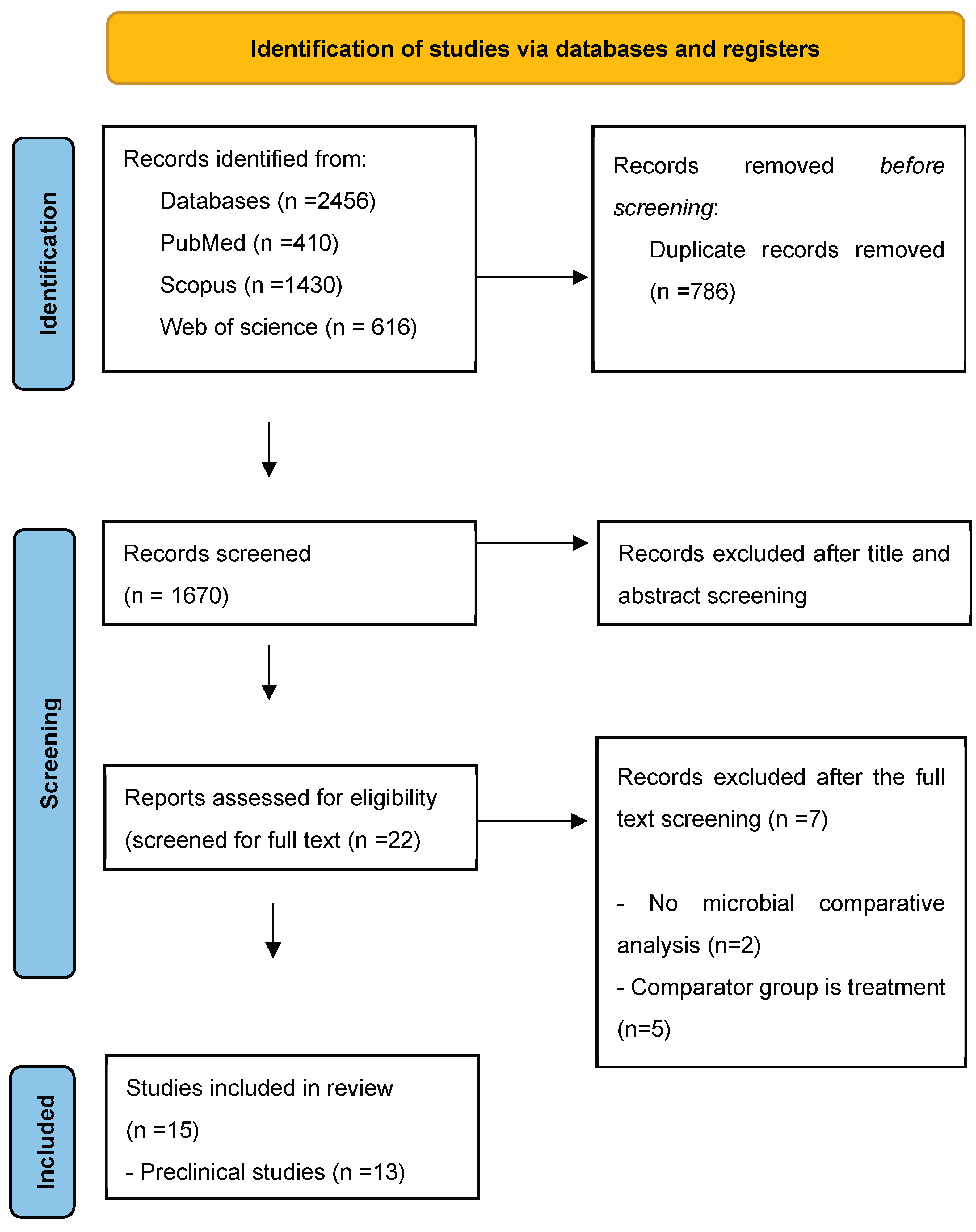
Figure 2.
Putative association between gut microbiota and cachexia in CRC models (figure created with BioRender).
Figure 2.
Putative association between gut microbiota and cachexia in CRC models (figure created with BioRender).
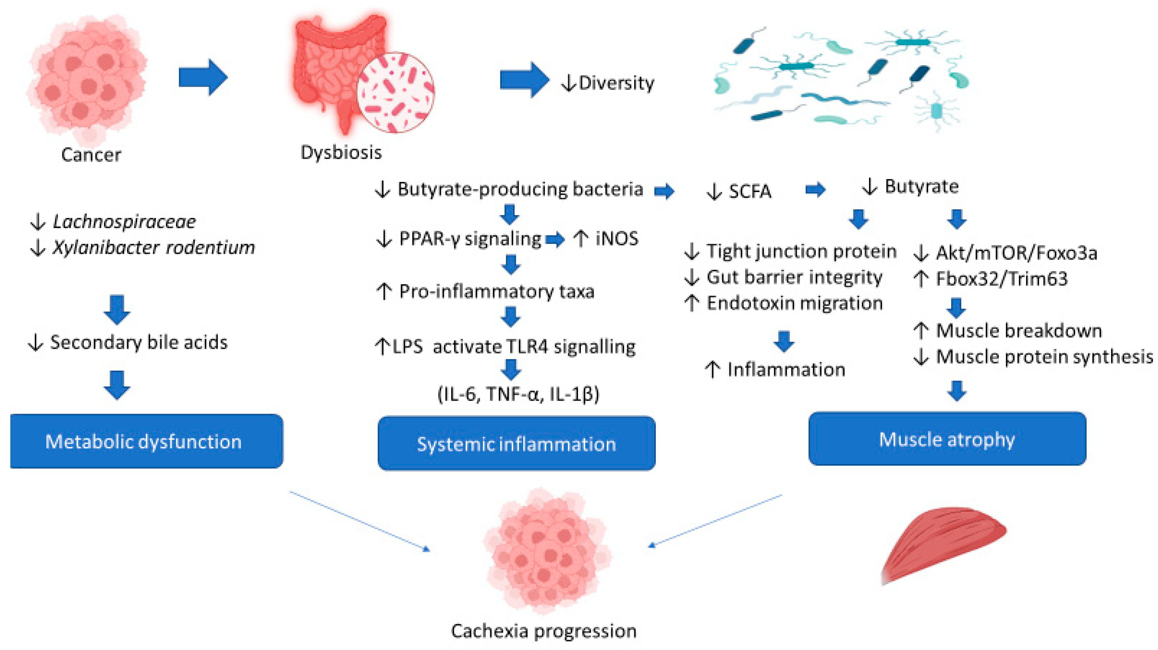

Table 1.
Characteristics of preclinical studies included in the systematic review.
| Author (Year) | Country | Study Design | Mouse Model (age) / Tumor Cell Line/ Tumor Model (Ectopic vs. Orthotopic) | Sample size | Cachexia Definition | Microbiota Analysis Method |
| Bindels (2016) | Belgium, United Kingdom, France & Canada | Experimental study | Male CD2F1 (8 weeks old) Colon carcinoma 26 (C26) cells Ectopic |
6 vs 8 mice | Loss of fat mass and muscle atrophy | 16s rRNA |
| Bindels (2018) | Belgium & France | Experimental study | Male CD2F1 (7 weeks old) Colon carcinoma 26 (C26) cells Ectopic |
7 vs 8 mice | Decreased food intake and loss of body weight, due to muscle atrophy and later on to adipose tissue loss. | 16s rRNA qPCR |
| Pötgens (2018) | Belgium | Experimental study | Male CD2F1 (7 weeks old) Colon carcinoma 26 (C26) cells Ectopic |
7 vs 8 mice | Body weight loss, weakness, muscle atrophy, fat depletion and decreased food intake | 16s rRNA qPCR |
| Pekkala (2019) | Finland | Experimental study | Male BALB/ cAnNCrl mice (5-6-week-old) Colon 26 carcinoma (C26) cell line Ectopic |
9 vs 7 mice | Body weight decreased, muscle atrophy and fat depletion | 16s rRNA |
| Feng (2021) | China | Experimental study | Male BALB/c mice (6–8 weeks old) C26 colon tumor cells Ectopic |
8 mice | >5% body weight loss, reduced muscle mass, fat depletion, elevated inflammatory markers (LPS, IL-6, TNF-α, IL-1β) | 16s rRNA |
| Pötgens (2021) | Belgium & United Kingdom | Experimental study | Male CD2F1 mice (6–7 weeks old) Colon carcinoma 26 (C26) cells Ectopic |
8 mice | The cachexia mouse model exhibited weight loss, muscle atrophy, and elevated inflammatory factors | 16s rRNA Metabolomics analysis |
| Kim (2023) | Korea | Experimental study | Male BALB/c mice (6 weeks old) C26 colon tumor cells Ectopic |
10 –13 mice | Loss of adipose and muscle tissues and decreased carcass weight (after tumor removal) | 16s rRNA |
| Liu (2023) a | China | Experimental study (in vivo & in vitro) | Male BALB/c mice (6–8 weeks old) C26 colon tumor cells Ectopic |
10 mice | Progressive weight loss, muscle atrophy, decreased grip strength and elevated inflammatory markers (LPS, IL-6, TNF-α, IL-1β). | 16s rRNA |
| Liu (2023) b | China | Experimental study | Male BALB/c mice (6–8 weeks) C26 colon tumor cells Ectopic |
6 mice | Skeletal muscle atrophy and weight loss | 16s rRNA |
| Thibaut (2025) | Belgium & United Kingdom | Experimental study | Male CD2F1 mice (7 weeks) / C26 colon tumor cells/ Ectopic; Male C57BL/6 mice (8 weeks)/ Murine MC38 colorectal cancer cells/Ectopic; Male NOD scid gamma (NSG) mice (8 weeks)/ Human HCT116 colorectal cancer cells/ Ectopic | 8 mice | Body weight and fat mass loss, muscle atrophy | 16s rRNA |
| Qiu (2025) | China | Experimental study | BALB/c mice CT26 tumor cell line Ectopic |
6 mice | Reduction in grip strength, loss of muscle mass | 16s rRNA |
| Jin (2025) | China | Experimental study | C57BL/6 mice (6-month-old) MC-38 colon cancer cells Orthotopic |
6 mice | Weight loss, muscle mass reduction | 16s rRNA |
| X Zou (2026) | China | Experimental study | Male BALB/c mice, (5 weeks old) CT26 tumor cell line Ectopic |
10 –12 mice | Reduced food intake, significant involuntary weight loss, loss of skeletal muscle and fat mass | shotgun metagenomics, metabolomic analysis |
Table 2.
Characteristics of clinical studies included in the systematic review.
| Author (Year) | Country | Study Design | Population | Cancer stage | Cachexia assessment | Microbiota Analysis Method |
| Ilozumba (2024) | USA and Germany | Prospective cohort (ColoCare) | Total : 87 patients Cachectic: 39 (45%) Non-cachectic: 48 (55%) |
Stages I–III CRC | Fearon criteria | 16S rRNA gene sequencing Fusobacterium nucleatum via qPCR |
| Ilozumba (2025) | USA and Germany | Prospective cohort (ColoCare) | Total : 103 patients Cachectic: 44 (43%) Non-cachectic: 59 (57%) |
Stages I–III CRC | Fearon criteria | 16S rRNA gene sequencing |
Table 3.
Gut microbiota changes in the cachectic mice compared to the healthy mice by phylum and family.
Table 3.
Gut microbiota changes in the cachectic mice compared to the healthy mice by phylum and family.
|
Taxonomic level Taxa |
Study | Direction of change |
Quantitative measures (cachectic vs control) |
| Phylum | |||
| Firmicutes | Pötgens (2018) | ↓ Decrease | NR |
| Pekkala (2019) | ↑ Increase | p= 0.046 | |
| Pötgens (2021) | ↓ Decrease | q < 0.05 | |
| Feng (2021) | ↑ Increase | NR | |
| Liu (2023) a | ↑ Increase | NR | |
| Thibaut (2025) | ↑ Increase | q < 0.05 | |
| Proteobacteria | Pekkala (2019) | ↑ Increase | p = 0.046 |
| Pötgens (2021) | ↑ Increase | q < 0.05 | |
| Thibaut (2025) | ↑ Increase | q < 0.05 | |
| Jin (2025) | ↑ Increase | NR | |
| Bacteroidetes | Pekkala (2019) | ↓ Decrease | p = 0.046 |
| Liu (2023) a | ↓ Decrease | NR | |
| Thibaut (2025) | ↓ Decrease | q = 0.002 | |
| Deferribacteres | Pekkala (2019) | ↑ Increase | p = 0.046 |
| Thibaut (2025) | ↑ Increase | q = 0.02 | |
| Family | |||
| Enterobacteriaceae | Bindels (2016) | ↑ Increase | p < 0.001 |
| Bindels (2018) | ↑ Increase | p < 0.05 | |
| Pötgens (2021) | ↑ Increase | q < 0.05 | |
| Feng (2021) | ↑ Increase | NR | |
| Thibaut (2025) | ↑ Increase | NR | |
| Jin (2025) | ↑ Increase | NR | |
| Lachnospiraceae | Pötgens (2018) | ↓ Decrease | p = 0.009 |
| Pekkala (2019) | ↑ Increase | p < 0.001 | |
| Feng (2021) | ↓ Decrease | p < 0.05 | |
| Liu (2023) b | ↓ Decrease | P < 0.05 | |
| Thibaut (2025) | ↓ Decrease | NR | |
| Ruminococcaceae | Pötgens (2018) | ↓ Decrease | p < 0.05 |
| Pötgens (2021) | ↓ Decrease | NR | |
| Liu (2023) b | ↓ Decrease | p < 0.05 | |
| Thibaut (2025) | ↓ Decrease | NR | |
| Lactobacillaceae | Pekkala (2019) | ↑ Increase | q <0.05 |
| Pötgens (2021) | ↓ Decrease | q < 0.05 | |
| Borkfalkiaceae (unclassified) | Thibaut (2025) | ↓ Decrease | p = 0.005 |
| Clostridiales incertae sedis XIII | Thibaut (2025) | ↑ Increase | *q < 0.01 |
| Deferribacteraceae | Thibaut (2025) | ↑ Increase | q < 0.05 |
| Eubacteriaceae | Feng (2021) | ↑ Increase | P < 0.05 |
| Streptococcaceae | Thibaut (2025) | ↑ Increase | *q < 0.01 |
Table 4.
Gut microbiota changes in the cachectic mice compared to the healthy mice by genus, species, and strain.
Table 4.
Gut microbiota changes in the cachectic mice compared to the healthy mice by genus, species, and strain.
|
Taxonomic level Taxa |
Study | Direction of change |
Quantitative measures (cachectic vs control) |
| Genus | |||
| Lactobacillus | Pekkala (2019) | ↑ Increase | p = 0.008 |
| Feng (2021) | ↓ Decrease | NR | |
| Liu (2023) a | ↑ Increase | p < 0.05 | |
| Kim (2023) | ↓ Decrease | NR | |
| Qiu (2025) | ↓ Decrease | p = 0.031 | |
| Jin (2025) | ↓ Decrease | NR | |
| Bacteroides | Pekkala (2019) | ↑ Increase | p = 0.030 |
| Zou (2026) | ↑ Increase | P < 0.05 | |
| Liu (2023) a | ↑ Increase | P < 0.05 | |
| Enterococcus | Pekkala (2019) | ↑ Increase | p = 0.011 |
| Liu (2023) a | ↑ Increase | p < 0.05 | |
| Eubacterium | Kim (2023) | ↓ Decrease | NR |
| Zou (2026) | ↓ Decrease | p < 0.05 | |
| Liu (2023) b | ↓ Decrease | p < 0.05 | |
| Intestinimonas | Feng (2021) | ↓ Decrease | NR |
| Kim (2023) | ↓ Decrease | NR | |
| Mucispirillum | Pekkala (2019) | ↑ Increase | p = 0.037 |
| Thibaut (2025) | ↑ Increase | q <0.05 | |
| Prevotella | Pekkala (2019) | ↓ Decrease | p = 0.011 |
| Thibaut (2025) | ↓ Decrease | q <0.05 | |
| Blautia | Feng (2021) | ↑ Increase | NR |
| Alistipes | Zou (2026) | ↓ Decrease | p < 0.05 |
|
Clostridia vadin BB60 gp |
Feng (2021) | ↓ Decrease | NR |
| Faecalibacterium | Liu (2023) b | ↓ Decrease | p < 0.05 |
| Lachnoclostridium | Liu (2023) a | ↓ Decrease | p < 0.05 |
| Muribaculum | Liu (2023) a | ↓ Decrease | p < 0.05 |
| Parabacteroides | Thibaut (2025) | ↑ Increase | q <0.05 |
| Roseburia | Liu (2023) b | ↓ Decrease | P < 0.05 |
| Zou (2026) | ↓ Decrease | P < 0.05 | |
| Sporobacter spp | Thibaut (2025) | ↓ Decrease | q <0.05 |
| Streptococcus | Thibaut (2025) | ↑ Increase | q <0.05 |
| Turicibacter | Feng (2021) | ↓ Decrease | NR |
| Species | |||
| Escherichia coli | Bindels (2016) | ↑ Increase | NR |
| Klebsiella oxytoca | Pötgens (2018) | ↑ Increase | p < 0.05 |
| Lactobacillus johnsonii/gasseri | Bindels (2016) | ↓ Decrease | p = 0.06 |
| Parabacteroides goldsteinii | Jin (2025) | ↑ Increase | NR |
| Xylanibacter rodentium | Thibaut (2025) | ↓ Decrease | q <0.05 |
| Strain | |||
| Parabacteroides goldsteinii/ASF 519 | Bindels (2016) | ↑ Increase | p < 0.001 |
| Uncultured | |||
|
Lachnospiraceae UCG-004 |
Kim (2023) | ↓ Decrease | NR |
Table 5.
Associations Between Pre-Surgery Gut Microbiota and Cachexia Onset at 6 Months Post-Surgery in CRC Patients (ColoCare Study).
Table 5.
Associations Between Pre-Surgery Gut Microbiota and Cachexia Onset at 6 Months Post-Surgery in CRC Patients (ColoCare Study).
|
Taxonomic Level Taxa |
Study | OR (95%CI) | Cachexia Risk | Notes |
| Species | ||||
| Fusobacterium nucleatum | Ilozumba et al. (2024) | 4.82 (1.15 - 20.10) | ↑ Increase | A priori CRC-associated |
| Genus | ||||
| Porphyromonas | Ilozumba et al. (2025) | 0.51 (0.26 - 0.89) | ↓ Decrease | Protective |
| Actinomyces | Ilozumba et al. (2025) | 0.72 (0.48 -1.03) | ↓ Decrease | Cachexia-relevant |
Disclaimer/Publisher’s Note: The statements, opinions and data contained in all publications are solely those of the individual author(s) and contributor(s) and not of MDPI and/or the editor(s). MDPI and/or the editor(s) disclaim responsibility for any injury to people or property resulting from any ideas, methods, instructions or products referred to in the content. |
© 2026 by the authors. Licensee MDPI, Basel, Switzerland. This article is an open access article distributed under the terms and conditions of the Creative Commons Attribution (CC BY) license (http://creativecommons.org/licenses/by/4.0/).
Copyright: This open access article is published under a Creative Commons CC BY 4.0 license, which permit the free download, distribution, and reuse, provided that the author and preprint are cited in any reuse.


