

Submitted:
29 June 2026
Posted:
01 July 2026
You are already at the latest version
Abstract
The Tat system is a general translocation machinery responsible for the transport of folded proteins across biological membranes that are energized by a proton or sodium motive force at these membranes. Protein substrates are characterized by usually amino-terminal signal peptides harboring a conserved twin-arginine sequence motif. Phylogenetic analyses suggest that a Tat system was already present in the last universal common ancestor (LUCA). Today, Tat systems are found in bacteria, archaea, and bacteria-derived organelles. Minimally, Tat systems are composed of one TatC-family component and one TatA/B-family component, each present in multiple copies, but more complex Tat systems exist that require additional homologs of TatA/B or TatC. Due to its ability to translocate folded proteins with bound cofactors, the Tat system is essential for a wide range of important physiological pathways, such as many anaerobic respirations or photosynthesis. With a focus on evolutionary aspects, and by including recent structural and mechanistic insights, we herein review our current knowledge on this unusual translocation pathway.
Keywords:
twin-arginine translocation
; protein transport
; membrane proteins
; protein interactions
; biological membranes
; last universal common ancestor
"Nothing in Biology Makes Sense Except in the Light of Evolution."Theodosius Dobzhansky [1973]
Introduction
The twin-arginine translocation (Tat) pathway has the challenging function to transport folded proteins across biological membranes. This is the key functional difference to the Sec pathway, the general translocation route for proteins that fold after transport [1]. Both systems date back to the last universal common ancestor (LUCA) and are found in all three domains of life [2,3,4]. So far analyzed Tat systems function in membranes that are energized by ion gradients, such as the cytoplasmic membrane of bacteria and archaea, the thylakoid membrane of plant plastids, as well as the inner membrane of mitochondria in plants and several other eukaryotic lineages [5,6,7]. Like the Sec system, the Tat system is a “general” translocation system in the sense that it is able to transport a wide range of proteins, even heterologous proteins that are recombinantly produced [6]. There are only few requirements to change a folded protein to a Tat substrate: It requires on one hand a usually amino (N)-terminal Tat-specific signal peptide, which includes the eponymous “twin-arginine (RR-)motif”, and on the other hand the absence of exposed larger hydrophobic regions, which is why the Tat system is suitable for the biotechnological production or genetic selection of proteins with correctly folded structures or improved solubility [8,9]. Natural Tat substrates are globularly folded proteins or protein domains ranging in size from less than 10 kDa, as in case of some monomeric high-potential iron sulfur proteins (HiPIPs), to over 140 kDa, as in case of the formate dehydrogenase α/β subunit complexes [10,11]. Thus, Tat substrates have often dimensions larger than the membrane thickness, and the high tolerance of Tat transport with respect to size and shape of the cargo proteins is astonishing and fascinates researchers since the beginning of the 1990ies [12]. However, not only the structural characteristics of the Tat translocon have been puzzling, but also the energization of the translocation process. While Sec transport is driven by nucleotide hydrolysis, Tat transport can be energized either by a proton motive force, such as in the model Tat systems of plant plastids or Escherichia coli, or by a sodium motive force, as in case of halophilic archaea [13,14,15]. Tat transport is energetically expensive and these high energetic expenses are an inherent property of Tat systems at energized membranes. In plant systems, the number of 78,000 consumed protons per transported protein has been experimentally determined [16]. From this perspective, it is understandable that Tat systems are thought to be used for protein translocation only when folding needs to occur prior to translocation, for example due to the requirement of cytoplasmic chaperones or cofactor insertion pathways [17,18], or when folding cannot be efficiently prevented, as is thought to be the case in halophilic archaea that have very high cytoplasmic potassium concentrations [19]. It could also be that easily achieved membrane energization may have contributed to a preferred use of Tat systems, as in cases of photosynthetic membranes in plant thylakoids or bacteriorhodopsin-containing membranes of haloarchaea [20,19]. However, due to the immense energetic costs of Tat transport, Tat systems were lost multiple times in evolution, especially when organisms no longer required a transport of folded redox proteins, as in case of many obligate fermentative organisms or obligate parasitic organisms [21]. In mitochondria, losses of Tat systems occurred multiple times as well [22]. Notably, in these mitochondria, nature evolved the specific ATPase Bcs1 of the AAA+ family that substituted Tat systems for transport of the mitochondrial Rieske protein [23,24], which was most probably the only remaining Tat substrate, and the evolution of a specific ATPase surely was energetically cheaper than a Tat system with only one substrate.
In this review, we provide a comprehensive survey of the current knowledge on Tat system components and the associated translocation mechanism, before we focus in more depth on the evolutionary aspects. Our extensive search into the origin of Tat systems and their evolutionary diversity led to surprising points and considerations that we expect to shape future research.
The Tat System Components
The Tat system minimally consists of multiple copies of a TatC and a TatA/B family protein [25]. TatA/B family proteins have an N-terminal transmembrane helix (TM) with an N-out-topology that is followed by a short hinge that connects this helix to an amphipathic helix on the cis side of the membrane [26,27,28] (Figure 1A). The carboxy- (C-)terminal region of TatA/B family proteins is neither conserved in sequence nor required for translocation, whereas the membrane-anchor, the amphipathic helix and several neighboring residues are essential [29,30]. TatC is a polytopic membrane protein with, in most cases, six transmembrane helices (TM1-TM6) and both termini on the cis side [31,32,33].
Proteins of the TatA/B family are found in two different complexes: they either form a transient homooligomeric complex that can associate with TatC, or they form a stable complex with TatC [1] (Figure 1A). The transient complex is formed out of and disassembles into highly mobile monomers or small oligomers that are distributed in the membrane [34,35,36]. Three-component Tat systems possess distinct TatA/B-family proteins for the two complexes, then called TatA and TatB, respectively, which is why these systems are also termed “TatABC systems” [1]. In two-component Tat systems (also called “minimal Tat systems” or “TatAC systems”), the same TatA can fulfil both functions, and is therefore termed “bifunctional TatA” [37]. Bacillus subtilis is the model organism for the analysis of two-component Tat systems. In this Gram-positive bacterium, two separate operon-encoded systems exist, TatAyCy and TatAdCd, that function in parallel for specific substrates [25,38,39]. Complementation analyses with overproduced components showed that TatCd tolerated all tested Tat substrates, whereas TatCy did not, and TatAd could function with both TatC paralogs, whereas TatAy functioned only with TatCy, pointing to an evolved diversity of TatA, TatC, and Tat substrate compatibility [40]. The situation in B. subtilis is even more complex, as a monocistronic gene encodes a third TatA paralog called TatAc. TatAc can specifically improve transport of the TatAyCy-containing system, where it interacts with TatAy [41]. A TatAdCd-harboring complex has been detected as a diffuse ca. 230 kDa band in blue native polyacrylamide gel electrophoresis (BN-PAGE) analyses, suggesting that in this case the expected TatAC complex is indeed formed [42]. In case of the TatAyCy system, a co-purification was demonstrated, which may be due to TatAy that is either laterally associated with or incorporated into TatCy-containing complexes [43]. Considering the yet limited information about TatC-harboring complexes, especially in phylogenetically distant organisms, it cannot be excluded that functional TatC complexes exist that lack inside integrated TatA/B family proteins. The exact purpose of TatA/B family proteins in TatC complexes has not yet been clearly defined. However, it has been shown with model three-component Tat systems that the TatB function is essential, and in fact the first mutants in plant and bacterial systems that abolished Tat transport turned out to be in TatB [44,45,46].
The Five Classes of Tat Substrates
As mentioned above, TatC-containing complexes of two- or three-component Tat systems recognize specific signal peptides of the transported Tat substrates [47,48,49]. Such signal peptides minimally consist of a hydrophilic n-region with a positive net-charge that is followed by a hydrophobic h-region, with a “twin-arginine” (RR-) motif at the interface of the n- and h-regions. This RR-motif has the consensus pattern “ZRRXΦΦ”, in which “Z” denotes a polar or charged residue that is followed by two arginines, a non-conserved position (X), and two hydrophobic residues (Φ) [1,50]. This is the general sequence motif that is recognized by TatC-containing complexes of Tat systems that have been analyzed so far, including archaeal, bacterial, and plant organelle systems [12]. This indicates that this motif evolved very early, before the separation of archaeal and bacterial lineages. If signal peptides are cleavable, they are often cleaved at an “AXA”-like recognition motif that specifies a cleavage site for a type I signal peptidase in a C-terminal region of the signal peptide (c-region) [51,52]. Alternatively, signal peptides can be cleaved by a type II signal peptidase at a lipid-modified cysteine in a lipobox near the end of the h-region [53,54].
Based on the position of folded domains, and the optional cleavage of the signal peptides, five classes of Tat substrates can be distinguished (Figure 1B): “Class I Tat substrates” have N-terminal signal peptides that are cleaved by type I signal peptidases, as found in most known cases [52], “Class II Tat substrates” are lipoproteins with N-terminal signal peptides that are cleaved by type II signal peptidases [55], “Class III Tat substrates” have N-terminal signal peptides that are not cleaved, as found for example in N-terminally membrane-anchored Rieske proteins [56,57,58,59], “Class IV Tat substrates” have an internal non-cleaved Tat signal peptide for the transport C-terminal domains of already membrane-inserted polytopic membrane proteins, as found in Rieske proteins of streptomycetes [60,61], and “Class V Tat substrates” have an internal non-cleaved Tat signal peptide that is preceded by an N-terminal folded globular domain on the cis-side and enables transport of a C-terminal folded domain to the trans-side, as found in the ferric citrate regulator FecR [62]. Some translocated Tat substrates can be integrated into membrane protein complexes, and even with cleaved signal peptides they can remain membrane-attached by interaction with at least one integral membrane protein and/or by a C-terminal membrane anchor [53,63,64].
Structural Aspects of TatC-Containing Complexes
Recently, CryoEM structures of several TatBC complexes with and without bound signal peptides have been solved [48,49]. These structures were surprising, showing that three TatC subunits assemble together with three TatB to an inverted cup-like structure that is open towards the cis-side and closed at the trans-side (Figure 2A). The amphipathic helices of TatB formed a triangle-shaped structure on the open surface of the cup-shaped TatC structure. The TatC transmembrane helices TM5 and TM6, together with the membrane-anchors of the TatB subunits, were highly tilted and therefore buried inside the complex. Binding of Tat substrates had only subtle effect on the overall structure of the complexes. The twin-arginine motif was seen to interact with TatC while the signal peptide was sandwiched between a transmembrane helix of one TatB and an amphipathic helix (APH) of another TatB [48,49]. In one of these studies, a dimeric Tat substrate was found to be bound and translocated by only one of the two signal peptides, which solved a long-standing open question in Tat transport [48]. In the articles that reported the first TatBC complexes, either a lateral passage through a TatA assembly or a central passage through TatC were favored [48,49]. Since a central passage is not consistent with available biochemical data, it was concluded that transport rather occurs through laterally recruited TatA [48,65]. There are strong arguments for a structural symmetry in these Tat complexes, although an asymmetry has been proposed in one of the structure reports [48,65]. However, based on the presence of two very different TatC-types in haloarchaea, it was suggested that these form complexes with a structural asymmetry [65], which will be discussed later in this review.
Roles of Homooligomeric TatA Assemblies
The role of TatA in membrane permeabilization for the passage of Tat substrates is based on manyfold evidence: (i) TatA is recruited to TatBC complexes to achieve functional translocation [66], (ii) with partially unfolded substrates, translocation arrest occurred in immediate vicinity of TatA [67], (iii) Tat substrates could be cross-linked to TatA during active transport [68], (iv) TatA can form large homooligomers [69,70,71,72], and TatA assemblies can destabilize biological membranes, which has been called “membrane weakening” [73,74,75]. A model that suggested a requirement of only a few TatA protomers for active plant Tat systems has later been attributed to methodologically difficult quantifications [76,77,78]. In E. coli, analyses of lacZ fusions at tatA, tatB or tatC positions in the respective operon suggested a 50:2:1 TatA:TatB:TatC ratio [79], while quantitative immunoblots were reported to indicate an approximately 20:1 TatA:TatB ratio [80]. YFP-fused TatA formed complexes of on average 25 TatA, though with a wide range of sizes [72], and metallothionein-tagged TatA generated at wild type level clusters of in average 17 +/- 6 TatA subunits [75]. In plant plastids (pea), TatA homooligomers of at least 16 protomers assembled [81], and an updated determination provided a 21:4:1 TatA:TatB:TatC stoichiometry [78]. Taken together, these analyses support the view that TatA is present in excess over the other Tat system components, which is expected for a membrane-permeabilizing function [82].
TatA has been demonstrated to interact with Tat substrates already in TatBC-deficient membranes of an E. coli mutant strain [83,74]. This points to potential functionalities in substrate-targeting [65]. Accordingly, molecular dynamics (MD) simulations indicated that membrane-interacting Tat substrates can associate with TatA, and that Tat substrates can stabilize TatA assemblies [83,84]. In agreement with this view, evenly distributed TatA-XFP fusion proteins move to TatBC complexes under conditions of recombinant substrate production [85,86]. A substrate-targeting role would explain why TatA does not remain close to TatBC complexes, which are near the cell poles [71,85]. A targeting role for TatA has been described in early Tat research also for B. subtilis and Streptomyces lividans, which can form soluble TatA complexes that may target substrates to the translocation site in the membrane [87,88,89,90].
If there are several TatA paralogs in an organism with three-component Tat systems, as often found in Pseudomonadota (the former Proteobacteria), they are sometimes termed TatE if they are not encoded in an operon together with tatBC [91]. As TatE can functionally replace TatA, it can be regarded as a second TatA paralog [91,92]. In E. coli, TatA and TatE appear to have some preference for specific substrates [91], and the tatE gene is induced under biofilm conditions [93], supporting the idea that TatE is a TatA paralog that is optimized for specific substrates and therein resembles TatAd and TatAy of Bacillus species [41]. It has not yet been analyzed whether this preference is related to targeting, translocation or both potential TatA functions.
Proposed Structures of TatA Assemblies
Several structures have been postulated to be formed by TatA alone, which always had a design that was thought to permit the passage of folded proteins. In an early model of the year 2000, termed “sea anemone model”, the groups of Berks and Palmer proposed that TatA forms rings that adapt to the changing diameter of the folded substrate during transport in order to prevent proton leakage [94]. This adaptation of ring size has been suggested to be achieved either by a “sliding” of individual subunits against each other (“iris mechanism”), or by exchanges of individual subunits during transport [94]. In 2005, the same groups suggested a pore that is gated by a “lid” formed by TatA C-termini [69]. In 2013, the group of Berks modified this model to a star-shaped structure of lidless TatA rings in which the TatA amphipathic helices and C-termini are radially directed outwards [73]. Thereby, they adapted their structural model to the “membrane-weakening and pulling model” that we had suggested in 2003, based on the idea that associations of TatA can destabilize membranes, which could permit a TatC-mediated pulling through these destabilized membrane regions [95]. It was also believed that TatA pore structures were based on an insertion of TatA amphipathic helices into the membrane, but the accessibility data and in vivo evidence argued against this model [96,97]. Another model suggested a trap-door-like mechanism in which Tat substrates push TatA to some extent deeper into the membrane without changing the TatA topology [68]. However, this model is not consistent with the observation that TatA hinge regions are more exposed upon substrate interactions [98,74]. Today, the “membrane-weakening” hypothesis as the basic explanation for Tat transport is acknowledged by most groups [82,99]. However, although we have originally postulated the “membrane-weakening” model [95], we recently extended it by adding a controlled local and transient membrane disruption step [65]. The rationale for this modification of our model was the observation of Tat transport with substrates that have a long flexible linker between the signal peptide and the globular mature domain, which demands a controlled and reversible membrane disruption rather than a proton-sealed membrane during transport [100,101]. Such a pore can be achieved by a conformational transition of an already membrane-weakening TatA ring, and this transition is proposed to be induced by the interaction of the TatA ring with a substrate-bound TatC-containing complex [65]. Notably, AlphaFold3 (AF3) predicts meaningful TatA ring structures [65,102] (Figure 2B). In these structures, the amphipathic helices align in a staggered and tilted way that agrees with biophysical studies [103]. The highly conserved “FK”-motif near the end of the amphipathic helix plays a key role in building these structures [12]. The ring structure as predicted by AF3 is actually generated by uniformly interacting staggered amphipathic helices, and this mode of ring-forming TatA interactions appears to be generally conserved [65].
The FK motif is proposed to be a prerequisite for the formation of regularly aligned TatA rings, which is supported by biochemical data [65,104]. The conservation of the FK motif in all TatA paralogs is indicative for the ability to form mixed complexes, such as the reported TatA/TatE complex in E. coli, or TatAy/TatAc-complex in B. subtilis [105,41]. In plastids and cyanobacteria, TatA paralogs have a conserved FQ motif instead of the FK motif in their amphipathic helix [65]. As the “K” of the FK motif is proposed to mediate phospholipid interactions, the “Q” of the FQ motif in cyanobacteria and plastids can be an adaptation to the abundant galactolipids in these cases [65]. Interestingly, plant TatB (Hcf106) and TatA (Tha4) have both this FQ motif, which implies that they can form mixed complexes. This would mean that “TatB” (Hcf106) of plant plastids is rather a bifunctional TatA than a TatB that only interacts with TatC. As TatA (Tha4) is essential for Tat transport in plastids, mixed Tha4/Hcf106 rings could be essential for plant plastid systems [106]. The additional function of TatB (Hcf106) in plastids would rationalize that the TatB:TatC ratio in plants has been determined to be 4:1 [78], and it would also explain that up to eight TatB subunits can be self-crosslinked via disulfide bridges in complexes that are not part of TatBC complexes [107]. E. coli TatB has no FK motif (“LR” is at this position), suggesting that this TatB is not as stably integrated into TatA rings and more specialized for TatC interactions. Notably, when the tatABC genes are overexpressed, traces of TatB could be co-purified with TatA, but BN-PAGE analyses indicated that these associations were small and thus likely not able to form large and stable mixed TatA/TatB rings [69,70].
The Molecular Mechanistic Model for Tat Transport
Based on the TatA ring model, the TatB3C3 complex structure, and available biochemical data, a molecular model for Tat transport has recently been proposed [65] (Figure 2C). This model postulates a signal peptide insertion function by the TatC-containing complex as initial step of Tat transport (phase I), followed by a TatA-dependent local membrane disruption and depolarization for the passage of the folded region of the protein (phase II) and a substrate-release, TatA ring disassembly, TatBC complex reset, and local membrane potential recovery (phase III). In contrast to the aforementioned older models, this new model explains that (i) flexible linkers between the signal peptide and the folded mature domain are tolerated by Tat systems [101,100], and that (ii) translocation-incompatible exposed hydrophobic regions (such as unfolded globular proteins) interrupt transport without blocking the translocon [108,100]. If the translocation efficiency of heterologous Tat substrates is low due to used linkers or exposed hydrophobic regions that can be trapped by interactions with the disrupted membrane, translocation is interrupted and the inserted signal peptide is released into the membrane where it can be cleaved by a signal peptidase [109]. Thus, the system is not blocked by translocation interruption (108, 100, 109, 110). This explains specific properties that had been termed “folding quality control”, which is extensively used in biotechnology (9, 111–114). C-terminal TatB domains have chaperone-like activities, and mutations in that region can effect the transport efficiency of not fully folded globular proteins [115]. In other studies, it has been observed that not fully folded proteins and natively unfolded proteins with exposed hydrophobic regions terminated translocation at a late stage, resulting in growth defects that were not prevented by TatB [108,110]. Furthermore, unfolded Tat substrates or long unstructured linkers without exposed hydrophobic regions are tolerated by Tat systems, suggesting that only exposed hydrophobic regions are problematic for Tat transport, and globular structures are not needed [100,101,110].
The newest mechanistic model postulates that the observed signal peptide insertase activity is a major function of TatC [116]. More specifically, it suggests that a ΔΨ-energized movement of the TM5/TM6 domain within TatC is required for the signal peptide insertion (Figure 2AC). This conformational change of the TatC TM5/TM6 domain is triggered by a conserved charge (in E. coli D211; from now on dubbed in any TatC “D211”) [15]. Although “D211” is highly conserved in TatCs and is classified as essential [117], some mutations of this residue in E. coli have residual activity that can complement transport with certain substrates, especially with multicopy suppression [118]. This suggests a primarily substrate-specific essential role of the TM5/TM6 movement, which can be the N-terminal unfolding during signal peptide insertion, while the TM5/TM6 domain surface exposure could also be achieved by the signal peptide insertion alone [118,65]. In this scenario, the “D211”-triggered ΔΨ-energized movement may be needed for the partial unfolding at the mature domain’s N-terminus that is often required for signal peptide insertion. Tat substates in which this region next to the signal peptide is already unstructured may not need the “D211”-triggered ΔΨ-energized movement of the TatC TM5/TM6 domain [117].
The mechanistic model includes the AF3-predicted TatA ring and suggests a mechanism for a controlled and transient local membrane disruption, based on membrane-weakening and easily extractable N-terminal TatA membrane anchors (74, 119–121). When docking to the TatC TM5/TM6 domain, the N-terminal TatA transmembrane anchors are proposed to be extracted from the membrane by a concerted conformational transition of the interacting APHs, resulting in a controlled local membrane disruption through which the substrate then passes the membrane [65]. According to this model, the directionality and efficiency result from the fact that the C-terminal region of the signal peptide is already at the trans-side of the membrane, and that the N-terminal part of the globular domain is partially unfolded during the signal peptide insertion process conducted by TatC. The transient pore permits a completion of folding, which moves the globular domain toward the c-region of the signal peptide that is already on the trans side. Linker sequences sandwiched between the signal peptide and the globular domain are tolerated but reduce the transport efficiency [100,101].
It is proposed that the closure of the transient membrane disruption is spontaneous, and that association with the membrane-stabilizing proteins of the PspA family can prevent extended membrane damage [65]. An association of PspA family proteins with Tat system components has been reported for E. coli and B. subtilis [122,123]. E. coli PspA was found to relieve saturation of Tat transport, which agrees with a function in support of rapid membrane closure and re-energization after transport [124]. In plant plastids the PspA homologue Vipp1 stimulated Tat transport as well, while a direct Vipp1/Tat interaction could not be demonstrated [125].
Asymmetric Tat Systems in Halophilic Archaea
In symmetric TatC-containing complexes, such as found in B. subtilis or E. coli, the local and transient membrane depolarization during Tat transport makes a regulation for a coordinated function of the three equivalent signal peptide binding sites necessary [65,16]. The alternative to a regulated coordination is an asymmetry in TatC-containing complexes that results in just one Tat substrate binding site. As single TatCs can only assemble to symmetric complexes with identical binding sites for TatA/B family proteins and Tat substrates, we searched for Tat systems with two or more TatC components. Among bacteria, strains with two TatCs often occur in Chloroflexota and Bacillota, but all these TatCs possess the typical conserved residues of the RR-motif binding site, indicating normal functionality. They thus can be expected to be part of separate Tat systems that can function in parallel, as known for B. subtilis [25]. However, already more than 20 years ago, it has been recognized that halophilic archaea such as Haloferax mediterranei or Halobacterium salinarium, possess an unusual TatC with 14 TMs (4, 126–128). In all of these initial descriptions, it was recognized that the 14 TM-TatCs represented two fused TatCs, connected by a domain with two additional transmembrane helices (Figure 3A). Next to the gene of the 14 TM-TatC was a gene encoding a second TatC with a usual 6 TM topology. The Tat system of haloarchaea is responsible for the large majority of secreted proteins, which is unique and proposed to relate to effects on folding by the high intracytoplasmic concentration of potassium in theses prokaryotes [19,129]. The Pohlschröder-group was the first that showed in a mutational analysis that in H. mediterranei both tatC genes were essential for this organism, as was one of its two tatA genes. They termed the long 14 TM TatC “TatCt” and the genetically linked 6 TM TatC “TatCo” [128]. In the same study, they also recognized that in Haloarcula species the 6 TM TatC found in other halophilic archaea had a C-terminal extension of 4 TMs, resulting in a 10 TM TatC. Later, the TatC proteins of halophilic archaea were classified into three combinatory groups, in all of which a 14 TM TatCt is combined with either a 6 TM TatCo (group I), a 6 TM TatC with an N-terminal extension (TatCn) (group II), or a 10 TM TatCx, in which a 6 TM TatC is C-terminally extended by 4 TMs (group III) [130]. It was proposed by Thomas and Bolhuis that the two different TatCs form a functional unit [131].
A closer look at the 14 TM TatCt indicates that the RR-motif binding sites of the two “fused” TatC are both inactive, as evidenced by the absence of the otherwise in archaea and bacteria highly conserved F94 and E103 residues (E. coli numbering), which are the key residues for the RR-motif binding [48] (Figure 3AB). In contrast, this RR-motif binding site of the 6 TM TatCo (as in the 10 TM TatCX) appears to be functional, as both residues are present, and also their direct environment is highly conserved (Figure 3AB). These characteristics strongly suggest that the 14 TM TatCt must interact with a 6 TM TatCo or 10 TM TatCx to form active TatC-containing complexes, which then would have a similar overall structure as the trimeric bacterial TatC-based complexes recently published [48,49] (Figure 3C).
Why did haloarchaea evolve asymmetric Tat complexes? One reason could be that the extremely high (several molar) salt concentrations in the habitat and inside the cytoplasm of these species interfere with the regulatory mechanism for activity coordination at symmetric Tat complexes, which may rely on salt bridges [132].
Protein Targeting in Early Evolution – Is TatC the Most Ancient Translocase?
How did Tat systems evolve? When first membranes appeared in evolution, proteins needed to be targeted into or across membranes to facilitate selective uptake or release of compounds, and to place proteins on the outside when this was advantageous for the evolving cell [133,134]. Three types of translocases are known to have ancestors that evolved before archaea and bacteria separated, and all of them can function as insertases of signal peptides: SecY-based systems, YidC, and TatC-based systems [2,3,4] (Figure 4A). SecY-family proteins are essential for all known organisms, showing monophyletic and highly divergent evolution in archaea and bacteria [2]. SecY associates laterally with more components, such as SecE or SecG, but SecY forms the core of the translocon and its ancestor likely catalyzed membrane protein insertion alone in early evolution [3]. A similarly evolutionary ancient targeting system consisting of a signal recognition particle (SRP) and an SRP receptor coevolved with the SecY-based systems to permit a cotranslational targeting that prevents aggregation of hydrophobic transmembrane helices [135,136]. SecY evolved from a fused duplication of an ancestor that was also the ancestor of today’s YidC [3]. All these systems recognize a positively charged region, followed by an insertion of an adjacent hydrophobic region. While YidC can insert a hydrophobic region on either side, Sec systems as well as TatC insert only hydrophobic regions that are on the C-terminal side of the positively charged region (Figure 4A). YidC thus cooperates with Sec-systems when N-out topologies are generated or when transmembrane helices are released from SecY [137,138]. However, YidC is also capable to function independently in membrane protein insertion [3,137].
Membrane protein biogenesis is considered the only function of ancient SecY/YidC-based systems, although today’s Sec machineries can translocate proteins across the membrane as well. The ATPase SecA is required for cotranslational or posttranslational translocation of soluble proteins or protein domains in bacteria, and there is no SecA homolog in archaea, indicating a late evolution of Sec-dependent translocation of soluble proteins in bacteria (5, 139–141) . Although an archaeal SecA homolog does not exist, there is evidence for an analogous SecA-like pathway in archaea [142]. These considerations suggest that the Tat system may have been the first translocation apparatus for soluble proteins in evolution. This assumption makes sense, as processes that permit translocation of unfolded proteins and extracytoplasmic folding first needed to evolve before such a transport was possible. Moreover, many ancient proteins required cofactors for their basic functions, and biogenesis and insertion pathways for these cofactors were located inside the cytoplasm. Since a cytoplasmic cofactor insertion machinery already existed, it could be used for translocated enzymes in case of Tat transport. Until today, one main reason for the use of Tat transport is still a cytoplasmic assembly of cofactors that are required for proteins functional in extracytoplasmic spaces. Such cofactors that have been experimentally demonstrated to be inserted into Tat protein substrates prior to transport include Fe-S-clusters ([4Fe-4S], [2Fe-2S]) [59,56,143], molybdenum cofactors (Mo-bis-MGD, Mo-MCD) [144,145], nickel-iron complexes [146], flavins (FAD) [147], coenzyme F420 [148], NADP [149], heme [150,151], and pyridoxal phosphate (PLP) [152,153]. In addition, enzymes with cobalt-containing cofactors (norpseudo-B12) that can insert their cofactor inside the cytoplasm, contain Tat signal peptides and are functional outside the cytoplasm [154]. However, copper cofactors are exceptions: as copper is highly toxic inside the cytoplasm, Tat-dependently translocated copper proteins fold already without copper assembly in the cytoplasm, and copper is inserted after transport across the cytoplasmic membrane [155].
Based on the current views on early evolution of metabolism and membranes, the hypothetical appearance of Tat systems can be ordered into the sequence of events during pre-LUCA evolution as follows (Figure 4B): When the RNA-based “ribozyme” world, in which small and simple peptides are believed to have played an assisting role, was extended to an “ribozyme/enzyme” world by the invention of ribozyme-based translation, more and more functions were transferred to or invented by proteins [156]. Due to the complex biosynthetic requirements of DNA, proteins are anticipated to have preceded DNA, and translation of imprecisely replicated RNA yielded manifold mutated protein sequences that were positively selected when conferring advantageous functions [157,158]. When first membranes emerged, which were leaky as they consisted of abiotically formed long chain amphipathic compounds like fatty acids [159,160], hydrophobic peptides could partition into such membranes. This permitted already a selection for those peptides that conferred certain advantages, such as selective permeability of needed compounds, leading to the evolution of the first membrane proteins. The evolution of lipid bilayers and membrane proteins went hand in hand, as only proteins – not RNA – could be stably placed into lipid environments to provide the necessary selective permeabilization of lipid bilayers. When biological fatty acid and phospholipid synthesis pathways evolved, membranes became thicker, tighter and more stable, while the insertion of proteins gradually required more and more assistance by insertases, which became the ancestors of today’s protein insertion and translocation systems that include the SecY, YidC, and TatC components.
How can the origin of Tat systems be imagined? As mentioned above, TatC inserts signal peptides with a N-in/C-out topology into membranes, much like SecY [116]. However, while SecY laterally inserts any transmembrane domains into the membrane [161,162,163], the insertion by TatC stops when an intermediate is formed that is with its N-terminus deeply buried in the complex [164]. Instead of releasing the bound signal peptide into the membrane, which happens in the absence of TatB, TatC of complete Tat systems permits the lateral passage of the globular Tat substrate domain through a TatA-disrupted membrane [116]. A function of TatB in translocation is therefore the prevention of signal peptide release prior to transport. It is possible that initially, at times of leaky membranes, folded domains could easily flip through the membrane, and thus TatC alone may have had “flippase” activity, rapidly transferring folded proteins from the cis- to the trans- side. However, when membranes became stronger, TatC surely required the assistance of TatA/B family peptides that evolved to transiently disrupt the membranes when interacting with TatC. This is why TatA/B family proteins are part of all Tat systems from archaea to bacteria and their derived organelles [4]. After this passage, the signal peptide is released together with the translocated globular protein domain.
To become translocated by Tat, the existing globular enzymes did not need to be kept in an unfolded state, which was an advantage of the Tat pathway. However, to facilitate and selectively enable the transport by Tat, these globular enzymes acquired early in evolution specific N-terminal transport signals – the signal peptides.
The Evolution of Signal Peptides – from Membrane-Protein Insertion to Tat-Transport
According to the “positive-inside-rule” of membrane proteins, the membrane protein regions next to the inner side of the membrane have a positive net charge [165,166]. The same rule is true for signal peptides of Sec- or Tat-dependently transported soluble proteins, which have the same targeting function as the first membrane-inserted helices of membrane proteins [136,167,168]. It is likely that these characteristics originated from the ability to associate with negatively charged lipid head groups via the n-region, and to anchor to these membranes via the h-region, which facilitated membrane insertion in early evolution.
Tat substrates just needed such a targeting module at their N-terminus. Still today, Tat substrates can be targeted to the membranes and can be transported thereafter [169]. However, the signal peptides need to be differentiated from Sec signal peptides, for that only Tat substrates are targeted by the Tat system. Tat signal peptides have a lower average hydrophobicity, which can influence the specificity [170]. However, this cannot be the only criterium, as there is significant overlap between the distributions of Sec and Tat signal peptide hydrophobicities. A second criterium could have been the RR-motif in Tat signal peptides. However, when a RR-motif was introduced into a Sec signal peptide, transport of the mutated protein was still Sec-dependent [10]. Even when the entire signal peptide of a Sec substrate was exchanged by a Tat signal peptide, transport remained Sec dependent unless the protein was allowed to fold inside the cytoplasm [171]. Moreover, complete inactivation of RR-motifs has been shown to still permit a targeting to the Tat translocon [172,173]. A third criterium could have been a Sec-avoidance motif, which has been defined as positively charged c-region of Tat signal peptides [174]. However, positive charges are only present in a subset of Tat substrate c-regions, and it has been shown that most Tat signal peptides can mediate Sec transport when fused to a Sec substrate [175]. In summary, it is likely an interplay of mature domain characteristics, signal peptide hydrophobicities, and charge distributions that together determine the targeting pathway to the correct translocon. One important aspect could be that Sec-targeted proteins are either already recognized by SRP or SecA at the ribosome for cotranslational targeting, or they are fully translated and then kept in an unfolded Sec-compatible state by chaperones such as SecB [136,139,176]. In other words: Sec substrates are never freely inserted into membranes prior to the translocon interaction, whereas Tat substrates can be inserted into the membrane and transported thereafter, which could be the key difference in the targeting pathways [169,177]. Notably, Tat substrates could be bound to purified solubilized TatC-containing complexes, showing that a membrane-independent targeting pathway may exist, but it is not known yet whether such a direct binding of Tat substrates to such complexes occurs in vivo with physiological substrate concentrations [48].
Many signal peptide-binding chaperones have been identified that were thought to be able to target Tat substrates to their translocon. Several of these chaperones are already genetically linked to specific substrates that contain for example Ni-Fe-centers or molybdenum cofactors [178]. These are specific chaperones, termed “redox enzyme maturation proteins” (REMPs), that use signal peptides as platforms to support the insertion of the required cofactor into the mature domain of these redox proteins [179,180,181]. Early models that suggested a direct targeting function for such chaperones to the Tat system could not be confirmed [144,182].
In contrast to REMPs, some general chaperones, DnaK and SlyD, have been identified that bind a broader range of Tat signal peptides [183,17]. DnaK is an ATP-dependent chaperone that is highly abundant and involved in folding of many proteins, often in cooperation with other proteins [184,185,186]. SlyD is less abundant, has minimally a prolyl isomerase and chaperone domain, and has been shown to be a folding chaperone [187,188]. Some SlyD homologs have a C-terminal nickel-binding domain that is involved in intracellular nickel storage. However, SlyD binds a broad range of Tat signal peptides without preference for nickel-containing Tat substrates [189]. E. coli SlyD bound the Tat signal peptide of the model Tat substrate HiPIP of Allochromatium vinosum with high affinity (KD ca. 0.5 µM), which is in the range of substrate-specific REMPs [189]. The twin-arginines in the Tat signal peptide were shown to contribute to the high affinity [189]. As SlyD is not involved in cofactor insertion, it might play a role in signal peptide protection or membrane targeting. Interestingly, SlyD is hijacked by phage ΦX174, targets its lysis protein to the membrane and incorporates together with this protein into an cell-wall-biosynthesis inhibitory complex that promotes lysis of infected cells [190]. The requirement of this host protein for cell lysis by ΦX174 is actually the original reason for its name (sensitivity to lysis: Sly) [191].
Early Tat Transport Without ΔΨ? Acidophilic Archaea Show What Is Possible
So far, this review discussed our knowledge about today’s Tat systems and the evolution of Tat systems in context of the evolution of cytoplasmic membranes, but we did not consider the energetic situation during early evolution. There seems to be a contradiction that on one hand Tat systems evolved in leaky membranes of pre-LUCA times, while on the other hand Tat transport is known to depend on the membrane energization. Today, this membrane energization can be either a pmf or a smf [13,14,15]. Both have the electric field (ΔΨ) in common, which is postulated to be the driving force for signal peptide insertion by energizing a movement of the TatC-TM5/TM6 domain, which can be accomplished by the residue “D211” in that domain [65].
However, as noted above, early membranes were leaky and required the evolution of fatty acid and phospholipid synthesis to become more sealed [159]. The diversification of membrane lipids in archaea and bacteria followed later [192,193]. It is anticipated that initially natural pH gradients, which were possibly converted to sodium gradients that are more stable at the leaky early membranes, were used to energize ancient membranes [159,194,195]. In any case, it is unlikely that a ΔΨ of these membranes was comparable to today’s membranes. Therefore, the basic question remains, whether Tat systems could have functioned in membranes without significant ΔΨ.
A closer look at the phylogenetic tree of TatC sequences in archaea and bacteria helped to answer this question. Saier et al. had already reported such a tree, which was the first evidence for an ancient origin of Tat systems in LUCA [4]. Our extended tree with exemplary representatives of known phyla with Tat systems shows that indeed TatC of archaea and bacteria are clearly separated and thus diverged early in evolution, supporting the existence of the Tat system in LUCA (Figure 5).
Interestingly, the extreme acidophiles among the archaea evolved TatC variants that clearly escape the organismic phylogeny grouping (Figure 5). All these archaea have optimum growth below pH 3, and some of them even below pH 1. To protect the cytoplasm from uncontrolled entry of protons at such growth conditions, the ΔΨ at the cytoplasmic membrane is very low or even inverted in this extremely acidophilic archaeal group, with the outside being negative and the inside positive [196,197] (Figure 6A). This has an enormous impact on the Tat mechanism: In an inverted electric field at extremely low pH, the negative charge would inhibit a required movement of the TM5/TM6-domain. It is thus revealing to see that these extremely acidophilic archaea lost the negative charge in this region of the TM5/TM6 domain (Figure 6C). In normal archaeal TatAC systems, the “D211”-equivalent charge is two positions away from the position found in bacteria, mitochondria and plastids, which may suggest an independent convergent evolution for the use of ΔΨ in archaea and bacteria, and hence a “post-LUCA” development (red dots in Figure 6C). The absence of the conserved “D211” in extremely acidophilic archaea suggests that (i) these systems must function without using a ΔΨ, and that (ii) a selection against the negative charge in the “D211” position of the TM5/TM6 domain occurred due to a required movement of this domain.
This in turn means that the electric field is not strictly needed for the mechanism, although it likely became necessary for almost all Tat systems that exist today. But if TatC of extremely acidophilic archaea does not use a ΔΨ, how is unfolding at the mature region next to the signal peptide energized and what can trigger the exposure of the TM5/TM6 domain at the surface to permit the docking of TatA? If the Tat substrates are not stably folded in the region next to the signal peptide, the binding energy may suffice to promote signal peptide insertion, and there can be several ways to achieve the coupling of signal peptide insertion to the exposure of the TM5/TM6 domain. Signal peptide insertion per se might be able to push this domain to the surface in phase I. Also, supportive structural adaptations may have evolved. We noted that the AF3-predicted TatC structures in these acidophilic archaea have unusual beta-sheet hairpins at the end of TM1, which might play such a role (Figure 6B). Another aspect is the energy requirement in phase II, the TatA-dependent membrane disruption. Here, the extreme pH gradient at the very stable and proton-tight cytoplasmic membrane of these acidophiles could facilitate the process.
As a side aspect, we would like to note that TatC of extremely acidophilic bacteria, unlike the extremely acidophilic archaea, can still use the ΔΨ as they possess the conserved “D211”. This is possible because these bacteria have to locally generate small inside negative membrane potentials, even though on average they may have an inverted membrane potential when growing below pH 3 (198). They need this ability, as bacterial F-type ATP synthases require at least a small but normal oriented (negative inside) membrane potential, even when artificially large proton gradients are imposed [199,200,201]. It is thus not surprising that extremely acidophilic bacteria can also use the membrane potential for Tat transport. The A-type ATP synthases of archaea do not strictly require a membrane potential and hence extremely acidophilic archaea likely do not establish local membrane regions with significant negative inside oriented membrane potentials [202]. This is why the Tat system in these archaea needs to function without the use of a membrane potential.
The Special Case of Ignicoccus – Another TatC that Works Without ΔΨ?
After finding that extremely acidophilic archaea lost the conserved aspartic acid (“D211”) in the TM5/TM6 domain, which could be explained by an adaptation to an inverted membrane potential, we examined the conservation of this negatively charged position in the complete set of bacteria and archaea that we had used for the phylogenetic tree. To our surprise, we found only members of one other genus that showed the absence of this negatively charged position: Ignicoccus (Figure 6C). The hyperthermophilic genus Ignicoccus differs from all other known prokaryotes by having an “outer cellular membrane” (OCM) that is proton-tight and energized by a redox-driven proton gradient for fueling ATP synthase at this membrane, whereas an “inner membrane” (IM) surrounds a compartment containing DNA and all information processing machinery (including replication, transcription, and translation) [203,204] (Figure 6D). The exact function of the inner membrane is yet unknown, but as the inner membrane is not involved in energy transduction and ATP synthesis, it is likely that the inner membrane is only weakly energized, which would explain the presence of a ΔΨ-independent Tat system in Ignicoccus. The characteristics of the translocated substrates and the IM of this genus may demand only little energization for Tat transport phases I and II. As outlined above, one simple explanation for ΔΨ-independence could be the presence of an unfolded region at the N-terminus of the mature domain in Tat substrates, making ΔΨ-energized unfolding that depends on “D211” unnecessary. However, this hypothesis needs to be experimentally addressed in future.
In view of the unusual abilities of Tat systems in extremely acidophilic archaea and Ignicoccus, the in other organisms usually observed ΔΨ-energization of phase I can be an evolutionary adaptation of Tat systems to the advantageous use of the existing significant ΔΨ in those membranes. For the pmf- or smf- dependent energization of phase II, which is much less understood, small chemical gradients (ΔpH, ΔNa+) may suffice, depending on the membrane rigidity [65,15].
A protective Function of Membranes and Tat Systems in Early Evolution?
It is simple to say that membranes “evolved”, and that initial coincidental abiotic leaky membranes were over time substituted by synthesized membrane lipids, but the selective pressure needs to be rationalized, the advantage that led to the evolution of membranes and Tat transport. This initial selective pressure could not have been the use of membranes for the generation of proton or sodium gradients to generate ATP, because this bioenergetic use requires the synthesis, targeting, and functional assembly of a highly complex nanomachine called ATP synthase, which evolved by a combination of a hexameric P-loop ATPase, a membrane-integral ion channel, and other interaction partners in the membrane [195,205]. Earlier simpler systems could have been based on ion-gradient driven pyrophosphate synthesis [206]. In any case, ancient SRP/SecY/YidC-assisted pathways were needed for the evolution of ion gradient-utilizing membrane-integral synthases of high-energy products. After an initial use of natural gradients, membrane-associated electron transport chains (ETCs) evolved, which led to the self-determined generation of ion gradients that was a prerequisite for the liberation of the first cell from its presumed mineral cavity deep in an alkaline vent of the sea (Figure 4B) [207]. Tat transport surely played a crucial role for these first ETCs, such as in the electron flow from hydrogen to polysulfides or N-oxides, as enzymes like nickel-containing hydrogenases were Tat-dependently placed on the outside of the membranes for these ETCs [208,146]. In fact, hydrogenases were the first Tat substrates, whose translocation was biochemically shown, long before the pathway was discovered, with two interacting subunits being transported by means of only one signal peptide [209].
However, initial selective advantages of membrane evolution in pre-LUCA times must have been immediate effects on the cellular processes. We suggest that – besides their function in the capture of synthesized biomolecules – membrane compartments also protected against harmful substances, especially the thiol- and Fe-S-cluster-reactive arsenite, which could inactivate key enzymes and cofactors that were needed for ancient biosynthetic pathways [210,211].
The important detoxification-aspect needs here some more detailed discussion, as it directly relates to the evolution of the Tat system. Arsenite is abundant in submarine geothermal habitats such as black smokers or alkaline vents, which are currently discussed as most likely habitats of pre-LUCA evolution [194]. Due to its thiol- and Fe-S-cluster reactivity, arsenite detoxification by oxidation likely played an important role in early evolution [212]. Aio-type arsenite oxidases were the only arsenite oxidases that were present in LUCA [213]. Today’s Aio-type arsenite oxidases detoxify arsenite outside the cytoplasm. Before membranes existed, Aio-type arsenite oxidases may have already been present to protect thiol-involving metabolism. They are minimally composed of two subunits, the catalytic subunit with a molybdopterin cofactor and an [3Fe-4S]1+/2+ cluster, and the Rieske subunit with its [2Fe-2S]1+/2+ cluster [214]. The Rieske subunit can be regarded as ancestor of Rieske proteins of cytochrome bc1/b6f type complexes [212], while the catalytic subunit belongs to the DMSO-reductase family and was likely an ancestor of today’s molybdopterin-containing terminal reductases [213]. Aio-type arsenite oxidases have a Tat signal peptide at the N-terminus of their Rieske subunit, and arsenite oxidases are thus translocated as a heterodimer by use of a signal peptide of the small subunit [215], which parallels the arrangement found in heterodimeric hydrogenases but is unusual for molybdopterin-containing Tat substrates [146,209]. The signal peptide of the Rieske protein likely anchored the first Aio-type arsenite oxidases to the outer face of the membrane, a topology that is still found in many Aio-type arsenite oxidases today [216,217,212]. Interestingly, membrane-anchoring by an uncleaved Tat signal peptide is evolutionarily conserved in Rieske proteins of cytochrome bc1/b6f type complexes [218].
Due to unfavorable redox potentials, arsenite oxidation cannot be coupled to CO2 reduction or polysulfide reduction [213]. Arsenite oxidases were therefore probably reoxidized by electron flow to early N-oxide reductases. N-oxides, especially nitrate and nitrite, are considered to have been abundant in pre-LUCA times and the only high-potential electron acceptors available in archaean oceans before the appearance of significant free oxygen [219]. Nitrate-dependent anaerobic arsenite oxidation still exists today [220]. The evolutionary oldest N-oxide reductases are those of the Nar-type [221], which have highly similar cofactors as arsenite oxidases. Thus, an electron flow from arsenite at the outside to nitrate in the inside of the early cells may have been a first ETC at membranes, which served as a detoxification pathway. This pathway had an immediate positive effect on the cells, prior to the evolution of an ATP synthase that could use ΔpH or ΔNa+ for ATP synthesis, and prior to the evolution of known ancient ETCs that served the purpose of self-controlled ion gradient formation, such as electron flow from hydrogen to polysulfides, which were likely present in abundance in hydrothermal vents. Later in evolution, when more oxidized compounds appeared on earth, alternative terminal electron acceptors could be used and new respiratory pathways emerged. Especially more extracytoplasmic Tat-dependently translocated molybdopterin-containing oxidoreductases evolved, which permitted the use of a multitude of alternative terminal electron acceptors for diverse anaerobic respirations that are known today, such as arsenate, selenate, thiosulfate, tetrathionate, polysulfide, perchlorate, chlorate, dimethyl sulfoxide, or trimethylamine N-oxide respirations [222]. This is why Tat transport is still closely associated with the translocation of cofactor-containing proteins, and this is why it is used whenever a general translocation pathway for folded proteins is needed.
As outlined in this review, the diversity of today’s Tat systems shows a remarkable adaptivity during evolution. Tat is much more than the initially believed ΔpH pathway [223]. The electric potential at the membrane is used by Tat in most cases, but functional Tat transport without significant membrane potentials was made possible in extremely acidophilic archaea and some exceptional hyperthermophilic species with uncommon cellular compartments (Figure 6).
Concluding Remarks and Future Directions
Considering the diverse Tat systems that evolved, it becomes conceivable that Tat transport is only expensive due to a requirement of a transient membrane disruption. In principle, Tat systems can function at hardly energized membranes or even at membranes with an inverted membrane potential. Uncovering the bioenergetics of this phenomenon will be a fascinating future research goal. For the transient membrane disruption in phase II, a ΔpH or ΔNa+ is likely important, and small gradients may suffice, depending on the membrane characteristics. The understanding of these effects similarly demands advanced studies. Due to the unexpected adaptability of Tat systems to diverse energization conditions, TatC-based transport was possible very early in evolution, which explains why Tat systems date back to LUCA. The use of the membrane potential possibly evolved later in archaea and bacteria. In view of the energetic expenses, will Tat systems persist at energized membranes in future evolution? As long as there is no “cheaper” alternative that works with a broad range of folded proteins, Tat transport is the pathway without alternative. In that sense, Tat transport is always a compromise. It is like an old machine that is still used, because a better transport machine for multiple folded substrates has not yet been invented by nature.
Funding
This work was supported by the German Research Foundation (DFG) grant BR 2285/8-1 to TB.
Conflicts of interest
The authors have no conflicts of interest.
References
- Natale, P.; Brüser, T.; Driessen, A. J. M. Sec- and Tat-mediated protein secretion across the bacterial cytoplasmic membrane - distinct translocases and mechanisms. Biochim Biophys. Acta 2008, 1778, 1735–1756. [Google Scholar] [CrossRef] [PubMed]
- Cao, T. B.; Saier, M. H. The general protein secretory pathway: phylogenetic analyses leading to evolutionary conclusions. Biochim Biophys. Acta 2003, 1609, 115–125. [Google Scholar] [CrossRef] [PubMed]
- Lewis, A. J. O.; Hegde, R. S. A unified evolutionary origin for the ubiquitous protein transporters SecY and YidC. BMC Biol. 19 2021, 266, 34911545. [Google Scholar] [CrossRef] [PubMed]
- Yen, M.-R.; Tseng, Y.-H.; Nguyen, E. H.; Wu, L.-F.; Saier, M. H. Sequence and phylogenetic analyses of the twin-arginine targeting (Tat) protein export system. Arch. Microbiol. 2002, 177, 441–450. [Google Scholar] [CrossRef] [PubMed]
- Pohlschröder, M.; Dilks, K.; Hand, N. J.; Wesley Rose, R. Translocation of proteins across archaeal cytoplasmic membranes. FEMS Microbiol. Rev. 2004, 28, 3–24. [Google Scholar] [CrossRef] [PubMed]
- Frain, K. M.; Robinson, C.; van Dijl, J. M. Transport of folded proteins by the Tat system. Protein J. 2019, 38, 377–388. [Google Scholar] [CrossRef] [PubMed]
- New, C. P.; Ma, Q.; Dabney-Smith, C. Routing of thylakoid lumen proteins by the chloroplast twin arginine transport pathway. Photosynth Res. 2018, 138, 289–301. [Google Scholar] [CrossRef] [PubMed]
- Brüser, T. The twin-arginine translocation system and its capability for protein secretion in biotechnological protein production. Appl. Microbiol. Biotechnol. 2007, 76, 35–45. [Google Scholar] [CrossRef] [PubMed]
- Fisher, A. C.; Rocco, M. A.; DeLisa, M. P. Genetic selection of solubility-enhanced proteins using the twin-arginine translocation system. Methods Mol. Biol. 2011, 705, 53–67. [Google Scholar] [CrossRef] [PubMed]
- Brüser, T.; Deutzmann, R.; Dahl, C. Evidence against the double-arginine motif as the only determinant for protein translocation by a novel Sec-independent pathway in Escherichia coli. FEMS Microbiol. Lett. 1998, 164, 329–336. [Google Scholar] [CrossRef] [PubMed]
- Stanley, N. R.; Sargent, F.; Buchanan, G.; Shi, J.; Stewart, V.; Palmer, T.; Berks, B. C. Behaviour of topological marker proteins targeted to the Tat protein transport pathway. Mol. Microbiol. 2002, 43, 1005–1021. [Google Scholar] [CrossRef] [PubMed]
- Hou, B.; Brüser, T. The Tat-dependent protein translocation pathway. Biomol. Concepts 2011, 2, 507–523. [Google Scholar] [CrossRef] [PubMed]
- Theg, S. M.; Cline, K.; Finazzi, G.; Wollman, F. A. The energetics of the chloroplast Tat protein transport pathway revisited. Trends Plant Sci. 2005, 10, 153–154. [Google Scholar] [CrossRef] [PubMed]
- Kwan, D. C.; Thomas, J. R.; Bolhuis, A. Bioenergetic requirements of a Tat-dependent substrate in the halophilic archaeon Haloarcula hispanica. FEBS J. 2008, 275, 6159–6167. [Google Scholar] [CrossRef] [PubMed]
- Bageshwar, U. K.; Musser, S. M. Two electrical potential-dependent steps are required for transport by the Escherichia coli Tat machinery. J. Cell Biol. 2007, 179, 87–99. [Google Scholar] [CrossRef] [PubMed]
- Alder, N. N.; Theg, S. M. Energetics of protein transport across biological membranes. a study of the thylakoid ΔpH-dependent/cpTat pathway. Cell 2003, 112, 231–242. [Google Scholar] [CrossRef]
- Graubner, W.; Schierhorn, A.; Brüser, T. DnaK plays a pivotal role in Tat targeting of CueO and functions beside SlyD as a general Tat signal binding chaperone. J. Biol. Chem. 2007, 282, 7116–7124. [Google Scholar] [CrossRef] [PubMed]
- Berks, B. C. A common export pathway for proteins binding complex redox cofactors? Mol. Microbiol. 1996, 22, 393–404. [Google Scholar] [CrossRef] [PubMed]
- Rose, R. W.; Brüser, T.; Kissinger, J. C.; Pohlschröder, M. Adaptation of protein secretion to extremely high-salt conditions by extensive use of the twin-arginine translocation pathway. Mol. Microbiol. 2002, 45, 943–950. [Google Scholar] [CrossRef] [PubMed]
- Peltier, J.-B.; Emanuelsson, O.; Kalume, D. E.; Ytterberg, J.; Friso, G.; Rudella, A.; Liberles, D. A.; Söderberg, L.; Roepstorff, P.; von Heijne, G.; van Wijk, K. J. Central functions of the lumenal and peripheral thylakoid proteome of Arabidopsis determined by experimentation and genome-wide prediction. Plant Cell 2002, 14, 211–236. [Google Scholar] [CrossRef] [PubMed]
- Wu, L. F.; Ize, B.; Chanal, A.; Quentin, Y.; Fichant, G. Bacterial twin-arginine signal peptide-dependent protein translocation pathway: evolution and mechanism. J. Mol. Microbiol. Biotechnol. 2000, 2, 179–189. [Google Scholar] [PubMed]
- Petrů, M.; Wideman, J.; Moore, K.; Alcock, F.; Palmer, T.; Doležal, P. Evolution of mitochondrial TAT translocases illustrates the loss of bacterial protein transport machines in mitochondria. BMC Biol. 16 2018, 141, 30466434. [Google Scholar] [CrossRef] [PubMed]
- Wagener, N.; Neupert, W. Bcs1, a AAA protein of the mitochondria with a role in the biogenesis of the respiratory chain. J. Struct. Biol. 2012, 179, 121–125. [Google Scholar] [CrossRef] [PubMed]
- Rosales-Hernandez, C.; Thoms, M.; Berninghausen, O.; Becker, T.; Beckmann, R. Mechanistic insights into Bcs1-mediated mitochondrial membrane translocation of the folded Rieske protein. EMBO J. 2025, 44, 3720–3741. [Google Scholar] [CrossRef] [PubMed]
- Jongbloed, J. D. H.; Grieger, U.; Antelmann, H.; Hecker, M.; Nijland, R.; Bron, S.; van Dijl, J. M. Two minimal Tat translocases in Bacillus. Mol. Microbiol. 2004, 54, 1319–1325. [Google Scholar] [CrossRef] [PubMed]
- Koch, S.; Fritsch, M. J.; Buchanan, G.; Palmer, T. Escherichia coli TatA and TatB proteins have N-out, C-in topology in intact cells. J. Biol. Chem. 2012, 287, 14420–14431. [Google Scholar] [CrossRef] [PubMed]
- Hu, Y.; Zhao, E.; Li, H.; Xia, B.; Jin, C. Solution NMR structure of the TatA component of the twin-arginine protein transport system from gram-positive bacterium Bacillus subtilis. J. Am. Chem. Soc. 2010, 10. [Google Scholar]
- Zhang, Y.; Wang, L.; Hu, Y.; Jin, C. Solution structure of the TatB component of the twin-arginine translocation system. Biochim Biophys. Acta 2014, 1838, 1881–1888. [Google Scholar] [CrossRef] [PubMed]
- Lee, P. A.; Buchanan, G.; Stanley, N. R.; Berks, B. C.; Palmer, T. Truncation analysis of TatA and TatB defines the minimal functional units required for protein translocation. J. Bacteriol. 2002, 184, 5871–5879. [Google Scholar] [CrossRef] [PubMed]
- Warren, G.; Oates, J.; Robinson, C.; Dixon, A. M. Contributions of the transmembrane domain and a key acidic motif to assembly and function of the TatA complex. J. Mol. Biol. 2009, 388, 122–132. [Google Scholar] [CrossRef] [PubMed]
- Behrendt, J.; Standar, K.; Lindenstrauss, U.; Brüser, T. Topological studies on the twin-arginine translocase component TatC. FEMS Microbiol. Lett. 2004, 234, 303–308. [Google Scholar] [CrossRef] [PubMed]
- Rollauer, S. E.; Tarry, M. J.; Graham, J. E.; Jaaskelainen, M.; Jager, F.; Johnson, S.; Krehenbrink, M.; Liu, S.-M.; Lukey, M. J.; Marcoux, J.; McDowell, M. A.; Rodriguez, F.; Roversi, P.; Stansfeld, P. J.; Robinson, C. V.; Sansom, M. S. P.; Palmer, T.; Hogbom, M.; Berks, B. C.; Lea, S. M. Structure of the TatC core of the twin-arginine protein transport system. Nature 2012, 492, 210–214. [Google Scholar] [CrossRef] [PubMed]
- Ramasamy, S.; Abrol, R.; Suloway, C. J. M.; Clemons, W. M. The glove-like structure of the conserved membrane protein TatC provides insight into signal sequence recognition in twin-arginine translocation. Structure 2013, 21, 777–788. [Google Scholar] [CrossRef] [PubMed]
- de Leeuw, E.; Porcelli, I.; Sargent, F.; Palmer, T.; Berks, B. C. Membrane interactions and self-association of the TatA and TatB components of the twin-arginine translocation pathway. FEBS Lett. 2001, 506, 143–148. [Google Scholar] [CrossRef]
- Berthelmann, F.; Brüser, T. Localization of the Tat translocon components in Escherichia coli. FEBS Lett. 2004, 569, 82–88. [Google Scholar] [CrossRef] [PubMed]
- Varadarajan, A.; Oswald, F.; Lill, H.; Peterman, E. J.; Bollen, Y. J. M. Rapid diffusion of large TatA complexes detected using single particle tracking microscopy. BioRxiv 2020. [Google Scholar] [CrossRef]
- Jongbloed, J. D.; van der Ploeg, R.; van Dijl, J. M. Bifunctional TatA subunits in minimal Tat protein translocases. Trends Microbiol. 2006, 14, 2–4. [Google Scholar] [CrossRef] [PubMed]
- Pop, O.; Martin, U.; Abel, C.; Müller, J. P. The twin-arginine signal peptide of PhoD and the TatAd/Cd proteins of Bacillus subtilis form an autonomous Tat translocation system. J. Biol. Chem. 2002, 277, 3268–3273. [Google Scholar] [CrossRef] [PubMed]
- Jongbloed, J. D.; Martin, U.; Antelmann, H.; Hecker, M.; Tjalsma, H.; Venema, G.; Bron, S.; van Dijl, J. M.; Müller, J. TatC is a specificity determinant for protein secretion via the twin-arginine translocation pathway. J. Biol. Chem. 2000, 275, 41350–41357. [Google Scholar] [CrossRef] [PubMed]
- Eijlander, R. T.; Jongbloed, J. D. H.; Kuipers, O. P. Relaxed specificity of the Bacillus subtilis TatAdCd translocase in Tat-dependent protein secretion. J. Bacteriol. 2009, 191, 196–202. [Google Scholar] [CrossRef] [PubMed]
- Goosens, V. J.; De-San-Eustaquio-Campillo, A.; Carballido-Lopez, R.; van Dijl, J. M. A Tat menage a trois--The role of Bacillus subtilis TatAc in twin-arginine protein translocation. Biochim Biophys. Acta 2015, 1853, 2745–2753. [Google Scholar] [CrossRef] [PubMed]
- Barnett, J. P.; Eijlander, R. T.; Kuipers, O. P.; Robinson, C. A minimal Tat system from a gram-positive organism: a bifunctional TatA subunit participates in discrete TatAC and TatA complexes. J. Biol. Chem. 2008, 283, 2534–2542. [Google Scholar] [CrossRef] [PubMed]
- Barnett, J. P.; van der Ploeg, R.; Eijlander, R. T.; Nenninger, A.; Mendel, S.; Rozeboom, R.; Kuipers, O. P.; van Dijl, J. M.; Robinson, C. The twin-arginine translocation (Tat) systems from Bacillus subtilis display a conserved mode of complex organization and similar substrate recognition requirements. FEBS J. 2009, 276, 232–243. [Google Scholar] [CrossRef] [PubMed]
- Settles, A. M.; Yonetani, A.; Baron, A.; Bush, D. R.; Cline, K.; Martienssen, R. Sec-independent protein translocation by the maize Hcf106 protein. Science 1997, 278, 1467–1470. [Google Scholar] [CrossRef] [PubMed]
- Weiner, J. H.; Bilous, P. T.; Shaw, G. M.; Lubitz, S. P.; Frost, L.; Thomas, G. H.; Cole, J. A.; Turner, R. J. A novel and ubiquitous system for membrane targeting and secretion of cofactor-containing proteins. Cell 1998, 93, 93–101. [Google Scholar] [CrossRef]
- Bogsch, E. G.; Sargent, F.; Stanley, N. R.; Berks, B. C.; Robinson, C.; Palmer, T. An essential component of a novel bacterial protein export system with homologues in plastids and mitochondria. J. Biol. Chem. 1998, 273, 18003–18006. [Google Scholar] [CrossRef] [PubMed]
- Gerard, F.; Cline, K. Efficient twin arginine translocation (Tat) Pathway transport of a precursor protein covalently anchored to its initial cpTatC binding site. J. Biol. Chem. 2006, 281, 6130–6135. [Google Scholar] [CrossRef] [PubMed]
- Deme, J. C.; Bryant, O. j.; Batista, M.; Stansfeld, P. J.; Berks, B. C.; Lea, S. M. Structure and substrate recognition by the Twin-arginine translocation (Tat) pathway core complex. BioRxiv 2025. [Google Scholar] [CrossRef]
- Zhao, Z.; Sazanov, L. A. Structure of E. coli Twin-arginine translocase (Tat) complex with bound cargo. BioRxiv 2025. [Google Scholar] [CrossRef]
- Yuan, J.; Zweers, J. C.; van Dijl, J. M.; Dalbey, R. E. Protein transport across and into cell membranes in bacteria and archaea. Cell Mol. Life Sci. 2010, 67, 179–199. [Google Scholar] [CrossRef] [PubMed]
- von Heijne, G. Patterns of amino acids near signal-sequence cleavage sites. Eur. J. Biochem 1983, 133, 17–21. [Google Scholar] [CrossRef] [PubMed]
- Lüke, I.; Handford, J. I.; Palmer, T.; Sargent, F. Proteolytic processing of Escherichia coli twin-arginine signal peptides by LepB. Arch. Microbiol. 2009, 191, 919–925. [Google Scholar] [CrossRef] [PubMed]
- Giménez, M. I.; Dilks, K.; Pohlschröder, M. Haloferax volcanii twin-arginine translocation substates include secreted soluble, C-terminally anchored and lipoproteins. Mol. Microbiol. 2007, 66, 1597–1606. [Google Scholar] [CrossRef] [PubMed]
- Widdick, D. A.; Hicks, M. G.; Thompson, B. J.; Tschumi, A.; Chandra, G.; Sutcliffe, I. C.; Brülle, J. K.; Sander, P.; Palmer, T.; Hutchings, M. I. Dissecting the complete lipoprotein biogenesis pathway in Streptomyces scabies. Mol. Microbiol. 2011, 80, 1395–1412. [Google Scholar] [CrossRef] [PubMed]
- Shruthi, H.; Babu, M. M.; Sankaran, K. TAT-pathway-dependent lipoproteins as a niche-based adaptation in prokaryotes. J. Mol. Evol. 2010, 70, 359–370. [Google Scholar] [CrossRef] [PubMed]
- Bachmann, J.; Bauer, B.; Zwicker, K.; Ludwig, B.; Anderka, O. The Rieske protein from Paracoccus denitrificans is inserted into the cytoplasmic membrane by the twin-arginine translocase. FEBS J. 2006, 273, 4817–4830. [Google Scholar] [CrossRef] [PubMed]
- de Buck, E.; Vranckx, L.; Meyen, E.; Maes, L.; Vandersmissen, L.; Anne, J.; Lammertyn, E. The twin-arginine translocation pathway is necessary for correct membrane insertion of the Rieske Fe/S protein in Legionella pneumophila. FEBS Lett. 2007, 581, 259–264. [Google Scholar] [CrossRef] [PubMed]
- Goosens, V. J.; Monteferrante, C. G.; van Dijl, J. M. Co-factor insertion and disulfide bond requirements for twin-arginine translocase-dependent export of the Bacillus subtilis Rieske protein QcrA. J. Biol. Chem. 2014, 289, 13124–13131. [Google Scholar] [CrossRef] [PubMed]
- Molik, S.; Karnauchov, I.; Weidlich, C.; Herrmann, R. G.; Klösgen, R. B. The Rieske Fe/S protein of the cytochrome b6/f complex in chloroplasts: missing link in the evolution of protein transport pathways in chloroplasts? J. Biol. Chem. 2001, 276, 42761–42766. [Google Scholar] [PubMed]
- Hopkins, A.; Buchanan, G.; Palmer, T. Role of the twin arginine protein transport pathway in the assembly of the Streptomyces coelicolor cytochrome bc1 complex. J. Bacteriol. 2014, 196, 50–59. [Google Scholar] [CrossRef] [PubMed]
- Tooke, F. J.; Babot, M.; Chandra, G.; Buchanan, G.; Palmer, T. A unifying mechanism for the biogenesis of membrane proteins co-operatively integrated by the Sec and Tat pathways. eLife 2017, 6, 28513434. [Google Scholar] [CrossRef] [PubMed]
- Passmore, I. J.; Dow, J. M.; Coll, F.; Cuccui, J.; Palmer, T.; Wren, B. W. Ferric citrate regulator FecR is translocated across the bacterial inner membrane via a unique twin-arginine transport-dependent mechanism. J. Bacteriol. 2020, 202, 32015149. [Google Scholar] [CrossRef] [PubMed]
- Hatzixanthis, K.; Palmer, T.; Sargent, F. A subset of bacterial inner membrane proteins integrated by the twin-arginine translocase. Mol. Microbiol. 2003, 49, 1377–1390. [Google Scholar] [CrossRef] [PubMed]
- Gallego-Parrilla, J. J.; Severi, E.; Chandra, G.; Palmer, T. Identification of novel tail-anchored membrane proteins integrated by the bacterial twin-arginine translocase. Microbiology 2024, 38363712. [Google Scholar] [CrossRef] [PubMed]
- Brüser, T.; Sanders, C. The mechanism of Tat-dependent protein translocation. Microb. Cell 2026, 13, 169–185. [Google Scholar] [CrossRef] [PubMed]
- Cline, K.; Mori, H. Thylakoid ΔpH-dependent precursor proteins bind to a cpTatC-Hcf106 complex before Tha4-dependent transport. J. Cell Biol. 2001, 154, 719–729. [Google Scholar] [CrossRef] [PubMed]
- Panahandeh, S.; Maurer, C.; Moser, M.; DeLisa, M. P.; Müller, M. Following the path of a twin-arginine precursor along the TatABC translocase of Escherichia coli. J. Biol. Chem. 2008, 283, 33267–33275. [Google Scholar] [CrossRef] [PubMed]
- Pal, D.; Fite, K.; Dabney-Smith, C. Direct interaction between a precursor mature domain and transport component Tha4 during twin arginine transport of chloroplasts. Plant Physiol. 2013, 161, 990–1001. [Google Scholar] [CrossRef] [PubMed]
- Gohlke, U.; Pullan, L.; McDevitt, C. A.; Porcelli, I.; de Leeuw, E.; Palmer, T.; Saibil, H. R.; Berks, B. C. The TatA component of the twin-arginine protein transport system forms channel complexes of variable diameter. Proc. Natl. Acad. Sci. U S A 2005, 102, 10482–10486. [Google Scholar] [CrossRef] [PubMed]
- Oates, J.; Barrett, C. M. L.; Barnett, J. P.; Byrne, K. G.; Bolhuis, A.; Robinson, C. The Escherichia coli twin-arginine translocation apparatus incorporates a distinct form of TatABC complex, spectrum of modular TatA complexes and minor TatAB complex. J. Mol. Biol. 2005, 346, 295–305. [Google Scholar] [CrossRef] [PubMed]
- Berthelmann, F.; Mehner, D.; Richter, S.; Lindenstrauss, U.; Lünsdorf, H.; Hause, G.; Brüser, T. Recombinant expression of tatABC and tatAC results in the formation of interacting cytoplasmic TatA tubes in Escherichia coli. J. Biol. Chem. 2008, 283, 25281–25289. [Google Scholar] [CrossRef] [PubMed]
- Leake, M. C.; Greene, N. P.; Godun, R. M.; Granjon, T.; Buchanan, G.; Chen, S.; Berry, R. M.; Palmer, T.; Berks, B. C. Variable stoichiometry of the TatA component of the twin-arginine protein transport system observed by in vivo single-molecule imaging. Proc. Natl. Acad. Sci. U S A 2008, 105, 15376–15381. [Google Scholar] [CrossRef] [PubMed]
- Rodriguez, F.; Rouse, S. L.; Tait, C. E.; Harmer, J.; de Riso, A.; Timmel, C. R.; Sansom, M. S.; Berks, B. C.; Schnell, J. R. Structural model for the protein-translocating element of the twin-arginine transport system. Proc. Natl. Acad. Sci. U S A 2013, 110, E1092-101. [Google Scholar] [CrossRef] [PubMed]
- Hou, B.; Heidrich, E. S.; Mehner-Breitfeld, D.; Brüser, T. The TatA component of the twin-arginine translocation system locally weakens the cytoplasmic membrane of Escherichia coli upon protein substrate binding. J. Biol. Chem. 2018, 293, 7592–7605. [Google Scholar] [CrossRef] [PubMed]
- Mehner-Breitfeld, D.; Ringel, M. T.; Tichy, D. A.; Endter, L. J.; Stroh, K. S.; Lünsdorf, H.; Risselada, H. J.; Brüser, T. TatA and TatB generate a hydrophobic mismatch important for the function and assembly of the Tat translocon in Escherichia coli. J. Biol. Chem. 2022, 298, 102236. [Google Scholar] [CrossRef]
- Jakob, M.; Kaiser, S.; Gutensohn, M.; Hanner, P.; Klösgen, R. B. Tat subunit stoichiometry in Arabidopsis thaliana challenges the proposed function of TatA as the translocation pore. Biochim Biophys. Acta 2009, 1793, 388–394. [Google Scholar] [CrossRef] [PubMed]
- Hauer, R. S.; Schlesier, R.; Heilmann, K.; Dittmar, J.; Jakob, M.; Klösgen, R. B. Enough is enough: TatA demand during Tat-dependent protein transport. Biochim Biophys. Acta 2013, 1833, 957–965. [Google Scholar] [CrossRef] [PubMed]
- Celedon, J. M.; Cline, K. Stoichiometry for binding and transport by the twin arginine translocation system. J. Cell Biol. 2012, 197, 523–534. [Google Scholar] [CrossRef] [PubMed]
- Jack, R. L.; Sargent, F.; Berks, B. C.; Sawers, G.; Palmer, T. Constitutive expression of Escherichia coli tat genes indicates an important role for the twin-arginine translocase during aerobic and anaerobic growth. J. Bacteriol. 2001, 183, 1801–1804. [Google Scholar] [CrossRef] [PubMed]
- Sargent, F.; Gohlke, U.; de Leeuw, E.; Stanley, N. R.; Palmer, T.; Saibil, H. R.; Berks, B. C. 2001] Purified components of the Escherichia coli Tat protein transport system form a double-layered ring structure. Eur. J. Biochem 268, 3361–3367. [CrossRef] [PubMed]
- Dabney-Smith, C.; Cline, K. Clustering of C-terminal stromal domains of Tha4 homo-oligomers during translocation by the Tat protein transport system. Mol. Biol. Cell 2009, 10. [Google Scholar]
- Cline, K. Mechanistic aspects of folded protein transport by the twin arginine translocase (Tat). J. Biol. Chem. 2015, 290, 16530–16538. [Google Scholar] [CrossRef] [PubMed]
- Taubert, J.; Hou, B.; Risselada, H. J.; Mehner, D.; Lünsdorf, H.; Grubmüller, H.; Brüser, T. TatBC-independent TatA/Tat substrate interactions contribute to transport efficiency. PLoS ONE 2015, 10, 25774531. [Google Scholar] [CrossRef] [PubMed]
- Tran, V.; Bricker, T. M.; Theg, S. M. Molecular dynamic studies on the interaction of a TatA oligomer with Tat translocon substrates. Biochim Biophys. Acta Biomembr. 2026, 1868, 184510. [Google Scholar] [CrossRef] [PubMed]
- Rose, P.; Fröbel, J.; Graumann, P. L.; Müller, M. Substrate-dependent assembly of the Tat translocase as observed in live Escherichia coli cells. PLoS ONE 2013, 8, 23936332. [Google Scholar] [CrossRef] [PubMed]
- Alcock, F.; Baker, M. A. B.; Greene, N. P.; Palmer, T.; Wallace, M. I.; Berks, B. C. Live cell imaging shows reversible assembly of the TatA component of the twin-arginine protein transport system. Proc. Natl. Acad. Sci. U S A 2013, 110, E3650-9. [Google Scholar] [CrossRef] [PubMed]
- Westermann, M.; Pop, O. I.; Gerlach, R.; Appel, T. R.; Schlormann, W.; Schreiber, S.; Müller, J. P. The TatA(d) component of the Bacillus subtilis twin-arginine protein transport system forms homo-multimeric complexes in its cytosolic and membrane embedded localisation. Biochim Biophys. Acta 2006, 1758, 443–451. [Google Scholar] [CrossRef] [PubMed]
- Schreiber, S.; Stengel, R.; Westermann, M.; Volkmer-Engert, R.; Pop, O. I.; Müller, J. P. Affinity of TatCd for TatAd elucidates its receptor function in the Bacillus subtilis Tat translocase system. J. Biol. Chem. 2006, 281, 19977–19984. [Google Scholar] [CrossRef] [PubMed]
- de Keersmaeker, S.; van Mellaert, L.; Schaerlaekens, K.; van Dessel, W.; Vrancken, K.; Lammertyn, E.; Anne, J.; Geukens, N. Structural organization of the twin-arginine translocation system in Streptomyces lividans. FEBS Lett. 2005, 579, 797–802. [Google Scholar] [CrossRef] [PubMed]
- de Keersmaeker, S.; van Mellaert, L.; Lammertyn, E.; Vrancken, K.; Anne, J.; Geukens, N. Functional analysis of TatA and TatB in Streptomyces lividans. Biochem Biophys. Res. Commun. 2005, 335, 973–982. [Google Scholar] [CrossRef] [PubMed]
- Sargent, F.; Bogsch, E. G.; Stanley, N. R.; Wexler, M.; Robinson, C.; Berks, B. C.; Palmer, T. Overlapping functions of components of a bacterial Sec-independent protein export pathway. EMBO J. 1998, 17, 3640–3650. [Google Scholar] [CrossRef] [PubMed]
- Yahr, T. L.; Wickner, W. T. Functional reconstitution of bacterial Tat translocation in vitro. EMBO J. 2001, 20, 2472–2479. [Google Scholar] [CrossRef] [PubMed]
- Beloin, C.; Valle, J.; Latour-Lambert, P.; Faure, P.; Kzreminski, M.; Balestrino, D.; Haagensen, J. A.; Molin, S.; Prensier, G.; Arbeille, B.; Ghigo, J. M. Global impact of mature biofilm lifestyle on Escherichia coli K-12 gene expression. Mol. Microbiol. 2004, 51, 659–674. [Google Scholar] [CrossRef] [PubMed]
- Berks, B. C.; Sargent, F.; Palmer, T. The Tat protein export pathway. Mol. Microbiol. 2000, 35, 260–274. [Google Scholar] [CrossRef] [PubMed]
- Brüser, T.; Sanders, C. An alternative model of the twin arginine translocation system. Microbiol. Res. 2003, 158, 7–17. [Google Scholar] [CrossRef] [PubMed]
- Walther, T. H.; Gottselig, C.; Grage, S. L.; Wolf, M.; Vargiu, A. V.; Klein, M. J.; Vollmer, S.; Prock, S.; Hartmann, M.; Afonin, S.; Stockwald, E.; Heinzmann, H.; Nolandt, O. V.; Wenzel, W.; Ruggerone, P.; Ulrich, A. S. Folding and self-assembly of the TatA translocation pore based on a charge zipper mechanism. Cell 2013, 152, 316–326. [Google Scholar] [CrossRef] [PubMed]
- Alcock, F.; Damen, M. P.; Levring, J.; Berks, B. C. In vivo experiments do not support the charge zipper model for Tat translocase assembly. eLife 6 2017, 28857741. [Google Scholar] [CrossRef] [PubMed]
- Aldridge, C.; Ma, X.; Gerard, F.; Cline, K. Substrate-gated docking of pore subunit Tha4 in the TatC cavity initiates Tat translocase assembly. J. Cell Biol. 2014, 205, 51–65. [Google Scholar] [CrossRef] [PubMed]
- Palmer, T.; Berks, B. C. The twin-arginine translocation (Tat) system. Curr. Biol. 2024, 34, R267–R268. [Google Scholar] [CrossRef] [PubMed]
- Cline, K.; McCaffery, M. Evidence for a dynamic and transient pathway through the TAT protein transport machinery. EMBO J. 2007, 26, 3039–3049. [Google Scholar] [CrossRef] [PubMed]
- Lindenstrauss, U.; Brüser, T. Tat transport of linker-containing proteins in Escherichia coli. FEMS Microbiol. Lett. 2009, 295, 135–140. [Google Scholar] [CrossRef] [PubMed]
- Abramson, J.; Adler, J.; Dunger, J.; Evans, R.; Green, T.; Pritzel, A.; Ronneberger, O.; Willmore, L.; Ballard, A. J.; Bambrick, J.; Bodenstein, S. W.; Evans, D. A.; Hung, C.-C.; O'Neill, M.; Reiman, D.; Tunyasuvunakool, K.; Wu, Z.; Žemgulytė, A.; Arvaniti, E.; Beattie, C.; Bertolli, O.; Bridgland, A.; Cherepanov, A.; Congreve, M.; Cowen-Rivers, A. I.; Cowie, A.; Figurnov, M.; Fuchs, F. B.; Gladman, H.; Jain, R.; Khan, Y. A.; Low, C. M. R.; Perlin, K.; Potapenko, A.; Savy, P.; Singh, S.; Stecula, A.; Thillaisundaram, A.; Tong, C.; Yakneen, S.; Zhong, E. D.; Zielinski, M.; Žídek, A.; Bapst, V.; Kohli, P.; Jaderberg, M.; Hassabis, D.; Jumper, J. M. Accurate structure prediction of biomolecular interactions with AlphaFold 3. Nature 2024, 630, 493–500. [Google Scholar] [CrossRef] [PubMed]
- Walther, T. H.; Grage, S. L.; Roth, N.; Ulrich, A. S. Membrane alignment of the pore-forming component TatA(d) of the twin-arginine translocase from Bacillus subtilis resolved by solid-state NMR spectroscopy. J. Am. Chem. Soc. 2010, 132, 15945–15956. [Google Scholar] [CrossRef] [PubMed]
- Barrett, C. M.; Mangels, D.; Robinson, C. Mutations in subunits of the Escherichia coli twin-arginine translocase block function via differing effects on translocation activity or Tat complex structure. J. Mol. Biol. 2005, 347, 453–463. [Google Scholar] [CrossRef] [PubMed]
- Eimer, E.; Fröbel, J.; Blümmel, A.-S.; Müller, M. TatE as a regular constituent of bacterial twin-arginine protein translocases. J. Biol. Chem. 2015, 290, 29281–29289. [Google Scholar] [CrossRef] [PubMed]
- Jessup, S.; Arachchi, R. S. M.; Carrie, C. Arabidopsis thaliana loss-of-function chloroplast tata mutants display an albino phenotype indicating a chloroplast-only function. Biochim Biophys. Acta Mol. Cell Res. 2026, 1873, 120068. [Google Scholar] [CrossRef] [PubMed]
- Ma, Q.; Fite, K.; New, C. P.; Dabney-Smith, C. Thylakoid-integrated recombinant Hcf106 participates in the chloroplast twin arginine transport system. Plant Direct 2018, 2, e00090. [Google Scholar] [CrossRef] [PubMed]
- Richter, S.; Brüser, T. Targeting of unfolded PhoA to the TAT translocon of Escherichia coli. J. Biol. Chem. 2005, 280, 42723–42730. [Google Scholar] [CrossRef] [PubMed]
- Di Cola, A.; Robinson, C. Large-scale translocation reversal within the thylakoid Tat system in vivo. J. Cell Biol. 2005, 171, 281–289. [Google Scholar] [CrossRef] [PubMed]
- Richter, S.; Lindenstrauss, U.; Lücke, C.; Bayliss, R.; Brüser, T. Functional Tat transport of unstructured, small, hydrophilic proteins. J. Biol. Chem. 2007, 282, 33257–33264. [Google Scholar] [CrossRef] [PubMed]
- DeLisa, M. P.; Tullman, D.; Georgiou, G. Folding quality control in the export of proteins by the bacterial twin-arginine translocation pathway. Proc. Natl. Acad. Sci. U S A 2003, 100, 6115–6120. [Google Scholar] [CrossRef] [PubMed]
- Sutherland, G. A.; Grayson, K. J.; Adams, N. B. P.; Mermans, D. M. J.; Jones, A. S.; Robertson, A. J.; Auman, D. B.; Brindley, A. A.; Sterpone, F.; Tuffery, P.; Derreumaux, P.; Dutton, P. L.; Robinson, C.; Hitchcock, A.; Hunter, C. N. Probing the quality control mechanism of the Escherichia coli twin-arginine translocase with folding variants of a de novo-designed heme protein. J. Biol. Chem. 2018, 293, 6672–6681. [Google Scholar] [CrossRef] [PubMed]
- Fisher, A. C.; DeLisa, M. P. A little help from my friends: quality control of presecretory proteins in bacteria. J. Bacteriol. 2004, 186, 7467–7473. [Google Scholar] [CrossRef] [PubMed]
- Fisher, A. C.; Kim, W.; DeLisa, M. P. Genetic selection for protein solubility enabled by the folding quality control feature of the twin-arginine translocation pathway. Protein Sci. 2006. [Google Scholar] [CrossRef] [PubMed]
- Taw, M. N.; Boock, J. T.; Sotomayor, B.; Kim, D.; Rocco, M. A.; Waraho-Zhmayev, D.; DeLisa, M. P. Twin-arginine translocase component TatB performs folding quality control via a chaperone-like activity. Sci. Rep. 2022, 12, 14862. [Google Scholar] [CrossRef] [PubMed]
- Fröbel, J.; Rose, P.; Lausberg, F.; Blümmel, A.-S.; Freudl, R.; Müller, M. Transmembrane insertion of twin-arginine signal peptides is driven by TatC and regulated by TatB. Nat. Commun. 2012, 3, 23250441. [Google Scholar] [PubMed]
- Buchanan, G.; Leeuw, E.; Stanley, N. R.; Wexler, M.; Berks, B. C.; Sargent, F.; Palmer, T. Functional complexity of the twin-arginine translocase TatC component revealed by site-directed mutagenesis. Mol. Microbiol. 2002, 43, 1457–1470. [Google Scholar] [CrossRef] [PubMed]
- Holzapfel, E.; Eisner, G.; Alami, M.; Barrett, C. M. L.; Buchanan, G.; Luke, I.; Betton, J.-M.; Robinson, C.; Palmer, T.; Moser, M.; Müller, M. The entire N-terminal half of TatC is involved in twin-arginine precursor binding. Biochemistry 2007, 46, 2892–2898. [Google Scholar] [CrossRef] [PubMed]
- Mehner-Breitfeld, D.; Schwarzkopf, J. M. F.; Young, R.; Kondabagil, K.; Brüser, T. The phage T4 antiholin RI has a cleavable signal peptide, not a SAR domain. Front Microbiol. 2021, 12, 34456892. [Google Scholar] [CrossRef] [PubMed]
- Hao, B.; Zhou, W.; Theg, S. M. Hydrophobic mismatch is a key factor in protein transport across lipid bilayer membranes via the Tat pathway. J. Biol. Chem. 2022, 298, 101991. [Google Scholar] [CrossRef] [PubMed]
- Stockwald, E. R.; Steger, L. M. E.; Vollmer, S.; Gottselig, C.; Grage, S. L.; Bürck, J.; Afonin, S.; Fröbel, J.; Blümmel, A.-S.; Setzler, J.; Wenzel, W.; Walther, T. H.; Ulrich, A. S. Length matters: Functional flip of the short TatA transmembrane helix. Biophys. J. 2022, 122, 2125–2146. [Google Scholar] [CrossRef] [PubMed]
- Mehner, D.; Osadnik, H.; Lunsdorf, H.; Brüser, T. The Tat system for membrane translocation of folded proteins recruits the membrane-stabilizing Psp machinery in Escherichia coli. J. Biol. Chem. 2012, 287, 27834–27842. [Google Scholar] [CrossRef] [PubMed]
- Bernal-Cabas, M.; Miethke, M.; Antelo-Varela, M.; Aguilar Suárez, R.; Neef, J.; Schön, L.; Gabarrini, G.; Otto, A.; Becher, D.; Wolf, D.; van Dijl, J. M. Functional association of the stress-responsive LiaH protein and the minimal TatAyCy protein translocase in Bacillus subtilis. Biochim Biophys. Acta 2020, 1867, 118719. [Google Scholar] [CrossRef] [PubMed]
- DeLisa, M. P.; Lee, P.; Palmer, T.; Georgiou, G. Phage shock protein PspA of Escherichia coli relieves saturation of protein export via the Tat pathway. J. Bacteriol. 2004, 186, 366–373. [Google Scholar] [CrossRef] [PubMed]
- Lo, S. M.; Theg, S. M. Role of vesicle-inducing protein in plastids 1 in cpTat transport at the thylakoid. Plant J. 2012, 71, 656–668. [Google Scholar] [CrossRef] [PubMed]
- Bolhuis, A. Protein transport in the halophilic archaeon Halobacterium sp. NRC-1: a major role for the twin-arginine translocation pathway? Microbiology 2002, 148, 3335–3346. [Google Scholar] [CrossRef] [PubMed]
- Hutcheon, G. W.; Bolhuis, A. The archaeal twin-arginine translocation pathway. Biochem Soc. Trans. 2003, 31, 686–689. [Google Scholar] [CrossRef] [PubMed]
- Dilks, K.; Gimenez, M. I.; Pohlschröder, M. Genetic and biochemical analysis of the twin-arginine translocation pathway in halophilic archaea. J. Bacteriol. 2005, 187, 8104–8113. [Google Scholar] [CrossRef] [PubMed]
- Pohlschröder, M.; Gimenez, M. I.; Jarrell, K. F. Protein transport in Archaea: Sec and twin arginine translocation pathways. Curr. Opin. Microbiol. 2005, 8, 713–719. [Google Scholar] [CrossRef] [PubMed]
- Ghosh, D.; Boral, D.; Vankudoth, K. R.; Ramasamy, S. Analysis of haloarchaeal twin-arginine translocase pathway reveals the diversity of the machineries. Heliyon 2019, 5, e01587. [Google Scholar] [CrossRef] [PubMed]
- Thomas, J. R.; Bolhuis, A. The tatC gene cluster is essential for viability in halophilic archaea. FEMS Microbiol. Lett. 2006, 256, 44–49. [Google Scholar] [CrossRef] [PubMed]
- Christian, J.; Waltho, J. Solute concentrations within cells of halophilic and non-halophilic bacteria. Biochim Biophys. Acta 1962, 65, 506–508. [Google Scholar] [CrossRef] [PubMed]
- Mansy, S. S. Membrane transport in primitive cells. Cold Spring Harb. Perspect. Biol. 2010, 2, a002188. [Google Scholar] [CrossRef] [PubMed]
- Schrum, J. P.; Zhu, T. F.; Szostak, J. W. The origins of cellular life. Cold Spring Harb. Perspect. Biol. 2010, 2, a002212. [Google Scholar] [CrossRef] [PubMed]
- Harris, A. J.; Goldman, A. D. The very early evolution of protein translocation across membranes. PLoS Comput Biol. 2021, 17, 33684113. [Google Scholar] [CrossRef] [PubMed]
- Saraogi, I.; Shan, S. Co-translational protein targeting to the bacterial membrane. Biochim Biophys. Acta 2014, 1843, 1433–1441. [Google Scholar] [CrossRef] [PubMed]
- Kuhn, A.; Kiefer, D. Membrane protein insertase YidC in bacteria and archaea. Mol. Microbiol. 2017, 103, 590–594. [Google Scholar] [CrossRef] [PubMed]
- Hegde, R. S.; Keenan, R. J. A unifying model for membrane protein biogenesis. Nat. Struct. Mol. Biol. 2024, 31, 1009–1017. [Google Scholar] [CrossRef] [PubMed]
- Zhu, Z.; Wang, S.; Shan, S. Ribosome profiling reveals multiple roles of SecA in cotranslational protein export. Nat. Commun. 2022, 13, 3393. [Google Scholar] [CrossRef] [PubMed]
- Neumann-Haefelin, C.; Schäfer, U.; Müller, M.; Koch, H. G. SRP-dependent co-translational targeting and SecA-dependent translocation analyzed as individual steps in the export of a bacterial protein. EMBO J. 2000, 19, 6419–6426. [Google Scholar] [CrossRef] [PubMed]
- Sääf, A.; Andersson, H.; Gafvelin, G.; Heijne, G. von SecA-dependence of the translocation of a large periplasmic loop in the Escherichia coli MalF inner membrane protein is a function of sequence context. Mol. Membr. Biol. 1995, 12, 209–215. [Google Scholar] [CrossRef] [PubMed]
- Irihimovitch, V.; Eichler, J. Post-translational secretion of fusion proteins in the halophilic archaea Haloferax volcanii. J. Biol. Chem. 2003, 278, 12881–12887. [Google Scholar] [CrossRef] [PubMed]
- Brüser, T.; Yano, T.; Brune, D. C.; Daldal, F. Membrane targeting of a folded and cofactor-containing protein. Eur. J. Biochem 2003, 270, 1211–1221. [Google Scholar] [CrossRef] [PubMed]
- Ray, N.; Oates, J.; Turner, R. J.; Robinson, C. DmsD is required for the biogenesis of DMSO reductase in Escherichia coli but not for the interaction of the DmsA signal peptide with the Tat apparatus. FEBS Lett. 2003, 534, 156–160. [Google Scholar] [CrossRef]
- Neumann, M.; Mittelstädt, G.; Iobbi-Nivol, C.; Saggu, M.; Lendzian, F.; Hildebrandt, P.; Leimkühler, S. A periplasmic aldehyde oxidoreductase represents the first molybdopterin cytosine dinucleotide cofactor containing molybdo-flavoenzyme from Escherichia coli. FEBS J. 2009, 276, 2762–2774. [Google Scholar] [CrossRef] [PubMed]
- Rodrigue, A.; Chanal, A.; Beck, K.; Müller, M.; Wu, L. F. Co-translocation of a periplasmic enzyme complex by a hitchhiker mechanism through the bacterial Tat pathway. J. Biol. Chem. 1999, 274, 13223–13228. [Google Scholar] [CrossRef] [PubMed]
- Panasia, G.; Drees, S. L.; Fetzner, S.; Philipp, B. A complex of LaoA and LaoB acts as a Tat-dependent dehydrogenase for long-chain alcohols in Pseudomonas aeruginosa. Appl. Env. Microbiol. 2021, 87, e0076221. [Google Scholar] [CrossRef] [PubMed]
- Bashiri, G.; Perkowski, E. F.; Turner, A. P.; Feltcher, M. E.; Braunstein, M.; Baker, E. N. Tat-dependent translocation of an F420-binding protein of Mycobacterium tuberculosis. PLoS ONE 2012, 7, 23110042. [Google Scholar] [CrossRef] [PubMed]
- Halbig, D.; Wiegert, T.; Blaudeck, N.; Freudl, R.; Sprenger, G. A. The efficient export of NADP-containing glucose-fructose oxidoreductase to the periplasm of Zymomonas mobilis depends both on an intact twin-arginine motif in the signal peptide and on the generation of a structural export signal induced by cofactor binding. Eur. J. Biochem 1999, 263, 543–551. [Google Scholar] [CrossRef] [PubMed]
- Sturm, A.; Schierhorn, A.; Lindenstrauss, U.; Lilie, H.; Brüser, T. YcdB from Escherichia coli reveals a novel class of Tat-dependently translocated hemoproteins. J. Biol. Chem. 2006, 281, 13972–13978. [Google Scholar] [CrossRef] [PubMed]
- Miethke, M.; Monteferrante, C. G.; Marahiel, M. A.; van Dijl, J. M. The Bacillus subtilis EfeUOB transporter is essential for high-affinity acquisition of ferrous and ferric iron. Biochim Biophys. Acta 2013, 1833, 2267–2278. [Google Scholar] [CrossRef] [PubMed]
- Ringel, M. T.; Dräger, G.; Brüser, T. The periplasmic transaminase PtaA of Pseudomonas fluorescens converts the glutamic acid residue at the pyoverdine fluorophore to α-ketoglutaric acid. J. Biol. Chem. 2017. [Google Scholar] [CrossRef] [PubMed]
- Ringel, M. T.; Dräger, G.; Brüser, T. PvdN enzyme catalyzes a periplasmic pyoverdine modification. J. Biol. Chem. 2016, 291, 23929–23938. [Google Scholar] [CrossRef] [PubMed]
- John, M.; Schmitz, R. P.; Westermann, M.; Richter, W.; Diekert, G. Growth substrate dependent localization of tetrachloroethene reductive dehalogenase in Sulfurospirillum multivorans. Arch. Microbiol. 2006, 186, 99–106. [Google Scholar] [CrossRef] [PubMed]
- Stolle, P.; Hou, B.; Brüser, T. The Tat substrate CueO is transported in an incomplete folding state. J. Biol. Chem. 2016, 13520–13528. [Google Scholar] [CrossRef] [PubMed]
- Fried, S. D.; Fujishima, K.; Makarov, M.; Cherepashuk, I.; Hlouchova, K. Peptides before and during the nucleotide world: an origins story emphasizing cooperation between proteins and nucleic acids. J. R Soc. Interface 2022, 19, 20210641. [Google Scholar] [CrossRef] [PubMed]
- Forterre, P. The last universal common ancestor of ribosome-encoding organisms: Portrait of LUCA. J. Mol. Evol. 2024, 92, 550–583. [Google Scholar] [CrossRef] [PubMed]
- Goldman, A. D.; Samudrala, R.; Baross, J. A. The evolution and functional repertoire of translation proteins following the origin of life. Biol. Direct 2010, 5, 15. [Google Scholar] [CrossRef] [PubMed]
- Sojo, V.; Pomiankowski, A.; Lane, N. A bioenergetic basis for membrane divergence in archaea and bacteria. PLoS Biol. 2014, 12, 25116890. [Google Scholar] [CrossRef] [PubMed]
- Chen, I. A.; Walde, P. From self-assembled vesicles to protocells. Cold Spring Harb. Perspect. Biol. 2010, 2, a002170. [Google Scholar] [CrossRef] [PubMed]
- Gumbart, J.; Schulten, K. Structural determinants of lateral gate opening in the protein translocon. Biochemistry 2007, 46, 11147–11157. [Google Scholar] [CrossRef] [PubMed]
- Hou, B.; Lin, P.-J.; Johnson, A. E. Membrane protein TM segments are retained at the translocon during integration until the nascent chain cues FRET-detected release into bulk lipid. Mol. Cell 2012, 48, 398–408. [Google Scholar] [CrossRef] [PubMed]
- Rapoport, T. A.; Goder, V.; Heinrich, S. U.; Matlack, K. E. Membrane-protein integration and the role of the translocation channel. Trends Cell Biol. 2004, 14, 568–575. [Google Scholar] [CrossRef] [PubMed]
- Gerard, F.; Cline, K. The thylakoid proton gradient promotes an advanced stage of signal peptide binding deep within the Tat pathway receptor complex. J. Biol. Chem. 2007. [Google Scholar] [CrossRef] [PubMed]
- Heijne, G. The distribution of positively charged residues in bacterial inner membrane proteins correlates with the trans-membrane topology. EMBO J. 1986, 5, 3021–3027. [Google Scholar] [CrossRef] [PubMed]
- Heijne, G. von Control of topology and mode of assembly of a polytopic membrane protein by positively charged residues. Nature 1989, 341, 456–458. [Google Scholar] [CrossRef] [PubMed]
- Ullers, R. S.; Houben, E. N.; Brunner, J.; Oudega, B.; Harms, N.; Luirink, J. Sequence specific interactions of nascent E. coli polypeptides with trigger factor and SRP. J. Biol. Chem. 2006. [Google Scholar] [CrossRef] [PubMed]
- Schibich, D.; Gloge, F.; Pöhner, I.; Björkholm, P.; Wade, R. C.; von Heijne, G.; Bukau, B.; Kramer, G. Global profiling of SRP interaction with nascent polypeptides. Nature 2016, 536, 219–223. [Google Scholar] [CrossRef] [PubMed]
- Bageshwar, U. K.; Whitaker, N.; Liang, F. C.; Musser, S. M. Interconvertibility of lipid- and translocon-bound forms of the bacterial Tat precursor pre-SufI. Mol. Microbiol. 2009, 74, 209–226. [Google Scholar] [CrossRef] [PubMed]
- Cristóbal, S.; de Gier, J. W.; Nielsen, H.; Heijne, G. von Competition between Sec- and TAT-dependent protein translocation in Escherichia coli. EMBO J. 1999, 18, 2982–2990. [Google Scholar] [CrossRef] [PubMed]
- Sanders, C.; Wethkamp, N.; Lill, H. Transport of cytochrome c derivatives by the bacterial Tat protein translocation system. Mol. Microbiol. 2001, 41, 241–246. [Google Scholar] [CrossRef] [PubMed]
- Ulfig, A.; Freudl, R. The early mature part of bacterial twin-arginine translocation (Tat) precursor proteins contributes to TatBC receptor binding. J. Biol. Chem. 2018, 293, 7281–7299. [Google Scholar] [CrossRef] [PubMed]
- Huang, Q.; Alcock, F.; Kneuper, H.; Deme, J. C.; Rollauer, S. E.; Lea, S. M.; Berks, B. C.; Palmer, T. A signal sequence suppressor mutant that stabilizes an assembled state of the twin arginine translocase. Proc. Natl. Acad. Sci. U S A 2017, 28223511. [Google Scholar] [CrossRef] [PubMed]
- Bogsch, E.; Brink, S.; Robinson, C. Pathway specificity for a ΔpH-dependent precursor thylakoid lumen protein is governed by a 'Sec-avoidance' motif in the transfer peptide and a 'Sec-incompatible' mature protein. EMBO J. 1997, 16, 3851–3859. [Google Scholar] [CrossRef] [PubMed]
- Tullman-Ercek, D.; DeLisa, M. P.; Kawarasaki, Y.; Iranpour, P.; Ribnicky, B.; Palmer, T.; Georgiou, G. Export pathway selectivity of Escherichia coli twin-arginine translocation signal peptides. J. Biol. Chem. 2007. [Google Scholar] [CrossRef] [PubMed]
- Driessen, A. J. SecB, a molecular chaperone with two faces. Trends Microbiol. 2001, 9, 193–196. [Google Scholar] [CrossRef]
- Shanmugham, A.; Wong Fong Sang, H. W.; Bollen, Y. J.; Lill, H. Membrane binding of twin arginine preproteins as an early step in translocation. Biochemistry 2006, 45, 2243–2249. [Google Scholar] [CrossRef] [PubMed]
- Turner, R. J.; Papish, A. L.; Sargent, F. Sequence analysis of bacterial redox enzyme maturation proteins (REMPs). Can. J. Microbiol. 2004, 50, 225–238. [Google Scholar] [CrossRef] [PubMed]
- Li, H.; Chang, L.; Howell, J. M.; Turner, R. J. DmsD, a Tat system specific chaperone, interacts with other general chaperones and proteins involved in the molybdenum cofactor biosynthesis. Biochim Biophys. Acta 2010, 1804, 1301–1309. [Google Scholar] [CrossRef] [PubMed]
- Genest, O.; Neumann, M.; Seduk, F.; Stöcklein, W.; Méjean, V.; Leimkühler, S.; Iobbi-Nivol, C. Dedicated metallochaperone connects apoenzyme and molybdenum cofactor biosynthesis components. J. Biol. Chem. 2008, 283, 21433–21440. [Google Scholar] [CrossRef] [PubMed]
- Schubert, T.; Lenz, O.; Krause, E.; Volkmer, R.; Friedrich, B. Chaperones specific for the membrane-bound [NiFe]-hydrogenase interact with the Tat signal peptide of the small subunit precursor in Ralstonia eutropha H16. Mol. Microbiol. 2007, 66, 453–467. [Google Scholar] [CrossRef] [PubMed]
- Bageshwar, U. K.; DattaGupta, A.; Musser, S. M. Influence of the TorD signal peptide chaperone on Tat-dependent protein translocation. PLoS ONE 2021, 16, 34499687. [Google Scholar] [CrossRef] [PubMed]
- Oresnik, I. J.; Ladner, C. L.; Turner, R. J. Identification of a twin-arginine leader-binding protein. Mol. Microbiol. 2001, 40, 323–331. [Google Scholar] [CrossRef] [PubMed]
- Hesterkamp, T.; Bukau, B. Role of the DnaK and HscA homologs of Hsp70 chaperones in protein folding in E. coli. EMBO J. 1998, 17, 4818–4828. [Google Scholar] [CrossRef] [PubMed]
- Deuerling, E.; Schulze-Specking, A.; Tomoyasu, T.; Mogk, A.; Bukau, B. Trigger factor and DnaK cooperate in folding of newly synthesized proteins. Nature 1999, 400, 693–696. [Google Scholar] [CrossRef] [PubMed]
- Deuerling, E.; Patzelt, H.; Vorderwulbecke, S.; Rauch, T.; Kramer, G.; Schaffitzel, E.; Mogk, A.; Schulze-Specking, A.; Langen, H.; Bukau, B. Trigger Factor and DnaK possess overlapping substrate pools and binding specificities. Mol. Microbiol. 2003, 47, 1317–1328. [Google Scholar] [CrossRef] [PubMed]
- Weininger, U.; Haupt, C.; Schweimer, K.; Graubner, W.; Kovermann, M.; Brüser, T.; Scholz, C.; Schaarschmidt, P.; Zoldak, G.; Schmid, F. X.; Balbach, J. NMR solution structure of SlyD from Escherichia coli: spatial separation of prolyl isomerase and chaperone function. J. Mol. Biol. 2009, 387, 295–305. [Google Scholar] [CrossRef] [PubMed]
- Scholz, C.; Eckert, B.; Hagn, F.; Schaarschmidt, P.; Balbach, J.; Schmid, F. X. SlyD proteins from different species exhibit high prolyl isomerase and chaperone activities. Biochemistry 2006, 45, 20–33. [Google Scholar] [CrossRef] [PubMed]
- Lindemeier, D.; Graubner, W.; Mehner-Breitfeld, D.; Malešević, M.; Brüser, T. Positive charges promote the recognition of proteins by the chaperone SlyD from Escherichia coli. PLoS ONE 2024, 19, 38917203. [Google Scholar] [CrossRef] [PubMed]
- Orta, A. K.; Riera, N.; Li, Y. E.; Tanaka, S.; Yun, H. G.; Klaic, L.; Clemons, W. M. The mechanism of the phage-encoded protein antibiotic from ΦX174. Science 2023, 381, eadg9091. [Google Scholar] [CrossRef] [PubMed]
- Roof, W. D.; Young, R. Phi X174 lysis requires slyD, a host gene which is related to the FKBP family of peptidyl-prolyl cis-trans isomerases. FEMS Microbiol. Rev. 1995, 17, 213–218. [Google Scholar] [CrossRef] [PubMed]
- Koga, Y. From promiscuity to the lipid divide: on the evolution of distinct membranes in Archaea and Bacteria. J. Mol. Evol. 2014, 78, 234–242. [Google Scholar] [CrossRef] [PubMed]
- Lombard, J.; López-García, P.; Moreira, D. The early evolution of lipid membranes and the three domains of life. Nat. Rev. Microbiol. 2012, 10, 507–515. [Google Scholar] [CrossRef] [PubMed]
- Martin, W. F. Early evolution without a tree of life. Biol. Direct 2011, 6, 36. [Google Scholar] [CrossRef] [PubMed]
- Mulkidjanian, A. Y.; Makarova, K. S.; Galperin, M. Y.; Koonin, E. V. Inventing the dynamo machine: the evolution of the F-type and V-type ATPases. Nat. Rev. Microbiol. 2007, 5, 892–899. [Google Scholar] [CrossRef] [PubMed]
- Baker-Austin, C.; Dopson, M. Life in acid: pH homeostasis in acidophiles. Trends Microbiol. 2007, 15, 165–171. [Google Scholar] [CrossRef] [PubMed]
- Schäfer, G.; Engelhard, M.; Müller, V. 1999] Bioenergetics of the Archaea. Microbiol. Mol. Biol. Rev. 63, 570–620. [CrossRef] [PubMed]
- Bakker, E. P. The role of alkali-cation transport in energy coupling of neutrophilic and acidophilic bacteria: An assessment of methods and concepts. FEMS Microbiol. Lett. 1990, 75, 319–334. [Google Scholar] [CrossRef]
- Dimroth, P.; Kaim, G.; Matthey, U. Crucial role of the membrane potential for ATP synthesis by F[1]F(o) ATP synthases. J. Exp. Biol. 2000, 203, 51–59. [Google Scholar] [CrossRef] [PubMed]
- von Ballmoos, C.; Wiedenmann, A.; Dimroth, P. Essentials for ATP synthesis by F1F0 ATP synthases. Annu Rev. Biochem 2009, 78, 649–672. [Google Scholar] [CrossRef] [PubMed]
- Kaim, G.; Dimroth, P. ATP synthesis by F-type ATP synthase is obligatorily dependent on the transmembrane voltage. EMBO J. 1999, 18, 4118–4127. [Google Scholar] [CrossRef] [PubMed]
- Gloger, C.; Born, A.-K.; Antosch, M.; Müller, V. The a subunit of the A1AO ATP synthase of Methanosarcina mazei Gö1 contains two conserved arginine residues that are crucial for ATP synthesis. Biochim Biophys. Acta 2015, 1847, 505–513. [Google Scholar] [CrossRef] [PubMed]
- Heimerl, T.; Flechsler, J.; Pickl, C.; Heinz, V.; Salecker, B.; Zweck, J.; Wanner, G.; Geimer, S.; Samson, R. Y.; Bell, S. D.; Huber, H.; Wirth, R.; Wurch, L.; Podar, M.; Rachel, R. A complex endomembrane system in the archaeon Ignicoccus hospitalis tapped by Nanoarchaeum equitans. Front Microbiol. 2017, 8, 28659892. [Google Scholar] [CrossRef] [PubMed]
- Küper, U.; Meyer, C.; Müller, V.; Rachel, R.; Huber, H. Energized outer membrane and spatial separation of metabolic processes in the hyperthermophilic Archaeon Ignicoccus hospitalis. Proc. Natl. Acad. Sci. U S A 2010, 107, 3152–3156. [Google Scholar] [CrossRef] [PubMed]
- Mahendrarajah, T. A.; Moody, E. R. R.; Schrempf, D.; Szánthó, L. L.; Dombrowski, N.; Davín, A. A.; Pisani, D.; Donoghue, P. C. J.; Szöllősi, G. J.; Williams, T. A.; Spang, A. ATP synthase evolution on a cross-braced dated tree of life. Nat. Commun. 2023, 14, 7456. [Google Scholar] [CrossRef] [PubMed]
- Baykov, A. A.; Anashkin, V. A.; Malinen, A. M.; Bogachev, A. V. The mechanism of energy coupling in H+/Na+-pumping membrane pyrophosphatase-possibilities and probabilities. Int. J. Mol. Sci. 2022, 23, 36012762. [Google Scholar] [CrossRef] [PubMed]
- Goldman, A. D.; Weber, J. M.; LaRowe, D. E.; Barge, L. M. Electron transport chains as a window into the earliest stages of evolution. Proc. Natl. Acad. Sci. U S A 2023, 120, e2210924120. [Google Scholar] [CrossRef] [PubMed]
- Gross, R.; Simon, J.; Kroger, A. The role of the twin-arginine motif in the signal peptide encoded by the hydA gene of the hydrogenase from Wolinella succinogenes. Arch. Microbiol. 1999, 172, 227–232. [Google Scholar] [CrossRef] [PubMed]
- van Dongen, W.; Hagen, W.; van den Berg, W.; Veeger, C. Evidence for an unusual mechanism of membrane translocation of the periplasmic hydrogenase of Desulfovibrio vulgaris (Hildenborough), as derived from expression in Escherichia coli. FEMS Microbiol. Lett. 1988, 50, 5–9. [Google Scholar] [CrossRef]
- van Lis, R.; Nitschke, W.; Duval, S.; Schoepp-Cothenet, B. Arsenics as bioenergetic substrates. Biochim Biophys. Acta 2013, 1827, 176–188. [Google Scholar] [CrossRef] [PubMed]
- Kirschning, A. S-Adenosylmethionine: considerations on its role in the origin and evolution of life. Nat. Prod. Rep. 2026, 10, 41549749. [Google Scholar]
- Lebrun, E.; Brugna, M.; Baymann, F.; Muller, D.; Lièvremont, D.; Lett, M.-C.; Nitschke, W. Arsenite oxidase, an ancient bioenergetic enzyme. Mol. Biol. Evol. 2003, 20, 686–693. [Google Scholar] [CrossRef] [PubMed]
- Szyttenholm, J.; Chaspoul, F.; Bauzan, M.; Ducluzeau, A.-L.; Chehade, M. H.; Pierrel, F.; Denis, Y.; Nitschke, W.; Schoepp-Cothenet, B. The controversy on the ancestral arsenite oxidizing enzyme; deducing evolutionary histories with phylogeny and thermodynamics. Biochim Biophys. Acta Bioenerg. 2020, 1861, 148252. [Google Scholar] [CrossRef] [PubMed]
- Watson, C.; Niks, D.; Hille, R.; Vieira, M.; Schoepp-Cothenet, B.; Marques, A. T.; Romão, M. J.; Santos-Silva, T.; Santini, J. M. Electron transfer through arsenite oxidase: Insights into Rieske interaction with cytochrome c. Biochim Biophys. Acta Bioenerg. 2017, 1858, 865–872. [Google Scholar] [CrossRef] [PubMed]
- van Lis, R.; Nitschke, W.; Warelow, T. P.; Capowiez, L.; Santini, J. M.; Schoepp-Cothenet, B. Heterologously expressed arsenite oxidase: a system to study biogenesis and structure/function relationships of the enzyme family. Biochim Biophys. Acta 2012, 1817, 1701–1708. [Google Scholar] [CrossRef] [PubMed]
- Duquesne, K.; Lieutaud, A.; Ratouchniak, J.; Muller, D.; Lett, M.-C.; Bonnefoy, V. Arsenite oxidation by a chemoautotrophic moderately acidophilic Thiomonas sp.: from the strain isolation to the gene study. Env. Microbiol. 2008, 10, 228–237. [Google Scholar] [CrossRef] [PubMed]
- Prasad, K. S.; Subramanian, V.; Paul, J. Purification and characterization of arsenite oxidase from Arthrobacter sp. Biometals 2009, 22, 711–721. [Google Scholar] [CrossRef] [PubMed]
- Dibrova, D. V.; Cherepanov, D. A.; Galperin, M. Y.; Skulachev, V. P.; Mulkidjanian, A. Y. Evolution of cytochrome bc complexes: from membrane-anchored dehydrogenases of ancient bacteria to triggers of apoptosis in vertebrates. Biochim Biophys. Acta 2013, 1827, 1407–1427. [Google Scholar] [CrossRef] [PubMed]
- Ducluzeau, A.-L.; van Lis, R.; Duval, S.; Schoepp-Cothenet, B.; Russell, M. J.; Nitschke, W. Was nitric oxide the first deep electron sink? Trends Biochem Sci. 2009, 34, 9–15. [Google Scholar] [CrossRef] [PubMed]
- Wang, J.; Wan, J.; Li, H.; Li, H.; Dagot, C.; Wang, Y. Biological arsenite oxidation with nitrate as sole electron acceptor. Env. Technol. 2017, 38, 2070–2076. [Google Scholar] [CrossRef] [PubMed]
- Stolz, J. F.; Basu, P. Evolution of nitrate reductase: molecular and structural variations on a common function. ChemBioChem 2002, 3, 198–206. [Google Scholar] [CrossRef]
- Wells, M.; Kanmanii, N. J.; Al Zadjali, A. M.; Janecka, J. E.; Basu, P.; Oremland, R. S.; Stolz, J. F. Methane, arsenic, selenium and the origins of the DMSO reductase family. Sci. Rep. 2020, 10, 10946. [Google Scholar] [CrossRef] [PubMed]
- Henry, R.; Carrigan, M.; McCaffrey, M.; Ma, X.; Cline, K. Targeting determinants and proposed evolutionary basis for the Sec and the ΔpH protein transport systems in chloroplast thylakoid membranes. J. Cell Biol. 1997, 136, 823–832. [Google Scholar] [CrossRef] [PubMed]
- Goddard, T. D.; Huang, C. C.; Meng, E. C.; Pettersen, E. F.; Couch, G. S.; Morris, J. H.; Ferrin, T. E. UCSF ChimeraX: Meeting modern challenges in visualization and analysis. Protein Sci. 2018, 27, 14–25. [Google Scholar] [CrossRef] [PubMed]
- Crooks, G. E.; Hon, G.; Chandonia, J.-M.; Brenner, S. E. WebLogo: a sequence logo generator. Genome Res. 2004, 14, 1188–1190. [Google Scholar] [CrossRef] [PubMed]
- Sievers, F.; Wilm, A.; Dineen, D.; Gibson, T. J.; Karplus, K.; Li, W.; Lopez, R.; McWilliam, H.; Remmert, M.; Söding, J.; Thompson, J. D.; Higgins, D. G. Fast, scalable generation of high-quality protein multiple sequence alignments using Clustal Omega. Mol. Syst. Biol. 2011, 7, 21988835. [Google Scholar] [PubMed]
- van den Berg, B.; Clemons, W. M., Jr.; Collinson, I.; Modis, Y.; Hartmann, E.; Harrison, S. C.; Rapoport, T. A. X-ray structure of a protein-conducting channel. Nature 2004, 427, 36–44. [Google Scholar] [CrossRef] [PubMed]
Figure 1.
Tat translocon components and Tat substrate classes. (A) TatA/B family proteins either assemble to complexes without integrated TatC (left), or they form a stable complex with TatC, such as the illustrated TatB3C3 complex. Color code: blue, TatA/B family protein; dark blue, membrane anchor; light blue, amphipathic helix; orange, TatC; dark orange, transmembrane helices of TatC; Only one TatC and one TatB in the shown complex is colored. The complex is based on PDB 9DZZ [48] and drawn by use of ChimeraX [224]. Dotted lines: non-essential C-terminal regions [31,29]. (B) Tat substrates can be classified according to specific characteristics of the not transported domain and the signal peptide. n-, h-, c-regions of signal peptides are indicated; Color code: green, not transported domain; beige, h-region of a signal peptide; blue, c-region of signal peptide; brown, lipobox; pink, transported domain. The positions of RR-motifs are indicated, as are the cleavage sites of class I and class II signal peptidases.
Figure 1.
Tat translocon components and Tat substrate classes. (A) TatA/B family proteins either assemble to complexes without integrated TatC (left), or they form a stable complex with TatC, such as the illustrated TatB3C3 complex. Color code: blue, TatA/B family protein; dark blue, membrane anchor; light blue, amphipathic helix; orange, TatC; dark orange, transmembrane helices of TatC; Only one TatC and one TatB in the shown complex is colored. The complex is based on PDB 9DZZ [48] and drawn by use of ChimeraX [224]. Dotted lines: non-essential C-terminal regions [31,29]. (B) Tat substrates can be classified according to specific characteristics of the not transported domain and the signal peptide. n-, h-, c-regions of signal peptides are indicated; Color code: green, not transported domain; beige, h-region of a signal peptide; blue, c-region of signal peptide; brown, lipobox; pink, transported domain. The positions of RR-motifs are indicated, as are the cleavage sites of class I and class II signal peptidases.
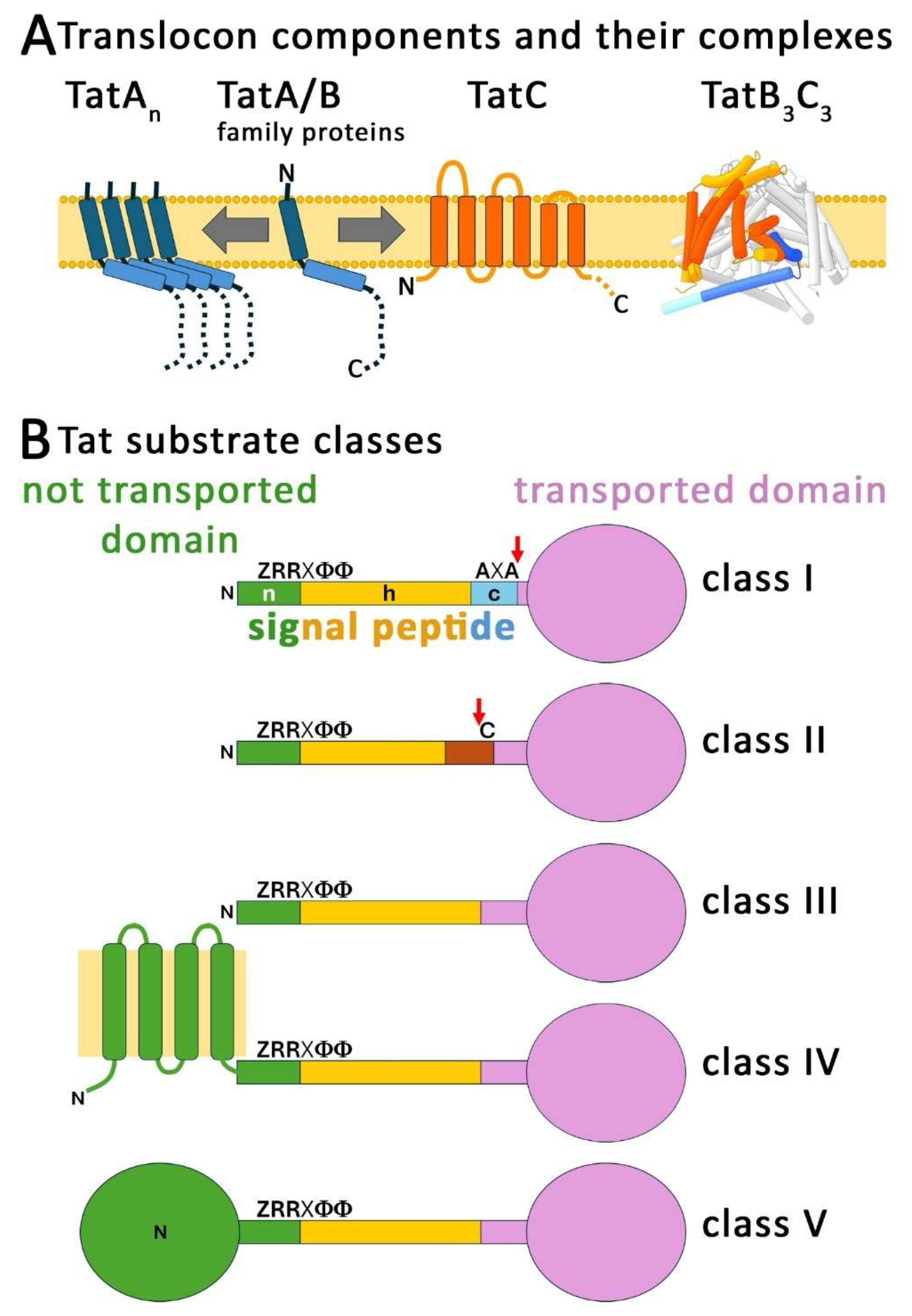
Figure 2.
Tat system complexes. (A) Structure of the TatB3C3 complex with one bound signal peptide, based on PDB 9E01 [48]. Shown are three views of the complex: the bottom (left), the side (middle) and the top (right). The position of the TatC TM5/TM6 domain is indicated and highlighted by more intensive colors of the TatC subunits. Each amphipathic helix of the three TatB subunits is drawn as a ribbon for a better view into the TatC cavity. Color code: turquoise, ochre, light pink: TatC subunits; blue, yellow, salmon pink: TatB subunits; red: Tat signal peptide (of MdoD). (B) AF3 model of the TatA ring that is formed by staggered amphipathic helices, as recently postulated [65]. Shown is the model of the Thermoplasma volcanium TatA1-49 ring with 17 subunits (each colored differently), which exemplifies the general ring structure. The positions of the FK-motif residues are indicated, as are the TatA membrane anchors that align the pore. (C) The postulated translocation process. Phase I: signal peptide (red) binding and insertion at TatC (grey), accompanied with unfolding of the N-terminal region of the mature substrate domain (dark grey) under use of the moving TM5/TM6 domain (turquoise, with “D211” highlighted). The amphipathic helix of TatA/B family proteins is also shown (blue). Phase II: TatA ring docking (transparent green) to the exposed TM5 and membrane anchor extraction, resulting in transient membrane disruption that allows transport (arrow). Phase III: closure of the membrane disruption, release and disassembly of TatA rings, release of the substrate, reset of the TatC-containing complex and recovery of membrane energization. ΔΨ: membrane potential; pmf: proton motive force; smf: sodium motive force (in halophilic archaea).
Figure 2.
Tat system complexes. (A) Structure of the TatB3C3 complex with one bound signal peptide, based on PDB 9E01 [48]. Shown are three views of the complex: the bottom (left), the side (middle) and the top (right). The position of the TatC TM5/TM6 domain is indicated and highlighted by more intensive colors of the TatC subunits. Each amphipathic helix of the three TatB subunits is drawn as a ribbon for a better view into the TatC cavity. Color code: turquoise, ochre, light pink: TatC subunits; blue, yellow, salmon pink: TatB subunits; red: Tat signal peptide (of MdoD). (B) AF3 model of the TatA ring that is formed by staggered amphipathic helices, as recently postulated [65]. Shown is the model of the Thermoplasma volcanium TatA1-49 ring with 17 subunits (each colored differently), which exemplifies the general ring structure. The positions of the FK-motif residues are indicated, as are the TatA membrane anchors that align the pore. (C) The postulated translocation process. Phase I: signal peptide (red) binding and insertion at TatC (grey), accompanied with unfolding of the N-terminal region of the mature substrate domain (dark grey) under use of the moving TM5/TM6 domain (turquoise, with “D211” highlighted). The amphipathic helix of TatA/B family proteins is also shown (blue). Phase II: TatA ring docking (transparent green) to the exposed TM5 and membrane anchor extraction, resulting in transient membrane disruption that allows transport (arrow). Phase III: closure of the membrane disruption, release and disassembly of TatA rings, release of the substrate, reset of the TatC-containing complex and recovery of membrane energization. ΔΨ: membrane potential; pmf: proton motive force; smf: sodium motive force (in halophilic archaea).
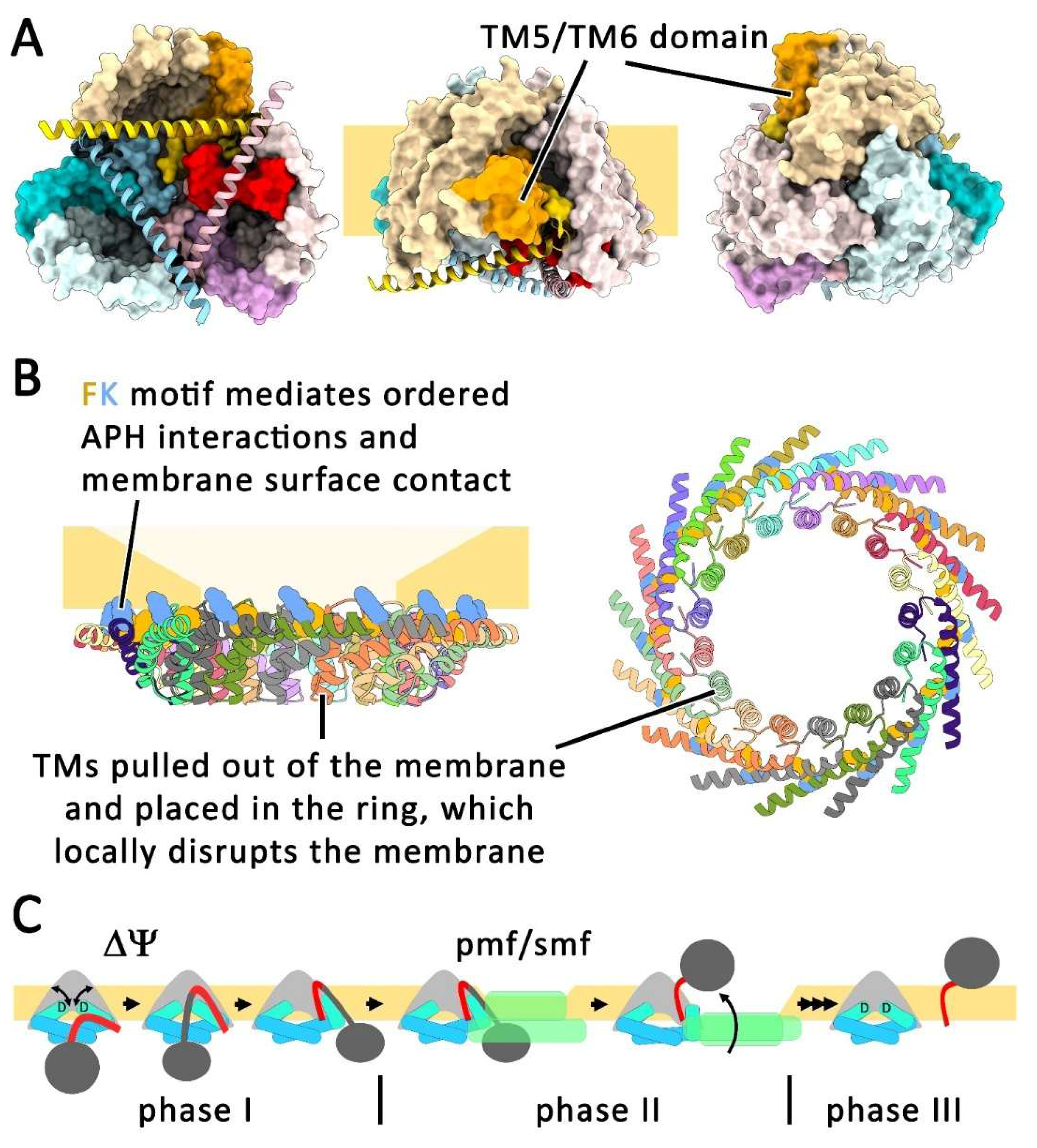
Figure 3.
Asymmetric TatC-containing complexes in haloarchaea. (A) Topology of TatCt and TatCo of Halobacterium. TMs of TatCt that correspond to TM 1-4 of 6TM TatCs are in purple, TMs that correspond to TM5 and TM6 of 6TM TatCs are in light and dark blue. TMs of TatCo that correspond to TM 1-4 of 6TM TatCs are in orange, TMs that correspond to TM5 and TM6 of 6TM TatCs are in yellow. The first cytoplasmic loop that contains the functional RR-motif binding site is in magenta. The positions of the two inactivated and one conserved binding sites are indicated. (B) WebLogo [225] plots of the conserved binding site in TatCo, TatCn, and TatCx (upper plot), and of the mutated binding sites 1 and 2 of TatCt (middle and lower plot, respectively). Color code: hydrophobic residues, orange; polar uncharged residues, green; positively charged residues, blue; negatively charged residues, red; glycine, black. Black bars: insertions; arrows indicate positions corresponding to F94 and E103 in E. coli TatC. Crossed arrows indicate mutated F94/E103 positions. The plots are based on Clustal Omega alignments of sequences from 13 genera of haloarchaea (Haloarcula, Halobacterium, Haloferax, Halogeometricum, Halomicrobium, Haloquadratum, Halorhabdus, Halorubrum, Haloterrigena, Halovivax, Natrialba, Natronobacterium, Natronomonas) [226]. (C) Structure of the heterodimeric TatCt/TatCo complex of Halobacterium salinarium, as predicted by AF3, without the potential inclusion of TatAs. Color code as in (A).
Figure 3.
Asymmetric TatC-containing complexes in haloarchaea. (A) Topology of TatCt and TatCo of Halobacterium. TMs of TatCt that correspond to TM 1-4 of 6TM TatCs are in purple, TMs that correspond to TM5 and TM6 of 6TM TatCs are in light and dark blue. TMs of TatCo that correspond to TM 1-4 of 6TM TatCs are in orange, TMs that correspond to TM5 and TM6 of 6TM TatCs are in yellow. The first cytoplasmic loop that contains the functional RR-motif binding site is in magenta. The positions of the two inactivated and one conserved binding sites are indicated. (B) WebLogo [225] plots of the conserved binding site in TatCo, TatCn, and TatCx (upper plot), and of the mutated binding sites 1 and 2 of TatCt (middle and lower plot, respectively). Color code: hydrophobic residues, orange; polar uncharged residues, green; positively charged residues, blue; negatively charged residues, red; glycine, black. Black bars: insertions; arrows indicate positions corresponding to F94 and E103 in E. coli TatC. Crossed arrows indicate mutated F94/E103 positions. The plots are based on Clustal Omega alignments of sequences from 13 genera of haloarchaea (Haloarcula, Halobacterium, Haloferax, Halogeometricum, Halomicrobium, Haloquadratum, Halorhabdus, Halorubrum, Haloterrigena, Halovivax, Natrialba, Natronobacterium, Natronomonas) [226]. (C) Structure of the heterodimeric TatCt/TatCo complex of Halobacterium salinarium, as predicted by AF3, without the potential inclusion of TatAs. Color code as in (A).
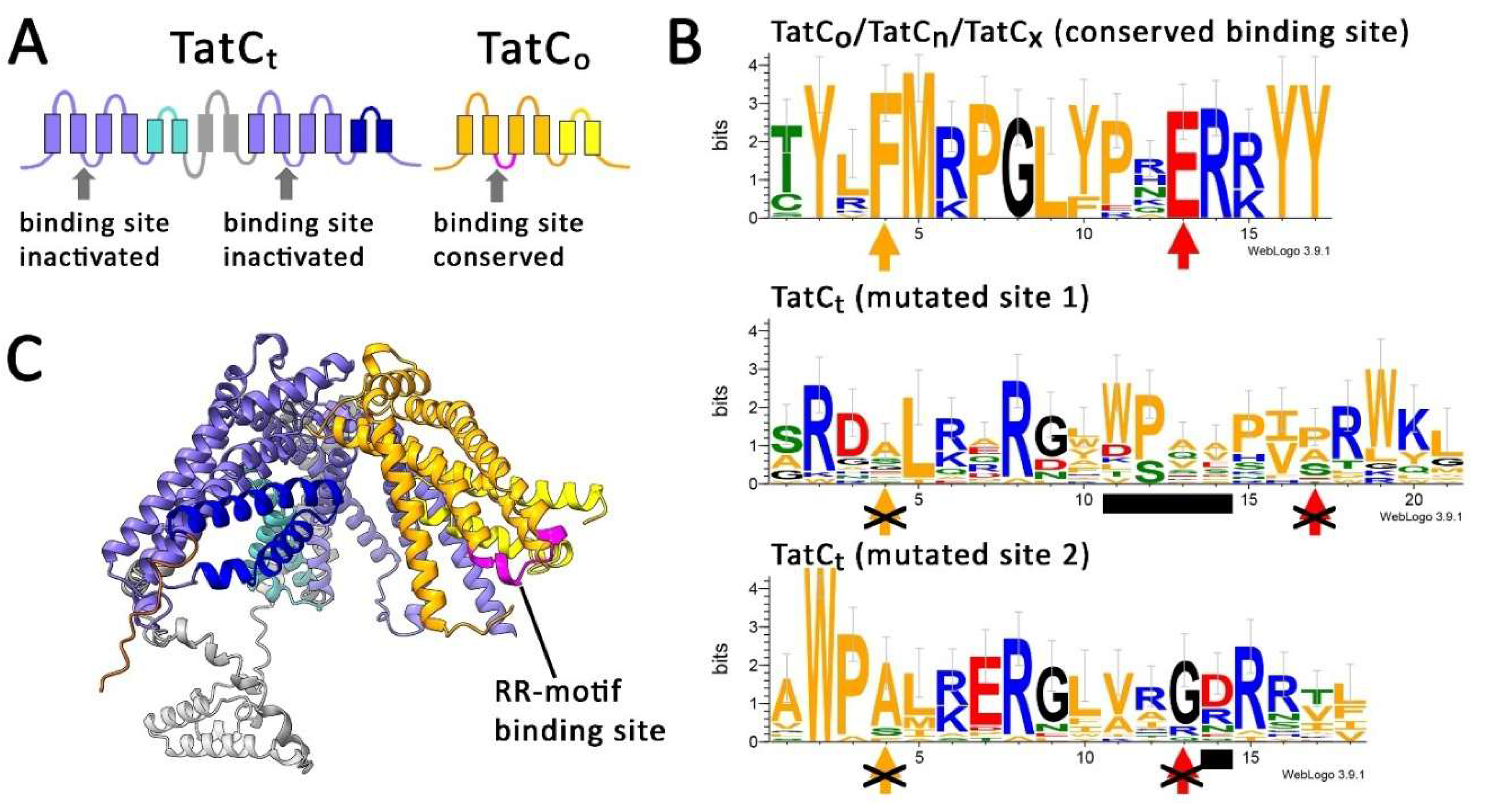
Figure 4.
Placing the origin of TatC within the current understanding of pre-LUCA evolution. (A) Activities of the ancient signal peptide insertases YidC, SecY, and TatC. The structures show surface characteristics in terms of hydrophobicity (ochre, hydrophobic; turquoise, hydrophilic), generated by ChimeraX [224]. The YidC structure is based on the AF3 data base (AF-Q7MQK5-F1-v6), the SecY structure on PDB 1RHZ [227], and the TatC3 complex model was generated by AF3 (based on E. coli TatC). The insertion of a hydrophobic region (dark ochre) next to a positively charged region (blue) is catalyzed in the indicated orientations. (B) Current hypothetical view of the pre-LUCA evolution, with the evolved components, conditions and processes ordered according to an arbitrary time scale (arrow). DNA may have evolved later [157]; Tat systems are associated with “translocation”.
Figure 4.
Placing the origin of TatC within the current understanding of pre-LUCA evolution. (A) Activities of the ancient signal peptide insertases YidC, SecY, and TatC. The structures show surface characteristics in terms of hydrophobicity (ochre, hydrophobic; turquoise, hydrophilic), generated by ChimeraX [224]. The YidC structure is based on the AF3 data base (AF-Q7MQK5-F1-v6), the SecY structure on PDB 1RHZ [227], and the TatC3 complex model was generated by AF3 (based on E. coli TatC). The insertion of a hydrophobic region (dark ochre) next to a positively charged region (blue) is catalyzed in the indicated orientations. (B) Current hypothetical view of the pre-LUCA evolution, with the evolved components, conditions and processes ordered according to an arbitrary time scale (arrow). DNA may have evolved later [157]; Tat systems are associated with “translocation”.
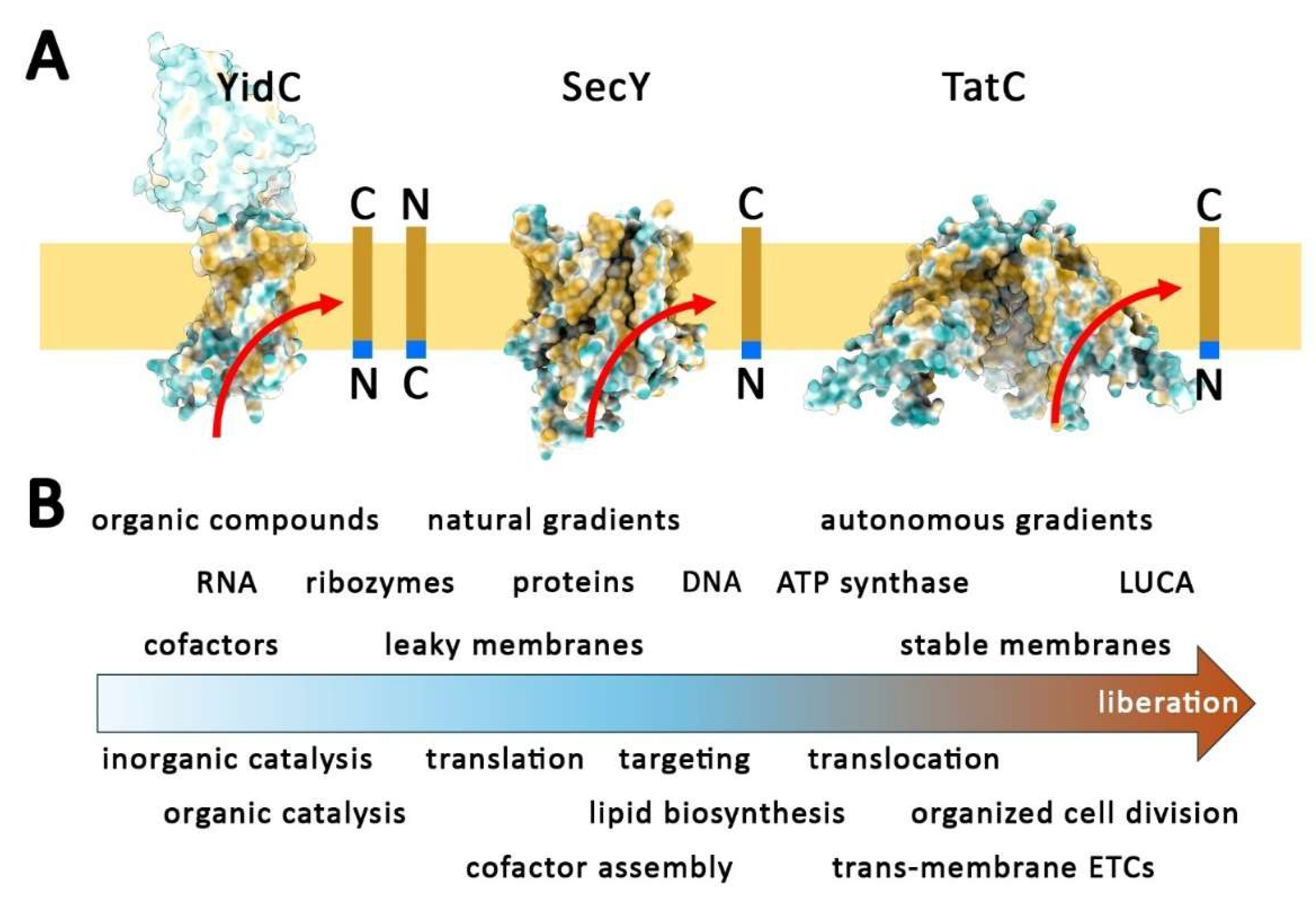
Figure 5.
Adaptation of TatC to acidic habitats dominates over organismic clustering in archaea. Phylogenetic tree of 108 TatC sequences from representatives of Tat-system-containing bacteria, archaea, plastids and mitochondria, highlighting the separate clades of the extremely acidophilic archaea that do not group according to the organismic phylogeny. For the analysis, the TatC alignment was generated by use of MAFFT7 with Smith-Waterman-based local pairwise alignment to account for the highly divergent sequences with conserved domains, variable N- and C-termini and insertions and deletions. Up to 1000 iterations were allowed to increase accuracy. Trimming was done using CLIPKIT software in the kpic-smart-gap mode. The final tree was generated by IQ-TREE with automated model finder, ultrafast bootstrapping with 1000 replicates, and an SH-aLERT-Test with 1000 replicates. Color code: red, extremely acidophilic archaeal TatCs; ochre, other archaeal TatCs; blue, bacteria (except cyanobacteria); purple, mitochondria; green, cyanobacteria and plastids; α, Rickettsia (closest relative to mitochondria); The inset shows the bootstrap-implemented tree. Most nodes are supported by high bootstrap values, indicating a generally robust and reliable tree topology. Low support values at several deep bacterial nodes indicate limited confidence in the resolution of these branches. However, this is irrelevant for the purpose of this tree. [End of Figure 5 caption].
Figure 5.
Adaptation of TatC to acidic habitats dominates over organismic clustering in archaea. Phylogenetic tree of 108 TatC sequences from representatives of Tat-system-containing bacteria, archaea, plastids and mitochondria, highlighting the separate clades of the extremely acidophilic archaea that do not group according to the organismic phylogeny. For the analysis, the TatC alignment was generated by use of MAFFT7 with Smith-Waterman-based local pairwise alignment to account for the highly divergent sequences with conserved domains, variable N- and C-termini and insertions and deletions. Up to 1000 iterations were allowed to increase accuracy. Trimming was done using CLIPKIT software in the kpic-smart-gap mode. The final tree was generated by IQ-TREE with automated model finder, ultrafast bootstrapping with 1000 replicates, and an SH-aLERT-Test with 1000 replicates. Color code: red, extremely acidophilic archaeal TatCs; ochre, other archaeal TatCs; blue, bacteria (except cyanobacteria); purple, mitochondria; green, cyanobacteria and plastids; α, Rickettsia (closest relative to mitochondria); The inset shows the bootstrap-implemented tree. Most nodes are supported by high bootstrap values, indicating a generally robust and reliable tree topology. Low support values at several deep bacterial nodes indicate limited confidence in the resolution of these branches. However, this is irrelevant for the purpose of this tree. [End of Figure 5 caption].
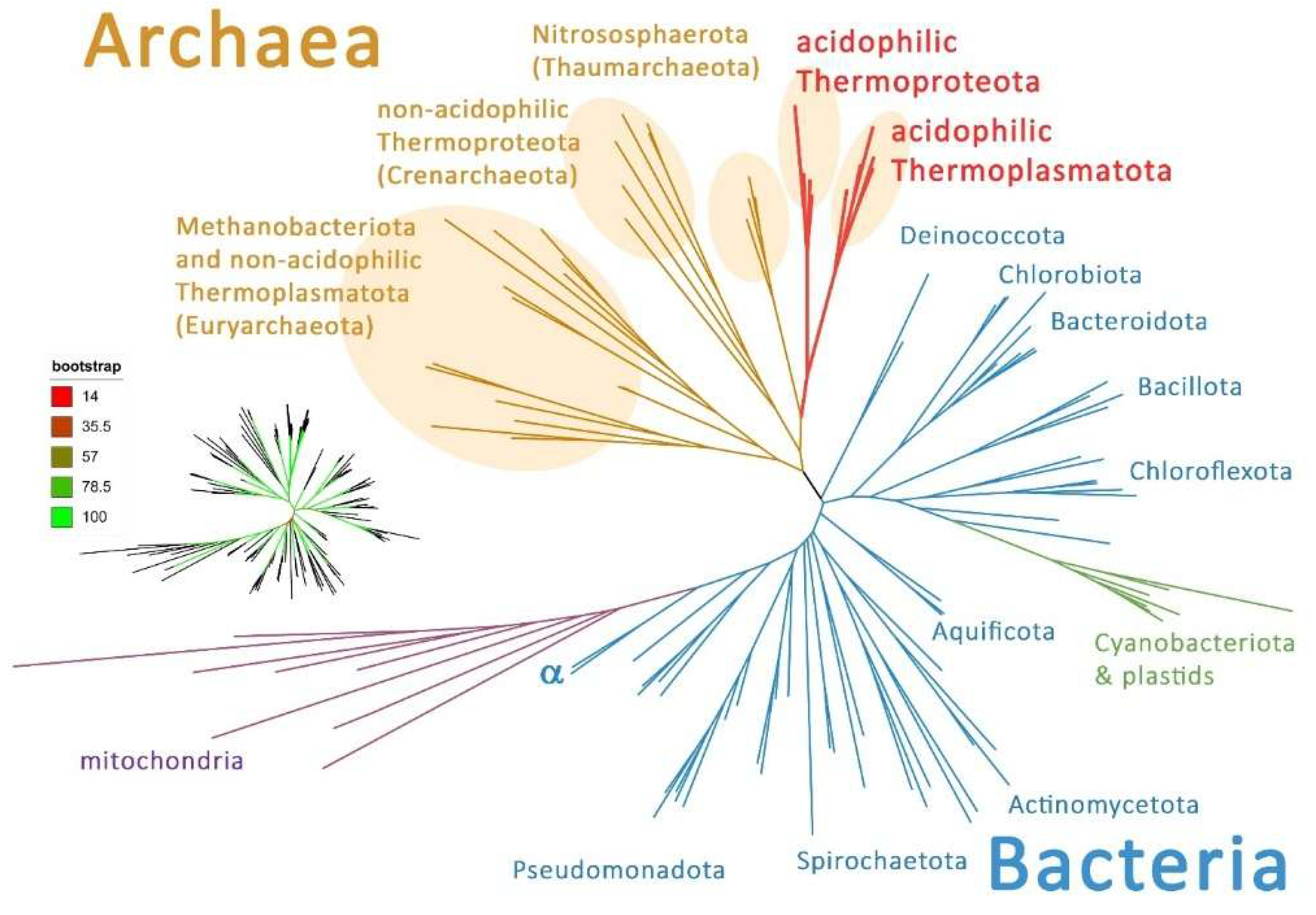
Figure 6.
Tat transport can function in membranes with no significant ΔΨ. (A) Scheme of an extremely acidophilic archaeon, highlighting the enormous pH gradient and the inverted membrane potential at the cytoplasmic membrane. The A-type ATP synthase of these organisms is entirely triggered by the ΔpH. (B) AF3 model of a representative TatC3 complex from an extremely acidophilic archaeon, highlighting the hairpin at the end of TM1 (red), as well as the position of the TM5/TM6 (orange) with its connecting turn (grey). (C) WebLogo plots of TatC-TM5/TM6 domains of the TatC alignment used for the phylogenetic tree. The regions of TM5, the turn and TM6 are indicated at the bottom (colors as in (B)). Upper plot: TatCs from bacteria, mitochondria and plastids, middle plot: TatCs from normal archaeal systems, lower plot: TatCs from extremely acidophilic archaea and Ignicoccus hospitalis. (D) Scheme of an Ignicoccus cell with its outer cellular membrane (OCM) and inner membrane (IM). Shown are also a key membrane-energizing ETC and the A-type ATP synthase in the OCM. No membrane energization is known for the IM, nor are ATP synthases in that membrane.
Figure 6.
Tat transport can function in membranes with no significant ΔΨ. (A) Scheme of an extremely acidophilic archaeon, highlighting the enormous pH gradient and the inverted membrane potential at the cytoplasmic membrane. The A-type ATP synthase of these organisms is entirely triggered by the ΔpH. (B) AF3 model of a representative TatC3 complex from an extremely acidophilic archaeon, highlighting the hairpin at the end of TM1 (red), as well as the position of the TM5/TM6 (orange) with its connecting turn (grey). (C) WebLogo plots of TatC-TM5/TM6 domains of the TatC alignment used for the phylogenetic tree. The regions of TM5, the turn and TM6 are indicated at the bottom (colors as in (B)). Upper plot: TatCs from bacteria, mitochondria and plastids, middle plot: TatCs from normal archaeal systems, lower plot: TatCs from extremely acidophilic archaea and Ignicoccus hospitalis. (D) Scheme of an Ignicoccus cell with its outer cellular membrane (OCM) and inner membrane (IM). Shown are also a key membrane-energizing ETC and the A-type ATP synthase in the OCM. No membrane energization is known for the IM, nor are ATP synthases in that membrane.
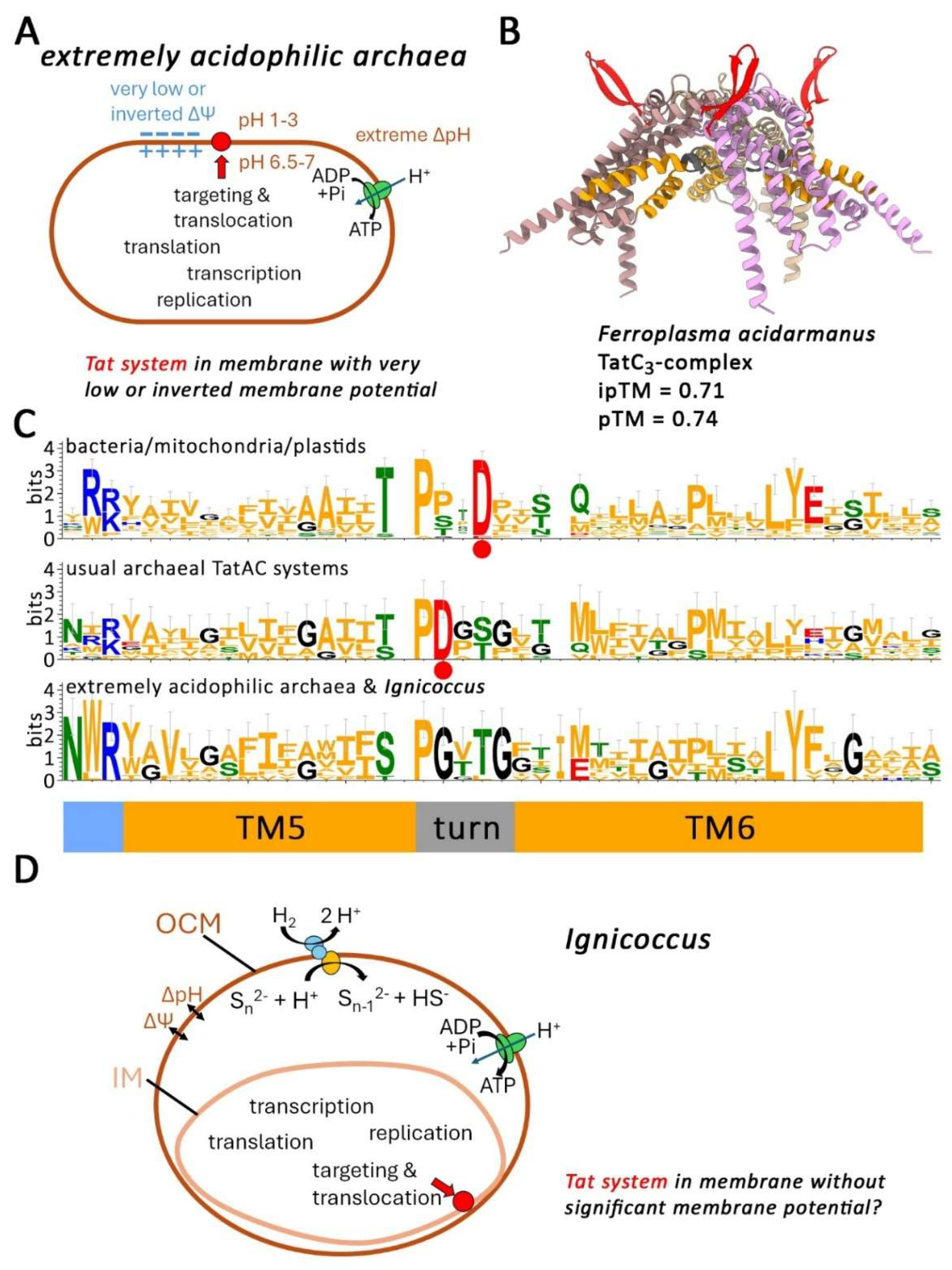
Disclaimer/Publisher’s Note: The statements, opinions and data contained in all publications are solely those of the individual author(s) and contributor(s) and not of MDPI and/or the editor(s). MDPI and/or the editor(s) disclaim responsibility for any injury to people or property resulting from any ideas, methods, instructions or products referred to in the content. |
© 2026 by the authors. Licensee MDPI, Basel, Switzerland. This article is an open access article distributed under the terms and conditions of the Creative Commons Attribution (CC BY) license (http://creativecommons.org/licenses/by/4.0/).
Copyright: This open access article is published under a Creative Commons CC BY 4.0 license, which permit the free download, distribution, and reuse, provided that the author and preprint are cited in any reuse.