

Submitted:
30 June 2026
Posted:
30 June 2026
You are already at the latest version
Abstract
Background
Non-massive hemoptysis is generally considered low-risk and manageable with conservative treatment. However, some patients progress to massive hemoptysis during follow-up. Identifying high-risk patients early in the emergency department matters for calibrating monitoring intensity, guiding timely intervention, and allocating acute care resources.
This study aimed to identify clinical, laboratory, and radiological predictors of progression to massive hemoptysis within the first 72 hours in emergency department patients presenting with non-massive hemoptysis who were managed conservatively.
Methods
This prospective cohort study enrolled patients at a tertiary university hospital emergency department between November 2023 and June 2025. Adult patients presenting with non-massive hemoptysis were enrolled consecutively. The primary outcome was development of massive hemoptysis within 72 hours of admission. Patients were divided into two groups: those who developed massive hemoptysis within 72 hours and those who did not. Demographic, bleeding, laboratory, imaging, and bronchoscopy data were recorded for all patients. Multivariate logistic regression was used to identify independent predictors.
Results
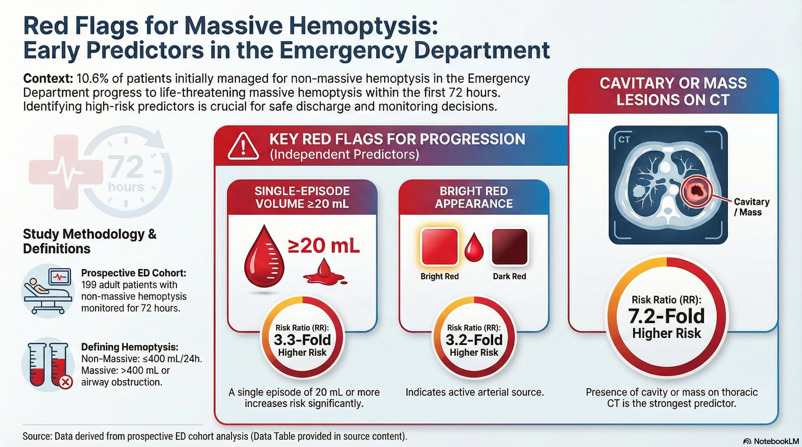
Of 199 patients, 10.6% developed massive hemoptysis within the first 72 hours. On multivariate analysis, bright red hemoptysis (3.17-fold increase in risk), a cavity or mass on chest CT (7.13-fold increase in risk), and bleeding volume ≥20 mL in a single episode (3.3-fold increase in risk) were independent predictors of massive hemoptysis.
Conclusion
A meaningful proportion of patients presenting with non-massive hemoptysis go on to develop massive hemoptysis in the early period. Simple clinical and radiological parameters available at admission can support early risk stratification and inform decisions about monitoring intensity and timely intervention.
Keywords:
hemoptysis
; risk factor
; clinical prognosis
; emergency department
; risk stratification
1. Introduction
Hemoptysis, the expectoration of blood from the lung parenchyma or tracheobronchial tree, is a common emergency department admission and a significant diagnostic challenge for clinicians. Severity is most commonly gauged by the volume of blood expectorated in the preceding 24 hours, and bleeding is broadly divided into two categories: non-massive (mild to moderate) hemoptysis and life-threatening massive hemoptysis [1,2,3].
However, the definition of massive hemoptysis remains highly heterogeneous in the literature. Reported threshold volumes range from 100 to 1,000 mL over 24 hours [4,5,6,7,8,9,10]. This variability reflects differences in clinical context, study populations, and how severity itself has been conceptualized over time. Some expert recommendations shift the focus from absolute volume to clinical impact, defining massive hemoptysis by its life-threatening consequences. Under this framework, massive hemoptysis encompasses bleeding that becomes life-threatening due to airway obstruction, hypoxemia, hemodynamic instability, or the need for interventions such as blood transfusion, hospitalization, or mechanical ventilation [10,11,12]. Either way, the clinical impact of bleeding may matter as much as the volume itself when assessing patient risk.
Massive hemoptysis remains one of the most feared emergencies in pulmonary and emergency medicine, given its potential to cause life-threatening complications such as airway obstruction and respiratory failure. Although it accounts for only about 5% of all hemoptysis cases, reported mortality rates range from 6.5% to 38% [13,14,15]. Most cases originate from the bronchial circulation; bleeding under systemic arterial pressure can turn life-threatening within minutes.[16] In the emergency department, distinguishing patients likely to remain stable from those at risk of rapid deterioration is therefore a real challenge. Identifying high-risk patients early is essential for timely decisions about monitoring, admission, and treatment.
Unlike massive hemoptysis, non-massive hemoptysis is generally considered low-risk and is usually managed conservatively. However, its clinical course can be unpredictable, and it may be the first sign of a serious underlying disease [1,3,17,18]. Prior studies have shown that recurrence rates can reach 17–19% over long-term follow-up, and some patients go on to develop increasingly severe bleeding episodes [12,19]. These findings suggest that non-massive hemoptysis should not always be treated as a benign condition.
Although existing clinical scoring systems aim to predict early poor outcomes, no prospective data evaluate progression from non-massive to massive hemoptysis within the first 72 hours of admission [1,18]. This gap is particularly relevant for emergency department discharge decisions and for identifying patients at risk of early deterioration.
This study aimed to assess how often non-massive hemoptysis progresses to massive hemoptysis within the first 72 hours in emergency department patients, and to identify the clinical, laboratory, and radiological risk factors associated with that progression.
2. Material and Methods
2.1. Study Design and Setting
This prospective cohort study was conducted in the emergency department of a university hospital in Turkey between November 1, 2023 and June 1, 2025, with follow-up carried out in collaboration with the pulmonology department. The study protocol was reviewed and approved by the institutional ethics committee (Approval No: 1235, October 11, 2023). The study was conducted in accordance with the ethical principles of the 1964 Declaration of Helsinki and its subsequent amendments. Written informed consent was obtained from all participants prior to enrollment.
All adult patients presenting to the emergency department with hemoptysis during the study period were consecutively evaluated (n=278). Excluded were patients with a prior history of massive hemoptysis (n=16), those with bleeding from the oral cavity, upper airway, or gastrointestinal tract that could mimic hemoptysis (n=48), those with an international normalized ratio (INR) >2 (n=10), and individuals with missing data (n=5). After applying these exclusion criteria, 199 patients who completed all clinical assessments were included in the final analysis. Patients were followed prospectively for 30 days. The primary outcome was development of massive hemoptysis within the first 72 hours of admission.
The dataset included demographic characteristics, medical history, laboratory findings, flexible fiberoptic bronchoscopy (FOB) results, multi-detector contrast-enhanced thoracic computer tomography (CT) angiography findings, bleeding volume and characteristics, hemoptysis recurrence, hospitalization, interventional procedures, and early and late prognostic outcomes.
2.2. Definitions and Assessment of Bleeding Volume
Massive and non-massive hemoptysis were classified using volume-based thresholds widely used in the literatüre [4,5,6,7,8,9,10]. Bleeding exceeding 400 mL in the 24 hours before admission was defined as massive hemoptysis; bleeding ≤400 mL over the same period was classified as non-massive.
To improve standardization of patient-reported bleeding volume estimates, household volume references were used. Patients were shown common household measures, including a teaspoon (2.5 mL), dessertspoon (5 mL), tablespoon (10 mL), tea glass (100 mL), and water glass (200 mL), and asked to identify which container and what proportion of it best matched their total expectorated blood volume.
2.3. Imaging Techniques
All posteroanterior chest radiographs were acquired by radiologic technologists with a minimum of three years of experience using a Samsung® XGEO GC80 system, in accordance with the European Guidelines on Quality Criteria for Diagnostic Radiographic Image [20]. Thoracic CT examinations were performed by technologists with at least three years of experience using a GE Optima® CT660® 128-detector spiral CT scanner, following an aortic-phase thoracic CT angiography protocol [21]. During thoracic CT angiography, 50 mL of an iohexol-based intravenous contrast agent was administered as a bolus injection via an antecubital vein, followed by a saline flush at a rate of 4.5 mL/s.
Bronchoscopy Standardization
All fiberoptic bronchoscopy procedures were performed by two experienced bronchoscopists using a Fujifilm® EB-580T flexible fiberoptic bronchoscope. The procedure was performed for diagnostic purposes, to identify the bleeding source and evaluate endobronchial pathologies, with no intent for emergency therapeutic intervention. In most cases, bronchoscopy was performed electively based on clinical assessment. Active bleeding, bloody secretions, clots, and other endobronchial findings were systematically recorded during bronchoscopy.
2.4. Follow-Up and Primary Outcome
Patients were followed clinically for 30 days after admission. Although follow-up continued for the full 30 days, development of massive hemoptysis within the first 72 hours of admission was pre-specified as the primary outcome and defined as the early period. Patients were divided into two groups based on whether massive hemoptysis developed within this period. The two groups were compared by clinical characteristics, laboratory parameters, and imaging findings (Figure 1).
2.5. Statistical Analysis
Statistical analyses were performed using SPSS version 25 (IBM Corp., Armonk, NY, USA). Categorical variables are summarized as counts and percentages; continuous variables are reported as mean±standard deviation or median (25th–75th percentile) depending on their distribution. The distribution of continuous variables was assessed using the Kolmogorov-Smirnov test. For between-group comparisons of categorical variables, the chi-square test was used; Fisher's exact test was applied when chi-square assumptions were not met. For multi-cell contingency tables where the Pearson chi-square test was inappropriate, Fisher's exact test with Monte Carlo simulation was used and two-sided Monte Carlo significance values were reported. Non-normally distributed continuous variables were compared using the Mann-Whitney U test. A p-value <0.05 was considered statistically significant.
Multivariate logistic regression analysis was performed to identify independent predictors of massive hemoptysis within the first 72 hours. Variables significant at p<0.10 in univariate analyses were included as candidates for the multivariate model. To reduce the risk of multicollinearity, only one variable from each pair representing the same clinical construct or showing high intercorrelation was retained in the model (e.g., INR/PT, total 24-hour bleeding volume/single-episode bleeding volume, SpO₂/oxygen requirement). All candidate variables were entered into the full model initially, and the final model was built using purposeful backward elimination.
3. Results
Of the 199 patients included, 68.3% (n=136) were male, with a mean age of 59.4±16.3 years. Active smoking was present in 42.2% (n=84), while 19.1% (n=38) had never smoked. At least one chronic comorbidity was present in 66.3% (n=132); the most common were hypertension (43.7%), coronary artery disease (28.1%), and diabetes mellitus (24.1%). A history of tuberculosis was documented in 7.0% (n=14). Anticoagulant therapy was used by 11.1% (n=22), while 25.1% (n=50) were on antiplatelet therapy.
62.8% of patients reported a first episode of hemoptysis. Recurrent bleeding during emergency department observation occurred in 33.7% (n=67). 10.6% of patients developed massive hemoptysis within the first 72 hours. On assessment of hemoptysis appearance, 45.7% (n=91) described blood-streaked sputum, 43.7% (n=87) reported bright red bleeding, and 10.6% (n=21) reported dark red or black bleeding. The median time from bleeding onset to admission was 24 hours (IQR: 8–48). The median blood volume expectorated in the 24 hours before admission was 40 mL (IQR: 15–100), with patients reporting a median of five bleeding episodes (IQR: 3–5) over the same period. Median bleeding volume per episode was 10 mL (IQR: 5–20).
Pathological findings were present on posteroanterior chest radiographs in 58.8% (n=117) of patients, and in 82.6% (n=161) of the 195 who underwent thoracic CT angiography. The most common CT findings were ground-glass opacities (48.2%) and consolidation (22.1%). FOB was performed in 55.8% (n=111) of patients; additional abnormal endobronchial findings beyond active bleeding were identified in 81.1% (n=90) of these. The three most common bronchoscopic findings were residual blood (62.2%), petechiae (33.3%), and endobronchial lesions (16.2%).
The median hospital stay was 5 days (IQR: 2–8). Thirty-day mortality was 4.0% (n=8). Of the eight deaths, four occurred in the massive hemoptysis group and four in those who did not develop it. Of these eight deaths, four were attributed to underlying malignancy, three to hospital-acquired infections, and one to acute renal failure. Recurrent bleeding was observed in 44.7% (n=89) of patients within the first 72 hours. Of these, 23.6% (n=21) subsequently developed massive hemoptysis.
Among patients who developed massive hemoptysis within the first 72 hours after admission, the median age was 59 years (IQR:55-72), compared with 62 years (IQR:47-70) in those who did not; however, this difference was not statistically significant (p=0.952). Massive hemoptysis incidence was similar in women and men (9.5% vs. 11.0%, p=0.748). On admission, patients who subsequently developed massive hemoptysis had significantly lower oxygen saturation (SpO₂) and significantly higher heart rate compared with those who did not develop massive hemoptysis (p=0.012 and p=0.041, respectively). In addition, INR, prothrombin time (PT), and urea levels were significantly higher in patients who developed massive hemoptysis (p=0.012, p=0.023, and p=0.047, respectively) (Table 1).
At admission, patients who subsequently developed massive hemoptysis within the first 72 hours reported a higher median total bleeding volume during the preceding 24 hours compared with those who did not develop massive hemoptysis (100 mL [IQR: 40–200] vs. 30 mL [IQR: 15–100], p=0.003). The median bleeding volume per single episode at admission was also higher in patients who later progressed to massive hemoptysis (20 mL [IQR: 5–50] vs. 10 mL [IQR: 5–20], p=0.008). These patients also had a longer median hospital stay (7 vs. 5 days, p=0.002). Thirty-day mortality was significantly higher in patients who developed massive hemoptysis within the first 72 hours (19.0% [n=4] vs. 2.3% [n=4], p<0.001). Furthermore, intensive care unit admission was observed in 52.4% of patients with massive hemoptysis, compared with only 6.7% in those without massive hemoptysis (p<0.001). Detailed information regarding bleeding characteristics and prognostic outcomes is presented in Table 2.
The two groups did not differ significantly in smoking status and comorbidity burden (p=0.771 and p=0.346, respectively). Pathological findings on posteroanterior chest radiograph were not significantly associated with massive hemoptysis (p=0.438). In contrast, any pathological finding on thoracic CT, and cavitary and mass lesions in particular, were significantly associated with an increased risk of massive hemoptysis within the first 72 hours (p=0.028). In addition, the need for oxygen support at admission, bright red bleeding, and a history of previous hemoptysis were also significantly associated with massive hemoptysis during the first 72 hours (p=0.002, p=0.008, and p=0.045, respectively). The associations between clinical characteristics and the risk of massive hemoptysis within the first 72 hours are detailed in Table 3.
On multivariate logistic regression, bright red hemoptysis, bleeding volume per episode, and a cavitary or mass lesion on thoracic CT were independent predictors of massive hemoptysis within the first 72 hours after admission. Each 1 mL increase in bleeding volume per episode was associated with a 1.7% increase in the odds of developing massive hemoptysis. Total bleeding volume over 24 hours, oxygen requirement at admission, history of hemoptysis, urea level, heart rate, and SpO₂ were all entered into the multivariate model but none emerged as independent predictors. These findings are summarized in Table 4.
ROC analysis identified an optimal threshold of ≥17.5 mL for bleeding volume per episode in predicting massive hemoptysis within the first 72 hours (Youden index: 0.27) (Figure 2). At this threshold, sensitivity was 52.4% and specificity was 74.7%. Discriminative performance was moderate, with an area under the curve (AUC) of 0.672 (95% CI: 0.549–0.796). Since bleeding volume in clinical practice is usually estimated rather than precisely measured, and a more practical threshold was needed for decision-making, the threshold was rounded to 20 mL for clinical use. Additional analyses showed that patients with a bleeding volume per episode of ≥20 mL had approximately 3.3 times higher odds of developing massive hemoptysis than those with <20 mL (OR=3.25; 95% CI: 1.29–8.16).
Figure 1.
Flow chart.
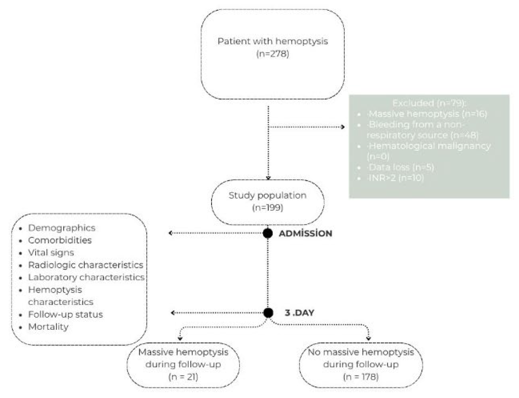
Figure 2.
Receiver operating characteristic (ROC) curve illustrating the predictive performance of single-episode bleeding volume for massive hemoptysis.
Figure 2.
Receiver operating characteristic (ROC) curve illustrating the predictive performance of single-episode bleeding volume for massive hemoptysis.
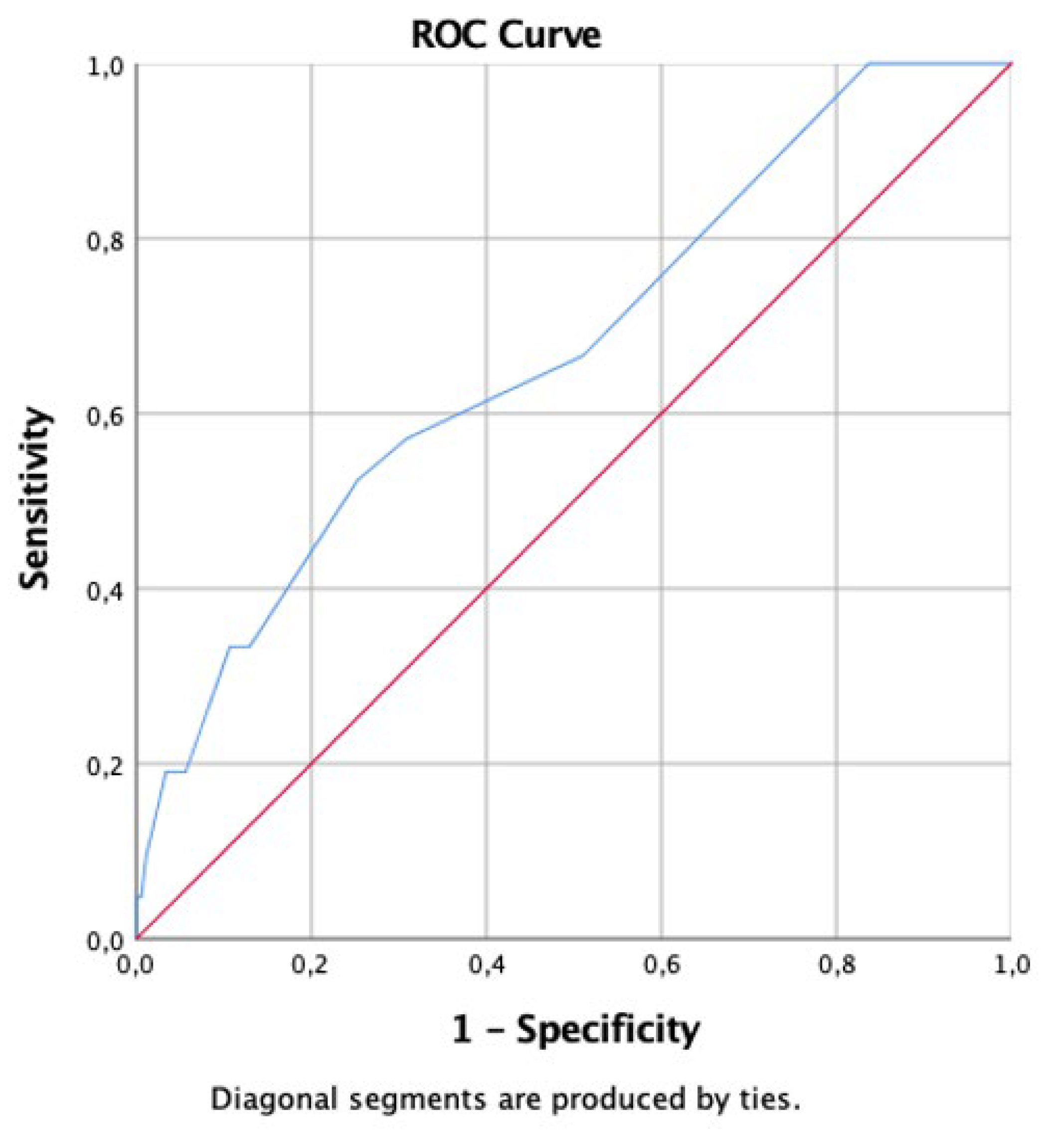

Table 1.
Comparison of laboratory and vital parameters between the groups.
| Variables (median [IQR]) |
No massive hemoptysis (n = 178) | Massive hemoptysis (n = 21) | p value |
|---|---|---|---|
| Systolic blood pressure (mmHg) | 130 (120–152) | 137 (100–157) | 0.904 |
| Diastolic blood pressure (mmHg) | 75 (70–80) | 80 (66–90) | 0.576 |
| Heart rate (beats/min) | 85 (80–95) | 94 (82–110) | 0.041 |
| SpO₂ (%) | 97 (95–98) | 95 (88–97) | 0.012 |
| White blood cell count (cells/µL) | 8,550 (7,230–10,650) | 8,240 (7,470–11,400) | 0.997 |
| Neutrophil count (cells/µL) | 5,540 (4,250–7,900) | 5,660 (4,770–6,270) | 0.966 |
| Lymphocyte count (cells/µL) | 1,860 (1,360–2,440) | 1,600 (1020–2,320) | 0.388 |
| Hemoglobin (g/dL) | 13.1 (11.3–14.3) | 12.6 (11.2–14.5) | 0.778 |
| Hematocrit (%) | 39.2 (35.4–42.8) | 38.5 (34.7–43.6) | 0.949 |
| Platelet count (×10³/µL) | 253 (200–299) | 234 (209–401) | 0.994 |
| Urea (mg/dL) | 32 (25–41.5) | 37.3 (29.4–56.0) | 0.047 |
| AST (U/L) | 18 (14–23) | 15 (13–23) | 0.445 |
| ALT (U/L) | 14 (10–22) | 15 (12–20) | 0.997 |
| CRP (mg/L) | 8 (2.5–39.3) | 8 (3.8–47.1) | 0.533 |
| INR | 1.0 (0.9–1.1) | 1.1 (1.0–1.2) | 0.012 |
| PT (s) | 12.2 (11.5–13.2) | 13.1 (12.3–14.0) | 0.023 |
| aPTT (s) | 25.2 (23.4–27.1) | 26.4 (24.4–28.3) | 0.242 |
SpO₂: Oxygen saturation, AST: Aspartate aminotransferase, ALT: Alanine aminotransferase, CRP: C-reactive protein, INR: International normalized ratio, PT: Prothrombin time, aPTT: Activated partial thromboplastin time, IQR: Interquartile range.
Table 2.
Comparison of bleeding characteristics and length of hospital stay between the groups.
| Variables (median [IQR]) |
No massive hemoptysis (n = 178) |
Massive hemoptysis (n = 21) |
p value |
|---|---|---|---|
| Total bleeding volume within 24 hours (mL) | 30 (15–100) | 100 (40–200) | 0.003 |
| Single-episode bleeding volume (mL) | 10 (5–20) | 20 (5–50) | 0.008 |
| Time since bleeding onset (hours) | 22 (8–48) | 24 (12–72) | 0.151 |
| Number of bleeding episodes, | 5 (3–5) | 4 (3–5) | 0.683 |
| Length of hospital stay (days) | 5 (0–7) | 7 (6–9) | 0.002 |
| 30-day mortality, n (%) | 4 (2.3) | 4 (19.0) | <0.001 |
IQR: Interquartile range.
Table 3.
Association between clinical characteristics and the risk of developing massive hemoptysis within the first 72 hours.
Table 3.
Association between clinical characteristics and the risk of developing massive hemoptysis within the first 72 hours.
| Clinical characteristics | Category | Massive hemoptysis (n = 21), % (n) |
p value |
|---|---|---|---|
| O2 requirement at admission | No | 7.8 (14) | 0.002 † |
| Yes | 35.0 (7) | ||
| Type of bleeding | Blood-streaked sputum | 4.4 (4) | 0.008 ‡ |
| Bright red blood | 18.4 (16) | ||
| Dark red/black blood | 4.8 (1) | ||
| Presence of bleeding in the emergency department | No | 7.6 (10) | 0.055 |
| Yes | 16.4 (11) | ||
| History of hemoptysis | No | 7.2 (9) | 0.045 |
| Yes | 16.2 (12) | ||
| Pathological findings on chest X-ray | No | 8.5 (7) | 0.438 |
| Yes | 12.0 (14) | ||
| Pathological findings on chest CT | No | 0 | 0.028 † |
| Yes | 13.0 (21) | ||
| Cavitary lesion on chest CT | No | 8.4 (15) | 0.004 † |
| Yes | 35.3 (6) | ||
| Mass lesion on chest CT | No | 8.3 (13) | 0.037 † |
| Yes | 21.1 (8) |
CT: computer tomography, O2: Oxygen; †Fisher’s exact test was used.; ‡ Due to low expected cell frequencies, the assumptions of the Pearson chi-square test were not met. Therefore, Fisher’s exact test with Monte Carlo simulation was applied, and the Monte Carlo two-sided significance value was reported.
Table 4.
Multivariable logistic regression analysis for the development of massive hemoptysis.
| Variable | OR | 95% CI | p value |
|---|---|---|---|
| Bright red hemoptysis | 3.174 | 1.022–9.851 | 0.046 |
| Single-episode bleeding volume (per mL) | 1.017 | 1.004–1.031 | 0.012 |
| Presence of cavitary or mass lesion on chest CT | 7.135 | 2.060–24.715 | 0.002 |
CT: Computer tomography, OR: Odds ratio CI: Confidence interval; Hosmer–Lemeshow p = 0.240; Nagelkerke R²:0.256; Reference category: dark red/black hemoptysis and blood-streaked sputum.
4. Discussion
Hemoptysis is associated with a risk of recurrence and clinical deterioration even when the initial bleeding volume is limited. Previous studies have reported recurrence rates of up to 19% during long-term follow-up among patients presenting with mild hemoptysis, and approximately 9% of patients who re-present may subsequently develop massive hemoptysis [10,17,19]. These findings show that some patients initially classified as non-massive may progress to more severe bleeding over time. While prior studies have focused mainly on long-term outcomes, our findings show that progression to massive hemoptysis can occur as early as the first 72 hours after admission. This suggests that non-massive hemoptysis should not always be considered low-risk, and highlights the importance of early risk stratification and close monitoring in the period immediately after admission.
Studies evaluating long-term mortality in hemoptysis report an overall rate of approximately 13.7%, with lung malignancy as the strongest predictor [10]. The 30-day mortality rate in our study was comparable to the in-hospital rates reported by Hirshberg et al. and Fartoukh et al., despite differences in follow-up duration [8,22]. Mortality was significantly higher among patients who developed massive hemoptysis within the first 72 hours than among those who did not. Although bleeding was not the direct cause of death in every case, early progression to massive hemoptysis appears to identify a subgroup with markedly worse short-term outcomes. Even in patients presenting with non-massive hemoptysis, the first 72 hours therefore call for close monitoring and careful consideration of discharge decisions.
Variability in the emergency department management of hemoptysis, the lack of consensus regarding the definition of massive hemoptysis, and the inherent unreliability of bleeding volume estimation have led clinicians to favor risk stratification approaches based on “impact severity,” such as airway obstruction, hemodynamic instability, and the need for intensive care, rather than absolute volume alone [1,18]. Risk stratification tools such as the Florence Hemoptysis Score (FLHASc) have been developed to predict early poor outcomes. The FLHASc incorporates easily assessed variables including systolic blood pressure <100 mmHg, a history of malignancy, and more than two hemoptysis episodes in the preceding 24 hours, and suggests that patients with a score of 0 and a normal chest radiograph can be safely discharged from the emergency department [1]. In our study, bright red hemoptysis, higher bleeding volume per episode, and cavitary or mass lesions on thoracic CT were each independently associated with massive hemoptysis within the first 72 hours. These findings suggest that imaging features and bleeding characteristics can provide prognostic information beyond existing clinical risk tools and may help refine early risk stratification in patients presenting with non-massive hemoptysis.
In the FLHASc study, 5.8% of patients discharged with mild hemoptysis returned to the emergency department within three months due to recurrent hemoptysis, and 1.5% re-presented within the first 72 hours after discharge [1]. However, in that study and in others, the definition and timing of massive hemoptysis have generally relied on 24-hour or longer windows, and none specifically reported the rate of progression from non-massive to massive hemoptysis [1,14,15,18]. In this respect, the present study distinguishes itself from prior work by providing original data on early progression from non-massive to massive hemoptysis.
Previous studies have proposed different threshold values for 24-hour total bleeding volume as a prognostic marker in hemoptysis [1,23]. In the FLHASc study, prognosis was not associated with the total bleeding volume over 24 hours but rather with the frequency of bleeding episodes during this period (>2 episodes/24 h) [1,11]. In our study, although a significant difference was observed between groups in terms of total bleeding volume during the 24 hours preceding emergency department admission, this variable did not emerge as an independent predictor in multivariable analysis. Likewise, the number of bleeding episodes within 24 hours was not associated with the development of massive hemoptysis. In contrast, a single-episode bleeding volume of ≥20 mL was associated with an approximately threefold increase in the odds of developing massive hemoptysis within the first 72 hours. These findings suggest that the volume of bleeding during an individual episode may provide clinically relevant prognostic information beyond total 24-hour bleeding volume and episode frequency.
Macroscopic characteristics of hemoptysis, including color, appearance, and odor, may provide insights not only into etiology but also into prognosis [24]. However, no study to date has directly assessed the relationship between the macroscopic characteristics of expectorated blood and clinical outcomes. In a study with a mean follow-up of approximately 2.5 years, the presence of active bleeding and residual blood on flexible fiberoptic bronchoscopy was shown to increase the risk of long-term recurrence by 3.3-fold and 2.7-fold, respectively.[19] In our study, bronchoscopic findings were not associated with early progression to massive hemoptysis; however, bright red hemoptysis emerged as an independent predictor within the first 72 hours. Bright red blood at admission likely reflects more active or ongoing bleeding, which may explain this finding. Simple visual features of hemoptysis may therefore carry clinically meaningful prognostic information and should be factored into the initial assessment of patients presenting to the emergency department.
Pulmonary tuberculosis and bronchogenic carcinoma are well-known causes of massive hemoptysis. One reason is that both can produce cavitary or mass lesions in the lung parenchyma, which raises the risk of serious bleeding [14,25]. In keeping with this, our study found that cavitary or mass lesions on thoracic CT were independently associated with massive hemoptysis within the first 72 hours. These findings suggest that these CT features may carry value not only for identifying the underlying etiology but also for early prognostic assessment.
Although SpO₂ has not been consistently shown as an independent predictor of outcomes in hemoptysis, respiratory failure and the need for mechanical ventilation have been linked to poor prognosis in several studies [1,19,22]. In our cohort, SpO₂ at admission was significantly lower in patients who subsequently developed massive hemoptysis. However, SpO₂ did not emerge as an independent predictor in multivariate analysis. This aligns with prior work and suggests that oxygen desaturation reflects the physiological effects of bleeding rather than independently driving clinical deterioration. That said, SpO₂ is quick to measure at the bedside and can add useful clinical context during the initial assessment of patients presenting with hemoptysis.
Uremia impairs hemostasis by disrupting platelet function and endothelial integrity, and bleeding complications are more common in patients with elevated urea levels [26]. In our study, urea levels were significantly higher in patients who developed massive hemoptysis within the first 72 hours, but this association did not survive multivariate analysis after adjusting for other clinical variables. Elevated urea may reflect a higher-risk clinical profile overall; our findings, however, do not support it as an independent predictor of early progression to massive hemoptysis.
This prospective cohort study shows that patients presenting to the emergency department with non-massive hemoptysis can progress to massive hemoptysis within the first 72 hours, and that 30-day mortality is significantly higher in those who do. These findings indicate that some patients presenting with non-massive hemoptysis are at risk of early clinical deterioration and therefore need close monitoring in the first 72 hours after admission. Bright red hemoptysis, bleeding volume per episode exceeding 20 mL, and cavitary or mass lesions on thoracic CT were each independently associated with early progression to massive hemoptysis. Multicenter studies are needed to validate these findings and establish their role in future risk assessment models.
5. Limitations
This study has several limitations. First, the study was conducted at a single university hospital, and patient characteristics and diagnostic and therapeutic approaches may have been shaped by local institutional practices. This may limit the generalizability of the findings. Second, the relatively small number of patients who developed massive hemoptysis within the first 72 hours may have reduced statistical power and introduced greater uncertainty into the multivariate estimates.
Because bleeding volume was assessed through patient self-report and household volume references, some degree of subjectivity was unavoidable. Bronchoscopy was also not performed in all patients, and diagnostic evaluations followed clinical need rather than a predefined standardized protocol. The >400 mL/24-hour threshold used to define massive hemoptysis was chosen from among the heterogeneous definitions in the literature, which may limit direct comparability with studies using different criteria.
Finally, given the observational design, the associations identified should be interpreted as associative rather than causal. External validation in larger, multicenter cohorts is needed before these findings can be integrated into routine clinical risk stratification.
Author Contributions
All authors have read and approved the final version of the manuscript. The authors confirm that the requirements for authorship, as stated in the journal guidelines, have been met, and that each author believes the manuscript represents honest, original, and scientifically sound work.
Funding Statement
This research received no specific grant from any funding agency in the public, commercial, or not-for-profit sectors.
Institutional Review Board Statement
The study protocol was reviewed and approved by the Ethics Committee of İzmir Bakırçay University (Approval No: 1235, Date: October 11, 2023). The study was conducted in accordance with the principles of the Declaration of Helsinki and its subsequent amendments.
Informed Consent Statement
Written informed consent was obtained from all participants prior to enrollment in the study.
Data Availability Statement
The datasets generated and/or analyzed during the current study are available from the corresponding author on reasonable request.
Acknowledgments
An AI-assisted language tool (ChatGPT-4o) was used solely to improve the language, fluency, and readability of the manuscript. They were not used for study design, data collection, data analysis, interpretation of findings, or literature review. All scientific content, analyses, and conclusions were generated and verified by the authors. Furthermore, NotebookLM was used to generate the visual content presented in graphical abstract.
Prior Presentation
This study was previously presented at the 46. Turkish Respiratory Research Association Congress 2024, held on November 21, 2024, in Antalya, Türkiye.
Conflicts of Interest Disclosure
The authors declare that they have no conflicts of interest related to this work.
References
- Vanni S, Bianchi S, Bigiarini S, Casula C, Brogi M, Orsi S, Acquafresca M, Corbetta L, Grifoni S. Management of patients presenting with haemoptysis to a Tertiary Care Italian Emergency Department: the Florence Haemoptysis Score (FLHASc). Intern Emerg Med. 2018 Apr;13(3):397-404. [CrossRef]
- Lee MK, Kim SH, Yong SJ, Shin KC, Kim HS, Yu TS, Choi EH, Lee WY. Moderate hemoptysis: recurrent hemoptysis and mortality according to bronchial artery embolization. Clin Respir J. 2015;9(1):53–64. [CrossRef]
- Ketai LH, Mohammed TLH, Kirsch J, Kanne JP, Chung JH, Donnelly EF, Ginsburg ME, Heitkamp DE, Henry TS, Kazerooni EA, et al. ACR Appropriateness Criteria® hemoptysis. J Thorac Imaging. 2014;29(3):W19–W22. [CrossRef]
- Knott-Craig CJ, Oostuizen JG, Rossouw G, Joubert JR, Barnard PM. Management and prognosis of massive hemoptysis: recent experience with 120 patients. J Thorac Cardiovasc Surg. 1993;105(3):394–397.
- Earwood JS, Thompson TD. Hemoptysis: evaluation and management. Am Fam Physician. 2015 Feb 15;91(4):243–249.
- Ong ZYT, Chai HZ, How CH, Koh J, Low TB. A simplified approach to haemoptysis. Singapore Med J. 2016 Aug;57(8):415–418. [CrossRef]
- Karlafti E, Tsavdaris D, Kotzakioulafi E, Kougias L, Tagarakis G, Kaiafa G, Netta S, Savopoulos C, Michalopoulos A, Paramythiotis D. Which is the best way to treat massive hemoptysis? A systematic review and meta-analysis of observational studies. J Pers Med. 2023;13(12):1649. [CrossRef]
- Hirshberg B, Biran I, Glazer M, Kramer MR. Hemoptysis: etiology, evaluation, and outcome in a tertiary referral hospital. Chest. 1997 Aug;112(2):440–444. [CrossRef]
- Garzon A, Gourin A. Surgical management of massive hemoptysis: a ten-year experience. Ann Surg. 1978 Mar;187(3):267–271. [CrossRef]
- Mondoni M, Carlucci P, Cipolla G et al. Long-term prognostic outcomes in patients with haemoptysis. Respir Res. 2021 Aug;22(1):219. [CrossRef]
- Radchenko C, Alraiyes AH, Shojaee S. A systematic approach to the management of massive hemoptysis. J Thorac Dis. 2017 Sep;9(Suppl 10):S1069-1086. [CrossRef]
- Larici AR, Franchi P, Occhipinti M, Contegiacomo A, del Ciello A, Calandriello L, Storto ML, Marano R, Bonomo L. Diagnosis and management of hemoptysis. Diagn Interv Radiol. 2014;20(4):299–309. [CrossRef]
- Davidson K, Shojaee S. Managing Massive Hemoptysis. Chest. 2020 Jan;157(1):77-88. [CrossRef]
- Jin F, Li Q, Bai C, Wang H, Li S, Song Y, Zeng Y, Zhou R, Li W, Hu C, et al. Chinese expert recommendation for diagnosis and treatment of massive hemoptysis. Respiration. 2020;99(1):83–92. [CrossRef]
- Ittrich H, Klose H, Adam G. Radiologic Management of Haemoptysis: Diagnostic and Interventional Bronchial Arterial Embolisation. Rofo. 2015 Apr;187(4):248-59. [CrossRef]
- Atchinson PRA, Hatton CJ, Roginski MA, Backer ED, Long B, Lentz SA. The emergency department evaluation and management of massive hemoptysis. Am J Emerg Med. 2021;50:148-155. [CrossRef]
- Gavelli F, Patrucco F, Statti G, Balbo PE. Mild-to-moderate hemoptysis: a diagnostic and clinical challenge. Minerva Med. 2018 Jun;109(3):239-247. [CrossRef]
- Jean-Baptiste E. Management of Hemoptysis in the Emergency Department. Crit Care Med. 2000 May;28(5):1642-1647. [CrossRef]
- Choi J, Baik JH, Kim CH, Song SH, Kim SK, Kim M, Yun S. Long-term outcomes and prognostic factors in patients with mild hemoptysis. Am J Emerg Med. 2018;36(7):1160–1165. [CrossRef]
- European Commission. European Guidelines on Quality Criteria for Diagnostic Radiographic Images. EUR 16260 EN. Luxembourg: Office for Official Publications of the European Communities; 1996.
- Bhalla AS, Das A, Naranje P, Irodi A, Raj V, Goyal A. Imaging protocols for CT chest: A recommendation. Indian J Radiol Imaging. 2019;29(3):236-246. [CrossRef]
- Fartoukh M, Khoshnood B, Parrot A, Khalil A, Carette MF, Stoclin A, Mayaud C, Cadranel J, Ancel PY. Early prediction of in-hospital mortality of patients with hemoptysis: an approach to defining severe hemoptysis. Respiration. 2012;83(2):106–114. [CrossRef]
- Ibrahim WH. Massive haemoptysis: the definition should be revised. Eur Respir J. 2008 Oct;32(4):1131-2. [CrossRef]
- Corey R. Hemoptysis. Walker HK, Hall WD, Hurst JW ed(s). In: Clinical Methods: The History, Physical, and Laboratory Examinations. Boston: Butterworths; 1990. Chapter 39. P. 211-213.
- Kaur H, Pandhi N, Kajal NC. A Prospective Study of the Clinical Profile of Hemoptysis and Its Correlation with Radiological and Microbiological Findings. Int J Mycobacteriol. 2022 Oct-Dec;11(4):394-399. [CrossRef]
- Diaz C, Quintero JA, Zarama V, Bustamante-Cristancho LA. Bleeding Complications in Uremic Patients After Ultrasound-Guided Central Venous Catheter Placement. Open Access Emerg Med. 2023 Jan;15:21-28. [CrossRef]
Disclaimer/Publisher’s Note: The statements, opinions and data contained in all publications are solely those of the individual author(s) and contributor(s) and not of MDPI and/or the editor(s). MDPI and/or the editor(s) disclaim responsibility for any injury to people or property resulting from any ideas, methods, instructions or products referred to in the content. |
© 2026 by the authors. Licensee MDPI, Basel, Switzerland. This article is an open access article distributed under the terms and conditions of the Creative Commons Attribution (CC BY) license (http://creativecommons.org/licenses/by/4.0/).
Copyright: This open access article is published under a Creative Commons CC BY 4.0 license, which permit the free download, distribution, and reuse, provided that the author and preprint are cited in any reuse.


