

Submitted:
09 July 2026
Posted:
10 July 2026
You are already at the latest version
Abstract
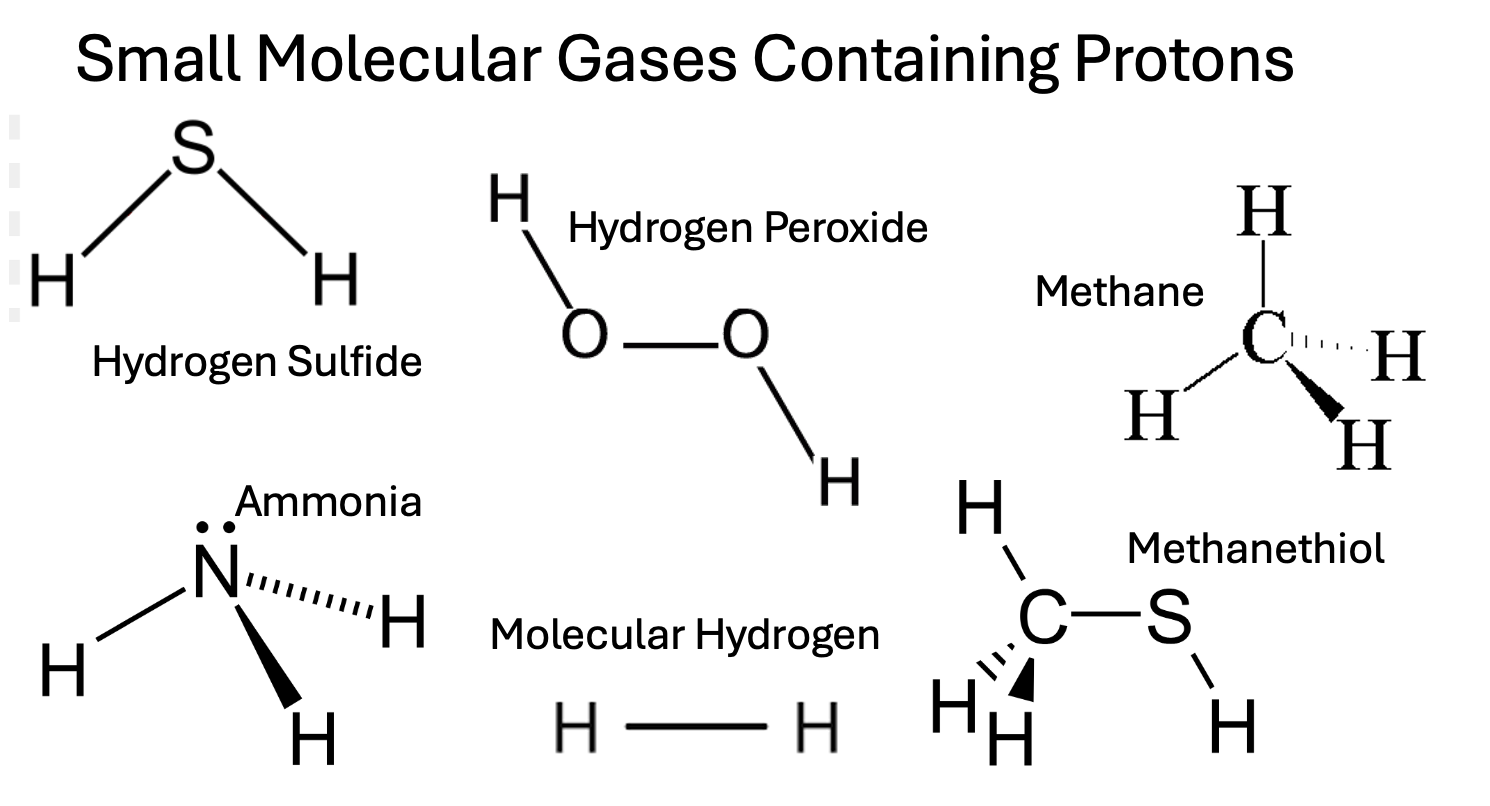
Deuterium is a natural heavy isotope of hydrogen, containing a neutron as well as a proton, making it twice as heavy. Mitochondrial ATP synthase nanomotors are highly sensitive to deuterium, causing them to release reactive oxygen species (ROS) and reducing ATP synthesis. Metabolic processes have evolved to devise ways to reduce the deuterium load in the mitochondria, primarily, we argue, by exploiting several small hydrogen-containing molecular gases. When a gas is produced, deuterium, due to its extra weight, tends to stay behind in the aqueous phase, so the gas becomes deuterium-depleted (deupleted). Furthermore, many of the enzymes that synthesize these small gas molecules are designed to exclude deuterium by exploiting proton tunneling. The gut microbes play a crucial role in producing deupleted gas molecules, such as hydrogen gas (H2), methane (CH4), ammonia (NH3), and hydrogen sulfide (H2S). During inflammatory bowel disease (IBD), activated immune cells upregulate NADPH oxidase (NOX) to produce superoxide, which is converted to hydrogen peroxide (H2O2) by superoxide dismutase. Some microbes can convert H2O2 into two molecules of (likely deupleted) water through anaerobic respiration. H2O2 readily crosses the mitochondrial membrane, and human mitochondrial glutathione peroxidase can also convert H2O2 to water. The glutamate-glutamine exchange that takes place between astrocytes and neurons plausibly capitalizes on NH3 to safely deliver deupleted protons to neuronal mitochondria. In this paper, we investigate how small molecular hydrogen-containing gases are handled in metabolism, from a perspective that considers the roles that deuterium might play in metabolic policy.
Keywords:
deuterium
; molecular hydrogen
; mitochondria
; ammonia
; microbiome
; hydrogen peroxide
; glutamine
; NADPH oxidase
1. Introduction
Deuterium is a heavy isotope of hydrogen, having a neutron as well as a proton in the nucleus, making it essentially twice as heavy. Deuterium is pervasive in nature, found in seawater at a concentration of 156 parts per million (ppm). There is a growing awareness in the research community that deuterium has biological properties that can disrupt metabolism [1]. In particular, the ATPase nanomotors in the mitochondria are very sensitive to deuterium, as it reduces the efficiency of ATP production and increases the release of reactive oxygen species (ROS) [2]. While 156 ppm may seem rare, because hydrogen is so common, deuterium levels in the blood are five or six times as high as calcium levels.
Human metabolic policy has evolved to have a sophisticated strategy for maintaining very low deuterium in the mitochondrial intermembrane space, to minimize exposure of the ATPase nanomotors to deuterium. The process likely depends heavily on the gut microbiome to recycle nutrients by producing abundant molecular hydrogen gas and using the hydrogen gas as a reducing agent to create short chain fatty acids (SCFAs) and methyl groups to fuel methylation pathways [3]. It has been suggested that these microbial products based on recycling hydrogen gas would likely have significantly reduced deuterium content, due to the properties of specialized flavoproteins that exploit proton tunneling, which deuterons are less capable of, and to the fact that deuterium tends to stay behind in the aqueous phase when a gas is formed [4].
A seminal paper published in 1961 showed that molecular hydrogen produced by a marine pseudomonad contained 80% less deuterium than the deuterium fraction in the seawater [5]. The hydrogenase enzyme expressed by this microbe bears significant homology with hydrogenase enzymes expressed by multiple microbes inhabiting the human gut. It is therefore likely that the hydrogen gas produced in the gut is similarly depleted in deuterium.
Interestingly, methane gas formed at high temperature during volcanic eruptions has also been found to be extremely depleted in deuterium [6]. This suggests a pattern that is plausible, given that deuterium is twice as heavy as hydrogen. Like methane and molecular hydrogen, trimethylamine (TMA) is also a gas at room temperature. This may mean that it is also naturally reduced in deuterium content, since we believe this may be a general property of hydrogen-containing gases, due to deuterium’s tendency to resist leaving the aqueous phase. We will have more to say about TMA later in this paper, since gut microbes produce it from dietary sources such as L-carnitine, choline, and betaine. Other hydrogen-containing gases produced by gut microbes include hydrogen sulfide (H2S), hydrogen peroxide (H2O2), methanethiol (CH3SH), and dimethyl sulfide (CH3) 2SH. Might it be the case that all these small molecular gases are enriched in 1H? Furthermore, does human biology exploit this property? This is the topic that this paper addresses.
Protons bound to carbon, nitrogen, oxygen, and sulfur differ in their ability to freely exchange with deuterons in the medium, and their ability to exchange also depends on the surrounding molecular context. Protons bound to oxygen in carboxyl groups or hydroxyl groups exchange with deuterons from the aqueous medium much more readily than protons bound to nitrogen in amide groups, such as the C(=O)NH2 amide group in glutamine [7]. Glutamine is the most abundant free amino acid in human plasma [8], and it plays a powerful role in protecting from ammonia toxicity [9,10]. We will present an argument in this paper that glutamine’s amide group may be enriched in 1H, a feature that may be exploited by human biology.
Generally, carbon atoms are very good at holding onto protons. The protons bound to carbon atoms in organic molecules for the most part remain securely bound and never exchange with deuterons or protons from the aqueous medium, although there are some interesting exceptions such as the protons in bis-allylic carbon atoms and in rings containing carbon and nitrogen, such as the pyrrolidine ring in proline and the imidazole ring in histidine [11].
2. Methanogens, Methanotrophs, Methylotrophs, and Trimethylamine
Gut microbes ferment complex carbohydrates such as dietary fiber into small organic molecules such as glucose and formate that are then metabolized anaerobically to produce hydrogen gas [12]. This hydrogen gas can then be used by methanogens (anaerobic archaea) to generate methane gas directly by reduction of carbon dioxide (CO2). [13]. For an excellent review on the unique metabolic profile of methanogenic archaea, please see Costa and Whitman (2023) [14]. Methane oxidizing genes are a common feature of the human gut microbiome. Methanotrophs such as Methylocystis intestini help reduce methane content in the gut while producing useful energy in the form of nicotinamide adenine dinucleotide (NADH) [15]. Due to its origins, this NADH is likely a good source of 1H rather than 2H protons released into metabolic water by the action of NADH dehydrogenase in the mitochondria.
Methylotrophs metabolize methylated amines, most notably, TMA, to produce formaldehyde (derived from the methyl groups) and ammonia as a byproduct. Formaldehyde can then be processed through assimilatory pathways to yield biomass or through dissimilatory pathways, producing carbon dioxide gas and useful energy as NADH [16]. Methylotrophs significantly outcompete methanogens for hydrogen gas. Methylotrophs have a much lower threshold for hydrogen gas, consuming it at partial pressures as low as 0.1 Pascal, whereas methanogens require a range from 2.8 to 10 Pascals [17]. The activity of methylotrophs is thus primarily limited by the availability of methyl groups. The proton in NADH that they produce is unlikely to be a deuteron.
Together with Professor László Boros, I recently published a paper hypothesizing that hydrogen gas produced by gut microbes plays an essential role in reducing the deuterium load in host mitochondria. Furthermore, we argued that trimethylamine oxide (TMAO) a metabolite produced by the liver through oxidation of TMA, may serve as a marker for deuterium overload in the mitochondria, systemically [18]. TMA that remains unmetabolized by gut microbes is an indicator of a deficiency in methylotrophs. Methylotrophs likely play a powerful role in supplying deupleted methyl groups to the methylation pathways. Therefore, elevated TMAO is a potential indicator of deuterium overload in the mitochondria as well as a disrupted microbiome. TMAO has been well recognized as a risk factor for cardiovascular disease as well as several other chronic diseases [19,20].
3. Do Hydrogenases Produce Low-Deuterium Molecular Hydrogen?
Hydrogenase enzymes are found in thousands of genomes from over 30 different phyla [21,22]. They are common in aquatic, terrestrial, and even host-associated ecosystems. They are all metalloenzymes that use iron and nickel as catalysts, with [NiFe]-hydrogenases, [FeFe]-hydrogenases and, less commonly, [Fe]-hydrogenases all having been identified. Their reversible reaction catalyzes the oxidation of molecular hydrogen (H2) into 2H+ and 2e-. It has been hypothesized that, during prehistoric times on Earth, the atmosphere was rich in hydrogen gas, which has been proposed to have been the first electron donor leading to ATP synthesis. H2 is ubiquitous throughout the biosphere, and it readily diffuses through microbial cells and has a low activation energy requirement. H2 metabolizing microbes are keystone species driving carbon cycling along oxygen gradients in hydrogen-rich soil ecosystems [23].
The reaction catalyzed by hydrogenases is reversible, and our interest is in the use of this enzyme to produce molecular hydrogen from protons and electrons by gut microbes. As documented in a paper published in 1961, MI Krichevsky et al. studied a hydrogenase produced by a marine pseudomonad species and found that it was capable of fractionating hydrogen isotopes to a remarkable degree when producing hydrogen gas. The hydrogen gas it produced was greatly depleted in deuterium compared to ocean water, having a deuterium content that was reduced by a factor of five. This microbe produced molecular hydrogen when it was grown with glucose, pyruvate, or formate as nutrients [5]. While the species investigated was a marine microbe rather than a colonizer of the human gut, its hydrogenase closely resembles hydrogenases synthesized by gut microbes. Furthermore, other studies have shown that hydrogenase enzymes can have extremely high deuterium kinetic isotope effects (KIEs), as high as 43 in an acidic environment [24]. Part of the reason for this may be the biophysical property that deuterium, being twice as heavy, tends to stay behind in solution in the water and resists the gas phase.
4. Microbial Production and Utilization of Hydrogen Gas
Molecular hydrogen is increasingly being recognized as a therapy that has widespread benefits. Yıldız et al., in a review paper published in 2025, wrote in the abstract: “H2 demonstrates numerous biologically therapeutic properties, including anti-inflammatory, antioxidant, anti-cancer, anti-stress, anti-apoptotic, anti-allergic effects, signaling molecule functions, regulation of redox balance, modulation of antioxidant enzyme gene expression, improvement of blood vessel function, down-regulation of pro-inflammatory cytokines, stimulation of energy metabolism, and protection of the nervous system” [25].
A metagenomics survey revealed that Firmicutes and Bacteroides are the most prevalent hydrogen-producing bacteria in the human large intestine, accounting for 92% of the bacterial species [26,27]. These species break down dietary fiber into small molecules and then extract hydrogen atoms from those molecules. Interestingly, Bacteroides fragilis, an important symbiont, anchor themselves in the colonic mucosa, mediated by immunoglobulin A, where they constantly produce hydrogen gas and also protect from pathogens [26,28]. Every day, as many as ten liters of hydrogen gas are produced in the human intestines [29]. A study based in Japan showed that patients with dementia had a reduced Bacteroides population compared to controls [30].
Seventy percent of the microbial species in the gut have the capacity to metabolize H2. The three major pathways for disposal of H2 in the gut are methanogenesis, acetogenesis and sulfate reduction, with methanogenic archaea being the most abundant in the healthy gut [31]. Hydrogen gas that is not captured by the microbes freely enters the blood stream, and it easily penetrates the blood-brain barrier, where it may serve a role in protecting from oxidative stress [26].
Anaerobic metabolism of non-digestible fiber by human gut microbes converts complex carbohydrates into carbon dioxide and hydrogen gas, that, as we have argued, is likely severely depleted in deuterium. Methanogenic archaea then use the hydrogen gas to reduce carbon dioxide to methane gas. This prevents acidification of the gut and facilitates the breakdown of complex carbohydrates, fibers, and resistant starches, increasing the total energy harvest from food [32]. Methanobrevibacter smithii is the dominant archaeon in the human gut, accounting for over 90% of the archaeal species in the gut [33]. If, as we argue, the hydrogen gas has lost 80% of the deuterium present in the aquatic medium, then the methane gas will also be significantly depleted in deuterium.
Methane is further processed in the gut in order to capture the gas as a significant nutrient. After monooxygenase converts methane to methanol, two subsequent steps involving microbial dehydrogenases transform methanol into formaldehyde and, finally, formate, where NAD+ picks up 1H to become NADH (which is then a source of 1H to fuel the ATPase nanomotors).
methane → methanol → formaldehyde → formate
Formate is widespread as a respiratory energy source in natural habitats [34]. Campylobacter jejuni, a common cause of diarrhea, has a periplasmic formate dehydrogenase enzyme which allows formate oxidation to be coupled with microaerobic oxygen reduction to water [35]. The water is released in the periplasmic space rather than the cytoplasm, and the outer membrane of the bacterium is highly permeable to water. This essentially means that the reaction delivers deuterium-depleted water to the host’s gut lumen.
In addition to its conversion to formate via a dehydrogenase, formaldehyde can also be a source of methyl groups in the methylation pathways. A study investigating metabolism of a methylotrophic bacterium, Methylobacterium extorquens, when fed methanol as a fuel source, revealed that methanol was reduced to formaldehyde which was then utilized to synthesize methylene tetrahydrofolate from tetrahydrofolate, either through a direct nonenzymatic transfer of the methylene group from formaldehyde to tetrahydrofolate, or through an indirect metabolic pathway involving several steps [36]. Formaldehyde is highly reactive, and, by being directed down these pathways, it can be quickly metabolized prior to causing harm through other spontaneous reactions. However, a further implication is that the methyl groups in methyltetrahydrofolate will be enriched in 1H. The fact that methylation pathways are so important in biology may be connected to their ability to deliver 1H to mitochondria [3,18].
Acetogenic bacteria also use the hydrogen gas to produce acetate from carbon dioxide, according to the Wood-Ljungdahl pathway:
2CO2 +4H2 → CH3COO- +H+ + 2H2O
Both the acetate and the water that are produced in this way are likely to be enriched in 1H. Acetate is a precursor to the four-carbon fatty acid, butyrate, the primary nutrient consumed by the colonocytes lining the gut barrier [37]. Butyrate stimulates human enteroendocrine L-cells to release glucagon-like peptide-1 (GLP-1), the hormone that suppresses appetite to maintain an ideal body weight [38]. GLP-1 analogues (receptor agonists) are widely prescribed to help people lose weight [39]. Butyrate’s beneficial effects in the gut may be primarily due to its low deuterium content.
Finally, hydrogen gas is also used by sulfate-reducing bacteria such as Desulfovibrio sp. to reduce sulfate to sulfide according to the following equation: [40].
4H2 + SO4-2 + H+ → HS- + 4H2O
The four water molecules are likely enriched in 1H compared to 2H. Mitochondria eagerly take up H2S (a gas) and oxidize it through the enzyme sulfide quinone oxidoreductase (SQR), which donates electrons to the electron transport chain (ETC) to produce ATP, while releasing (preferentially 1H?) protons into the mitochondrial water [41]. We will return to a discussion on H2S in a later section.
We predict that these products of molecular hydrogen metabolism - acetate, butyrate, methane and its derivatives, including formaldehyde, formate, and the methyl groups carried by the methylation pathways, as well as the water produced in reactions involving oxygen reduction, will all be sources of 1H that can be used to fuel mitochondria in the human host cells.
5. Methane Gas: A Broader View
Many toxic metal and chemical exposures can alter the composition of the gut mi- crobiome, including pesticides, toxic metals, persistent organic polutants, and artificial sweeteners [42]. Gut dysbiosis results in a decrease in beneficial microbiota, such as Faecalibacterium and Roseburia, along with an increase in pathogenic species, such as adherent-invasive Escherichia coli (AIEC). This results in a reduction in SCFAs along with an increase in detrimental microbial metabolites [43]. Pathogen overgrowth triggers the immune system to release ROS to neutralize the threat, but this can also be harmful to host cells [44].
Excessive methane in the breath, linked to constipation issues, is considered to be a marker for gut dysbiosis, whereby the metabolism of methane gas cannot keep up with its production by methanogenic archaea [45]. However, since 2012, there has been growing interest in the idea of the potential for methane gas administration to have therapeutic benefits. Beneficial effects have been shown in treating ischemia and reperfusion injury, as well as inflammatory diseases in general. It appears to have anti-inflammatory effects and to protect cells from apoptosis [46,47]. This could be simply because it is a low-deuterium nutrient.
While it had long been believed that methane is primarily synthesized by methanogenic archaea, which reduce carbon dioxide using hydrogen gas as a reducing agent, it has recently become clear that methane can also be generated nonenzymatically from certain common compounds containing methyl groups bound to sulfur or nitrogen atoms. This abiotic mechanism, catalyzed by light and heat, occurs in all living organisms, including cyanobacteria, algae, fungi, plants, and mammals, and it can even take place outside of living cells [48].
Most notably, dimethyl sulfoxide (DMSO) and methionine can be spontaneously oxidized to produce methane under conditions of oxidative stress. Keppler et al. wrote in 2023: “In summary, the reaction of methylated sulfur compounds, such as DMSO and methionine with Fenton-type chemistry involving ROS, carbonate radicals, or oxo-iron(IV) results in the formation of methyl radicals (•CH3), of which a portion reacts to CH4 through abstraction of a hydrogen atom from hydrocarbons, hydrogen peroxide, or hydrogen carbonate” [49]. In an earlier paper by the same authors, it was stated that “An in-depth understanding of the various factors that control cellular methane formation and consumption and a thorough understanding of its dual function and bioactivity may bring significant benefits to the domain of human health and disease” [50].
It is intriguing that, under conditions of oxidative stress, which develops in response to pathogen overgrowth, methane can be regenerated nonenzymatically from methylated nitrogen or sulfur sourced from common organic molecules, via the Fenton reaction. This may be especially important because oxidative stress interferes with the enzymatic production of methane by strict anaerobes [51].
6. Does Hydrogen Peroxide Play a Central Role in Deuterium Homeostasis?
Hydrogen peroxide (H2O2) production, signaling, and detoxification are central in metabolism, and H2O2 acts as a vital second messenger in cellular processes, including growth, inflammation, and wound healing, but it can also cause damage at high levels. Key enzymes like NADPH oxidases (NOX) generate superoxide (O2•-) from oxygen [52], and superoxide dismutase (SOD) isoforms convert O2•- to H2O2 [53]. Catalase and glutathione peroxidase break down H2O2, maintaining a balance that dictates whether H2O2 signals or damages, impacting gene expression, cell fate, and mitochondrial function [54].
Metabolically generated H2O2 has been identified as a central hub in redox signaling and oxidative stress. Major sources include the NOX proteins and complex III of the mitochondrial respiratory chain. H2O2 is also generated in the peroxisome during fatty acid oxidation. H2O2 serves as a second messenger in insulin signaling and several signaling cascades induced by growth factors [55]. H2O2, as a small gas molecule, can easily cross lipid membranes, and its transport across membranes is also facilitated by aquaporins, especially peroxiporins [56]. Peroxiporins are dysregulated in cancer and other diseases associated with inflammatory conditions [57]. O2•-, produced in Complex I and Complex III of the respiratory chain, is rapidly dismutated to H2O2 and oxygen by mitochondrial superoxide dismutase (SOD2) [58]. H2O2, in turn, is metabolized to two molecules of water (DDW) by glutathione peroxidase and peroxiredoxins, particularly PRDX3 [59,60,61,62].
The point we want to emphasize here is that H2O2 is a gas, and therefore it is likely to be deupleted. We hypothesize that it essentially selectively removes hydrogen rather than deuterium from the aqueous medium where it is formed, and then it can migrate to another locale where it deposits the deupleted protons that it extracted, as metabolic water. In this way, it fractionates deuterium out and delivers DDW, whenever it is reduced by catalase, glutathione peroxidase, or peroxiredoxins.
Various signaling events due to stressors trigger ROS generation by NADPH oxidases (NOX), whose primary and deliberate product is O2•-. Adequate antioxidant systems are essential for protecting cells from tissue damage due to overabundant ROS [63]. Under physiological conditions, a baseline, normal level of ROS formation supports a wide range of processes. NOX is no longer viewed as solely involved with phagocyte antimicrobial activity, but rather as being vital to broad cellular functions and homeostasis [64]. Tissue damage associated with many diseases is attributed to an imbalance between ROS formation and its clearance via antioxidant defenses.
6.1. NOX2 and SOD3
NOX2 is a key enzyme that promotes atherosclerosis, due to the fact that it releases O2•- into the extracellular space. O2•- spontaneously dismutates to produce H2O2 and O2. NOX2 was originally identified in phagocytes, where it generates the respiratory burst to kill pathogens [65]. Subsequent studies have demonstrated that NOX2 is also expressed in endothelial cells, cardiomyocytes, hematopoietic stem cells, and platelets [66]. Interestingly, macrophages release exosomes containing NOX2 that can then deliver NOX2 to distant cells [67].
Via NOX2, macrophages and endothelial cells release O2•- into the vascular wall, where it can react with nitric oxide (NO) to form peroxynitrite (ONOO-), a highly reactive but short-lived molecule that causes widespread tissue damage by initiating lipid peroxidation chain reactions. The rate constant of the reaction of O2•- with NO to form peroxynitrite is 10,000 times faster than spontaneous dismutation [67].
SOD enzymes play an essential role in accelerating the rate of conversion of O2•- to H2O2, to reduce exposure to ONOO-. SOD-enhanced catalysis of the dismutation reaction increases the rate of dismutation by 10,000-fold or more [68]. SOD3 (also known as EC-SOD) is the only SOD isoform found predominantly in the extracellular space, anchored to the extracellular matrix through a heparin-binding domain. It is secreted by vascular smooth muscle cells and fibroblasts. It catalyzes the dismutation of extracellular O2•- into H2O2 and O2.
O2•- cannot cross plasma membranes due to its negative charge, but H2O2 can freely enter the cell via diffusion. Furthermore, aquaporins, especially AQP8, actively support the uptake of H2O2 by the cells [67]. Internally, H2O2 also crosses membranes to reach the peroxisome and the mitochondria. In the peroxisome, it is rapidly converted to oxygen and water through catalase:
2H2O2 → 2H2O + O2
In the mitochondria, glutathione peroxidase converts H2O2 to two molecules of DDW:
2GSH + H2O2 → GSSG + 2H2O
Thioredoxin catalyzes a similar reaction which results in a disulfide bond between two of its cysteine residues. Since H2O2 is a gas, it is highly likely that its protons are predominantly 1H rather than 2H. When H2O2 is formed from O2•- in the extracellular space, we propose that deuterons are much less likely to leave the aqueous medium, just as is the case for H2. Thus, the import of H2O2 into the cell is a clever way to decrease the deuterium content in the interior while increasing it in the extracellular space. We hypothesize that both the peroxisomes and the mitochondria capitalize on this feature to synthesize DDW. However, we are unaware of any studies that have measured the amount of deuterium in metabolically produced H2O2.
SOD3 critically depends on copper as a catalyst, and the copper transporter ATP7A plays an essential role in providing copper to SOD3. Interestingly, in experiments on diabetic mice, it was shown that ATP7A depends on insulin signaling. The studies on these mice showed that ATP7A activity is severely reduced, and this is associated with increased oxidative stress and increased levels of O2•- in the vasculature, along with endothelial dysfunction. External administration of insulin ameliorated the effect. In humans, copper deficiency and/or impaired copper transport are associated with cardiac hypertrophy, heart failure, ischemic heart disease, and diabetes mellitus cardiomyopathy [69].
Ceruloplasmin is a protein that is synthesized in the liver and released into the circulation. It is a multi-copper oxidase enzyme, and copper incorporation is crucial for its ability to oxidize Fe+2 (which triggers the Fenton reaction when H2O2 is present) to the nontoxic form, Fe+3, which can then be stored in transferrin for safe transport. Copper deficiency would thus also impair ceruloplasmin function, greatly increasing the risk of damage due to the Fenton reaction when H2O2 reacts with Fe+2 [70].
6.2. NOX4
NOX4 is the only NOX isoform that is expressed at the mitochondrial membrane, and it is primarily localized to internal membranes, including in the mitochondria, the ER, and the nuclear envelope. NOX4 is unique among NADPH oxidases because it primarily directly produces H2O2 rather than O2•-, due to a longer E-loop (the third extracytosolic loop) with a histidine residue that acts as a proton source to facilitate the dismutation of O2•- [71]. NOX4 is upregulated in cardiomyocytes under stress conditions such as pressure overload or hypoxia. Nox4-null mice developed exaggerated contractile dysfunction, hypertrophy, and cardiac dilatation during exposure to chronic overload [72]. NOX4 expression is correlated with aging and increased in human atherosclerotic arteries [73].
NOX4 has significant anti-atherosclerotic functions. Deletion or inhibition of NOX4 often leads to increased inflammation and atherosclerosis. Endothelial NOX4 promotes angiogenesis and recovery from hypoxia [74]. In a mouse model of ischemia/reperfusion injury, NOX4 knockout surprisingly revealed that NOX4, despite producing ROS, protects the heart from damage during ischemia [75]. Expression of vascular NOX4 is increased in association with hypertension, atherosclerosis, diabetes, restenosis, ischemia reperfusion injury, fibrosis, vascular aging, and pulmonary hypertension [74]. It is conceivable that all these conditions are associated with excess deuterium in the mitochondria, and that NOX4 helps to ameliorate that problem.
Figure 1 illustrates schematically the processes by which the cell is able to import H2O2 into the mitochondria and the peroxisome as a way to deliver 1H to these organelles.
6.3. NOX4 in Cancer Cells
NOX4 acts as a key cellular energy sensor, linking mitochondrial ATP levels to metabolic states, especially during stress adaptation and in drug resistance in tumor cells. NOX4 is upregulated in many types of cancer, and this leads to chemotherapy drug-resistance in ovarian cancer cells [76]. A well-designed study investigating the role of NOX4 upregulation in renal carcinoma cells revealed that NOX4 in these cells is localized to the mitochondria, and it is inactivated by ATP, through ATP binding to a Walker A binding motif (AXXXXGKT). None of the other NOX isoforms have this motif. When cancer cells switch to aerobic glycolysis as a source of ATP, ATP levels drop in the mitochondria, and this leads to increased production of ROS via NOX4 activation. Pyruvate kinase catalyzes the last step within glycolysis. Pyruvate kinase M2 (PKM2) is the main isoform expressed in cancer cells. Lysine acetylation mediates its clearance through lysosomal degradation. The ROS generated through NOX4 expression suppress acetylation, thereby protecting it from degradation [77]. PKM2 promotes cancer cell migration and invasion, by redirecting metabolism towards the Warburg effect [78]. It is conceivable that deuterium overload impairs mitochondrial ATP synthesis in cancer cells, leading to upregulation of NOX4, both as a source of ROS and as a potential source of DDW.
7. Hydrogen Sulfide Gas
Hydrogen sulfide (H2S) has long been recognized as a toxic and dangerous gas, but it was much more recently that it also became recognized as an endogenous regulatory transmitter. H2S, produced by several human enzymes, affects a variety of biological processes, including transcriptional and posttranslational gene modification, activation of signaling pathways, and alterations in metabolic processes. New efforts are being considered around the use of H2S as a gas transmitter with therapeutic drug potential [79].
7.1. Sulfate Reducing Bacteria in the Gut
Desulfovibrio (DSV) are a resident commensal bacterium present in the human gastrointestinal (GI) tract, and the most common sulfur-reducing bacterium. They are known to be opportunistic pathogens that frequently overgrow under dysbiotic conditions, e.g., in association with inflammatory bowel disease (IBD) [80] and neurodegenerative diseases such as Parkinson’s disease (PD) [81]. They are a gram-negative bacterium that produces H2S via the dissimilatory sulfate reduction pathway, using molecular hydrogen as a reducing agent [82]. The nematode Caenorhabditis elegans exhibits dietary preferences, often favoring bacterial strains that support its growth and health [83]. In an elegant study involving a transgenic strain of C. elegans which expresses human α-synuclein, researchers found that, when presented with DSV strains from various sources as food, C. elegans greatly preferred the environmental strain D. vulgaris DSM 644 over two human isolates, one of which was obtained from a PD patient. Furthermore, colonization with the environmental strain led to reduced aggregation of α-synuclein, reduced oxidative stress, and increased lifespan (by a median of 36 days). The authors proposed that sublethal stressors may activate protective molecular pathways such as antioxidant defenses that increase the capacity of the host to cope with toxic exposures that could cause cellular damage [84].
Desulfovibrio strains are generally considered to be strict anaerobes. however, interestingly, it has been found that a strain of Desulfovibrio (D. vulgaris Hildenborough) obtained from anoxic ditch sludge could tolerate up to 0.04% oxygen in the medium, which could even support their growth. When exposed to an oxygen gradient, these microbes are drawn to an optimal location where the oxygen levels are favorable to growth, i.e., a hypoxic but not an anoxic environment [85]. This suggests that they colonize the gut wall where the environment is hypoxic, and they may serve a useful role in clearing the oxygen present in the gut wall, where there is a strong oxygen gradient, benefiting other strict anaerobes colonizing the gut. Singhal et al. noted in a paper published in 2020 that the richly vascularized subepithelial mucosa harbor significantly more oxygen than the anaerobic gut lumen, especially in the more distal, colonic regions of the GI tract [86]. These authors wrote: “The pO2 drops precipitously along the radial axis from the intestinal submucosa to the lumen, which is home to trillions of anaerobic microbes” [86].
D. vulgaris Hildenborough possess membrane-bound terminal oxygen reductases (members of the respiratory cytochrome bd family) that serve as an oxygen scavenger [87,88]. They also express soluble rubredoxin oxidases, which reduce oxygen to water, coupling NADH oxidation to oxygen consumption. Oxygen exposure increases their resistance to killing by H2O2 [89]. Bacterioferritin plays a crucial role in protecting D. vulgaris Hildenborough from oxygen toxicity, by sequestering iron. Its gene expression increases dramatically upon oxygen exposure. It also protects both the host cells and other microbes from oxidative stress due to the Fenton reaction [90].
7.2. Mitochondria Metabolize H2S
Sulfate-reducing bacteria, common in shallow marine sediments and hydrothermal vents, utilize sulfate as the terminal electron acceptor rather than oxygen, and generate H2S as the main respiratory by-product. The endosymbiotic hypothesis of mitochondrial origin proposes that mitochondria originate from the integration of an endosymbiotic bacterium into the host cell, a primordial Asgard archaeon [91]. This may be why eukaryotic mitochondria are able to oxidize H2S efficiently to generate ATP. A mitochondrial sulfide oxidation pathway rapidly eliminates H2S by oxidizing it to sulfite and thiosulfate. Sulfide quinone oxidoreductase (SQR) and persulfide dioxidase, operating in tandem, yield sulfite from H2S. The overall equation for the reaction is:
Quinone + H2S + O2 + H2O → Quinol + Sulfite + H+
where one proton is picked up by the quinone in the inner membrane and the other is released into the aqueous medium. Both should be deupleted, given that H2S is a gas, derived from molecular hydrogen produced by anaerobic hydrogenogens. Glutathione is involved as an intermediary to temporarily form S-sulfanylglutathione (GSSH). Sulfite is then oxidized to sulfate by sulfite oxidase or to thiosulfate by rhodenase. The electrons that are released by SQR are transferred to the ETC at complex III, promoting ATP synthesis [92].
7.3. H2S Is a Powerful Signaling Gas
Occupational exposure to high levels of H2S is toxic, because H2S blocks the respiratory chain by inhibiting cytochrome c oxidase. However, H2S is an endogenous signaling gas, acting as a cytoprotectant and promoting cardiovascular homeostasis and health [93]. It is produced in human cells primarily by three key enzymes: cystathionine β-synthase (CBS), Cystathionine γ-lyase (CSE), and 3-mercaptopyruvate sulfurtransferase (3-MST), using the sulfur-containing amino acids, L-cysteine and homocysteine as substrates [94].
The administration of physiological or pharmacological levels of H2S attenuates myocardial injury, protects blood vessels, limits inflammation, and regulates blood pressure [93]. Like NO, H2S is a potent vasodilator. In mice, genetic deletion of CSE markedly reduces H2S levels in the serum, heart, aorta, and other tissues [95]. H2S plays a role in wound healing, and endogenously produced H2S mediates angiogenesis induced by vascular endothelial growth factor (VEGF) [96]. In in vitro studies, exogenously administered H2S at physiologically relevant concentrations induced angiogenesis and stimulated endothelial cell proliferation and migration [96,97].
8. Methanethiol, SELENBP1 and Cancer
Hydrogen sulfide, methanethiol, and dimethyl sulfide are all volatile sulfur compounds (VSCs) characterized by their low odor thresholds, significant role in halitosis (bad breath), and toxicity at high concentrations. They act as crucial biological mediators and signaling molecules (gasotransmitters) in mammals, influencing blood pressure, inflammation, and neuronal signaling [98]. Methanethiol (CH3SH) is a toxic foul-smelling gas that plays interesting but poorly characterized roles in human physiology. It can be derived from methionine or through methylation of H2S. It is overproduced by cancer cells and often found in excess amounts in tumor microenvironments, associated with accelerated disease progression [99].
Selenium-binding protein 1 (SELENBP1) is a methanethiol oxidase, and it is a critical enzyme for metabolizing methanethiol [100]. It catalyzes the conversion of methanethiol to H2S, H2O2, and formaldehyde.
H2O + CH3SH + O2 → H2CO + H2O2 + H+ + H2S
It depends on both selenium and copper as catalysts [101]. It plays a protective role in ulcerative colitis [102]. Both mouse and human SELENBP1 are abundantly expressed in the liver, lungs, prostate, colon, and pancreas [103,104].
Deficiencies in SELENBP1 cause high levels of methanethiol, leading to a “cabbage-like” breath odor known as extraoral halitosis [105]. SELENBP1 is widely recognized as a tumor suppressor, and it is often downregulated in various epithelial cancers, including lung, colorectal, ovarian, liver, and prostate cancers [103].
Human methyltransferase-like protein 7B (METTL7B; also known as TMT1B) catalyzes the transfer of a methyl group from S-adenosyl-l-methionine to H2S, yielding methanethiol, and to other exogenous thiol-containing small molecules [106]. METTL7B is consistently upregulated in various cancers, acting as a promoter of tumor progression and a marker of poor prognosis [107]. In a study on lung adenocarcinoma cells, high expression levels were linked to enhanced cancer cell proliferation, migration, and invasion, and increased resistance to therapies [108].
Both glucose and methionine are enriched in cancer cells, and the nonenzymatic Maillard reaction between these two metabolites yields methanethiol. Gut microbes also break down methionine via γ-methionine-lyase, to produce methanethiol. Methanethiol-producing strains such as Fusobacterium nucleatum are highly abundant in the gut microbiome of colorectal carcinoma (CRC) patients [99]. Colorectal carcinoma (CRC) and hepatocellular carcinoma (HCC) cells produce and release high levels of methanethiol (CH3SH) into the tumor microenvironment (TME) due to their dysregulated sulfur metabolism. Elevated levels of methionine, along with reduced expression of SELENBP1 and increased expression of METTL7B, lead to high endogenous production of this volatile compound, which serves as a scent-based marker for cancer [99].
We hypothesize that the methanethiol that is produced in abundance in association with colon cancer can be exploited by normal cells expressing SELENBP1 in the gut to produce H2S, H2O2, and formaldehyde, all of which are likely readily available sources of deupleted water to support mitochondrial health. This hypothesis aligns with the general theory we have developed that cancer cells hoard deuterium and supply the resident immune cells with deupleted resources [4,109]. Indeed, it has been confirmed experimentally that SELENBP1 expression levels are significantly lower in colorectal cancer cells than in adjacent healthy tissue cells [110]. Given that it is a gas, it is likely that, like the other small organic gas molecules, methanethiol has reduced levels of deuterium.
Figure 2 illustrates the pathways by which methanethiol is produced, through both microbial and human enzymes, and the pathway by which it is broken down into potentially valuable deupleted molecules by SELENBP1.
9. A Role for Ammonia in Deupletion?
Ammonia management in the body is a critical detoxification process centered on converting ammonia, which can be toxic in elevated amounts, into non-toxic urea in the liver (the urea cycle) to be recycled via the gut microbiome or finally for renal excretion. The kidneys play a dual role: managing acid-base balance by excreting ammonia as ammonium (NH4+) in the urine and producing ammonia from glutamine in the proximal tubules to regulate body pH.
In the remainder of this paper, we will refer to “total” ammonia as T-ammonia, representing the mixture of NH3 and NH4+. NH3, being a gas, is likely enriched in 1H relative to 2H. A question we address here is whether the organism exploits this feature to fractionate deuterium, preferentially retaining NH3 and exporting NH4+.
T-ammonia is no longer viewed merely as metabolic waste. It is a critical driver of tumor progression. T-ammonia accumulates in the tumor microenvironment, where it is a source of nitrogen to support amino acid and nucleic acid synthesis. It stimulates growth pathways and fuels an immunosuppressive state [111]. Cancer cells actively export ammonia as a gas (NH3) which is likely low in deuterium simply because it is a gas. Resident cancer-associated fibroblasts take up NH3 and utilize it to synthesize glutamine, which is then delivered back to the tumor cells [112]. This exchange may serve as a way to preferentially export 1H over 2H to the external milieu, by taking advantage of the high pH in the tumor cytoplasm to promote the production and export of NH3 derived from T-ammonia [113].
9.1. Do Gut Microbes Convert Urea to Deupleted Ammonia?
Urea, produced primarily in the liver and excreted as a major component of urine, can be viewed as a storage form of T-ammonia. A reaction of urea with one molecule of water yields ammonia and carbon dioxide:
(NH2) 2CO + H2O → 2NH3 + CO2
The metabolism of urea by microbial urease [114], as well as the microbial metabolism of other nitrogen-containing molecules such as the amino acids, purines, and pyrimidines, are all sources of T-ammonia in the gut lumen. Interestingly, ammonia, the gas, (NH3) readily diffuses across plasma membranes, whereas ammonium, the ion (NH4+) does not. Furthermore, epithelial cells in the gut express transport proteins that actively take up NH3 but not NH4+ [115]. Villous epithelial cells throughout the gut express two proteins that facilitate the active transport of NH3 across the gut barrier into the circulation, namely the NH3 transporter family members Rh B glycoprotein (RhBG) and Rh C glycoprotein (RhCG) [115].
T-ammonia in the lumen is mainly found in solution as the cation NH4+. At equilibrium, only a small percentage is present as the gas, NH3, particularly in an acidic environment, because of the high acid dissociation constant (pKa). But only NH3 can passively cross the gut barrier. Various transport proteins exist to actively carry either NH3 or NH4+ across membranes, and in different circumstances a preference for transport of one or the other is probably strategically important.
It is likely that an ammonium ion containing deuterium (NH3D+) would leave behind in the lumen the deuteron rather than one of the protons when it gets converted to the gas, ammonia, via acid dissociation: NH3D+ → NH3 + D+. This means that ammonia as a gas is likely a source of low-deuterium protons. As the ammonia is syphoned off by the transporters, more ammonia is produced, as the two species restore chemical equilibrium.
9.2. What Is the Role of RhAG in Red Blood Cells?
RhAG, another member of the Rh glycoprotein ammonia transporter family, is only expressed in red blood cells (RBCs). It has not been easy for researchers to figure out what role this protein plays in RBCs, but it is becoming clear that it is involved in T-ammonia homeostasis in the vasculature [116]. RhAG acts as a scaffold for the Rh complex. It appears to transport T-ammonia exclusively as NH3 across the RBC membrane [117]. It has been suggested that RBCs act as a storage depot for T-ammonia to reduce the levels of free T-ammonia in the circulation [116]. Thus, RhAG’s role in RBCs may be to support T-ammonia buffering within the RBCs, which can help sequester T-ammonia and protect the plasma from hyperammonemia [118].
Notably, RhAG transports methylamine (CH3NH2) as well as ammonia (NH3). Like NH3, CH3NH2 is a gas, and this suggests that RhAG specifically acts as a gas channel. Ripoche et al. wrote: “The results show that RhAG facilitates CH3NH2/NH3 movement across the RBC membrane and represents a potential example of a gas channel in mammalian cells” [117]. In fact, sequestration of methylamine may be a beneficial role of RhAG. The concentration of methylamine in RBCs is five times that found in plasma. Interestingly, methylamine is not efficiently cleared during dialysis treatment, due to the fact that RBCs sequester it [119]. An analysis of the crystallographic structure of the related bacterial NH3 transport channel AmtB showed, through reconstitution into vesicles, that AmtB conducts uncharged NH3 [120], consistent with the analysis of Ripoche et al. for RhAG.
The concentration of T-ammonia in RBCs is significantly higher than that in the plasma. RhAG expressed in RBCs forms a structural unit with anion exchanger 1 (eAE1; a sodium- independent anion exchanger) within the ankyrin core complex. RBCs swell and lyse in isotonic NH4+ buffer. This effect is tightly regulated by chloride concentration. When chloride was replaced by glutamate, NH4+-induced swelling was inhibited, preventing hemolysis. Inhibition of eAE1 also completely inhibited hemolysis [121]. It has been shown that NH3 transport parallels RhAG expression in RBCs [117].
Westhoff et al. wrote: “Erythrocytes are ideally positioned to then transport ammonium to be exchanged in the liver and kidney, where other Rh-related proteins (RhBG and RhCG) are expressed” [122]. We take this idea one step further: we suggest that RBCs actively import NH3 as a gas from the medullary interstitium in the kidneys, syphoning it off before NH4+ is exported via the urine, as explained in Section 9.3.
9.3. Renal Tubules Preferentially Export NH4+
The ammonium ion (NH4+) and potassium ion (K+) show almost identical biophysical characteristics. Since NH4+ is very similar in size and charge to K+, cells can exploit K+ transport channels to actively transport NH4+ across membranes. The kidneys utilize a complex process to selectively export NH4+ into the urine and retain NH3 in the interstitium. Cells in the proximal tubules of the kidneys synthesize abundant NH4+ from glutamine, which is secreted into the luminal fluid through active transport by both an apical Na+/H+ exchanger NHE3 and by various K+ transport proteins [123]. However, a substantial amount of ammonia (NH3) is reabsorbed later in the thick ascending limb of the loop of Henle. Approximately 40% of the NH4+ that enters the loop of Henle is passed to the interstitium as NH3.
Tubule cells in the thick ascending limb of Henle have a remarkable apical impermeability to NH3, due to the specific lipid composition of the bilayer [124,125]. However, they actively import NH4+ from the lumen against a concentration gradient, utilizing the Na+/H+ ion exchanger NHE4, substituting NH4+ for the proton. Defective versions of NHE4 lead to metabolic acidosis [126]. On the basolateral side, these cells release NH3 but not NH4+ into the medullary interstitial space, to facilitate acid-base balance and to create a concentration gradient used for final waste excretion [127]. The Rhesus glcyoproteins, RhBG and RhCG act as NH3 transporters in the distal tubule and collecting duct [128]. We hypothesize that RBCs may actively import NH3 in the medullary interstitium, syphoning it off to capture 1H for return to the circulation. As we’ve discussed, red blood cells actively take up NH3 but not NH4+ via their RhAG proteins [117].
The Inner Medullary Collecting Duct (IMCD), located in the deep, inner medulla, is the final, terminal segment of the renal nephron responsible for maximal urine concentration and final electrolyte adjustment before urine enters the renal pelvis. In the IMCD, basolateral Na+-K+ ATPase enables active basolateral uptake of NH4+ but not NH3 [128]. The renal excretion of NH4+ acidifies the urine, playing a crucial role in acid/base balance.
However, this process of RBC uptake of NH3 and export of NH4+ into the urine presumably also selectively retains protium that is returned to the circulation, while deuterium is then selectively excreted in the urine via NH4+. Essentially, through this complex process, deupleted protons reenter the circulation and deuterium has become enriched in the aqueous medium in the loop of Henle. These deuterium-enriched fluids are finally excreted as urine.
10. Does Glutamine Supply Deupleted Protons to the Neuronal Mitochondria?
Glutamate is not only the most abundant excitatory neurotransmitter, but it is also the precursor to γ-aminobutyric acid (GABA), the predominant inhibitory neurotransmitter. Abnormal levels of extracellular glutamate impair neurotransmission and cause neurodegeneration and neuronal apoptosis. One of the most important roles of astrocytes is to assure that glutamate does not reach toxic levels in the synapse [129].
Astrocytes also play an essential role in the brain to clear excess T-ammonia, protecting the neurons from T-ammonia toxicity [130]. In achieving this goal, they also protect from glutamate neuroexcitotoxicity, because they take up glutamate that is released into the synapse and use the T-ammonia to convert the glutamate to glutamine. The so-called astrocyte-neuron glutamate-glutamine cycle prevents overstimulation of neuronal N-Methyl-D-aspartate (NMDA)/ α-amino-3-hydroxy-5-methyl-4-isoxazolepropionic acid (AMPA) receptors, which can lead to cell death [131]. The process of glutamate uptake by astrocytes, conversion to glutamine, and transport back to neurons is fast, happening in the order of seconds [132]. A question is whether it is fast enough to predominantly avoid picking up a deuteron via exchange with protons in the side chain amide group while in transit. If so, then this whole process might be a mechanism to deliver 1H to the mitochondria. Note that the amide group in glutamine exchanges protons with deuterons in the aqueous medium at a much lower rate than do protons bound to oxygen in hydroxyl and carboxyl groups [7].
10.1. The Glutamate/GABA-Glutamine Cycle
Glutamate and GABA are the principal excitatory and inhibitory neurotransmitters in the brain. They are both extensively recycled between neurons and astrocytes. Astrocytes take up these neurotransmitters from the synapse following their release by neurons. In the cytoplasm, astrocytes convert glutamate to glutamine via glutamine synthetase, and then return the glutamine to the neurons, which, in turn, resynthesize glutamate from glutamine, in their mitochondria. GABAergic neurons further metabolize glutamate via glutamic acid decarboxylase to produce GABA [133]. Infants with a genetically defective version of glutamine synthetase suffer from severe brain malformations, multi-organ failure and neonatal death, attesting to the importance of this enzyme to metabolism [134].
10.2. Aquaporin 4 and Potassium Channels in Astrocytes
Aquaporin 4 (AQP4) is the primary water channel in the central nervous system, and it is concentrated in the end feet of astrocytes. Curiously, AQP4 channels are localized in close proximity to inward rectifier potassium channels (Kir 4). Potassium is released from neurons into the extracellular space during repolarization of the action potential and is then rapidly taken up by astrocytes via Kir 4. Glutamate is released into the synapse during synaptic activity and is taken up by astrocytes via glutamate transporters on astrocyte processes [135].
AQP4 channels import not only water but also NH3, because it is an uncharged molecule of similar size. Similarly, Kir 4 takes up NH4+ in addition to K+, again, because of similar size and charge properties. So, both the AQP4 channels and the Kir 4 channels can import T-ammonia.
Experiments on cultured astrocytes have shown that AQP4 is upregulated in the presence of high levels of T-ammonia [136]. This explains the feature of brain swelling due to the uptake of excess water by astrocytes in association with hepatic encephalopathy. But it also implies that astrocytes actively take up more NH3 via upregulated aquaporin channels. An experiment on cultured astrocytes exposed to ammonium chloride found that AQP4 was sharply upregulated in response to ammonium exposure, and this was mediated through phosphorylation of p38 [137]. On the other hand, both Kir4.1 and Kir5.1 were downregulated in hyperammonemic mice, suppressing the uptake of NH4+ but also disturbing potassium homeostasis [138,139]. We hypothesize that the upregulation of AQP4 and the simultaneous downregulation of Kir4.1 and Kir5.1 in response to excess T-ammonia in the vasculature is a strategy to selectively import deupleted NH3 rather than deuterium-rich NH4+ when T-ammonia levels are high.
10.3. Astrocytes Clear Glutamate from the Synapse
Astrocytes convert glutamate to glutamine via glutamine synthetase (GS). The reaction catalyzed by GS is carried out through a two-step mechanism. In the first step, γ-glutamylphosphate is formed as an intermediate from glutamate, where phosphate is sourced from ATP. In the presence of this intermediate, NH4+ binds to the active site and, in the second step, it is deprotonated, releasing NH3 near the γ-glutamyl phosphate intermediate. The ammonia molecule then displaces phosphate, yielding free phosphate and glutamine [140]. We hypothesize that any ammonium ion containing deuterium would preferentially lose the deuteron over a proton in this step, thus rarely delivering an NH3 molecule containing 2H.
Interestingly, astrocytic density is reduced in major depressive disorder (MDD). In particular, blood vessels in gray matter from the orbitofrontal area of brains of patients suffering from MDD were specifically significantly impoverished in AQP4 expression compared to normal controls [141]. Low levels of AQP4 could impair glutamate/glutamine cycling, and this could be a factor in symptoms of depression, by reducing the ability of neurons to maintain low deuterium in their mitochondria. Mitochondrial dysfunction in neurons is a prevalent feature of depression [142].
Figure 3 illustrates the glutamate-glutamine exchange between neurons and astrocytes that may serve as a mechanism for glutamine to safely deliver a deupleted ammonia molecule to the mitochondria in the neuron.
10.4. T-Ammonia Handling in the Mitochondrial Matrix: Supplying 1H to the Intermembrane Space?
Aquaporin-8 (AQP8) is the primary aquaporin expressed in the inner mitochondrial membrane (IMM). It plays a key role in mediating water transport into and out of the mitochondrial matrix, contributing to the organelle’s volume regulation. Because AQP8 is highly expressed in the inner membrane, water freely moves from the matrix into the intermembrane space and vice versa [143]. AQP8 is known as a “peroxiporin” because it can transport not only water but also ammonia and hydrogen peroxide across the membrane. It exclusively transports NH3 but not NH4+ [144]. The mitochondrial matrix has a very high pH of around 7.9, which means that∼6% of the T-ammonia is present in the gas form. The porous membrane then allows the ammonia to be siphoned off into the intermembranes space, delivering an overabundance of 1H to it (we hypothesize). As the ammonia is removed, more ammonia appears while dissociation equilibrium is continuously reestablished. Because the intermembrane space is much more acidic, very little of the T-ammonia remains as a gas on that side of the membrane, so ammonia naturally crosses from the matrix into the intermembrane space.
10.5. Liver Failure and Hepatic Encephalopathy
Liver failure results in elevated levels of T-ammonia in the blood, because the damaged liver is unable to detoxify T-ammonia via the production of urea [145]. This T-ammonia readily crosses the blood-brain barrier and causes toxic effects in the brain. Increased T-ammonia concentrations in the brain significantly alter brain glutamate levels, glutamate uptake, and glutamate receptor function. In rat models, acute liver failure causes an upregulation of extracellular glutamate levels. T-ammonia is likely a major contributing factor to the brain edema and seizures associated with hepatic encephalopathy [146].
Glutamate plays a critical role in inducing brain swelling. Activation of the metabolomic glutamate receptors (mGluR) increases the rate of tissue swelling in rat hippocampal slices. During cerebral edema, excessive amounts of glutamate are released, and it causes increased expression of AQP4 [147], promoting the uptake of not only water but also ammonia into astrocytes. The glutamate can then be combined with the ammonia to produce glutamine, which, in our view, is a carrier of deupleted protons. The removal of both the stressor ammonia and the neurotoxin glutamate from the circulation is achieved by the astrocytes through this process.
During hepatic encephalopathy, astrocytes upregulate AQP4 [137] and downregulate Kir4.1 channel expression [138]. This causes both edema (which is primarily due to astrocyte swelling) and excess potassium in the external space, due to impaired potassium uptake by astrocytes [148]. But it also means that T-ammonia is taken up by the astrocytes primarily as the gas rather than the dissolved cation. The fact that glutamate induces these changes in expression makes sense if one acknowledges that glutamate can be converted to glutamine both as a way to detoxify ammonia and to create a carrier molecule for 1H, which the astrocytes immediately deliver to adjacent neurons. In other words, might it be the case that astrocytes are able to capitalize on excess T-ammonia in the circulation to facilitate the delivery of deupleted protons to neurons?
11. Discussion
In this paper, we have carefully examined metabolism that involves small hydrogen-containing gas molecules, from the perspective of deuterium homeostasis. We suggest that much of the complexity found in the metabolism of these gas molecules revolves around strategies to reduce deuterium content in mitochondrial water. We hypothesize that, when the methanogenic archaea that derive small organic molecules by using molecular hydrogen to reduce carbon dioxide are scarce in the gut, various processes increase the abundance of H2O2, as a compensatory mechanism to supply 1H protons to mitochondria. This results in inflammatory bowel disease that can lead to colon cancer in extreme conditions [149]. Methanogenic archaea are depleted in association with IBD [150] . Hydrogen gas administration is being widely recognized as a treatment option for many diseases and conditions [25].
Chronic inflammation is a leading driver in the progression of many long-term health conditions [151], and it may be that its positive role is to heal deuterium-overloaded mitochondria through the delivery of deupleted H2O2 to them to compensate for the insufficiency of deupleted nutrients such as SCFAs and methyl groups supplied by the gut microbes. Mitochondria express both glutathione peroxidase and especially peroxiredoxins [62] that can readily convert H2O2 to two molecules of H2O. High H2 concentration in the gut stimulates the production of butyrate, an essential nutrient for the colonocytes that is probably deupleted [12].
H2S , and other sulfur-containing gases, including DMSO and CH3SH, also appear to play an important role in supplying 1H protons to the mitochondria. Gut microbes are also heavily involved in this process. H2S-producing bacteria are upregulated in the gut in association with Crohn’s disease [152]. Cancer cells actively produce CH3SH and deliver it to tumor-resident cells, potentially as a resource to help restore their mitochondrial health [99].
Ammonia is another gas that can be exploited for its low-deuterium content, but this is a complicated process that involves selective transport proteins to optimize the export of deuterons via NH4+ and the retention of protons in NH3. Gut microbes play an essential role through their expression of urease that converts urea produced in the liver to T-ammonia. Gut epithelial cells actively take up NH3, and deliver it back to the liver [115]. Bacterial urease activity is strongly upregulated in association with gut dysbiosis and Crohn’s disease [153]. The kidneys appear to preferentially retain NH3, the gas, and export NH4+, the cation, into the urine.
Finally, the complex interaction between neurons and astrocytes in glutamate/glutamine exchange may also be a mechanism to deliver 1H to neuronal mitochondria. The glutamate/glutamine cycle is dysregulated in association with various tauopathies present in neurodegenerative disease [154].
We hope that this paper will inspire others to conduct research to clarify the role of deuterium in metabolic policy and the processes by which organisms protect mitochondria from deuterium overload. If our ideas around the role of small molecular gases, many of which are produced by the gut microbes, in deuterium homeostasis can be proven, it would revolutionize our understanding of the mechanisms of disease processes.
Author Contributions
SS wrote the first draft and many rounds of editing and refinement.
Funding
This research was funded in part by Quanta Computer, Inc., in Taoyuan City, Taiwan, under grant number 6950759.
Conflicts of Interest
The author declares that there are no conflicts of interest to report.
Generative AI
The author declares that generative AI was not used in the creation of this manuscript.
References
- Répás Z, Győri Z, Buzás-Bereczki O, Boros LG. The biological effects of deuterium present in food. Discov Food. 2025; 5: 57. [CrossRef]
- Olgun A. Biological effects of deuteronation: ATP synthase as an example. Theoretical Biology and Medical Modelling. 2007; 4: 9. [CrossRef]
- Seneff S, Nigh G, Kyriakopoulos AM. Melatonin in health and disease and its metabolism by the gut microbes: implications for deuterium homeostasis? Current Medicinal Chemistry. May 16, 2026. [Epub ahead of print]. [CrossRef]
- Seneff S, Kyriakopoulos A. Cancer, deuterium, and gut microbes: a novel perspective. Endocrine and Metabolic Science 2025; 17: 100215. [CrossRef]
- Krichevsky MI, Friedman I, Newell MF, Sisler FD. Deuterium fractionation during molecular hydrogen formation in marine pseudomonad. J Biol Chem. 1961 Sep;236:2520-5.
- Ricci A, Fiebig J, Tassi F, Hofmann S, Capecchiacci F, Vaselli O. Extremely deuterium depleted methane revealed in high-temperature volcanic gases. Geochimica et Cosmochimica Acta. 2024; 364: 148-165. [CrossRef]
- Grocholska P, Bąchor R. Trends in the hydrogen-deuterium exchange at the carbon centers. Preparation of internal standards for quantitative analysis by LC-MS. Molecules. 2021 May 18;26(10):2989. [CrossRef]
- Cruzat V, Macedo Rogero M, Noel Keane K, Curi R, Newsholme P. Glutamine: metabolism and immune function, supplementation and clinical translation. Nutrients. 2018 Oct 23;10(11):1564. [CrossRef]
- Nakamura E, Hagen SJ. Role of glutamine and arginase in protection against ammonia-induced cell death in gastric epithelial cells. Am J Physiol Gastrointest Liver Physiol. 2002 Dec;283(6):G1264-75. [CrossRef]
- Bassini-Cameron A, Monteiro A, Gomes A, Werneck-de-Castro JP, Cameron L. Glutamine protects against increases in blood ammonia in football players in an exercise intensity-dependent way. Br J Sports Med. 2008 Apr;42(4):260-6. [CrossRef]
- Seneff S, Nigh G, Kyriakopoulos AM. Is deuterium sequestering by reactive carbon atoms an important mechanism to reduce deuterium content in biological water? FASEB Bioadv. 2025 May 14;7(6):e70019. [CrossRef]
- Campbell A, Gdanetz K, Schmidt AW, Schmidt TM. H2 generated by fermentation in the human gut microbiome influences metabolism and competitive fitness of gut butyrate producers. Microbiome. 2023 Jun 15;11(1):133. [CrossRef]
- Buan NR. Methanogens: pushing the boundaries of biology. Emerg Top Life Sci. 2018 Dec 14;2(4):629-646. [CrossRef]
- Costa KC, Whitman WB. Model organisms to study methanogenesis, a uniquely archaeal metabolism. J Bacteriol. 2023 Aug 24;205(8):e0011523. [CrossRef]
- Zhao Y, Chen H, Huang J, Chistoserdova L, Yu Z. The gut methanotroph Methylocystis intestini modulates intestinal peristalsis and fat metabolism via reducing methane levels. Nat Commun. 2026; 17: 2. [CrossRef]
- Klein VJ, Irla M, Gil López M, Brautaset T, Fernandes Brito L. Unravelling formaldehyde metabolism in bacteria: road towards synthetic methylotrophy. Microorganisms. 2022 Jan 20;10(2):220. [CrossRef]
- Feldewert C, Lang K, Brune A. The hydrogen threshold of obligately methyl-reducing methanogens. FEMS Microbiology Letters. 2020; 367(17): fnaa137. [CrossRef]
- Seneff S, Boros LG. The essential role of hydrogen gas recycling by gut microbes in reducing deuterium load in host mitochondria: Is trimethylamine oxide a deuterium sensor? Metabolomics. 2026; 22:63. [CrossRef]
- Canyelles M, Borràs C, Rotllan N, Tondo M, Escol-Gil JC, Blanco-Vaca F. Gut microbiota-derived TMAO: a causal factor promoting atherosclerotic cardiovascular disease? Int J Mol Sci. 2023 Jan 18;24(3):1940. [CrossRef]
- Gatarek P, Kaluzna-Czaplinska J. Trimethylamine N-oxide (TMAO) in human health. EXCLI J. 2021 Feb 11;20:301-319. [CrossRef]
- Greening C, Biswas A, Carere CR, Jackson CJ, Taylor MC, Stott MB, Cook GM, Morales SE. Genomic and metagenomic surveys of hydrogenase distribution indicate H2 is a widely utilised energy source for microbial growth and survival. ISME J. 2016; 10:761777. [CrossRef]
- Vignais PM, Billoud B. Occurrence, classification, and biological function of hydrogenases: an overview. Chem Rev. 2007; 107:42064272. [CrossRef]
- Pich-Choquette S, Constant P. Molecular hydrogen, a neglected key driver of soil biogeochemical processes. Appl Environ Microbiol. 2019 Mar 6;85(6):e02418-18. [CrossRef]
- Greene BL, Wu CH, McTernan PM, Adams MW, Dyer RB. Proton-coupled electron transfer dynamics in the catalytic mechanism of a [NiFe]-hydrogenase. J Am Chem Soc. 2015; 137(13): 45584566. [CrossRef]
- Yıldız F, LeBaron TW, Alwazeer D. A comprehensive review of molecular hydrogen as a novel nutrition therapy in relieving oxidative stress and diseases: mechanisms and perspectives. Biochem Biophys Rep. 2025 Jan 25;41:101933. [CrossRef]
- Ichikawa Y, Yamamoto H, Hirano SI, Sato B, Takefuji Y, Satoh F. The overlooked benefits of hydrogen-producing bacteria. Med Gas Res. 2023 Jul-Sep;13(3):108-111. [CrossRef]
- Wolf PG, Biswas A, Morales SE, Greening C, Gaskins HR. H2 metabolism is widespread and diverse among human colonic microbes. Gut Microbes. 2016 May 3;7(3):235-45. [CrossRef]
- Donaldson GP, Ladinsky MS, Yu KB, Sanders JG, Yoo BB, Chou WC, Conner ME, Earl AM, Knight R, Bjorkman PJ, Mazmanian SK. Gut microbiota utilize immunoglobulin A for mucosal colonization. Science. 2018 May 18;360(6390):795-800. [CrossRef]
- Levitt MD. Production and excretion of hydrogen gas in man. N Engl J Med. 1969;281:122-127.
- Saji N, Murotani K, Hisada T, Tsuduki T, Sugimoto T, Kimura A, Niida S, Toba K, Sakurai T. The relationship between the gut microbiome and mild cognitive impairment in patients without dementia: a cross-sectional study conducted in Japan. Sci Rep. 2019 Dec 18;9(1):19227. [CrossRef]
- Nava GM, Carbonero F, Croix JA, Greenberg E, Gaskins HR. Abundance and diversity of mucosa-associated hydrogenotrophic microbes in the healthy human colon. ISME J. 2012 Jan;6(1):57-70. [CrossRef]
- Chen Z, Liang N, Zhang H, Li H, Guo J, Zhang Y, Chen Y, Wang Y, Shi N. Resistant starch and the gut microbiome: exploring beneficial interactions and dietary impacts. Food Chem X. 2024 Jan 3;21:101118. [CrossRef]
- Volmer JG, McRae H, Morrison M. The evolving role of methanogenic archaea in mammalian microbiomes. Front Microbiol. 2023 Sep 1;14:1268451. [CrossRef]
- Sawers RG. How FocA facilitates fermentation and respiration of formate by Escherichia coli. J Bacteriol. 2025 Feb 20;207(2):e0050224. [CrossRef]
- Kassem II, Candelero-Rueda RA, Esseili KA, Rajashekara G. Formate simultaneously reduces oxidase activity and enhances respiration in Campylobacter jejuni. Sci Rep. 2017; 7:40117. [CrossRef]
- Marx CJ, Van Dien SJ, Lidstrom ME. Flux analysis uncovers key role of functional redundancy in formaldehyde metabolism. PLoS Biol. 2005 Feb;3(2):e16. [CrossRef]
- Hodgkinson K, El Abbar F, Dobranowski P, Manoogian J, Butcher J, Figeys D, Mack D, Stintzi A. Butyrate’s role in human health and the current progress towards its clinical application to treat gastrointestinal disease. Clin Nutr. 2023 Feb;42(2):61-75. [CrossRef]
- Yadav H, Lee JH, Lloyd J, Walter P, Rane SG. Beneficial metabolic effects of a probiotic via butyrate-induced GLP-1 hormone secretion. J Biol Chem. 2013 Aug 30;288(35):25088-25097. [CrossRef]
- Ahmad N, Alruwayyes A, Alarjani A, Tawhari A, Aldosri A, Alahmari A, Balaha M. GLP-1 receptor agonists for weight loss: a systematic review and meta-analysis of randomized controlled trials. Medicine (Baltimore). 2026 Mar 13;105(11):e47994. [CrossRef]
- Rowland I, Gibson G, Heinken A, Scott K, Swann J, Thiele I, Tuohy K. Gut microbiota functions: metabolism of nutrients and other food components. Eur J Nutr. 2018 Feb;57(1):1-24. [CrossRef]
- Paul BD, Snyder SH, Kashfi K. Effects of hydrogen sulfide on mitochondrial function and cellular bioenergetics. Redox Biol. 2021 Jan;38:101772. [CrossRef]
- Claus SP, Guillou H, Ellero-Simatos S. The gut microbiota: a major player in the toxicity of environmental pollutants? NPJ Biofilms Microbiomes. 2016 May 4;2:16003. 10.1038/npjbiofilms.2016.3. Erratum in: NPJ Biofilms Microbiomes. 2017 Jun 22;3:17001. [CrossRef]
- Kou R, Guo Y, Qin Z, Xu X, Liu Y, Wei W, Chen Y, Jian Z, Lan B. Systemic dysregulation of the gut microenvironment plays a pivotal role in the onset and progression of inflammatory bowel disease. Front Immunol. 2025 Sep 25;16:1661386. [CrossRef]
- Novaes RD, Teixeira AL, de Miranda AS. Oxidative stress in microbial diseases: pathogen, host, and therapeutics. Oxid Med Cell Longev. 2019 Jan 10;2019:8159562. p. [CrossRef]
- Mehravar S, Takakura W, Wang J, Pimentel M, Nasser J, Rezaie A. Symptom profile of patients with intestinal methanogen overgrowth: a systematic review and meta-analysis. Clin Gastroenterol Hepatol. 2025 Jun;23(7):1111-1122.e9. [CrossRef]
- Jia Y, Li Z, Liu C, Zhang J. Methane medicine: a rising star gas with powerful anti-inflammation, antioxidant, and antiapoptosis properties. Oxid Med Cell Longev. 2018 Mar 18;2018:1912746. [CrossRef]
- Ye ZH, Ning K, Ander BP, Sun XJ. Therapeutic effect of methane and its mechanism in disease treatment. J Zhejiang Univ Sci B. 2020 Aug.;21(8):593-602. [CrossRef]
- Ernst L, Barayeu U, Hdeler J, Dick TP, Klatt JM, Keppler F, Rebelein JG. Methane formation driven by light and heat prior to the origin of life and beyond. Nat Commun. 2023;14: 4364. [CrossRef]
- Keppler F, Boros M, Polag D. Radical-driven methane formation in humans evidenced by exogenous isotope-labeled DMSO and methionine. Antioxidants (Basel). 2023 Jul 4;12(7):1381. [CrossRef]
- Keppler F, Ernst L, Polag D, Zhang J, Boros M. ROS-driven cellular methane formation: potential implications for health sciences. Clin Transl Med. 2022; 12(7): e905. [CrossRef]
- Lyu Z, Shao N, Akinyemi T, Whitman WB. Methanogenesis. Current Biology. 2018; 28: R719R736.
- Katsuyama M, Matsuno K, Yabe-Nishimura C. Physiological roles of NOX/NADPH oxidase, the superoxide-generating enzyme. J Clin Biochem Nutr. 2012 Jan;50(1):9-22. [CrossRef]
- Faraci FM, Didion SP. Vascular protection: superoxide dismutase isoforms in the vessel wall. Arterioscler Thromb Vasc Biol. 2004 Aug;24(8):1367-73. [CrossRef]
- Giorgio M, Trinei M, Migliaccio E, Pelicci PG. Hydrogen peroxide: a metabolic by-product or a common mediator of ageing signals? Nat Rev Mol Cell Biol. 2007 Sep;8(9):722-8. [CrossRef]
- Sies H. Role of metabolic H2O2 generation: redox signaling and oxidative stress. J Biol Chem. 2014 Mar 28;289(13):8735-41. [CrossRef]
- Almasalmeh A, Krenc D, Wu B, Beitz E. Structural determinants of the hydrogen peroxide permeability of aquaporins. FEBS J. 2014 Feb;281(3):647-56. [CrossRef]
- da Silva IV, Mlinarić M, Lourenço AR, Pérez-Garcia O, Čipak Gašparović A, Soveral G.Peroxiporins and oxidative stress: promising targets to tackle inflammation and cancer. Int J Mol Sci. 2024 Aug 1;25(15):8381. [CrossRef]
- Fukai T, Ushio-Fukai M. Superoxide dismutases: role in redox signaling, vascular function, and diseases. Antioxid Redox Signal. 2011 Sep 15;15(6):1583-606. [CrossRef]
- Molavian H, Madani Tonekaboni A, Kohandel M, Sivaloganathan S. The synergetic coupling among the cellular antioxidants glutathione peroxidase/peroxiredoxin and other antioxidants and its effect on the concentration of H2O2. Sci Rep. 2015 Sep 1;5:13620. [CrossRef]
- Ahn B, Ranjit R, Kneis P, Xu H, Piekarz KM, Freeman WM, Kinter M, Richardson A, Ran Q, Brooks SV, Van Remmen H. Scavenging mitochondrial hydrogen peroxide by peroxiredoxin 3 overexpression attenuates contractile dysfunction and muscle atrophy in a murine model of accelerated sarcopenia. Aging Cell. 2022 Mar;21(3):e13569. [CrossRef]
- Perkins A, Nelson KJ, Parsonage D, Poole LB, Karplus PA. Peroxiredoxins: guardians against oxidative stress and modulators of peroxide signaling. Trends Biochem Sci. 2015 Aug;40(8):435-45. [CrossRef]
- Drechsel DA, Patel M. Respiration-dependent H2O2 removal in brain mitochondria via the thioredoxin/peroxiredoxin system. J Biol Chem. 2010 Sep 3;285(36):27850-8. [CrossRef]
- Malinouski M, Zhou Y, Belousov VV, Hatfield DL, Gladyshev VN. Hydrogen peroxide probes directed to different cellular compartments. PLoS One. 2011 Jan 21;6(1):e14564. [CrossRef]
- Kračun D, Lopes LR, Cifuentes-Pagano E, Pagano PJ. NADPH oxidases: redox regulation of cell homeostasis and disease. Physiol Rev. 2025 Jul 1;105(3):1291-1428. [CrossRef]
- Huang J, Canadien V, Lam GY, Steinberg BE, Dinauer MC, Magalhaes MA, Glogauer M, Grinstein S, Brumell JH. Activation of antibacterial autophagy by NADPH oxidases. Proc Natl Acad Sci U S A. 2009 Apr 14;106(15):6226-31. [CrossRef]
- Violi F, Pignatelli P. Clinical application of NOX activity and other oxidative biomarkers in cardiovascular disease: a critical review. Antioxid Redox Signal. 2015 Aug 10;23(5):514-32. [CrossRef]
- Petersen SV, Poulsen NB, Linneberg Matthiesen C, Vilhardt F. Novel and converging ways of NOX2 and SOD3 in trafficking and redox signaling in macrophages. Antioxidants (Basel). 2021 Jan 25;10(2):172. [CrossRef]
- Andrés CMC, Pérez de la Lastra JM, Andrés Juan C, Plou FJ, Pérez-Lebeña E. Superoxide anion chemistry -- its role at the core of the innate immunity. Int J Mol Sci. 2023 Jan 17;24(3):1841. [CrossRef]
- Liu Y, Miao J. An emerging role of defective copper metabolism in heart disease. Nutrients. 2022 Feb 7;14(3):700. [CrossRef]
- Liu Z, Wang M, Zhang C, Zhou S, Ji G. Molecular functions of ceruloplasmin in metabolic disease pathology. Diabetes Metab Syndr Obes. 2022 Mar 3;15:695-711. [CrossRef]
- Takac I, Schroeder K, Zhang L, Lardy B, Anilkumar N, Lambeth JD, Shah AM, Morel F, Brandes RP. The E-loop is involved in hydrogen peroxide formation by the NADPH oxidase Nox4. J Biol Chem. 2011 Apr 15;286(15):13304-13. [CrossRef]
- Zhang M, Brewer AC, Schröder K, Santos CX, Grieve DJ, Wang M, Anilkumar N, Yu B, Dong X, Walker SJ, Brandes RP, Shah AM. NADPH oxidase-4 mediates protection against chronic load-induced stress in mouse hearts by enhancing angiogenesis. Proc Natl Acad Sci U S A. 2010 Oct 19;107(42):18121-6. [CrossRef]
- Canugovi C, Stevenson MD, Vendrov AE, Hayami T, Robidoux J, Xiao H, Zhang YY, Eitzman DT, Runge MS, Madamanchi NR. Increased mitochondrial NADPH oxidase 4 (NOX4) expression in aging is a causative factor in aortic stiffening. Redox Biol. 2019 Sep;26:101288. [CrossRef]
- Touyz RM, Montezano AC. Vascular Nox4: a multifarious NADPH oxidase. Circ Res. 2012 Apr 27;110(9):1159-61. [CrossRef]
- Matsushima S, Kuroda J, Ago T, Zhai P, Ikeda Y, Oka S, Fong GH, Tian R, Sadoshima J. Broad suppression of NADPH oxidase activity exacerbates ischemia/reperfusion injury through inadvertent downregulation of hypoxia-inducible factor-1alpha and upregulation of peroxisome proliferator-activated receptor-alpha. Circ Res. 2013; 112:1135-1149. [CrossRef]
- Liu WJ, Huang YX, Wang W, Zhang Y, Liu BJ, Qiu JG, Jiang BH, Liu LZ. NOX4 signaling mediates cancer development and therapeutic resistance through HER3 in ovarian cancer cells. Cells. 2021 Jun 30;10(7):1647. [CrossRef]
- Shanmugasundaram K, Nayak BK, Friedrichs WE, Kaushik D, Rodriguez R, Block K. NOX4 functions as a mitochondrial energetic sensor coupling cancer metabolic reprogramming to drug resistance. Nat Commun. 2017 Oct 19;8(1):997. [CrossRef]
- Zhu S, Guo Y, Zhang X, Liu H, Yin M, Chen X, Peng C. Pyruvate kinase M2 (PKM2) in cancer and cancer therapeutics. Cancer Lett. 2021 Apr 10;503:240-248. [CrossRef]
- Jin YQ, Yuan H, Liu YF, Zhu YW, Wang Y, Liang XY, Gao W, Ren ZG, Ji XY, Wu DD. Role of hydrogen sulfide in health and disease. MedComm. 2020;5(9):e661. [CrossRef]
- Singh SB, Carroll-Portillo A, Lin HC. Desulfovibrio in the gut: The enemy within? Microorganisms. 2023 Jul 7;11(7):1772. [CrossRef]
- Murros KE, Huynh VA, Takala TM, Saris PEJ. Desulfovibrio bacteria are associated with Parkinson’s disease. Front Cell Infect Microbiol. 2021 May 3;11:652617. [CrossRef]
- Barton LL, Fardeau ML, Fauque GD. Hydrogen sulfide: a toxic gas produced by dissimilatory sulfate and sulfur reduction and consumed by microbial oxidation. Met Ions Life Sci. 2014;14:237-77. [CrossRef]
- Shtonda BB, Avery L. Dietary choice behavior in Caenorhabditis elegans. J Exp Biol. 2006 Jan;209(Pt 1):89-102. [CrossRef]
- Mohammadi K, Zhang D, Saris PEJ. Strain-specific effects of Desulfovibrio on neurodegeneration and oxidative stress in a Caenorhabditis elegans PD model. npj Parkinsons Dis. 2025; 11: 236. [CrossRef]
- Johnson MS, Zhulin IB, Gapuzan ME, Taylor BL. Oxygen-dependent growth of the obligate anaerobe Desulfovibrio vulgaris Hildenborough. J Bacteriol. 1997 Sep;179(17):5598-601. [CrossRef]
- Singhal R, Shah YM. Oxygen battle in the gut: Hypoxia and hypoxia-inducible fac- tors in metabolic and inflammatory responses in the intestine. J Biol Chem. 2020 Jul 24;295(30):10493-10505. [CrossRef]
- Ramel F, Amrani A, Pieulle L, Lamrabet O, Voordouw G, Seddiki N, Brthes D, Company M, Dolla A, Brasseur G. Membrane-bound oxygen reductases of the anaerobic sulfate-reducing Desulfovibrio vulgaris Hildenborough: roles in oxygen defence and electron link with periplasmic hydrogen oxidation. Microbiology (Reading). 2013 Dec;159(Pt 12):2663-2673. [CrossRef]
- Borisov VB, Gennis RB, Hemp J, Verkhovsky MI. The cytochrome bd respiratory oxygen reductases. Biochim Biophys Acta. 2011 Nov;1807(11):1398-413. [CrossRef]
- Wildschut JD, Caffrey SM, Voordouw JK, Voordouw G. Oxygen exposure increases resistance of Desulfovibrio vulgaris Hildenborough to killing by hydrogen peroxide. Antonie Van Leeuwenhoek. 2012 Feb;101(2):303-11. [CrossRef]
- Figueiredo MC, Lobo SA, Carita JN, Nobre LS, Saraiva LM. Bacterioferritin protects the anaerobe Desulfovibrio vulgaris Hildenborough against oxygen. Anaerobe. 2012 Aug;18(4):454-8. [CrossRef]
- Roger AJ, Muñoz-Gómez SA, Kamikawa R. The origin and diversification of mitochondria. Curr Biol. 2017 Nov 6;27(21):R1177-R1192. [CrossRef]
- Murphy B, Bhattacharya R, Mukherjee P. Hydrogen sulfide signaling in mitochondria and disease. FASEB J. 2019 Dec;33(12):13098-13125. [CrossRef]
- Polhemus DJ, Lefer DJ. Emergence of hydrogen sulfide as an endogenous gaseous signaling molecule in cardiovascular disease. Circ Res. 2014 Feb 14;114(4):730-7. [CrossRef]
- Pan LL, Liu XH, Gong QH, Yang HB, Zhu YZ. Role of cystathionine γ-lyase/hydrogen sulfide pathway in cardiovascular disease: a novel therapeutic strategy? Antioxid Redox Signal. 2012 Jul 1;17(1):106-18. [CrossRef]
- Yang G, Wu L, Jiang B, Yang W, Qi J, Cao K, Meng Q, Mustafa AK, Mu W, Zhang S, Snyder SH, Wang R. H2S as a physiologic vasorelaxant: hypertension in mice with deletion of cystathionine gamma-lyase. Science. 2008 Oct 24;322(5901):587-90. [CrossRef]
- Papapetropoulos A, Pyriochou A, Altaany Z, Yang G, Marazioti A, Zhou Z, Jeschke MG, Branski LK, Herndon DN, Wang R, and Szabo C. Hydrogen sulfide is an endogenous stimulator of angiogenesis. Proc Natl Acad Sci U S A. 2009 Dec 22;106(51):21972-7. [CrossRef]
- Cai WJ, Wang MJ, Moore PK, Jin HM, Yao T, and Zhu YC. The novel proangiogenic effect of hydrogen sulfide is dependent on Akt phosphorylation. Cardiovasc Res. 2007 Oct 1;76(1):29-40. [CrossRef]
- Tangerman A. Measurement and biological significance of the volatile sulfur compounds hydrogen sulfide, methanethiol and dimethyl sulfide in various biological matrices. J. Chromatogr. B. Analyt. Technol. Biomed. Life Sci. 2009; 877: 3366-77. [CrossRef]
- Philipp TM, Scheller AS, Krafczyk N, Klotz LO, Steinbrenner H. Methanethiol: a scent mark of dysregulated sulfur metabolism in cancer. Antioxidants (Basel). 2023 Sep 19;12(9):1780. [CrossRef]
- Philipp TM, Will A, Richter H, Winterhalter PR, Pohnert G, Steinbrenner H, Klotz LO. A coupled enzyme assay for detection of selenium-binding protein 1 (SELENBP1) methanethiol oxidase (MTO) activity in mature enterocytes. Redox Biol. 2021 Jul;43:101972. [CrossRef]
- Philipp TM, Gernoth L, Will A, Schwarz M, Ohse VA, Kipp AP, et al. Selenium-binding protein 1 (SELENBP1) is a copper-dependent thiol oxidase. Redox Biol. 2023; 65: 102807. [CrossRef]
- Fonseca-Camarillo G, Furuzawa-Carballeda J, Priego-Ranero ÁA, Barreto-Zúñiga R, Martínez-Benítez B, Yamamoto-Furusho JK. Protective role of selenium-binding protein 1 (SELENBP1) in patients with ulcerative colitis. Metabolites. 2024 Dec 1;14(12):662. [CrossRef]
- Zhang Y, He Q. The role of SELENBP1 and its epigenetic regulation in carcinogenic progression. Front Genet. 2022 Nov 1;13:1027726. [CrossRef]
- Elhodaky M, Diamond AM. Selenium-binding protein 1 in human health and disease. Int J Mol Sci. 2018 Nov 2;19(11):3437. [CrossRef]
- Pol A, Renkema GH, Tangerman A, Winkel EG, Engelke UF, de Brouwer APM, Lloyd KC, Araiza RS, van den Heuvel L, Omran H, Olbrich H, Oude Elberink M, Gilissen C, Rodenburg RJ, Sass JO, Schwab KO, Schfer H, Venselaar H, Sequeira JS, Op den Camp HJM, Wevers RA. Mutations in SELENBP1, encoding a novel human methanethiol oxidase, cause extraoral halitosis. Nat Genet. 2018 Jan;50(1):120-129. [CrossRef]
- Maldonato BJ, Russell DA, Totah RA. Human METTL7B is an alkyl thiol methyltransferase that metabolizes hydrogen sulfide and captopril. Sci Rep. 2021 Mar 1;11(1):4857. [CrossRef]
- Denford SE, Wilhelm BT. Defining the elusive oncogenic role of the methyltransferase TMT1B. Front Oncol. 2023 Jun 29;13:1211540. [CrossRef]
- Li R, Mu C, Cao Y, Fan Y. METTL7B serves as a prognostic biomarker and promotes metastasis of lung adenocarcinoma cells. Ann Transl Med. 2022 Aug;10(16):895. [CrossRef]
- Kyriakopoulos A, Seneff S. Explaining deuterium-depleted water as a cancer therapy: a narrative review. Eur J Cancer Prev. 2026; 35(1): 87-96. [CrossRef]
- Zhang X, Hong R, Bei L, Hu Z, Yang X, Song T, Chen L, Meng H, Niu G, Ke C. SELENBP1 inhibits progression of colorectal cancer by suppressing epithelial-mesenchymal transition. Open Med (Wars). 2022 Sep 1;17(1):1390-1404. [CrossRef]
- Chen G, Wang C, Huang S, Yang S, Su Q, Wang Y, Dai W. Novel roles of ammonia in physiology and cancer. J Mol Cell Biol. 2025 Jun 28;17(1):mjaf007. [CrossRef]
- Hinow P, Pinter G, Yan W, Wang SE. Modeling the bidirectional glutamine/ammonium conversion between cancer cells and cancer-associated fibroblasts. PeerJ. 2021 Jan 13;9:e10648. [CrossRef]
- Seneff S, Kyriakopoulos AM. Cancer, deuterium, and autophagy: a complex story unraveled. Biocell 2026. In Press.
- Shen TC, Albenberg L, Bittinger K, Chehoud C, Chen YY, Judge CA, Chau L, Ni J, Sheng M, Lin A, Wilkins BJ, Buza EL, Lewis JD, Daikhin Y, Nissim I, Yudkoff M, Bushman FD, Wu GD. Engineering the gut microbiota to treat hyperammonemia. J Clin Invest. 2015 Jul 1;125(7):2841-50. [CrossRef]
- Handlogten ME, Hong SP, Zhang L, Vander AW, Steinbaum ML, Campbell-Thompson M, Weiner ID. Expression of the ammonia transporter proteins Rh B glycoprotein and Rh C glycoprotein in the intestinal tract. Am J Physiol Gastrointest Liver Physiol. 2005 May;288(5):G1036-47. [CrossRef]
- Weiner ID, Verlander JW. Molecular physiology of the Rh ammonia transport proteins. Curr Opin Nephrol Hypertens. 2010 Sep;19(5):471-7. [CrossRef]
- Ripoche P, Bertrand O, Gane P, Birkenmeier C, Colin Y, Cartron JP. Human Rhesus-associated glycoprotein mediates facilitated transport of NH3 into red blood cells. Proc. Natl. Acad. Sci. USA. 2004;101:1722217227. [CrossRef]
- Sudnitsyna JS, Skvertchinskaya EA, Dobrylko IA Nikitina ER, Krivchenko AI, Gambaryan SP, Mindukshev V. Human erythrocyte ammonium transport is mediated by functional interaction of ammonium (RhAG) and anion (AE1) transporters. Biochem. Moscow Suppl. Ser. A. 2016; 10: 301310. [CrossRef]
- Ponda MP, Quan Z, Melamed ML, Raff A, Meyer TW, Hostetter TH. Methylamine clearance by haemodialysis is low. Nephrol Dial Transplant. 2010 May;25(5):1608-13. [CrossRef]
- Khademi S, O’Connell J 3rd, Remis J, Robles-Colmenares Y, Miercke LJ, Stroud RM. Mechanism of ammonia transport by Amt/MEP/Rh: structure of AmtB at 1.35 A. Science. 2004 Sep 10;305(5690):1587-94. [CrossRef]
- Sudnitsyna J, Ruzhnikova TO, Panteleev MA, Kharazova A, Gambaryan S, Mindukshev IV. Chloride gradient is involved in ammonium influx in human erythrocytes. Int J Mol Sci. 2024 Jul 5;25(13):7390. [CrossRef]
- Westhoff CM, Dang B, Daniel M, Foskett J, Kevin FJ. Ammonium transport by the erythrocyte Rh-associated protein. Blood. 2004; 104(11): 1594. [CrossRef]
- Weiner ID, Hamm LL. Molecular mechanisms of renal ammonia transport. Annu Rev Physiol. 2007;69:317-40. [CrossRef]
- Kikeri D, Sun A, Zeidel ML, Hebert SC. Cell membranes impermeable to NH3. Nature. 1989 Jun 8;339(6224):478-80. [CrossRef]
- Watts BA 3rd, Good DW. Effects of ammonium on intracellular pH in rat medullary thick ascending limb: mechanisms of apical membrane NH4+ transport. J Gen Physiol. 1994 May;103(5):917-36. [CrossRef]
- Houillier P, Bourgeois S. More actors in ammonia absorption by the thick ascending limb. Am J Physiol Renal Physiol. 2012 Feb 1;302(3):F293-7. [CrossRef]
- Bourgeois S, Houillier P. State of knowledge on ammonia handling by the kidney. Pflugers Arch. 2024 Apr;476(4):517-531. [CrossRef]
- Weiner ID, Verlander JW. Role of NH3 and NH4+ transporters in renal acid-base transport. Am J Physiol Renal Physiol. 2011 Jan;300(1):F11-23. [CrossRef]
- Mangia S, Giove F, Dinuzzo M. Metabolic pathways and activity-dependent modulation of glutamate concentration in the human brain. Neurochem Res. 2012 Nov;37(11):2554-61. [CrossRef]
- Rose CF, Verkhratsky A, Parpura V. Astrocyte glutamine synthetase: pivotal in health and disease. Biochem Soc Trans. 2013 Dec;41(6):1518-24. [CrossRef]
- Bröer S, Brookes N. Transfer of glutamine between astrocytes and neurons. J Neurochem. 2001 May;77(3):705-19. [CrossRef]
- Uwechue NM, Marx MC, Chevy Q, Billups B. Activation of glutamate transport evokes rapid glutamine release from perisynaptic astrocytes. J Physiol. 2012 May 15;590(10):2317-31. [CrossRef]
- Andersen JV. The glutamate/GABA-glutamine cycle: Insights, updates, and advances. J Neurochem. 2025 Mar;169(3):e70029. [CrossRef]
- Häberle J, Görg B, Rutsch F, Schmidt E, Toutain A, Benoist JF, Gelot A, Suc AL, Höhne W, Schliess F, Häussinger D, Koch HG. Congenital glutamine deficiency with glutamine synthetase mutations. N Engl J Med. 2005 Nov 3;353(18):1926-33. [CrossRef]
- Xiao M, Hu G. Involvement of aquaporin 4 in astrocyte function and neuropsychiatric disorders. CNS Neurosci Ther. 2014 May;20(5):385-90. [CrossRef]
- Rama Rao KV, Chen M, Simard JM, Norenberg MD. Increased aquaporin-4 expression in ammonia-treated cultured astrocytes. Neuroreport. 2003 Dec 19;14(18):2379-82. [CrossRef]
- Pan CF, Zhu SM, Zheng YY. Ammonia induces upregulation of aquaporin-4 in neocortical astrocytes of rats through the p38 mitogen-activated protein kinase pathway. Chin Med J (Engl). 2010 Jul;123(14):1888-92.
- Lichter-Konecki U, Mangin JM, Gordish-Dressman H, Hoffman EP, Gallo V. Gene expression profiling of astrocytes from hyperammonemic mice reveals altered pathways for water and potassium homeostasis in vivo. Glia. 2008 Mar;56(4):365-77. [CrossRef]
- Rangroo Thrane V, Thrane AS, Wang F, Cotrina ML, Smith NA, Chen M, Xu Q, Kang N, Fujita T, Nagelhus EA, Nedergaard M. Ammonia triggers neuronal disinhibition and seizures by impairing astrocyte potassium buffering. Nat Med. 2013 Dec;19(12):1643-8. [CrossRef]
- Issoglio FM, Campolo N, Zeida A, Grune T, Radi R, Estrin DA, Bartesaghi S. Exploring the catalytic mechanism of human glutamine synthetase by computer simulations. Biochemistry. 2016 Oct 25;55(42):5907-5916. [CrossRef]
- Rajkowska G, Hughes J, Stockmeier CA, Javier Miguel-Hidalgo J, Maciag D. Coverage of blood vessels by astrocytic endfeet is reduced in major depressive disorder. Biol Psychiatry. 2013 Apr 1;73(7):613-21. [CrossRef]
- Bansal Y, Kuhad A. Mitochondrial dysfunction in depression. Curr Neuropharmacol. 2016;14(6):610-8. [CrossRef]
- Calamita G, Ferri D, Gena P, Liquori GE, Cavalier A, Thomas D, Svelto M. The inner mitochondrial membrane has aquaporin-8 water channels and is highly permeable to water. J Biol Chem. 2005 Apr 29;280(17):17149-53. [CrossRef]
- Saparov SM, Liu K, Agre P, Pohl P. Fast and selective ammonia transport by aquaporin-8. J Biol Chem. 2007 Feb 23;282(8):5296-301. [CrossRef]
- Vilstrup H, Eriksen PL, Kjrgaard K, Srensen M, Thomsen KL, Ott P. Down the road towards hepatic encephalopathy. Urea synthesis - the liver workhorse of nitrogen metabolism. Metab Brain Dis. 2024 Dec 2;40(1):49. [CrossRef]
- Chan H, Butterworth RF. Cell-selective effects of ammonia on glutamate transporter and receptor function in the mammalian brain. Neurochem Int. 2003 Sep-Oct;43(4-5):525-32. [CrossRef]
- Gunnarson E, Zelenina M, Axehult G, Song Y, Bondar A, Krieger P, Brismar H, Zelenin S, Aperia A. Identification of a molecular target for glutamate regulation of astrocyte water permeability. Glia. 2008 Apr 15;56(6):587-96. [CrossRef]
- Obara-Michlewska M, Pannicke T, Karl A, Bringmann A, Reichenbach A, Szeliga M, Hilgier W, Wrzosek A, Szewczyk A, Albrecht J. Down-regulation of Kir4.1 in the cerebral cortex of rats with liver failure and in cultured astrocytes treated with glutamine: Implications for astrocytic dysfunction in hepatic encephalopathy. J Neurosci Res. 2011 Dec;89(12):2018-27. [CrossRef]
- Sato Y, Tsujinaka S, Miura T, Kitamura Y, Suzuki H, Shibata C. Inflammatory bowel disease and colorectal cancer: epidemiology, etiology, surveillance, and management. Cancers (Basel). 2023 Aug 17;15(16):4154. [CrossRef]
- Cisek AA, Szymańska E, Aleksandrzak-Piekarczyk T, Cukrowska B. The role of methanogenic archaea in inflammatory bowel disease-a review. J Pers Med. 2024 Feb 10;14(2):196. [CrossRef]
- Chavda VP, Feehan J, Apostolopoulos V. Inflammation: the cause of all diseases. Cells. 2024 Nov 18;13(22):1906. [CrossRef]
- Mottawea W, Chiang CK, Mühlbauer M, Starr AE, Butcher J, Abujamel T, Deeke SA, Brandel A, Zhou H, Shokralla S, Hajibabaei M, Singleton R, Benchimol EI, Jobin C, Mack DR, Figeys D, Stintzi A. Altered intestinal microbiota-host mitochondria crosstalk in new onset Crohn’s disease. Nat Commun. 2016 Nov 23;7:13419. [CrossRef]
- Ni J, Shen TD, Chen EZ, Bittinger K, Bailey A, Roggiani M, Sirota-Madi A, Friedman ES, Chau L, Lin A, Nissim I, Scott J, Lauder A, Hoffmann C, Rivas G, Albenberg L, Baldassano RN, Braun J, Xavier RJ, Clish CB, Yudkoff M, Li H, Goulian M, Bushman FD, Lewis JD, Wu GD. A role for bacterial urease in gut dysbiosis and Crohn’s disease. Sci Transl Med. 2017 Nov 15;9(416):eaah6888. [CrossRef]
- Sidoryk-Węgrzynowicz M, Adamiak K, Strużyńska L. Astrocyte-neuron interaction via the glutamate-glutamine cycle and its dysfunction in tau-dependent neurodegeneration. Int J Mol Sci. 2024 Mar 6;25(5):3050. [CrossRef]
Figure 1.
Schematic of the process by which cells import H2O2, derived from oxygen in the extracellular space, and convert it to water and oxygen in the peroxisome and to two molecules of water in the mitochondria. We propose that the H2O2 is enriched in 1H compared to 2H, because it is a gas. NOX: NADPH oxidase; SOD3: superoxide dismutase 3; GPX: glutathione peroxidase; CAT: catalase; PRDX: peroxiredoxin.
Figure 1.
Schematic of the process by which cells import H2O2, derived from oxygen in the extracellular space, and convert it to water and oxygen in the peroxisome and to two molecules of water in the mitochondria. We propose that the H2O2 is enriched in 1H compared to 2H, because it is a gas. NOX: NADPH oxidase; SOD3: superoxide dismutase 3; GPX: glutathione peroxidase; CAT: catalase; PRDX: peroxiredoxin.
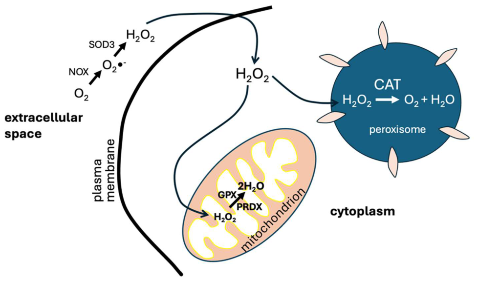
Figure 2.
The chemical reactions that involve the production and metabolism of methanethiol. Methanethiol is a major breakdown product of methionine, catalyzed by gut microbes. Human cells can produce methanethiol by methylating hydrogen sulfide gas. Methanethiol is degraded to formaldehyde, hydrogen sulfide, and hydrogen peroxide by SELENBP1. These three metabolites are all likely to be enriched in 1H compared to 2H. MGL: methionine-γ-lyase; METTL7B: methyltransferase-like protein 7B; SELENBP1: methanethiol oxidase.
Figure 2.
The chemical reactions that involve the production and metabolism of methanethiol. Methanethiol is a major breakdown product of methionine, catalyzed by gut microbes. Human cells can produce methanethiol by methylating hydrogen sulfide gas. Methanethiol is degraded to formaldehyde, hydrogen sulfide, and hydrogen peroxide by SELENBP1. These three metabolites are all likely to be enriched in 1H compared to 2H. MGL: methionine-γ-lyase; METTL7B: methyltransferase-like protein 7B; SELENBP1: methanethiol oxidase.
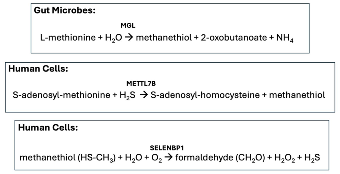
Figure 3.
Schematic of the glutamate-glutamine cycle, whereby neurons release glutamate into the synapse from vesicles, and astrocytes take up the glutamate and convert it to glutamine, by combining it with NH3. The glutamate receptors in the dendrite transmit a signal by binding to glutamate, but do not take it up. The glutamine is transported back to the neuron and taken up by mitochondria, which convert it back to glutamate in the matrix, releasing NH3. The NH3 is actively imported into the intermembrane space. It is likely enriched in 1H relative to 2H. The glutamate is then transported back into the vesicles and released into the synapse for a repeat cycle. Glu: glutamate; Gln: glutamine; NH3: Ammonia; AQP4: aquaporin-4; AQP8:aquaporin-8; GLS: glutaminase; GS: glutamine synthetase; AGC1: aspartate/glutamate carrier 1; VGLUT: vesicular glutamate transporter; EAAT: Excitatory Amino Acid Transporter; SAT1: system A amino acid transporter 1; MTS-SLC1A5: mitochondrial glutamine transporter; SNAT3: sodium-coupled neutral amino acid transporter 3.
Figure 3.
Schematic of the glutamate-glutamine cycle, whereby neurons release glutamate into the synapse from vesicles, and astrocytes take up the glutamate and convert it to glutamine, by combining it with NH3. The glutamate receptors in the dendrite transmit a signal by binding to glutamate, but do not take it up. The glutamine is transported back to the neuron and taken up by mitochondria, which convert it back to glutamate in the matrix, releasing NH3. The NH3 is actively imported into the intermembrane space. It is likely enriched in 1H relative to 2H. The glutamate is then transported back into the vesicles and released into the synapse for a repeat cycle. Glu: glutamate; Gln: glutamine; NH3: Ammonia; AQP4: aquaporin-4; AQP8:aquaporin-8; GLS: glutaminase; GS: glutamine synthetase; AGC1: aspartate/glutamate carrier 1; VGLUT: vesicular glutamate transporter; EAAT: Excitatory Amino Acid Transporter; SAT1: system A amino acid transporter 1; MTS-SLC1A5: mitochondrial glutamine transporter; SNAT3: sodium-coupled neutral amino acid transporter 3.
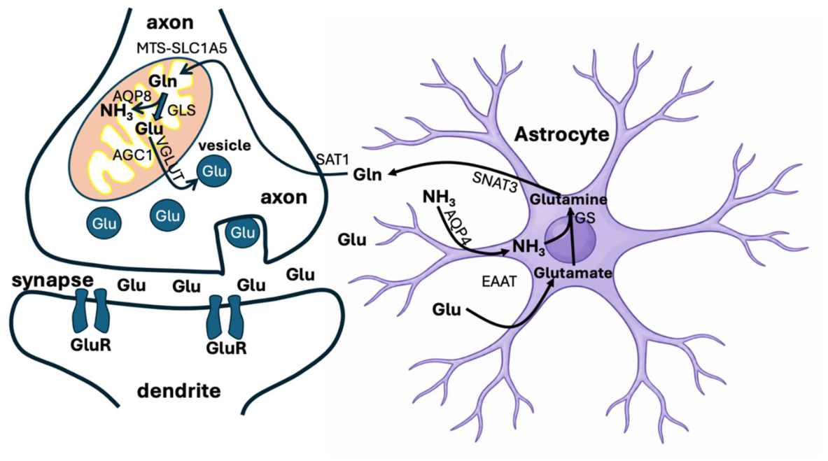
Disclaimer/Publisher’s Note: The statements, opinions and data contained in all publications are solely those of the individual author(s) and contributor(s) and not of MDPI and/or the editor(s). MDPI and/or the editor(s) disclaim responsibility for any injury to people or property resulting from any ideas, methods, instructions or products referred to in the content. |
© 2026 by the authors. Licensee MDPI, Basel, Switzerland. This article is an open access article distributed under the terms and conditions of the Creative Commons Attribution (CC BY) license (http://creativecommons.org/licenses/by/4.0/).
Copyright: This open access article is published under a Creative Commons CC BY 4.0 license, which permit the free download, distribution, and reuse, provided that the author and preprint are cited in any reuse.


