

Submitted:
26 June 2026
Posted:
29 June 2026
You are already at the latest version
Abstract
eranylgeranoic acid (GGA) is an endogenous acyclic diterpenoid metabolite of the mevalonate pathway that has been implicated in programmed cell death in hepatoma cells and may contribute to the elimination of premalignant hepatocytes. Recent metabolomic studies have identified 2,3-dihydrogeranylgeranoic acid (2,3-diGGA), an α-saturated derivative of GGA with reduced cell-death-inducing activity compared with GGA. This concise review examines the hypothesis that diversion of bioactive GGA toward 2,3-diGGA formation represents a metabolic shift that may attenuate hepatic tumor surveillance and influence hepatocellular carcinoma susceptibility. The review summarizes current evidence for endogenous GGA biosynthesis through the mevalonate pathway, MAO-B- and CYP3A4-associated GGA formation, and the tissue- and age-associated distribution of GGA and 2,3-diGGA. It also discusses the proposed 2,3-diGGA-forming activity, whose molecular identity remains unknown, and evaluates the potential utility of the GGA/2,3-diGGA ratio as a candidate biomarker. Finally, future experimental strategies are outlined to identify the responsible enzyme, clarify causality in cellular and animal models, and validate this metabolic framework in human liver tissues, chronic liver disease cohorts, and HCC-associated settings.
Keywords:
geranylgeranoic acid
; 2
; 3-dihydrogeranylgeranoic acid
; hepatocellular carcinoma
; hepatic tumor surveillance
; monoamine oxidase B
; metabolic checkpoint
1. Introduction
Geranylgeranoic acid (GGA) is an acyclic diterpenoid that was initially developed as a synthetic acyclic retinoid[1,2,3,4] and was later identified as a naturally occurring metabolite of the mevalonate pathway[5,6,7]. Early studies showed that micromolar concentrations of GGA induce differentiation[2,8] and cell death in human hepatoma cell lines. This biological activity, which differs from that of canonical retinoids, has drawn attention to GGA as a potential chemopreventive agent against hepatocellular carcinoma (HCC)[3,4]. Subsequent studies demonstrated the presence of GGA in certain edible plants and plant-derived foods[5,7] and showed that oral intake increases circulating GGA levels[9]. These findings support the concept that GGA has a dual origin from endogenous biosynthesis and habitual dietary intake[10,11,12]. They also raise the possibility that GGA may exert physiological functions beyond its role as a dietary or pharmacological compound, particularly within liver-centered metabolic networks[12,13].
Isotope-tracing studies have shown that GGA is synthesized through the mevalonate pathway. In this process, geranylgeranyl pyrophosphate (GGPP) and geranylgeraniol (GGOH) act as key upstream intermediates, while hepatic monoamine oxidase B (MAO-B) functions as a major geranylgeraniol oxidase[6,14]. In hepatocytes, MAO-B-dependent GGA formation is proposed, based primarily on in vitro and preclinical evidence, to sustain levels of GGA as a putative tumor-suppressive lipid mediator capable of promoting the elimination of premalignant cells through apoptosis[15,16,17], autophagy[18,19,20], or pyroptosis-like inflammatory cell death[21]. Age-related declines in hepatic MAO-B expression and GGA levels, together with the protective effects of oral GGA supplementation in murine models of spontaneous hepatocarcinogenesis, have led to the hypothesis of “GGA insufficiency” as a potential metabolic vulnerability associated with aging and chronic liver disease[22,23,24].
Despite these advances in understanding GGA biosynthesis and function, the downstream metabolism of GGA remains incompletely characterized. In addition to conjugation and oxidation, GGA can undergo α-saturation at the 2,3-double bond to form 2,3-dihydrogeranylgeranoic acid (2,3-diGGA), a structurally related metabolite with reduced cell-death-inducing activity in hepatoma cells[25,26,27]. An early biochemical study demonstrated the enzymatic formation of 2,3-diGGA from GGOH in rat thymus homogenates[26]. A subsequent study showed that endogenous 2,3-diGGA accumulates at relatively high levels in the thymus compared with other organs[6]. Together, these observations support the possibility that a specific, as-yet-unidentified enzymatic activity contributes to 2,3-diGGA formation. They also raise the hypothesis that diversion of metabolic flux from GGA toward 2,3-diGGA may limit the availability of bioactive GGA for tumor-suppressive signaling.
This concise review summarizes current knowledge of GGA metabolism, spanning its biosynthesis from mevalonate to its downstream conversion into 2,3-diGGA, with particular focus on hepatic tumor surveillance and HCC susceptibility. First, the biochemical steps leading to the synthesis of GGA and 2,3-diGGA are outlined, and the preclinical evidence supporting the putative tumor-suppressive role of GGA in hepatocytes is reviewed. Experimental findings regarding 2,3-diGGA formation and tissue distribution are then discussed. A working model is proposed in which metabolic diversion from GGA to 2,3-diGGA may represent a hypothesis-generating biochemical checkpoint that could influence liver cancer susceptibility. Key future directions include identifying the enzyme responsible for 2,3-diGGA formation, evaluating the GGA/2,3-diGGA ratio as a candidate biomarker, and validating this metabolic framework in human liver tissues, chronic liver disease cohorts, and HCC-associated settings.
2. Mevalonate Pathway to GGA and 2,3-diGGA in Hepatic Metabolism
The biosynthesis of GGA proceeds through the classical mevalonate pathway, which generates a range of isoprenoid intermediates[28,29,30]. In this pathway, mevalonate derived from acetyl-CoA is phosphorylated and decarboxylated to produce isopentenyl pyrophosphate and dimethylallyl pyrophosphate[31,32,33]. These intermediates are sequentially condensed to form farnesyl pyrophosphate and ultimately GGPP[34,35,36]. This intermediate serves as a donor for protein geranylgeranylation and as a precursor for other isoprenoid end products. However, a fraction of this pool is dephosphorylated by phosphatase activities to GGOH[37,38], an allylic alcohol that serves as the immediate substrate for oxidative conversion to GGA[39,40,41].
In hepatocytes, GGOH is oxidized to GGA through sequential dehydrogenation and aldehyde oxidation[39,40]. Experimental and biochemical studies suggest that MAO-B[42,43], localized to the outer mitochondrial membrane, acts as a major geranylgeraniol oxidase, generating the corresponding geranylgeranial[14]. This aldehyde is then converted to GGA by cytosolic aldehyde dehydrogenases. In parallel, cytochrome P450 enzymes, particularly CYP3A4 in human liver, can also oxidize GGOH to GGA and may provide a compensatory pathway when MAO-B activity is reduced[44]. Beyond these oxidative steps, GGA itself can undergo further structural modification at its polyprenyl backbone. In this context, α-saturation of the 2,3-double bond represents a downstream modification that generates 2,3-diGGA[6]. The enzyme or enzyme complex responsible for this conversion has not yet been identified, and the underlying mechanism remains unclear. Nevertheless, this reaction preserves the overall diterpenoid skeleton while altering the electronic and conformational properties of the molecule, and has been reported to reduce its capacity to induce cell death in hepatoma cells[25,26,27]. From a metabolic perspective, α-saturation may represent a GGA-consuming pathway that could limit the availability of bioactive GGA for tumor-suppressive signaling.
Taken together, hepatic GGA metabolism can be viewed as a sequential pathway with several potential regulatory branch points. Upstream, flux into GGOH depends on GGPP abundance and on phosphatases that release the alcohol from its pyrophosphate form. At the level of GGOH, MAO-B and CYP3A4 may influence the rate of oxidative conversion to GGA. Further downstream, the putative 2,3-diGGA-forming activity may introduce an additional branch that diverts GGA toward a less active product. Changes in the expression or activity of these enzymes during aging[22,45,46], or modulation by commonly used drugs[47,48,49,50], could therefore alter metabolite distribution within this pathway and may influence hepatic tumor surveillance and HCC susceptibility (Figure 1).
3. GGA as a Putative Hepatocellular Tumor-Suppressive Lipid Mediator
GGA was initially characterized by its ability to induce growth arrest and cell death in human hepatoma cell lines[2,12,51]. Unlike canonical retinoids, which primarily act through nuclear receptor–dependent transcriptional programs, GGA has been reported to exert comparatively rapid cytotoxic effects at micromolar concentrations[21,25,40]. These effects include mitochondrial dysfunction[52,53], activation of caspase-dependent apoptosis[12,17], and induction of non-apoptotic cell death pathways[20,21], depending on cellular context and dose. In several models, GGA has been reported to eliminate transformed cells more efficiently than non-transformed counterparts, suggesting preferential sensitivity of transformed hepatocytes under the tested conditions[5,22,23].
Mechanistic studies suggest that GGA activates multiple, partially overlapping cell death pathways[54,55,56]. In many hepatoma models, GGA has been shown to induce classical apoptotic hallmarks, including chromatin condensation, DNA fragmentation, and caspase-3 activation[5,17]. In other settings, it promotes autophagic vacuolization[57] and cell death that is only partially suppressed by caspase inhibitors[21], consistent with autophagy-associated cell elimination[58,59,60]. More recent studies suggest the involvement of pyroptosis-like inflammatory cell death, characterized by loss of membrane integrity, release of inflammatory mediators, and upstream signaling associated with peroxisomal function and oxidative stress[51,61,62]. Although the relative contribution of these pathways likely varies across experimental systems, a consistent observation is that GGA sensitizes hepatoma cells to cell death signals, favoring cell elimination rather than simple growth inhibition in experimental settings[63,64].
The putative tumor-suppressive role of GGA is further supported by in vivo studies in murine models of spontaneous hepatocarcinogenesis. In aged mice predisposed to liver tumor development, hepatic GGA levels decline with age in parallel with reduced MAO-B expression[22]. Oral administration of GGA or its 4,5-didehydro analog from midlife onward significantly reduced the incidence and multiplicity of HCC in these models, without apparent systemic toxicity under the reported experimental conditions[22,23]. Collectively, these findings suggest that maintaining or restoring hepatic GGA levels may reinforce a chemopreventive barrier against age-associated clonal expansion, at least in preclinical models[22,23,45].
From a metabolic perspective, GGA may be viewed as an endogenous lipid mediator linking the mevalonate pathway and MAO-B activity to hepatocellular homeostasis. According to this working model, sufficient MAO-B-dependent GGA production may increase the susceptibility of premalignant hepatocytes to GGA-induced cell death, thereby potentially limiting progression to overt carcinoma. Conversely, age- or disease-related declines in MAO-B expression, together with enhanced downstream metabolism of GGA, could create a state of “GGA insufficiency” in which this proposed surveillance mechanism is weakened[45]. In this context, metabolic conversion of GGA to less active derivatives, such as 2,3-diGGA, which exhibits markedly reduced cytotoxic activity in hepatoma cells, becomes particularly relevant. Thus, metabolic processes that divert GGA toward 2,3-diGGA may reduce the pool of bioactive GGA and could shift the metabolic balance toward a state permissive for hepatocarcinogenesis[65].
The next section focuses on the formation and tissue distribution of 2,3-diGGA, with particular emphasis on the enzymatic activity detected in rat thymus. It also examines how this pathway may intersect with hepatic GGA homeostasis and how the putative 2,3-diGGA–forming enzyme could represent a future target for preserving GGA-mediated tumor-suppressive activity.
4. Formation and Tissue Distribution of 2,3-diGGA
Although GGA has been studied in some detail as an endogenous lipid mediator, its downstream metabolite, 2,3-diGGA, remains much less well characterized. Structurally, 2,3-diGGA preserves the acyclic diterpenoid framework of GGA but contains a saturated C2–C3 bond, consistent with α,β-reduction of the 2,3-double bond. This seemingly minor structural modification has notable functional consequences in experimental systems: in hepatoma cell lines, 2,3-diGGA exhibits a markedly reduced capacity to induce cell death compared with GGA[25,26,27]. Thus, conversion of GGA to 2,3-diGGA may represent a metabolic attenuation step that reduces the cell-death-inducing activity of GGA in hepatoma models.
Biochemical studies suggest that 2,3-diGGA can be generated enzymatically in a tissue-dependent manner. Sagami et al. first detected 2,3-diGGA formation when GGOH was incubated with rat thymus homogenates under aerobic conditions, suggesting the presence of a thymus-associated enzyme system capable of catalyzing α-saturation during late-stage polyprenyl metabolism[26,27]. Building on this observation, a subsequent isotope-tracing study using 13C-labeled mevalonolactone in human hepatoma HuH-7 cells clarified the precursor-product relationship within GGA metabolism[6]. Newly synthesized GGA accumulated rapidly, whereas 13C incorporation into 2,3-diGGA increased more slowly and reached levels comparable to those of GGA only at later time points[6]. These kinetics support the interpretation that 2,3-diGGA is formed downstream of GGA rather than being generated primarily through direct conversion from GGOH.
The potential physiological relevance of this reaction is supported by tissue distribution data. Previous analyses showed that the molar ratio of 2,3-diGGA to GGA is high in the thymus, whereas it remains low in healthy liver[6], suggesting that the relative flux toward 2,3-diGGA formation may be limited in the liver under physiological conditions. This pattern raises the possibility that hepatic GGA homeostasis may be altered under pathological conditions that enhance metabolic flux toward the 2,3-diGGA branch. In incubation experiments, 2,3-diGGA formation was time-dependent and required both substrate and tissue homogenate, consistent with an enzymatic mechanism[26,27]. Although the identity of the enzyme responsible for 2,3-diGGA formation and the detailed reaction mechanism remain unresolved, current evidence suggests that this pathway may act as a GGA-consuming branch that reduces the availability of bioactive GGA.
5. Proposed Metabolic Diversion from GGA to 2,3-diGGA and Implications for HCC Susceptibility
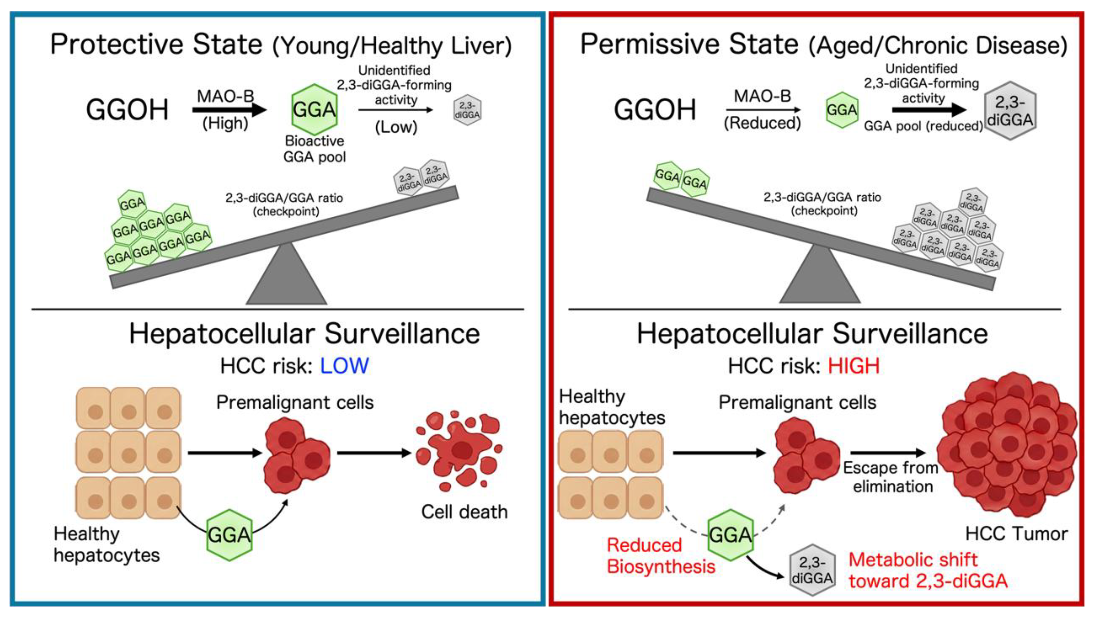
The data summarized above suggest that GGA and 2,3-diGGA may constitute a functionally relevant branch of late-stage mevalonate-derived metabolism[6,12]. Conceptually, this branch may be viewed as a proposed metabolic balance between a GGA-dominant state, in which bioactive GGA may contribute to the elimination of premalignant hepatocytes, and a more permissive state, in which GGA is diverted toward the less active 2,3-diGGA pool[65,66]. In the liver, where MAO-B–dependent GGA synthesis is proposed to contribute to a chemopreventive barrier[45], shifts in the balance between these two metabolites could influence HCC susceptibility. This proposed metabolic framework is illustrated in Figure 2, in which GGA dominance is depicted as a putatively protective state, whereas increased diversion toward 2,3-diGGA is depicted as a more permissive state for hepatocarcinogenesis.
In a putatively “protective” state, hepatic MAO-B expression and activity may be sufficient to sustain GGA production from GGOH. Under these conditions, premalignant hepatocytes that acquire oncogenic alterations may remain susceptible to GGA-induced apoptosis, autophagy-associated death, or pyroptosis-like inflammatory cell death. Although downstream metabolism of GGA through conjugation, oxidation, or conversion to 2,3-diGGA may proceed in parallel, it appears to represent a relatively minor route under physiological liver conditions[67]. As a result, steady-state GGA concentrations may be maintained within a range that supports GGA-mediated cell death responses in experimental models. Dietary intake of GGA-containing foods[5,7] or exogenous supplementation[9] may further support this state by increasing systemic or hepatic GGA availability, although direct evidence for this effect in human liver remains limited[22].
In contrast, several age- or disease-related changes may favor a more “permissive” metabolic state in which diversion from GGA toward 2,3-diGGA is enhanced. First, reduced MAO-B expression, as observed in aging and chronic liver injury[68,69,70,71], may lower the rate of GGA formation[22,72]. Second, induction of certain drug-metabolizing enzymes by pharmacologic exposure may divert the upstream precursor GGOH toward alternative metabolic fates, thereby potentially limiting GGA availability for tumor-suppressive signaling[73,74,75]. Third, upregulation of the putative 2,3-diGGA-forming activity could shift the downstream balance toward the dihydro metabolite, thereby converting a fraction of bioactive GGA into a less active form. Together, these changes may create a proposed state of “functional GGA deficiency,” in which hepatic tumor surveillance is attenuated and premalignant cells may become more likely to escape elimination and contribute to hepatocarcinogenesis.
This framework generates several testable predictions for future experimental and translational studies. If the balance between GGA and 2,3-diGGA is related to HCC susceptibility, higher hepatic or circulating 2,3-diGGA/GGA ratios would be expected to correlate with increased HCC incidence or poorer outcomes in chronic liver disease cohorts. Experimentally reducing the activity of the 2,3-diGGA–forming pathway, through genetic knockdown or targeted inhibition, would be predicted to preserve GGA levels and enhance GGA-mediated cell death responses[76]. Conversely, interventions that increase MAO-B–dependent synthesis would be predicted to decrease relative flux into the 2,3-diGGA branch and restore GGA-mediated chemopreventive activity in preclinical models.
From a translational perspective, the GGA/2,3-diGGA balance suggests two possible future intervention points: enhancing GGA availability, either by supporting endogenous synthesis or by administering exogenous GGA, and limiting GGA loss by restraining its conversion to 2,3-diGGA[77,78]. In principle, combining these approaches could allow lower doses of supplemental GGA to achieve meaningful increases in effective hepatic exposure, potentially improving chemopreventive efficiency while minimizing toxicity; however, this possibility remains to be tested experimentally. At the same time, any pharmacologic strategy would need to consider the physiological roles of 2,3-diGGA in other tissues, such as the thymus, as well as potential off-target effects and drug-interaction risks associated with modulating MAO-B or CYP activity[73,79,80,81].
Taken together, these considerations support a working hypothesis in which metabolic diversion from GGA to 2,3-diGGA may represent a biochemical checkpoint that influences the putative tumor-suppressive barrier in the liver. However, this model requires further enzymological, preclinical, and clinical validation. Whether maintaining the GGA/2,3-diGGA balance within a protective range contributes to long-term hepatocellular surveillance remains to be determined. In the following sections, strategies are considered to identify the enzyme responsible for 2,3-diGGA formation and to evaluate whether targeting this proposed metabolic branch could be translated into future approaches for HCC chemoprevention.
6. Future Directions and Experimental Strategies
The hypothesis that diversion of GGA toward 2,3-diGGA may influence HCC susceptibility raises several key experimental questions. A central unresolved issue is that the enzyme responsible for 2,3-diGGA formation has not yet been identified. Activity-guided purification from rat thymus, where 2,3-diGGA formation appears to be relatively prominent, represents a reasonable starting point[6,26,27]. Fractionation of subcellular compartments followed by incubation with GGA or GGOH, together with LC-MS/MS detection of 2,3-diGGA, could help identify the organelle in which this activity resides. Proteomic analysis of active fractions, supported by inhibitor profiling and candidate-gene validation, may clarify whether this activity belongs to a known enzyme family, such as short-chain dehydrogenases/reductases, or represents a previously uncharacterized reductive activity acting on prenylated lipid metabolites[12,82,83,84].
Once candidate genes have been nominated, genetic approaches in cell and animal models will be important for defining their physiological roles. In hepatoma cell lines and primary hepatocytes, loss- and gain-of-function studies of the putative 2,3-diGGA–forming enzyme could test its impact on the intracellular GGA/2,3-diGGA balance and on susceptibility to GGA-induced cell death. In vivo, liver-specific knockout or transgenic overexpression in mice could be combined with models of spontaneous or chemically induced hepatocarcinogenesis to examine whether altering this pathway affects tumor incidence, multiplicity, or latency[85,86]. Parallel measurements of MAO-B expression, GGA content, and the 2,3-diGGA/GGA ratio in these models would help place this enzyme within the broader network of mevalonate-derived lipid metabolism and redox regulation[87,88].
Translational studies in humans represent another major line of inquiry. Quantitative profiling of GGA and 2,3-diGGA in liver tissue, serum, and potentially bile from patients at different stages of chronic liver disease could evaluate whether the 2,3-diGGA/GGA ratio correlates with fibrosis stage, inflammatory activity, or subsequent development of HCC[89,90]. Retrospective analyses of biobanked samples from surveillance cohorts may be particularly informative[91,92]. In parallel, pharmacological and clinical factors that influence this metabolic pathway need to be defined. Commonly used drugs that modulate MAO-B, CYP3A4, or lipid metabolism could alter GGA homeostasis and potentially favor 2,3-diGGA formation; systematic pharmacometabolomic studies may help identify such interactions and possible modifiable risk conditions[93,94,95,96].
If the GGA/2,3-diGGA balance proves to be both measurable and amenable to intervention, it could provide a basis for future chemopreventive strategies[97,98]. One possible approach would be the rational design of small-molecule inhibitors directed against the 2,3-diGGA-forming enzyme, with selectivity optimized to avoid interference with related reductases. Another possible approach would be to develop reduction-resistant GGA analogs that preserve GGA-like cell-death-inducing activity while maintaining acceptable safety[99]. In both cases, careful evaluation of immunological consequences would be essential, given the high 2,3-diGGA levels in the thymus and the possibility that this metabolite contributes to T-cell development or function[6,26,27].
Furthermore, while current evidence highlights the presence of GGA and 2,3-diGGA in cytosolic or triglyceride-associated pools, future studies could investigate whether these diterpenoids are incorporated into specific membrane phospholipids[100]. Such incorporation, if confirmed, could alter membrane dynamics or serve as a reservoir for novel lipid mediators, adding another layer of complexity to their physiological functions.
In summary, this concise review proposes that the metabolic pathway from mevalonate to GGA and 2,3-diGGA may represent a regulated metabolic branch that influences the putative tumor-suppressive capacity of the liver. Defining the enzymology of 2,3-diGGA formation, clarifying the determinants of the GGA/2,3-diGGA balance in liver disease, and exploring whether this balance can be stabilized in favor of GGA may help establish new directions for HCC chemoprevention research.
7. Conclusions
This concise review proposes that GGA-centered metabolism may represent a regulatory node that influences the putative tumor-suppressive capacity of the liver. GGA may be viewed as a putative tumor-suppressive lipid mediator linking the mevalonate pathway and MAO-B activity to the elimination of premalignant hepatocytes in preclinical models. In contrast, its downstream metabolite 2,3-diGGA appears to represent a less active metabolic product with markedly reduced cell-death-inducing activity in hepatoma models. Accordingly, conversion of GGA to 2,3-diGGA may represent a GGA-consuming branch that, if enhanced, could reduce the bioactive GGA pool and contribute to functional GGA insufficiency during aging or chronic liver disease.
Key priorities for future research include elucidating the enzymology of 2,3-diGGA formation, defining the clinical determinants of the GGA/2,3-diGGA balance, and evaluating whether this balance can be stabilized in favor of GGA. If this pathway proves to be both measurable and amenable to intervention in humans, approaches aimed at maintaining endogenous GGA levels or selectively limiting its conversion to 2,3-diGGA may provide a basis for future HCC chemoprevention research.
Author Contributions
Tabata Y conceived the idea, reviewed the literature, developed the hypothesis, and wrote the manuscript.
Funding
This work was supported by JSPS KAKENHI Grant Numbers JP23K16802 and JP26K21093.
Acknowledgments
Not applicable.
Conflicts of Interest
The author declares that the research was conducted in the absence of any commercial or financial relationships that could be construed as a potential conflict of interest.
References
- Woo HY, Yoo SY, Heo J. Peretinoin, an Acyclic Retinoid, for the Secondary Prevention of Hepatocellular Carcinoma. Molecules 2021;26:295. [CrossRef]
- Araki H, Shidoji Y, Yamada Y, Moriwaki H, Muto Y. Retinoid Agonist Activities of Synthetic Geranyl Geranoic Acid Derivatives. Biochemical and Biophysical Research Communications 1995;209:66–72. [CrossRef]
- Muto Y, Moriwaki H, Ninomiya M, Adachi S, Saito A, Takasaki KT, et al. Prevention of second primary tumors by an acyclic retinoid, polyprenoic acid, in patients with hepatocellular carcinoma. Hepatoma Prevention Study Group. The New England Journal of Medicine 1996;334:1561–7. [CrossRef]
- Muto Y, Moriwaki H, Saito A. Prevention of second primary tumors by an acyclic retinoid in patients with hepatocellular carcinoma. The New England Journal of Medicine 1999;340:1046–7. [CrossRef]
- Shidoji Y, Ogawa H. Natural occurrence of cancer-preventive geranylgeranoic acid in medicinal herbs. Journal of Lipid Research 2004;45:1092–103. [CrossRef]
- Shidoji Y, Tabata Y. Unequivocal evidence for endogenous geranylgeranoic acid biosynthesized from mevalonate in mammalian cells. Journal of Lipid Research 2019;60:579–93. [CrossRef]
- Tabata Y. Quantitative Screening of Geranylgeranoic Acid in Selected Plant-Based Foods Using LC/MS/MS. Front Nutr 2025;12:1652270. [CrossRef]
- Yasuda I, Shiratori Y, Adachi S, Obora A, Takemura M, Okuno M, et al. Acyclic retinoid induces partial differentiation, down-regulates telomerase reverse transcriptase mRNA expression and telomerase activity, and induces apoptosis in human hepatoma-derived cell lines. Journal of Hepatology 2002;36:660–71. [CrossRef]
- Mitake M, Ogawa H, Uebaba K, Shidoji Y. Increase in plasma concentrations of geranylgeranoic Acid after turmeric tablet intake by healthy volunteers. J Clin Biochem Nutr 2010;46:252–8. [CrossRef]
- Tabata Y. Endogenous and dietary geranylgeranoic acid: A mini review on its role in hepatic tumor suppression. Eurasian Journal of Medicine and Oncology 2025;9:356–61. [CrossRef]
- Faulkner R, Jo Y. Synthesis, function, and regulation of sterol and nonsterol isoprenoids. Front Mol Biosci 2022;9:1006822. [CrossRef]
- Shidoji Y. Geranylgeranoic acid, a bioactive and endogenous fatty acid in mammals: a review. Journal of Lipid Research 2023;64:100396. [CrossRef]
- Duttenhefner JN, Reindl KM. Mevalonate pathway in pancreatic ductal adenocarcinoma: Mechanisms driving metabolic and cellular plasticity. Cancer Pathog Ther 2026;4:110–23. [CrossRef]
- Tabata Y, Shidoji Y. Hepatic monoamine oxidase B is involved in endogenous geranylgeranoic acid synthesis in mammalian liver cells. Journal of Lipid Research 2020;61:778–89. [CrossRef]
- Nakamura N, Shidoji Y, Moriwaki H, Muto Y. Apoptosis in human hepatoma cell line induced by 4,5-didehydro geranylgeranoic acid (acyclic retinoid) via down-regulation of transforming growth factor-alpha. Biochemical and Biophysical Research Communications 1996;219:100–4. [CrossRef]
- Nakamura N, Shidoji Y, Yamada Y, Hatakeyama H, Moriwaki H, Muto Y. Induction of apoptosis by acyclic retinoid in the human hepatoma-derived cell line, HuH-7. Biochemical and Biophysical Research Communications 1995;207:382–8. [CrossRef]
- Shidoji Y, Okamoto K, Muto Y, Komura S, Ohishi N, Yagi K. Prevention of geranylgeranoic acid-induced apoptosis by phospholipid hydroperoxide glutathione peroxidase gene. Journal of Cellular Biochemistry 2006;97:178–87. [CrossRef]
- Iwao C, Shidoji Y. Induction of nuclear translocation of mutant cytoplasmic p53 by geranylgeranoic acid in a human hepatoma cell line. Scientific Reports 2014;4:4419. [CrossRef]
- Iwao C, Shidoji Y. Upregulation of energy metabolism-related, p53-target TIGAR and SCO2 in HuH-7 cells with p53 mutation by geranylgeranoic acid treatment. Biomedical Research (Tokyo, Japan) 2015;36:371–81. [CrossRef]
- Shidoji Y, Iwao C. A rapid increase in lysophospholipids after geranylgeranoic acid treatment in human hepatoma-derived HuH-7 cells revealed by metabolomics analysis. Biochemistry and Biophysics Reports 2021;28. [CrossRef]
- Yabuta S, Shidoji Y. TLR4-mediated pyroptosis in human hepatoma-derived HuH-7 cells induced by a branched-chain polyunsaturated fatty acid, geranylgeranoic acid. Bioscience Reports 2020;40:BSR20194118. [CrossRef]
- Tabata Y, Omori M, Shidoji Y. Age-Dependent Decrease in Hepatic Geranylgeranoic Acid Content in C3H/HeN Mice and Its Oral Supplementation Prevents Spontaneous Hepatoma. Metabolites 2021;11:634. [CrossRef]
- Omori M, Shidoji Y, Moriwaki H. Inhibition of Spontaneous Hepatocarcinogenesis by 4,5-Didehydrogeranylgeranoic Acid: Effects of Small-Dose and Infrequent Administration. International Journal of Translational Medicine 2023;3:487–95. [CrossRef]
- Nair B, Menon A, Khader MA, Sethi G, Nath LR. Linking necroptosis with liver aging and chronic inflammation in hepatic pathology. Life Sci 2025;379:123871. [CrossRef]
- Iwao C, Shidoji Y. Polyunsaturated branched-chain fatty acid geranylgeranoic acid induces unfolded protein response in human hepatoma cells. PLoS One 2015;10:1–20. [CrossRef]
- Kodaira Y, Usui K, Kon I, Sagami H. Formation of (R)-2,3-dihydrogeranylgeranoic acid from geranylgeraniol in rat thymocytes. J Biochem 2002;132:327–34. [CrossRef]
- Kodaira Y, Kusumoto T, Takahashi T, Matsumura Y, Miyagi Y, Okamoto K, et al. Formation of lipid droplets induced by 2,3-dihydrogeranylgeranoic acid distinct from geranylgeranoic acid. Acta Biochimica Polonica 2007;54:777–82. [CrossRef]
- Goldstein JL, Brown MS. Regulation of the mevalonate pathway. Nature 1990;343:425–30. [CrossRef]
- Avalos M, Garbeva P, Vader L, van Wezel GP, Dickschat JS, Ulanova D. Biosynthesis, evolution and ecology of microbial terpenoids. Nat Prod Rep 2022;39:249–72. [CrossRef]
- Lange BM, Rujan T, Martin W, Croteau R. Isoprenoid biosynthesis: the evolution of two ancient and distinct pathways across genomes. Proc Natl Acad Sci U S A 2000;97:13172–7. [CrossRef]
- Kuzuyama T, Seto H. Two distinct pathways for essential metabolic precursors for isoprenoid biosynthesis. Proceedings of the Japan Academy, Series B 2012;88:41–52. [CrossRef]
- Holstein SA, Hohl RJ. Isoprenoids: remarkable diversity of form and function. Lipids 2004;39:293–309. [CrossRef]
- Faylo JL, Ronnebaum TA, Christianson DW. Assembly-Line Catalysis in Bifunctional Terpene Synthases. Acc Chem Res 2021;54:3780–91. [CrossRef]
- Verdaguer IB, Crispim M, Hernández A, Katzin AM. The Biomedical Importance of the Missing Pathway for Farnesol and Geranylgeraniol Salvage. Molecules 2022;27:8691. [CrossRef]
- Ericsson J, Greene JM, Carter KC, Shell BK, Duan DR, Florence C, et al. Human geranylgeranyl diphosphate synthase: isolation of the cDNA, chromosomal mapping and tissue expression. Journal of Lipid Research 1998;39:1731–9. [CrossRef]
- Tong H, Wiemer AJ, Neighbors JD, Hohl RJ. Quantitative determination of farnesyl and geranylgeranyl diphosphate levels in mammalian tissue. Analytical Biochemistry 2008;378:138–43. [CrossRef]
- Sagami H, Ishii K, Ogura K. Geranylgeranylpyrophosphate synthetase of pig liver. Methods in Enzymology 1985;110:184–8. [CrossRef]
- Sagami H, Ishii K, Ogura K. Occurrence and unusual properties of geranylgeranyl pyrophosphate synthetase of pig liver. Bicohemistry International 1981;3:669–75. [CrossRef]
- Mitake M, Shidoji Y. Geranylgeraniol oxidase activity involved in oxidative formation of geranylgeranoic acid in human hepatoma cells. Biomedical Research (Tokyo, Japan) 2012;33:15–24. [CrossRef]
- Muraguchi T, Okamoto K, Mitake M, Ogawa H, Shidoji Y. Polished rice as natural sources of cancer-preventing geranylgeranoic acid. Journal of Clinical Biochemistry and Nutrition 2011;49:8–15. [CrossRef]
- Verdaguer IB, Hernández A, Torres S, Rodríguez-Peiris M, Arenas F, Martinez DE, et al. Prenols and prenoic acids in metabolism, disease, and aging. J Lipid Res 2026;67:101009. [CrossRef]
- Ramsay RR. Molecular aspects of monoamine oxidase B. Progress in Neuro-Psychopharmacology and Biological Psychiatry 2016;69:81–9. [CrossRef]
- Gaweska H, Fitzpatrick PF. Structures and mechanism of the monoamine oxidase family. Biomolecular Concepts 2011;2:365–77. [CrossRef]
- Tabata Y, Shidoji Y. Hepatic CYP3A4 Enzyme Compensatively Maintains Endogenous Geranylgeranoic Acid Levels in MAOB-Knockout Human Hepatoma Cells. Metabolites 2022;12:140. [CrossRef]
- Tabata Y. Geranylgeranoic acid and the MAOB–CYP3A4 axis: a metabolic shift underlying age-related liver cancer risk. Front Aging 2025;6:1680031. [CrossRef]
- Ren J, Tong Y-M, Cui R-X, Wang Z, Li Q-L, Liu W, et al. Comparison of survival between adolescent and young adult vs older patients with hepatocellular carcinoma. World Journal of Gastrointestinal Oncology 2020;12:1394–406. [CrossRef]
- Zonfa CE, Thyagarajan A, Sahu RP. Repurposing Itraconazole in Combination with Chemotherapy and Immune Checkpoint Inhibitor for Cancer. Med Sci (Basel) 2026;14:55. [CrossRef]
- Olawale ME, Lazarus P. Inhibition of CYP2A6-mediated nicotine metabolism: A potential strategy for smoking cessation therapy. J Pharmacol Exp Ther 2026;393:103792. [CrossRef]
- Rached G, Campana A, Fiani D, Nguyen C, Van den Eynde V, Gillman PK, et al. Safety and Efficacy of Monoamine Oxidase Inhibitors in Patients Who Use Psychoactive Substances: Potential Drug Interactions and Substance Use Disorder Treatment Data. CNS Drugs 2026;40:359–417. [CrossRef]
- Eyal S, Alfasi S, Ben Zaken K, Sawaid IO, Segev L, Mesfin S, et al. Consensus Molecules Associated with Parkinson’s Disease. Neurol Int 2026;18:23. [CrossRef]
- Shidoji Y. Induction of Hepatoma Cell Pyroptosis by Endogenous Lipid Geranylgeranoic Acid-A Comparison with Palmitic Acid and Retinoic Acid. Cells 2024;13:809. [CrossRef]
- Shidoji Y, Nakamura N, Moriwaki H, Muto Y. Rapid loss in the mitochondrial membrane potential during geranylgeranoic acid-induced apoptosis. Biochem Biophys Res Commun 1997;230:58–63. [CrossRef]
- Shimonishi S, Muraguchi T, Mitake M, Sakane C, Okamoto K, Shidoji Y. Rapid downregulation of Cyclin D1 induced by geranylgeranoic acid in human hepatoma cells. Nutrition and Cancer 2012;64:473–80. [CrossRef]
- Ajoolabady A, Tang D, Kroemer G, Ren J. Ferroptosis in hepatocellular carcinoma: mechanisms and targeted therapy. Br J Cancer 2023;128:190–205. [CrossRef]
- Wu X, Cao J, Wan X, Du S. Programmed cell death in hepatocellular carcinoma: mechanisms and therapeutic prospects. Cell Death Discov 2024;10:356. [CrossRef]
- García-Pras E, Fernández-Iglesias A, Gracia-Sancho J, Pérez-del-Pulgar S. Cell Death in Hepatocellular Carcinoma: Pathogenesis and Therapeutic Opportunities. Cancers (Basel) 2021;14:48. [CrossRef]
- Okamoto K, Sakimoto Y, Imai K, Senoo H, Shidoji Y. Induction of an incomplete autophagic response by cancer-preventive geranylgeranoic acid (GGA) in a human hepatoma-derived cell line. Biochemical Journal 2011;440:63–71. [CrossRef]
- Liu L, Liao J-Z, He X-X, Li P-Y. The role of autophagy in hepatocellular carcinoma: friend or foe. Oncotarget 2017;8:57707–22. [CrossRef]
- Bialik S, Dasari SK, Kimchi A. Autophagy-dependent cell death - where, how and why a cell eats itself to death. J Cell Sci 2018;131:jcs215152. [CrossRef]
- Huang X, Yan H, Xu Z, Yang B, Luo P, He Q. The inducible role of autophagy in cell death: emerging evidence and future perspectives. Cell Commun Signal 2025;23:151. [CrossRef]
- Liu Y, Pan R, Ouyang Y, Gu W, Xiao T, Yang H, et al. Pyroptosis in health and disease: mechanisms, regulation and clinical perspective. Sig Transduct Target Ther 2024;9:245. [CrossRef]
- Tan Y, Chen Q, Li X, Zeng Z, Xiong W, Li G, et al. Pyroptosis: a new paradigm of cell death for fighting against cancer. J Exp Clin Cancer Res 2021;40:153. [CrossRef]
- Koren E, Fuchs Y. Modes of Regulated Cell Death in Cancer. Cancer Discov 2021;11:245–65. [CrossRef]
- Sato K, Nakajima T, Choyke PL, Kobayashi H. Selective cell elimination in vitro and in vivo from tissues and tumors using antibodies conjugated with a near infrared phthalocyanine. RSC Adv 2015;5:25105–14. [CrossRef]
- Drapela S, Ilter D, Gomes AP. Metabolic reprogramming: a bridge between aging and tumorigenesis. Mol Oncol 2022;16:3295–318. [CrossRef]
- Raskov H, Gaggar S, Tajik A, Orhan A, Gögenur I. Metabolic switch in cancer – Survival of the fittest. European Journal of Cancer 2023;180:30–51. [CrossRef]
- Yang J, Xiao H, Zhang Z, Zhang L, Zhang X. Regulatory roles of bioactive lipids in vascular homeostasis and remodeling. Med Rev (2021) 2026;6:35–56. [CrossRef]
- Edmondson DE. Hydrogen peroxide produced by mitochondrial monoamine oxidase catalysis: biological implications. Curr Pharm Des 2014;20:155–60. [CrossRef]
- Fowler JS, Volkow ND, Wang GJ, Logan J, Pappas N, Shea C, et al. Age-related increases in brain monoamine oxidase B in living healthy human subjects. Neurobiology of Aging 1997;18:431–5. [CrossRef]
- Liu Y, Li D, Xie C, Pan Y, Shi Q. Monoamine Oxidase B in Cancers: Implications for Therapeutics and Prognosis. JIR 2025;Volume 18:15555–86. [CrossRef]
- Nicotra A, Pierucci F, Parvez H, Senatori O. Monoamine oxidase expression during development and aging. Neurotoxicology 2004;25:155–65. [CrossRef]
- Tabata Y. Monoamine oxidase B as a context-dependent metabolic switch in hepatocellular carcinoma. Eurasian Journal of Medicine and Oncology 2026:In press. [CrossRef]
- Tu D-Z, Hu X-Y, Lei J-X, Liu S-Y, Xiao Z-P, Yang L, et al. A patent review of CYP3A4 inhibitors (2018 - present). Expert Opin Ther Pat 2025;35:503–13. [CrossRef]
- Mo H, Jeter R, Bachmann A, Yount ST, Shen C-L, Yeganehjoo H. The Potential of Isoprenoids in Adjuvant Cancer Therapy to Reduce Adverse Effects of Statins. Front Pharmacol 2019;9. [CrossRef]
- Riederer P, Müller T. Monoamine oxidase-B inhibitors in the treatment of Parkinson’s disease: clinical-pharmacological aspects. J Neural Transm (Vienna) 2018;125:1751–7. [CrossRef]
- Ren J, Yan G, Yang L, Kong L, Guan Y, Sun H, et al. Cancer chemoprevention: signaling pathways and strategic approaches. Sig Transduct Target Ther 2025;10:113. [CrossRef]
- Chen M, Lan H, Yao S, Jin K, Chen Y. Metabolic Interventions in Tumor Immunity: Focus on Dual Pathway Inhibitors. Cancers (Basel) 2023;15:2043. [CrossRef]
- Punnasseril JMJ, Auwal A, Gopalan V, Lam AK, Islam F. Metabolic Reprogramming of Cancer Cells and Therapeutics Targeting Cancer Metabolism. Cancer Med 2025;14:e71244. [CrossRef]
- Galli S, Pacilio P, Bianchini E, Alborghetti M, De Carolis L, Lombardo P, et al. The Role of MAO-B Inhibitors in Fatigue in Parkinson’s Disease: A Narrative Review. J Clin Med 2025;14:2598. [CrossRef]
- Diep YN, Park HJ, Kwon J-H, Tran M, Ko HY, Jo H, et al. Astrocytic scar restricting glioblastoma via glutamate–MAO-B activity in glioblastoma-microglia assembloid. Biomaterials Research 2023;27:71. [CrossRef]
- Junkes L, Quagliato LA, Appolinario JC, Shader RI, Nardi AE. MAO inhibitors for treatment-resistant depression: Bringing an updated perspective on pioneering drugs. Pharmacol Res 2025;219:107876. [CrossRef]
- Kavanagh KL, Jörnvall H, Persson B, Oppermann U. Medium- and short-chain dehydrogenase/reductase gene and protein families. Cell Mol Life Sci 2008;65:3895. [CrossRef]
- Rodriguez SB, Leyh TS. An Enzymatic Platform for the Synthesis of Isoprenoid Precursors. PLoS One 2014;9:e105594. [CrossRef]
- Pang X-Y, Wang S, Jurczak MJ, Shulman GI, Moise AR. RETINOL SATURASE MODULATES LIPID METABOLISM AND THE PRODUCTION OF REACTIVE OXYGEN SPECIES. Arch Biochem Biophys 2017;633:93–102. [CrossRef]
- Zhang HE, Henderson JM, Gorrell MD. Animal models for hepatocellular carcinoma. Biochimica et Biophysica Acta (BBA) - Molecular Basis of Disease 2019;1865:993–1002. [CrossRef]
- Zheng H, Xue H, Yun W-J. An overview of mouse models of hepatocellular carcinoma. Infect Agent Cancer 2023;18:49. [CrossRef]
- Guerra B, Recio C, Aranda-Tavío H, Guerra-Rodríguez M, García-Castellano JM, Fernández-Pérez L. The Mevalonate Pathway, a Metabolic Target in Cancer Therapy. Front Oncol 2021;11:626971. [CrossRef]
- Li S, Yuan H, Li L, Li Q, Lin P, Li K. Oxidative Stress and Reprogramming of Lipid Metabolism in Cancers. Antioxidants (Basel) 2025;14:201. [CrossRef]
- Polpichai N, Maneenil C, Danpanichkul P, Rattananukrom C, Choudhury A, Wong YJ, et al. Current and new strategies for hepatocellular carcinoma surveillance. Gastroenterol Rep (Oxf) 2025;13:goaf045. [CrossRef]
- Beudeker BJB, Boonstra A. Circulating biomarkers for early detection of hepatocellular carcinoma. Therap Adv Gastroenterol 2020;13:1756284820931734. [CrossRef]
- Parikh ND, Tayob N, Singal AG. Blood-based biomarkers for hepatocellular carcinoma screening: Approaching the end of the ultrasound era? J Hepatol 2023;78:207–16. [CrossRef]
- Singal AG, Yang JD, Parikh ND. Blood-based Biomarkers for HCC Surveillance: Ready for Center Stage? Am J Gastroenterol 2024;119:2147–50. [CrossRef]
- Salolin Vargas VP, Gasbarra M, Calderon-Martinez E, Shah YR, Dahiya DS, Saenz de Sicilia MG. Non-alcoholic fatty liver disease and drug induced liver injury: A metabolic storm waiting to happen. World J Hepatol 2025;17:105255. [CrossRef]
- Stolaki EV, Psatha K, Aivaliotis M. Metabolomics and Pharmacometabolomics: Advancing Precision Medicine in Drug Discovery and Development. Metabolites 2025;15:750. [CrossRef]
- Palatini P, De Martin S. Pharmacokinetic drug interactions in liver disease: An update. World J Gastroenterol 2016;22:1260–78. [CrossRef]
- Tanaka Y. Metabolomics in liver diseases: A novel alternative for liver biopsy? World J Hepatol 2024;16:12–6. [CrossRef]
- Molendijk J, Robinson H, Djuric Z, Hill MM. Lipid mechanisms in hallmarks of cancer. Molecular Omics 2020;16:6. [CrossRef]
- Schwenk S, Kaplan DE. Chemoprevention of hepatocellular carcinoma. Hepatol Commun 2025;9:e0836. [CrossRef]
- Park J, Choi J, Kim D-D, Lee S, Lee B, Lee Y, et al. Bioactive Lipids and Their Derivatives in Biomedical Applications. Biomol Ther (Seoul) 2021;29:465–82. [CrossRef]
- Robichaud PP, Surette ME. Polyunsaturated fatty acid-phospholipid remodeling and inflammation. Curr Opin Endocrinol Diabetes Obes 2015;22:112–8. [CrossRef]
Figure 1.
Endogenous biosynthetic pathway of geranylgeranoic acid (GGA) and its conversion to 2,3-dihydrogeranylgeranoic acid (2,3-diGGA). This pathway originates from the classical mevalonate pathway, in which acetyl-CoA is converted to mevalonate (MVA), followed by the formation of the C5 isoprenoid units isopentenyl pyrophosphate (IPP) and dimethylallyl pyrophosphate (DMAPP). Subsequent condensation of these isoprenoid units yields geranylgeranyl pyrophosphate (GGPP), a key branch point in isoprenoid metabolism. Dephosphorylation of GGPP produces geranylgeraniol (GGOH), which then undergoes sequential oxidation, catalyzed by monoamine oxidase B (MAO-B) and aldehyde dehydrogenases, to form geranylgeranial (GGal) and geranylgeranoic acid (GGA). Finally, GGA can be converted to 2,3-diGGA through α-saturation of its 2,3-double bond, a modification associated with reduced cell-death-inducing activity in hepatoma cells. The molecular structures illustrate conservation of the acyclic diterpenoid backbone during these late-stage modifications. Solid arrows indicate established metabolic steps, whereas dashed arrows indicate proposed or incompletely characterized steps.
Figure 1.
Endogenous biosynthetic pathway of geranylgeranoic acid (GGA) and its conversion to 2,3-dihydrogeranylgeranoic acid (2,3-diGGA). This pathway originates from the classical mevalonate pathway, in which acetyl-CoA is converted to mevalonate (MVA), followed by the formation of the C5 isoprenoid units isopentenyl pyrophosphate (IPP) and dimethylallyl pyrophosphate (DMAPP). Subsequent condensation of these isoprenoid units yields geranylgeranyl pyrophosphate (GGPP), a key branch point in isoprenoid metabolism. Dephosphorylation of GGPP produces geranylgeraniol (GGOH), which then undergoes sequential oxidation, catalyzed by monoamine oxidase B (MAO-B) and aldehyde dehydrogenases, to form geranylgeranial (GGal) and geranylgeranoic acid (GGA). Finally, GGA can be converted to 2,3-diGGA through α-saturation of its 2,3-double bond, a modification associated with reduced cell-death-inducing activity in hepatoma cells. The molecular structures illustrate conservation of the acyclic diterpenoid backbone during these late-stage modifications. Solid arrows indicate established metabolic steps, whereas dashed arrows indicate proposed or incompletely characterized steps.
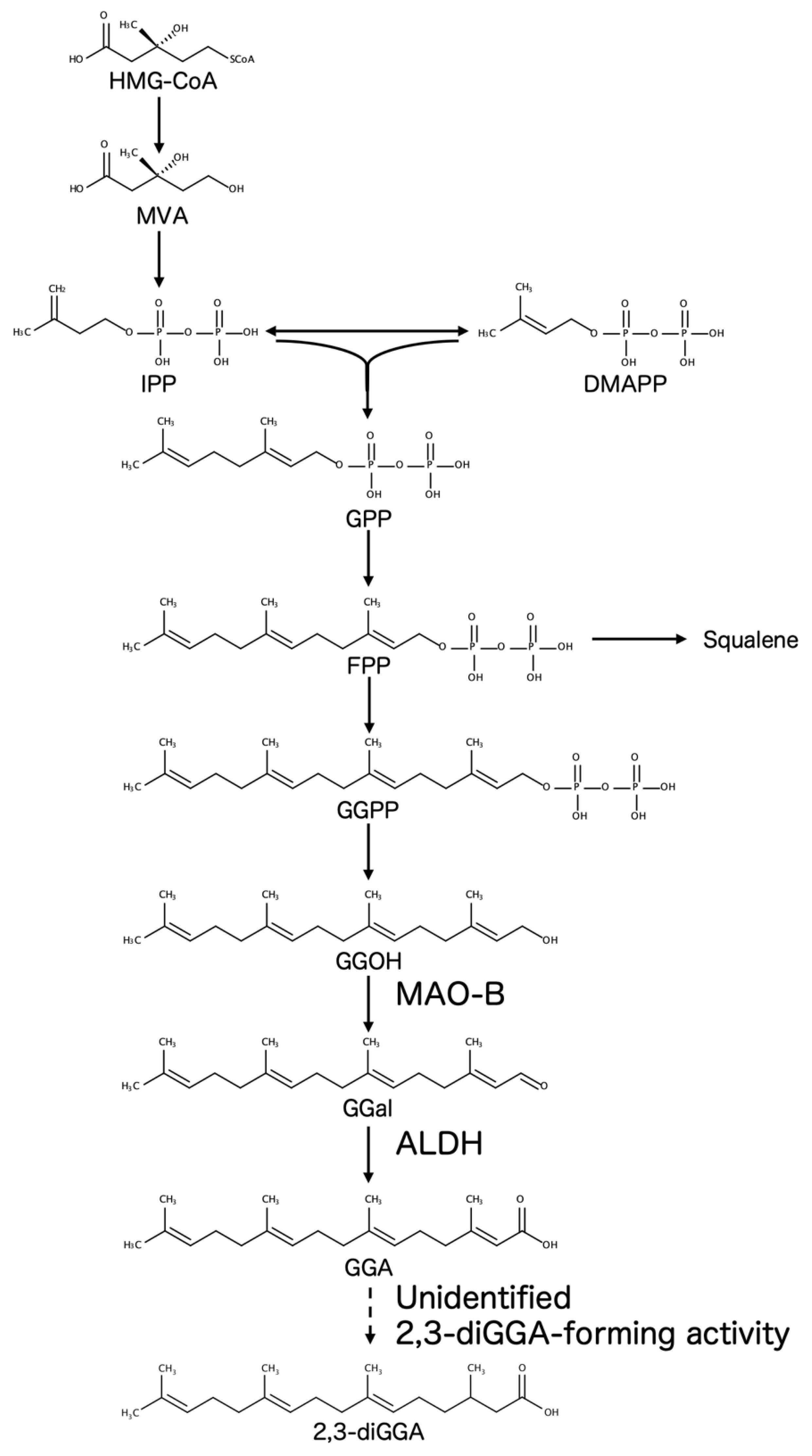
Figure 2.
Proposed model in which the 2,3-diGGA/GGA ratio may influence hepatocellular tumor surveillance. In a protective state (young/healthy liver), MAO-B–dependent oxidation of geranylgeraniol (GGOH) maintains a bioactive GGA pool, whereas flux toward the formation of 2,3-dihydrogeranylgeranoic acid (2,3-diGGA) remains limited. This balance is proposed to support hepatocellular tumor surveillance by promoting the elimination of premalignant hepatocytes through programmed cell-death pathways, thereby resulting in a lower risk of hepatocellular carcinoma (HCC). In a permissive state (aged/chronic disease), reduced MAO-B activity, together with increased diversion of GGA toward 2,3-diGGA via an as-yet-unidentified 2,3-diGGA–forming activity, is proposed to decrease functional GGA availability and shift the balance toward 2,3-diGGA, thereby facilitating the escape and progression of premalignant hepatocytes and increasing HCC risk. 2,3-diGGA is depicted as a pool with reduced bioactivity relative to GGA, based on currently available evidence; however, the definitive biological functions of 2,3-diGGA and the identity of the enzyme(s) responsible for its formation remain to be established.
Figure 2.
Proposed model in which the 2,3-diGGA/GGA ratio may influence hepatocellular tumor surveillance. In a protective state (young/healthy liver), MAO-B–dependent oxidation of geranylgeraniol (GGOH) maintains a bioactive GGA pool, whereas flux toward the formation of 2,3-dihydrogeranylgeranoic acid (2,3-diGGA) remains limited. This balance is proposed to support hepatocellular tumor surveillance by promoting the elimination of premalignant hepatocytes through programmed cell-death pathways, thereby resulting in a lower risk of hepatocellular carcinoma (HCC). In a permissive state (aged/chronic disease), reduced MAO-B activity, together with increased diversion of GGA toward 2,3-diGGA via an as-yet-unidentified 2,3-diGGA–forming activity, is proposed to decrease functional GGA availability and shift the balance toward 2,3-diGGA, thereby facilitating the escape and progression of premalignant hepatocytes and increasing HCC risk. 2,3-diGGA is depicted as a pool with reduced bioactivity relative to GGA, based on currently available evidence; however, the definitive biological functions of 2,3-diGGA and the identity of the enzyme(s) responsible for its formation remain to be established.
Disclaimer/Publisher’s Note: The statements, opinions and data contained in all publications are solely those of the individual author(s) and contributor(s) and not of MDPI and/or the editor(s). MDPI and/or the editor(s) disclaim responsibility for any injury to people or property resulting from any ideas, methods, instructions or products referred to in the content. |
© 2026 by the authors. Licensee MDPI, Basel, Switzerland. This article is an open access article distributed under the terms and conditions of the Creative Commons Attribution (CC BY) license (http://creativecommons.org/licenses/by/4.0/).
Copyright: This open access article is published under a Creative Commons CC BY 4.0 license, which permit the free download, distribution, and reuse, provided that the author and preprint are cited in any reuse.


