

Submitted:
26 June 2026
Posted:
29 June 2026
You are already at the latest version
Abstract
Immune checkpoint inhibitor (ICI)-associated hemophagocytic lymphohistiocytosis (HLH) is a rare, life-threatening hyperinflammatory toxicity increasingly described in solid tumors, but evidence remains limited to case reports and small series. We systematically reviewed adult cases of ICI-associated HLH in solid malignancies identified through PubMed, Cochrane Library, Google Scholar, and reference-list screening from database inception through March 3, 2026. Case-level data were extracted for tumor type, ICI regimen, timing, diagnostic findings, HLH-directed therapy, ICU-level care, and outcomes; because of heterogeneity, findings were summarized descriptively. Eighty adult cases were included. Median age was 63 years; the most common cancers were lung cancer, melanoma, breast cancer, and renal tumors. PD-1 inhibitors predominated, mainly pembrolizumab and nivolumab. Among cases with reported timing, median time from last ICI dose to HLH onset was 17 days, with most occurring within 30 days of the last dose and within the first four ICI-containing cycles. Corticosteroids were used in nearly all cases; additional therapies included tocilizumab, etoposide, anakinra, IVIG, mycophenolate, ruxolitinib, calcineurin inhibitors, and plasma exchange. ICI-associated HLH often occurs early after exposure and may mimic sepsis, malignancy progression, or other immune-related adverse events; prospective data are needed to define risk factors, diagnostic thresholds, and treatment strategies.
Keywords:
hemophagocytic lymphohistiocytosis
; HLH
; immune checkpoint inhibitor
; ICI
; solid tumor
; systematic review
1. Introduction
The advent of immune checkpoint inhibitors (ICIs) has fundamentally changed how we treat solid tumors. Agents targeting PD-1, PD-L1, and CTLA-4 have produced responses, and in some cases durable remissions beyond those historically achieved with chemotherapy alone [1]. Their use has expanded rapidly across melanoma, non-small cell lung cancer, renal cell carcinoma, and beyond, and they are now a cornerstone of first-line treatment for many advanced malignancies [1].
This enhanced immune activity, however, carries significant toxicity. By releasing the brakes on the immune system, ICIs can trigger a wide spectrum of inflammatory toxicities. Skin rashes, colitis, thyroiditis, and pneumonitis are now familiar territory for oncologists, with immune related adverse events (irAEs) reported in up to 90% of patients receiving anti-CTLA-4 and up to 70% of those on anti-PD-1/PD-L1 agents [2]. Less common but potentially life-threatening are hyperinflammatory toxicities such as hemophagocytic lymphohistiocytosis (HLH), characterized by dysregulated systemic immune activation.
HLH is not a new disease. Hematologists have long recognized it in the context of infections, autoimmune conditions, and hematologic malignancies, where it carries a mortality rate approaching 40% in adults [3]. It is classified as primary, typically driven by genetic defects in cytotoxic lymphocyte function, or secondary, arising in response to an external trigger such as infection, rheumatologic disease, or malignancy [4].
More recently, HLH has emerged as a rare complication of ICI therapy in patients with solid tumors, a population in whom the syndrome has been underrecognized and poorly studied.
Pharmacovigilance data suggest HLH is reported seven times more frequently with ICI therapy than with other drug classes, pointing to a real biological signal rather than incidental association [5].
Recognizing ICI-associated HLH in patients with solid tumors is particularly challenging, as its clinical features, including fever, cytopenias, hyperfferitinemia, and hepatitis, overlap substantially with sepsis, disease progression, and other irAEs such as immune-mediated hepatitis There is no single test that makes the diagnosis, and delayed recognition may allow progression to critical illness. In patients receiving combination immunotherapy or concurrent chemotherapy, the picture is even murkier, as treatment-related cytopenias and malignancy-associated inflammation can each independently mimic or obscure the syndrome [6].
Despite increasing recognition, the published literature on ICI-associated HLH in solid tumors remains almost entirely case reports and small series, with treatment strategies variable and no clear consensus on management [7]. There is no consolidated analysis focused specifically on the solid tumor population characterizing clinical presentation, onset timing, treatment patterns, and outcomes. Clinicians managing these cases are working without evidence-based guidance.
To address this gap, we conducted a systematic review of published cases of ICI-associated HLH in adult patients with solid malignancies. Our objective was to summarize published case-level data to better characterize the clinical features, timing of onset, diagnostic findings, therapeutic strategies, and outcomes of this rare but serious complication, with the aim of earlier recognition and more standardized management.
2. Methods
This systematic review was conducted in accordance with the Preferred Reporting Items for Systematic Reviews and Meta-Analyses (PRISMA) guidelines.
2.1. Eligibility Criteria
Studies were eligible for inclusion if they reported cases of HLH associated with ICI therapy in adult patients (≥18 years) with solid malignancies. Eligible study designs included case reports and case series with extractable patient-level data. Studies were required to report clinical presentation, treatment, or outcomes related to HLH.Studies were excluded if they involved pediatric populations, HLH associated with hematologic malignancies, or non-English publications without sufficient extractable data.
2.2. Search Strategy
A systematic literature search was conducted in PubMed, Cochrane Library, and Google Scholar from database inception through March 3, 2026. The study selection process is summarized in Figure 1. The PubMed search strategy combined terms related to hemophagocytic lymphohistiocytosis (HLH) and immune checkpoint inhibitors, including specific agent names. Searches were limited to English-language studies involving adult populations. No publication year restrictions were applied.
The PubMed search strategy was as follows, yielding 140 records, which were added in to the initial manuscript pool:
(
hemophagocytic lymphohistiocytosis[tiab]
OR HLH[tiab]
OR hemophagocytic syndrome[tiab]
)
AND
(
immune checkpoint inhibitor*[tiab]
OR checkpoint inhibitor*[tiab]
OR immunotherapy[tiab]
OR pembrolizumab[tiab]
OR nivolumab[tiab]
OR cemiplimab[tiab]
OR dostarlimab[tiab]
OR atezolizumab[tiab]
OR durvalumab[tiab]
OR avelumab[tiab]
OR ipilimumab[tiab]
OR tremelimumab[tiab]
)
NOT
(
leukemia[tiab]
OR lymphoma[tiab]
OR myeloma[tiab]
OR “hematologic malignan*”[tiab]
OR “haematologic malignan*”[tiab]
)
AND english[lang]
NOT (
infant[mesh]
OR child[mesh]
OR adolescent[mesh]
)
NOT (
leukemia[mesh]
OR lymphoma[mesh]
)
The Cochrane Library was additionally searched using the following exact terms:
hemophagocytic lymphohistiocytosis, and also, HLH, yielding 74 records which were added to the initial manuscript pool.
Google Scholar was searched using the following this exact search term:
“hemophagocytic lymphohistiocytosis”, HLH, immunotherapy, ici, solid tumors
This yielded 47 records after manual screening by title, which were included in the initial manuscript pool.
2.3. Study Selection and Data Extraction
Two reviewers independently screened titles and abstracts for eligibility, followed by a full-text review of potentially relevant articles. Discrepancies were resolved through discussion and consensus.
Data were extracted at the individual case level. Extracted variables included patient demographics (age, sex), malignancy type, type of ICI used, number of ICI cycles, time from ICI exposure to HLH onset, programmed death (PD-1/PD-L1) or cytotoxic T-lymphocyte-associated protein 4 (CTLA-4) expression (when available), HLH diagnostic criteria, intensive care unit (ICU) admission, treatment modalities for HLH, and clinical outcomes, including mortality.
2.4. Study Selection Results
A total of 261 articles were identified through database searching. An additional 9 articles were identified through manual review of the reference lists. 144 studies remained after duplicate removal. All 144 studies underwent title and abstract screening, leaving 72 articles. After screening and application of eligibility criteria, 70 articles were included in the final analysis, leading to a total of 80 cases (Figure 1).
2.5. Data Synthesis
Given the rarity of ICI-associated HLH and the heterogeneity of available data, a quantitative meta-analysis was not feasible. Therefore, a descriptive synthesis was performed. Case-level data were tabulated to identify patterns in patient characteristics, clinical presentation, treatment strategies, and outcomes.
2.6. Risk of Bias and Certainty of Evidence
Given that the available literature on ICI-associated HLH consists entirely of case reports and small case series, formal risk of bias assessment using standardized tools such as the Cochrane Risk of Bias tool or GRADE framework was not applicable to this dataset. Individual case reports are inherently subject to selection bias, reporting bias, and publication bias favoring positive or survival outcomes. These limitations are acknowledged throughout and are reflected in our descriptive approach to data synthesis. No formal certainty of evidence rating was assigned, as the heterogeneity of reporting and absence of comparative data preclude such assessment. This review was not prospectively registered given its descriptive, hypothesis-generating nature and the absence of a pre-specified quantitative analysis plan.
3. Results
3.1. Selection of Studies and Data Analysis
The present case-level dataset consisted of 80 adult patients with solid tumors who developed HLH after ICI therapy [8,9,10,11,12,13,14,15,16,17,18,19,20,21,22,23,24,25,26,27,28,29,30,31,32,33,34,35,36,37,38,39,40,41,42,43,44,45,46,47,48,49,50,51,52,53,54,55,56,57,58,59,60,61,62,63,64,65,66,67,68,69,70,71,72,73,74,75]. (Table 1) Most included reports were case reports or small case series; therefore, results were summarized descriptively. Not all reports fully reported timing, diagnosis, or outcome, therefore the denominators differed by variables.
3.2. Patient and Tumor Characteristics
Median age was 63 years (IQR, 51.5–73.3; range, 32–80) and sex distribution was balanced with 41 male patients (51.3%) and 39 female patients (48.8%). The most common tumor type was lung cancer (22/80, 27.5%), followed by melanoma (15/80, 18.8%), breast cancer (9/80, 11.3%), kidney/renal tumors (8/80, 10.0%), and gynecologic malignancies (6/80, 7.5%). The most frequent individual tumor types were melanoma (15/80, 18.8%), lung adenocarcinoma (14/80, 17.5%), triple-negative breast cancer (7/80, 8.8%), clear cell renal cell carcinoma (6/80, 7.5%), and lung squamous cell carcinoma (5/80, 6.3%). These findings are summarized in Table 2.
3.3. ICI Exposure and Timing of HLH Presentation
Most of the exposure was to PD-1 inhibitors and was noted in 69 of 80 cases (86.3%). Exposure to PD-L1 inhibitor and CTLA-4 inhibitor was reported in 10 (12.5%) and 16 (20.0%) cases, respectively. Due to the combination checkpoint blockade, the categories were not mutually exclusive. Sixty-five patients (81.3%) received a single checkpoint inhibitor, and 15 (18.8%) received combination checkpoint blockade. Pembrolizumab was the most common agent (38/80, 47.5%), followed by nivolumab (25/80, 31.3%) and ipilimumab (14/80, 17.5%). Sixty-five cases reported the time from last exposure to ICI to HLH onset. Median time to onset was 17 days (interquartile range 10 to 30; range 0 to 360). HLH occurred within 30 days of the last ICI dose in 49 of 65 cases (75.4%) and within 60 days in 57 of 65 cases (87.7%). Seventy cases reported the number of ICI-containing cycles before HLH onset, with a median of 2 cycles (IQR, 1–4; range, 1–17). HLH occurred after the first ICI-containing cycle in 27/70 cases (38.6%) and within the first four ICI-containing cycles in 53/70 cases (75.7%). These findings are summarized in Table 3.
3.4. Diagnosis - Characteristics and Severity
HScore reporting was incomplete. 41 of 80 cases (51.3%) had an HScore or qualitative HScore estimate; 40 cases had extractable numeric scores. For numeric reports, the median HScore was 237 (interquartile range, 207-267; range, 105-319). HLH-2004 diagnostic criteria, defined as meeting at least 5 of 8 criteria when explicitly reported, were documented as fulfilled in 74 of 80 cases. Among 79 cases with reported bone marrow findings, hemophagocytosis was identified in 53 (67.1%). In 6 of 79 (7.6%) reported cases, a concomitant infection trigger was identified. ICU admission or ICU-level care was reported in 15/80 patients (18.8%). These findings are summarized in Table 4.
3.5. HLH-Directed Therapy Treatment and Outcomes
Treatment was heterogeneous, but corticosteroids were used in 78 of 80 cases (97.5%). Other immunomodulatory or HLH-directed therapies included tocilizumab (19/80 [23.8%]), etoposide (12/80 [15.0%]), anakinra (10/80 [12.5%]), mycophenolate mofetil (8/80 [10.0%]), IVIG (6/80 [7.5%]), ruxolitinib (3/80 [3.8%]), cyclosporine or tacrolimus (3/80 [3.8%]), and plasma exchange (3/80 [3.8%]). Outcomes were reported in 78 cases. Clinical recovery or improvement was observed in 68/78 cases (87.2%) and 10 patients died (12.8%). No formal testing was performed because of the descriptive design, small sample size, and heterogeneous case reporting. Mortality was higher in patients requiring ICU admission or ICU-level care (4/15, 26.7%) vs. non-ICU cases with known outcomes (6/63, 9.5%). These findings are summarized in Table 5.
4. Discussion
ICI-associated HLH is increasingly recognized as a rare but potentially life-threatening irAE. The available literature remains dominated by case reports and small case series, limiting estimates of incidence, prognosis and optimal treatment. To our knowledge, our study represents one of the largest consolidated analyses of ICI-associated HLH in adult patients with solid malignancies. In this systemic review of published adult solid tumor cases, we identified several clinically relevant patterns: PD-1/PD-L1 inhibitor exposure was most frequent, onset was often early after ICI exposure, corticosteroids were used in nearly all reported cases, and reported clinical improvement was common.
In our cohort of 80 patients, ICI-associated HLH most commonly occurred in patients with lung cancer and was predominantly associated with PD-1/PD-L1 inhibitor exposure, particularly pembrolizumab. Importantly, most cases developed early during treatment, with over three-quarters of reported cases occurring within the first four cycles of therapy. This finding suggests that clinicians should maintain a high index of suspicion for HLH early after ICI initiation. Treatment approaches varied, but corticosteroids were used in nearly all reported cases, with a majority of patients demonstrating clinical improvement or recovery following immunosuppressive therapy.
Among cases with reported timing, most occurred within 30 days of the last ICI dose and within the first four ICI-containing cycles. This pattern suggests that ICI-associated HLH may represent an early hyperinflammatory toxicity phenotype after checkpoint inhibition and supports heightened clinical vigilance when compatible inflammatory features develop.
The recognition of HLH in patients receiving ICIs is challenging due to significant overlap with other oncologic and treatment-related conditions. ICI-associated HLH may present as unexplained fever, cytopenias, and elevated transaminases [7]. Additional findings that may support the diagnosis include hyperferritinemia, hypertriglyceridemia, and organomegaly.
These clinical and laboratory abnormalities may closely resemble other serious conditions encountered in this patient population, including sepsis, disseminated intravascular coagulation, acute liver failure, and progressive malignancy. In addition, other irAEs, such as ICI-associated autoimmune hepatitis, may present with similar inflammatory and hepatic manifestations, further complicating timely diagnosis [76].
Given the potentially rapid progression and high morbidity associated with HLH, early recognition is critical, particularly as historical adult mortality is substantial [77]. Suspicion should be maintained in patients presenting with unexplained systemic inflammatory symptoms shortly after initiation of immunotherapy, particularly when accompanied by cytopenias, hyperferritinemia, or evidence of organ dysfunction. Early diagnostic evaluation and prompt initiation of immunosuppressive therapy may improve clinical outcomes and reduce progression to severe or ICU-level disease.
While the precise mechanisms leading to HLH remain incompletely understood, the condition is characterized by severe immune activation and dysregulation. A hallmark feature is the uncontrolled release of proinflammatory cytokines, including IL-1, IL-6, TNF-a, and IFN-y, alongside aberrant activation of cytotoxic cells [78,79,80]. The pathogenesis of HLH is thought to involve excessive activation and impaired regulation of cytotoxic T lymphocytes (CTLs) and natural killer (NK) cells, resulting in persistent immune stimulation and cytokine amplification. Dysregulation of regulatory T-cell function may further contribute to the uncontrolled inflammatory response [81].
ICIs enhance antitumor immunity by promoting T-cell activation and proliferation through blockade of inhibitory immune checkpoints such as PD-1/PD-L1 and CTLA-4 pathways. Although this mechanism improves immune-mediated tumor recognition, it may also disrupt peripheral immune tolerance and permit excessive activation of autoreactive or hyperinflammatory immune responses. [82]. The resulting cytokine-driven inflammatory cascade may contribute to macrophage activation and hemophagocytosis, the pathologic hallmark of HLH [83]. In severe cases, this hyperinflammatory state can ultimately lead to rapid tissue injury, multiorgan dysfunction, and death.
The HLH-2004 criteria provide a commonly used diagnostic framework that incorporates clinical, laboratory, and pathologic findings, including fever, splenomegaly, cytopenias, hypertriglyceridemia and/or hypofibrinogenemia, hemophagocytosis, reduced or absent natural killer (NK) cell activity, hyperferritinemia, and elevated soluble CD25 levels [84]. Current recommendations suggest that fulfillment of at least five of eight HLH-2004 criteria supports the diagnosis of HLH. However, given the potentially rapid progression and high mortality associated with untreated disease, initiation of therapy may be warranted in patients with high clinical suspicion even before complete diagnostic criteria are fulfilled [85].
Another commonly utilized diagnostic tool is the H-Score, which incorporates additional clinical and laboratory variables such as underlying immunosuppression, hepatomegaly, and elevated aspartate aminotransferase (AST) levels. In the original validation study, patients diagnosed with secondary hemophagocytic syndrome demonstrated substantially higher H-Score values compared with patients without HLH, supporting its utility as an adjunctive diagnostic tool in adult populations.
The HLH-2004 treatment protocol was originally developed for pediatric HLH and primarily incorporate corticosteroids, cyclosporine A, intrathecal therapy, and etoposide, followed by hematopoietic stem cell transplantation in selected cases [86]. However, the management of adult ICI-associated HLH remains less clearly defined. The HLH group published expert opinions on the management of adult ICI-associated HLH, with recommendations for treatment with corticosteroids and anti-IL-6R therapy (tocilizumab) in patients with suspected HLH, with escalation to etoposide to be considered if insufficient response is seen after 48 hours [86,87].
In our review, corticosteroids were used in nearly all reported cases, supporting their continued role as the backbone of therapy for ICI-associated HLH. Tocilizumab was among the most commonly utilized steroid-sparing or adjunctive agents, reflecting increasing interest in targeted cytokine-directed therapy for hyperinflammatory syndromes associated with immunotherapy. Prior case reports and small case series have reported rapid clinical improvement and normalization of inflammatory markers following tocilizumab administration in selected patients with ICI-associated HLH [88].
Additional therapies reported in refractory cases included intravenous immunoglobulin (IVIG), anakinra, ruxolitinib, and plasmapheresis, although evidence supporting these approaches remains limited to isolated reports and small series. Given the high morbidity and mortality associated with HLH, as well as the expanding use of ICIs, there exists a need for standardized diagnostic and treatment guidelines tailored specifically to adult patients with ICI-associated HLH.
When compared with secondary HLH arising from more traditional etiologies such as infection or malignancy, ICI-associated HLH in our cohort appeared to demonstrate several distinct clinical patterns. Patients in our study had a higher mean age at presentation (63 years) compared with previously reported cohorts of secondary HLH, in which the average age is often closer to 50 years [89]. Similar age distributions have also been reported in other pharmacovigilance and systematic reviews of ICI-associated HLH. The studies by Diaz and Noseda reported incidences at mean ages of 64 and 63 years old, respectively [5,89]. This difference may partially reflect the older baseline age of patients receiving immunotherapy for solid malignancies, particularly lung cancer, which represented the most common underlying malignancy in our cohort.
Our findings also demonstrated similarities compared with classical secondary HLH. Bone marrow hemophagocytosis was identified in 67.1% of cases in our review, which is generally consistent with previously reported rates in adult HLH cohorts [90].
Reported mortality in published ICI-associated HLH cases our cohort was lower than historically reported mortality rates in adult secondary HLH cohorts (12.8% versus 40%) [91]. Similar findings have been observed in other reviews of ICI-associated HLH. Several factors may contribute to this difference, including earlier recognition of immune-related toxicities in patients receiving ICIs, prompt initiation of immunosuppressive therapy, publication bias favoring successful outcomes, and potential biological differences between ICI-associated HLH and classical malignancy-associated HLH.
Our study has several limitations that should be acknowledged. First, the available literature on ICI-associated HLH is largely limited to case reports and small case series, which are subject to publication bias and heterogeneous reporting practices. Consequently, the reported case counts and outcomes of ICI-associated HLH cannot be interpreted as true incidence, population-level mortality or comparative prognosis estimates. The lower mortality observed in our cohort compared with historically reported mortality rates in adult HLH may, in part, reflect underreporting of fatal cases, publication bias toward successful outcomes, or increased recognition and earlier treatment of ICI-associated disease.
Additionally, many reports lacked complete patient-level clinical data, including comorbid conditions, infectious evaluations, detailed diagnostic workup, and standardized reporting of HLH criteria or H-Score components. This limited our ability to fully assess contributing factors, disease severity, and predictors of clinical outcomes. Treatment reporting was also variable, with incomplete information regarding corticosteroid dosing, duration, tapering strategies, and timing of escalation therapies. Furthermore, our review was limited to English-language publications and excluded patients with hematologic malignancies, which may affect the generalizability of our findings. In addition, the search strategy incorporated terms intended to exclude hematologic malignancies. Although this approach was designed to focus the review on solid tumor populations, it may have inadvertently excluded otherwise relevant reports involving solid tumors that also referenced hematologic malignancies in mixed cohorts or within the manuscript text. As a result, definitive conclusions regarding optimal management strategies for ICI-associated HLH cannot be established from the currently available literature.
5. Future Directions
Future studies should prioritize prospective multicenter registries and standardized case reporting for ICI-associated HLH. Minimum reporting elements should include ICI agent and regimen, timing from last dose and cycle number, concurrent infection or other triggers, HLH-2004 criteria, HScore components when available, ferritin/triglycerides/fibrinogen/sCD25, marrow findings, ICU-level care, treatment sequence and dosing and clinical outcome. Such data is needed to define risk factors, refine diagnostic thresholds in oncology patients, and guide treatment escalation.
6. Conclusion
ICI-associated hemophagocytic lymphohistiocytosis (HLH) is a rare but potentially life-threatening immune-related adverse event. In this review of published adult solid tumor cases, ICI-associated HLH was most often reported in patients with lung cancer and most commonly followed PD-1 inhibitor exposure, particularly pembrolizumab. Onset was often early, typically within the first month and within the first four treatment cycles, supporting heightened clinical vigilance early after ICI initiation when compatible inflammatory features develop. Clinical presentation may overlap with sepsis, progressive malignancy, and other immune-related toxicities, making early diagnosis challenging. Persistent fever, cytopenias, hyperferritinemia, transaminitis, or unexplained systemic inflammation after ICI exposure should prompt consideration of HLH and early diagnostic evaluation. Most patients improved with corticosteroids with tocilizumab used as an adjunctive agent in some cases; reported mortality was greater among patients who requiring ICU-level care. Because the available evidence consists largely of case reports and small series with heterogeneous reporting, these findings should be interpreted descriptively rather than as estimates of incidence or prognosis. Current evidence remains insufficient to define optimal treatment algorithms or predictors of outcome. Increasing recognition of this syndrome with expanding ICI use highlights the need for standardized reporting, prospective data collection, and management protocols for adult patients with ICI-associated HLH.
Author Contributions
Conceptualization, A.N. (Areeba Nayyer) and M.J.; Methodology, S.C.; Data Curation, S.C., Z.A. and F.K.; Investigation, Z.A. and F.K.; Formal Analysis, A.N. (Ashish Nepal); Visualization, A.N. (Ashish Nepal); Writing – Original Draft Preparation, S.R., Z.A. and H.D.; Writing – Review & Editing, Z.A. and A.N. (Areeba Nayyer); Project Administration, A.N. (Areeba Nayyer); Supervision, M.J. All authors have read and agreed to the published version of the manuscript.
Funding
This research received no external funding.
Data Availability Statement
No new data were created or analyzed in this study. Data sharing is not applicable to this article.
Conflicts of Interest
None.
References
- Fitzsimmons, T.S.; Singh, N.; Walker, T.D.; Newton, C.; Evans, D.G.; Crosbie, E.J.; et al. Immune checkpoint inhibitors efficacy across solid cancers and the utility of PD-L1 as a biomarker of response: A systematic review and meta-analysis. Front. Med. 2023, 10. [Google Scholar] [CrossRef] [PubMed]
- Schneider, B.J.; Naidoo, J.; Santomasso, B.D.; Lacchetti, C.; Adkins, S.; Anadkat, M.; et al. Management of immune-related adverse events in patients treated with immune checkpoint inhibitor therapy: ASCO guideline update. J. Clin. Oncol. 2021, 39(36), 4073–126. [Google Scholar] [CrossRef] [PubMed]
- Ramos-Casals, M.; Brito-Zerón, P.; López-Guillermo, A.; Khamashta, M.A.; Bosch, X. Adult haemophagocytic syndrome. The Lancet 2014, 383(9927), 1503–16. [Google Scholar] [CrossRef] [PubMed]
- Koumadoraki, E.; Madouros, N.; Sharif, S.; Saleem, A.; Jarvis, S.; Khan, S. Hemophagocytic lymphohistiocytosis and infection: A literature review. Cureus 2022. [Google Scholar] [CrossRef] [PubMed]
- Diaz, L.; Jauzelon, B.; Dillies, A.-C.; Le Souder, C.; Faillie, J.-L.; Maria, A.; et al. Hemophagocytic lymphohistiocytosis associated with immunological checkpoint inhibitors: A pharmacovigilance study. J. Clin. Med. 2023, 12(5), 1985. [Google Scholar] [CrossRef] [PubMed]
- Daver, N.; McClain, K.; Allen, C.E.; Parikh, S.A.; Otrock, Z.; Rojas-Hernandez, C.; et al. A consensus review on malignancy-associated hemophagocytic lymphohistiocytosis in adults. Cancer 2017, 123(17), 3229–40. [Google Scholar] [CrossRef] [PubMed]
- Walmsley, C.S.; Schoepflin, Z.; De Brabandt, C.; Rangachari, D.; Berwick, S.; Patell, R. Hemophagocytic lymphohistiocytosis associated with immune checkpoint inhibitor use: A review of the current knowledge and future directions. Blood Cells Mol. Dis. 2025, 110, 102896. [Google Scholar] [CrossRef] [PubMed]
- Alhouri, A.; Johal, H.; Frazer, R. Hemophagocytic Lymphohistiocytosis Secondary to Immunotherapy Toxicity in a Patient With Breast Cancer: A Case Report. Cureus 2026, 18(1), e101206. [Google Scholar] [CrossRef] [PubMed]
- Pinto Valdivia, R.; Posado-Domínguez, L.; Iglesias, M.E.; Antúnez Plaza, P.; Fonseca-Sánchez, E. Pembrolizumab-Associated Hemophagocytic Lymphohistiocytosis in Clear Cell Renal Carcinoma: Case Report and Literature Review. Reports (MDPI) 2025, 8(4), 256. [Google Scholar] [CrossRef] [PubMed]
- Virdi, R.K.; Hussain, U.; Bitran, J. Nivolumab-Induced Hemophagocytic Lymphohistiocytosis (HLH) in a Patient With Metastatic Melanoma: A Case Report and Literature Review. Cureus 2025, 17(10), e95260. [Google Scholar] [CrossRef] [PubMed]
- Khan, A.W.; Chandra, S.; Mahadevia, H.; Subramanian, J.; Ponvilawan, B.; Bansal, D. Immune checkpoint inhibitor-induced hemophagocytic lymphohistiocytosis in lung cancer: a case series. Explor Target Antitumor Ther. 2025, 6, 1002347. [Google Scholar] [CrossRef] [PubMed]
- Kanayama, Y.; Hatanaka, T.; Tanaka, Y.; Yoshinaga, T.; Iino, H.; Hatsumi, N.; et al. Tocilizumab for steroid-refractory hemophagocytic syndrome after durvalumab and tremelimumab for advanced hepatocellular carcinoma. Clin. J. Gastroenterol. 2026, 19(1), 144–50. [Google Scholar] [CrossRef] [PubMed]
- Rollet, E.; Puigrenier, S.; Najem, A.; Desgrousilliers, B.; Riviere, P. Successful Immunotherapy Rechallenge Without Recurrence of Hemophagocytic Lymphohistiocytosis in a Patient With Metastatic Endometrial Cancer Previously Treated With Dostarlimab. World J. Oncol. 2025, 16(6), 661–3. [Google Scholar] [CrossRef] [PubMed]
- Cooksley, T.; Weaver, J.M.J.; Nagy, B.; Armstrong, A. Immune checkpoint inhibitor-related haemophagocytic lymphohistiocytosis (HLH) in triple negative breast cancer: a case series. QJM 2026, 119(2), 166–8. [Google Scholar] [CrossRef] [PubMed]
- Chow, K.V.C.; Mui, K.; Hunt, S.; Gan, C.L. Treatment-refractory immune checkpoint inhibitor-induced hemophagocytic lymphohistiocytosis in the setting of adjuvant pembrolizumab for resected stage IIC melanoma: a case report. J. Med. Case Rep. 2025, 19(1), 535. [Google Scholar] [CrossRef] [PubMed]
- Salazar-Zuniga, B.; Thurner, L.; Mohr, T.; Staber, P.; Hecht, M.; Fleser, O. Hemophagocytic lymphohistiocytosis induced by radio-chemo-immunotherapy: a case report. Strahlenther. Onkol. 2026, 202(5), 539–43. [Google Scholar] [CrossRef] [PubMed]
- Dommaraju, S.R.; Stefanoudakis, D.; Hui, G.; Drakaki, A. Unchecked Immunity to Unleashed Anarchy: A Case of Hemophagocytic Lymphohistiocytosis Following Combination Immune Checkpoint Inhibitor Therapy. Cureus 2025, 17(8), e90533. [Google Scholar] [CrossRef] [PubMed]
- Jiao, P.; Wang, S.; Wu, J.; Zhao, Y.; Han, J.; Sha, Z. Tislelizumab-induced hemophagocytic lymphohistiocytosis in a patient with microsatellite instability-high colon cancer and coexisting systemic lupus erythematosus: A case report and literature review. Front. Oncol. 2025, 15. [Google Scholar] [CrossRef] [PubMed]
- Peres, C.; Willner, C. Hemophagocytic Lymphohistiocytosis After Treatment With Checkpoint Inhibitor Therapy. J. Med. Cases 2025, 16(7), 267–70. [Google Scholar] [CrossRef] [PubMed]
- Olmes, G.L.; Nigdelis, M.P.; Hamoud, B.H.; Solomayer, E.F.; Bewarder, M.; Bittenbring, J.T.; et al. Secondary hemophagocytic lymphohistiocytosis associated with adjuvant pembrolizumab therapy in a young patient with triple-negative breast cancer: a case report with literature review. Arch. Gynecol. Obstet. 2025, 312(5), 1813–20. [Google Scholar] [CrossRef] [PubMed]
- Honda, T.; Naito, H.; Osaki, Y.; Tohi, Y.; Matsuoka, Y.; Kato, T.; et al. A case of hemophagocytic lymphohistiocytosis during immune checkpoint inhibitor treatment for metastatic renal cell carcinoma, complicated by pancytopenia attributed to cytomegalovirus infection. IJU Case Rep. 2025, 8(4), 423–6. [Google Scholar] [CrossRef] [PubMed]
- Kawamura, Y.; Shimomura, A.; Taniyama, T.; Hirai, H.; Hashimoto, K.; Honda, Y.; et al. Preoperative pembrolizumab-induced hemophagocytic lymphohistiocytosis in a patient with breast cancer: A case report. Oncol. Lett. 2025, 29(3), 136. [Google Scholar] [CrossRef] [PubMed]
- Gajagowni, S.; Wang, E.; Wang, J.; Campbell, M.T.; Siddiqui, B.A. Hemophagocytic Lymphohistiocytosis (HLH) Following Immune Checkpoint Therapy (ICT). Case Rep. Oncol. Med. 2025, 2025, 5582848. [Google Scholar] [CrossRef] [PubMed]
- Alan, O.; Bulbul, M.C.; Enlice, M.A.; Mandel, N.M. Immunotherapy-related secondary hemophagocytosis in a glioblastoma patient: response to cytokine-directed therapy. Immunotherapy 2025, 17(1), 11–7. [Google Scholar] [CrossRef] [PubMed]
- Blazevic, I.; Fares, N.; Delaunay, B.; Gomez-Roca, C. Regorafenib-Triggered Hemophagocytic Lymphohistiocytosis: A Report of Two Cases. Cureus 2024, 16(10), e72721. [Google Scholar] [CrossRef] [PubMed]
- Hidaka, H.; Kamata, H.; Uojima, H.; Iwasaki, S.; Iida, J.; Nakazawa, T.; et al. A case of immune checkpoint inhibitor-associated hemophagocytosis after initiation of atezolizumab plus bevacizumab therapy for advanced hepatocellular carcinoma. Clin. J. Gastroenterol. 2024, 17(6), 1075–9. [Google Scholar] [CrossRef] [PubMed]
- Rollet, E.; Puigrenier, S.; Najem, A.; Desgrousilliers, B.; Riviere, P. Secondary Hemophagocytic Lymphohistiocytosis Following Dostarlimab Treatment in a Patient With Metastatic Endometrial Cancer. World J. Oncol. 2024, 15(5), 844–50. [Google Scholar] [CrossRef] [PubMed]
- Wei, S.; Chen, H.; Deng, X.; Jiang, P.; Wang, J. A case report of hemophagocytic lymphohistiocytosis induced by toripalimab plus chemoradiotherapy in cervical cancer. Heliyon 2024, 10(13), e33816. [Google Scholar] [CrossRef] [PubMed]
- Ozaki, T.; Yumita, S.; Ogasawara, S.; Fujiya, M.; Tsuchiya, T.; Yoshino, R.; et al. Cytokine release syndrome following durvalumab and tremelimumab in advanced hepatocellular carcinoma: A case report with cytokine and damage-associated molecular pattern analysis. Hepatol. Res. 2024. [Google Scholar] [CrossRef] [PubMed]
- Patton, L.; Monteith, B.; Heffernan, P.; Herzinger, T.; Wilson, B.E. Hemophagocytic lymphohistiocytosis/cytokine release syndrome secondary to neoadjuvant pembrolizumab for triple-negative breast cancer: a case study. Front Oncol. 2024, 14, 1394543. [Google Scholar] [CrossRef] [PubMed]
- Rossignon, P.; Nguyen, L.D.K.; Boegner, P.; Ku, J.; Herpain, A. Refractory insulin resistance and hemophagocytic lymphohistiocytosis following enfortumab vedotin treatment: A case report. Mol. Clin. Oncol. 2024, 20(6), 44. [Google Scholar] [CrossRef] [PubMed]
- Ota, H.; Munechika, M.; Tobino, K.; Uchida, K.; Muarakami, Y. A Case Report of Hemophagocytic Lymphohistiocytosis and Meningitis Due to Atezolizumab Treatment for Lung Adenocarcinoma. Cureus 2024, 16(4), e58253. [Google Scholar] [CrossRef] [PubMed]
- Long, C.; Al-Abdulmalek, A.; Lai, J.; Haegert, D.G.; Isnard, S.; Cournoyer, D.; et al. Haemophagocytic Lymphohistiocytosis Following the anti-PD-1 Nivolumab in a Patient with Gastric Cancer and Ankylosing Spondylitis. Eur. J. Case Rep. Intern Med. 2024, 11(5), 004370. [Google Scholar] [CrossRef] [PubMed]
- Walmsley, C.S.; Andersen, M.; Lam, B.; Rangachari, D.; Berwick, S.; Patell, R. CRE24-035: Pembrolizumab-Induced Hemophagocytic Lymphohistiocytosis in a Patient With Breast Cancer: A Case Report and Systematic Review of the Literature. J. Natl. Compr. Canc Netw. 2024, 22(2.5), CRE24–035. [Google Scholar] [CrossRef] [PubMed]
- Hagiwara, S.; Tanizaki, J.; Hayashi, H.; Komeda, Y.; Nishida, N.; Yoshida, A.; et al. Hemophagocytic lymphohistiocytosis induced by nivolumab/ipilimumab combination therapy: A case of lung adenocarcinoma that responded to early steroid pulse therapy. Cancer Rep. 2024, 7(2), e1960. [Google Scholar] [CrossRef] [PubMed]
- Zhai, C.; Jin, X.; You, L.; Yan, N.; Dong, J.; Qiao, S.; et al. Hemophagocytic lymphohistiocytosis following pembrolizumab and bevacizumab combination therapy for cervical cancer: a case report and systematic review. BMC Geriatr. 2024, 24(1), 32. [Google Scholar] [CrossRef] [PubMed]
- Harada, M.; Tomisaki, I.; Shimajiri, S.; Kuretake, K.; Harada, K.; Fujimoto, N. Small cell carcinoma of the kidney treated with immune checkpoint inhibitor/tyrosine kinase inhibitor. IJU Case Rep. 2023, 6(6), 386–9. [Google Scholar] [CrossRef] [PubMed]
- Oyama, S.; Shirai, T.; Abe, Y.; Tsuchiya, M.; Inui, T.; Suhara, K.; et al. Immune checkpoint inhibitor-related haemophagocytic lymphohistiocytosis in a patient with non-small cell lung carcinoma. Respirol. Case Rep. 2023, 11(4), e01117. [Google Scholar] [CrossRef] [PubMed]
- Long, C.; Al-Abdulmalek, A.; Lai, J.; Haegert, D.G.; Isnard, S.; Cournoyer, D.; et al. Haemophagocytic Lymphohistiocytosis Following the anti-PD-1 Nivolumab in a Patient with Gastric Cancer and Ankylosing Spondylitis. Eur. J. Case Rep. Intern Med. 2024, 11(5), 004370. [Google Scholar] [CrossRef] [PubMed]
- Rubio-Perez, J.; Rodríguez-Perez, Á.R.; Díaz-Blázquez, M.; Moreno-García, V.; Dómine-Gómez, M. Treatment-related hemophagocytic lymphohistiocytosis due to atezolizumab: a case report and review of the literature. J. Med. Case Rep. 2022, 16(1), 365. [Google Scholar] [CrossRef] [PubMed]
- Marar, R.; Prathivadhi-Bhayankaram, S.; Krishnan, M. Immune Checkpoint Inhibitor-Induced Hemophagocytic Lymphohistiocytosis in a Patient With Squamous Cell Carcinoma. J. Hematol. 2022, 11(4), 142–7. [Google Scholar] [CrossRef] [PubMed]
- Wei, Y.; He, W.; Sun, W.; Wu, C.; Ren, D.; Wang, X.; et al. Hemophagocytic lymphohistiocytosis in two patients following treatment with pembrolizumab: two case reports and a literature review. Transl. Cancer Res. 2022, 11(8), 2960–6. [Google Scholar] [CrossRef] [PubMed]
- Holmes, Z.; Courtney, A.; Hiong, A. Haemophagocytic lymphohistiocytosis as a complication of combination anti-PD-1 and anti-CTLA-4 checkpoint inhibitor immunotherapy for metastatic melanoma, and the outcome of rechallenge with single-agent anti-PD-1 immunotherapy. BMJ Case Rep. 2022, 15(8), e251052. [Google Scholar] [CrossRef] [PubMed]
- Tiu, C.; Shinde, R.; Pal, A.; Biondo, A.; Lee, A.; Tunariu, N.; et al. A Wolf in Sheep’s Clothing: Systemic Immune Activation Post Immunotherapy. J. Immunother. Precis Oncol. 2021, 4(4), 189–95. [Google Scholar] [CrossRef] [PubMed]
- Pacholczak-Madej, R.; Grela-Wojewoda, A.; Lompart, J.; Żuchowska-Vogelgesang, B.; Ziobro, M. Effective Treatment of a Melanoma Patient with Hemophagocytic Lymphohistiocytosis after Nivolumab and Ipilimumab Combined Immunotherapy. Prague Med. Rep. 2022, 123(1), 35–42. [Google Scholar] [CrossRef] [PubMed]
- Endo, Y.; Inoue, Y.; Karayama, M.; Nagata, Y.; Hozumi, H.; Suzuki, Y.; et al. Marked, Lasting Disease Regression and Concomitantly Induced Autoimmune Hemolytic Anemia and Hemophagocytic Lymphohistiocytosis in a Patient With Lung Adenocarcinoma and Autoantibodies Receiving Atezolizumab Plus Chemotherapy: A Case Report. JTO Clin. Res. Rep. 2022, 3(1), 100263. [Google Scholar] [CrossRef] [PubMed]
- Masood, A.; Wahab, A.; Clifford, T.; Weaver, E.J.; Ehsan, H.; El Ayass, W. Secondary hemophagocytic lymphohistiocytosis due to nivolumab/ipilimumab in a renal cell cancer patient-A case report. Clin. Case Rep. 2021, 9(12), e05184. [Google Scholar] [CrossRef] [PubMed]
- Doyle, J.J.; Hall, J.A.; Reely, K.; Dodlapati, J. Hemophagocytic lymphohistiocytosis due to pembrolizumab therapy for adenocarcinoma of the lung. Proc (Bayl Univ Med Cent) 2021, 34(6), 729–30. [Google Scholar] [CrossRef] [PubMed]
- Ghous, G.; Shoukat, H.M.H.; Tarar, Z.I.; Zafar, M.U.; McGreevy, J.W. Encephalitis Associated With Hemophagocytic Lymphohistiocytosis Secondary to Immune Checkpoint Inhibitors: An Unfamiliar Spin-Off. Cureus 2021, 13(6), e16079. [Google Scholar] [CrossRef] [PubMed]
- Sackstein, P.; Zaemes, J.; Kim, C. Pembrolizumab-induced cytokine release syndrome in a patient with metastatic lung adenocarcinoma: a case report. J. Immunother. Cancer 2021, 9(7), e002855. [Google Scholar] [CrossRef] [PubMed]
- Costa, C.M.; Gadotti, L.L.; Seiwald, M.C.; Salgues, A.C.R.; Ganem, F.; Nascimento, E.C.T.; et al. Ganglionar tuberculosis infection evolving to hemophagocytic lymphohistiocytosis after anti-programmed cell death 1 treatment for high-risk melanoma: a case report. J. Med. Case Rep. 2021, 15(1), 350. [Google Scholar] [CrossRef] [PubMed]
- Ramos-Ruperto, L.; Busca-Arenzana, C.; Valdivieso, J.; López-Granados, E.; Robles-Marhuenda, Á. COVID-19 and Pembrolizumab-Induced Secondary Hemophagocytic Lymphohistiocytosis: a Case Report. SN Compr. Clin. Med. 2021, 3(6), 1412–5. [Google Scholar] [CrossRef] [PubMed]
- Kurozumi, A.; Takahashi, H.; Watanabe, T.; Iwasaki, Y. Two cases of lung cancer with hemophagocytic lymphohistiocytosis caused by immune checkpoint inhibitors. Thorac. Cancer 2021, 12(10), 1625–8. [Google Scholar] [CrossRef] [PubMed]
- Olivares-Hernández, A.; Figuero-Pérez, L.; Amores Martín, M.A.; Bellido Hernández, L.; Mezquita, L.; Vidal Tocino, M.D.R.; et al. Response to Treatment with an Anti-Interleukin-6 Receptor Antibody (Tocilizumab) in a Patient with Hemophagocytic Syndrome Secondary to Immune Checkpoint Inhibitors. Case Rep. Oncol. Med. 2021, 2021, 6631859. [Google Scholar] [CrossRef] [PubMed]
- Azari, A.E.; Stratton, R.; Singh, A. First case of hemophagocytic lymphohistiocytosis secondary to cabozantinib with checkpoint inhibitors. Rheumatology 2021, 60(5), e167–8. [Google Scholar] [CrossRef] [PubMed]
- Özdemir, B.C.; Latifyan, S.; Perreau, M.; Fenwick, C.; Alberio, L.; Waeber, G.; et al. Cytokine-directed therapy with tocilizumab for immune checkpoint inhibitor-related hemophagocytic lymphohistiocytosis. Ann. Oncol. 2020, 31(12), 1775–8. [Google Scholar] [CrossRef] [PubMed]
- Thummalapalli, R.; Heumann, T.; Stein, J.; Khan, S.; Priemer, D.S.; Duffield, A.S.; et al. Hemophagocytic Lymphohistiocytosis Secondary to PD-1 and IDO Inhibition in a Patient with Refractory Glioblastoma. Case Rep. Oncol. 2020, 13(2), 508–14. [Google Scholar] [CrossRef] [PubMed]
- Takahashi, H.; Koiwa, T.; Fujita, A.; Suzuki, T.; Tagashira, A.; Iwasaki, Y. A case of pembrolizumab-induced hemophagocytic lymphohistiocytosis successfully treated with pulse glucocorticoid therapy. Respir. Med. Case Rep. 2020, 30, 101097. [Google Scholar] [CrossRef] [PubMed]
- Mizuta, H.; Nakano, E.; Takahashi, A.; Koyama, T.; Namikawa, K.; Yamazaki, N. Hemophagocytic lymphohistiocytosis with advanced malignant melanoma accompanied by ipilimumab and nivolumab: A case report and literature review. Dermatol. Ther. 2020, 33(3), e13321. [Google Scholar] [CrossRef] [PubMed]
- Kalmuk, J.; Puchalla, J.; Feng, G.; Giri, A.; Kaczmar, J. Pembrolizumab-induced Hemophagocytic Lymphohistiocytosis: an immunotherapeutic challenge. Cancers Head. Neck 2020, 5, 3. [Google Scholar] [CrossRef] [PubMed]
- Akagi, Y.; Awano, N.; Inomata, M.; Kuse, N.; Tone, M.; Yoshimura, H.; et al. Hemophagocytic Lymphohistiocytosis in a Patient with Rheumatoid Arthritis on Pembrolizumab for Lung Adenocarcinoma. Intern Med. 2020, 59(8), 1075–80. [Google Scholar] [CrossRef] [PubMed]
- Honjo, O.; Kubo, T.; Sugaya, F.; Nishizaka, T.; Kato, K.; Hirohashi, Y.; et al. Severe cytokine release syndrome resulting in purpura fulminans despite successful response to nivolumab therapy in a patient with pleomorphic carcinoma of the lung: a case report. J. Immunother. Cancer 2019, 7(1), 97. [Google Scholar] [CrossRef] [PubMed]
- Laderian, B.; Koehn, K.; Holman, C.; Lyckholm, L.; Furqan, M. Association of Hemophagocytic Lymphohistiocytosis and Programmed Death 1 Checkpoint Inhibitors. J. Thorac. Oncol. 2019, 14(4), e77–8. [Google Scholar] [CrossRef] [PubMed]
- Okawa, S.; Kayatani, H.; Fujiwara, K.; Ozeki, T.; Takada, K.; Iwamoto, Y.; et al. Pembrolizumab-induced Autoimmune Hemolytic Anemia and Hemophagocytic Lymphohistiocytosis in Non-small Cell Lung Cancer. Intern Med. 2019, 58(5), 699–702. [Google Scholar] [CrossRef] [PubMed]
- Hantel, A.; Gabster, B.; Cheng, J.X.; Golomb, H.; Gajewski, T.F. Severe hemophagocytic lymphohistiocytosis in a melanoma patient treated with ipilimumab + nivolumab. J. Immunother. Cancer 2018, 6(1), 73. [Google Scholar] [CrossRef] [PubMed]
- Sadaat, M.; Jang, S. Hemophagocytic lymphohistiocytosis with immunotherapy: Brief review and case report. J. Immunother. Cancer 2018, 6(1). [Google Scholar] [CrossRef] [PubMed]
- Sasaki, K.; Uehara, J.; Iinuma, S.; Doi, H.; Honma, M.; Toki, Y.; et al. Hemophagocytic lymphohistiocytosis associated with dabrafenib and trametinib combination therapy following pembrolizumab administration for advanced melanoma. Ann. Oncol. 2018, 29(7), 1602–3. [Google Scholar] [CrossRef] [PubMed]
- Shah, D.; Shrestha, R.; Ramlal, R.; Hatton, J.; Saeed, H. Pembrolizumab associated hemophagocytic lymphohistiocytosis. Ann. Oncol. 2017, 28(6), 1403. [Google Scholar] [CrossRef] [PubMed]
- Takeshita, M.; Anai, S.; Mishima, S.; Inoue, K. Coincidence of immunotherapy-associated hemophagocytic syndrome and rapid tumor regression. Ann. Oncol. 2017, 28(1), 186–9. [Google Scholar] [CrossRef] [PubMed]
- Lorenz, G.; Schul, L.; Bachmann, Q.; Angermann, S.; Slotta-Huspenina, J.; Heemann, U.; et al. Hemophagocytic lymphohistiocytosis secondary to pembrolizumab treatment with insufficient response to high-dose steroids. Rheumatology 2019, 58(6), 1106–9. [Google Scholar] [CrossRef] [PubMed]
- Dudda, M.; Mann, C.; Heinz, J.; Schmidgen, I.; Weid, F.; Kühn, M.; et al. Hemophagocytic lymphohistiocytosis of a melanoma patient under BRAF/MEK-inhibitor therapy following anti-PD1 inhibitor treatment: a case report and review to the literature. Melanoma Res. 2021, 31(1), 81–4. [Google Scholar] [CrossRef] [PubMed]
- Kenworthy, C.; Di, M.; Deshpande, H. Hemophagocytic Lymphohistiocytosis as a complication of immune checkpoint inhibitor therapy for sarcoma. Curr. Probl. Cancer Case Rep. 2022, 8, 100202. [Google Scholar] [CrossRef]
- Kaudlay Sathyanarayana, P.K.; Mathew, K.; Chacko, S.; Dilhani, N.; Ilyas, M. Steroid-Refractory Pembrolizumab-Induced Haemophagocytic Lymphohistiocytosis (HLH) Successfully Treated With Anakinra: A Case Report. Cureus 2026. [Google Scholar] [CrossRef] [PubMed]
- Shi, L.; Li, X.; Xie, R.; Zhu, X.; Huang, Q.; Jiang, H.; et al. A case report of hemophagocytic lymphohistiocytosis secondary to tislelizumab and bevacizumab combination therapy for renal cell carcinoma. Transl. Cancer Res. 2026, 15(1), 69. [Google Scholar] [CrossRef] [PubMed]
- Powers, J.S.; Foley, O.W.; Wang, C.C.; Marcus, J.; Hinchcliff, E. Pembrolizumab-associated hemophagocytic lymphohistiocytosis in recurrent metastatic squamous cell carcinoma of the ovary arising in a teratoma. Gynecol. Oncol. Rep. 2026, 64, 102059. [Google Scholar] [CrossRef] [PubMed]
- Rajapakse, P.; Andanamala, H. Hemophagocytic lymphohistiocytosis secondary to immune checkpoint inhibitor therapy. World J. Oncol. 2022, 13(2), 49–52. [Google Scholar] [CrossRef] [PubMed]
- La Marle, S.; Richard-Colmant, G.; Fauvernier, M.; Ghesquières, H.; Hot, A.; Sève, P.; et al. Mortality and associated causes in hemophagocytic lymphohistiocytosis: A multiple-cause-of-death analysis in France. J. Clin. Med. 2023, 12(4), 1696. [Google Scholar] [CrossRef] [PubMed]
- Schmid, J.P.; Ho, C.; Chrétien, F.; Lefebvre, J.M.; Pivert, G.; Kosco-Vilbois, M.; et al. Neutralization of IFNgamma defeats haemophagocytosis in lcmv-infected perforin- and rab27a-deficient mice. EMBO Mol. Med. 2009, 1(2), 112–24. [Google Scholar] [CrossRef] [PubMed]
- Krei, J.M.; Møller, H.J.; Larsen, J.B. The role of interleukin-18 in the diagnosis and monitoring of hemophagocytic lymphohistiocytosis/macrophage activation syndrome – A systematic review. Clin. Exp. Immunol. 2020, 203(2), 174–82. [Google Scholar] [CrossRef] [PubMed]
- Griffin, G.; Shenoi, S.; Hughes, G.C. Hemophagocytic lymphohistiocytosis: An update on pathogenesis, diagnosis, and therapy. Best Pract. Res. Clin. Rheumatol. 2020, 34(4), 101515. [Google Scholar] [CrossRef] [PubMed]
- Kubo, T.; Hirohashi, Y.; Tsukahara, T.; et al. Immunopathological basis of immune-related adverse events induced by immune checkpoint blockade therapy. Immunol. Med. 2022, 45(2), 108–118. [Google Scholar] [CrossRef] [PubMed]
- Lee, D.J.; Lee, H.J., Jr.; Farmer, J.R.; Reynolds, K.L. Mechanisms Driving Immune-Related Adverse Events in Cancer Patients Treated with Immune Checkpoint Inhibitors. Curr. Cardiol. Rep. Published. 2021, 23(8), 98. [Google Scholar] [CrossRef] [PubMed]
- Geusens, D.; Dierickx, D.; Carton, S.; Van Cutsem, E.; Dekervel, J. Immunotherapy Rechallenge After Checkpoint Inhibitor Induced Hemophagocytic Lymphohistiocytosis: A Case Report and Literature Review. Clin. Colorectal Cancer 2024, 23(2), 194–197. [Google Scholar] [CrossRef] [PubMed]
- Henter, J.I.; Horne, A.; Aricó, M.; et al. HLH-2004: Diagnostic and therapeutic guidelines for hemophagocytic lymphohistiocytosis. Pediatr. Blood Cancer 2007, 48(2), 124–131. [Google Scholar] [CrossRef] [PubMed]
- Khan, A.W.; Chandra, S.; Mahadevia, H.; Subramanian, J.; Ponvilawan, B.; Bansal, D. Immune checkpoint inhibitor-induced hemophagocytic lymphohistiocytosis in lung cancer: a case series. Explor Target Antitumor Ther. Published. 2025, 6, 1002347. [Google Scholar] [CrossRef] [PubMed]
- La Rosée, P.; Horne, A.; Hines, M.; et al. Recommendations for the management of hemophagocytic lymphohistiocytosis in adults. Blood 2019, 133(23), 2465–2477. [Google Scholar] [CrossRef] [PubMed]
- Neelapu, S.S.; Tummala, S.; Kebriaei, P.; et al. Chimeric antigen receptor T-cell therapy - assessment and management of toxicities. Nat. Rev. Clin. Oncol. 2018, 15(1), 47–62. [Google Scholar] [CrossRef] [PubMed]
- Özdemir, B.C.; Latifyan, S.; Perreau, M.; et al. Cytokine-directed therapy with tocilizumab for immune checkpoint inhibitor-related hemophagocytic lymphohistiocytosis. Ann. Oncol. 2020, 31(12), 1775–1778. [Google Scholar] [CrossRef] [PubMed]
- Konkol, S.; Killeen, R.B.; Rai, M. Hemophagocytic lymphohistiocytosis. In StatPearls [Internet]; StatPearls Publishing: Treasure Island (FL), 3 May 2025; Available online: https://www.ncbi.nlm.nih.gov/books/NBK557776/.
- Noseda, R.; Bertoli, R.; Müller, L.; Ceschi, A. Haemophagocytic lymphohistiocytosis in patients treated with immune checkpoint inhibitors: analysis of WHO global database of individual case safety reports. J. Immunother. Cancer Published. 2019, 7(1), 117. [Google Scholar] [CrossRef] [PubMed]
- Fukaya, S.; Yasuda, S.; Hashimoto, T.; et al. Clinical features of haemophagocytic syndrome in patients with systemic autoimmune diseases: analysis of 30 cases. Rheumatology 2008, 47(11), 1686–1691. [Google Scholar] [CrossRef] [PubMed]
Figure 1.
PRISMA flow diagram summarizing study identification through database searching, screening, eligibility assessment, and final study inclusion.
Figure 1.
PRISMA flow diagram summarizing study identification through database searching, screening, eligibility assessment, and final study inclusion.
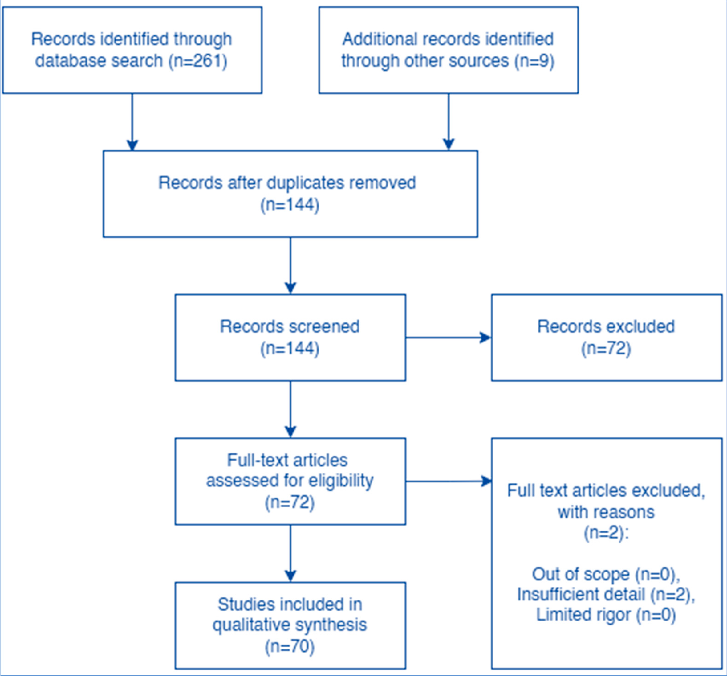

Table 2.
Patient and tumor characteristics (n=80).
| Variable | n/N (%) or median (IQR; range) |
|---|---|
| Total analyzable cases | 80 |
| Age, years | Median 63.0 (IQR 51.5–73.3; range 32–80) |
| Female sex | 39/80 (48.8%) |
| Male sex | 41/80 (51.3%) |
| Tumor group: Lung cancer | 22/80 (27.5%) |
| Tumor group: Melanoma | 15/80 (18.8%) |
| Tumor group: Breast cancer | 9/80 (11.3%) |
| Tumor group: Kidney/renal tumors | 8/80 (10.0%) |
| Tumor group: Gynecologic malignancies | 6/80 (7.5%) |
| Tumor group: Bladder/urinary tract | 3/80 (3.8%) |
| Tumor group: Hepatobiliary/hepatocellular carcinoma | 3/80 (3.8%) |
| Tumor group: Gastrointestinal malignancies | 3/80 (3.8%) |
| Tumor group: Non-melanoma squamous cell carcinoma/skin | 2/80 (2.5%) |
| Tumor group: CNS/glioblastoma | 2/80 (2.5%) |
| Tumor group: Thymic/mediastinal | 2/80 (2.5%) |
| Tumor group: Sarcoma/soft tissue/bone | 2/80 (2.5%) |
| Tumor group: Prostate | 1/80 (1.3%) |
| Tumor group: Head and neck | 1/80 (1.3%) |
| Tumor group: Small cell carcinoma, unspecified primary | 1/80 (1.3%) |
| Most common individual tumor types | Melanoma 15 (18.8%); lung adenocarcinoma 14 (17.5%); triple-negative breast cancer 7 (8.8%); clear cell renal cell carcinoma 6 (7.5%); lung squamous cell carcinoma 5 (6.3%) |
Percentages are calculated using all 80 analyzable cases. Tumor groups were consolidated from free-text tumor-type entries.
Table 3.
ICI exposure and timing of HLH onset.
| Variable | n/N (%) or median (IQR; range) |
|---|---|
| ICI monotherapy | 65/80 (81.3%) |
| Combination checkpoint blockade | 15/80 (18.8%) |
| PD-1 inhibitor exposure | 69/80 (86.3%) |
| PD-L1 inhibitor exposure | 10/80 (12.5%) |
| CTLA-4 inhibitor exposure | 16/80 (20.0%) |
| Pembrolizumab | 38/80 (47.5%) |
| Nivolumab | 25/80 (31.3%) |
| Ipilimumab | 14/80 (17.5%) |
| Atezolizumab | 5/80 (6.3%) |
| Unspecified anti-PD-L1 ICI | 3/80 (3.8%) |
| Durvalumab | 2/80 (2.5%) |
| Tremelimumab | 2/80 (2.5%) |
| Dostarlimab | 2/80 (2.5%) |
| Tislelizumab | 2/80 (2.5%) |
| Toripalimab | 2/80 (2.5%) |
| Time from last ICI dose to HLH onset, days | Median 17.0 (IQR 10.0–30.0; range 0–360), n=65 reported |
| HLH onset ≤30 days from last ICI dose | 49/65 (75.4%) |
| HLH onset ≤60 days from last ICI dose | 57/65 (87.7%) |
| ICI-containing cycles before HLH onset | Median 2.0 (IQR 1.0–4.0; range 1–17), n=70 reported |
| HLH after first ICI-containing cycle | 27/70 (38.6%) |
| HLH within first four ICI-containing cycles | 53/70 (75.7%) |
Drug-class and agent counts are not mutually exclusive because 15 patients received combination checkpoint blockade.
Table 4.
HLH diagnostic features and severity.
| Variable | n/N (%) or median (IQR; range) |
|---|---|
| HScore reported or qualitatively estimated | 41/80 (51.3%) |
| Numeric HScore available | 40/80 (50.0%) |
| HScore among numeric reports | Median 237.0 (IQR 207.0–267.0; range 105–319), n=40 |
| HLH criteria documented as met | 74/80 (92.5%) |
| Bone marrow hemophagocytosis present | 53/79 (67.1%) |
| Concurrent infection trigger identified | 6/79 (7.6%) |
| ICU admission or ICU-level care | 15/80 (18.8%) |
Denominators reflect the number of cases with extractable reporting for each variable.
Table 5.
HLH-directed treatments and outcomes.
| Variable | n/N (%) |
|---|---|
| Corticosteroids | 78/80 (97.5%) |
| Tocilizumab | 19/80 (23.8%) |
| Etoposide | 12/80 (15.0%) |
| Anakinra | 10/80 (12.5%) |
| Mycophenolate mofetil | 8/80 (10.0%) |
| IVIG | 6/80 (7.5%) |
| Ruxolitinib | 3/80 (3.8%) |
| Cyclosporine or tacrolimus | 3/80 (3.8%) |
| Plasmapheresis or therapeutic plasma exchange | 3/80 (3.8%) |
| Thrombomodulin/rhTM | 3/80 (3.8%) |
| Siltuximab | 1/80 (1.3%) |
| Infliximab | 1/80 (1.3%) |
| Outcome reported | 78/80 (97.5%) |
| Recovery or clinical improvement | 68/78 (87.2%) |
| Death | 10/78 (12.8%) |
| Death among ICU/ICU-level care cases | 4/15 (26.7%) |
| Death among non-ICU cases with known outcomes | 6/63 (9.5%) |
Treatment categories are not mutually exclusive. Outcomes are calculated among cases with known outcomes unless otherwise stated. Abbreviations: CNS, central nervous system; CTLA-4, cytotoxic T-lymphocyte–associated antigen 4; HLH, hemophagocytic lymphohistiocytosis; ICI, immune checkpoint inhibitor; ICU, intensive care unit; IQR, interquartile range; IVIG, intravenous immunoglobulin; PD-1, programmed cell death protein 1; PD-L1, programmed death ligand 1; rhTM, recombinant human thrombomodulin.
Disclaimer/Publisher’s Note: The statements, opinions and data contained in all publications are solely those of the individual author(s) and contributor(s) and not of MDPI and/or the editor(s). MDPI and/or the editor(s) disclaim responsibility for any injury to people or property resulting from any ideas, methods, instructions or products referred to in the content. |
© 2026 by the authors. Licensee MDPI, Basel, Switzerland. This article is an open access article distributed under the terms and conditions of the Creative Commons Attribution (CC BY) license.
Copyright: This open access article is published under a Creative Commons CC BY 4.0 license, which permit the free download, distribution, and reuse, provided that the author and preprint are cited in any reuse.


