

Submitted:
28 June 2026
Posted:
30 June 2026
You are already at the latest version
Abstract
Engineered mesenchymal stem cells (MSCs) have emerged as a promising therapeutic platform for extending healthspan. As agents of tissue repair and modulators of biological aging, MSCs have been widely studied for their capacity to enhance regeneration, restore immune homeostasis, and reduce chronic inflammation associated with age-related decline. This review examines emerging bioengineering strategies designed to overcome key age-related limitations in MSC homing, survival, and paracrine signaling, which have historically constrained their in vivo efficacy. We discuss major engineering approaches, including genetic modification, surface engineering, metabolic reprogramming, and preconditioning, with particular attention to their contributions to longevity-focused applications. Preclinical studies have demonstrated that engineered MSCs and their extracellular vesicles (EVs) yield measurable improvements in therapeutic performance. Reported benefits include prolonged persistence in inflamed tissues, partial reversal of senescence-associated phenotypes, and modulation of pro-aging inflammatory pathways. While MSC-derived EVs appear to offer a safer alternative with lower immunological risk, significant challenges persist in terms of manufacturing scalability, cargo consistency, and process standardization. The literature collectively supports that engineered MSC platforms have significant potential to contribute to healthspan extension. However, successful clinical translation will require a standardized manufacturing process to ensure therapeutic safety, reproducibility and efficacy in age-related conditions.
Keywords:
healthspan
; aging
; mesenchymal stem cells
; cellular engineering
; regenerative medicine
; immunomodulation
; senescence
1. Introduction
Healthspan, defined as the period of life spent in good health without chronic disease or disability, is a central focus of aging research. Mesenchymal stem/stromal cells (MSCs) are multipotent stromal cells with immunomodulatory, regenerative, and trophic properties, making them promising candidates for therapeutic approaches that target age-related pathologies and extend healthspan [1]. These properties have attracted substantial attention in anti-aging research, with evidence showing that MSCs regulate oxidative stress, attenuate endothelial cell senescence, and promote angiogenesis [2].
However, the therapeutic efficacy of wild-type MSCs is limited by poor engraftment, low survival in harsh microenvironments, and reduced functional potency, particularly when cells are derived from older donors [3]. The relevance of healthspan to MSC biology is highlighted owing to shared tissue-level mechanisms underlying many age-associated disorders, including chronic low-grade inflammation, vascular insufficiency, impaired proteostasis, mitochondrial dysfunction, stem/progenitor cell exhaustion, extracellular matrix stiffening, and the accumulation of senescent cells.
Together, these changes impair tissue repair and injury responses by reducing stem cell self-renewal and differentiation while promoting a pro-inflammatory microenvironment through senescence-associated secretory phenotype (SASP) factors. MSC-based interventions are therefore appealing because they may replenish depleted progenitor pools, attenuate inflammaging, and enhance endogenous repair mechanisms in age-related conditions. Rather than functioning solely as replacement cells, MSCs act as dynamic, microenvironment-responsive signaling platforms that modulate multiple interconnected hallmarks of aging by adjusting their paracrine secretome in response to local inflammatory and senescent cues [2,4,5].
Nevertheless, MSCs remain vulnerable to replicative and stress-induced senescence, which often alters their secretory profiles and decreases proliferative capacity [6]. A variety of engineering strategies have been developed to address these limitations and improve MSC survival, proliferation, and pro-regenerative capacity, thereby enhancing clinical performance [7,8]. These approaches include genetic and epigenetic modifications that increase secretion of regenerative factors or strengthen resilience to adverse conditions, as well as preconditioning methods that enhance immunomodulatory activity [9]. For example, preconditioning MSCs with cytokines or hypoxic conditions improves their secretory profile and increases production of anti-inflammatory cytokines [10].
The rationale for engineering MSCs is based on the well-documented age-related decline in stem cell potency, which compromises tissue homeostasis and limits the healing capacity of endogenous cell populations [11]. These limitations are intensified in pathological microenvironments. For example, ischemic tissues are characterized by hypoxia and nutrient deprivation, which reduce MSC survival; diabetic wounds contain high levels of advanced glycation end products, reactive oxygen species, proteases, and inflammatory cytokines, which impair proliferation and differentiation; and fibrotic tissues exhibit abnormal stiffness and disrupted integrin signaling, which promote maladaptive cell behavior [12,13,14,15].
Current strategies, therefore, focus on optimizing MSC viability through targeted priming and genetic enhancement to counteract the senescence-associated secretory phenotype and functional exhaustion observed in vivo [16,17]. Senescent MSCs exhibit altered secretome compositions that can negatively impact tissue repair and homeostasis, necessitating strategies to counteract age-related functional decline [5]. This issue is significant because donor age is inversely correlated with MSC proliferation, differentiation, and immunoregulatory capacity, which may lead to detrimental “inflammaging” outcomes if aged MSCs are used therapeutically [14,18]. Therefore, investigating approaches that improve MSC function and mitigate the negative effects of cellular senescence is essential, especially in therapies targeting older populations [19].
This review examines advanced bioengineering strategies to optimize MSC characteristics for improved outcomes in age-related diseases, with an emphasis on overcoming age-associated functional decline and extending healthspan. Strategies to address donor age-dependent reductions in MSC functionality include deriving MSC-like cells from induced pluripotent stem cells, applying senolytics to remove senescent subpopulations, and enriching juvenile cell fractions through sorting, all of which aim to restore proliferative and immunomodulatory capacities [1,15,20]. In addition, biomaterial-based priming, pharmacological interventions, and customized culture conditions are being investigated to modulate MSC behavior and enhance therapeutic properties [21,22].
This review highlights three major strategies to improve MSC trafficking: increasing chemokine receptor expression, such as CXCR4, through genetic approaches; using cytokine or hypoxia preconditioning to enhance migratory signaling; and engineering the cell surface with biomaterials to facilitate targeted endothelial adhesion [23,24,25]. Understanding these distinctions is essential for selecting the most appropriate strategy to address age-related declines in MSC homing efficiency, including reduced CXCR4 expression and impaired VCAM-1-mediated endothelial tethering in senescent microenvironments [4,15,26]. Careful consideration of each approach’s durability, safety, and compatibility with pathological conditions is necessary for optimal implementation.
2. Unmodified MSCs: Limitations and Challenges
The therapeutic utility of primary mesenchymal stromal cells is often limited by poor engraftment and survival after systemic administration because culture-expanded cells lose critical migratory receptors such as CXCR4 [27]. Native MSCs rely on intrinsic homing receptors, stress-response pathways, and paracrine cues to reach target tissues. However, after transplantation into aged, damaged, or inflamed tissues, these mechanisms may be insufficient. As a result, MSCs often show limited retention, short persistence, and inconsistent functional benefit despite favorable safety profiles [15,17,26]. Furthermore, these cells are rapidly cleared by the mononuclear phagocyte system and frequently become trapped in the pulmonary vasculature, restricting their distribution to distal target organs [28,29].
Disrupted homing mechanisms are especially important for healthspan-focused applications, where therapeutic cells must function within chronically diseased tissues characterized by persistent inflammation, oxidative stress, and fibrotic remodeling that can worsen pulmonary entrapment and phagocyte clearance [15,30]. Donor-to-donor heterogeneity and tissue-source heterogeneity significantly increase variability in MSC potency due to differences in age, genetics, health status, and origin-specific secretory profiles, undermining batch consistency and clinical predictability [9,31,32]. Extensive ex vivo expansion required to achieve therapeutic cell numbers often leads to loss of homing receptor expression and reduced metabolic plasticity [33].
The age of the donor, tissue source, expansion protocols, oxygen tension, and inflammatory stimuli strongly influence MSC potency by altering proliferative capacity, homing receptor expression, and immunomodulatory functions [31,34,35]. These variations complicate the standardization of MSC products and challenge the development of reliable potency tests for preclinical studies [30,31,32]. In older donors, MSCs often experience accelerated senescence during expansion, leading to the loss of functional subpopulations and an increase in clones with reduced regenerative and immunomodulatory potential [4,14,32]. Serial passaging can further select for rapidly dividing but less effective clones, diminishing both heterogeneity and therapeutic versatility.
These age-related changes impair the ability to maintain a pro-regenerative secretome and immunomodulatory activity, as replicative senescence shifts the secretory profile toward proinflammatory SASP components that hinder tissue repair [4,5,36]. Unmodified MSCs exhibit inefficient homing and engraftment after systemic or local administration due to age- and expansion-related downregulation of chemokine receptors such as CXCR4 and impaired VCAM-1-mediated endothelial interactions. These factors lead to pulmonary entrapment and clearance by mononuclear phagocytes [15,23,25,26,30,31].
Following intravenous infusion, MSCs are rapidly sequestered in the pulmonary capillaries due to their size and reduced deformability, triggering an acute inflammatory response and facilitating phagocytic clearance within hours, thereby limiting redistribution to sites of injury or inflammation [24,30,37]. Metabolic stress from pulmonary trapping further impairs the secretory function of aged MSCs, reducing the systemic availability of essential growth factors and cytokines needed for immune modulation [38]. This impairment, combined with replicative senescence, diminishes sustained immunomodulatory interactions and exacerbates inflammaging in chronically inflamed environments [15,39,40].
These challenges hinder clinical translation by preventing standardized manufacturing, regulatory potency validation, and safe systemic delivery in elderly patients. Donor heterogeneity and expansion-induced senescence increase the risk of inconsistent efficacy and may worsen inflammaging through impaired biodistribution and clearance [14,15,17,31]. Rigorous quality control and standardized isolation and cryopreservation protocols are essential to reduce variability [41]. Conventional two-dimensional monolayer expansion exacerbates these issues by accelerating senescence, downregulating homing receptors such as CXCR4, and shifting the secretory profile toward proinflammatory mediators [31,32,36]. Emerging research suggests that preconditioning strategies, including hypoxic exposure or specific growth factor administration, have the ability to enhance CXCR4 expression and improve MSC homing efficiency before clinical use [42].
3. Engineered MSCs: Mechanisms and Therapeutic Potential
Multiple strategies have been designed to improve MSC performance, including genetic modification, microRNA treatment, preconditioning, and epigenetic reprogramming, each offering a distinct route to improved healthspan-related outcomes (Figure 1) [43]. These approaches aim to enhance key MSC characteristics, including homing ability, viability in challenging physiological environments, and targeted paracrine signaling. Genetic modification often uses viral vectors to introduce genes encoding growth factors or anti-apoptotic proteins, thereby increasing MSC survival and regenerative factor production [24,31]. Non-viral methods, such as nucleofection and electroporation, are also being refined to deliver therapeutic genes or regulatory RNAs, offering safer alternatives for clinical applications [44].
Surface modification strategies that use bio-orthogonal chemistry or lipid-based coatings should be matched to the dominant biological challenge in each disease. For example, in ischemic cardiovascular conditions, these modifications can reduce pulmonary entrapment, whereas in inflammatory environments, they can decrease immune clearance. This approach may prolong systemic circulation and improve adhesion and function at target tissues [45,46,47]. In ischemic limb disease and chronic wounds, impaired SDF-1/CXCR4 signaling limits cell recruitment, and poor endothelial adhesion delays angiogenesis and tissue repair. Disease-tailored surface modifications may therefore enhance MSC retention and improve recovery [7,48].
For immunosenescence and inflammatory disorders, the major challenge is rapid immune clearance and inflammation driven by insufficient receptor engagement; in these settings, surface modifications should support durable anti-inflammatory effects and improved communication with immune cells [14,15,39]. For neurodegenerative diseases and frailty syndromes, surface engineering can be designed to deliver neurotrophic factors, support mitochondrial function, and provide anti-senescent vesicles. These examples underscore the need for mechanism-driven, disease-specific MSC platforms rather than a one-size-fits-all approach [43,49,50].
3.1. Genetic, RNA, and Genome-Editing Strategies
In addition to genetic methods, small molecules and growth factors can modulate MSC behavior by activating signaling pathways that support proliferation, differentiation, and stress resistance [51]. Engineering the culture environment with defined physical, chemical, or biological cues can also guide MSC development and enhance therapeutic efficacy [52]. Epigenetic reprogramming, including reversal of age-associated methylation patterns, may rejuvenate aged MSCs and improve their therapeutic utility [4].
CRISPR-Cas9 enables precise genetic modifications that can enhance MSC survival, differentiation, and immunomodulatory function [53]. For example, CRISPR/Cas9-mediated Keap1 knockout in adipose-derived MSCs activates the Nrf2 pathway, strengthening antioxidant defenses and improving cell survival [53,54]. Similarly, insertion of SV40T at the Rosa26 locus can increase the proliferative capacity of bone marrow MSCs without compromising key cellular features [55]. Upregulation of chemokine receptors such as CXCR1, CXCR4, and CXCR7 can improve MSC migration and expansion, thereby enhancing outcomes in disease models [56].
Engineering MSCs to express IL-10, HGF, IDO, Foxp3, or Bcl-2 can strengthen immunomodulation, reduce inflammation, and improve resistance to apoptosis [34,57]. Likewise, increasing expression of CXCR4, IL-10, BDNF, or IFN-beta using viral vectors or CRISPR/Cas9 can enhance MSC immune, antiviral, and neurotrophic functions [50,58]. Finally, post-transcriptional modification strategies may increase long-term MSC resistance to hostile microenvironments [59].
3.2. Preconditioning, Metabolic Reprogramming, and Biomaterial Priming
Pharmacological approaches using small molecules, cytokines, growth factors, hypoxia, extracellular matrix signals, and biomaterials can prime MSCs to maintain stronger secretory, migratory, and immunomodulatory traits under hostile conditions [14,22,60]. For example, diabetes impairs the reparative function of bone marrow–derived stem/progenitor cells, including MSCs (BMD-MSCs). Ex vivo priming with SDF-1α can functionally activate these cells before transplantation. In the referenced study, SDF-1α–primed diabetic BMD-MSCs significantly accelerated wound closure, increased neovascularization, and enhanced endothelial progenitor cell recruitment, while improving cell proliferation without reducing viability [61]. Mechanistically, SDF-1α priming upregulated pro-healing and proangiogenic mediators, including plasminogen and EphB4, and promoted adhesion-related pathways that may support endothelial progenitor cell interaction and vascular engraftment. Importantly, because SDF-1α exposure was performed ex vivo and cells were washed before implantation, this approach improved wound repair without increasing systemic SDF-1α levels [61]. These findings support SDF-1α preconditioning as a practical strategy to enhance the regenerative potency of MSCs/BMD-MSCs for diabetic wound healing.
Exposure to hypoxia or inflammatory cytokines such as IFN-gamma and TNF-alpha can improve the MSC secretome and increase release of cytoprotective and immunomodulatory factors that are often reduced under standard culture conditions [35,62]. In addition, TGF-alpha priming improves MSC survival in ischemic myocardium, demonstrating that targeted biochemical preconditioning can help overcome barriers to engraftment and expansion in vivo [63].
Metabolic reprogramming, such as increasing NAD+ levels or activating AMPK, improves MSC function in aged tissues by restoring mitochondrial health, balancing glycolysis, and increasing resistance to age-related decline [39,64]. These approaches can also precondition MSCs, reducing variability and improving clinical consistency [65,66]. Conversely, poorly controlled culture conditions can increase MSC heterogeneity, reduce immunomodulatory function, and decrease potency consistency [34,51,56]. To ensure reliable clinical outcomes, potency assays for engineered MSCs should include mechanism-specific functional tests, such as immunomodulatory secretome profiling, migration assays, and measurement of critical quality attributes [30,67,68].
3.3. Surface Engineering, Glycoengineering, and HCELL
Cell-surface engineering methods, including lipid-anchored palmitated protein G for antibody or ligand attachment, have been developed to address poor homing and tissue targeting in MSC therapy. These strategies display adhesion molecules that enhance selectin- or integrin-mediated binding, improving MSC trafficking to specific target sites or inflamed tissues [1,24]. Because MSCs commonly lack sufficient selectin ligands for effective capture under physiological flow, surface modifications such as enzymatic fucosylation of cell-surface glycans can increase vascular tethering and tissue entry without altering the cell’s internal program [1,69]. Glycoengineering provides a targeted strategy to increase selectin binding and improve MSC trafficking to inflamed tissues, thereby enhancing delivery efficiency [70].
Specialized scaffolds can also help maintain therapeutic activity and prolong MSC survival in inflamed environments [71]. Fucosylation of MSCs by converting CD44 to the HCELL glycoform has been shown to enhance vascular adhesion and transendothelial migration into damaged tissues by activating integrin alpha4beta1 [72,73]. This modification helps MSCs attach and roll along blood vessels under flow, overcoming their natural lack of E-selectin ligands [74,75]. Because native MSCs lack the sialofucosylated determinants required for E-selectin recognition, HCELL glycoengineering addresses poor bone targeting and limited vascular extravasation [76,77,78].
As a transient, non-genetic modification, this approach improves MSC homing to bone and inflamed sites while preserving cell viability and function and avoiding risks associated with permanent genetic alteration. These features have supported early clinical translation, including phase I trials in osteoporosis [70,72,73]. However, HCELL generation via glycosyltransferase-programmed stereosubstitution is limited to existing glycoproteins, such as CD44, and may unintentionally alter other surface molecules. It may also transiently shift the secretome toward anti-inflammatory effects, which could restrict broader therapeutic applications [1,76].
3.4. MSC-Derived Extracellular Vesicles as Cell-Free Engineering Platforms
MSC-derived extracellular vesicles (EVs) are promising cell-free engineering platforms that may overcome key limitations of cellular therapy, including tumorigenic risk and poor survival in hostile host microenvironments. These vesicles deliver concentrated bioactive proteins, lipids, and nucleic acids [79,80,81]. Their surface can be modified or their cargo can be loaded with specific therapeutic agents to improve targeting and signaling, allowing them to mimic beneficial functions of parent MSCs while reducing risks associated with systemic cell administration [82,83].
In aging studies, vesicles from young or rejuvenated MSCs reduced senescence markers such as p16 and p21, decreased oxidative stress, and increased healthspan in Ercc1-deficient and naturally aged mice by delivering anti-senescence signals that restore stem cell function without the risks of direct cell therapy [84,85,86]. However, clinical translation of engineered EVs remains challenging due to batch variability across different cell sources and culture conditions, the lack of reliable large-scale isolation methods, and the absence of standardized potency assays to ensure consistent biodistribution and safety [79,80,87,88].
Its utility could be advanced through standardized proteomic analysis, functional assays, and quality attributes directly linked to treatment outcomes [80,89]. Preclinical studies provide strong evidence that engineered MSCs, including senescence-resistant cells generated through CRISPR/Cas9 or epigenetic modification, can improve healthspan-related outcomes in aged animal models. These cells reduce cellular senescence, support tissue repair, and slow age-related functional decline. For example, intravenous administration of senescence-resistant mesenchymal progenitor cells improved memory, bone strength, and reproductive health in non-human primates without evident adverse effects [90,91]. Similarly, extracellular vesicles from rejuvenated MSCs increased healthspan in progeroid mice, producing effects comparable to those of young cell infusions [85].
Together, these advanced engineering approaches highlight the potential of MSCs as a flexible therapeutic platform for age-related diseases and healthspan improvement [92]. Because many engineering strategies are available, careful comparisons of efficacy and risk will be essential to identify the most suitable options for clinical use [93].
4. Supercharged MSCs with E-Selectin
Genetic engineering can enhance MSCs by introducing adhesion molecules such as E-selectin, which improves their capacity to home to and persist at target sites. In this approach, adeno-associated virus (AAV)-mediated transduction is used to induce surface expression of E-selectin in murine bone marrow-derived MSCs. E-selectin gene modification markedly enhances both the therapeutic phenotype and function of MSCs. Unmodified MSCs do not express E-selectin; however, after AAV-mediated E-selectin engineering, MSCs acquire a stronger MSC phenotype, including robust upregulation of the key MSC markers CD44 and CD105, while retaining expression of other standard MSC markers such as CD29, CD73, and Sca-1 [94]. Importantly, E-selectin-engineered MSCs preserve their fundamental stem/stromal cell properties, including colony-forming capability, proliferative capacity, and trilineage differentiation potential into chondrogenic, osteogenic, and adipogenic lineages. Thus, E-selectin engineering enhances MSC identity and potency without compromising their native multipotency [93,94]. Figure 2 summarizes this strategy, tracing the transition from conventional MSCs through AAV-mediated E-selectin engineering to the resulting supercharged phenotype and its enhanced therapeutic function in ischemic tissue.
Functionally, E-selectin-engineered MSCs displayed superior regenerative and proangiogenic activity compared with control MSCs. In an ischemic wound model, treatment with E-selectin-engineered MSCs significantly accelerated wound closure, promoted complete re-epithelialization, increased collagen deposition, and enhanced vascular density within ischemic wounds [94] (Figure 2). These cells also showed improved survival and persistence in ischemic tissue, suggesting that E-selectin modification strengthens MSC viability and therapeutic durability in a hostile ischemic microenvironment. Together, these findings indicate that E-selectin engineering transforms conventional MSCs into a more potent regenerative cell product with enhanced tissue repair and angiogenic capacity. Therefore, E-selectin-positive MSCs are appropriately termed “supercharged MSCs” [94,95].
These cells interact strongly with endothelial ligands in ischemic tissues, which improves cell retention, promotes neovascularization, and increases the expression of proangiogenic genes such as Cxcl2 [45,94,95]. Studies show that E-selectin engineering increases expression of nine proangiogenic genes in vitro, including Cxcl2, and reduces muscle atrophy while enhancing neovascularization in limb ischemia models [13,45,94]. In addition to enhancing tissue adhesion, E-selectin also acts as a signaling molecule that increases expression of proangiogenic genes, including Cxcl2, and reduces inflammatory mediators such as TNF. These effects promote release of beneficial paracrine factors, improve cell survival, and enhance neovascularization in ischemic tissues [13,45,94].
These cells interact strongly with endothelial ligands in ischemic tissues, which improves cell retention, promotes neovascularization, and increases the expression of proangiogenic genes such as Cxcl2 [13,45,95]. Studies show that E-selectin engineering increases expression of nine proangiogenic genes in vitro, including Cxcl2, and reduces muscle atrophy while enhancing neovascularization in limb ischemia models [96,97]. In addition to enhancing tissue adhesion, E-selectin also acts as a signaling molecule that increases expression of proangiogenic genes, including Cxcl2, and reduces inflammatory mediators such as TNF. These effects promote the release of beneficial paracrine factors, improve cell survival, and enhance neovascularization in ischemic tissues [13,45,96].
By reshaping the molecular microenvironment, this approach addresses key limitations of standard MSC therapies in severe tissue loss [98]. Future studies should determine whether E-selectin alters the MSC secretome, recruits host endothelial or immune cells, acts primarily through cell retention or paracrine activation, and whether its benefits persist after transgene expression declines. These studies will help determine whether supercharging should focus primarily on adhesion, paracrine activation, or both [13,94,96].
5. Manufacturing, Potency, and Regulatory Considerations for Engineered MSC Products
Clinical translation of engineered MSCs depends as much on manufacturing strategy as on biological rationale. During production, MSCs can undergo replicative aging and clonal selection, reducing multipotency and shifting the secretome toward a pro-inflammatory profile even when canonical surface markers remain stable [32,99]. Each additional engineering step introduces potential variability. Therefore, standardized culture methods and rigorous bioequivalence testing across batches are essential to reduce phenotypic drift and preserve therapeutic potency consistent with the intended mechanism of action [100,101].
Because engineered MSCs may still home inefficiently, effective delivery can require high cell doses. This increases the risk of pulmonary embolism, microthrombi, and vascular occlusion, particularly when infusion parameters are not optimized [30,102,103,104]. Age-related changes in both donors and recipients may further worsen biodistribution and reduce regenerative effects [30]. Because only a small fraction of administered cells typically engraft, repeated treatments may be required; however, repeated expansion and administration can accelerate MSC aging and reduce signaling capacity [105,106].
Hypoxic preconditioning and optimized delivery strategies may help mitigate these limitations [106]. Nevertheless, donor variability and prolonged ex vivo expansion remain major obstacles to producing consistent, high-potency MSC products for reliable clinical outcomes [107]. Potency testing should extend beyond surface-marker analysis and include functional, multi-parameter assays that reflect how engineered cells interact with diseased tissue [108]. Future validation should incorporate real-time monitoring of secretome profiles and cell-surface ligand density to keep batch variability within safe and effective limits [5].
Regulators generally have higher expectations for genetically engineered MSCs than for minimally manipulated products. When viral vectors are used, testing must detect replication-competent viruses and carefully assess the risk of integration to reduce the potential for insertional mutagenesis or tumorigenesis [88,109]. Genome-edited products require thorough off-target analysis, integration-site assessment, and clonal-expansion monitoring to ensure genetic stability and safety [88,110].
For surface-engineered MSCs, the main regulatory focus is confirmation of transient surface changes, such as improved selectin binding, through functional assays demonstrating improved homing. These products generally do not require the same genomic integration or off-target analyses as genome-edited cells [88,111,112]. For MSC-derived EV products, oversight focuses on vesicle identity, purity, sterility after filtration, and potency assays that demonstrate consistent secretome activity [87,113,114]. In all cases, product development should include a clear control strategy with defined acceptance ranges for critical quality attributes.
6. Strategies to Resolve Limitations of Engineered Mesenchymal Stem Cells
Precise delivery of engineered MSCs to target tissues and organs remains a major challenge. Genetic modifications, such as adding chemokine receptors including CCR7, can guide MSCs to lymphoid tissues for improved targeting [115]. Preconditioning MSCs with cytokines such as IFN-gamma can enhance their intrinsic immunosuppressive activity and survival in inflamed environments without requiring extensive genetic manipulation [116]. Using the MSC secretome, particularly extracellular vesicles, as a cell-free therapeutic platform may avoid risks associated with live-cell transplantation, including tumor formation and poor cell survival [87].
It will also be important to determine the optimal method for delivering human E-selectin, whether through genetic modification or protein engineering, to achieve effective surface expression without compromising MSC function or therapeutic potential. Inducible expression systems allow temporal and spatial control of therapeutic protein production. These systems enable precise adjustment of gene expression, reducing off-target effects and limiting long-term immune reactions [117]. They also permit fine-tuning of adhesion molecule expression so that homing can be improved while minimizing immune activation [118]. In addition, inducible systems can adapt protein expression levels to disease-specific conditions, making MSC therapies more precise.
Optimizing MSC culture conditions can help preserve function and therapeutic potency. Appropriate media, scaffold materials, and bioreactor configurations can support immunomodulatory and differentiation capacity, as well as expression of engineered surface molecules [109]. Controlling the timing and duration of engineered molecule expression may also reduce immune responses while preserving native MSC benefits. Transient fucosylation of surface proteins, for example, can improve MSC targeting and retention in inflamed tissues and may increase therapeutic efficacy [73,76].
Biomaterial scaffolds and directed delivery systems can localize MSCs to target tissues, reducing systemic dispersion and improving therapeutic outcomes. For instance, injectable hydrogels and polymer-nanoparticle composites can retain engineered MSCs at local treatment sites. By mimicking aspects of the extracellular matrix, these materials support MSC survival and function while reducing genetic risks and potentially improving scalability and immune compatibility [1,25,119]. They can also be loaded with immunosuppressive agents to shape the local microenvironment and prolong MSC retention and activity [9].
Advanced gene-editing tools such as CRISPR/Cas9 enable precise modifications that can improve MSC therapeutic performance. These tools allow targeted insertion or deletion of genes to enhance immunomodulation, survival, or tissue targeting while reducing limitations associated with older viral approaches [115]. Such modifications can create stable, heritable changes that may improve the safety and efficacy of MSC products for clinical use [120]. In addition, engineering MSCs to produce immunosuppressive factors or molecules that inhibit complement and NK-cell activity may further reduce immune rejection [83].
7. Future Solutions and Clinical Translation for Healthspan Extension
Future MSC engineering approaches for healthspan extension will likely combine strategies to improve targeting, persistence, and resistance to aging. Genetic modifications that increase senescence resistance, combined with biomaterials that support rejuvenation, may prevent cell decline while supporting regeneration and immune function in age-related diseases [41,52,90].
For diabetic wounds, which are characterized by hyperglycemia, oxidative stress, and impaired angiogenesis, MSCs may need to be engineered to release higher levels of angiogenic factors such as VEGF or ANG1. Targeted surface modifications or hydrogel encapsulation can help retain these cells locally and improve vascularization, while transient or cell-free formats may help maintain safety [13,110,121,122]. Another promising direction is the development of cell-free therapies using bioengineered MSC-derived extracellular vesicles (EVs).
These vesicles can be designed for targeted drug delivery or increased stability in the bloodstream, which may improve therapeutic utility [123]. MSC-derived EVs may avoid risks of tumorigenicity and immune reactions associated with live-cell transplantation [9]. For frailty and aging-related conditions, EV-rich products with anti-inflammatory activity may be particularly valuable. MSC-EVs are also attractive because they can be produced at scale and characterized more precisely than parental cell products, helping ensure clinical consistency [124]. To further improve precision, researchers are also using CRISPR-Cas9 to create immortalized cell lines with stable activity and reduced donor-to-donor variability [87,125].
Although these approaches are promising, clinical healthspan extension remains challenging. Long-term monitoring is essential to ensure safety, particularly regarding tumorigenic risk [9,126]. Building on the representative clinical trials and translational experience with MSC-based therapies to date (Table 1), large randomized trials are still needed to define effective dosing and confirm reproducibility. Standardized methods for measuring the secretome and biophysical features of extracellular vesicles are also required for quality control before broad clinical use [31,127]. Finally, regulatory guidelines that account for age-related recipient biology and batch-to-batch variability in stem cell products will be critical for reducing the risk of treatment failure in older patients [35,39].
8. Safety, Study Design, and Clinical Endpoint Considerations
Thorough preclinical testing and standardized manufacturing are necessary to preserve consistency of cell therapy products and reduce risk. New genetic modification tools, including advanced viral vectors and RNA-silencing approaches, are improving MSC engineering and may support the development of broadly available off-the-shelf MSC therapies [138]. The safety of unmodified MSCs has been evaluated in more than 1,200 clinical trials across multiple conditions [30,139,140].
To use engineered MSCs safely, consistent manufacturing and rigorous preclinical validation are required so that enhanced features can be translated into healthspan-focused therapies. Study design should consider that many MSC products have limited persistence in vivo, whereas age-related diseases are typically chronic. Therefore, trials should include surrogate biomarkers of biological aging and tissue recovery rather than relying only on mortality-based endpoints. Functional measures such as the six-minute walk test may be useful in selected contexts [141].
Studies should also assess polypharmacy and possible drug-cell interactions that may influence MSC efficacy in older patients with comorbidities [142]. Finally, data-driven analyses of gene-environment interactions may help identify new therapeutic targets and maintain immune regulation during aging. In healthspan-focused trials, primary outcomes should be both clinically meaningful and disease-specific.
These endpoints should include objective measures of physical function and biological aging, such as reduction of senescence-associated secretory phenotypes or reversal of age-related immune remodeling, to evaluate therapeutic efficacy in older adults [14,15]. Studies should also monitor senescent-cell clearance and systemic consequences, because interactions between MSC therapies and pre-existing secretory phenotypes remain an important determinant of therapeutic response [19,51].
9. Conclusion
Current research and clinical studies suggest that engineered MSCs may benefit conditions such as immune disorders, bone and cartilage injury, and neurological diseases [126,143]. Nevertheless, unmodified MSCs often show poor tissue targeting, limited persistence, donor-to-donor variability, and functional decline in aged or inflamed environments. Engineering approaches can address these limitations by improving survival, homing, paracrine signaling, senescence resistance, and manufacturing consistency.
Important challenges remain in making MSC-based therapies safe, effective, and reproducible. These challenges require strong quality control, standardized manufacturing, and risk-mitigation strategies to improve clinical outcomes [144,145,146]. Additional preclinical and clinical studies are needed to understand long-term effects and optimize delivery of engineered MSCs, particularly for inflammation and age-related metabolic disorders [39].
E-selectin-supercharged MSCs illustrate how modifying cell adhesion can enhance tissue repair, whereas HCELL glycoengineering provides a non-genetic strategy by adding an E-selectin ligand to CD44. Both approaches emphasize the importance of cell trafficking in MSC therapy and can inform the design of more effective regenerative treatments. Progress in this field will require integration of engineering, aging biology, potency testing, scalable manufacturing, and well-designed clinical trials.
Safety features such as inducible suicide systems may improve the safety of genetically engineered MSCs by allowing clinicians to control cell growth and persistence after administration [145]. Despite these advances, improved non-viral gene delivery methods, including lipid nanoparticles and polymer polyplexes, are still needed. These methods avoid genomic integration and may be safer than viral vectors, but they must remain efficient in MSCs, which are difficult to transfect [44,147].
Engineered MSCs and MSC-derived EVs may support tissue repair and healthspan extension, but their success will depend on well-defined mechanisms of action, reliable manufacturing, rigorous safety evaluation, and clinically meaningful endpoints. Inflammation-responsive or doxycycline-inducible promoters may allow cells to release immunomodulatory factors only when needed, reducing off-target effects, immune reactions, and toxicity [148,149]. With improved plasmid designs and cell preparation methods, these advances can make engineered MSC therapies more precise and clinically effective [150,151].
Author Contributions
Conceptualization, A-I.S.R; resources, A-I.S.R and Z.-J.L.; writing—original draft preparation, A-I.S.R, Y.Y.O, and M.L; writing—review and editing, A-I.S.R., Y.Y.O., N.L, Y.L, M.L, D.A.R, Z.-J.L and O.C.V.; visualization, A-I.S.R and M.L; supervision, O.C.V. and Z.-J.L. All authors have read and agreed to the published version of the manuscript.
Funding
This research was funded by National Institutes of Health—NIH/NHLBI Catalyze R33 HL156141; Philanthropy: Eloise & David Kimmelman Foundation.
Institutional Review Board Statement
Not applicable.
Informed Consent Statement
Not applicable.
Data Availability Statement
No new data were created or analyzed in this study. Data sharing is not applicable to this article.
Acknowledgments
The authors thank all members of the Surgical Vascular Research Labs at DeWitt Daughtry Family Department of Surgery, University of Miami Miller School of Medicine, for their helpful discussion and suggestions.
Conflicts of Interest
The authors Anne-Isabelle S. Reme, Yulexi Y. Ortiz, Nga Le, Yan Li, Mela Lew, Daniela Alexandra Ramos, declare no conflicts of interest. Authors Zhao-Jun Liu (Z.-J.L.) and Omaida C. Velazquez (O.C.V.) declare the following potential conflicts of interest with respect to the research, authorship, and/or presentation and/or publication of some aspects that are indirectly related to this work: E-selectin gene modification technologies aimed at pro-neovascularization technologies were developed in our research laboratory and patented/licensed by the University of Miami. These E-Selectin technologies are currently under pre-clinical development by Ambulero Inc., a new start-up company out of the University of Miami that focuses on developing new vascular treatments for ischemic tissue conditions and limb salvage. Z.-J.L. and O.C.V. serve as Ambulero Inc. consultants and chief scientific and medical advisory officers, respectively, and are co-inventors of the E-Selectin technologies and are minority shareholders in Ambulero Inc. Z.-J.L. and O.C.V. are also funded by the NIH/NHLBI and philanthropy in preclinical investigations of E-Selectin technologies and other pro-neovascularization technologies.
Abbreviations
The following abbreviations are used in this manuscript:
| AAV | Adeno-associated virus |
| ALS | Amyotrophic lateral sclerosis |
| AMPK | AMP-activated protein kinase |
| ANG1 | Angiopoietin-1 |
| ARDS | Acute respiratory distress syndrome |
| BDNF | Brain-derived neurotrophic factor |
| BM-MSC | Bone marrow-derived mesenchymal stem cell |
| BMD-MSCs | Bone marrow-derived mesenchymal stem/progenitor cells |
| BMSCs | Bone marrow stromal stem cells |
| Cas9 | CRISPR-associated protein 9 |
| CCR7 | C-C chemokine receptor type 7 |
| CD29 | Cluster of differentiation 29 (integrin β1) |
| CD44 | Cluster of differentiation 44 |
| CD62E | E-selectin |
| CD73 | Cluster of differentiation 73 |
| CD105 | Cluster of differentiation 105 (endoglin) |
| CLI | Critical limb ischemia |
| CLTI | Chronic limb-threatening ischemia |
| CRISPR | Clustered regularly interspaced short palindromic repeats |
| CXCR1 | C-X-C chemokine receptor type 1 |
| CXCR4 | C-X-C chemokine receptor type 4 |
| CXCR7 | C-X-C chemokine receptor type 7 |
| Cxcl2 | C-X-C motif chemokine ligand 2 |
| DNA | Deoxyribonucleic acid |
| EphB4 | Ephrin type-B receptor 4 |
| EV / EVs | Extracellular vesicle(s) |
| Foxp3 | Forkhead box protein P3 |
| GMP | Good manufacturing practice |
| GPS | Glycosyltransferase-programmed stereosubstitution |
| GVHD | Graft-versus-host disease |
| HCELL | Hematopoietic cell E-/L-selectin ligand |
| HGF | Hepatocyte growth factor |
| IDO | Indoleamine 2,3-dioxygenase |
| IFN-β | Interferon beta |
| IFN-γ | Interferon gamma |
| IL-10 | Interleukin-10 |
| Keap1 | Kelch-like ECH-associated protein 1 |
| MRI | Magnetic resonance imaging |
| MSC / MSCs | Mesenchymal stem cell(s) |
| MSC-EVs | MSC-derived extracellular vesicles |
| MSC-NTF | Mesenchymal stem cell-derived neurotrophic factor (cells) |
| NAD / NADH | Nicotinamide adenine dinucleotide |
| NK | Natural killer (cell) |
| Nrf2 | Nuclear factor erythroid 2-related factor 2 |
| PAD | Peripheral artery disease |
| PCNA | Proliferating cell nuclear antigen |
| RNA | Ribonucleic acid |
| SASP | Senescence-associated secretory phenotype |
| SDF-1 | Stromal cell-derived factor 1 |
| SV40T | Simian virus 40 large T antigen |
| TGF-α | Transforming growth factor alpha |
| TNF / TNF-α | Tumor necrosis factor (alpha) |
| VCAM-1 | Vascular cell adhesion molecule 1 |
| VEGF | Vascular endothelial growth factor |
References
- Park, J.S.; Suryaprakash, S.; Lao, Y.-H.; Leong, K.W. Engineering Mesenchymal Stem Cells for Regenerative Medicine and Drug Delivery. Methods 2015, 84, 3–16. [CrossRef]
- Wang, L.; Deng, Z.; Li, Y.; Wu, Y.; Yao, R.; Cao, Y.; Wang, M.; Zhou, F.; Zhu, H.; Kang, H. Ameliorative Effects of Mesenchymal Stromal Cells on Senescence Associated Phenotypes in Naturally Aged Rats. J. Transl. Med. 2024, 22, 722. [CrossRef]
- UPM-MAKNA Cancer Research Laboratory, Institute of Bioscience, Universiti Putra Malaysia, Selangor, Malaysia; Shamsul Kamal, A.A.; Department of Biomedical Sciences, Faculty of Medicine and Health Sciences, Universiti Putra Malaysia, Selangor, Malaysia; Fakiruddin, K.S.; Haematology Unit, Cancer Research Centre, Institute for Medical Research, National Institutes of Health, Ministry of Health Malaysia, Selangor, Malaysia; Bobbo, K.A.; UPM-MAKNA Cancer Research Laboratory, Institute of Bioscience, Universiti Putra Malaysia, Selangor, Malaysia; Department of Biomedical Sciences, Faculty of Medicine and Health Sciences, Universiti Putra Malaysia, Selangor, Malaysia; Ling, K.H.; Department of Biomedical Sciences, Faculty of Medicine and Health Sciences, Universiti Putra Malaysia, Selangor, Malaysia; et al. Engineered Mesenchymal Stem Cells as Treatment for Cancers: Opportunities, Clinical Applications and Challenges. Malays. J. Med. Sci. 2024, 31, 56–82. [CrossRef]
- Liu, J.; Ding, Y.; Liu, Z.; Liang, X. Senescence in Mesenchymal Stem Cells: Functional Alterations, Molecular Mechanisms, and Rejuvenation Strategies. Front. Cell Dev. Biol. 2020, 8, 258. [CrossRef]
- Siraj, Y.; Galderisi, U.; Alessio, N. Senescence Induces Fundamental Changes in the Secretome of Mesenchymal Stromal Cells (MSCs): Implications for the Therapeutic Use of MSCs and Their Derivates. Front. Bioeng. Biotechnol. 2023, 11, 1148761. [CrossRef]
- Yang, W.; Ji, W.; Liao, B.; Li, Z.; Wang, J.; Lin, H.; Wang, J.; He, Q. Genome-Wide Sequencing Identified Extrachromosomal Circular DNA as a Transcription Factor-Binding Motif of the Senescence Genes That Govern Replicative Senescence in Human Mesenchymal Stem Cells. Front. Cell. Neurosci. 2024, 18, 1421342. [CrossRef]
- Ocansey, D.K.W.; Pei, B.; Yan, Y.; Qian, H.; Zhang, X.; Xu, W.; Mao, F. Improved Therapeutics of Modified Mesenchymal Stem Cells: An Update. J. Transl. Med. 2020, 18, 42. [CrossRef]
- Damasceno, P.K.F.; De Santana, T.A.; Santos, G.C.; Orge, I.D.; Silva, D.N.; Albuquerque, J.F.; Golinelli, G.; Grisendi, G.; Pinelli, M.; Ribeiro Dos Santos, R.; et al. Genetic Engineering as a Strategy to Improve the Therapeutic Efficacy of Mesenchymal Stem/Stromal Cells in Regenerative Medicine. Front. Cell Dev. Biol. 2020, 8, 737. [CrossRef]
- Agha, H.M.; Abdalqader, M.A.; Hoh, J.; Wassouf, Y.; Al-Saadawi, K.A.A.; Mohammed, A.A.; Alsayadi, M.M.S. Mesenchymal Stem Cells as Cornerstones of Regenerative Medicine: Mechanisms, Applications, and Future Perspectives. AUIQ Complement. Biol. Syst. 2025, 2, 21–32. [CrossRef]
- Yang, S.; Li, K.; Wang, N.; Li, Z.; Zhang, Z.; Tuan, R.S.; Jiang, Y. Engineering Approaches to Modify Immunomodulatory Functions of Mesenchymal Stromal Cells (MSCs): Tissue Regeneration and Clinical Application. Adv. Sci. 2026, e22601. [CrossRef]
- Lunyak, V.V.; Amaro-Ortiz, A.; Gaur, M. Mesenchymal Stem Cells Secretory Responses: Senescence Messaging Secretome and Immunomodulation Perspective. Front. Genet. 2017, 8, 220. [CrossRef]
- Du, S.; Zeugolis, D.I.; O’Brien, T. Scaffold-Based Delivery of Mesenchymal Stromal Cells to Diabetic Wounds. Stem Cell Res. Ther. 2022, 13, 426. [CrossRef]
- Huerta, C.T.; Voza, F.A.; Ortiz, Y.Y.; Liu, Z.-J.; Velazquez, O.C. Mesenchymal Stem Cell-Based Therapy for Non-Healing Wounds Due to Chronic Limb-Threatening Ischemia: A Review of Preclinical and Clinical Studies. Front. Cardiovasc. Med. 2023, 10, 1113982. [CrossRef]
- Lee, B.-C.; Kang, K.-S. Functional Enhancement Strategies for Immunomodulation of Mesenchymal Stem Cells and Their Therapeutic Application. Stem Cell Res. Ther. 2020, 11, 397. [CrossRef]
- Yang, X.; Wang, Y.; Rovella, V.; Candi, E.; Jia, W.; Bernassola, F.; Bove, P.; Piacentini, M.; Scimeca, M.; Sica, G.; et al. Aged Mesenchymal Stem Cells and Inflammation: From Pathology to Potential Therapeutic Strategies. Biol. Direct 2023, 18, 40. [CrossRef]
- Fraile, M.; Eiro, N.; Costa, L.A.; Martín, A.; Vizoso, F.J. Aging and Mesenchymal Stem Cells: Basic Concepts, Challenges and Strategies. Biology 2022, 11, 1678. [CrossRef]
- Masterson, C.H.; Curley, G.F.; Laffey, J.G. Modulating the Distribution and Fate of Exogenously Delivered MSCs to Enhance Therapeutic Potential: Knowns and Unknowns. Intensive Care Med. Exp. 2019, 7, 41. [CrossRef]
- Kapetanos, K.; Asimakopoulos, D.; Christodoulou, N.; Vogt, A.; Khan, W. Chronological Age Affects MSC Senescence In Vitro—A Systematic Review. Int. J. Mol. Sci. 2021, 22, 7945. [CrossRef]
- Chen, H.; Liu, O.; Chen, S.; Zhou, Y. Aging and Mesenchymal Stem Cells: Therapeutic Opportunities and Challenges in the Older Group. Gerontology 2022, 68, 339–352. [CrossRef]
- Joint Shantou International Eye Center of Shantou University and The Chinese University of Hong Kong, Shantou, Guangdong, China; Chen, C.-B.; Bin, X.; Huang, Y.; Kin Ng, T.; S. Cheung, H. Age Effect on Mesenchymal Stem Cell Properties: A Concise Review. Aging Pathobiol. Ther. 2022, 4, 109–118. [CrossRef]
- Wong, R.S.Y.; Tan, E.W.; Goh, B.H. Mesenchymal Stem Cell-Based Therapies: Challenges and Enhancement Strategies. Cell Biochem. Biophys. 2025, 84, 131–147. [CrossRef]
- Noronha, N.D.C.; Mizukami, A.; Caliári-Oliveira, C.; Cominal, J.G.; Rocha, J.L.M.; Covas, D.T.; Swiech, K.; Malmegrim, K.C.R. Priming Approaches to Improve the Efficacy of Mesenchymal Stromal Cell-Based Therapies. Stem Cell Res. Ther. 2019, 10, 131. [CrossRef]
- Sajjad, U.; Ahmed, M.; Iqbal, M.Z.; Riaz, M.; Mustafa, M.; Biedermann, T.; Klar, A.S. Exploring Mesenchymal Stem Cells Homing Mechanisms and Improvement Strategies. Stem Cells Transl. Med. 2024, 13, 1161–1177. [CrossRef]
- García-Bernal, D.; García-Arranz, M.; Yáñez, R.M.; Hervás-Salcedo, R.; Cortés, A.; Fernández-García, M.; Hernando-Rodríguez, M.; Quintana-Bustamante, Ó.; Bueren, J.A.; García-Olmo, D.; et al. The Current Status of Mesenchymal Stromal Cells: Controversies, Unresolved Issues and Some Promising Solutions to Improve Their Therapeutic Efficacy. Front. Cell Dev. Biol. 2021, 9, 650664. [CrossRef]
- Wong, J.K.U.; Mehta, A.; Vũ, T.T.; Yeo, G.C. Cellular Modifications and Biomaterial Design to Improve Mesenchymal Stem Cell Transplantation. Biomater. Sci. 2023, 11, 4752–4773. [CrossRef]
- Lin, H.; Sohn, J.; Shen, H.; Langhans, M.T.; Tuan, R.S. Bone Marrow Mesenchymal Stem Cells: Aging and Tissue Engineering Applications to Enhance Bone Healing. Biomaterials 2019, 203, 96–110. [CrossRef]
- Li, C.; Zhao, H.; Wang, B. Challenges for Mesenchymal Stem Cell-Based Therapy for COVID-19. Drug Des. Devel. Ther. 2020, Volume 14, 3995–4001. [CrossRef]
- Song, Y.; Wu, J.; Liu, Y.; Xu, N.; Bai, H.; Wang, L.; Ai, J.; Li, K. The Remodeling of Ovarian Function: Targeted Delivery Strategies for Mesenchymal Stem Cells and Their Derived Extracellular Vesicles. Stem Cell Res. Ther. 2024, 15, 90. [CrossRef]
- Lopes-Pacheco, M.; Rocco, P.R.M. Functional Enhancement Strategies to Potentiate the Therapeutic Properties of Mesenchymal Stromal Cells for Respiratory Diseases. Front. Pharmacol. 2023, 14, 1067422. [CrossRef]
- Zhuang, W.-Z.; Lin, Y.-H.; Su, L.-J.; Wu, M.-S.; Jeng, H.-Y.; Chang, H.-C.; Huang, Y.-H.; Ling, T.-Y. Mesenchymal Stem/Stromal Cell-Based Therapy: Mechanism, Systemic Safety and Biodistribution for Precision Clinical Applications. J. Biomed. Sci. 2021, 28, 28. [CrossRef]
- Zhou, T.; Yuan, Z.; Weng, J.; Pei, D.; Du, X.; He, C.; Lai, P. Challenges and Advances in Clinical Applications of Mesenchymal Stromal Cells. J. Hematol. Oncol.J Hematol Oncol 2021, 14, 24. [CrossRef]
- Česnik, A.B.; Švajger, U. The Issue of Heterogeneity of MSC-Based Advanced Therapy Medicinal Products–a Review. Front. Cell Dev. Biol. 2024, 12, 1400347. [CrossRef]
- Ma, S.; Xie, N.; Li, W.; Yuan, B.; Shi, Y.; Wang, Y. Immunobiology of Mesenchymal Stem Cells. Cell Death Differ. 2014, 21, 216–225. [CrossRef]
- Costa, L.A.; Eiro, N.; Fraile, M.; Gonzalez, L.O.; Saá, J.; Garcia-Portabella, P.; Vega, B.; Schneider, J.; Vizoso, F.J. Functional Heterogeneity of Mesenchymal Stem Cells from Natural Niches to Culture Conditions: Implications for Further Clinical Uses. Cell. Mol. Life Sci. 2021, 78, 447–467. [CrossRef]
- Fan, X.-L.; Zhang, Y.; Li, X.; Fu, Q.-L. Mechanisms Underlying the Protective Effects of Mesenchymal Stem Cell-Based Therapy. Cell. Mol. Life Sci. 2020, 77, 2771–2794. [CrossRef]
- Miclau, K.; Hambright, W.S.; Huard, J.; Stoddart, M.J.; Bahney, C.S. Cellular Expansion of MSCs : Shifting the Regenerative Potential. Aging Cell 2023, 22, e13759. [CrossRef]
- Hernandez Pichardo, A.; Wilm, B.; Liptrott, N.J.; Murray, P. Intravenous Administration of Human Umbilical Cord Mesenchymal Stromal Cells Leads to an Inflammatory Response in the Lung. Stem Cells Int. 2023, 2023, 1–14. [CrossRef]
- Han, Y.; Yang, J.; Fang, J.; Zhou, Y.; Candi, E.; Wang, J.; Hua, D.; Shao, C.; Shi, Y. The Secretion Profile of Mesenchymal Stem Cells and Potential Applications in Treating Human Diseases. Signal Transduct. Target. Ther. 2022, 7, 92. [CrossRef]
- Planat-Benard, V.; Varin, A.; Casteilla, L. MSCs and Inflammatory Cells Crosstalk in Regenerative Medicine: Concerted Actions for Optimized Resolution Driven by Energy Metabolism. Front. Immunol. 2021, 12, 626755. [CrossRef]
- Lee, B.-C.; Yu, K.-R. Impact of Mesenchymal Stem Cell Senescence on Inflammaging. BMB Rep. 2020, 53, 65–73. [CrossRef]
- Wang, Y.; Gao, T.; Wang, B. Application of Mesenchymal Stem Cells for Anti-Senescence and Clinical Challenges. Stem Cell Res. Ther. 2023, 14, 260. [CrossRef]
- Hocking, A.M. The Role of Chemokines in Mesenchymal Stem Cell Homing to Wounds. Adv. Wound Care 2015, 4, 623–630. [CrossRef]
- Zhou, X.; Hong, Y.; Zhang, H.; Li, X. Mesenchymal Stem Cell Senescence and Rejuvenation: Current Status and Challenges. Front. Cell Dev. Biol. 2020, 8, 364. [CrossRef]
- Hamann, A.; Nguyen, A.; Pannier, A.K. Nucleic Acid Delivery to Mesenchymal Stem Cells: A Review of Nonviral Methods and Applications. J. Biol. Eng. 2019, 13, 7. [CrossRef]
- Huerta, C.T.; Voza, F.A.; Ortiz, Y.Y.; Liu, Z.-J.; Velazquez, O.C. Targeted Cell Delivery of Mesenchymal Stem Cell Therapy for Cardiovascular Disease Applications: A Review of Preclinical Advancements. Front. Cardiovasc. Med. 2023, 10, 1236345. [CrossRef]
- Ye, T.; Liu, X.; Zhong, X.; Yan, R.; Shi, P. Nongenetic Surface Engineering of Mesenchymal Stromal Cells with Polyvalent Antibodies to Enhance Targeting Efficiency. Nat. Commun. 2023, 14, 5806. [CrossRef]
- Zhong, X.; Luo, L.; Wu, J.; Li, W.; Liu, X.; Ye, T.; Li, Z.; Shi, P. Adhesion-Assisted Antioxidant-Engineered Mesenchymal Stromal Cells for Enhanced Cardiac Repair in Myocardial Infarction. ACS Nano 2025, 19, 11412–11426. [CrossRef]
- Lee, D.Y.; Cha, B.-H.; Jung, M.; Kim, A.S.; Bull, D.A.; Won, Y.-W. Cell Surface Engineering and Application in Cell Delivery to Heart Diseases. J. Biol. Eng. 2018, 12, 28. [CrossRef]
- Palanisamy, C.P.; Pei, J.; Alugoju, P.; Anthikapalli, N.V.A.; Jayaraman, S.; Veeraraghavan, V.P.; Gopathy, S.; Roy, J.R.; Janaki, C.S.; Thalamati, D.; et al. New Strategies of Neurodegenerative Disease Treatment with Extracellular Vesicles (EVs) Derived from Mesenchymal Stem Cells (MSCs). Theranostics 2023, 13, 4138–4165. [CrossRef]
- Carbone-Schellman, J.; Fontecilla-Escobar, J.; Sales-Salinas, N.; Chaparro-Pico, W.F.; Molina-Berríos, A.; Ruete, M.C.; González, P.A.; Machuca, M.A.; Opazo, Ma.C.; Ezquer, M.E.; et al. Therapeutic Challenges in Central Nervous System Viral Infections: Advancing Mesenchymal Stem Cell-Based Strategies for Treating Neuroinflammation and Promoting Tissue Repair. Front. Immunol. 2025, 16, 1677433. [CrossRef]
- Mark F. Pittenger; Pittenger, M.F.; Mark F. Pittenger; Dennis E. Discher; Discher, D.E.; Bruno Péault; Péault, B.; Donald G. Phinney; Phinney, D.G.; Joshua M. Hare; et al. Mesenchymal Stem Cell Perspective: Cell Biology to Clinical Progress. Npj Regen. Med. 2019, 4, 22–22. [CrossRef]
- Feng, Y.; Qiu, Y.; Zhang, S.; Dai, K.; Wang, J.; Liu, C. Materiobiology-Guided Regulation of Mesenchymal Stromal Cell Fate for Aging-Related Diseases: From Basic Parameter Design to Clinical Application. Bioact. Mater. 2026, 61, 577–600. [CrossRef]
- Sarsenova, M.; Kim, Y.; Raziyeva, K.; Kazybay, B.; Ogay, V.; Saparov, A. Recent Advances to Enhance the Immunomodulatory Potential of Mesenchymal Stem Cells. Front. Immunol. 2022, 13, 1010399. [CrossRef]
- Hu, Y.; Liu, S.; Zhu, B.-M. CRISPR/Cas9-Induced Loss of Keap1 Enhances Anti-Oxidation in Rat Adipose-Derived Mesenchymal Stem Cells. Front. Neurol. 2020, 10, 1311. [CrossRef]
- Hu, X.; Li, L.; Yu, X.; Zhang, R.; Yan, S.; Zeng, Z.; Shu, Y.; Zhao, C.; Wu, X.; Lei, J.; et al. CRISPR/Cas9-Mediated Reversibly Immortalized Mouse Bone Marrow Stromal Stem Cells (BMSCs) Retain Multipotent Features of Mesenchymal Stem Cells (MSCs). Oncotarget 2017, 8, 111847–111865. [CrossRef]
- Mendiratta, M.; Mendiratta, M.; Garg, D.; Mohanty, S.; Kumar Sahoo, R. The Integral Role of Mesenchymal Stem Cells in Stem Cell Transplantation from Promotion of Stem Cell Engraftment to Immunomodulation. In Biochemistry; Paolo Piccaluga, P., Visani, G., Salaheldin Khattab, S., Eds.; IntechOpen, 2024; Vol. 62 ISBN 978-0-85466-524-2.
- Huerta, C.T.; Ortiz, Y.Y.; Liu, Z.-J.; Velazquez, O.C. Methods and Limitations of Augmenting Mesenchymal Stem Cells for Therapeutic Applications. Adv. Wound Care 2023, 12, 467–481. [CrossRef]
- Recent Update on Mesenchymal Stem Cells; Ahmed Al-Anazi, K., Ed.; IntechOpen, 2024; ISBN 978-1-83769-697-0.
- Zheng, C.; Chen, J.; Liu, S.; Jin, Y. Stem Cell-Based Bone and Dental Regeneration: A View of Microenvironmental Modulation. Int. J. Oral Sci. 2019, 11, 23. [CrossRef]
- Ferreira, J.R.; Teixeira, G.Q.; Santos, S.G.; Barbosa, M.A.; Almeida-Porada, G.; Gonçalves, R.M. Mesenchymal Stromal Cell Secretome: Influencing Therapeutic Potential by Cellular Pre-Conditioning. Front. Immunol. 2018, 9, 2837. [CrossRef]
- Castilla, D.M.; Liu, Z.-J.; Tian, R.; Li, Y.; Livingstone, A.S.; Velazquez, O.C. A Novel Autologous Cell-Based Therapy to Promote Diabetic Wound Healing. Ann. Surg. 2012, 256, 560–572. [CrossRef]
- Calligaris, M.; Zito, G.; Busà, R.; Bulati, M.; Iannolo, G.; Gallo, A.; Carreca, A.P.; Cuscino, N.; Castelbuono, S.; Carcione, C.; et al. Proteomic Analysis and Functional Validation Reveal Distinct Therapeutic Capabilities Related to Priming of Mesenchymal Stromal/Stem Cells with IFN-γ and Hypoxia: Potential Implications for Their Clinical Use. Front. Cell Dev. Biol. 2024, 12, 1385712. [CrossRef]
- M. Alhattab, D.; Alshehri, S.; Jamali, F. Clinical Relevance of Mesenchymal Stromal Cells from Various Sources: Insights into Transcriptome Analysis for Identifying Inherent Potential. In Recent Update on Mesenchymal Stem Cells; Ahmed Al-Anazi, K., Ed.; IntechOpen, 2023 ISBN 978-1-83769-697-0.
- Yuan, X.; Liu, Y.; Bijonowski, B.M.; Tsai, A.-C.; Fu, Q.; Logan, T.M.; Ma, T.; Li, Y. NAD+/NADH Redox Alterations Reconfigure Metabolism and Rejuvenate Senescent Human Mesenchymal Stem Cells in Vitro. Commun. Biol. 2020, 3, 774. [CrossRef]
- Kahrizi, M.S.; Mousavi, E.; Khosravi, A.; Rahnama, S.; Salehi, A.; Nasrabadi, N.; Ebrahimzadeh, F.; Jamali, S. Recent Advances in Pre-Conditioned Mesenchymal Stem/Stromal Cell (MSCs) Therapy in Organ Failure; a Comprehensive Review of Preclinical Studies. Stem Cell Res. Ther. 2023, 14, 155. [CrossRef]
- Kaundal, U.; Bagai, U.; Rakha, A. Immunomodulatory Plasticity of Mesenchymal Stem Cells: A Potential Key to Successful Solid Organ Transplantation. J. Transl. Med. 2018, 16, 31. [CrossRef]
- Robb, K.P.; Audet, J.; Gandhi, R.; Viswanathan, S. Putative Critical Quality Attribute Matrix Identifies Mesenchymal Stromal Cells with Potent Immunomodulatory and Angiogenic “Fitness” Ranges in Response to Culture Process Parameters. Front. Immunol. 2022, 13, 972095. [CrossRef]
- Jayaraman, P.; Lim, R.; Ng, J.; Vemuri, M.C. Acceleration of Translational Mesenchymal Stromal Cell Therapy Through Consistent Quality GMP Manufacturing. Front. Cell Dev. Biol. 2021, 9, 648472. [CrossRef]
- Lo, C.Y.; Weil, B.R.; Palka, B.A.; Momeni, A.; Canty, J.M.; Neelamegham, S. Cell Surface Glycoengineering Improves Selectin-Mediated Adhesion of Mesenchymal Stem Cells (MSCs) and Cardiosphere-Derived Cells (CDCs): Pilot Validation in Porcine Ischemia-Reperfusion Model. Biomaterials 2016, 74, 19–30. [CrossRef]
- Le, B.; Cressman, A.; Morales, D.; Fierro, F.A. First Clinical Experiences Using Preconditioning Approaches to Improve MSC-Based Therapies. Curr. Stem Cell Rep. 2024, 10, 1–7. [CrossRef]
- Li, P.; Ou, Q.; Shi, S.; Shao, C. Immunomodulatory Properties of Mesenchymal Stem Cells/Dental Stem Cells and Their Therapeutic Applications. Cell. Mol. Immunol. 2023, 20, 558–569. [CrossRef]
- Soria-Juan, B.; Escacena, N.; Capilla-González, V.; Aguilera, Y.; Llanos, L.; Tejedo, J.R.; Bedoya, F.J.; Juan, V.; De La Cuesta, A.; Ruiz-Salmerón, R.; et al. Cost-Effective, Safe, and Personalized Cell Therapy for Critical Limb Ischemia in Type 2 Diabetes Mellitus. Front. Immunol. 2019, 10, 1151. [CrossRef]
- Gil-Chinchilla, J.I.; Bueno, C.; Martínez, C.M.; Ferrández-Múrtula, A.; García-Hernández, A.M.; Blanquer, M.; Molina-Molina, M.; Zapata, A.G.; Sackstein, R.; Moraleda, J.M.; et al. Optimizing Cryopreservation Conditions for Use of Fucosylated Human Mesenchymal Stromal Cells in Anti-Inflammatory/Immunomodulatory Therapeutics. Front. Immunol. 2024, 15, 1385691. [CrossRef]
- Sackstein, R. Glycoengineering of HCELL, the Human Bone Marrow Homing Receptor: Sweetly Programming Cell Migration. Ann. Biomed. Eng. 2012, 40, 766–776. [CrossRef]
- Sackstein, R. Glycosyltransferase-programmed Stereosubstitution (GPS) to Create HCELL: Engineering a Roadmap for Cell Migration. Immunol. Rev. 2009, 230, 51–74. [CrossRef]
- García-Bernal, D.; García-Arranz, M.; García-Guillén, A.I.; García-Hernández, A.M.; Blanquer, M.; García-Olmo, D.; Sackstein, R.; Moraleda, J.M.; Zapata, A.G. Exofucosylation of Adipose Mesenchymal Stromal Cells Alters Their Secretome Profile. Front. Cell Dev. Biol. 2020, 8, 584074. [CrossRef]
- Sackstein, R.; Merzaban, J.S.; Cain, D.W.; Dagia, N.M.; Spencer, J.A.; Lin, C.P.; Wohlgemuth, R. Ex Vivo Glycan Engineering of Membrane CD44 To Create HCELL Programs Human Mesenchymal Stem Cell Trafficking to Bone. Blood 2007, 110, 218–218. [CrossRef]
- Buettner, M.J.; Shah, S.R.; Saeui, C.T.; Ariss, R.; Yarema, K.J. Improving Immunotherapy Through Glycodesign. Front. Immunol. 2018, 9, 2485. [CrossRef]
- Kou, M.; Huang, L.; Yang, J.; Chiang, Z.; Chen, S.; Liu, J.; Guo, L.; Zhang, X.; Zhou, X.; Xu, X.; et al. Mesenchymal Stem Cell-Derived Extracellular Vesicles for Immunomodulation and Regeneration: A next Generation Therapeutic Tool? Cell Death Dis. 2022, 13, 580. [CrossRef]
- Gowen, A.; Shahjin, F.; Chand, S.; Odegaard, K.E.; Yelamanchili, S.V. Mesenchymal Stem Cell-Derived Extracellular Vesicles: Challenges in Clinical Applications. Front. Cell Dev. Biol. 2020, 8, 149. [CrossRef]
- Toh, W.S.; Lai, R.C.; Zhang, B.; Lim, S.K. MSC Exosome Works through a Protein-Based Mechanism of Action. Biochem. Soc. Trans. 2018, 46, 843–853. [CrossRef]
- Goodman, S.B.; Lin, T. Modifying MSC Phenotype to Facilitate Bone Healing: Biological Approaches. Front. Bioeng. Biotechnol. 2020, 8, 641. [CrossRef]
- Ankrum, J.A.; Ong, J.F.; Karp, J.M. Mesenchymal Stem Cells: Immune Evasive, Not Immune Privileged. Nat. Biotechnol. 2014, 32, 252–260. [CrossRef]
- Sanz-Ros, J.; Jorge Sanz-Ros; Nekane Romero-García; Nekane Romero-García; Mas-Bargues, C.; Cristina Mas-Bargues; Monleon, D.; Daniel Monleón; Juozas Gordevicius; Juozas Gordevičius; et al. Small Extracellular Vesicles from Young Adipose-Derived Stem Cells Prevent Frailty, Improve Health Span, and Decrease Epigenetic Age in Old Mice. Sci. Adv. 2022, 8. [CrossRef]
- Akaitz Dorronsoro; Akaitz Dorronsoro; Dorronsoro, A.; Fernando E. Santiago; Santiago, F.E.; Diego Grassi; Grassi, D.; Tianpeng Zhang; Tianpeng Zhang; Zhang, T.; et al. Mesenchymal Stem Cell-Derived Extracellular Vesicles Reduce Senescence and Extend Health Span in Mouse Models of Aging. Aging Cell 2021, 20. [CrossRef]
- Lei, Q.; Gao, F.; Liu, T.; Ren, W.; Chen, L.; Cao, Y.; Chen, W.; Guo, S.; Zhang, Q.; Chen, W.; et al. Extracellular Vesicles Deposit PCNA to Rejuvenate Aged Bone Marrow–Derived Mesenchymal Stem Cells and Slow Age-Related Degeneration. Sci. Transl. Med. 2021, 13, eaaz8697. [CrossRef]
- Maumus, M.; Rozier, P.; Boulestreau, J.; Jorgensen, C.; Noël, D. Mesenchymal Stem Cell-Derived Extracellular Vesicles: Opportunities and Challenges for Clinical Translation. Front. Bioeng. Biotechnol. 2020, 8, 997. [CrossRef]
- Fernández-Santos, M.E.; Garcia-Arranz, M.; Andreu, E.J.; García-Hernández, A.M.; López-Parra, M.; Villarón, E.; Sepúlveda, P.; Fernández-Avilés, F.; García-Olmo, D.; Prosper, F.; et al. Optimization of Mesenchymal Stromal Cell (MSC) Manufacturing Processes for a Better Therapeutic Outcome. Front. Immunol. 2022, 13, 918565. [CrossRef]
- Witwer, K.W.; Van Balkom, B.W.M.; Bruno, S.; Choo, A.; Dominici, M.; Gimona, M.; Hill, A.F.; De Kleijn, D.; Koh, M.; Lai, R.C.; et al. Defining Mesenchymal Stromal Cell (MSC)-derived Small Extracellular Vesicles for Therapeutic Applications. J. Extracell. Vesicles 2019, 8, 1609206. [CrossRef]
- Cai, Y.; Ji, Z.; Wang, S.; Zhang, W.; Qu, J.; Belmonte, J.C.I.-; Liu, G.-H. Genetic Enhancement: An Avenue to Combat Aging-Related Diseases. Life Med. 2022, 1, 307–318. [CrossRef]
- Lin, L.; Xue, Q.; Melino, G.; Shi, Y. Rewriting Destiny—Gene-Hacked Stem Cells Ignite a Revolution against Aging. Cell Death Dis. 2025, 16, 635. [CrossRef]
- Nowakowski, A.; Walczak, P.; Lukomska, B.; Janowski, M. Genetic Engineering of Mesenchymal Stem Cells to Induce Their Migration and Survival. Stem Cells Int. 2016, 2016, 4956063. [CrossRef]
- Ding, Y.; Li, Y.; Sun, Z.; Han, X.; Chen, Y.; Ge, Y.; Mao, Z.; Wang, W. Cell-Derived Extracellular Vesicles and Membranes for Tissue Repair. J. Nanobiotechnology 2021, 19, 368. [CrossRef]
- Huerta, C.T.; Ortiz, Y.Y.; Li, Y.; Ribieras, A.J.; Voza, F.; Le, N.; Dodson, C.; Wang, G.; Vazquez-Padron, R.I.; Liu, Z.-J.; et al. Novel Gene-Modified Mesenchymal Stem Cell Therapy Reverses Impaired Wound Healing in Ischemic Limbs. Ann. Surg. 2023, 278, 383–395. [CrossRef]
- Quiroz, H.J.; Valencia, S.F.; Shao, H.; Li, Y.; Ortiz, Y.Y.; Parikh, P.P.; Lassance-Soares, R.M.; Vazquez-Padron, R.I.; Liu, Z.-J.; Velazquez, O.C. E-Selectin-Overexpressing Mesenchymal Stem Cell Therapy Confers Improved Reperfusion, Repair, and Regeneration in a Murine Critical Limb Ischemia Model. Front. Cardiovasc. Med. 2022, 8, 826687. [CrossRef]
- Quiroz, H.J.; Shao, H.J.; Li, Y.; Ortiz, Y.Y.; Parikh, P.P.; Lassance-Soares, R.M.; Liu, Z.-J.J.; Velazquez, O.C. O6 Analysis of Angiogenic Gene Profiling after E-Selectin + Stem Cell Therapy in Murine Ischemic Limb. Br. J. Surg. 2021, 108, znab282.011. [CrossRef]
- Quiroz, H.J.; Shao, H.; Parikh, P.P.; Regueiro, M.M.; Li, Y.; Soares, R.M.; Liu, Z.-J.; Velazquez, O.C. Abstract 376: Supercharging Mesenchymal Stem Cells with E-Selectin/AAV to Augment Postnatal Neovascularization in the Murine Ischemic Hindlimb. Arterioscler. Thromb. Vasc. Biol. 2019, 39. [CrossRef]
- Huerta, C.T.; Ortiz, Y.; Li, Y.; Ribieras, A.; Voza, F.; Le, N.; Liu, Z.-J.; Velazquez, O.C. Abstract 594: E-Selectin-Overexpressing Mesenchymal Stem Cell-Based Therapy Augments Cutaneous Wound Healing In Ischemic Limbs. Arterioscler. Thromb. Vasc. Biol. 2023, 43. [CrossRef]
- Barekzai, J.; Petry, F.; Zitzmann, J.; Czermak, P.; Salzig, D. Bioprocess Development for Human Mesenchymal Stem Cell Therapy Products. In New Advances on Fermentation Processes; María Martínez-Espinosa, R., Ed.; IntechOpen, 2020 ISBN 978-1-78985-313-1.
- Mebarki, M.; Abadie, C.; Larghero, J.; Cras, A. Human Umbilical Cord-Derived Mesenchymal Stem/Stromal Cells: A Promising Candidate for the Development of Advanced Therapy Medicinal Products. Stem Cell Res. Ther. 2021, 12, 152. [CrossRef]
- Shirbaghaee, Z.; Hassani, M.; Heidari Keshel, S.; Soleimani, M. Emerging Roles of Mesenchymal Stem Cell Therapy in Patients with Critical Limb Ischemia. Stem Cell Res. Ther. 2022, 13, 462. [CrossRef]
- Lo, C.Y.; Antonopoulos, A.; Dell, A.; Haslam, S.M.; Lee, T.; Neelamegham, S. The Use of Surface Immobilization of P-Selectin Glycoprotein Ligand-1 on Mesenchymal Stem Cells to Facilitate Selectin Mediated Cell Tethering and Rolling. Biomaterials 2013, 34, 8213–8222. [CrossRef]
- Caplan, H.; Olson, S.D.; Kumar, A.; George, M.; Prabhakara, K.S.; Wenzel, P.; Bedi, S.; Toledano-Furman, N.E.; Triolo, F.; Kamhieh-Milz, J.; et al. Mesenchymal Stromal Cell Therapeutic Delivery: Translational Challenges to Clinical Application. Front. Immunol. 2019, 10, 1645. [CrossRef]
- Cui, L.; Kerkelä, E.; Bakreen, A.; Nitzsche, F.; Andrzejewska, A.; Nowakowski, A.; Janowski, M.; Walczak, P.; Boltze, J.; Lukomska, B.; et al. The Cerebral Embolism Evoked by Intra-Arterial Delivery of Allogeneic Bone Marrow Mesenchymal Stem Cells in Rats Is Related to Cell Dose and Infusion Velocity. Stem Cell Res. Ther. 2015, 6, 11. [CrossRef]
- Kim, H.J.; Park, J.-S. Usage of Human Mesenchymal Stem Cells in Cell-Based Therapy: Advantages and Disadvantages. Dev. Reprod. 2017, 21, 1–10. [CrossRef]
- Haque, N.; Kasim, N.H.A.; Rahman, M.T. Optimization of Pre-Transplantation Conditions to Enhance the Efficacy of Mesenchymal Stem Cells. Int. J. Biol. Sci. 2015, 11, 324–334. [CrossRef]
- Kouroupis, D.; Sanjurjo-Rodriguez, C.; Jones, E.; Correa, D. Mesenchymal Stem Cell Functionalization for Enhanced Therapeutic Applications. Tissue Eng. Part B Rev. 2019, 25, 55–77. [CrossRef]
- Hofer, H.R.; Tuan, R.S. Secreted Trophic Factors of Mesenchymal Stem Cells Support Neurovascular and Musculoskeletal Therapies. Stem Cell Res. Ther. 2016, 7, 131. [CrossRef]
- Mundra, V.; Gerling, I.C.; Mahato, R.I. Mesenchymal Stem Cell-Based Therapy. Mol. Pharm. 2013, 10, 77–89. [CrossRef]
- Srifa, W.; Kosaric, N.; Amorin, A.; Jadi, O.; Park, Y.; Mantri, S.; Camarena, J.; Gurtner, G.C.; Porteus, M. Cas9-AAV6-Engineered Human Mesenchymal Stromal Cells Improved Cutaneous Wound Healing in Diabetic Mice. Nat. Commun. 2020, 11, 2470. [CrossRef]
- De Wolf, C.; Van De Bovenkamp, M.; Hoefnagel, M. Regulatory Perspective on in Vitro Potency Assays for Human Mesenchymal Stromal Cells Used in Immunotherapy. Cytotherapy 2017, 19, 784–797. [CrossRef]
- Galipeau, J.; Krampera, M.; Barrett, J.; Dazzi, F.; Deans, R.J.; DeBruijn, J.; Dominici, M.; Fibbe, W.E.; Gee, A.P.; Gimble, J.M.; et al. International Society for Cellular Therapy Perspective on Immune Functional Assays for Mesenchymal Stromal Cells as Potency Release Criterion for Advanced Phase Clinical Trials. Cytotherapy 2016, 18, 151–159. [CrossRef]
- Giebel, B.; Kordelas, L.; Börger, V. Clinical Potential of Mesenchymal Stem/Stromal Cell-Derived Extracellular Vesicles. Stem Cell Investig. 2017, 4, 84–84. [CrossRef]
- Marolt Presen, D.; Traweger, A.; Gimona, M.; Redl, H. Mesenchymal Stromal Cell-Based Bone Regeneration Therapies: From Cell Transplantation and Tissue Engineering to Therapeutic Secretomes and Extracellular Vesicles. Front. Bioeng. Biotechnol. 2019, 7, 352. [CrossRef]
- Gao, F.; Chiu, S.M.; Motan, D.A.L.; Zhang, Z.; Chen, L.; Ji, H.-L.; Tse, H.-F.; Fu, Q.-L.; Lian, Q. Mesenchymal Stem Cells and Immunomodulation: Current Status and Future Prospects. Cell Death Dis. 2016, 7, e2062–e2062. [CrossRef]
- Lee, M.W.; Ryu, S.; Kim, D.S.; Sung, K.W.; Koo, H.H.; Yoo, K.H. Strategies to Improve the Immunosuppressive Properties of Human Mesenchymal Stem Cells. Stem Cell Res. Ther. 2015, 6, 179. [CrossRef]
- Ewa Choy, Y.W.; Choy, K.W.; Woon, K.S.; Wafi, M.A.; Then, K.Y.; Then, K.L. Genetically Engineered Mesenchymal Stem Cells Using Viral Vectors: A New Frontier in Anti-Angiogenic Therapy. Sains Malays. 2024, 53, 63–86. [CrossRef]
- Ali Khodadoust, M.; Boroumand, A.; Sedaghat, A.; Reihani, H.; Kaffash Farkhad, N.; Tavakol Afshari, J. A Closer Look at Mesenchymal Stem Cells (MSCs), Their Potential and Function as Game-Changers of Modern Medicine. In Recent Update on Mesenchymal Stem Cells; Ahmed Al-Anazi, K., Ed.; IntechOpen, 2023 ISBN 978-1-83769-697-0.
- Grosskopf, A.K.; Roth, G.A.; Smith, A.A.A.; Gale, E.C.; Hernandez, H.L.; Appel, E.A. Injectable Supramolecular Polymer–Nanoparticle Hydrogels Enhance Human Mesenchymal Stem Cell Delivery. Bioeng. Transl. Med. 2020, 5, e10147. [CrossRef]
- Yang, G.; Fan, X.; Liu, Y.; Jie, P.; Mazhar, M.; Liu, Y.; Dechsupa, N.; Wang, L. Immunomodulatory Mechanisms and Therapeutic Potential of Mesenchymal Stem Cells. Stem Cell Rev. Rep. 2023, 19, 1214–1231. [CrossRef]
- Deng, Q.; Du, F.; Pan, S.; Xia, Y.; Zhu, Y.; Zhang, J.; Li, C.; Yu, S. Activation of Angiopoietin-1 Signaling with Engineering Mesenchymal Stem Cells Promoted Efficient Angiogenesis in Diabetic Wound Healing. Stem Cell Res. Ther. 2025, 16, 75. [CrossRef]
- Zhao, X.; Fu, L.; Zou, H.; He, Y.; Pan, Y.; Ye, L.; Huang, Y.; Fan, W.; Zhang, J.; Ma, Y.; et al. Optogenetic Engineered Umbilical Cord MSC-Derived Exosomes for Remodeling of the Immune Microenvironment in Diabetic Wounds and the Promotion of Tissue Repair. J. Nanobiotechnology 2023, 21, 176. [CrossRef]
- Sitbon, A.; Delmotte, P.-R.; Pistorio, V.; Halter, S.; Gallet, J.; Gautheron, J.; Monsel, A. Mesenchymal Stromal Cell-Derived Extracellular Vesicles Therapy Openings New Translational Challenges in Immunomodulating Acute Liver Inflammation. J. Transl. Med. 2024, 22, 480. [CrossRef]
- Boulestreau, J.; Maumus, M.; Rozier, P.; Jorgensen, C.; Noël, D. Mesenchymal Stem Cell Derived Extracellular Vesicles in Aging. Front. Cell Dev. Biol. 2020, 8, 107. [CrossRef]
- Johnson, J.; Shojaee, M.; Mitchell Crow, J.; Khanabdali, R. From Mesenchymal Stromal Cells to Engineered Extracellular Vesicles: A New Therapeutic Paradigm. Front. Cell Dev. Biol. 2021, 9, 705676. [CrossRef]
- Liu, P.; An, Y.; Zhu, T.; Tang, S.; Huang, X.; Li, S.; Fu, F.; Chen, J.; Xuan, K. Mesenchymal Stem Cells: Emerging Concepts and Recent Advances in Their Roles in Organismal Homeostasis and Therapy. Front. Cell. Infect. Microbiol. 2023, 13, 1131218. [CrossRef]
- Ding, J.-Y.; Chen, M.-J.; Wu, L.-F.; Shu, G.-F.; Fang, S.-J.; Li, Z.-Y.; Chu, X.-R.; Li, X.-K.; Wang, Z.-G.; Ji, J.-S. Mesenchymal Stem Cell-Derived Extracellular Vesicles in Skin Wound Healing: Roles, Opportunities and Challenges. Mil. Med. Res. 2023, 10, 36. [CrossRef]
- Gupta, P.K.; Krishna, M.; Chullikana, A.; Desai, S.; Murugesan, R.; Dutta, S.; Sarkar, U.; Raju, R.; Dhar, A.; Parakh, R.; et al. Administration of Adult Human Bone Marrow-Derived, Cultured, Pooled, Allogeneic Mesenchymal Stromal Cells in Critical Limb Ischemia Due to Buerger’s Disease: Phase II Study Report Suggests Clinical Efficacy. Stem Cells Transl. Med. 2017, 6, 689–699. [CrossRef]
- Pollak, R.; Anderson, J.; Altmanshofer, B.; Caporusso, J.; Fantini, G.; Koppisetti, S.; Brigido, S.; Hariri, R. Human Placenta-Derived Cells ( PDA -002) in Diabetic Foot Ulcer Patients With and Without Peripheral Artery Disease: A Phase 2 Multi-Center, Randomised, Double-Blind, Placebo-Controlled Trial. Int. Wound J. 2025, 22, e70769. [CrossRef]
- Kurtzberg, J.; Abdel-Azim, H.; Carpenter, P.; Chaudhury, S.; Horn, B.; Mahadeo, K.; Nemecek, E.; Neudorf, S.; Prasad, V.; Prockop, S.; et al. A Phase 3, Single-Arm, Prospective Study of Remestemcel-L, Ex Vivo Culture-Expanded Adult Human Mesenchymal Stromal Cells for the Treatment of Pediatric Patients Who Failed to Respond to Steroid Treatment for Acute Graft-versus-Host Disease. Biol. Blood Marrow Transplant. 2020, 26, 845–854. [CrossRef]
- Zhao, X.; Ruan, J.; Tang, H.; Li, J.; Shi, Y.; Li, M.; Li, S.; Xu, C.; Lu, Q.; Dai, C. Multi-Compositional MRI Evaluation of Repair Cartilage in Knee Osteoarthritis with Treatment of Allogeneic Human Adipose-Derived Mesenchymal Progenitor Cells. Stem Cell Res. Ther. 2019, 10, 308. [CrossRef]
- Lozano-Rivas, N.; Linares, L.F.; Marras-Fernandez-Cid, C.; Garcia-Hernandez, A.M.; Algueró, M.D.C.; Iniesta, F.; Sanchez-Salinas, D.; López-Lucas, M.D.; Rodriguez-Valiente, M.; Cabañas, V.; et al. AB1011 Clinical Trial of Intravenous Infusion of Fucosylated Bone Marrow Mesenchymal Stem Cells in Patients with Osteoporosis. Ann. Rheum. Dis. 2018, 77, 1625. [CrossRef]
- Cudkowicz, M.E.; Lindborg, S.R.; Goyal, N.A.; Miller, R.G.; Burford, M.J.; Berry, J.D.; Nicholson, K.A.; Mozaffar, T.; Katz, J.S.; Jenkins, L.J.; et al. A Randomized placebo-controlled Phase 3 Study of Mesenchymal Stem Cells Induced to Secrete High Levels of Neurotrophic Factors in Amyotrophic Lateral Sclerosis. Muscle Nerve 2022, 65, 291–302. [CrossRef]
- Ruiz, J.G.; Oliva, A.A.; Ramdas, K.N.; Javier, J.; Rosen, J.; Perry, R.; Blanco, A.; Ylisastigui, P.; Walston, J.; Arai, H.; et al. Randomized Phase 2b Dose-Escalation Trial of Stem Cell Therapy with Laromestrocel for Aging Frailty. Cell Stem Cell 2026, 33, 393-404.e4. [CrossRef]
- Hare, J.M.; Fishman, J.E.; Gerstenblith, G.; DiFede Velazquez, D.L.; Zambrano, J.P.; Suncion, V.Y.; Tracy, M.; Ghersin, E.; Johnston, P.V.; Brinker, J.A.; et al. Comparison of Allogeneic vs Autologous Bone Marrow–Derived Mesenchymal Stem Cells Delivered by Transendocardial Injection in Patients With Ischemic Cardiomyopathy: The POSEIDON Randomized Trial. JAMA 2012, 308, 2369. [CrossRef]
- Matthay, M.A.; Calfee, C.S.; Zhuo, H.; Thompson, B.T.; Wilson, J.G.; Levitt, J.E.; Rogers, A.J.; Gotts, J.E.; Wiener-Kronish, J.P.; Bajwa, E.K.; et al. Treatment with Allogeneic Mesenchymal Stromal Cells for Moderate to Severe Acute Respiratory Distress Syndrome (START Study): A Randomised Phase 2a Safety Trial. Lancet Respir. Med. 2019, 7, 154–162. [CrossRef]
- Dehghani, L. Allogeneic Mesenchymal Stem Cell-Derived Exosome in Patients with Acute Ischemic Stroke (Randomized, Single-Blind, Placebo-Controlled, Phase 1/2); Isfahan University of Medical Sciences, 2021;
- Hodgkinson, C.P.; Gomez, J.A.; Mirotsou, M.; Dzau, V.J. Genetic Engineering of Mesenchymal Stem Cells and Its Application in Human Disease Therapy. Hum. Gene Ther. 2010, 21, 1513–1526. [CrossRef]
- Wu, X.; Jiang, J.; Gu, Z.; Zhang, J.; Chen, Y.; Liu, X. Mesenchymal Stromal Cell Therapies: Immunomodulatory Properties and Clinical Progress. Stem Cell Res. Ther. 2020, 11, 345. [CrossRef]
- Galderisi, U.; Peluso, G.; Di Bernardo, G. Clinical Trials Based on Mesenchymal Stromal Cells Are Exponentially Increasing: Where Are We in Recent Years? Stem Cell Rev. Rep. 2022, 18, 23–36. [CrossRef]
- Rolland, Y.; Sierra, F.; Ferrucci, L.; Barzilai, N.; De Cabo, R.; Mannick, J.; Oliva, A.; Evans, W.; Angioni, D.; De Souto Barreto, P.; et al. Challenges in Developing Geroscience Trials. Nat. Commun. 2023, 14, 5038. [CrossRef]
- Garay, R.P. Recent Clinical Trials with Stem Cells to Slow or Reverse Normal Aging Processes. Front. Aging 2023, 4, 1148926. [CrossRef]
- Bertolo, A.; Baur, M.; Guerrero, J.; Pötzel, T.; Stoyanov, J. Autofluorescence Is a Reliable in Vitro Marker of Cellular Senescence in Human Mesenchymal Stromal Cells. Sci. Rep. 2019, 9, 2074. [CrossRef]
- Sun, W.; Lv, J.; Guo, S.; Lv, M. Cellular Microenvironment: A Key for Tuning Mesenchymal Stem Cell Senescence. Front. Cell Dev. Biol. 2023, 11, 1323678. [CrossRef]
- Kim, N.; Cho, S.-G. Clinical Applications of Mesenchymal Stem Cells. Korean J. Intern. Med. 2013, 28, 387. [CrossRef]
- Li, J.; Huang, Y.; Sun, H.; Yang, L. Mechanism of Mesenchymal Stem Cells and Exosomes in the Treatment of Age-Related Diseases. Front. Immunol. 2023, 14, 1181308. [CrossRef]
- Carballo-Pedrares, N.; Ponti, F.; Lopez-Seijas, J.; Miranda-Balbuena, D.; Bono, N.; Candiani, G.; Rey-Rico, A. Non-Viral Gene Delivery to Human Mesenchymal Stem Cells: A Practical Guide towards Cell Engineering. J. Biol. Eng. 2023, 17, 49. [CrossRef]
- Rowland, C.R.; Glass, K.A.; Ettyreddy, A.R.; Gloss, C.C.; Matthews, J.R.L.; Huynh, N.P.T.; Guilak, F. Regulation of Decellularized Tissue Remodeling via Scaffold-Mediated Lentiviral Delivery in Anatomically-Shaped Osteochondral Constructs. Biomaterials 2018, 177, 161–175. [CrossRef]
- Selich, A.; Fleischauer, J.; Roepke, T.; Weisskoeppel, L.; Galla, M.; Von Kaisenberg, C.; Maus, U.A.; Schambach, A.; Rothe, M. Inflammation-Inducible Promoters to Overexpress Immune Inhibitory Factors by MSCs. Stem Cell Res. Ther. 2023, 14, 270. [CrossRef]
- Kozisek, T.; Hamann, A.; Nguyen, A.; Miller, M.; Plautz, S.; Pannier, A.K. High-Throughput Screening of Clinically Approved Drugs That Prime Nonviral Gene Delivery to Human Mesenchymal Stem Cells. J. Biol. Eng. 2020, 14, 16. [CrossRef]
- Kozisek, T.; Hamann, A.; Samuelson, L.; Fudolig, M.; Pannier, A.K. Comparison of Promoter, DNA Vector, and Cationic Carrier for Efficient Transfection of hMSCs from Multiple Donors and Tissue Sources. Mol. Ther. - Nucleic Acids 2021, 26, 81–93. [CrossRef]
Figure 1.
Engineering strategies to improve MSC performance for potential healthspan applications. Ten approaches: genetic modification, anti-inflammatory and chemokine-receptor engineering, hypoxic/cytokine preconditioning, biomaterial scaffolding, metabolic senescence engineering, HCell/CD44 glycoengineering, MSC-derived extracellular vesicle (EV) engineering, E-selectin supercharging, and combinatorial methods. Each is paired with its potential healthspan benefit, ranging from tissue regeneration and alleviation of inflammaging to delayed cellular aging and multimodal regeneration. Created in BioRender. Reme, A. (2026) https://BioRender.com/m6igdzm.
Figure 1.
Engineering strategies to improve MSC performance for potential healthspan applications. Ten approaches: genetic modification, anti-inflammatory and chemokine-receptor engineering, hypoxic/cytokine preconditioning, biomaterial scaffolding, metabolic senescence engineering, HCell/CD44 glycoengineering, MSC-derived extracellular vesicle (EV) engineering, E-selectin supercharging, and combinatorial methods. Each is paired with its potential healthspan benefit, ranging from tissue regeneration and alleviation of inflammaging to delayed cellular aging and multimodal regeneration. Created in BioRender. Reme, A. (2026) https://BioRender.com/m6igdzm.
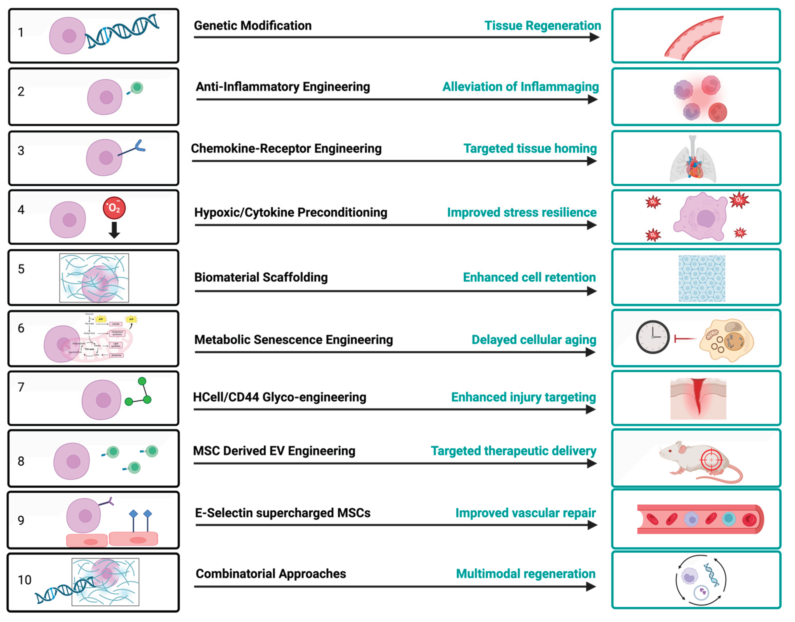
Figure 2.
AAV-mediated E-selectin engineering of MSCs. An AAV vector drives surface expression of E-selectin (CD62E), converting conventional MSCs into E-selectin⁺ “supercharged” MSCs that upregulate CD44 and CD105 while retaining core MSC identity and multipotency. In ischemic tissue, these cells adhere to activated endothelium, improving cell retention, angiogenesis, and wound repair. Created in BioRender. Reme, A. (2026) https://BioRender.com/hsqc8g7.
Figure 2.
AAV-mediated E-selectin engineering of MSCs. An AAV vector drives surface expression of E-selectin (CD62E), converting conventional MSCs into E-selectin⁺ “supercharged” MSCs that upregulate CD44 and CD105 while retaining core MSC identity and multipotency. In ischemic tissue, these cells adhere to activated endothelium, improving cell retention, angiogenesis, and wound repair. Created in BioRender. Reme, A. (2026) https://BioRender.com/hsqc8g7.
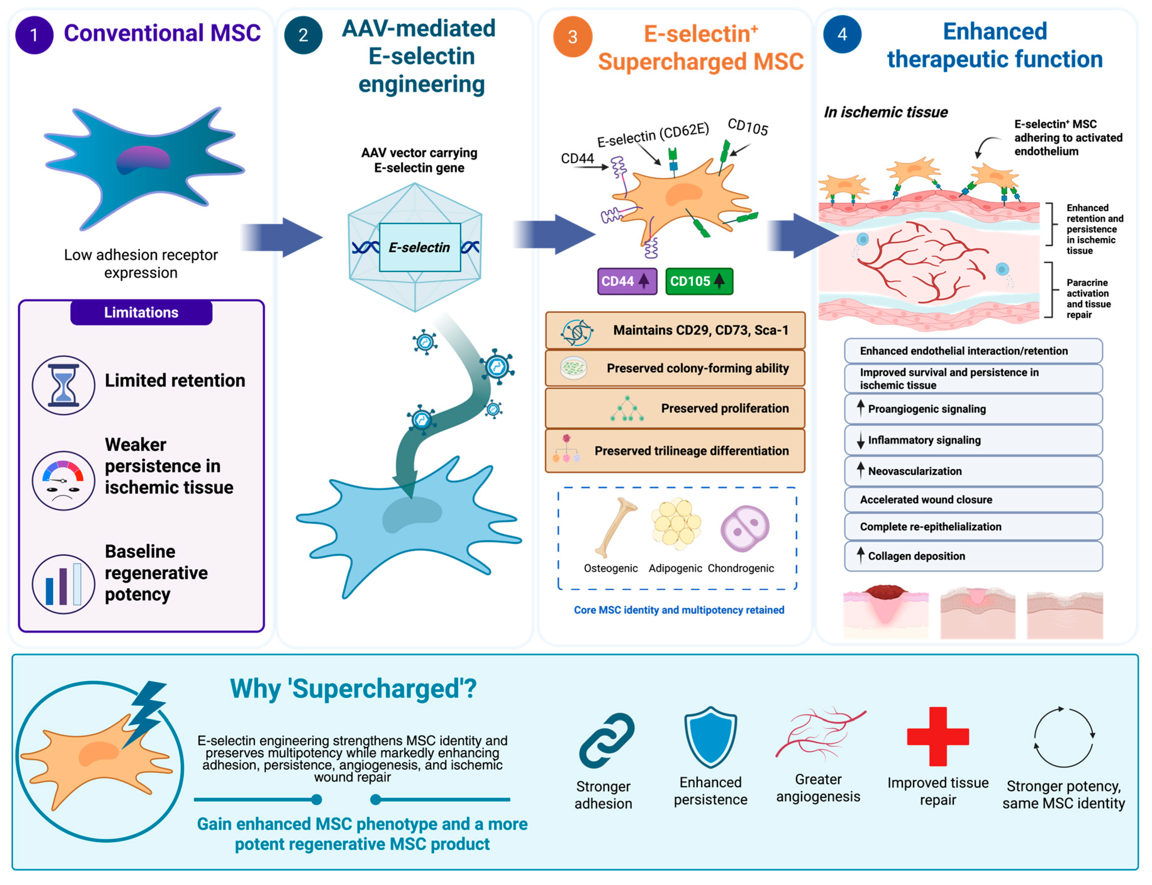

Table 1.
Representative clinical trials and translational experience with MSC-based therapies.
| Indication | MSC Strategy | Clinical endpoints | Main translational message | Registered trial (ClinicalTrials.gov) |
Registry status |
|---|---|---|---|---|---|
|
CLTI / PAD / Ischemic ulcers (Phase 2) |
Mostly unmodified autologous or allogeneic MSCs | Limb salvage, amputation-free survival, perfusion, and ulcer healing | Benefits have been modest or variable, supporting the need for more potent engineered MSCs | Stempeucel-CLI — allogeneic BM-MSC, CLI / Buerger’s disease (NCT01484574) [128] | Completed |
|
Chronic and diabetic wounds (Phase 2) |
MSCs or bone marrow–derived stromal/cell preparations | Wound closure, granulation tissue, angiogenesis, and safety | MSCs are promising, but poor survival and weak retention in hostile wound tissue limit efficacy | PDA-002 — placenta-derived stromal cells, diabetic foot ulcer ± PAD (NCT02264288) [129] | Terminated |
|
Immune and inflammatory diseases, including GVHD (Phase 3) |
Allogeneic MSCs; immunomodulatory MSC approaches | Response rate, inflammation, steroid-sparing effect, and safety | MSCs can suppress inflammation, but patient response is inconsistent | Remestemcel-L (Prochymal) — allogeneic MSC, pediatric steroid-refractory acute GVHD (NCT02336230) [130] | Completed |
|
Bone/cartilage injury and Osteoarthritis (Phase 1) |
Bone marrow– or adipose-derived MSCs; local injection or scaffold-supported delivery | Pain, joint function, cartilage repair, mobility, and safety | MSCs may improve symptoms, but durable structural regeneration remains uncertain | Allogeneic adipose-derived mesenchymal progenitor cells, knee osteoarthritis (NCT02641860) [131] | Completed |
|
Osteoporosis / Bone targeting (Phase 1) |
HCELL/CD44 glycoengineered or fucosylated MSCs | Early clinical translation, including phase I studies | Surface engineering can improve MSC homing without permanent genetic modification | Autologous fucosylated BM-MSC, established osteoporosis (NCT02566655) [132] | Completed |
|
Neurological and Neurodegenerative diseases (Phase 3) |
MSCs, neurotrophic-factor-enhanced MSCs, or MSC-EVs | Neurological function, disease progression, neuroinflammation, and safety | MSCs may provide trophic and anti-inflammatory effects, but targeting and potency remain barriers | NurOwn (MSC-NTF) — autologous neurotrophic-factor-secreting MSCs, ALS (NCT03280056) [133] | Completed |
|
Frailty / aging-related decline (Phase 2b) |
Allogeneic MSCs, rejuvenated MSCs, senescence-resistant cells, or MSC-EVs | Physical function, six minute walk test, inflammatory markers, and aging biomarkers | Healthspan trials require functional and biological aging endpoints | Lomecel-B — allogeneic BM-MSC, aging frailty (NCT03169231) [134] | Completed |
|
Cardiovascular ischemic disease (Phase 1/2) |
MSCs, preconditioned MSCs, or chemokine-receptor-enhanced MSCs | Cardiac function, perfusion, exercise capacity, and safety | Poor engraftment and survival limit efficacy, supporting strategies that improve homing and persistence | POSEIDON — autologous vs. allogeneic BM-MSC, chronic ischemic cardiomyopathy (NCT01087996) [135] | Completed |
|
Respiratory inflammatory injury / ARDS (Phase 2a) |
Systemic MSCs or MSC-EVs | Oxygenation, inflammatory cytokines, ventilator-free days, survival, and safety | Pulmonary trapping and variable potency remain important limitations | START — allogeneic BM-MSC, moderate–severe ARDS (NCT02097641) [136] | Completed |
| E-selectin “supercharged” MSCs for ischemic wounds | AAV-mediated E-selectin-engineered MSCs | Preclinical translational development | E-selectin engineering enhances MSC phenotype, survival, angiogenesis, and ischemic wound repair | Not registered — preclinical [95] | Pre-clinical |
| MSC-derived extracellular vesicles | Native or engineered MSC-EVs | Emerging clinical translation; many studies remain preclinical | EVs may reduce live-cell risks but require standardized manufacturing and potency assays | Allogeneic MSC-derived exosomes, acute ischemic stroke (NCT03384433)[137] | Completed |
Disclaimer/Publisher’s Note: The statements, opinions and data contained in all publications are solely those of the individual author(s) and contributor(s) and not of MDPI and/or the editor(s). MDPI and/or the editor(s) disclaim responsibility for any injury to people or property resulting from any ideas, methods, instructions or products referred to in the content. |
© 2026 by the authors. Licensee MDPI, Basel, Switzerland. This article is an open access article distributed under the terms and conditions of the Creative Commons Attribution (CC BY) license.
Copyright: This open access article is published under a Creative Commons CC BY 4.0 license, which permit the free download, distribution, and reuse, provided that the author and preprint are cited in any reuse.


