

Submitted:
05 July 2026
Posted:
06 July 2026
Read the latest preprint version here
Abstract
Atherosclerosis, the leading cause of death, is the slow accumulation of oxidized lipids in the artery walls, eventually leading to heart attacks and stroke. Despite much study, the underlying pathology remains poorly understood. We hypothesize that atherosclerosis develops due to deuterium overload in mitochondria, systemically, and that the accumulation of lipids in the atheroma ameliorates this problem. We show the critical role that gut microbes play in supplying deuterium-depleted nutrients to the mitochondria, in large part through the supply of methyl groups and short chain fatty acids. Deuterium (2H), a natural heavy isotope of protium, damages the ATP synthase nanomotors that produce ATP. Gut microbes exploit hydrogen gas recycling to deplete deuterium in derived metabolites. Gut dysbiosis impairs this process. The bis-allylic carbon atoms in polyunsaturated fatty acids (PUFAs) have a unique property that allows them to trap and sequester deuterium. A defective methylation system, reflected in high plasma trimethylamine oxide (TMAO), leads, through complex signaling processes, to the trapping of lipids in the artery wall and the induction of an inflammatory response to promote lipid peroxidation chain reactions. Deficiencies in aldehyde dehydrogenases lead to increased exposure to formaldehyde, acetaldehyde, and malondialdehyde, causing tissue damage. Over time, deuterium loading at the bis-allylic carbon atoms quenches the cascade, stabilizing the plaque. This theory can explain most of the markers associated with cardiovascular disease (CVD), including TMAO, low-density lipoprotein (LDL), homocysteine, malondialdehyde, psychological and emotional stress, dimethylglycine, and deficiencies in glutathione, zinc and B vitamins.
Keywords:
mitochondria
; deuterium
; atherosclerosis
; lipid peroxidation
; homocysteine
; methylation pathways
; gut dysbiosis
; formaldehyde
1. Introduction
Cardiovascular disease (CVD) is the most common cause of mortality, and it is responsible for over one third of global deaths. As for many other diseases, mitochondrial dysfunction is strongly associated with CVD [1]. Dysfunctional mitochondria release excessive amounts of reactive oxygen species (ROS), damaging mitochondrial membranes, mitochondrial DNA, and proteins. Lipid peroxidation cascades can trigger further dysfunction, leading to a cell death spiral [2]. Many of the drugs taken to decrease risk of CVD (β-blockers, angiotensin-converting enzyme inhibitors, and angiotensin receptor-II blockers) are protective against mitochondrial damage [1].
There is a long list of biomarkers that are known to be associated with increased risk of CVD, although researchers have struggled to find a clear explanation for why these markers are elevated and whether they have a causal or just an associative role. These include elevated serum low-density lipoprotein (LDL) [3], trimethylamine N-oxide (TMAO) [4], homocysteine (Hcy) [5], and dimethylglycine (DMG) [6], among others, as well as chronic emotional and psychological stress (chronic elevated adrenaline) [7]. One striking common element shared among many of these markers is their crucial role in methylation pathways.
We hypothesize that, foundationally, cardiovascular disease develops in response to an excess burden of deuterium loading in the mitochondrial water. Deuterium (heavy hydrogen, 2H) is a natural isotope of hydrogen (protium, 1H), found at a concentration of 155 parts per million (ppm) in seawater. While this may seem small, deuterium levels in the circulation are five or six times as high as calcium levels. The ATP synthase (ATPase) nanomotors in mitochondria that are responsible for the production of adenosine triphosphate (ATP), the energy currency of the cell, are highly sensitive to deuterium, which damages the nanomotors, leading to inefficiencies in ATP production and an increase in ROS [8].
The gut microbiome plays an essential role in supplying deuterium depleted (deupleted) nutrients to the host via hydrogen recycling, in the form of both short chain fatty acids (SCFAs; predominantly acetate, propionate and butyrate) and the methyl groups in the methylation pathways [9]. The hydrogen gas produced by gut microbes is likely 80% depleted in deuterium [10]. It is used by the microbes as a reducing agent to convert carbon dioxide (CO2) into small deupleted organic molecules (e.g., methyl groups and SCFAs).
Furthermore, many other small hydrogen-containing molecular gas molecules are likely also enriched in 1H over 2H, due to a general property that deuterium tends to stay behind in the aqueous medium. These include methane (CH4), hydrogen sulfide (H2S), hydrogen peroxide (H2O2), and ammonia (NH3), among others [11,12]. Many of these gases play powerful roles in metabolism, and their enrichment in 1H over 2H may be the primary reason why they are produced during metabolic activity.
In this paper, we develop a novel hypothesis that gut dysbiosis is central to multiple pathologies linked to mitochondrial dysfunction, while our focus is on cardiovascular disease. The high-level view is that a shortage in the gastrointestinal tract of strict anaerobic bacteria that produce deupleted nutrients through hydrogen gas recycling necessitates a switch to H2O2 as an additional source of deupleted water, systemically. This shortage induces an inflammatory response, and H2O2, produced in the external milieu, is imported into human cells and into their mitochondria, where it is converted to deupleted water (DDW) via glutathione peroxidase [13] and the thioredoxin system [14]. Aquaporins, which are primarily viewed as water channels, also actively transport H2O2 across membranes [15]. Mitochondria have a vastly higher capacity to consume H2O2 than to produce it, especially via thioredoxin [16,17]. This frames inflammation in an entirely different light, and the pathology that arises is likely primarily due to defects in enzymes that would normally rapidly remove highly reactive intermediary molecules, such as superoxide, the hydroxyl radical, formaldehyde, and acetaldehyde, that are central to deuterium homeostasis.
Deuterium levels in foods are lowest in fats, with proteins in an intermediate range, and carbohydrates having the highest levels [48]. A fascinating case study involved a man who, due to ulcerative colitis, adopted an extremely high fat, low carbohydrate diet, and saw his serum LDL-cholesterol increase six-fold from 95 mg/dL to 574 mg/dL. He maintained this very high level for seven years yet failed to develop any evidence of atherosclerosis. The extreme diet also cured his ulcerative colitis [47]. His lean body type and extreme physical fitness may have played a role, but it is also clear that his diet was a low-deuterium diet, and it also included an abundant source of L-carnitine and choline from animal-based foods, to support methylation pathways.
2. Gut Microbes Provide Deuterium Depleted Nutrients to the Host via Fiber Fermentation and Methylation Pathways
Gut microbes produce molecular hydrogen (H2) from soluble fiber and resistant starches, such as inulin, pectin, and fructo-oligosaccharides, via fermentation in the colon [18]. Acetogenic bacteria produce acetate from carbon dioxide (CO2) and H2, and other short chain fatty acids (SCFAs) (e.g., propionate and butyrate) are derived from acetate. Methanogenic archaea use H2 as a reducing agent to synthesize methane gas from CO2. Methane is then oxidized to methanol by methanotrophs via methane monooxygenase enzymes [19]. All these metabolites are likely to also have an overabundance of 1H relative to 2H, due to their underlying source from H2.
Methylotrophic bacteria generate formaldehyde during the process of metabolizing methanol. Formaldehyde is the simplest of all aldehydes and the most toxic. However, it is also crucial in metabolism for its essential role in methylation pathways and for its role in producing reduced nicotinamide adenine dinucleotide (NADH) from NAD+.
Formaldehyde reacts spontaneously with tetrahydrofolate (THF) to produce methylene-tetrahydrofolate (CH2-THF), and there is also a microbial enzymatic pathway that creates CH2-THF from formaldehyde and THF [20]. CH2-THF is then reduced to methyl-THF (CH3-THF) via the enzyme methylene tetrahydrofolate reductase (MTHFR), an enzyme that likely has a high deuterium kinetic isotope effect (KIE), because it is a flavoprotein that can select protons over deuterons through proton tunneling [21,22]. Therefore, the methyl group in CH3-THF is an excellent source of 1H, which ultimately supplies deupleted water (DDW) to the host mitochondria. MTHFR depends on riboflavin (vitamin B2) as a cofactor.
Formaldehyde is highly reactive, creating DNA-DNA, protein-protein, and DNA-protein cross-links [23]. A crucial detoxification pathway, widely expressed in human cells, but also in methylotrophs and in many pathogens, utilizes a formaldehyde dehydrogenase that is dependent on both zinc and glutathione, with NAD+ serving as the electron acceptor, to convert formaldehyde to formate [24,25]. Formate can then be metabolized to CO2, via another dehydrogenase, also generating a molecule of NADH [26]. These two molecules of NADH will be useful for the human host, where NADH dehydrogenase (Complex I) supplies protons to the intermembrane space [27]. Because the formaldehyde is derived from methanol, which in turn is derived from molecular hydrogen, microbially produced formaldehyde is likely highly enriched in 1H relative to 2H. This may be the primary reason why it is produced endogenously, despite its high reactivity.
A 2026 meta-analysis of over 11K gut metagenomes found that the genus CAG-170, a strict anaerobe that cannot be cultured outside the body, stood out as being strongly associated with good health [28]. Members of this genus convert dietary fiber into deupleted SCFAs, and also produce cobalamin, an essential cofactor for methionine synthase, the enzyme that converts homocysteine to methionine, the universal methyl donor, pulling the methyl group from CH3-THF [29].
3. Methylotrophic Bacteria Produce L-Serine as a Safe Carrier of 1C Units that Support Fatty Acid Synthesis in the Liver
Methylotrophic bacteria, as exemplified by Methylobacterium extorquens AM1, are able to survive with methanol as a sole nutrient. They convert methanol to formaldehyde, which they then fully metabolize to CO2 to satisfy their energy needs, while also directing it down a pathway that involves THF as a carrier and ultimately produces serine, which serves as a central metabolic hub for the generation of many downstream metabolites to build cellular mass [30,31,32]. Serine synthesis becomes a way to capture the valuable 1H protons in the methanol as the hydroxymethyl side chain in serine, while also consuming formaldehyde, which is highly reactive.
The serine they produce can also serve as a deupleted resource for the host, as it can be used to regenerate CH2-THF, a feature that is exploited by the liver in the synthesis of fatty acids. Furthermore, the methylene unit in CH2-THF is then fully metabolized to CO2 in the liver to generate two molecules of NADPH. The 1H protons in these NADPH molecules then become incorporated into the synthesized fatty acids [33,34], which is likely a primary reason why these fatty acids are low in deuterium. Acetyl-CoA is the essential two-carbon building block for fatty acid synthesis [35], and it is also likely to be enriched in 1H protons if it was derived from acetate produced by gut microbes.
The liver relies heavily on folate-mediated serine catabolism to fuel lipogenesis. Serine is first converted to glycine in a reaction that produces 5,10-methylene-THF (CH2-THF) from THF. A reaction catalyzed by C1-tetrahydrofolate synthase (also known as methylene tetrahydrofolate dehydrogenase (MTHFD1)) then converts CH2-THF to formyl-THF, coupled to the reduction of NADP+ to NADPH. Aldehyde dehydrogenase 1 family member L1 (ALDH1L1) further catalyzes the conversion of 10-formyl-THF to THF and CO2, generating yet another molecule of NADPH [34], having fully metabolized the hydroxymethyl group in serine.
A case-control study of a Chinese population found that those whose blood serine concentration was above the median value had a significantly decreased risk for coronary heart disease (CHD), compared to those whose serine level was below the median value (OR 0.57, P = 0.004). For every 1 µg/mL increase in serine concentration, the risk of CHD significantly decreased by 6% [36]. L-serine has been found to induce antioxidant and cytoprotective effects on endothelial cells by activating the master transcription factor that upregulates an antioxidant response, nuclear factor erythroid 2-related factor 2 (Nrf2) [37]. It is conceivable that its ability to serve as a source of 1C units underlies its observed beneficial effects.
4. Is TMAO a Marker for Both Gut Dysbiosis and Deuterium Overload?
In a recent paper I published together with László Boros on the role that TMAO plays in cardiovascular disease, we argued that TMAO is a marker for both gut dysbiosis and excess deuterium in mitochondrial water. TMAO has a direct causal effect in the development of atherosclerosis, as well as many other diseases, through its powerful signaling mechanisms [12]. TMAO is derived from the metabolism of nutrients containing trimethylated nitrogen atoms, such as choline, L-carnitine, and betaine. The gut microbiome derives trimethylamine ((CH3)3N) from these nutrients, and the liver oxidizes it to TMAO, via the enzyme flavin-containing monooxygenase 3 (FMO3) [38].
The list of diseases linked to TMAO was enumerated in the abstract of a review paper published in 2021 by Gatarek and Kaluzna-Czaplinska: “Many studies report a positive relationship between the level of TMAO concentration and the development of various diseases, such as cardiovascular diseases and cardiorenal disorders, including atherosclerosis, hypertension, ischemic stroke, atrial fibrillation, heart failure, acute myocardial infarction, and chronic kidney disease, and also diabetes mellitus, metabolic syndrome, cancers (stomach, colon), as well as neurological disorders.”
In our review paper, we proposed that TMA is normally further metabolized by anaerobic archaea in the gut, converting the three methyl groups into either methane gas or formaldehyde, and producing ammonia (NH3) from the remaining nitrogen atom. It is only when this process is disrupted that TMAO levels rise. The enzymes that metabolize TMA have a strong deuterium kinetic isotope effect, meaning that their products are deuterium depleted and that any unmetabolized TMA left behind becomes enriched in deuterium. Both the methane and the formaldehyde can be subsequently used to produce CH3-THF that is further enriched in 1H. CH3-THF then becomes the source of the methyl group in methionine, the universal methyl donor, through the reaction catalyzed by methionine synthase, where the other substrate is homocysteine.
homocysteine + CH3-THF → methionine + THF
Methionine synthase depends on cobalamin (B12) as a cofactor [39], and B12 deficiency is linked to cardiovascular disease [40]. A crucial point that has been overlooked is that the methyl group that is transferred from methionine to many other molecules is likely to be highly enriched in 1H relative to 2H. These methyl groups are often ultimately fully metabolized to CO2 and either deupleted H2O or NAD(P)H that is enriched in 1H, both of which support mitochondrial health.
Interestingly, TMAO has been shown to inhibit an enzyme that plays a critical role in methylation pathways. This enzyme, S-adenosylhomocysteine hydrolase (SAHH), is the only enzyme that converts S-adenosylhomocysteine (SAH) to homocysteine. SAH is a potent inhibitor of the enzyme that restores methionine from homocysteine, and this inhibition results in a global reduction in methylation capacity [41].
What we have come to understand is that an initial gut imbalance resulting in fewer beneficial bacteria and a larger population of pathogens can lead to a downward spiral where the production of deupleted nutrients is reduced, requiring an increase in the release of reactive oxygen species (ROS) to control the pathogen population. This results in an environment that is stressful for the strict anaerobes, which cannot tolerate oxygen exposure, and which are the primary producers of the deupleted nutrients. There is a shift from relying on molecular hydrogen as a source of 1H protons to relying on hydrogen peroxide (H2O2) instead. This sets the stage for a chronic inflammatory condition, systemically, as a workaround strategy for keeping the mitochondria healthy.
5. Melatonin as a Facilitator of Methyl Group Metabolism
The hormone melatonin is inexpensive and well tolerated as a supplement. It is widely used as a sleep aid, but it has many other therapeutic benefits as well. In a review article on the potential for melatonin therapy to treat CVD, the authors wrote: “Studies have demonstrated that melatonin has significant effects on ischemia-reperfusion injury, myocardial chronic intermittent hypoxia injury, pulmonary hypertension, hypertension, valvular heart diseases, vascular diseases, and lipid metabolism” [42]. Melatonin suppresses the production of inflammatory cytokines by preventing the activation of nuclear factor-kappa B (NF-κB), which induces transcriptional expression of the NLRP3 inflammasome [43].
What is not well known is that melatonin is produced by many cells throughout the body. Enterochromaffin cells in the gastrointestinal tract produce 400 times as much melatonin as the pineal gland, by methylating N-acetylserotonin. This melatonin is transported to the liver, where a cytochrome P450 enzyme, CYP2C19, converts it back to N-acetylserotonin, releasing formaldehyde [44]. In fact, CYP2C19 is also widely expressed in the small intestine, so metabolism of melatonin there offers a tight loop where methyl groups sourced from melatonin can be easily metabolized, releasing formaldehyde that can be fed back to the microbes, which can then convert it to formate and ultimately to CO2 and deupleted H2 or NADPH for further deuterium fractionation [45].
We hypothesize that melatonin’s ability to facilitate the complete metabolism of a methyl group may be an important aspect of its beneficial role in keeping mitochondria healthy.
6. Adrenaline, Formaldehyde, Glutathione, and NADH
It is well established that psychological stress [46,47] and mental stress [48] are risk factors for cardiovascular disease. Both stressors activate the sympathetic nervous system, leading to the increased release of adrenaline (epinephrine) from the adrenal glands, to trigger the “fight or flight” response. A meta-analysis involving 33 studies and a total of 43,641 participants, published in 2024, examined the association between the levels of endogenous stress hormones and the risk of cardiovascular disease. They found a relative risk of 1.68 for norepinephrine (the inactive precursor of epinephrine) and 1.58 for epinephrine [49]. Norepinephrine is converted to epinephrine by methylation of the nitrogen atom, with S-adenosyl methionine serving as the methyl donor [50].
Adrenaline, a catecholamine, is metabolized by monoamine oxidase A (MAO-A), producing the two gases methylamine and H2O2 as reaction products, along with the aldehyde, DOPEGAL (3,4-Dihydroxyphenylglycolaldehyde). Methylamine can lead to endothelial injury through its enzymatic conversion to formaldehyde. Both formaldehyde and H2O2 have been shown to be toxic towards cultured endothelial cells [51]. Yu and Zuo wrote in 1996: “Uncontrolled deamination of methylamine may be a risk factor for initiation of endothelial injury, and subsequent genesis of atherosclerosis” [51].
Formaldehyde is highly reactive, and it readily cross-links both proteins and nucleic acids [52]. It is ubiquitous in human body fluids, tissues and cells. Endogenous formaldehyde is an often-overlooked toxic exposure. The primary source of endogenous formaldehyde is the oxidative deamination of methylamine catalyzed by semicarbazide-sensitive amine oxidase (SSAO). SSAO is an amine oxidase that is expressed in vascular smooth muscle cells, cartilage and plasma. It is also known as vascular adhesion protein-1. It is highly expressed in the endothelial cells of inflamed blood vessels, where it mediates leukocyte transmigration into the arterial intima, contributing to plaque formation [53]. Both vascular endothelial cells and smooth muscle cells highly express this enzyme, and formaldehyde becomes toxic to them unless it is rapidly removed [54].
The activity of SSAO was found to be significantly increased in association with insulin-dependent diabetes (p < 0.0005). It was also higher in subgroups with either retinopathy or nephropathy or both compared to those with diabetes but without these other complications (p < 0.005). The authors proposed that the conversion of endogenous amines like methylamine and aminoacetone into cytotoxic aldehydes (formaldehyde and acetaldehyde, respectively) might play a role in the development of microvascular complications associated with diabetes [55].
Brachial artery flow-mediated dilation is a technique that measures endothelial function, a key indicator of vascular health. In a study involving short-term acute exposure to formaldehyde in a cadaver dissection laboratory, it was shown experimentally that formaldehyde caused a decrease in brachial arm FMD% and induced oxidative stress (increased expression of malondialdehyde) [56].
Endogenous formaldehyde scavenges cellular glutathione, and this can disrupt the redox state, resulting in cytotoxicity, particularly when glutathione levels are low. The spontaneous reaction between the thiol group in glutathione and formaldehyde produces 5-hydroxymethylglutathione as the product. This blocks glutathione’s ability to protect from oxidative stress [57]. Glutathione-dependent formaldehyde dehydrogenase is the primary enzyme responsible for metabolizing formaldehyde, by converting 5-hydroxymethylglutathione to 5-formylglutathione, while converting NAD+ to NADH [58]. This enzyme is also known as class III alcohol dehydrogenase (ADH5), and it is highly conserved in all human tissues [59]. A second enzyme metabolizes 5-formylglutathione to form formate and restore glutathione. Finally, formate dehydrogenase converts formate to carbon dioxide, yielding yet another molecule of NADH. Thus, the methyl group, originally sourced from S-adenosylmethionine (SAMe), is fully metabolized to CO2 through this process, yielding two molecules of NADH.
These three reactions are very useful for restoring NADH, especially because the formaldehyde is likely highly enriched in 1H relative to 2H, given its origins. So, as long as there is sufficient glutathione and these enzymes are available, formaldehyde is a powerful nutrient, serving as a way to metabolize methyl groups to supply deupleted protons to the mitochondria via NADH. This is plausibly the primary reason why it is produced endogenously. Furthermore, the metabolism of the methyl group in adrenaline may similarly serve as an important source of 1H protons to mitochondria, when stress places excess demand for energy production.
7. An Essential Role for Oxidized PUFAs in Atherosclerosis
The hallmark of atherosclerosis is the buildup of oxidized LDL (Ox-LDL) within the walls of arteries, which leads to plaque development [60]. Ox-LDL is a fundamental player in endothelial dysfunction. It induces the migration of monocytes into the artery wall, which then take up the Ox-LDL and transform into foam cells, triggering an inflammatory cascade [61]. Foam cells are the primary cellular component of atherosclerotic plaque, and they are pivotal in driving plaque initiation, progression and, ultimately, rupture [62].
In response to Ox-LDL, monocytes leave the blood and enter the inner layer (intima) of the artery wall where they transform into macrophages. These macrophages then engulf Ox-LDL via scavenger receptors, accumulating lipids to become lipid-engorged foam cells [63]. Even in mildly oxidized LDL particles, oxidized phospholipids activate human aortic endothelial cells to bind monocytes, mediated by the connecting segment 1 in fibronectin [64]. These monocytes are then poised to become foam cells through further lipid accumulation. In atherosclerotic lesions, rapid unregulated uptake of Ox-LDL via scavenger receptors CD36 and Scavenger Receptor class B type 1 facilitates the transformation of macrophages into foam cells [60,62]. In addition to Ox-LDL, oxidized membrane vesicles and apoptotic blebs play a role in the progression of atherosclerosis [65]. Following the accumulation of lipids taken up from Ox-LDL, foam cells eventually die by apoptosis, releasing lipid-laden apoptotic bodies. Other macrophages or dendritic cells engulf these apoptotic bodies, limiting plaque growth. If clearance fails, dead foam cells become necrotic and release inflammatory signals, promoting plaque instability and advanced disease [62].
Endothelial cells and vascular smooth muscle cells can also transform into foam cells by a similar mechanism. In these cell types, upregulation of the lectin-like oxidized low-density lipoprotein receptor-1 (LOX-1) following exposure to proinflammatory signaling promotes the uptake of Ox-LDL. Mice overexpressing LOX-1 present an accelerated atherosclerotic lesion formation [66].
8. Bis-Allylic Carbon Atoms in PUFAs Mediate the Lipid Peroxidation Cascade: A Protective Role for Deuterium
Bis-allylic carbon atoms are methylene (CH2) groups situated between two carbon-carbon double bonds (C=C-CH2-C=C). They are common features of PUFAs such as linoleic acid and arachidonic acid. Their C-H bonds are weak, and this makes them susceptible to lipid peroxidation due to hydroxyl radicals, where the product is a lipid peroxyl radical (ROO•). This launches a free radical chain reaction, also known as autoxidation, which leads to the production of toxic metabolites such as malondialdehyde (MDA) and widespread cellular damage [67,68]. Interestingly, when the bis-allylic protons are replaced with deuterons, the chain reaction is arrested, due to the fact that deuterons, once they have substituted for protons, are highly resistant to removal by free radicals. It has been proposed that PUFAs that are deuterated at the bis-allylic sites could be of therapeutic use in treating neurodegenerative disease, by quenching the peroxidation cascade under oxidative stress conditions [69].
Isoprostanes are prostaglandin-like molecules that are induced from PUFAs nonenzymatically through free-radical-based peroxidation of several different PUFAs. Laboratory experiments have shown that deuterated PUFAs (D-PUFAs) drastically suppress the formation of Isoprostanes. Supplemented deuterated PUFAs have been shown to decrease lipid peroxidation products in hyperlipidemic mice and to reduce atherosclerotic lesion formation [70]. Experimentally, in an in vitro study, linoleic acid with only one of the protons bound to C11 substituted by 2H worked as well as fully deuterated C11 (11,11-D2-Lin) to protect mitochondria from oxidative stress through suppression of lipid peroxidation [71].
The deuterium KIE for the propagation rate constant of linoleic acid is 12.8. In an experiment involving yeast, researchers were able to show that yeast treated with 20%:80% deuterated:undeuterated bis-allylic carbon atoms in a PUFA were protected from cell death through lipid autooxidation cascades. These authors concluded that “the findings reported here show that inclusion of only a small fraction of PUFAs deuterated at the bis-allylic sites is sufficient to profoundly inhibit the chain reaction of non-deuterated PUFAs in yeast” [72].
9. A Role for Defective Acetaldehyde Dehydrogenase
The liver is primarily responsible for metabolizing ethanol, via a sequence of two enzymes: ethanol dehydrogenase and acetaldehyde dehydrogenase. While acetaldehyde is highly reactive and therefore the primary cause of the damaging effects of excess ethanol consumption, these two reactions yield two molecules of NADH and one molecule of acetate, which, as acetyl-Coenzyme A, is the primary feeder into the citric acid cycle [73]. NADH is the substrate for NADH dehydrogenase, Complex I in the mitochondria, which delivers four protons from the matrix into the intermembrane space. Acetate is also a vital metabolic precursor for fatty acid synthesis, especially under hypoxic conditions [74].
Mitochondrial aldehyde dehydrogenase 2 (ALDH2) is a crucial enzyme that not only metabolizes acetaldehyde to acetate but also detoxifies ROS-generated aldehyde adducts, including MDA and 4-hydroxy-2-nonenal (4-HNE) [75]. ALDH2 deficiency impairs cellular energy by causing toxic aldehyde accumulation. This buildup damages the electron transport chain, reduces ATP synthesis, and creates oxidative stress, leading to decreased mitochondrial respiration, abnormal calcium handling, and mitochondrial structural damage [76].
Excessive use of acetaminophen is associated with an increased risk to major cardiovascular events [77]. Inducible nitric oxide synthase (iNOS) is upregulated in the liver in response to acetaminophen. Nitric oxide in excessive amounts inhibits ALDH2 through nitrosylation of a cysteine residue, most likely Cys302 at the active site [78]. The result is an impaired ability to detoxify acetaldehyde, MDA, and 4-HNE. This could be the main reason for the adverse effects of acetaminophen.
Excess alcohol consumption is a risk factor for atrial fibrillation (AFIB) [79]. A common dysfunctional polymorphism in ALDH2 (ALDH2*2) has been linked to an increased risk to both cancer [80] and heart disease [81]. In a murine model of binge alcohol consumption, mice that were endowed through CRISPR technology with the human ALDH2*2 gene experienced increased 4-HNE accumulation and collagen deposition in the heart compared to control mice. Patients suffering from AFIB with the ALDH2*2 variant similarly had increased oxidative stress and atrial remodeling than AFIB patients without this variant [82].
10. An Important Role for 7-Ketocholesterol
In the presence of ROS, cholesterol undergoes spontaneous non-enzymatic oxidation to produce 7-ketocholesterol (7-KC), a powerful signaling molecule that can activate microglia in the brain to increase the levels of H2O2, creating a positive feedback loop. The presence of 7-KC in lipid rafts in neurons activates α- and β-secretase enzymes that produce amyloid β from amyloid precursor protein (APP), leading to progression of neurodegenerative diseases such as Alzheimer’s disease [83].
7-KC is a key player in the progression of atherosclerosis [84]. It significantly promotes lipid accumulation by driving macrophages to become foam cells, increasing cholesterol ester storage, inducing oxidative stress, and impairing the cell’s ability to clear lipids, thereby contributing to plaque growth and instability [85]. An in vitro study quantifying the oxidation products of LDL (LOPs) exposed to copper found that 7-KC was a major LOP [86]. 7-KC is abundant in atherosclerotic plaque deposits. Exposure of cell cultures of cardiomyocytes to 7-KC led to increased levels of lysophospholipids and enhanced the production of cholesteryl esters, which accumulate in the fatty deposits in the cells [85]. As a major component of Ox-LDL, 7-KC is both a biomarker and a therapeutic target [87].
Oxidized cholesteryl esters are reaction products of lipoxygenases acting on PUFAs bound to cholesterol that trigger inflammation and stimulate the binding of monocytes to endothelial cells, initiating atherosclerotic plaque formation. Upon oxidation, these molecules become more amphipathic due to the oxygen-containing groups, making them more mobile and less confined to the hydrophobic cores of lipid droplets [88]. They bind to lysine residues of various proteins, causing dysfunction. The binding of these oxidized lipids to proteins significantly contributes to the chronic inflammation and dysfunction observed in atherosclerotic lesions [89].
Oxidation products of cholesteryl linoleate and cholesteryl arachidonate, commonly found in lipid stores in the plaque, include aldehydes that are powerful signaling molecules and precursors to 7-KC core aldehydes [89]. Linoleic acid has one bis-allylic carbon atom (C11) and arachidonic acid has three: C7, C10, and C13. These are all potential sites where deuterium can become permanently trapped [90]. This feature also serves to reduce the deuterium load in the cell.
It can be said, then, that, when circulating LDL gets oxidized, possibly through exposure to toxic metals, it is taken up by macrophages within the vessel walls, and it induces further accumulation of lipids. Unoxidized LDL returns to the liver, where the cholesterol is converted to 7-hydroxycholesterol and transferred to the gut as bile acids. This places excessive oxidation of LDL as a primary driver of and key early event in atherosclerosis [91]. But it also suggests that atherosclerotic plaque may have a therapeutic role, by being a source of deupleted protons to supply to the aquatic medium and a trap for excess deuterium via the bis-allylic carbon atoms in PUFAs, ultimately reducing the deuterium load in the mitochondria.
11. ApoB-100 Modifications Influence whether LDL is Returned to the Liver or Accumulated in Atherosclerotic Plaque
Each LDL particle carries a single Apolipoprotein B-100 (ApoB-100) protein on its surface, which plays an essential role in dictating whether the particle will be incorporated into atherosclerotic plaque or returned to the liver for clearance [92]. Binding of ApoB-100 in LDL particles to heparan sulfate proteoglycans is essential as an initial step in LDL reuptake by the liver [93]. It is increasingly being recognized that lipid oxidation products (LOPs) play an essential role in influencing the ultimate destination of the LDL particle [94]. They are both markers and mediators of oxidative stress. Elevated levels are associated with obesity, ischemia-reperfusion injury, nervous system disorders and cancer [95]. Isoprostanes modify lysine residues on ApoB-100, altering its structure and reducing its ability to bind to both the proteoglycans [96] and the LDL receptor, which slows LDL clearance, increasing cardiovascular risk [97]. At the same time, such modification promotes ligand binding to macrophage scavenger receptors, facilitating foam cell formation [98]. Ox-LDL also stimulates the production of the monocyte chemotactic protein-1 in vascular cells, which leads to the recruitment of inflammatory immune cells to the atheroma [94].
MDA and 4-HNE also react with lysine residues in ApoB-100, forming Schiff bases that likely play a major role in the progression of atherosclerosis. Multiple studies by VZ Lankin et al. point to MDA and 4-HNE as the true toxic exposures linking lipid peroxidation to foam cell formation in the artery wall [99,100,101,102,103,104,105]. In a paper published in 2012, they demonstrated that macrophages actively accumulate MDA-modified LDL rather than enzymatically oxidized LDL [99], through strong binding to scavenger receptors.
A paper published by these same authors in 2019 focused on a hypothesis to explain why diabetes is a a risk factor for cardiovascular disease [100]. Methylglyoxal, a highly reactive α-dicarbonyl compound, can be formed nonenzymatically when glucose derivatives are attacked by peroxyl free radicals (ROO•). Under conditions of hyperglycemia, oxidative stress can cause non-enzymatic accumulation of methylglyoxal. Methylglyoxal further induces reactive oxygen in a positive feedback loop. Advanced glycation end products (AGEs), derived from methylglyoxal can induce increased NADPH oxidase (NOX) expression through the activation of RAGE in endothelial cells, leading to increased production of the superoxide radical in a downward spiral [101,102].
A follow-on paper published in 2022 by Lankin et al. showed that the interaction of the endothelial receptor LOX-1 with oxidatively modified LDL induces NOX expression, which generates hyperproduction of superoxide. This damages the glycocalyx and leads to apoptosis in the affected cells. They wrote: “On the whole, the accumulated evidence suggests that carbonyl modification of apoprotein B-100 of LDL is a key factor responsible for vascular wall damage leading to atherogenesis and endothelial dysfunction” [103]. ApoB-100 binding to methylglyoxal accelerates atherogenecity by increasing retention of LDL in the artery wall [104].
The fourth paper by Lankin et al., published in 2022, makes strong claims that it is malondialdehyde liganding to lysine residues in ApoB-100 that launches the entire cascade leading to atherosclerotic progression. They wrote: “Therefore, the initial stages of endothelial-dysfunction, which is an essential process in atherogenesis, are directly related to formation of oxidatively modified LDL (most probably, MDA-modified ones). Superoxide-dependent endotheliocyte membrane damage probably makes the vascular wall more permeable to MDA-LDL, causing formation of ‘foam cells’ during atherogenesis" [105].
A significant study published in 2010 revealed that exposure of human endothelial cells to glycated human serum albumin, a precursor of AGEs, upregulated Nox4 in these cells, causing the release of superoxide at the vascular wall. These authors wrote in the abstract: “The results revealed that a circulating and abundant modified glycated human serum albumin protein in diabetic patients induced a sustained reactive oxygen species production in human endothelial cells. This effect may have been due to an up-regulation of Nox4, the main subunit of NADPH oxidase in the endothelium.” [106].
All of this leads us to believe that the upregulation of Nox4 in the endothelial wall is a necessary response for the purpose of generating H2O2 to fuel the mitochondria with 1H protons. Superoxide dismutase 3 (SOD3) is a copper-dependent extracellular enzyme highly expressed by vascular smooth muscle cells and fibroblasts. It catalyzes the conversion of O2•- to H2O2, and is crucial for protection from damage due to ROS [107]. External H2O2 is actively imported into cells and into their mitochondria, where it is quickly converted to two molecules of water by glutathione peroxidase, assuming there is sufficient glutathione available in the reduced form (GSH as opposed to GSSG) [108].
However, this process requires exposure to both O2•- and reactive carbonyl compounds that can cause widespread damage if not rapidly cleared. Mitochondrial dysfunction is a core feature of Type II diabetes [109]. It may be that enrichment in 2H relative to 1H in the mitochondrial intermembrane space is the primary driver behind the dysfunction. An elegant biological system attempts to restore mitochondrial health through delivery of 1H-rich H2O2 to mitochondria, but this strategy is risky due to the reactive molecules it necessitates along the way, especially when antioxidant defenses are insufficient.
In an in vitro experiment, prior methylation of lysine residues in ApoB prevented the rapid uptake by macrophages following exposure to free radicals [110]. This suggests that reduced methylation capacity increases the risk of foam cell formation. However, it has not yet been verified that ApoB-100 is naturally subjected to lysine methylation. Histones are just a subset of the thousands of eukaryotic proteins marked by lysine methylation [111]. Research tools are being developed that can quantify global protein methylation patterns through mass spectrometry, which will eventually yield an answer as to whether or not natural lysine methylation of ApoB-100 is a protective mechanism in humans [112].
Figure 1 provides a schematic of the proposed processes by which atherosclerosis develops over time, subsequent to elevated TMAO levels in the vasculature due to an impaired microbiome, and the processes by which the plaque becomes stabilized due to deuterium trapping.
12. Homocysteine and Dimethylglycine as Risk Factors for Cardiovascular Disease
Kilmer McCully first proposed the idea that homocysteine (Hcy) might play a critical role in atherosclerosis in 1975 [113]. In a follow-on paper published in 1993, he argued that the reaction of homocysteine thiolactone with LDL causes “immediate aggregation and precipitation of thiolated LDL,” and that this “increases internalization of LDL by membrane receptors, degradation and cholesterol accumulation within cultured macrophages” [114]. Much later, in 2011, he developed a complex biochemical explanation for this link, proposing that the vulnerable plaque of arteriosclerosis originates from complexes of microbes with homocysteinylated lipoproteins. Obstruction of the vasa vasorum leads to arterial ischemia [115].
Homocysteine thiolactone (HTL) is a cyclic thioester generated from Hcy by methionyl-tRNA synthetase during repair of protein synthesis errors, where Hcy mistakenly substitutes for methionine. It is a chemically reactive metabolite that damages proteins through N-homocysteinylation of lysine residues, contributing to cardiovascular diseases, thrombosis, and neurodegeneration. Specifically, a carboxyl group of Hcy is N-linked to the epsilon-amino group of a protein lysine residue.
Elevated HTL is strongly associated with hyperhomocysteinemia [116,117]. In the abstract of a study published in 1999, the authors wrote: “Hyperhomocysteinemia is associated with numerous conditions, including coronary disease, stroke, peripheral vascular disease (carotid artery and cerebrovascular atherosclerosis), venous thrombosis, renal disease, diabetes mellitus, and organ transplant” [118]. There is a downward spiral in renal failure whereby HTL induces endothelial dysfunction in the kidneys that impairs acidification of the urine, and the excretion of HTL depends on the low urinary pH [119]. The leading cause of death for patients with end stage renal disease is CVD [120].
Interestingly, the enzyme paraoxonase 1 (PON1), a component of high-density lipoprotein (HDL), is able to convert HTL back to Hcy [121]. HTL is the only known endogenous ligand of PON1. High HDL is generally viewed as protective against heart disease risk, and PON1 may be the primary reason for its benefits [122]. HTL binds to critical lysine residues in ApoB-100 in low-density lipoproteins (LDL), creating homocysteinylated LDL. This modification leads to increased LDL aggregation, impaired clearance, and elevated macrophage uptake, contributing to foam cell formation and atherogenesis [123].
As noted by D. Diamond et al. in a review paper published in 2022, there is now a large body of literature showing that low HDL is a greater risk factor for heart disease than high LDL, and that it is the “small dense LDL particles” (sdLDL) that incur risk rather than the “large buoyant LDL particles” (lbLDL) [124] [5]. Furthermore, a low-carbohydrate/high-fat diet demonstrably has a strong beneficial effect, in terms of lowering many parameters linked to cardiovascular disease, including body mass index, inflammation, sdLDL, Lp(a), triglycerides, hyperglycemia, and hypertension, while raising levels of lbLDL. Such a diet is also a low deuterium diet, and this may be a primary factor in the beneficial effects.
Nearly all the B vitamins are involved in one-carbon metabolism, acting as cofactors, precursors, or substrates in the reactions [125] In particular, methionine synthase converts Hcy to methionine, obtaining the methyl group from CH3-THF. The enzyme depends on cobalamin (vitamin B12) as a cofactor as well as folate (B9) as a substrate. So, deficiencies in these B vitamins can lead to an accumulation of Hcy and a deficiency in methionine [126]. However, betaine-homocysteine methyl transferase (BHMT) provides a salvage pathway when folate is deficient, converting Hcy to methionine by replacing CH3-THF with betaine to supply the methyl group [127]. Hcy can also be metabolized to cysteine via the transsulfuration pathway, involving two enzymes, both of which depend on vitamin B6 (pyridoxal 5’-phosphate) as a cofactor. Mice with a defective version of one of these enzymes, cystathionine β synthase, had elevated plasma Hcy which led to endothelial dysfunction and decreased bioavailability of nitric oxide and reduced Gpx activity [128]. Cysteine is important as a precursor to glutathione. Vitamin B6 deficiency is another risk factor for hyperhomocysteinemia [129].
BHMT is a zinc metalloenzyme that converts betaine (trimethylglycine) to N,N dimethylglycine (DMG), while converting Hcy to methionine [130]. Zinc deficiency is a known risk factor for cardiovascular disease [131]. DMG accumulates during kidney failure due to impaired excretion, and it suppresses BHMT via product inhibition, further impairing the ability to reduce plasma Hcy [16]. Severe hyperhomocysteinemia inhibits glutathione peroxidase (Gpx), a mitochondrial enzyme that serves an essential antioxidant role by producing water from H2O2 in mitochondria [132].
Dimethylglycine dehydrogenase (DMGDH) is a mitochondrial matrix flavoenzyme that plays a significant role in choline and 1-carbon metabolism. DMGDH removes a methyl group from DMG to produce sarcosine, and a related enzyme, sarcosine dehydrogenase, similarly converts sarcosine to glycine. Both enzymes convert THF to CH2-THF. Concurrently, FAD is reduced to FADH2. These mitochondrial enzymes play a crucial role in methylation pathways, by restoring CH2-THF and supplying FAD with protons, while providing glycine as a substrate for glutathione synthesis. In the absence of THF, DMGDH produces free formaldehyde [133].
Figure 2 schematizes the various pathways involved in homocysteine homeostasis.
13. Lipoxins: Resolution of the Inflammatory Response
Eicosanoids are lipid peroxidation products that act as signaling molecules derived from arachidonic acid. They are formed enzymatically by lipoxygenase (LOX), cyclo-oxygenase (COX) and cytochrome P450 enzymes (CYPs). Eicosanoids include leukotrienes, prostaglandins, prostacyclins, and thromboxanes, all of which are powerful signaling molecules that can launch an inflammatory cascade [134]. Lipoxins (LXs; lipoxygenase interaction products) are powerful, endogenous anti-inflammatory, pro-resolving molecules produced through the enzymatic action of LOX, and they play a vital role in reducing excessive tissue injury and chronic inflammation [135]. They act as the body’s “off-switch” for inflammation, promoting its resolution by stopping immune cell traffic and helping tissues return to normal (homeostasis). Lipoxins both inhibit pro-inflammatory factors and stimulate anti-inflammatory factors. They have been shown to inhibit neutrophil activation and promote phagocytosis of apoptotic neutrophils by macrophages [136]. They are crucial in resolving acute inflammation and show great therapeutic potential for chronic diseases like arthritis, asthma, and neurological disorders, with synthetic versions being developed as anti-inflammatory drugs [137].
Interestingly, it has been discovered that deuteration of the C10 bis-allylic carbon atom in arachidonic acid causes a significant increase in the production of lipoxin A4/B4 (LXA4/LXB4) by macrophages, likely because deuteration suppresses COX activity due to a large deuterium KIE, redirecting the reaction towards LOX activity instead [138]. COX enzymes are no longer able to abstract the C10 deuteride, and this favors the LOX pathway, ultimately arresting the reaction cascade. LXA4 protects against cardiac dysfunction in diabetes, reduces fatty liver and kidney disease in obesity, and improves overall cardiac function by restoring tissue homeostasis. It promotes the transformation of macrophages from M1 to M2 phenotype [139].
M2 macrophages are highly phagocytic, able to clear apoptotic cells and debris. They release anti-inflammatory cytokines such as IL-10 and TGF-β, to resolve inflammation and promote tissue repair, as opposed to pro-inflammatory cytokines released by M1 macrophages [140]. Oral administration of lipoxins to obese mice attenuated inflammation in adipose tissues and increased the ratio of M2 to M1 macrophages. It also improved metrics of fatty liver disease and chronic kidney disease. The key mechanism of action appeared to be a reduction in inflammatory signaling in the visceral adipose tissue [141].
14. Does Deuterium Sequestering in Plaque Reduce Mitochondrial Deuterium Load?
While carbon atoms normally bind tightly to hydrogen and do not freely exchange protons with deuterons in the aquatic medium, there are several interesting exceptions, and these exceptions likely play an important role in deuterium homeostasis. Bis-allylic carbon atoms in PUFAs are one example. A bis-allylic carbon atom is a carbon atom that is located directly between two carbon-carbon double bonds in a PUFA. During reactions catalyzed by enzymes like LOX, COX and CYP enzymes, there is an opportunity for the proton bound to these carbon atoms to freely exchange with deuterons from the medium. Once this happens, the deuteron remains firmly bound to the carbon atom, so the molecule essentially has the ability to sequester deuterium indefinitely [138]. Other carbon atoms that share this unique property are found in rings that contain both carbon and nitrogen, including the imidazole ring in histidine and its derivatives and the pyrrolidine ring in proline [90]. Collagen is rich in proline, and it has been shown in a study on seals that they can sequester twice the normal concentration of deuterium in their bones, which contain large amounts of collagen [142]. It is likely that mammals exploit this feature to sequester deuterium in integuments, including fur, nails, teeth, and bone [143]. Peptidyl-prolyl isomerase (PPIase) may be able to catalyze the exchange in the endoplasmic reticulum [90], where it is highly expressed to facilitate the proper folding of collagen into its triple-helix structure [144].
Cardiolipin is a unique lipid that localizes in the inner membrane of the intermembrane space, closely associated with ATPase nanomotors. It has been proposed that it may trap deuterons to prevent them from entering the nanomotors. Under stress conditions, cardiolipin migrates to the outer membrane, where it binds to α-synuclein and becomes incorporated into Lewy bodies associated with misfolded α-synuclein. This may be a mechanism to trap and secure deuterium within Lewy bodies in association with Parkinson’s disease [145].
Here, we propose that cardiovascular plaque may be able to similarly trap and secure deuterons at the bis-allylic carbon atoms in the accumulating PUFAs. Inflammation and the associated oxidative stress provide the catalyst that supports the entrapment of deuterons in the plaque regions. After a sufficient number of deuterons accumulate, the chain reaction is quenched, and sufficient lipoxins are produced to signal resolution of the inflammatory response. This may be the process by which plaque becomes stabilized. It would be especially important to trap deuterium in the arteries fueling the heart, because the heart muscle heavily relies on mitochondria to meet its vast energy needs. Similarly, the plaque that builds up in the carotid arteries would serve a similar role to support the brain [146].
Figure 3 is a block diagram of the cascade of events that we hypothesize unfolds in the course of the development of cardiovascular disease. It begins with the loss of obligatory anaerobic archaea in the gut microbiome, which results in excess deuterium loading in the mitochondria and an increase in pathogens populating the gut. Immune cell infiltration to control the pathogens also results in the production of H2O2 that is transported into the mitochondria to help increase the population of 1H protons there. Meanwhile, the impaired ability to clear TMA in the gut results in its oxidation to TMAO in the liver and its circulation throughout the body, leading to a systemic inflammatory response that results in widespread peroxidation of lipids in LDL particles and lipid membranes. Immune cells deposit lipids in cardiovascular and carotid arteries, where they continue to experience a lipid peroxidation chain reaction, leading to more production of H2O2 and the trapping of deuterium at bis-allylic carbon atoms in the lipids. The H2O2 is imported into mitochondria in the heart to alleviate their deuterium overload problem. Eventually the plaque lesions become stabilized, once the peroxidation cascade is arrested due to sufficient deuterium doping.
15. Taurine’s Potential Role in Deuterium Clearance in Ischemia
Taurine is an unusual amino acid since it has a sulfonyl (-SO3H) group instead of a carboxyl (-COOH) group, and the amino group is on the β-carbon rather than the α-carbon, which makes it a non-coding amino acid. It is widely present in many tissues and is the most common free amino acid in the body. As a nutrient, it is found in high concentrations in animal-based products but is absent from all plant-based foods except Japanese seaweed. Its concentration is particularly high in the heart and skeletal muscles, making it an essential or nearly essential nutrient for most mammals [147].
A worldwide study found a strong inverse correlation between urinary excretion of taurine and ischemic heart disease mortality, and high urinary taurine was linked to a diet enriched in seafood [148]. Cats and dogs with taurine deficiency develop cardiomyopathy [149,150]. Taurine has become popular as a supplement because it acts as a powerful antioxidant and anti-inflammatory agent [151], and it has protective effects in cardiovascular disease [152]. Excessive ROS due to insufficient antioxidant support act as powerful signaling to induce the inflammatory response [153]. Inflammation is a risk factor not only for cardiovascular disease but also for type 2 diabetes [154].
The release of taurine from cardiomyocytes is a marker for ischemic injury. In a porcine heart-infarct model, it was shown that the amount of taurine released during an acute event is proportional to the degree of ischemic injury, as assessed by estimation of infarct size [155]. Taurine released from the heart is taken up by the liver and transferred to the gut via conjugation with the bile acids [156].
A paper published in 2025 developed a theory that gut microbial metabolism of taurine is a mechanism to reduce the deuterium burden in the body, and that the release of taurine during a major cardiac event is therefore a protective mechanism, systemically, for the mitochondria. Gut microbes express a remarkable enzyme called taurine-α-ketoglutarate dioxygenase (TauD) which uses dioxygen to oxidize taurine while simultaneously oxidizing α-ketoglutarate to produce succinate. TauD has a very high deuterium KIE – there is a 37-fold decrease in the rate of extraction of a C1 hydride ion from taurine when the hydride is replaced by deuteride. Sulfite and aminoacetaldehyde are also produced in the reaction [157]. Thus, the proton that is extracted from taurine is nearly always 1H rather than 2H, and the taurine that remains unmetabolized and is excreted through the feces is deuterium enriched.
Figure 1 in that paper presented a complex collaboration between gut microbes and hu- man colonocytes, whereby taurine processing by the gut microbes ultimately promotes the synthesis of bilirubin, a powerful antioxidant that yields deupleted water when it is oxidized by H2O2. At the same time, sulfate derived from taurine helps support the synthesis of sulfomucins by goblet cells, enhancing gut barrier function [157]. Bilirubin is further metabolized by gut microbes to produce urobilinogen, which contains a bis-allylic carbon atom at C10. Urobilinogen likely traps deuterium at C10 prior to its excretion in the feces, providing a role similar to that of C10 in arachidonic acid [90]. Urobilinogen acts as a powerful antioxidant through its release of a proton from its C10 methylene unit to quench reactive oxygen [158], but this also provides an opportunity to trap deuterium and target it for excretion.
16. Discussion
This paper presents a novel theory to explain the complex series of events that unfold slowly over time during the progression of atherosclerosis. We claim that a primary underlying cause of atherosclerosis is excess deuterium load in the mitochondria, systemically. It seems plausible that the mitochondria rely heavily on deupleted nutrients produced by gut microbes through the recycling of hydrogen gas [12].
Gut microbiota and their metabolites can interact in complex ways to influence the structural and epigenetic packaging of the DNA of host cells. Guo et al. wrote in 2025: “The dynamics of gut microbiota and its metabolites intricately align with DNA methylation, histone methylation, lactylation, glycosylation, and non-coding RNAs, which are key players in epigenetics” [159]. Metabolic dysfunctions induced by a disrupted gut microbiome can even be transferred to the next generation through epigenetic alterations [160].
Gut dysbiosis, often caused by toxic exposures, leads to IBD, which creates an environment that is not conducive to the strict anaerobic archaea which play an essential role in providing SCFAs and methyl groups to feed into methylation pathways. A lower prevalence of archaeal methanogens is linked to IBD [161]. Hydrogen peroxide gas, upregulated in response to pathogen overgrowth, becomes an alternative resource to fuel mitochondria with 1H via the reduction of 1H-enriched H2O2 to two molecules of H2O by thioredoxin and glutathione peroxidase in the mitochondria [16].
It is well established that diabetes is a risk factor for cardiovascular disease, and elevated serum glucose is likely the mediator that links these two conditions together [162]. It has been shown experimentally that hyperglycaemia causes an increase in the production of ROS in cultured bovine aortic endothelial cells. Methylglyoxal induces an increase in rat aortic contractility, and the mechanism appears to be mediated by the activation of protein kinase C (PKC) and NOX [163].
We hypothesize that the synthesis of superoxide by NOX4 is central to the need to produce H2O2 in the artery wall as a resource for supplying the mitochondria with deupleted water. Impaired methylation pathways lead over time to systemic mitochondrial stress, which then induces upregulation of NOX4 in the artery wall. NOX4 is upregulated in response to many different cardiovascular stressors, including diabetes, arrhythmias, myocardial infarction, and heart failure [164]. It is recently becoming clear that NOX4 can play a positive role in maintaining cardiac function. Labbé et al. wrote in the abstract of a paper published in 2025: “NOX4 is a dual-function enzyme that can both exacerbate and protect against cardiac pathology, making it a promising, though complex, therapeutic target for various cardiovascular diseases” [164]. Its protective role is likely mediated by its ability to indirectly supply mitochondria with deupleted water.
A series of papers by Lankin et al. clearly explain the mechanisms by which atherosclerotic plaque develops. Carbonyl modification of ApoB-100 via malondialdehyde in LDL particles directly leads to the accumulation of OxLDL in foam cells in the artery wall [99,100,103,105]. We hypothesize that further oxidation of the PUFAs in these lipid deposits eventually traps a sufficient number of deuterium atoms at bis-allylic carbons to resolve the lipid peroxidation chain reaction and stabilize the plaque.
In the presence of adequate extracellular SOD3 to convert superoxide to hydrogen peroxide, and with adequate support by aldehyde dehydrogenases to detoxify acetaldehyde and formaldehyde, and adequate levels of glutathione and reducing capacity in mitochondria, this strategy could work to restore mitochondrial health. However, deficiencies in critical vitamins and minerals lead to arterial damage and a vicious cycle that results in the further accumulation of oxidized lipids in the artery wall [165].
Our theory accounts for many of the factors that have been shown to be linked to increased risk to cardiovascular disease, including elevated serum LDL and homocysteine, elevated TMAO and dimethylglycine, defective aldehyde dehydrogenase, deficiencies in glutathione, zinc, B vitamins, and antioxidant defenses, diabetes, and psychological stress.
Conflicts of Interest
The author declares no competing financial interests.
Notice of Grant Support
This research was funded in part by Quanta Computer, Inc., in Taoyuan City, Taiwan, under grant number 6950759.
Abbreviations
-COOH: carboxyl group; -SO3H: sulfonyl group; 1H: protium; 2H: deuterium; 4-HNE: 4-hydroxy-2-nonenal; 7-KC: 7-Ketocholesterol; ADH5: alcohol dehydrogenase 5; AFIB: atrial fibrillation; AGEs: advanced glycation end products; ALDH1L1: aldehyde dehydrogenase 1 family member L1; ALDH2: aldehyde dehydrogenase 2; APP: amyloid precursor protein; ATP: adenosine triphosphate; ATPase: ATP synthase; ApoB-100: apolipoprotein B 100; BHMT: betaine-homocysteine methyl transferase; CH2-THF: methylene-tetrahydrofolate; CH3-THF: methyl-tetrahydrofolate; CHD: coronary heart disease; COX: cyclo-oxygenase; CVD: cardiovascular disease; CYPs: cytochrome P450 enzymes; D-PUFAs: deuterated PUFAs; DDW: deuterium depleted water; DMG: dimethylglycine DMGDH: dimethylglycine dehydrogenase; DOPEGAL: 3,4-Dihydroxyphenylglycolaldehyde; FMO3: flavin-containing monooxygenase 3; Gpx: glutathione peroxidase; H2: hydrogen gas; H2O2: hydrogen peroxide; H2S: hydrogen sulfide; HDL: high-density lipoprotein; HTL: homocysteine thiolactone; Hcy: homocysteine; KIE: kinetic isotope effect;a LDL: low-density lipoprotein; LOPs: LDL oxidation products; LOX: lipoprotein receptor; LXA4/LXB4: lipoxin A4/B4 LXs: lipoxins; MAO-A: monoamine oxidase A; MDA: malondialdehyde; MTHFD1: methylene tetrahydrofolate dehydrogenase; MTHFR: methylene tetrahydrofolate reductase; NADH: nicotinamide adenine dinucleotide; NF-κB: nuclear factor-kappa B; NH3: ammonia; NOX: NADPH oxidase; Nrf2: nuclear factor erythroid 2-related factor 2; Ox-LDL: oxidized LDL; PON1: paraoxonase 1; PPIase: peptidyl-prolyl isomerase; PUFAs: polyunsaturated fatty acids; ROO•: lipid peroxyl radical ROS: reactive oxygen species; SAH: S-adenosylhomocysteine; SAHH: S-adenosylhomocysteine hydrolase; SAMe: S-adenosyl methionine; SCFAs: short chain fatty acids; SOD3: superoxide dismutase 3; SSAO: semicarbazide-sensitive amine oxidase; THF: tetrahydrofolate; TMAO: trimetylamine oxide; TauD: taurine-α-ketoglutarate dioxygenase; deupleted: deuterium-depleted; iNOS: inducible nitric oxide synthase; lbLDL: large buoyant low-density lipoprotein; sdLDL: small dense low-density lipoprotein.
References
- Yang J, Guo Q, Feng X, Liu Y, Zhou Y. Mitochondrial dysfunction in cardiovascular diseases: potential targets for treatment. Front Cell Dev Biol (2022) 10:841523. [CrossRef]
- Kowalczyk P, Sulejczak D, Kleczkowska P, Bukowska-Oko I, Kucia M, Popiel M, et al. Mitochondrial oxidative stress – a causative factor and therapeutic target in many diseases. Int J Mol Sci (2021) 22(24):13384. [CrossRef]
- Mortensen MB, Dzaye O, Btker HE, Jensen JM, Maeng M, Bentzon JF, et al. Low-density lipoprotein cholesterol is predominantly associated with atherosclerotic cardiovascular disease events in patients with evidence of coronary atherosclerosis: The Western Denmark Heart Registry. Circulation (2023) 147(14):1053-1063. [CrossRef]
- Karaaa Y. Trimethylamine N-oxide as a potential biomarker for cardiovascular disease: Its association with dietary sources of trimethylamine N-oxide and microbiota. Eurasian J Med (2023) 55(1):S21-S26. [CrossRef]
- Held C, Sumner G, Sheridan P, McQueen M, Smith S, Dagenais G, et al. Correlations between plasma homocysteine and folate concentrations and carotid atherosclerosis in high-risk individuals: baseline data from the Homocysteine and Atherosclerosis Reduction Trial (HART). Vasc Med (2008) 13(4):245-53. [CrossRef]
- Svingen GF, Ueland PM, Pedersen EK, Schartum-Hansen H, Seifert R, Ebbing M, et al. Plasma dimethylglycine and risk of incident acute myocardial infarction in patients with stable angina pectoris. Arterioscler Thromb Vasc Biol (2013) 33(8):2041-8. [CrossRef]
- Gao S, Wang X, Meng LB, Zhang YM, Luo Y, Gong T, et al. Recent progress of chronic stress in the development of atherosclerosis. Oxid Med Cell Longev (2022) 2022:4121173. [CrossRef]
- Olgun A. Biological effects of deuteronation: ATP synthase as an example. Theor Biol Med Model (2007) 4:9. [CrossRef]
- Seneff S, Kyriakopoulos A. Cancer, deuterium, and gut microbes: a novel perspective. Endocrine and Metabolic Science (2025) 17: 100215. [CrossRef]
- Krichevsky MI, Friedman I,Newell MF, Sisler FD. Deuterium fractionation during molecular hydrogen formation in marine pseudomonad. J Biol Chem (1961) 236: 2520-2525.
- Seneff S, Kyriokopoulos AM. Deuterium trafficking, mitochondrial dysfunction, copper homeostasis, and neurodegenerative disease. Front Mol Biosci (2025) 12:1639327. [CrossRef]
- Seneff S, Boros LG. The essential role of hydrogen gas recycling by gut microbes in reducing deuterium load in host mitochondria: is trimethylamine oxide a deuterium sensor? Metabolomics (2026) 22(3):63. [CrossRef]
- Handy DE, Lubos E, Yang Y, Galbraith JD, Kelly N, Zhang YY, et al. Glutathione peroxidase-1 regulates mitochondrial function to modulate redox-dependent cellular responses. J Biol Chem (2009) 284(18):11913-21. [CrossRef]
- Harris-Gauthier N, Traa A, AlOkda A, Moldakozhayev A, Anglas U, Soo SK, et al. Mitochondrial thioredoxin system is required for enhanced stress resistance and extended longevity in long-lived mitochondrial mutants. Redox Biol (2022) 53:102335. [CrossRef]
- Bienert GP, Møller AL, Kristiansen KA, Schulz A, Møller IM, Schjoerring JK, et al. Specific aquaporins facilitate the diffusion of hydrogen peroxide across membranes. J Biol Chem (2007) 282(2):1183-92. [CrossRef]
- Drechsel DA, Patel M. Respiration-dependent H2O2 removal in brain mitochondria via the thioredoxin/peroxiredoxin system. J Biol Chem (2010) 285(36):27850-8. [CrossRef]
- Pannala VR, Dash RK. Mechanistic characterization of the thioredoxin system in the removal of hydrogen peroxide. Free Radic Biol Med (2015) 78:42-55. [CrossRef]
- Holscher HD. Dietary fiber and prebiotics and the gastrointestinal microbiota. Gut Microbes (2017) 8(2):172-184. [CrossRef]
- Hogendoorn C, Pol A, Nuijten GHL, Op den Camp HJM. Methanol production by "Methylacidiphilum fumariolicum” SolV under different growth conditions. Appl Environ Microbiol (2020) 86(18):e01188-20. [CrossRef]
- He H, Noor E, Ramos-Parra PA, Garca-Valencia LE, Patterson JA, Daz de la Garza RI, et al. In vivo rate of formaldehyde condensation with tetrahydrofolate. Metabolites (2020) 10(2):65. [CrossRef]
- Sutcliffe MJ, Scrutton NS. A new conceptual framework for enzyme catalysis: Hydrogen tunneling coupled to enzyme dynamics in flavoprotein and quinoprotein enzymes. Eur J Biochem (2002) 269(13): 3096-3102. [CrossRef]
- Klinman JP. An integrated model for enzyme catalysis emerges from studies of hydrogen tunneling. Chem Phys Lett (2009) 471(4-6):179-193. [CrossRef]
- Chen NH, Djoko KY, Veyrier FJ, McEwan AG. Formaldehyde stress responses in bacterial pathogens. Front Microbiol (2016) 7:257. [CrossRef]
- Lee B, Yurimoto H, Sakai Y, Kato N. Physiological role of the glutathione-dependent formaldehyde dehydrogenase in the methylotrophic yeast Candida boidinii. Microbiology (Reading) (2002) 148(Pt 9):2697-2704. [CrossRef]
- Liao Y, Chen S, Wang D, Zhang W, Wang S, Ding J, Wang Y, Cai L, Ran X, Wang X, Zhu H. Structure of formaldehyde dehydrogenase from Pseudomonas aeruginosa: the binary complex with the cofactor NAD(+). Acta Crystallogr Sect F Struct Biol Cryst Commun (2013) 69: 967972. [CrossRef]
- Ferry JG. Formate dehydrogenase. FEMS Microbiol Rev (1990) 7(3-4):377-82. [CrossRef]
- Sharma LK, Lu J, Bai Y. Mitochondrial respiratory complex I: structure, function and implication in human diseases. Curr Med Chem (2009) 16(10):1266-77. [CrossRef]
- da Silva AC, Lapkin J, Yin Q, Muller E, Almeida A. Meta-analysis of the uncultured gut microbiome across 11,115 global metagenomes reveals a candidate signature of health. Cell Host Microbe (2026) 34(3):379-392.e5. [CrossRef]
- Allen LH. Vitamin B-12. Adv Nutr (2012) 3(1):54-5. [CrossRef]
- Singh HB, Kang MK, Kwon M, Kim SW. Developing methylotrophic microbial platforms for a methanol-based bioindustry. Front Bioeng Biotechnol (2022) 10:1050740. [CrossRef]
- Head ML. How bacteria stopped worrying and learned to love formaldehyde. PLoS Biol (2005) 3(2):e55. [CrossRef]
- Marx CJ, Van Dien SJ, Lidstrom ME. Flux analysis uncovers key role of functional redundancy in formaldehyde metabolism. PLoS Biol (2005) 3(2):e16. [CrossRef]
- Fan J, Ye J, Kamphorst JJ, Shlomi T, Thompson CB, Rabinowitz JD. Quantitative flux analysis reveals folate-dependent NADPH production. Nature (2014) 510(7504):298-302. Erratum in: Nature (2014) 513(7519):574. [CrossRef]
- Zhang Z, TeSlaa T, Xu X, Zeng X, Yang L, Xing G, et al. Serine catabolism generates liver NADPH and supports hepatic lipogenesis. Nat Metab (2021) 3(12):1608-1620. [CrossRef]
- Chandel NS. Lipid metabolism. Cold Spring Harb Perspect Biol (2021) 13(9):a040576. [CrossRef]
- Fan F, Liang Z, Liu Z, Sun P, Hu L, Jia J, et al. Association between serine concentration and coronary heart disease: A case-control study. Int J Gen Med (2024) 17:2955-2965. [CrossRef]
- Maralani MN, Movahedian A, Javanmard ShH. Antioxidant and cytoprotective effects of L-serine on human endothelial cells. Res Pharm Sci (2012) 7(4):209-15.
- Zhu W, Buffa JA, Wang Z, Warrier M, Schugar R, Shih DM, et al. Flavin monooxygenase 3, the host hepatic enzyme in the metaorganismal trimethylamine N-oxide-generating pathway, modulates platelet responsiveness and thrombosis risk. J Thromb Haemost (2018) 16(9):1857-1872. [CrossRef]
- Froese DS, Fowler B, Baumgartner MR. Vitamin B12 , folate, and the methionine remethylation cycle-biochemistry, pathways, and regulation. J Inherit Metab Dis (2019) 42(4):673-685. [CrossRef]
- Pawlak R. Is vitamin B12 deficiency a risk factor for cardiovascular disease in vegetarians? Am J Prev Med (2015) 48(6):e11-26. [CrossRef]
- Han JH, Rey FE, Denu JM. Gut microbiota-derived metabolite trimethylamine N- oxide alters the host epigenome through inhibition of S-adenosylhomocysteine hydrolase. J Biol Chem (2025) 301(9):110521. [CrossRef]
- Sun H, Gusdon AM, Qu S. Effects of melatonin on cardiovascular diseases: progress in the past year. Curr Opin Lipidol (2016) 27(4):408-13. [CrossRef]
- Boaru SG, Borkham-Kamphorst E, Van de Leur E, Lehnen E, Liedtke C, Weiskirchen R. NLRP3 inflammasome expression is driven by NF-B in cultured hepatocytes. Biochem Biophys Res Commun (2015) 458(3):700-706. [CrossRef]
- Seneff S, Nigh G, Kyriakopoulos A. Melatonin in health and disease and its metabolism by the gut microbes: implications for deuterium homeostasis? Current Medicinal Chemistry (2026) May 16. [Epub ahead of print]. . [CrossRef]
- Clermont V, Grangeon A, Barama A, Turgeon J, Lallier M, Malaise J, et al. Activity and mRNA expression levels of selected cytochromes P450 in various sections of the human small intestine. Br J Clin Pharmacol (2019) 85(6):1367-1377. [CrossRef]
- Satyjeet F, Naz S, Kumar V, Aung NH, Bansari K, Irfan S, et al. Psychological stress as a risk factor for cardiovascular disease: A case-control study. Cureus (2020) 12(10):e10757. [CrossRef]
- Vaccarino V, Bremner JD. Stress and cardiovascular disease: an update. Nat Rev Cardiol (2024) 21(9):603-616. [CrossRef]
- Raggi P. Mental stress as a trigger of cardiovascular events: A narrative review. Eur J Clin Invest (2026) 56(1):e70128. [CrossRef]
- Tsai SY, Hsu JY, Lin CH, Kuo YC, Chen CH, Chen HY, et al. Association of stress hormones and the risk of cardiovascular diseases systematic review and meta-analysis. Int J Cardiol Cardiovasc Risk Prev (2024) 23:200305. [CrossRef]
- Nagatsu T. The catecholamine system in health and disease - Relation to tyrosine 3-monooxygenase and other catecholamine-synthesizing enzymes. Proc Jpn Acad Ser B Phys Biol Sci (2007) 82(10):388-415. [CrossRef]
- Yu PH, Zuo DM. Formaldehyde produced endogenously via deamination of methylamine. A potential risk factor for initiation of endothelial injury. Atherosclerosis (1996) 120(1-2):189-97. [CrossRef]
- Tayri-Wilk T, Slavin M, Zamel J, Blass A, Cohen S, Motzik A, et al. Mass spectrometry reveals the chemistry of formaldehyde cross-linking in structured proteins. Nat Commun (2020) 11(1):3128. [CrossRef]
- Unzeta M, Hernndez-Guillamon M, Sun P, Sol M. SSAO/VAP-1 in cerebrovascular disorders: A potential therapeutic target for stroke and Alzheimer’s disease. Int J Mol Sci (2021) 2(7):3365. [CrossRef]
- Zhang Y, Yang Y, He X, Yang P, Zong T, Sun P, et al. The cellular function and molecular mechanism of formaldehyde in cardiovascular disease and heart development. J Cell Mol Med (2021) 25(12):5358-5371. [CrossRef]
- Boomsma F, Derkx FH, van den Meiracker AH, Man in ’t Veld AJ, Schalekamp MA. Plasma semicarbazide-sensitive amine oxidase activity is elevated in diabetes mellitus and correlates with glycosylated haemoglobin. Clin Sci (Lond) (1995) 88(6):675-9. [CrossRef]
- Augenreich M, Stickford J, Stute N, Koontz L, Cope J, Bennett C, et al. Vascular dysfunction and oxidative stress caused by acute formaldehyde exposure in female adults. Am J Physiol Heart Circ Physiol (2020) 319(6):H1369-H1379. [CrossRef]
- Umansky C, Morellato AE, Rieckher M, Scheidegger MA, Martinefski MR, Fernndez GA, et al. Endogenous formaldehyde scavenges cellular glutathione resulting in redox disruption and cytotoxicity. Nat Commun (2022) 13(1):745. [CrossRef]
- Holmquist B, Vallee BL. Human liver class III alcohol and glutathione dependent formaldehyde dehydrogenase are the same enzyme. Biochem Biophys Res Commun (1991) 178(3):1371-7. [CrossRef]
- Koivusalo M, Baumann M, Uotila L. Evidence for the identity of glutathione-dependent formaldehyde dehydrogenase and class III alcohol dehydrogenase. FEBS Lett (1989) 257(1):105-9. [CrossRef]
- Babakr A. T. Scavenger receptors: Different classes and their role in the uptake of oxidized low-density lipoproteins. Biomed Pharmacol J (2024) 17(2).
- Babakr AT. Oxidized low-density lipoproteins and their contribution to atherosclerosis. Explor Cardiol (2025) 3:101246. [CrossRef]
- Gui Y, Zheng H, Cao RY. Foam cells in atherosclerosis: novel insights into its origins, consequences, and molecular mechanisms. Front Cardiovasc Med (2022) 9:845942. [CrossRef]
- Boullier A, Bird DA, Chang MK, Dennis EA, Friedman P, Gillotre-Taylor K, et al. Scavenger receptors, oxidized LDL, and atherosclerosis. Ann N Y Acad Sci (2001) 947:214-22.. [CrossRef]
- Cole AL, Subbanagounder G, Mukhopadhyay S, Berliner JA, Vora DK. Oxidized phospholipid-induced endothelial cell/monocyte interaction is mediated by a cAMP-dependent R-Ras/PI3-kinase pathway. Arterioscler Thromb Vasc Biol (2003) 23(8):1384-90. [CrossRef]
- Huber J, Vales A, Mitulovic G, Blumer M, Schmid R, Witztum JL, et al. Oxidized membrane vesicles and blebs from apoptotic cells contain biologically active oxidized phospholipids that induce monocyte-endothelial interactions. Arterioscler Thromb Vasc Biol (2002) 22(1):101-7. [CrossRef]
- Pirillo A, Norata GD, Catapano AL. LOX-1, OxLDL, and atherosclerosis. Mediators Inflamm (2013) 2013:152786. [CrossRef]
- Ng SCW, Furman R, Axelsen PH, Shchepinov MS. Free radical chain reactions and polyunsaturated fatty acids in brain lipids. ACS Omega (2022) 7(29):25337-25345. [CrossRef]
- Su LJ, Zhang JH, Gomez H, Murugan R, Hong X, Xu D, et al. Reactive oxygen species-induced lipid peroxidation in apoptosis, autophagy, and ferroptosis. Oxid Med Cell Longev (2019) 2019:5080843. [CrossRef]
- Shchepinov MS, Roginsky VA, Brenna JT, Molinari RJ, Hui Tsui RT, Clarke, CF, et al. "Deuterium Protection of Polyunsaturated Fatty Acids against Lipid Peroxidation: A Novel Approach to Mitigating Mitochondrial Neurological Diseases". In: Watson RR, De Meester F, Editors. Omega-3 Fatty Acids in Brain and Neurological Health. Academic Press (2014) p. 373-383. [CrossRef]
- Berbée JFP, Mol IM, Milne GL, Pollock E, Hoeke G, Lütjohann D, Monaco C, Rensen PCN, van der Ploeg LHT, Shchepinov MS. Deuterium-reinforced polyunsaturated fatty acids protect against atherosclerosis by lowering lipid peroxidation and hypercholesterolemia. Atherosclerosis (2017) 264:100-107. [CrossRef]
- Andreyev AY, Tsui HS, Milne GL, Shmanai VV, Bekish AV, Fomich MA, et al. Isotope-reinforced polyunsaturated fatty acids protect mitochondria from oxidative stress. Free Radic Biol Med (2015) 82:63-72. [CrossRef]
- Hill S, Lamberson CR, Xu L, To R, Tsui HS, Shmanai VV, et al. Small amounts of isotope-reinforced polyunsaturated fatty acids suppress lipid autoxidation. Free Radic Biol Med (2012) 53(4):893-906. [CrossRef]
- Hurley TD, Edenberg HJ. Genes encoding enzymes involved in ethanol metabolism. Alcohol Res (2012) 34(3):339-44. [CrossRef]
- Gao X, Lin SH, Ren F, Li JT, Chen JJ, Yao CB, et al. Acetate functions as an epigenetic metabolite to promote lipid synthesis under hypoxia. Nat Commun (2016) 7:11960. [CrossRef]
- Kimura M, Yokoyama A, Higuchi S. Aldehyde dehydrogenase-2 as a therapeutic target. Expert Opin Ther Targets (2019) 23(11):955-966. [CrossRef]
- Hu YF, Wu CH, Lai TC, Chang YC, Hwang MJ, Chang TY, et al. ALDH2 deficiency induces atrial fibrillation through dysregulated cardiac sodium channel and mitochondrial bioenergetics: A multiomics analysis. Biochim Biophys Acta Mol Basis Dis (2021) 1867(5):166088. [CrossRef]
- Chan AT, Manson JE, Albert CM, Chae CU, Rexrode KM, Curhan GC, et al. Nonsteroidal antiinflammatory drugs, acetaminophen, and the risk of cardiovascular events. Circulation (2006) 113(12):1578-87. [CrossRef]
- Moon KH, Kim BJ, Song BJ. Inhibition of mitochondrial aldehyde dehydrogenase by nitric oxide-mediated S-nitrosylation. FEBS Lett (2005) 579(27):6115-20. [CrossRef]
- Jiang H, Mei X, Jiang Y, Yao J, Shen J, Chen T, et al. Alcohol consumption and atrial fibrillation risk: An updated dose-response meta-analysis of over 10 million participants. Front Cardiovasc Med (2022) 9:979982. [CrossRef]
- Chen CH, Wang WL, Hsu MH, Mochly-Rosen D. Alcohol consumption, ALDH2 polymorphism as risk factors for upper aerodigestive tract cancer progression and prognosis. Life (Basel) (2022) 12(3):348. [CrossRef]
- Rui H, Yu H, Chi K, Han Z, Zhu W, Zhang J, et al. ALDH2 deficiency augments atherosclerosis through the USP14-cGAS-dependent polarization of proinflammatory macrophages. Redox Biol (2024) 76:103318. [CrossRef]
- Hsu LA, Tsai FC, Yeh YH, Chang CJ, Kuo CT, Chen WJ, et al. Aldehyde dehydrogenase 2 ameliorates chronic alcohol consumption-induced atrial fibrillation through detoxification of 4-HNE. Int J Mol Sci (2020) 21(18):6678. [CrossRef]
- Mahalakshmi K, Parimalanandhini D, Sangeetha R, Livya Catherene M, Beulaja M, Thiagarajan R, et al. Influential role of 7- ketocholesterol in the progression of Alzheimer’s disease. Prostaglandins Other Lipid Mediat (2021) 156:106582. [CrossRef]
- Dias IH, Petler N, Harihararajah K, Kremenska Y, Anderson AM, O'Connor MS. Emerging role of 7-ketocholesterol and hydroxylated 7-ketocholesterol in the pathophysiology of disease. J Steroid Biochem Mol Biol (2026) 260:106981. [CrossRef]
- Cheng ML, Tang HY, Wu PT, Yang CH, Lo CJ, Lin JF, et al. 7-Ketocholesterol induces lipid metabolic reprogramming and enhances cholesterol ester accumulation in cardiac cells. Cells (2021) 10(12):3597. [CrossRef]
- Kritharides L, Jessup W, Gifford J, Dean RT. A method for defining the stages of low-density lipoprotein oxidation by the separation of cholesterol- and cholesteryl ester-oxidation products using HPLC. Anal Biochem (1993) 213(1):79-89. [CrossRef]
- Sukumaran ES, S AN. Why 7-ketocholesterol matters now: A rapid review of its pathogenic and therapeutic relevance. J Steroid Biochem Mol Biol (2026) 255:106865. [CrossRef]
- Gonen A, Miller YI. From inert storage to biological activity – in search of identity for oxidized cholesteryl esters. Front Endocrinol (Lausanne) (2020) 11:602252. [CrossRef]
- Kawai Y, Saito A, Shibata N, Kobayashi M, Yamada S, Osawa T, et al. Covalent binding of oxidized cholesteryl esters to protein: implications for oxidative modification of low density lipoprotein and atherosclerosis. J Biol Chem (2003) 278(23):21040-9. [CrossRef]
- Seneff S, Nigh G, Kyriakopoulos A. Is deuterium sequestering by reactive carbon atoms an important biological mechanism to reduce deuterium content in biological water? FASEB BioAdvances (2025) 7(6): e70019. [CrossRef]
- Batty M, Bennett MR, Yu E. The role of oxidative stress in atherosclerosis. Cells (2022) 11(23):3843. [CrossRef]
- Kounatidis D, Vallianou NG, Poulaki A, Evangelopoulos A, Panagopoulos F, Stratigou T, Geladari E, Karampela I, Dalamaga M. ApoB100 and atherosclerosis: What’s new in the 21st century? Metabolites (2024) 14(2):123. [CrossRef]
- Mahley RW, Ji ZS. Remnant lipoprotein metabolism: key pathways involving cell-surface heparan sulfate proteoglycans and apolipoprotein E. J Lipid Res (1999) 40(1):1-16.
- Ahotupa M. Lipid oxidation products and the risk of cardiovascular diseases: Role of lipoprotein transport. Antioxidants (Basel) (2024) 13(5):512. [CrossRef]
- Milne GL, Dai Q, Roberts LJ 2nd. The isoprostanes – 25 years later. Biochim Biophys Acta (2015) 1851(4):433-45. [CrossRef]
- Oörni K, Pentikäinen MO, Annila A, Kovanen PT. Oxidation of low density lipoprotein particles decreases their ability to bind to human aortic proteoglycans. Dependence on oxidative modification of the lysine residues. J Biol Chem (1997) 272(34):21303-11. [CrossRef]
- Weisgraber KH, Innerarity TL, Mahley RW. Role of lysine residues of plasma lipoproteins in high affinity binding to cell surface receptors on human fibroblasts. J Biol Chem (1978) 253(24):9053-62.
- Gillotte KL, Hrkk S, Witztum JL, Steinberg D. Oxidized phospholipids, linked to apolipoprotein B of oxidized LDL, are ligands for macrophage scavenger receptors. J Lipid Res (2000) 41(5):824-33.
- Lankin VZ, Tikhaze AK, Kumskova EM. Macrophages actively accumulate malonyldialdehyde-modified but not enzymatically oxidized low density lipoprotein. Mol Cell Biochem (2012) 365(1-2):93-8. [CrossRef]
- Lankin VZ, Shadyro OI, Shumaev KB, Tikhaze AK, Sladkova AA. Non-enzymatic methylglyoxal formation from glucose metabolites and generation of superoxide anion radical during methylglyoxal-dependent cross-links reaction. Journal of Antioxidant Activity (2019) 1(4): 33-45. [CrossRef]
- Oliveira AL, de Oliveira MG, Mónica FZ, Antunes E. Methylglyoxal and advanced glycation end products (AGEs): Targets for the prevention and treatment of diabetes-associated bladder dysfunction? Biomedicines (2024) 12(5):939. [CrossRef]
- Cepas V, Collino M, Mayo JC, Sainz RM. Redox signaling and advanced glycation endproducts (AGEs) in diet-related diseases. Antioxidants (Basel) (2020) 9(2):142. [CrossRef]
- Lankin VZ, Tikhaze AK, Melkumyants AM. Dicarbonyl-dependent modification of LDL as a key factor of endothelial dysfunction and atherosclerotic vascular wall damage. Antioxidants (Basel) (2022) 11(8):1565. [CrossRef]
- Rabbani N, Godfrey L, Xue M, Shaheen F, Geoffrion M, Milne R, Thornalley PJ. Glycation of LDL by methylglyoxal increases arterial atherogenicity: a possible contributor to increased risk of cardiovascular disease in diabetes. Diabetes (2011) 60(7):1973-80. [CrossRef]
- Lankin VZ, Tikhaze AK, Melkumyants AM. Malondialdehyde as an important key factor of molecular mechanisms of vascular wall damage under heart diseases development. Int J Mol Sci (2022) 24(1):128. [CrossRef]
- Rodio-Janeiro BK, Gonzlez-Peteiro M, Ucieda-Somoza R, Gonzlez-Juanatey JR, Alvarez E. Glycated albumin, a precursor of advanced glycation end-products, up-regulates NADPH oxidase and enhances oxidative stress in human endothelial cells: molecular correlate of diabetic vasculopathy. Diabetes Metab Res Rev (2010) 26(7):550-8. [CrossRef]
- Itoh S, Ozumi K, Kim HW, Nakagawa O, McKinney RD, Folz RJ, Zelko IN, Ushio-Fukai M, Fukai T. Novel mechanism for regulation of extracellular SOD transcription and activity by copper: role of antioxidant-1. Free Radic Biol Med (2009) 46(1):95-104. [CrossRef]
- Veal EA, Day AM, Morgan BA. Hydrogen peroxide sensing and signaling. Mol Cell (2007) 26(1):1-14. [CrossRef]
- Rovira-Llopis S, Bauls C, Diaz-Morales N, Hernandez-Mijares A, Rocha M, Victor VM. Mitochondrial dynamics in type 2 diabetes: Pathophysiological implications. Redox Biol (2017) 11:637-645. [CrossRef]
- Steinbrecher UP. Oxidation of human low density lipoprotein results in derivatization of lysine residues of apolipoprotein B by lipid peroxide decomposition products. J Biol Chem (1987) 262(8):3603-8.
- Cornett EM, Ferry L, Defossez PA, Rothbart SB. Lysine methylation regulators moonlighting outside the epigenome. Mol Cell (2019) 75(6):1092-1101. [CrossRef]
- Lund PJ, Lehman SM, Garcia BA. Quantitative analysis of global protein lysine methylation by mass spectrometry. Methods Enzymol (2019) 626:475-498. [CrossRef]
- McCully KS, Wilson RB. Homocysteine theory of arteriosclerosis. Atherosclerosis (1975) 22(2):215-27. [CrossRef]
- McCully KS. Chemical pathology of homocysteine I. Atherogenesis. Ann Clin Lab Sci (1993) 23(6): 477-493.
- McCully KS. Chemical pathology of homocysteine. V. Thioretinamide, thioretinaco, and cystathionine synthase function in degenerative diseases. Ann Clin Lab Sci (2011) 41(4):301-14.
- Jakubowski H. Homocysteine thiolactone: metabolic origin and protein homocysteinylation in humans. J Nutr (2000) 130(2S Suppl):377S-381S. [CrossRef]
- Jakubowski H. Pathophysiological consequences of homocysteine excess. J Nutr (2006) 136(6 Suppl):1741S-1749S. [CrossRef]
- Prasad K. Homocysteine, a risk factor for cardiovascular disease. Int J Angiol (1999) 8(1):76-86. [CrossRef]
- Jakubowski H. Homocysteine modification in protein structure/function and human disease. Physiol Rev (2019) 99(1):555-604. [CrossRef]
- Jankowski J, Floege J, Fliser D, B¨ohm M, Marx N. Cardiovascular disease in chronic kidney disease: Pathophysiological insights and therapeutic options. Circulation (2021) 143(11):1157-1172. [CrossRef]
- Pera-Kajn J, Jakubowski H. Paraoxonase 1 and homocysteine metabolism. Amino Acids (2012) 43(4):1405-17. [CrossRef]
- Khalil A, Fulop T, Berrougui H. Role of paraoxonase1 in the regulation of high-density lipoprotein functionality and in cardiovascular protection. Antioxid Redox Signal (2021) 34(3):191-200. [CrossRef]
- Naruszewicz M, Mirkiewicz E,Olszewski AJ, McCully KS. Thiolation of low-density lipoprotein by homocysteine thiolactone causes increased aggregation and altered interaction with cultured macrophages. Nutr Metab Cardovasc Dis (1994) 4: 70-77.
- Diamond DM, Bikman BT, Mason P. Statin therapy is not warranted for a person with high LDL-cholesterol on a low-carbohydrate diet. Curr Opin Endocrinol Diabetes Obes (2022) 29(5):497-511. [CrossRef]
- Lyon P, Strippoli V, Fang B, Cimmino L. B vitamins and one-carbon metabolism: Implications in human health and disease. Nutrients (2020) 12(9):2867. [CrossRef]
- Oh R, Brown DL. Vitamin B12 deficiency. Am Fam Physician (2003) 67(5):979-86.
- Maree KA, van der Westhuyzen J, Metz J. Interrelationship between serum concentrations of methionine, vitamin B12 and folate. Int J Vitam Nutr Res (1990) 60(2):136-41.
- Weiss N, Heydrick S, Zhang YY, Bierl C, Cap A, Loscalzo J. Cellular redox state and endothelial dysfunction in mildly hyperhomocysteinemic cystathionine beta-synthase-deficient mice. Arterioscler Thromb Vasc Biol (2002) 22: 3441. [CrossRef]
- Ubbink JB, van der Merwe A, Delport R, Allen RH, Stabler SP, Riezler R, et al. The effect of a subnormal vitamin B-6 status on homocysteine metabolism. J Clin Invest (1996) 98(1):177-84. [CrossRef]
- McGregor DO, Dellow WJ, Lever M, George PM, Robson RA, Chambers ST. Dimethylglycine accumulates in uremia and predicts elevated plasma homocysteine concentrations. Kidney Int (2001) 59(6):2267-72. [CrossRef]
- Hara T, Yoshigai E, Ohashi T, Fukada T. Zinc in cardiovascular functions and diseases: Epidemiology and molecular mechanisms for therapeutic development. Int J Mol Sci (2023) 24(8):7152. [CrossRef]
- Durmaz A, Dikmen N. Homocysteine effects on cellular glutathione peroxidase (GPx- 1) activity under in vitro conditions. J Enzyme Inhib Med Chem (2007) 22(6):733-8. [CrossRef]
- McAndrew RP, Vockley J, Kim JJ. Molecular basis of dimethylglycine dehydrogenase deficiency associated with pathogenic variant H109R. J Inherit Metab Dis (2008) 31(6):761-8. [CrossRef]
- Lone AM, Taskn K. Proinflammatory and immunoregulatory roles of eicosanoids in T cells. Front Immunol (2013) 4:130. [CrossRef]
- Chandrasekharan JA, Sharma-Walia N. Lipoxins: nature’s way to resolve inflammation. J Inflamm Res (2015) 8:181-92. [CrossRef]
- Chi J, Cheng J, Wang S, Li C, Chen M. Promising anti-inflammatory tools: Biomedical efficacy of lipoxins and their synthetic pathways. Int J Mol Sci (2023) 24(17):13282. [CrossRef]
- Saqib U, Pandey M, Vyas A, Patidar P, Hajela S, Ali A, et al. Lipoxins as modulators of diseases. Cells (2025) 14(16):1244. [CrossRef]
- Navratil AR, Shchepinov MS, Dennis EA. Lipidomics reveals dramatic physiological kinetic isotope effects during the enzymatic oxygenation of polyunsaturated fatty acids ex vivo. J Am Chem Soc (2018) 140(1):235-243. [CrossRef]
- Fu T, Mohan M, Brennan EP, Woodman OL, Godson C, Kantharidis P, et al. Therapeutic potential of lipoxin A4 in chronic inflammation: Focus on cardiometabolic disease. ACS Pharmacol Transl Sci (2020) 3(1):43-55. [CrossRef]
- Luo M, Zhao F, Cheng H, Su M, Wang Y. Macrophage polarization: an important role in inflammatory diseases. Front Immunol (2024) 15:1352946. [CrossRef]
- Börgeson E, Johnson AM, Lee YS, Till A, Syed GH, Ali-Shah ST, et al. Lipoxin A4 attenuates obesity-induced adipose inflammation and associated liver and kidney disease. Cell Metab (2015) 22(1):125-37. [CrossRef]
- Gharibi H, Chernobrovkin AL, Eriksson G, Saei AA, Timmons Z, Kitchener AC, et al. Abnormal (hydroxy)proline deuterium content redefines hydrogen chemical mass. J Am Chem Soc (2022) 144(6):2484-2487. [CrossRef]
- Uzoigwe CE. Doubting doubly labeled water. Proc Natl Acad Sci U S A (2025) 122(45):e2520143122. [CrossRef]
- Ishikawa Y, Boudko S, Bchinger HP. Ziploc-ing the structure: Triple helix formation is coordinated by rough endoplasmic reticulum resident PPIases. Biochim Biophys Acta (2015) 1850(10):1983-93. [CrossRef]
- Seneff S, Nigh G, Kyriakopoulos AM. Mitochondrial dysfunction in Parkinson’s disease: Is impaired deuterium depleted nutrient supply by gut microbes a primary factor? Biocell (2025) 49(9): 1545-1572. 10.32604/biocell.2025.066687.
- Ismail A, Ravipati S, Gonzalez-Hernandez D, Mahmood H, Imran A, Munoz EJ, Naeem S, et al. Carotid artery stenosis: a look into the diagnostic and management strategies, and related complications. Cureus (2023) 15(5):e38794. [CrossRef]
- Schaffer SW, Jong CJ, Ramila KC, Azuma J. Physiological roles of taurine in heart and muscle. J Biomed Sci (2010) 17 Suppl 1(Suppl 1):S2. [CrossRef]
- Yamori Y, Liu L, Ikeda K, Miura A, Mizushima S, Miki T, Nara Y; WHO-Cardiovascular Disease and Alimentary Comparison (CARDIAC) Study Group. Distribution of twenty-four hour urinary taurine excretion and association with ischemic heart disease mortality in 24 populations of 16 countries: results from the WHO-CARDIAC study. Hypertens Res (2001) 24(4):453-7. [CrossRef]
- Kaplan JL, Stern JA, Fascetti AJ, Larsen JA, Skolnik H, Peddle GD, et al. Taurine deficiency and dilated cardiomyopathy in golden retrievers fed commercial diets. PLoS One (2018) 13(12):e0209112. Erratum in: PLoS One (2018) 13(12):e0210233. doi: 10.1371/journal.pone.0210233. [CrossRef]
- Pion PD, Kittleson MD, Rogers QR, Morris JG. Myocardial failure in cats as- sociated with low plasma taurine: a reversible cardiomyopathy. Science (1987) 237(4816):764-8. [CrossRef]
- Ma Y, Zhang Y, Li R, Deng S, Qin Q, Ran C, et al.Mechanism of taurine reducing inflammation and organ injury in sepsis mice. Cell Immunol (2022) 375:104503. [CrossRef]
- Wójcik OP, Koenig KL, Zeleniuch-Jacquotte A, Costa M, Chen Y. The potential protec- tive effects of taurine on coronary heart disease. Atherosclerosis (2010) 208(1):19-25. [CrossRef]
- Mittal M, Siddiqui MR, Tran K, Reddy SP, Malik AB. Reactive oxygen species in inflammation and tissue injury. Antioxid Redox Signal (2014) 20(7):1126-67. [CrossRef]
- Bosevski M, Stojanovska L, Apostolopoulos V. Inflammatory biomarkers: impact for diabetes and diabetic vascular disease. Acta Biochim Biophys Sin (Shanghai) (2015) 47(12):1029-31. [CrossRef]
- Kavianipour M, Wikström G, Ronquist G, Waldenström A. Validity of elevated interstitial levels of taurine as a predictor of myocardial ischemic injury. Amino Acids (2004) 27(1):107111. [CrossRef]
- Duszka K. Versatile Triad Alliance: Bile acid, taurine and microbiota. Cells (2022) 11(15):2337. [CrossRef]
- Seneff S, Kyriakopoulos AM. Taurine prevents mitochondrial dysfunction and protects mitochondria from reactive oxygen species and deuterium toxicity. Amino Acids (2025) 57: 6. [CrossRef]
- Nakamura T,Sato K, Akiba M, Masao Ohnishi M. Urobilinogen, as a bile pigment metabolite, has an antioxidant function. J Oleo Sci (2006)55(4): 191-197. [CrossRef]
- Guo X, Li J, Xu J, Zhang L, Huang C, Nie Y, Zhou Y. Gut microbiota and epigenetic inheritance: implications for the development of IBD. Gut Microbes (2025) 17(1):2490207. [CrossRef]
- Mostafavi Abdolmaleky H, Zhou JR. Gut microbiota dysbiosis, oxidative stress, inflammation, and epigenetic alterations in metabolic diseases. Antioxidants (Basel) (2024) 13(8):985. [CrossRef]
- Cisek AA, Szymaska E, Aleksandrzak-Piekarczyk T, Cukrowska B. The role of methanogenic archaea in inflammatory bowel disease – a review. J Pers Med (2024) 14(2):196. [CrossRef]
- Poznyak AV, Litvinova L, Poggio P, Sukhorukov VN, Orekhov AN. Effect of glucose levels on cardiovascular risk. Cells (2022) 11(19):3034. [CrossRef]
- Eid BG, Abu-Sharib AT, El-Bassossy HM, Balamash K, Smirnov SV. Enhanced calcium entry via activation of NOX/PKC underlies increased vasoconstriction induced by methylglyoxal. Biochem Biophys Res Commun (2018) 506(4):1013-1018. [CrossRef]
- Labbé P, Thorin E, Thorin-Trescases N. The Dual Role of NOX4 in Cardiovascular diseases: driver of oxidative stress and mediator of adaptive remodeling. Antioxidants (Basel) (2025) 14(9):1137. [CrossRef]
- Ouyang Y, Jiang W, Long X, Mao P, He P, Ouyang X. The Role of Micronutrients in Atherosclerosis: Mechanisms and Clinical Application. J Cell Mol Med (2025) 29(21):e70920. [CrossRef]
Figure 1.
Schematic of the proposed processes that unfold over many years whereby a disrupted microbiome leads to increased presence of TMAO in the circulation. TMAO signaling launches an inflammatory response that leads to a lipid peroxidation chain reaction. Oxidized LDL is taken up by macrophages that infiltrate the artery wall. The lipids in the macrophages experience a continued peroxidation chain reaction until enough deuterium has been trapped in the bis-allylic carbon atoms to quench the reaction cascade. The extracellular H2O2 is imported into the mitochondria of nearby cells and reduced to two molecules of water enriched in 1H. Deuterium trapping in the plaque also reduces the organism's overall deuterium load. OxLDL: oxidized low-density lipoprotein; TMA: trimethylamine; TMAO: trimethylamine oxide; SOD3: superoxide dismutase 3; O2•−: Superoxide; H2O2: hydrogen peroxide.
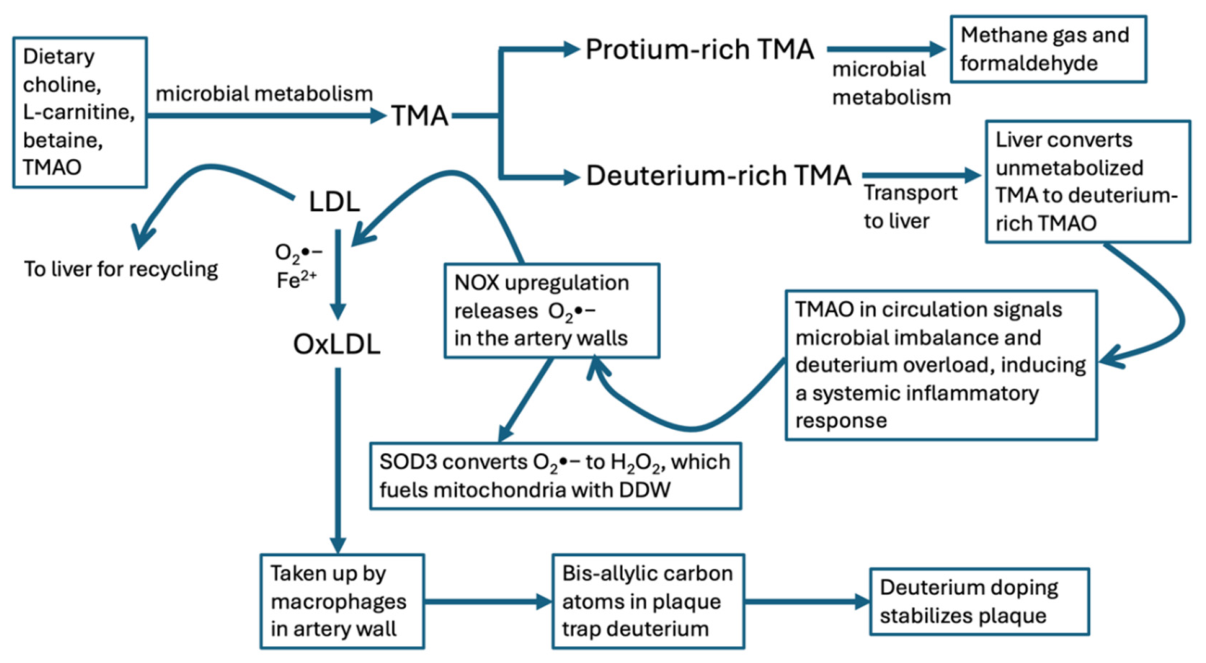
Figure 2.
Schematic of the various pathways involving homocysteine that lead to restoration of methylation capacity, synthesis of glutathione, or plaque accumulation via the process of homocysteinylation of ApoB-100 in LDL. PON1: paraoxenase 1 ; MAT: methionine adenosyltransferase; SAHH: S-adenosyl homocysteine hydrolase; ApoB: apolipoprotein B.
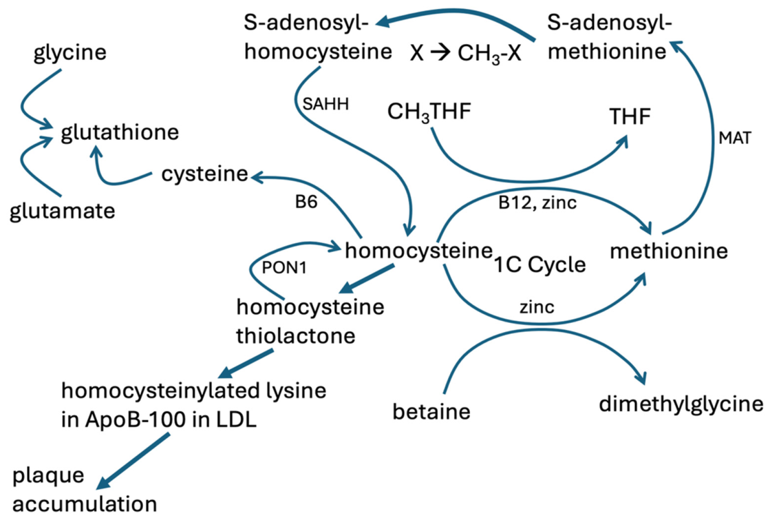
Figure 3.
A block diagram of the processes by which atherosclerosis develops, according to our hypothesis, beginning with a reduction in the population of obligatory anaerobic archaea in the gut, and ending with the stabilization of plaque lesions via deuterium doping. See text for details.
Figure 3.
A block diagram of the processes by which atherosclerosis develops, according to our hypothesis, beginning with a reduction in the population of obligatory anaerobic archaea in the gut, and ending with the stabilization of plaque lesions via deuterium doping. See text for details.
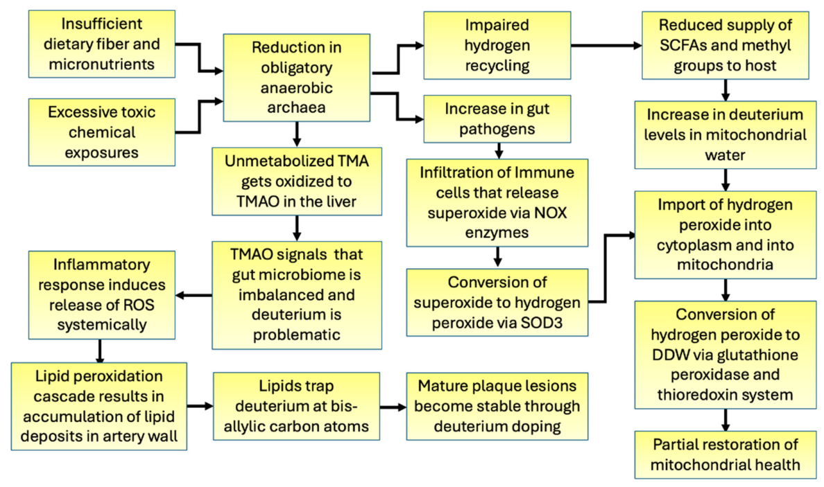
Disclaimer/Publisher’s Note: The statements, opinions and data contained in all publications are solely those of the individual author(s) and contributor(s) and not of MDPI and/or the editor(s). MDPI and/or the editor(s) disclaim responsibility for any injury to people or property resulting from any ideas, methods, instructions or products referred to in the content. |
© 2026 by the authors. Licensee MDPI, Basel, Switzerland. This article is an open access article distributed under the terms and conditions of the Creative Commons Attribution (CC BY) license (http://creativecommons.org/licenses/by/4.0/).
Copyright: This open access article is published under a Creative Commons CC BY 4.0 license, which permit the free download, distribution, and reuse, provided that the author and preprint are cited in any reuse.