
Submitted:
23 June 2026
Posted:
24 June 2026
You are already at the latest version
Abstract
Psychological stress arises when individuals perceive demands as exceeding their capacity to cope. Social stress can be acute, such as giving a presentation, producing transient increases in heart rate, blood pressure, and neuroendocrine activation, or chronic, in which stressors persist over extended periods. Chronic psychosocial stress disrupts immune homeostasis and contributes to anxiety, depression, and post traumatic stress, while also increasing risk for infections, inflammatory and autoimmune diseases, cardiovascular disease, metabolic dysfunction, and cancer. Persistent stress engages interconnected neuroendocrine–immune networks, including the hypothalamic–pituitary–adrenal axis, the sympathetic–adrenomedullary system, and the microbiota–gut–brain–immune axis, leading to sustained glucocorticoid and catecholamine signaling, gut dysbiosis, impaired barrier integrity, and altered microbial metabolite profiles. In this review, we summarize the molecular and cellular mechanisms by which these axes remodel innate and adaptive immunity and shape disease susceptibility. Using social defeat and social isolation in mice as model systems, we highlight how chronic social stress reprograms immune responses in key tissues, including the lung, brain, and gut.
Keywords:
social disruption stress
; glucocorticoids
; adrenergic receptors
; epinephrine
; norepinephrine
; macrophages infections
1. Introduction
The psychological stress is a state of mental and physiological tension that arises in response to challenging or threatening situations. It can be acute, such as during an exam or job interview and is typically transient, or chronic, when stressors are persistent and perceived as overwhelming. Common sources of chronic stress include high pressure occupations, financial instability, and adverse social relationships. Unlike acute stress, which can be adaptive, chronic stress is detrimental to health due to sustained activation of the stress response. Prolonged stress exposure increases the risk of cardiovascular disease, metabolic disorders such as diabetes, and neuropsychiatric conditions including depression, while also exacerbating pre-existing pathologies.
The physiological response to stress is mediated primarily through the hypothalamic-pituitary-adrenal (HPA) axis, the sympathetic-adreno-medullar (SAM) axis, and the microbiota-gut-brain-immune axis. The HPA and SAM axes are central regulators of immune function under both homeostatic and stress conditions. Activation of the HPA axis culminates in the secretion of glucocorticoids (cortisol in humans and corticosterone in rodents), whereas activation of the SAM axis leads to the release of catecholamines, including epinephrine from the adrenal medulla and norepinephrine from sympathetic nerve terminals that innervate all peripheral tissues.
Under homeostatic conditions, glucocorticoids and catecholamines release follows a circadian rhythm, peaking during the active phase (morning for humans and evening for mice) and decreased during the inactive phase. In response to acute psychological stress, these mediators are rapidly elevated to initiate the “fight or flight” response. Sympathetic nervous system activation increases heart rate, dilates airways, and redistributes blood flow to skeletal muscle, while HPA axis activation enhances metabolic output by increasing blood glucose levels and suppressing non-essential processes such as digestion and certain aspects of immune function. Following resolution of an acute stressor, these systems typically return to baseline. In contrast, chronic stress results in sustained activation of both the HPA and SAM axes, leading to prolonged neuroendocrine and immune alterations.
This review focuses on the role of HPA and Sam axes in regulating innate and adaptive immunity during both homeostasis and chronic stress. We discussed the cellular and molecular pathways that mediate these effects and how their dysregulation contributes to disease. In addition, we examine the microbiota-gut-brain axis. A bi-directional communication network in which stress-induced activation of the sympathetic nervous system alters gut microbial composition (dysbiosis), while microbial metabolites influence central nervous system function. Emerging evidence suggests that this axis extends to immune regulation during chronic stress, where sympathetic activation promotes microbial translocation and primes myeloid cells in peripheral tissues such as the spleen (Figure 1).
We also review experimental models of chronic stress, with a particular focus on the mouse social disruption stress (SDR) model. We describe how SDR activates both HPA and SAM axes and alters gut microbiome composition. Furthermore, we compare SDR with chronic restraint stress models to highlight distinct neuroendocrine, immune, and behavioral outcomes. Finally, we discuss studies of social isolation in adult mice, which induce depression and anxiety-like behaviors alongside gut dysbiosis, providing additional insight into the interplay between social stress, neuroendocrine signaling, and immune function.
2. Hypothalamic-Pituitary-Adrenal Axis
The hypothalamic-pituitary-adrenal (HPA) axis and the sympathetic-adreno-medullar (SAM) axis are activated in parallel in response to stress, resulting in the coordinated release of glucocorticoids and catecholamines, respectively (Figure 1). HPA axis activation is initiated by corticotropin-releasing hormone (CRH), also known as corticotropin-releasing factor produced by parvocellular neurons in the paraventricular nucleus (PVN) of the hypothalamus[1]. CRH is secreted into the hypophyseal portal circulation and transported to the anterior pituitary, where it binds corticotropin-releasing factor receptor-1 (CRF1) on corticotropes to stimulate the synthesis and secretion of adrenocorticotropic hormone (ACTH) into the systemic circulation. Circulating ACTH acts on melanocortin type 2 receptors (MC-R2) in the adrenal cortex to drive the synthesis and release of glucocorticoids, cortisol in humans and corticosterone in rodents, which are the principal hormonal-effectors of the HPA axis in the context of stress(reviewed in[2]). These glucocorticoids exert widespread effects on metabolism, cardiovascular function, and the immune system, and provide negative feedback at the level of the hypothalamus and pituitary to terminate the stress response.
2.1. Glucocorticoids
Glucocorticoids (GCs) are regulators of energy metabolism, brain function, and immune responses (Figure 1). Under homeostasis conditions, GC secretion follows a circadian rhythm that is controlled by the suprachiasmatic nucleus (SCN); (i) SC-driven activation of the HPA axis, (ii) autonomic innervation of the adrenal gland via the splanchnic nerve, and (iii) intrinsic adrenal clocks[3,4]. GCs signal through two receptors, the mineralocorticoids receptors (MR), and the glucocorticoid receptors (GR). Because MRs have a higher affinity for GCs than GRs, MRs ae occupied at basal hormone levels, whereas GRs become engaged at the circadian peak or during stress, when GC concentrations are elevated [5,6]. GR is a ligand-activated nuclear receptor that, upon GC binding, translocate to the nucleus and modulates gene transcription via binding to negative glucocorticoid response elements (GREs) in target genes and via positive GREs to induce suppressive mediator genes. GR can also regulate transcription indirectly through protein-protein interactions with transcription factors. GCs participate in a classic negative-feedback loop that constrains HPA axis activity and limits the consequences of excessive GC exposure[7]. At exceptionally low GC concentrations, macrophages and T cells secrete macrophage migration inhibitory factors (MIF), which can counteract GC-mediated suppression of cytokine production; as GC levels rise, MIF decreases, so that MIF restricts GC anti-inflammatory effects at low but not high GC concentrations[8,9].
Following HPA axis activation, circulating GC levels increase rapidly and typically peak within approximately 30 minutes. GC interaction with GRs in anterior pituitary corticotropes suppresses ACTH secretion by inhibiting transcription of its precursor gene Pomc[6]. Under physiological conditions, GC release occurs in ultradian pulses with a period of roughly 90 minutes, superimposed on the circadian rhythm that peaks at the onset of the active phase (morning in humans and dark in mice) [6].
2.2. Regulation of the Immune System by Glucocorticoids
Glucocorticoids are potent mediators of anti-inflammatory and immunosuppressive responses (Figure 1). They exert these effects both by directly repressing pro-inflammatory genes through binding to negative GREs and by inducing a range of anti-inflammatory mediators. A study by Surjit and colleagues identified negative GREs in more than 1,000 mouse-/human ortholog genes, highlighting the breadth of direct GC-mediated transcriptional repression[10]. Toll-like receptor (TLR) signaling induces the production of pro-inflammatory cytokines such as IL-1, IL-6, GM-CSF, and TNFα through activation of NF-κβ and AP-1. GCs dampen TLR signaling via three main mechanisms. First, GC activated GR directly interacts with NF-κβ and AP-1, inhibiting their transcriptional activity[11,12]. Second, GCs induce expression of Iκβ, which binds NF-κB and retains it in the cytoplasm, thereby preventing nuclear translocation and target gene activation[13]. Third, GCs upregulate dual-specificity phosphatase1 (DUPS1, also known as MAPK phosphatase 1), which dephosphorylates and inactivates MAPKs; consequently, GC-mediated suppression of pro-inflammatory genes such as TNFα, IL-1α, IL-1β, and cyclooxygenase 2 was impaired in DUSP1-/- mouse macrophages[14]. GCs also induce glucocorticoid-induced leucine zipper (GILZ), a key mediator of GC-dependent immunosuppression which diverse roles across immune cell types. GILZ inhibits NF-κB, and interacts with other regulators, including AP-1, Raf-1, and Ras, thereby modulating multiple signaling pathways[15]. In macrophages, GLIZ-mediated inhibition of NF-κβ reduces the production of proinflammatory cytokines and co-stimulatory molecules[16]. Overexpression of GILZ in macrophages has been reported to enhance phagocytosis, protect against pyroptosis, decrease mitochondrial function, and increase matrix metalloproteinase activity[17]. In dendritic cells, GILZ expression promotes an immature, tolerogenic phenotype characterized by increased IL-10 production and reduced IL-12 and IL-23 secretion[18]. GCs also induce the zinc finger protein tristetraprolin TTTP which destabilizes pro-inflammatory cytokine mRNA by binding AU-rich elements in their 3” untranslated regions and targeting them for degradation[19]. Thus, GCs suppress inflammatory responses by converging on both transcriptional and post-transcriptional regulatory mechanisms.
Despite their overall anti-inflammatory profile, GCs can have both inhibitory and permissive effects on immune function. While GCs suppress the production of many cytokines, they can simultaneously increase expression of certain cytokine receptors, thereby enhancing cellular responsiveness to those cytokines[20]. For example, GCs upregulates IL-2Rα expression on T cell receptor activated CD4 T cells, leading to accelerated cell -cycle progression and increased T cell proliferation, even in the context of reduced IL-2 production[21]. Similarly, IL-7Rα expression on human and mouse T cells is enhanced by GCs[22,23]. In mice, increased IL7-Rα expression is driven by GC binding to a conserved noncoding sequence located 3.6 kb from the promoter[23]. Because IL-7R signaling is essential for thymocyte development and maturation[24] and for peripheral T cell survival[25], GC-induced IL-7Rα expression enhances IL-7- responsive gene programs and can protect T cells from GC-induced apoptosis[22]. GCs also regulate naïve T cell trafficking between blood and secondary lymph organs. Binding of GCs to mineralocorticoid receptors on naïve T cells induces CXCR4 expression, which promotes their migration from blood into secondary lymphoid organs such as spleen, lymph nodes, and Peyer’s patches[26]. As circulating GC levels decline, naïve T cells egress from lymphoid tissues back into the blood, generating a GC-dependent circadian rhythm of naïve T cell recirculation. The dominant immunological effects of GCs, however, are suppression of T cell effector function, mediated in part by direct actions on TCR signaling. Early TCR signaling events involved activation of the Src family kinases Lck and Fyn, which phosphorylates the TCR complex. Lowenberg et al. demonstrated that GCs inhibit Lck and Fyn activity in a GR-dependent manner, thereby attenuating TCR signaling[27]. In addition, GCs upregulate the inhibitory co-receptor programmed cell death 1 (PD-1) by binding a GRE located 2,525bp upstream of the Pdcd1 transcription start site, suggesting that GC-induced PD1 expression contributes to T cell suppression[28]. GCs also reshape CD4 T helper cell differentiation. GCs inhibits CD4 Th1 differentiation by reducing IL-12 and IL-18 production by macrophages and monocytes[29] and by suppressing gene expression of the Th1-lineage transcription factor T-bet, and through direct GR-T-bet interactions [30]. Conversely, GCs promote a shift toward Th2 differentiation by enhancing production of IL-4, IL-10, and IL-13[30]. GILZ is an important mediator of these T cell-intrinsic effects of GCs. Up-regulation of GILZ in T cells inhibits activation and proliferation via suppression of NF-κβ, and AP-1 by binding Raf and Ras, thereby blocking downstream MAPK1 activation[16] . Through these pathways, GILZ skews naïve T cell differentiation towards Th2, while suppressing Th-1 and Th-17 lineages, GILZ also promotes regulatory T cell development and activity, at least in part via augmentation of TGF-β signaling.
In summary, GCs modulate both innate and adaptive immunity through a combination of direct transcriptional repression, induction of anti-inflammatory mediators, altered cytokine receptor expression, and reprogramming of T cell signaling and differentiation (Figure 1). Much of our mechanistic understanding derives from studies in mice treated with synthetic GCs such as dexamethasone and from in vitro experiments in GC-treated immune cells. Under homeostatic conditions, GC release is tightly regulated by circadian rhythms entrained to the light-dark cycle, producing corresponding oscillations in innate and adaptive immunity (reviewed in[31]). During chronic stress, this circadian pattern is overridden by sustained release of GCs. The extent to which prolonged GC elevations suppress immune function in humans depends on GC concentrations, which are shaped by stressor intensity, subjective stress perception, and duration of exposure, as well as on cell type specific sensitivity to GCs.
3. Sympathetic-Adreno-Medullary Axis
The sympathetic nervous system (SNS) consists of preganglionic and postganglionic neurons that together mediate rapid physiological responses to stress. Preganglionic sympathetic neurons arise from the thoracic spinal cord and upon activation, transmit electric impulses through the splanchnic nerve to adrenal medulla, where acetylcholine stimulates chromaffin cells to secrete the catecholamines epinephrine and norepinephrine into the circulation. These catecholamines initiate the classic fight-or- flight response, increasing heart rate, cardiac output, blood pressure and blood glucose levels, while redistributing blood flow to support immediate physical activity[32] . In parallel, preganglionic neurons synapse with postganglionic neurons in sympathetic ganglia located along the spinal column (Figure 1). Postganglionic fibers extend into peripheral tissues, including primary and secondary lymphoid organs, where they release norepinephrine from axonal varicosities and regulate the activity of multiple nearby cell types through adrenergic receptors[32]. . Through this dense innervation, the SNS provides a major neural pathway by which the brain can modulate immune function in peripheral tissues and lymphoid organs[33]. . The SNS also contributes to physiological homeostasis under basal conditions. Circadian signals from the suprachiasmatic nucleus help to regulate daily rhythmic sympathetic tone, which is elevated during the active phase to maintain basal physiological readiness. During acute stress, this system is strongly activated to generate a rapid adaptive response, whereas prolonged SNS activation can disrupt immune regulation and has been linked to anxiety, depression, and other stress-associated disorders[34,35].
4. SNS Innervation of Lymphoid Tissue
SNS innervation of lymphoid tissue provides a major efferent pathway by which the brain rapidly modulates immune function. Sympathetic fibers densely innervate primary and secondary lymphoid organs, releasing norepinephrine onto stromal and immune cells and thereby shaping leukocyte trafficking, cytokine production, and the overall tone of innate and adaptive immunity.
4.1. Bone Marrow
Blood cells, including erythrocytes, leukocytes and platelets, are generated by hematopoiesis, which takes place primarily in the bone marrow. Hematopoietic stem cells (HSCs), endowed with self-renewal capacity and multilineage potential, give rise to progenitor cells that differentiate into all mature blood cell types[36]. HSCs reside in specialized niches formed by stromal and endothelial cells, which provide essential molecular and cellular cues for HSC maintenance and function[37]. Both stromal cells and HSCs are molecularly heterogeneous[36,38,39,40,41], supporting the concept that distinct HSC subpopulations are regulated by anatomically and functionally specialized niches[42] (Figure 2) . At steady state, the SNS regulates the proliferation and differentiation of hematopoietic stem and progenitor cells (HSPCs), and the migration of HSPCs, as well as the trafficking of lymphocytes, in a circadian manner[42]. One study showed that, in the morning, light induced norepinephrine and TNF secretion promotes HSPC proliferation and differentiation, whereas in the evening, darkness-induced TNF enhances melatonin secretion, which favors HSPC renewal and maintenance[43]. Under homeostatic conditions, HSCs and their progeny periodically enter the bloodstream, migrate to sites of extramedullary hematopoiesis such as the spleen, or return to the bone marrow to occupy vacant niches.
HSC and HSPC retention in the bone marrow is governed by CXCL12 produced by stromal cells and its receptor CXCR4 expressed on HSCs and HSPCs. CXCL12 expressions in bone marrow stromal cells oscillates in antiphase with the release of HSCs and HSPCs into the circulation, peaking approximately 5 hours after light onset and reaching a nadir about 5 hours after darkness[44]. Circadian release of norepinephrine from sympathetic fibers leads to activation of β3-adrenergic receptors on stromal cells, which reduces expression of the transcription factor Sp1 and down-regulation of CXCL12. Reduced CXCL2 levels permit HSCs and HSPCs to egress into the bloodstream, whereas in the evening, when norepinephrine levels fall, CXCL12 expression rises and HSCs/HSPCs are retained in the bone marrow[44] (Figure 2).
In response to infections, the hematopoietic system rapidly increases production of innate myeloid cells. HSCs and progenitors express Toll-like receptors (TLRs) and can directly sense pathogen-associated molecular patterns (PAMPS)[45,46]. Microbial recognition by HSPCs drives proliferation and a shift toward myelopoiesis (reviewed in[47]). For example, TLR4 signaling induces IL-6, which promotes phosphorylation of C/EBPα and CEBPβ, thereby enhancing myeloid differentiation[48]. TLR4 and NOD-like receptor signaling synergistically induce HSC migration to the spleen in a granulocyte colony-stimulating factor (G-CSF) – dependent manner[49]. In both settings TLR2 and TLR4 signaling are associated with loss of HSC self-renewal capacity. Macrophages derived from TLR2 stimulated HSCs and progenitor cells produce lower levels of inflammatory cytokines (TNFα, IL-6, and IL-1β) and reactive oxygen species upon Pam3CSK4 stimulation than bone marrow-derived macrophages generated in vitro with M-CSF[50,51]. Infection with the parasite Leishmania donovani also induces HSC expansion and myelopoiesis; notably, this infection skews myeloid differentiation toward non-classical progenitors that are more permissive to infection[52].
Chronic psychological stress is major factor in cardiovascular disease. It drives increased bone marrow production of monocytes and neutrophils, which are recruited to atherosclerotic plaques and promote lesion[53]. Mechanistically, chronic stress elevates norepinephrine release form sympathetic fibers, leading to β3-adrenegic receptor mediated downregulation of CXCL12 in the bone marrow and activation of HSCs[54]. Enhanced HSC proliferation increases the output of neutrophils and monocytes into the circulation. In atherosclerosis-prone Apoe-/- mice, chronic stress-induced hematopoiesis augments plaque features associated with vulnerable lesions, whose rupture can trigger myocardial infarction and stroke. Following myocardial infarction, Apoe-/- mice develop larger atherosclerotic lesions with increased monocyte recruitment, which has been linked to sympathetic signaling that mobilizes HSCs to the spleen and enhances splenic monocyte production The monocyte recruitment was shown to be due to sympathetic nervous signaling which induced migration of hematopoietic stems to the spleen, resulting in increased monocyte production in the spleen[55].
4.2. Thymus
The thymus is the primary lymphoid organ responsible for the development of mature T cells. It is organized into an outer cortex and an inner medulla. Bone marrow-derived T cells precursors enter the outer cortex, where they undergo T cell lineage commitment, T cell receptor (TCR) gene rearrangement, and positive selection. Thymocytes then migrate to the inner medulla, where self-reactive cells are eliminated by negative selection, and the T cells exit the thymus as mature but naïve T cells.
Sympathetic innervation of the thymus has been characterized using immunofluorescence and confocal microscopy in mice which revealed that a meshwork of nerve fibers is present in all -thymic compartments, including the capsule, subcapsular region, cortex, cortico-medullary junction, and medulla (Figure 2) [56]. In the outer cortex and cortico-medullary junction, CD4+ CD8+ thymocytes are strongly associated with nerve fibers, whereas in the medulla CD4+ CD8- and CD4- CD8+ thymocytes are in close apposition to nerve fibers[56]. This intimate association of developing thymocytes with nerve elements suggests that neural signals contribute to the regulation of T cell development. The same study also examined myeloid populations, finding that most dendritic cells (DCs) localized to medullary regions, with a subset in contact with nerve fibers, and that CD11b+ and F4/80+ macrophages were likewise associated with nerve fibers, indicating broad neuroimmune interactions with the thymic microenvironment.
Immature thymocytes express β-adrenergic receptors, and mature medullary thymocytes express higher numbers of β-adrenergic receptors per cell, consistent with an increase in β-adrenergic responsiveness during thymocyte maturation[57]. Thymic epithelial cells, which provide critical stromal support for T cell development, also express β1- and β-2 adrenergic receptors[58,59] . Chronic treatment of rats with β-adrenergic blockers altered thymic differentiation and maturation[60,61] (Figure 2), indicating that β-adrenergic signaling regulates thymocyte differentiation into mature T cells both directly, via receptors on thymocytes, and indirectly, via modulation of thymic epithelial cells. In addition, α1-adrenegic receptors are expressed on immature thymocytes, thymic epithelial cells and CD68+ macrophages in the rat thymus Chronic treatment of the α1-blocker urapidil disrupts thymocyte differentiation and lineage commitment, further supporting a role for a α1-adrenegic signaling in thymic T cell development[62].. Collectively, these findings suggest that, under steady-state conditions, adrenergic signaling within the thymus supports adaptive immunity by promoting thymocyte differentiation and maturation.
By contrast, chronic stress can impair thymic function and T cell development. Chronic immobilization stress in C57BL/6 mice markedly reduced thymocyte numbers and increased thymocyte apoptosis[63]. The proportion of immature thymocytes was particularly diminished, consistent with enhanced negative selections and/or impaired progression through early developmental stages under sustained stress exposure. Together, these observations indicate that while physiological sympathetic signaling in the thymus supports T cell maturation, chronic stress-associated SNS activation can shift this balance toward thymic atrophy and reduced T cell output.
4.3. Spleen and Lymph Nodes
The spleen filters blood, removing blood-borne debris and pathogens, and participates in iron recycling from senescent erythrocytes (reviewed in[64]). The spleen is divided into red pulp and white pulp, separated by the marginal zone (reviewed in[65] ). Blood enters the marginal zone from open ends of the splenic artery, percolates through the red pulp, and exits via the splenic vein (Figure 2). Macrophages in the marginal zone and red pulp phagocytose and clear blood- borne pathogens (reviewed in[66] ). Marginal zone B cells and dendritic cells capture antigens, migrate into the white pulp, and present antigens to T cells (reviewed in[64]). Adaptive immune responses are organized within the white pulp, which contains distinct B and T cells areas[65]. After antigen recognition, activated T cells migrate to the B cell follicles to provide help to B cells. The red pulp also serves as a reservoir for Ly6Chi monocytes[67] , which can be rapidly mobilized to sites of injury, such as ischemic myocardial injury[67] or corneal epithelial abrasion[68]. In addition, the red pulp is a site of myelopoiesis, supported by hematopoietic stem and progenitor cells (HSCs/HSPCs) that migrate from bone marrow to the spleen under the control of sympathetic signaling, as discussed above.
Sympathetic nerve fibers reach the spleen via the splenic nerve. Immunofluorescent staining has shown nerve fibers in all spleen compartments, in close association with macrophages, DCs and lymphocytes[69]. Norepinephrine release from these fibers in response to corneal epithelial injury induces rapid egress of reservoir monocytes from the red pulp into the circulation. Pharmacological activation of the β-2 adrenergic receptor further enhances monocyte recruitment to the injured cornea[68] . Sympathetic activation also increases proliferation and differentiation of myeloid progenitors during spleen myelopoiesis, an affect that is blocked by β2-adrenergic receptor antagonist[70] . Thus, sympathetic signaling in the spleen not only mobilizes monocytes from existing reservoirs but also supports local myeloid production.
Sympathetic fibers also innervate the white pulp[69]. In response to antigen, germinal centers (GCs) form in the white pulp of the spleen and in lymph nodes, providing specialized microenvironments in which mature B cells proliferate, undergo somatic hypermutation, and differentiate into plasma cells or memory B cells. Chemical depletion of sympathetic nerve fibers in mice impairs GC formation after Staphylococcus aureus infection, and such mice fail to develop protective immunity[66] . These findings indicate that sympathetic innervation is required for efficient GC formation, although the underlying mechanisms remain incompletely defined. Lymph nodes filter lymphatic fluid, which contain microbes, cellular debris, dead cells, and tumor cells. Antigen-laden DCs, macrophages, and monocytes migrate from inflamed tissues to draining lymph node via CCR7-depndent mechanisms[71,72]. Within lymph nodes, these antigen-presenting cells stimulate naïve CD4 T cells via MHC class II and cross-present antigen to CD8 T cells via MHC class I , triggering proliferation and differentiation of the T cells. CD4 T cells that acquire a Th1 phenotype exit via the bloodstream and home to inflamed tissues, whereas other CD4 T cells upregulate CXCR5, migrate into B cell follicles, and differentiate into T follicular helper cells (Tfh), which provide essential help for GC formation and antibody affinity maturation (reviewed in [73]).
Immunofluorescence and confocal microcopy studies have demonstrated that mouse lymph nodes are densely innervated, with DCs, macrophages, and B and T lymphocytes closely associated with nerve fibers[74] . Consistent with the spleen, sympathetic innervation is required for GC formation in lymph nodes although the precise mechanisms are not yet defined. One proposed mechanism involves β-adrenergic regulation of Tfh cells. Supporting this idea, β-adrenergic blockade in humans decreased the specific IgG responses to seasonal influenza vaccine and decreased the frequency of Tfh cells in the spleen and lymph nodes[73].
Lymphocyte circulation through lymph nodes is necessary for immune surveillance. In a study by Naki et al.[75] , treatment of mice with a β2-adrenergic receptor caused a rapid decline in circulating B and T lymphocytes accompanied by reduced lymphocyte counts in lymph (Figure 2). The authors showed that β2-adrenergic receptors on lymphocytes physically interacted with chemokine receptors CCR7 and CXCR4, which promote lymphocyte retention in lymph nodes. Activation of β2-adrenergic receptors on T cells resulted in CCR7 dependent retention, whereas B cell retention depended on CXCR4[75]. These findings suggest that chronic SNS activation during prolonged stress could impair immune surveillance by excessive retention of lymphocytes within lymph nodes and limiting the egress of effector CD4 Th1 cells from draining nodes to inflamed tissues.
5. Adrenergic Receptors
Epinephrine and norepinephrine signal through adrenergic receptorsa family of G-protein-coupled receptors divided into five major subtypes: α1, α2, β1, β2, and β3. Each subtype couples to heterotrimeric G proteins, thereby engaging different intracellular signaling pathways (Figure 3). β-adrenergic receptors primarily couple to Gs, stimulating adenylate cyclase and increasing cAMP production. In contrast, α2-adrenergic receptors couple to Gi to inhibit adenylate cyclase and reduce cAMP levels, whereas α1-adrenergic receptors couple to Gq to stimulate phospholipase C-β, leading to IP3 and DAG generation and downstream protein kinase C activation. Consequently, the cellular effects of epinephrine and norepinephrine depend on the adrenergic receptor repertoire expressed by each cell type. Among immune cells, the β2-adrenergic receptor is the most prominently expressed subtype and represents a major conduit for sympathetic control of immune function[76].
5.1. Adrenergic Receptors and Macrophages
Stimulation of macrophage adrenergic receptors by norepinephrine and epinephrine produces both anti- and pro-inflammatory outcomes, depending on receptor subtype engagement, catecholamine concentration, and macrophage activation state. In LPS- activated macrophages, norepinephrine and β-2-Adrenergic receptor agonists downregulate expression of the pro-inflammatory cytokines TNFα and IL-6 (PMID:36284839) and the chemokine CCL2[77,78] and suppress anti-mycobacterial activity by inhibiting nitric oxide production[77,78,79]. . Stimulation of β2-Adrenergic receptor also induces the anti-inflammatory cytokine IL-10, a potent negative regulator[80] [81] of both innate and adaptive immunity that limits macrophage production of pro-inflammatory cytokines[82,83,84] , reduces MHC class II and co-stimulator CD86[82,85,86], and inhibits macrophage production of IL-12 required for CD4 Th1 differentiation (Figure 3)[87].
Macrophages also express α2-adrenergic receptors, and several studies indicate that α-2 signaling can enhance inflammatory and antimicrobial responses (Figure 3). Spengler and colleagues reported that α2-adrenergic receptor activation increases the production of TNF-α by LPS stimulated macrophages and induces IL-12, a cytokine required for IFN-γ -induction by CD4 T cells[88,89], Consistent with a role in host defense, we have shown that α2-adrenergic receptor engagement augments anti-mycobacterial activity in peritoneal and spleen macrophages[90]. Using the RAW264.7 macrophage cell line, we further found that increased resistance to Mycobacterium avium following stimulation with the α2-agonist clonidine was due to increased production of nitric oxide (NO) and superoxide (O2-) and the formation of peroxynitrite (ONOO-)[91] .
More recent work suggests that α2-adrenergic receptor can also exert anti-inflammatory effects, depending on context. In an air pouch model of inflammation, the α2-A agonist guanfacine reduced myeloid cell accumulation and decreased levels of TNFα, CCL-2 and IL-10 but not Il-6, in the inflammatory exudate [92]. The authors proposed that α-2adrenergic signaling dampens NF-κβ driven responses by reducing activity of the transcription factor c-Rel, a key promoter of inflammatory cytokine expression upon LPS stimulation[93]. In line with its capacity to modulate antigen-presenting function, activation of macrophage α2-adrenergic receptor has also been reported to enhance T cell stimulation and promote tumor rejection (Figure 3)[94].
Thus, the downstream signaling and functional outcome of macrophage stimulation with norepinephrine depends critically on which adrenergic receptor subtype is engaged. β2-adrenergic receptor activation promotes exchange of GDP to GTP on Gas and dissociation of Gas from Gβy subunits. Gαs then stimulates adenylate cyclase to convert ATP to cAMP, which binds the regulatory subunit of protein kinase A (PKA) and releases the catalytic subunit to phosphorylate serine and threonine residues on target proteins. The anti-inflammatory effects of β2-drenergic receptor agonists are mediated via the Iκβ/NF-κβ pathway and MAP kinase pathways. NF-κβ is normally sequestered in the cytoplasm by the inhibitor Iκβ-α. Phosphorylation and degradation of Iκβ-α results in nuclear translocation of NF-κβ and binding of NF-κβ to promoters of inflammatory genes. Activation of the cAMP/PKA pathway by β2-adrenegic receptor agonists up-regulates Iκβ- expression, thereby retaining NF-κβ in the cytoplasm[95]. β2-adrenegic agonists also suppress cytokine gene expression by cAMP-dependent inhibition of the ERK MAP Kinase[96] and cAMP-dependent up-regulation of mitogen-activatedprotein kinase phosphatase 1 (MKP1), which constrains p38 MAP kinase activity[97] . By contrast, α2- adrenergic receptor activation in macrophages releases Gαi, leading to inhibition of cAMP production. α-2 adrenergic receptor signaling also suppresses voltage-gated Ca2+ currents, activates receptor-operated K+ channels, and engages p38 MAP kinase[98] , providing a distinct signaling profile that can support either pro- or anti-inflammatory outcomes depending on context.
Although selective α2 and β2 agonists clearly define receptor-specific signaling cascades, an important unresolved question is how macrophages con-expressing α2 and β2 -adrenergic receptors respond to physiological catecholamines. The net outcome depends on relative receptor expression and catecholamine concentration. For example, we observed that low dose epinephrine (10−8 M) inhibited increased bacterial growth[99] . These data suggest that in resident peritoneal macrophages, α2-adrenergic receptor signaling is predominant at low catecholamine levels and promotes antimicrobial activity, whereas at higher epinephrine concentrations the β2-adrenergic signaling becomes dominant and suppresses anti-mycobacterial functions. Little is known about how adrenergic receptor expression is regulated in macrophages, but Hernandez et al. [92] found that monocytes express low levels of the α-2A-adrenergic receptor and that the J774.2 monocyte/macrophage cells upregulates the α-2A adrenergic receptor upon LPS stimulation. Similarly, Hanlon et al. reported that resting microglia, which primarily express β2-adrenergic receptors, switch to α-2A-adrenergic receptor expression after LPS stimulation[100], indicating that inflammatory conditions can dynamically reprogram the adrenergic receptor repertoire of myeloid cells.
5.2. Adrenergic Receptors and Dendritic Cells
Dendritic cells (DCs) link innate and adaptive immunity by processing and presenting antigens to T lymphocytes. DCs express both α-and β-adrenergic receptors [101]. Immature DCs, (iDC) but not mature DCs (mDC), express the α1B-adrenergic receptor, which regulates iDC migration from sites of inflammation to draining lymph nodes [102]. Adrenergic stimulation of bone marrow derived DCs with norepinephrine can enhance antigen uptake via α2-adrenergic receptor- dependent activation of PI3K[103].
The β2-adrenergic receptor is highly expressed functionally active in iDCs, as norepinephrine stimulation induces cAMP and PKA-dependent phosphorylation of CREB[101]. β2-adrenergic receptor activation does not markedly alter DC differentiation or their capacity to drive naïve T cell proliferation[101]. However, it profoundly reshapes DC cytokine production by suppressing pro-inflammatory cytokines TNFα, IL-6, IL-12p70, and IL-23 while increasing IL-10[104] , and enhancing IL-33, which functions as an inflammatory cytokine in Th2-type immune responses[105]. β2-adrenergic stimulation also reduces the ability to activated DCs to promote Th1 differentiation and IFN-γ production, while favoring Th17 differentiation and increased IL-17production[106] . Mechanistically, this shift reflects β2-adrenergic receptor-mediated inhibition of NF-κB and AP-1 signaling, resulting in downregulation of IL-12p70 and an altered IL-12/IL-23 balance[101]. The decrease in IL-12p70 (p35/p40) together with preserved or enhanced IL-23 (p19/p40) favors IL-23-driven differentiation of naïve CD4 T cells into IL-17–producing Th17 cells at the expense of IL-12-driven Th1 differentiation and IFN-γ production. Consistent with this, another study reported that β2-adrenergic agonists skew Th differentiation toward a Th17 profile by increasing DC production of IL-23p19 and IL-6 while inhibiting IL-12p35 and IL-12p40 (Figure 3)[107].
In addition to modulating effector responses, β2-adrenergic receptor signaling in DCs supports regulatory pathways. β2-adrenergic activation in bone marrow–derived DCs enhances the induction of Foxp3+ suppressive regulatory T cells (Tregs)[104] . Moreover, β2-adrenergic receptor signaling in Foxp3+ Tregs themselves has been shown to increase their suppressive function[108]. Together, these findings indicate that adrenergic signaling in DCs not only modulates antigen uptake and migration but also reprograms DC cytokine output and T cell–polarizing capacity, shifting adaptive responses away from Th1 and toward Th17 and regulatory pathways in the context of sympathetic nervous system activation.
5.3. Adrenergic Receptors and CD4 T Cells
Mouse naïve CD4 T cells and effector Th1 cells express the β2-adrenergic receptor, whereas effector Th2 cells lack β2-adrenergic receptor expression[109,110] . This differential expression is driven by epigenetic regulation involving histone acetylation, histone methylation, and DNA methylation at the β2-adrenergic receptor promoter [111] . During Th1 differentiation, naïve CD4 T cells acquire increased β2-adrenergic receptor expression associated with histone-3 H3/H4 acetylation and H3K4 methylation, whereas Th2 differentiation is accompanied by reduced β2-adrenergic receptor expression, decreased H3/H4 acetylation and H3K4 methylation, increased H3K9 and H3K27 methylation, and enhanced DNA methylation at the locus[111].
Functionally, β2-adrenergic signaling modulates CD4 T cell polarization in a cytokine-context–dependent manner. Stimulation of naïve CD4 T cells with anti-CD3/anti-CD28 in the presence of norepinephrine and IL-12 promotes Th1 differentiation and yields Th1 cell that produce 2 to 4-fold more IFN-γ; this effect requires Il-12, as neutralization of IL-12 abrogates the increase in IFN-γ[112]. Thus, β2-adrenergic stimulation can augment IFN-γ production when adequate IL-12 is provided by antigen-presenting cells. However, as outlined above, β2-adrenergic signaling in dendritic cells and macrophages suppresses IL-12 production and favors IL-23 and IL-6, thereby indirectly constraining Th1 differentiation and promoting Th17 polarization[101,106] .
Consistent with this, β2-adrenergic stimulation has been shown to reduce Th1 responses and enhance Th17 differentiation in both mice and humans[101,106] . In human peripheral blood mononuclear cells, a subset of Th17 cells expresses β2-adrenergic receptors, and differentiation in the presence of β2 agonist terbutaline decreases IFN-γ production while increasing IL-17A[113] . A separate study similarly reported terbutaline-induced upregulation of IL-17A in human PBMCs and memory Th17 cells via a cAMP/PKA-dependent pathway[114] .In summary, β2-adrenergic stimulation tends to reduce Th1 differentiation and favor Th17 differentiation. Th1 cells are critical for protection against intracellular pathogens such as Mycobacterium tuberculosis through IFN-γ production, whereas Th17 cells protect against extracellular bacteria such as Staphylococcus aureus by secreting IL-17A, which promotes recruitment of neutrophil recruitment[115] . Prolonged β-adrenergic activation is therefore expected to alter the Th1/Th17 balance in a way that may increase susceptibility to intracellular infections while enhancing Il-17 driven inflammatory pathways. This shift can contribute to the pathogenesis or exacerbation of autoimmune and inflammatory diseases in which IL-17 plays a central role, including autoimmune arthritis in mice[116] , psoriatic disease[117] , and asthma[118] .
5.4. Adrenergic Receptors and CD8 T Cells
The β2-adrenergic receptor is differentially expressed in human CD8 T cell subsets, with higher expression on effector memory CD8 T cells than on central memory or naïve CD8 T cells[119,120,121]. Stimulation of mouse and human CD8 T cells with norepinephrine reduces IFN-γ and TNF-α secretion and cytolytic activity in response to TCR engagement but does not necessarily impair proliferative capacity in all settings[120,122] During activation, CD8 T cells normally increase glucose uptake and undergo metabolic reprogramming to support effector function. β2- adrenergic receptor signaling during CD8 T cell activation interferes with this process by downregulating glucose transporter 1 expression and decreasing glucose uptake and glycolysis[123] . In human CD8 T cell subsets, activation with anti-CD3/CD28 in the presence of norepinephrine enhances expression of IL-6 and chemokines CXCL2, CXCL3, and CCL2 in memory CD8 T cells compared to naïve and central memory T cells, while reducing IL-2 and IFN-γ production; in this context, expansion of memory CD* T cells is diminished, indicating that norepinephrine can also impose a suppressive effect on CD8 T cell proliferation[121] .
CD8 T cell exhaustion is a differentiation state driven by chronic antigen exposure, characterized by progressive loss of effector function and upregulation of inhibitory receptors such as PD-1, LAG-3, 2B4, CD160, and TIGIT. In this context, adrenergic signaling appears to contribute directly to the exhausted phenotype. Expression of the β1-adrenergic receptor is increased on terminally exhausted CD8 T cells, whereas β2-adrenergic receptor expression is not similarly upregulated[124] (Figure 3). In mouse models of chronic LCMV infection and pancreatic cancer, exhausted CD8 T cells are located near sympathetic nerve fibers[124]. In vitro, antigen-specific CD8 T cells engineered to overexpress the β1-adrenergic receptor show impaired proliferation and cytokine production when stimulated through the TCR in the presence of catecholamines, whereas genetic deletion of β1-adrenergic receptors protects CD8 T cells from progressing to terminal exhaustion during chronic viral infection[124] . In a pancreatic cancer model, β-blocker treatment synergizes with immune checkpoint blockade to enhance CD8 T cell responses and promote the development of memory-like CD8 T cells, thereby linking sympathetic signaling to the quality of antitumor and antiviral T cell immunity[124] (Figure 3) .
Several studies further support the concept that β-adrenergic signaling promotes tumor growth and impairs the efficacy of antitumor immunotherapy. In mice bearing Eμ-myc lymphoma, β-adrenergic stimulation increased tumor growth, and reduced survival; mechanistically, β-adrenergic signaling inhibited CD8 T cell proliferation, IFN-γ production, and cytolytic activity via direct effects on CD8 T cells and diminished the[125] effectiveness of immunotherapy with anti-PD-1 and anti-4-1BB antibodies . In a vaccine-based tumor model using the STxB-E7 vaccine (composed of the non-toxic B subunit of Shiga toxin coupled to an HPV16-derived E7 peptide), efficacy was enhanced by concomitant treatment with the β-blocker propranolol, which increased CD8 T cell infiltration into tumors[126,127,128] . This increase in tumor-infiltrating CD8 T cells was attributed to improved priming in tumor-draining lymph nodes under β-blockade. Notably, tumor-infiltrating CD8 T cells in this setting became insensitive to β-adrenergic signaling due to downregulation of the β2-adrenergic receptor upon activation, whereas naïve CD8 T cells remained extremely sensitive to β-adrenergic modulation[128] . Together, these findings indicate that adrenergic signaling—particularly via β-adrenergic receptors—can dampen CD8 T cell effector function, promote exhaustion, and reduce the efficacy of antitumor and antiviral immune responses, while β-blockade can partially reverse these effects and improve immunotherapy outcomes.
5.5. Adrenergic Receptors and B Cells
B cells exclusively express β2-adrenergic receptors. T cell activation of B cells requires two signals. Signal 1 is engagement of the B cell receptor (BCR) by antigen., followed by antigen processing and presentation of peptide-MHC class II complexes to CD4+ Th2 cells. This interaction induces upregulation of the co-stimulatory molecule B7-2 (CD86) on B cells, which binds CD28 on helper T cells. Signal 2 is provided by CD40-CD40L interactions together with Th2-derived cytokines (IL-4, IL-5, and IL-6), driving B cell proliferation, class-switch recombination, and differentiation into plasma cells. Stimulation of the B cell β2-adrenergic receptors enhances humoral responses by increasing antibody production[129,130] and promoting B cell proliferation[130] . Resting B cells express incredibly low level of B7-2; concurrent stimulation of the BCR and β2-adrenergic receptor synergistically upregulates B7-2 expression[131,132]. This increase is mediated both by NF-κβ dependent transcriptional activation and by enhanced B7-2 mRNA stability[131] . Co-engagement of CD86 on B cells with CD28 on Th2 cells and the B cells β2-adrenergic receptor with β2-agonist augments production of IgG1 and IgE, independently of IL-4-induced class switching[132]. Because Th2 cells lack β2-adrenergic receptors, β2-agonists act directly on B cells. At the level of IgG1 producing B cells, β2-adrenergic receptor signaling and CD86 signaling converge on the 3” IgH enhancer via activation of the transcription factors OCA-B and Oct-2, leading to an additive increase in IgG1 mRNA[133]. β2-adrenergic receptor signaling activates a cAMP-PKA-CREB pathway that promotes OCA-B phosphorylation[133], whereas CD86 stimulation induces Oct-2 expression through NF-κβ activation via PI3K/Akt and PLCγ2/PKC-NF-κβ p65 pathway[134] and a Lyn-CD19-Akt-NFκB p50/p65 pathway[134] (Figure 3) IgG1is the most abundant IgG subclass and is central to defense against bacterial and viral pathogens through opsonization, complement activation, and neutralization of toxins and viral particles. Thus, β2-adrenergic enhancement of IgG1 production can potentiate protective antibacterial and antiviral immunity.
Class switching to IgE is initiated in germinal centers by CD40-CD40L interactions and IL-4 signaling through the B cell IL-4 receptor (reviewed in[135]). β2-adrenegric signaling further increases IgE production via a cAMP- and PKA-dependent mechanism (Figure 3) . In this context, the effect is independent of CREB and instead relies on activation of p38 MAPK[136]. The availability of p38 MAPK in B cells is regulated by the hematopoietic protein tyrosine phosphatase HePTB, which binds unphosphorylated p38 MAPK[137]. PKA-dependent phosphorylation of HePTP releases p38 MAPK, which can then be phosphorylated by the MAPK cascade downstream of CD40 engagement. Activated p38 MAPK drives transcriptional programs that increase IgE production[136] . Because IgE mediates allergic reactions, β2-adrenergic–driven enhancement of IgE may contribute to elevated IgE levels observed in allergic rhinitis, asthma, and atopic dermatitis. Chronic sympathetic activation and sustained β2-adrenergic signaling in B cells are therefore expected to not only amplify protective IgG1 responses to pathogens but also potentiate IgE-mediated allergic disease by selectively promoting IgG1 and IgE class switching and antibody production[138,139].
6. Microbiota-Gut-Brain-Immune Axis
The microbiota-gut-brain-immune axis describes bidirectional communication between the gut microbiota, the central nervous system, and the immune system. Studies in humans and animal models have linked alterations in gut microbial composition to depression and other stress-related disorders through this interconnected network. These studies indicate that linked changes in gut microbiota with depression occur via a microbiota-gut-brain axis (reviewed in[140] ). Activation of the gut-brain pathways by stress, including via vagal and neuroendocrine signaling, alters microbial metabolism and promotes the release of short-chain fatty acids (SCFAs) acetate, propionate, and butyrate generated by microbial fermentation of dietary fibers. SCFAs enter the circulation and have been detected in both blood and the central nervous system[141] . In the brain, SCFAs signal through receptors such as FFAR2 expressed on microglia, thereby regulating microglial maturation and function[142].
The influence of gut microbiota extends to systemic immunity. In addition to producing SCFAs, chronic stress can increase intestinal permeability and promote translocation of commensal bacteria from the gut into the circulation. We have reported that chronic social stress of mice induces translocation of Lactobacilli into the spleen, leading to priming of macrophages and neutrophils and increased IL-1β and IL-23 mRNA [143]. Chronic social stress has also been shown to drive bacterial translocation to secondary lymph nodes[144]. Macrophages and neutrophils express FFAR2[145], indicating that SCFAs can directly modulate their function. Knockdown or loss of FFAR2 increases susceptibility to pathogens such as Klebsiella pneumoniae, Citrobacter rodentium, and Staphylococcus aureus, supporting the role of SCFA-FFAR2 signaling in priming myeloid cells against bacterial infection (reviewed in[145]). Beyond SCFAs, microbiota-derived tryptophan and bile acid metabolites have been reported to promote differentiation of immunosuppressive cell populations and suppress pro-inflammatory cells, further linking microbial metabolism to systemic immune regulation (reviewed in[146] ).
The gut contains thousands of microbial species, many of which are restricted to the intestinal tract and have co-evolved with the host. Dominant gut phyla are Bacteroides, Firmicutes, Actinobacteria, Proteobacteria, and Verrucomicrobia. Distinct microbial communities occupy different gut habitats; Lactobacillaceae and Enterobacteriacae preferentially reside in the oxygen rich small intestine, whereas SCFA-producing Bacteroidetes, Firmicutes, and Actinobacteria predominate in the anaerobic colon[147] . Under homeostatic conditions, a multi-layer’s gastrointestinal barrier, comprising the mucus layer, intestinal epithelium, and intestinal immune system, prevents luminol microbiota from entering the underlying lamina propria and systemic circulation[148] 6. Psychological stress disrupts this barrier in part through corticotropin-releasing factor (CRF) released by enteric and central neurons in the small intestine and colon. CRF signaling stimulates mast cell derived proteases, causing epithelial barrier dysfunction and increased intestinal permeability, which permit leakage of bacterial metabolites and translocation of bacteria into the bloodstream[149] Barrier damage also exposes mucosal immune cells to microbial antigens, inducing IL-22 production by Th17 cells and elevating circulating IL-22. In the brain, IL-22 has been shown to dampen neuronal activation and protect against stress-induced anxiety in experimental models[150].
7. Mouse Social Disruption Stress Model of Chronic Stress
Chronic stress adversely affects human health and is associated with increased risk of depression, post-traumatic stress disorder (PTSD), cardiovascular disease, diabetes, cancer, and impaired wound healing. Chronic stress responses are mediated by sustained activation of the HPA axis and sympathetic nervous system, leading to elevated circulating myeloid cells, increased levels of pro-inflammatory cytokines, and alterations in gut microbiota composition. The mouse social disruption stress (SDR) model, also referred to as social defeat stress, is a well-established paradigm of chronic psychosocial stress based on repeated social defeat of subordinate male mice. In SDR, a cage of the three resident mice are exposed to an aggressive CD1 intruder mouse for 2 hours per day over six stress cycles. This procedure induces anxiety-like behavior in the resident mice elevates serum IL-1β and IL-6[151,152] , and increases plasma corticosterone, norepinephrine, and epinephrine[153](Figure 4). A related paradigm, termed chronic social defeat stress (CSDS), extends this model over a longer period. In CSDS, an aggressor CD1 mouse is housed on one side of a social defeat cage separated by a perforated Plexiglas divider. Experimental mice are introduced into the aggressor’s side for 10 minutes once daily for 10 consecutive days to experience physical defeat; they are then moved to the opposite side of the cage, where they remain in continuous sensory contact with the aggressor for the subsequent 24 hours[154]. This design produces sustained psychosocial stress with both physical and social components and is widely used to model stress-induced behavioral and neuroimmune alterations.
7.1. Effects of SDR Stress on Immune Cells in The Spleen and Bone Marrow
SDR induces splenomegaly and increases the numbers of CD11b+ monocytes and neutrophils in the spleen[155,156,157] . This expansion of splenic myeloid cells reflects enhanced myelopoiesis within the spleen[158] . Wohleb et al. reported that CD11b+ monocytes remain elevated in the spleen 8 days after the last SDR cycle but returned to baseline by 24 days[155] . Notably, SDR also increased the proportion of CD11b+ splenic monocytes expressing the progenitor cell marker CD34, and this CD34+ subset persisted for 24 days, suggesting that subsequent bouts of SDR could further amplify splenic myelopoiesis.
We and others have shown that SDR primes splenic myeloid cells toward a pro-inflammatory, hyperresponsive phenotype. Monocytes and neutrophils from SDR-exposed mice express higher levels of IL-1β mRNA than cells from non-stressed controls[143] , and monocytes from SDR mice produce increased IL-1β, IL-6, and TNFα in response to LPS stimulation[151,159] . CD11c+ splenic DCs from SDR mice display an activated phenotype, with increased expression of MHC class 1, CD80 and CD44, and produce TNFα, IL-6 and IL-10 upon in vitro stimulation with LPS and CpG[160]. Importantly, SDR-primed monocytes and DCs are resistant to glucocorticoid-mediated suppression, which explains their heightened activity despite elevated corticosterone during chronic stress[152] (Figure 4). Development of glucocorticoid resistance requires IL-1 signaling, as IL-1 receptor type1 knockout mice fail to develop glucocorticoid resistance under SDR[152] and depends on TLR4 signaling[157]. Because SDR often produces bite wounds, glucocorticoid resistance is observed primarily wounded mice[161] , suggesting that it represents an adaptive response that preserves wound-healing capacity in the face of high corticosterone levels. Consistent with this, the spleen serves as a monocyte reservoir that can rapidly deploy cells to inflamed tissues, including wounds[67]. We propose that sympathetic activation in response to wounding promotes both glucocorticoid resistance and the mobilization of monocytes, as glucocorticoid resistance can be induced via β-adrenergic receptor signaling[162], and sympathetic control of β-adrenergic pathways regulate Ly6Chigh monocytes recruitment to corneal epithelial wounds[68] (Figure 4).
SDR also enhances splenic macrophage microbicidal function. Macrophages from SDR-exposed mice show increased production of inducible nitric oxide synthases, superoxide anion, and peroxynitrite[163,164] . Their ability to kill Escherichia coli via a TLR4 dependent mechanism is augmented compared with controls[163] . At the transcription level, chronic stress remodels the chromatin landscape transcriptome of monocytes toward a hyperinflammatory state. Barrett et al. demonstrated that chronic stress alters chromatin accessibility and gene expression in myeloid cells, promoting changes in metabolism, cytokine production, and efferocytosis[165]. Another study by Powell et al. further showed that SDR causes broad transcriptional reprogramming in CD11b+ splenic monocytes, identifying 2,976 transcripts with>50% differential expression compared to non-stressed mice; 1,142 genes were and 1,730 downregulated[166] . Upregulated genes included genes involved in cell growth and differentiation, myeloid effector functions, prostaglandin synthases, reactive oxygen species-related molecules, pattern recognition receptors and their associated signaling molecules, Ig receptors, mediators of extracellular matrix remodeling, APC function (endocytosis, and exocytosis, lipid metabolism, and cytoskeletal remodeling), and signal transduction control (MAP kinase signaling). Promoter analysis of upregulated genes indicated increased activity of the transcription factor PU.1, which drives early myeloid lineage commitment, whereas transcription factors associated with terminal differentiation (cMaf, Mafb, CREB, AP-1) showed reduced activity[166] . These data support a model in which SDR expands a pool of immature, pro-inflammatory Ly-6Chigh monocytes at the expense of terminally differentiated myeloid cells (Figure 4).
Microbiota are required for full expression of SDR-induced myeloid priming. Allen et al. showed that SDR failed to enhance macrophage microbicidal activity in germ-free Swiss Webster mice, but that reconstitution with microbiota from CD1 mice restored SDR-induced killing of E. coli [164]. Similarly, broad-spectrum antibiotics in conventionally colonized CD1 mice abrogated SDR-induced enhancement of microbicidal function[164]. These observations led us to hypothesize that SDR promotes translocation of commensal bacteria from the gut to the spleen, where they prime myeloid cells[143]. Using RT-PCR for bacterial 16S rRNA, we detected sequences corresponding to Lactobacillus, Bacteroides, and Bifidobacterium in spleens from SDR-exposed mice; only Lactobacillus 16S rRNA increased significantly compared with non-stressed controls, and its abundance positively correlated with IL-1β and IL23p19 mRNA levels[143] (Figure 4). Sequencing of cloned PCR products identified Lactobacillus murinus/L. animalis, L. crispatus/L. gallinarum, and Dolosigranulum pigrum[143].Based on this findings, we propose the following model for SDR effects on splenic immune function: (1) SNS activation of β2-adrenergic signaling enhances myelopoiesis, increasing CD11b+ monocytes; (2) sympathetic activation increases intestinal permeability in the small intestine and colon, allowing commensal bacteria and their metabolites to escape; (3) commensal bacteria translocate to the spleen, where they prime monocytes and neutrophils for enhanced pro-inflammatory cytokine production and microbicidal activity; (4) elevated production of IL-1β, IL-6, TNFα, and IL-23 in the spleen contributes to increased systemic pro-inflammatory cytokine levels; and (5) primed monocytes migrate to distal tissues, including the brain and wounds, promoting chronic inflammation, a key precursor of many chronic diseases. One clinically relevant example is septic shock, a leading cause of mortality in intensive care units. Septic shock is driven by overwhelming cytokine release in response to bacterial endotoxins. SDR increases mortality after LPS challenge and is associated with higher IL-1β and TNFα expression in the brain, liver, lung, and spleen compared with non-stressed mice[167] . While splenic myelopoiesis maintains a reservoir of stress-responsive monocytes, bone marrow hematopoiesis sustains steady-state production of all blood lineages. SDR enhances myelopoiesis while reducing lymphopoiesis and erythropoiesis in the bone marrow[168]. Administration of the β-adrenergic agonist isoprenaline similarly increases bone marrow production of macrophages and granulocytes and decreases lymphocyte and erythrocyte output, indicating that β-adrenergic signaling alone is sufficient to reprogram bone marrow hematopoiesis toward myeloid bias[168] (Figure 4).
Psychological stress in humans is associated with iron-deficiency anemia[169]. Iron required for erythropoiesis is derived from dietary absorption by duodenal enterocytes and, to a greater extent, from macrophage-mediated recycling of iron from senescent red blood cells[170]. Systemic iron levels are controlled by hepcidin, a peptide hormone produced by hepatocytes[171] [172] and by infected macrophages, where it exerts antimicrobial activity[173]. Hepcidin regulates iron homeostasis by binding to ferroportin, the sole known cellular iron exporter; hepcidin binding to ferroportin on enterocytes and macrophages triggers internalization and degradation of ferroportin, thereby reducing iron efflux[174] . Kasahara et al. reported that SDR in mice reduces circulating red blood cell counts and plasma iron levels, while increasing plasma hepcidin and decreasing ferroportin expression in the spleen and duodenum[175] .These changes are consistent with impaired erythropoiesis under chronic stress (Figure 4). Hepcidin is activated by proteolytic processing from a precursor form by the proprotein convertase furin; SDR increased furin expression and activity, suggesting that stress may upregulate hepcidin by enhancing furin-mediated processing[175] . Chronic social disruption stress therefore reprograms splenic and bone marrow hematopoiesis toward a glucocorticoid-resistant, hyperinflammatory myeloid bias while simultaneously restricting iron availability for erythropoiesis, a combination that favors chronic inflammation, impaired host defense balance, and increased risk for cardiometabolic and stress-related disease.
7.2. Effects of SDR Stress on Gut Microbiota and Gut Barrier
The gut microbiota is a largely stable community comprising more than 1,000 species that has arisen through ecological successions, favoring microbes best adapted to the intestinal niche[176] . This stability is critical for host health, as abrupt perturbations can result in adverse outcomes such as diarrhea, opportunistic infections, and obesity[177,178] . Galley et al. used high-throughput pyrosequencing to examine the colonic microbiota of C57BL/6 mice exposed to SDR and found that even a single SDR cycle altered the colonic community[179] . SDR did not affect α-diversity, but significantly changed β-diversity, indicating shifts in community composition rather than overall richness. At the family level, Porphyromonadaceae were markedly reduced in SDR mice compared with home-cage controls, and at the genus level, Lactobacillus and Parabacteroides were significantly decreased[179] . Quantitative PCR showed that one SDR cycle reduced the relative abundance of Lactobacillus, whereas six cycles were required to significantly lower its absolute abundance, suggesting that SDR effects on the microbiota are cumulative; similar patterns were observed in both inbred C57BL/6 and outbred CD-1 mice[179] . In a related study, the same group reported that the colonic mucosal microbiome differs from the luminal microbiota and that restraint stress reduced Lactobacillus spp. in the colonic mucosa but not in the lumen, underscoring compartment-specific stress effects[180].
Our detection of Lactobacillus spp in the spleen of SDR stressed mice suggests that reduced abundance of Lactobacilli in the colonic mucosa may, at least in part, reflect translocation of these bacteria into the blood circulation and spleen[143]. :. In addition, sympathetic nervous system-driven alterations in gut physiology are likely to influence microbial community structure (reviewed in[181] ). Intestinal epithelial cells (IECs) play a significant role in maintaining the physical barrier between microbiota and host, relaying microbial signals to the immune system, and producing factors that shape the gut microbiota[182,183] . Allen et al. examined the impact of SDR on the colonic epithelium in conventionally raised and germ-free mice[184] . RNA sequencing of IECs isolated from conventionally raised SDR mice revealed a pro-inflammatory, pro-oxidative, and antimicrobial transcriptional profile, which coincided bacterial adhesion to IECs[184] . By contrast, IECs from germ-free SDR mice showed no significant transcriptional changes, indicating that microbiota is required for SDR-induced epithelial reprogramming. In conventionally raised mice, SDR increased expression of ROS generating enzymes such as Duox2 and NOS2 in IECs, paralleling shifts in microbiome composition and suggesting that SDR-induced oxidative stress is a key driver of gut microbiota dysbiosis[184] . In summary, SDR-induced social stress destabilizes the gut microbiota and damages the epithelial barrier, promoting oxidative, pro-inflammatory IEC responses and bacterial translocation, which together create dysbiosis, leaky gut environment that fuels systemic and neuroimmune inflammation (Figure 4).
7.3. Effects of SDR Stress on Brain Function
SDR induces anxiety-like behavior that coincides with increased circulation of bone marrow-derived CD11b+/Ly6Chigh monocytes and accumulation of CD11bhigh CD45high brain macrophages (Figure 4)[185]. Recruitment of these myeloid cells to the brain is driven by IL-1β and chemokines such as CCL2 and CX3CL1 [186]. SDR increases IL-1β and CCL2 mRNA expression in multiple brain regions, including the cortex, hypothalamus, basal ganglia, and hippocampus[185]. Using LysM-GFP and GFP+ bone marrow chimeric mice, Wohleb et al. showed that SDR recruits GFP+ macrophages into perivascular spaces and the parenchyma of the prefrontal cortex, amygdala, and hippocampus—regions implicated in fear, anxiety, and mood regulation[185] . Mice deficient in chemokine receptors CCR2 and CX3CR1, which are required for monocyte trafficking, failed to recruit monocytes to the brain after SDR and did not develop anxiety-like behavior, indicating that monocyte recruitment to specific brain regions is necessary for the behavioral phenotype and can occur in the absence of classical neuroinflammation[185]. SDR induced macrophage recruitment and anxiety-like behavior persists for at least 8 days after the final stress cycle but no longer evident by 24 days (PMID:2439304). However, a single additional SDR cycle administered 24 days after the initial series triggers renewed translocation of Ly6Chigh monocytes from the spleen to the brain and re-establishes anxiety-like behavior[156] . For monocytes to enter the brain parenchyma, they must traverse the cerebrovascular endothelium, a process regulated by endothelium adhesion molecules that mediate firm adhesion via integrins on monocytes[187,188] . Intracellular adhesion molecules ICAM-1and VCAM-1 are key adhesion molecules involved in monocyte-endothelium interactions[189]. Sawicki et al. demonstrated that SDR upregulates VACM-1 and ICAM-1 protein on the vasculature of brain regions that correspond to sites of macrophage trafficking and increases mRNA expression of E-selectin and chemokines CXCL1, CXCL2, and CCL2 in these regions[190]. These changes were localized to CD11b+ microglia/macrophages rather than astrocytes, indicating that SDR induces a region-specific neurovascular activation program that facilitates myeloid cell recruitment [190]. Interleukin-1 (IL-1) signaling plays a vital role in SDR-induced neuroinflammation and anxiety-like behavior. Studies using IL-1 receptor type-1 knockout (IL-1R1−/−) mice show that IL-1R1 expression is required for SDR-induced increases in circulating monocytes, recruitment of bone marrow–derived myeloid cells to the brain, and region-specific microglial activation[191,192]. Brain endothelial cells express high levels of IL-1R1 and participate in neuroimmune signaling[193]. Endothelial-specific IL-1R1 knockdown (eIL-1R1kd) preserved SDR-induced monocytosis, monocyte recruitment to the brain, and microglial activation, but markedly reduced induction of IL-1β, TNFα, and IL-6 in brain CD11b+ cells and prevented development of anxiety-like behavior[192] (Figure 4). These data indicate that IL-1 signaling in endothelial cells transduces macrophage-derived signals to amplify neuroinflammation and promote anxiety-like behavior. Complementary work using mice with selective deletion of IL-1R1 in glutamatergic neurons of the hippocampus (nIL-1R1−/−) showed that neuronal IL-1R1 is required for SDR-induced deficits in social interaction and working memory[194] .
RNA sequencing of hippocampal tissue revealed that SDR upregulates canonical inflammatory pathways and upstream regulators in wild-type mice, whereas these changes are abrogated in nL-1R1-/- mice, indicating that IL-1β signaling in neurons drives inflammatory transcriptional programs modulates social behavior and cognition[194] . IL-6 also contributes to SDR-induced monocyte priming and anxiety-like behavior (Figure 4). SDR elevates plasma IL-6[195]. Niraula et al. examined SDR responses in wild-type and IL-6 knockout mice[196] . SDR induced anxiety and social avoidance were absent in IL-6 knockout mice, despite normal SD-induced monocyte production in the bone marrow, release into the circulation, and recruitment of CD11b+/CD45high monocytes to the brain[196] . Notably, SDR-induced IL-1β expression in the brain was prevented in IL-6 knockout mice, suggesting that IL-6 shapes the activation profile of stress-responsive monocytes. nCounter NanoString analysis showed that blood CD11b+Ly6Chigh monocytes from SDR-exposed wild-type mice exhibited a primed transcriptional profile, which was attenuated in IL-6 knockout mice. Monocytes recruited to the brains of SDR wild-type mice displayed increased expression of inflammatory genes (Ager, Alox5, Ccr1, Cxcl2, Il1b, Mmp8, Myd88, Stat3), a signature that was absent in IL-6-deficient mice[196] . These findings indicate that IL-6 induced by SDR generates a primed monocyte phenotype that propagates IL-1–mediated neuroinflammation and anxiety and highlight two distinct priming pathways: bacterial translocation drives IL-1β-expressing monocytes in the spleen, whereas IL-6 primes monocytes in the bone marrow[196] .
Microglia, the resident immune cells of the brain, are critical for SDR-induced monocyte recruitment and anxiety-like behavior. Depleting microglia with the CSF1R antagonist (PLX5622) prior to SDR abolished monocyte recruitment to the brain and prevented anxiety-like behavior[197] . Transcriptomic profiling showed that SDR-activated microglia up-regulated CCl2, which recruits IL-1β-expressing monocytes; these monocytes then adhere to IL-1R1+ endothelial cells, activating the endothelium, a process blocked by microglial depletion[197] . Goodman et al. used single-cell RNA sequencing of hippocampal cells to examine SDR effects in the presence or absence of microglia[198] . SDR induced distinct microglial transcriptional profiles characterized by cytokine/chemokine signaling, cellular stress responses, and enhanced phagocytosis, which were associated with recruited IL-1β-expressing monocytes, macrophages, and neutrophils[198]. Microglial depletion attenuated the SDR associated transcriptional changes in leukocytes, endothelia, and astrocytes, and prevented SDR- induced social withdrawal and cognitive impairment[198] . Hippocampal neurons exhibited robust responses to SDR that were partly microglia dependent, including stress-induced CREB activation, calcium signaling, and changes in glutamatergic receptor signaling. These findings indicate that SDR-activated microglia orchestrate neuroimmune interactions that drive social avoidance and spatial memory deficits[199]. Together SDR-driven chronic social stress creates a feed-forward neuroimmune loop in which IL-6–primed, IL-1β-producing monocytes, activated microglia, and IL-1R1+ endothelial and neuronal networks cooperate to generate region-specific neuroinflammation that manifests as anxiety-like behavior, social withdrawal, and cognitive impairment (Figure 4) .
7.5. Effects of SDR Stress on Autoimmune Diseases
Patients with stress-related disorders frequently exhibit comorbid autoimmune conditions, suggesting that chronic stress can promote or exacerbate autoimmunity. Shimo et al investigated the relationship between chronic social stress and brain-directed autoimmunity by analyzing antibody responses in chronic social defeat stress (CSDS) mice and in patients with major depressive disorder (MDD)[200]. In CSDS-exposed mice, serum antibody concentrations were elevated, and T and B cell populations were expanded in the cervical lymph nodes that drain the brain. Sera from these socially defeated mice displayed increased reactivity against brain tissue, and levels of brain-reactive IgG positively correlated with the degree of social avoidance. Similarly, sera from MDD patients contained higher levels of brain-reactive antibodies, which correlated with anhedonia. These findings support a link between chronic psychosocial stress, the development of brain-directed autoantibodies, and stress-related behavioral symptoms, and suggest that stress-induced immune dysregulation may contribute to autoimmune processes targeting the central nervous system.
8. Effects of SDR Stress on Pathogen Infections
Compared with the extensive literature on SDR stress on brain function and depression, fewer studies have examined how SDR influences host responses to pathogen infections. In this section, we summarize work on viral infections with influenza A virus (IAV), herpes simplex virus type 1 (HSV-1), and Theiler’s murine encephalomyelitis virus (TMEV), as well as chronic lung infections with Mycobacterium tuberculosis (M.tb) and intestinal infection with Citrobacter rodentium.
8.1. Viral Infections
SDR prior to IAV infection enhances the primary antiviral immune response[201] . SDR-exposed mice terminate viral gene expression earlier than non-stressed controls and exhibit an augmented pulmonary IAV-specific CD8+ T cell response. Primary immunity to IAV is initiated when DCs present viral antigens to CD4+ and CD8+T lymphocytes. Powell et.al. showed that SDR generates DCs with increased immunogenicity that confer enhanced adaptive immunity to IAV/PR/8/34 infection[202] (Figure 5). In their study, DCs from SDR and control mice were adoptively transferred into naïve recipients, which were then infected with IAV 24 hours later. Mice receiving DCs from SDR donors developed higher numbers of Db NP366-74 specific CD8+ T cells and increased IFN-γ and IFN-α mRNA at the peak primary response (day 9 post-infection) compared with mice receiving control DCs. The effect of SDR on immunological memory to IAV was examined by Mays et al.[203]. SDR-exposed mice had significantly higher numbers of CD8+ T cells specific for the immunodominant NP366-74 epitope of A/PR/8/34 in lung and spleen 6–12 weeks after primary infection (resting memory). Upon viral rechallenge, SDR-memory mice terminated viral gene expression sooner and mounted a larger Db NP366-74 specific CD8+ T cell response than control-memory mice. Together, these studies indicate that social stress prior to IAV infection can enhance both primary and memory CD8+ T cell responses. HVS-1 is an oral pathogen, that initially replicates in oral epithelial cells and establishes latency in the trigeminal ganglia (TG). During primary infection, macrophages infiltrate TG 3-5 days post infection and produce cytokines that help control viral replication[204]. Dong-Newsom et al. investigated whether SDR enhances HSV-1 immunity by increasing macrophage migration to the cornea and TG, thereby increasing cytokine production, and decreasing viral replication[205]. BALB/c mice were exposed to 6 SDR cycles before ocular HVS-1 infection. SDR increased the percentage of CD11b+ macrophages and upregulated IFN-α and TNF-α gene expressions in the cornea and TG. Viral protein levels in the TG were reduced in SDR mice compared with controls, indicating that SDR-enhanced innate immunity can restrict HSV-1 replication (Figure 5). Theiler’s murine encephalomyelitis virus (TMEV) is a model of multiple sclerosis in mice[206] . Intracerebral TMEV infection produces a biphasic CNS disease, with an acute phase characterized by neuronal and glial infection and inflammation, followed by a chronic phase of immune-mediated demyelination[206,207] . SDR applied before TMEV infection exacerbates disease severity and CNS inflammation compared with non-stressed controls, whereas SDR applied concurrently with infection reduces disease severity and inflammation[208]. Meagher et al, examined the role of IL-6 in these adverse effects. SDR increased IL-6 levels in the brain and circulation; intracerebral infusion of an IL-6 neutralizing antibody before each SDR cycle reversed this increase. Prior SDR exposure augmented TMEV-induced sickness behavior, motor impairment, CNS viral titters, and CNS inflammation, all of which were prevented or markedly reduced by IL-6 neutralization . SDR has also been shown to impair virus specific adaptive immunity in this model[209]. SDR applied before infection reduced TMEV-induced CD4 and CD8 T cell responses in the brain and spleen, and decreased virus-specific CD4+ and CD8+ T cells in the CNS (but not in the spleen). Thus, in TMEV infection, SDR can enhance early inflammatory and IL-6-dependent responses while compromising virus-specific adaptive immunity and worsening disease when applied before infection (Figure 5) .
In summary, current studies suggest that SDR can enhance primary antiviral responses—particularly by augmenting innate immunity and antiviral CD8+ T cells in some viral infections—yet may also impair virus-specific adaptive responses and exacerbate disease in others. More mechanistic work is needed to define when stress-induced neuroendocrine and immune changes are protective versus detrimental across different pathogens and infection contexts.
8.2. Bacterial Infections in The Lung
Tuberculosis remains a major global health threat, causing an estimated 1.23 million deaths in 2024 and more than 10 million cases annually (World Health Organization, 2025). Individuals over 50 years of age constitute a high-risk group and account for over half of TB-related deaths[210]. Chronic low-grade inflammation in aging (“inflammaging”) has been proposed as a key factor predisposing older adults to progression from latent to active Mycobacterium tuberculosis (M.tb) infection[211] . Mouse studies support this concept: Older C57BL/6 mice (18 months) initially control M.tb infection more effectively than young mice (3 months) but gradually lose control over time[212,213].
We previously characterized age-related changes in alveolar macrophages (AMs) and their responses to M.tb. AMs from old mice expressed higher basal mRNA levels of IL-1β, IFN-β, IL10, IL12p40 and chemokine CCL2. We identified two AM subsets, a predominant CD11c+ CD11b- population and a minor CD11c+ CD11b+ population that was increased 4-fold in old mice. The CD11c+CD11b+ subset displayed a distinct inflammatory signature, with higher expression of CD206, TLR2, CD16/CD32, MHC class II, and CD86 than the CD11c+CD11b− population, which instead showed higher IFN-β and IL-10 expression. The CD11c+CD11b+ cells also expressed monocytic markers Ly6C, CX3CR1, and CD115, consistent with a monocytic origin. Functionally, CD11c+CD11b+ AMs phagocytosed more M.tb and supported higher expression of M.tb survival genes, indicating that this subset is more permissive to bacterial growth, whereas the resident CD11c+CD11b− AMs are more immunoregulatory. These findings indicate that inflammaging reshapes the alveolar macrophage compartment in ways that may favor chronic M.tb persistence[214]. We next examined how SDR modifies lung immunity in young and old C57BL/6 mice and their response to M.tb infection[215] . Consistent with our earlier work, SDR in young mice increased spleen weight and CD11b+ monocytes producing IL-1β[215] ; SDR in old mice produced similar splenic changes, indicating that the splenic response to SDR is comparable in young and old mice[214] . Also, SDR did not alter the abundance of the two AM subsets. We measured cytokines in bronchoalveolar lavage (BAL) fluid and cytokine mRNA in AMs. Unexpectedly, SDR reduced basal levels of IL-1β, TNFα, CXCL2 in BAL fluid in both age groups, with largest reduction in old mice. In AMs, SDR decreased IL-1β, TNFα, and CXCl2 mRNA in old but not young mice, suggesting that SDR creates a more immunosuppressive pulmonary milieu in aged lungs. Because SDR elevates corticosterone and norepinephrine (NE) in the blood[153,216] , we measured these mediators in BAL fluid. Corticosterone did not differ between groups, but NE was increased by SDR in both young and old mice, with significantly higher levels in old mice.
These findings suggest that locally produced NE contributes to an immunosuppressive environment in the aged lung. In contrast to splenic monocytes, which are primed by SDR to produce more cytokines in response to TLR agonists[143], AMs from old SDR mice showed reduced induction of IL-1β, TNFα, IL12p40, and IL6 mRNA after TLR2 stimulation with Pam3CSK4. Thus, in the aged lung, SDR appears to dampen rather than prime AM responsiveness to pathogen-associated signals.
To test the functional consequences for M.tb infection, young and old mice were subjected to six SDR cycles and infected with M.tb the day after the last stress cycle [215]. Bacterial burden was assessed at 30 days post-infection, representing early adaptive immunity, and at 60 days, when chronic granulomatous infection is established. In agreement with previous studies[212,213] , old non-stressed mice-controlled M.tb better than young mice at 30 days. SDR had no effect on bacterial loads at this early point. By 60 days, CFUs in young mice were only modestly increased compared with 30 days, suggesting a plateau of infection. In contrast, CFUs in old non-stressed mice rose significantly between 30 and 60 days, indicating loss of infection control with age. Notably, CFUs in old SDR-exposed mice at 60 days were significantly higher than in old non-stressed mice, showing that SDR further exacerbates age-related failure to control the chronic M.tb infection. Protective immunity against M.tb depends on IFN-γ secreting CD4+ T cells[217]. At 60 days post infection, lung IFN-γ mRNA were significantly lower in old mice than in young mice, with the lowest levels in old SDR mice. In contrast, IL-10 mRNA was significantly increased in lungs of old SDR mice, and confocal microscopy revealed an increased number of CD4 T cells expressing IL-10. We also observed more CD4+ T cells co-expressing LAG3 and CD49b, markers of type-1 regulatory T (Tr1) cells that produce elevated levels of IL-10[218]. These findings suggest that SDR in aged mice skews CD4 T cell differentiation away from IFN-γ–producing, host-protective T cells toward IL-10–producing regulatory T cells that impair M.tb control. In summary, SDR prior to M.tb infection establishes an immunosuppressive, NE-rich environment in the aged lung that blunts AM responsiveness, reduces IFN-γ and enhances IL-10 production by CD4+ T cells, and thereby undermines long-term control of chronic M.tb infection.
The lung harbors its own microbiome, including bacteria, fungi, and viruses, which derives partly from the upper airway and partly from distinct lower airway communities (reviewed in[219]). Dysbiosis of the lung and gut microbiomes is associated with respiratory diseases, and a bidirectional gut–lung axis has been proposed (reviewed in[220]). Enjeti et.al.[220] reviewed evidence that M.tb infection alters gut microbiota composition. For example, Winglee et.al., showed that aerosol M.tb infection changes intestinal bacterial communities by day 6, particularly within the Clostridiales and Bacteroidales orders[221]. Similarly, intranasal infection with Streptococcus pneumoniae alters gut microbiota within 24 hours in both young and old mice[222]. In old mice, this infection caused increased mortality and poor bacterial control, associated with robust neutrophil influx, at 24 hours and marked expansion of fecal Enterobacteriaceae (95% E. coli). By 48 hours, E. coli was detectable in blood and lungs of aged mice. The mechanisms linking lung infection to gut microbiome changes remain unclear. We hypothesize that lung sensory neurons—potentially dorsal root ganglion and nociceptor neurons expressing pattern recognition receptors (including TLRs) and IL-1β receptors—detect pulmonary infection and signal to the central nervous system, which activates the SNS. SNS activation could then increase intestinal permeability, driving gut dysbiosis and bacterial translocation, while simultaneously mobilizing neutrophils and monocytes from bone marrow and splenic reservoirs into the circulation.
8.3. Bacterial Infection in The Gut
Citrobacter rodentium is a murine colonic pathogen that induces colonic inflammation resembling human inflammatory disease[223]. Infection with C. rodentium elicits a Th1-mediated infectious colitis characterized by accumulation of monocytes/macrophages and neutrophils in the colon[223] .To determine how SDR affects this model, mice were exposed to SDR during oral challenge of C. rodentium [224]. SDR significantly increased colonic pathogen burden and promoted translocation of C. rodentium to the spleen, accompanied by elevated colonic macrophages and inflammatory cytokines. These stress-enhanced effects were abolished in CCL2-/-mice, demonstrating that CCL2 dependent monocyte recruitment is necessary for SDR driven exacerbation of colitis[224]. Commensal Lactobacillus spp. is known to reduce the severity of C. rodentium-induced colitis[225] . Consistent with this, treatment with probiotic Lactobacillus reuteri lowered colonic Ccl2 mRNA and attenuated SDR-enhanced colitis, indicating that L. reuteri can prevent stress-exacerbated colitis at least in part by downregulating CCL2 (PMID:26422754).
Galley et al. further examined how SDR shapes the colonic mucosa-associated microbiota during C. rodentium challenge[226] . Mice were exposed to SDR and then infected with C. rodentium or given vehicle control. SDR alone altered the composition of mucosa-associated microbiota; neither pathogen challenge nor vehicle exposure reversed these SDR-induced changes, which persisted for at least 19 days after the last SDR cycle[226] . These findings indicate that SDR induces long-lasting dysbiosis of the mucosal microbiome that is independent of, and not normalized by, subsequent enteric infection[227].To define the neuroendocrine pathways underlying SDR-enhanced colitis, mice were pretreated with antagonists targeting α2- and β-adrenergic (sympathetic) signaling or glucocorticoid and corticotropin-releasing hormone receptor 1 (CRHR1) (HPA axis) signaling prior to SDR and C. rodentium infection (PMID:41418891). SDR upregulated expression of Dual oxidase 2 (Duox2), Dual oxidase maturation factor 2 (Duoxa2), and inducible nitric oxide synthase 2 (Nos2) in intestinal epithelial cells (IECs), creating a pro-oxidative, antimicrobial profile; this induction was blocked by β-adrenergic antagonism but not by α2-adrenergic, CRH, or glucocorticoid receptor antagonists[228] . SDR also induced microbial dysbiosis that was reversed by β-adrenergic blockade, implicating sympathetic β-adrenergic signaling as a key driver of both epithelial oxidative responses and microbiota disruption[228] . The NADPH oxidase inhibitor apocynin reduced SDR-induced ROS/RNS production, lowered colonic expression of Duox2 and related oxidative enzymes, and diminished the severity of infectious colitis, indicating that SDR sensitizes the gut to inflammation through β-adrenergic receptor–dependent, NADPH oxidase–mediated oxidative stress[228] .
9. Other Mouse Models of Psychological Stress
A widely used mouse model of psychological stress is restraint stress, in which mice are placed in well-ventilated 50mL tubes. To induce chronic stress, mice typically undergo 2-6 hours of daily restraint for 7-21 days. This paradigm activates the HPA axis, producing high circulating corticosterone, and only raises splenic norepinephrine after prolonged exposure (21 days)[229]. Consequently, its effects on the immune system differ markedly from those of SDR. Rather than inducing splenomegaly, chronic restraint stress reduces spleen size and increases glucocorticoid receptors expression in the spleen[230]. During 7 days of restraint, splenic immune composition shifts, with increased CD3+CD8+ T cells and decreased red pulp C11b+ F4/80+ macrophages and decreased CD11b+Ly6G-Ly-6Chigh monocytic myeloid cells; these changes are prevented by the GR antagonist RU486, indicating mediation via HPA activation[229]. In contrast to SDR, which enhances myelopoiesis and suppresses erythropoiesis, chronic restraint stress promotes stress erythropoiesis in the spleen, accompanied by reduced blood iron and elevated circulating erythropoietin, and increased splenic c-Kit and BMP4 expression that drives BMP4-dependent erythropoiesis[231] . Social isolation is another important psychological stressor. In humans, it reflects a lack of social connections (e.g., absence of spouse, limited contact with family and friends) and is more common in older adults, contributing to increased morbidity and mortality via higher risk of cardiovascular disease, hypertension, infections, and cognitive decline. In mice, social isolation is modeled by single housing and compared with group housing. Here we focus on isolation initiated in adulthood. Magalhães et al. showed that 3 weeks of social isolation in middle-aged male C57BL/6J mice induced depressive-like and anxiety-like behaviors and impaired spatial memory (PMID:38266657). Another study found that 12 weeks of social isolation in middle-aged BALB/c mice produced depressive-like behavior and elevated plasma IL-6 and IL-1β[232]. Gut microbiota was also analyzed in this study by high-throughput sequencing of 16s RNA, which revealed significant changes in β-diversity and a reduced Firmicutes-to-Bacteroidetes ratio, a pattern also reported in patients with major depressive disorder209. Socially isolated mice showed expansion of disease-associated families (Staphylococcaceae, Corynebacteriaceae, Moraxellaceae, Aerococcaceae, Alcaligenaceae) and reductions in putatively beneficial taxa such as Ruminococcaceae, Akkermansiaceae, and Christensenellaceae. These findings suggest that chronic social isolation promotes overgrowth of potentially pathogenic bacteria and loss of probiotic populations, alongside changes in microbial metabolites that may influence the nervous system. Notably, responses to social isolation are sex-dependent: aged female mice isolated for 1 month did not develop depressive-like behavior or changes in IL-6, TNFα, IL-1β, or hippocampal microglial activation, but instead displayed increased hyperactivity and exploratory behavior[233]. Few studies have examined immune function in adult socially isolated mice. A recent report showed that social isolation promotes tumor progression[234]. Male C57BL/6J mice and BALB/c mice isolated 7 days before subcutaneous tumor cell injection exhibited accelerated tumor growth and reduced survival relative to group-housed controls. In a B16-F10 lung metastasis model, socially isolated mice developed more rapid metastasis and increased tumor migration from spleen to liver. Single-cell RNA sequencing of tumor-infiltrating CD45+ cells revealed a shift in CD8 T cell subsets, with more exhausted CD8+ T cells and fewer cytotoxic and memory CD8+ T cells in isolated mice, accompanied by reduced CD8+ anti-tumor activity. Pharmacological blockade of β2-adrenergic signaling reversed this CD8 T cell dysfunction, indicating that social isolation impairs CD8 T cell–mediated anti-tumor immunity via sympathetic nervous system activation of β2-adrenergic receptors.
Taken together, these models highlight that different forms of psychological stress engage distinct neuroendocrine pathways, HPA-dominant restraint stress, SNS/β-adrenergic–dominant SDR stress and social isolation, thereby imprinting qualitatively different immune phenotypes that can either bias toward myeloid inflammation, stress erythropoiesis, or impaired CD8 T cell–mediated tumor surveillance. This review concludes that chronic psychosocial stress exerts profound and highly context-dependent effects on the immune system by differentially engaging the HPA axis and sympathetic nervous system, reshaping hematopoiesis, barrier function, microbiota, and tissue-specific immunity. Across models such as social disruption stress, restraint stress, and social isolation, stress hormones and catecholamines reprogram myeloid and lymphoid compartments, driving glucocorticoid-resistant, hyperinflammatory monocytes in some settings, stress erythropoiesis, or iron restriction in others, and impaired CD8 T cell–mediated tumor surveillance in yet others. The gut–brain–immune axis and organ-specific microbiomes emerge as critical amplifiers of these effects: stress-induced dysbiosis, epithelial oxidative stress, and barrier breakdown foster bacterial translocation and metabolite signaling that prime myeloid cells and shape T cell differentiation, ultimately feeding forward into neuroinflammation, colitis, and altered control of pulmonary and enteric infections. Together, these findings underscore that the health consequences of chronic stress reflect a dynamic interplay between neuroendocrine circuits, immune effector pathways, and microbiota, and they highlight β-adrenergic and IL-1/IL-6–dependent signaling, as well as probiotic and barrier-protective strategies, as promising targets to mitigate stress-driven susceptibility to infection, autoimmunity, allergy, and neuropsychiatric disease.
References
- Bardin, C.W.; Brown, T.R.; Mills, N.C.; Gupta, C.; Bullock, L.P. The regulation of the beta-glucuronidase gene by androgens and progestins. Biol. Reprod. 1978, 18, 74–83. [Google Scholar] [CrossRef]
- Burford, N.G.; Webster, N.A.; Cruz-Topete, D. Hypothalamic-Pituitary-Adrenal Axis Modulation of Glucocorticoids in the Cardiovascular System. Int. J. Mol. Sci. 2017, 18. [Google Scholar] [CrossRef]
- Nicolaides, N.C.; Charmandari, E.; Kino, T.; Chrousos, G.P. Stress-Related and Circadian Secretion and Target Tissue Actions of Glucocorticoids: Impact on Health. Front Endocrinol. 2017, 8, 70. [Google Scholar] [CrossRef]
- Leliavski, A.; Dumbell, R.; Ott, V.; Oster, H. Adrenal clocks and the role of adrenal hormones in the regulation of circadian physiology. J. Biol. Rhythm. 2015, 30, 20–34. [Google Scholar] [CrossRef]
- de Kloet, E.R. From receptor balance to rational glucocorticoid therapy. Endocrinology 2014, 155, 2754–2769. [Google Scholar] [CrossRef]
- Olejniczak, I.; Oster, H.; Ray, D.W. Glucocorticoid circadian rhythms in immune function. Semin Immunopathol. 2022, 44, 153–163. [Google Scholar] [CrossRef]
- Uchoa, E.T.; Aguilera, G.; Herman, J.P.; Fiedler, J.L.; Deak, T.; de Sousa, M.B. Novel aspects of glucocorticoid actions. J. Neuroendocrinol. 2014, 26, 557–572. [Google Scholar] [CrossRef]
- Calandra, T.; Bernhagen, J.; Metz, C.N.; Spiegel, L.A.; Bacher, M.; Donnelly, T.; Cerami, A.; Bucala, R. MIF as a glucocorticoid-induced modulator of cytokine production. Nature 1995, 377, 68–71. [Google Scholar] [CrossRef]
- Calandra, T.; Bucala, R. Macrophage migration inhibitory factor: a counter-regulator of glucocorticoid action and critical mediator of septic shock. J. Inflamm. 1995, 47, 39–51. [Google Scholar]
- Surjit, M.; Ganti, K.P.; Mukherji, A.; Ye, T.; Hua, G.; Metzger, D.; Li, M.; Chambon, P. Widespread negative response elements mediate direct repression by agonist-liganded glucocorticoid receptor. Cell 2011, 145, 224–241. [Google Scholar] [CrossRef]
- Scheinman, R.I.; Gualberto, A.; Jewell, C.M.; Cidlowski, J.A.; Baldwin, A.S., Jr. Characterization of mechanisms involved in transrepression of NF-kappa B by activated glucocorticoid receptors. Mol. Cell Biol. 1995, 15, 943–953. [Google Scholar] [CrossRef]
- Jonat, C.; Rahmsdorf, H.J.; Park, K.K.; Cato, A.C.; Gebel, S.; Ponta, H.; Herrlich, P. Antitumor promotion and antiinflammation: down-modulation of AP-1 (Fos/Jun) activity by glucocorticoid hormone. Cell 1990, 62, 1189–1204. [Google Scholar] [CrossRef]
- Auphan, N.; DiDonato, J.A.; Rosette, C.; Helmberg, A.; Karin, M. Immunosuppression by glucocorticoids: inhibition of NF-kappa B activity through induction of I kappa B synthesis. Science 1995, 270, 286–290. [Google Scholar] [CrossRef]
- Abraham, S.M.; Lawrence, T.; Kleiman, A.; Warden, P.; Medghalchi, M.; Tuckermann, J.; Saklatvala, J.; Clark, A.R. Antiinflammatory effects of dexamethasone are partly dependent on induction of dual specificity phosphatase 1. J. Exp. Med. 2006, 203, 1883–1889. [Google Scholar] [CrossRef]
- Ayroldi, E.; Riccardi, C. Glucocorticoid-induced leucine zipper (GILZ): a new important mediator of glucocorticoid action. Faseb J. 2009, 23, 3649–3658. [Google Scholar] [CrossRef]
- Cannarile, L.; Delfino, D.V.; Adorisio, S.; Riccardi, C.; Ayroldi, E. Implicating the Role of GILZ in Glucocorticoid Modulation of T-Cell Activation. Front Immunol. 2019, 10, 1823. [Google Scholar] [CrossRef]
- Legroux, T.M.; Schymik, H.S.; Gasparoni, G.; Mohammadi, S.; Walter, J.; Libert, C.; Diesel, B.; Hoppstädter, J.; Kiemer, A.K. Immunomodulation by glucocorticoid-induced leucine zipper in macrophages: enhanced phagocytosis, protection from pyroptosis, and altered mitochondrial function. Front Immunol. 2024, 15, 1396827. [Google Scholar] [CrossRef]
- Vétillard, M.; Schlecht-Louf, G. Glucocorticoid-Induced Leucine Zipper: Fine-Tuning of Dendritic Cells Function. Front Immunol. 2018, 9, 1232. [Google Scholar] [CrossRef]
- Smoak, K.; Cidlowski, J.A. Glucocorticoids regulate tristetraprolin synthesis and posttranscriptionally regulate tumor necrosis factor alpha inflammatory signaling. Mol. Cell Biol. 2006, 26, 9126–9135. [Google Scholar] [CrossRef]
- Wiegers, G.J.; Reul, J.M. Induction of cytokine receptors by glucocorticoids: functional and pathological significance. Trends Pharmacol. Sci. 1998, 19, 317–321. [Google Scholar] [CrossRef]
- Wiegers, G.J.; Labeur, M.S.; Stec, I.E.; Klinkert, W.E.; Holsboer, F.; Reul, J.M. Glucocorticoids accelerate anti-T cell receptor-induced T cell growth. J. Immunol. 1995, 155, 1893–1902. [Google Scholar]
- Franchimont, D.; Galon, J.; Vacchio, M.S.; Fan, S.; Visconti, R.; Frucht, D.M.; Geenen, V.; Chrousos, G.P.; Ashwell, J.D.; O'Shea, J.J. Positive effects of glucocorticoids on T cell function by up-regulation of IL-7 receptor alpha. J. Immunol. 2002, 168, 2212–2218. [Google Scholar] [CrossRef]
- Lee, H.C.; Shibata, H.; Ogawa, S.; Maki, K.; Ikuta, K. Transcriptional regulation of the mouse IL-7 receptor alpha promoter by glucocorticoid receptor. J. Immunol. 2005, 174, 7800–7806. [Google Scholar] [CrossRef]
- Tani-ichi, S.; Shimba, A.; Wagatsuma, K.; Miyachi, H.; Kitano, S.; Imai, K.; Hara, T.; Ikuta, K. Interleukin-7 receptor controls development and maturation of late stages of thymocyte subpopulations. Proc. Natl. Acad. Sci. U S A 2013, 110, 612–617. [Google Scholar] [CrossRef]
- Koenen, P.; Heinzel, S.; Carrington, E.M.; Happo, L.; Alexander, W.S.; Zhang, J.G.; Herold, M.J.; Scott, C.L.; Lew, A.M.; Strasser, A.; et al. Mutually exclusive regulation of T cell survival by IL-7R and antigen receptor-induced signals. Nat. Commun. 2013, 4, 1735. [Google Scholar] [CrossRef]
- Besedovsky, L.; Linz, B.; Born, J.; Lange, T. Mineralocorticoid receptor signaling reduces numbers of circulating human naïve T cells and increases their CD62L, CCR7, and CXCR4 expression. Eur. J. Immunol. 2014, 44, 1759–1769. [Google Scholar] [CrossRef]
- Löwenberg, M.; Tuynman, J.; Bilderbeek, J.; Gaber, T.; Buttgereit, F.; van Deventer, S.; Peppelenbosch, M.; Hommes, D. Rapid immunosuppressive effects of glucocorticoids mediated through Lck and Fyn. Blood 2005, 106, 1703–1710. [Google Scholar] [CrossRef]
- Maeda, N.; Maruhashi, T.; Sugiura, D.; Shimizu, K.; Okazaki, I.M.; Okazaki, T. Glucocorticoids potentiate the inhibitory capacity of programmed cell death 1 by up-regulating its expression on T cells. J. Biol. Chem. 2019, 294, 19896–19906. [Google Scholar] [CrossRef]
- Blotta, M.H.; DeKruyff, R.H.; Umetsu, D.T. Corticosteroids inhibit IL-12 production in human monocytes and enhance their capacity to induce IL-4 synthesis in CD4+ lymphocytes. J. Immunol. 1997, 158, 5589–5595. [Google Scholar]
- Liberman, A.C.; Refojo, D.; Druker, J.; Toscano, M.; Rein, T.; Holsboer, F.; Arzt, E. The activated glucocorticoid receptor inhibits the transcription factor T-bet by direct protein-protein interaction. Faseb J. 2007, 21, 1177–1188. [Google Scholar] [CrossRef]
- Shimba, A.; Ikuta, K. Glucocorticoids Regulate Circadian Rhythm of Innate and Adaptive Immunity. Front Immunol. 2020, 11, 2143. [Google Scholar] [CrossRef]
- McCorry, L.K. Physiology of the autonomic nervous system. Am. J. Pharm. Educ. 2007, 71, 78. [Google Scholar] [CrossRef]
- Buijs, R.M.; Scheer, F.A.; Kreier, F.; Yi, C.; Bos, N.; Goncharuk, V.D.; Kalsbeek, A. Organization of circadian functions: interaction with the body. Prog. Brain Res. 2006, 153, 341–360. [Google Scholar] [CrossRef]
- de Diego, A.M.; Gandía, L.; García, A.G. A physiological view of the central and peripheral mechanisms that regulate the release of catecholamines at the adrenal medulla. Acta Physiol. (Oxf) 2008, 192, 287–301. [Google Scholar] [CrossRef]
- Chhatar, S.; Lal, G. Role of adrenergic receptor signalling in neuroimmune communication. Curr. Res. Immunol. 2021, 2, 202–217. [Google Scholar] [CrossRef]
- Laurenti, E.; Göttgens, B. From haematopoietic stem cells to complex differentiation landscapes. Nature 2018, 553, 418–426. [Google Scholar] [CrossRef]
- Morrison, S.J.; Scadden, D.T. The bone marrow niche for haematopoietic stem cells. Nature 2014, 505, 327–334. [Google Scholar] [CrossRef]
- Tikhonova, A.N.; Dolgalev, I.; Hu, H.; Sivaraj, K.K.; Hoxha, E.; Cuesta-Domínguez, Á.; Pinho, S.; Akhmetzyanova, I.; Gao, J.; Witkowski, M.; et al. The bone marrow microenvironment at single-cell resolution. Nature 2019, 569, 222–228. [Google Scholar] [CrossRef]
- Baccin, C.; Al-Sabah, J.; Velten, L.; Helbling, P.M.; Grünschläger, F.; Hernández-Malmierca, P.; Nombela-Arrieta, C.; Steinmetz, L.M.; Trumpp, A.; Haas, S. Combined single-cell and spatial transcriptomics reveal the molecular, cellular and spatial bone marrow niche organization. Nat. Cell Biol. 2020, 22, 38–48. [Google Scholar] [CrossRef]
- Dykstra, B.; Kent, D.; Bowie, M.; McCaffrey, L.; Hamilton, M.; Lyons, K.; Lee, S.J.; Brinkman, R.; Eaves, C. Long-term propagation of distinct hematopoietic differentiation programs in vivo. Cell Stem Cell 2007, 1, 218–229. [Google Scholar] [CrossRef]
- Yamamoto, R.; Morita, Y.; Ooehara, J.; Hamanaka, S.; Onodera, M.; Rudolph, K.L.; Ema, H.; Nakauchi, H. Clonal analysis unveils self-renewing lineage-restricted progenitors generated directly from hematopoietic stem cells. Cell 2013, 154, 1112–1126. [Google Scholar] [CrossRef]
- Fielding, C.; Méndez-Ferrer, S. Neuronal regulation of bone marrow stem cell niches. F1000Res 2020, 9. [Google Scholar] [CrossRef]
- Golan, K.; Kumari, A.; Kollet, O.; Khatib-Massalha, E.; Subramaniam, M.D.; Ferreira, Z.S.; Avemaria, F.; Rzeszotek, S.; García-García, A.; Xie, S.; et al. Daily Onset of Light and Darkness Differentially Controls Hematopoietic Stem Cell Differentiation and Maintenance. Cell Stem Cell 2018, 23, 572–585.e577. [Google Scholar] [CrossRef]
- Méndez-Ferrer, S.; Lucas, D.; Battista, M.; Frenette, P.S. Haematopoietic stem cell release is regulated by circadian oscillations. Nature 2008, 452, 442–447. [Google Scholar] [CrossRef]
- Iwasaki, A.; Medzhitov, R. Toll-like receptor control of the adaptive immune responses. Nat. Immunol. 2004, 5, 987–995. [Google Scholar] [CrossRef]
- Kawai, T.; Akira, S. The role of pattern-recognition receptors in innate immunity: update on Toll-like receptors. Nat. Immunol. 2010, 11, 373–384. [Google Scholar] [CrossRef]
- Sioud, M. Microbial sensing by haematopoietic stem and progenitor cells: Vigilance against infections and immune education of myeloid cells. Scand. J. Immunol. 2020, 92, e12957. [Google Scholar] [CrossRef]
- Sasaki, Y.; Guo, Y.M.; Goto, T.; Ubukawa, K.; Asanuma, K.; Kobayashi, I.; Sawada, K.; Wakui, H.; Takahashi, N. IL-6 Generated from Human Hematopoietic Stem and Progenitor Cells through TLR4 Signaling Promotes Emergency Granulopoiesis by Regulating Transcription Factor Expression. J. Immunol. 2021, 207, 1078–1086. [Google Scholar] [CrossRef]
- Burberry, A.; Zeng, M.Y.; Ding, L.; Wicks, I.; Inohara, N.; Morrison, S.J.; Núñez, G. Infection mobilizes hematopoietic stem cells through cooperative NOD-like receptor and Toll-like receptor signaling. Cell Host Microbe 2014, 15, 779–791. [Google Scholar] [CrossRef]
- Herman, A.C.; Monlish, D.A.; Romine, M.P.; Bhatt, S.T.; Zippel, S.; Schuettpelz, L.G. Systemic TLR2 agonist exposure regulates hematopoietic stem cells via cell-autonomous and cell-non-autonomous mechanisms. Blood Cancer J. 2016, 6, e437. [Google Scholar] [CrossRef]
- Yáñez, A.; Hassanzadeh-Kiabi, N.; Ng, M.Y.; Megías, J.; Subramanian, A.; Liu, G.Y.; Underhill, D.M.; Gil, M.L.; Goodridge, H.S. Detection of a TLR2 agonist by hematopoietic stem and progenitor cells impacts the function of the macrophages they produce. Eur. J. Immunol. 2013, 43, 2114–2125. [Google Scholar] [CrossRef]
- Abidin, B.M.; Hammami, A.; Stäger, S.; Heinonen, K.M. Infection-adapted emergency hematopoiesis promotes visceral leishmaniasis. PLoS Pathog. 2017, 13, e1006422. [Google Scholar] [CrossRef]
- Moore, K.J.; Tabas, I. Macrophages in the pathogenesis of atherosclerosis. Cell 2011, 145, 341–355. [Google Scholar] [CrossRef]
- Heidt, T.; Courties, G.; Dutta, P.; Sager, H.B.; Sebas, M.; Iwamoto, Y.; Sun, Y.; Da Silva, N.; Panizzi, P.; van der Laan, A.M.; et al. Differential contribution of monocytes to heart macrophages in steady-state and after myocardial infarction. Circ. Res. 2014, 115, 284–295. [Google Scholar] [CrossRef]
- Dutta, P.; Courties, G.; Wei, Y.; Leuschner, F.; Gorbatov, R.; Robbins, C.S.; Iwamoto, Y.; Thompson, B.; Carlson, A.L.; Heidt, T.; et al. Myocardial infarction accelerates atherosclerosis. Nature 2012, 487, 325–329. [Google Scholar] [CrossRef]
- Al-Shalan, H.A.M.; Hu, D.; Nicholls, P.K.; Greene, W.K.; Ma, B. Immunofluorescent characterization of innervation and nerve-immune cell neighborhood in mouse thymus. Cell Tissue Res. 2019, 378, 239–254. [Google Scholar] [CrossRef]
- Fuchs, B.A.; Albright, J.W.; Albright, J.F. Beta-adrenergic receptors on murine lymphocytes: density varies with cell maturity and lymphocyte subtype and is decreased after antigen administration. Cell Immunol. 1988, 114, 231–245. [Google Scholar] [CrossRef]
- Kurz, B.; Feindt, J.; von Gaudecker, B.; Kranz, A.; Loppnow, H.; Mentlein, R. Beta-adrenoceptor-mediated effects in rat cultured thymic epithelial cells. Br. J. Pharmacol. 1997, 120, 1401–1408. [Google Scholar] [CrossRef]
- von Patay, B.; Kurz, B.; Mentlein, R. Effect of transmitters and co-transmitters of the sympathetic nervous system on interleukin-6 synthesis in thymic epithelial cells. Neuroimmunomodulation 1999, 6, 45–50. [Google Scholar] [CrossRef]
- Rauski, A.; Kosec, D.; Vidić-Danković, B.; Plećas-Solarović, B.; Leposavić, G. Effects of beta-adrenoceptor blockade on the phenotypic characteristics of thymocytes and peripheral blood lymphocytes. Int. J. Neurosci. 2003, 113, 1653–1673. [Google Scholar] [CrossRef]
- Rauski, A.; Kosec, D.; Vidić-Danković, B.; Radojević, K.; Plećas-Solarović, B.; Leposavić, G. Thymopoiesis following chronic blockade of beta-adrenoceptors. Immunopharmacol. Immunotoxicol. 2003, 25, 513–528. [Google Scholar] [CrossRef]
- Pesić, V.; Kosec, D.; Radojević, K.; Pilipović, I.; Perisić, M.; Vidić-Danković, B.; Leposavić, G. Expression of alpha1-adrenoceptors on thymic cells and their role in fine tuning of thymopoiesis. J. Neuroimmunol. 2009, 214, 55–66. [Google Scholar] [CrossRef]
- Domínguez-Gerpe, L.; Rey-Méndez, M. Alterations induced by chronic stress in lymphocyte subsets of blood and primary and secondary immune organs of mice. BMC Immunol. 2001, 2, 7. [Google Scholar] [CrossRef]
- Bronte, V.; Pittet, M.J. The spleen in local and systemic regulation of immunity. Immunity 2013, 39, 806–818. [Google Scholar] [CrossRef]
- Mebius, R.E.; Kraal, G. Structure and function of the spleen. Nat. Rev. Immunol. 2005, 5, 606–616. [Google Scholar] [CrossRef]
- Borges da Silva, H.; Fonseca, R.; Pereira, R.M.; Cassado Ados, A.; Álvarez, J.M.; D'Império Lima, M.R. Splenic Macrophage Subsets and Their Function during Blood-Borne Infections. Front Immunol. 2015, 6, 480. [Google Scholar] [CrossRef]
- Swirski, F.K.; Nahrendorf, M.; Etzrodt, M.; Wildgruber, M.; Cortez-Retamozo, V.; Panizzi, P.; Figueiredo, J.L.; Kohler, R.H.; Chudnovskiy, A.; Waterman, P.; et al. Identification of splenic reservoir monocytes and their deployment to inflammatory sites. Science 2009, 325, 612–616. [Google Scholar] [CrossRef]
- He, S.; Liu, J.; Xue, Y.; Fu, T.; Li, Z. Sympathetic Nerves Coordinate Corneal Epithelial Wound Healing by Controlling the Mobilization of Ly6Chi Monocytes From the Spleen to the Injured Cornea. Invest Ophthalmol. Vis. Sci. 2023, 64, 13. [Google Scholar] [CrossRef]
- Hu, D.; Al-Shalan, H.A.M.; Shi, Z.; Wang, P.; Wu, Y.; Nicholls, P.K.; Greene, W.K.; Ma, B. Distribution of nerve fibers and nerve-immune cell association in mouse spleen revealed by immunofluorescent staining. Sci. Rep. 2020, 10, 9850. [Google Scholar] [CrossRef]
- Vasamsetti, S.B.; Florentin, J.; Coppin, E.; Stiekema, L.C.A.; Zheng, K.H.; Nisar, M.U.; Sembrat, J.; Levinthal, D.J.; Rojas, M.; Stroes, E.S.G.; et al. Sympathetic Neuronal Activation Triggers Myeloid Progenitor Proliferation and Differentiation. Immunity 2018, 49, 93–106.e107. [Google Scholar] [CrossRef]
- Kirby, A.C.; Coles, M.C.; Kaye, P.M. Alveolar macrophages transport pathogens to lung draining lymph nodes. J. Immunol. 2009, 183, 1983–1989. [Google Scholar] [CrossRef]
- Leirião, P.; del Fresno, C.; Ardavín, C. Monocytes as effector cells: activated Ly-6C(high) mouse monocytes migrate to the lymph nodes through the lymph and cross-present antigens to CD8+ T cells. Eur. J. Immunol. 2012, 42, 2042–2051. [Google Scholar] [CrossRef]
- Vinuesa, C.G.; Linterman, M.A.; Yu, D.; MacLennan, I.C. Follicular Helper T Cells. Annu Rev. Immunol. 2016, 34, 335–368. [Google Scholar] [CrossRef]
- Hu, D.; Nicholls, P.K.; Claus, M.; Wu, Y.; Shi, Z.; Greene, W.K.; Ma, B. Immunofluorescence characterization of innervation and nerve-immune cell interactions in mouse lymph nodes. Eur. J. Histochem 2019, 63. [Google Scholar] [CrossRef]
- Nakai, A.; Hayano, Y.; Furuta, F.; Noda, M.; Suzuki, K. Control of lymphocyte egress from lymph nodes through β2-adrenergic receptors. J. Exp. Med. 2014, 211, 2583–2598. [Google Scholar] [CrossRef]
- Nance, D.M.; Sanders, V.M. Autonomic innervation and regulation of the immune system (1987-2007). Brain Behav. Immun. 2007, 21, 736–745. [Google Scholar] [CrossRef]
- Haskó, G.; Szabó, C. Regulation of cytokine and chemokine production by transmitters and co-transmitters of the autonomic nervous system. Biochem Pharmacol. 1998, 56, 1079–1087. [Google Scholar] [CrossRef]
- Steinberger, K.J.; Bailey, M.T.; Gross, A.C.; Sumner, L.A.; Voorhees, J.L.; Crouser, N.; Curry, J.M.; Wang, Y.; DeVries, A.C.; Marsh, C.B.; et al. Stress-induced Norepinephrine Downregulates CCL2 in Macrophages to Suppress Tumor Growth in a Model of Malignant Melanoma. Cancer Prev. Res. (Phila) 2020, 13, 747–760. [Google Scholar] [CrossRef]
- Boomershine, C.S.; Lafuse, W.P.; Zwilling, B.S. Beta2-adrenergic receptor stimulation inhibits nitric oxide generation by Mycobacterium avium infected macrophages. J. Neuroimmunol. 1999, 101, 68–75. [Google Scholar] [CrossRef]
- Chen, L.; Boomershine, C.; Wang, T.; Lafuse, W.P.; Zwilling, B.S. Synergistic interaction of catecholamine hormones and Mycobacterium avium results in the induction of interleukin-10 mRNA expression by murine peritoneal macrophages. J. Neuroimmunol. 1999, 93, 149–155. [Google Scholar] [CrossRef]
- Ağaç, D.; Estrada, L.D.; Maples, R.; Hooper, L.V.; Farrar, J.D. The β2-adrenergic receptor controls inflammation by driving rapid IL-10 secretion. Brain Behav. Immun. 2018, 74, 176–185. [Google Scholar] [CrossRef]
- de Waal Malefyt, R.; Abrams, J.; Bennett, B.; Figdor, C.G.; de Vries, J.E. Interleukin 10(IL-10) inhibits cytokine synthesis by human monocytes: an autoregulatory role of IL-10 produced by monocytes. J. Exp. Med. 1991, 174, 1209–1220. [Google Scholar] [CrossRef]
- Rutz, S.; Ouyang, W. Regulation of Interleukin-10 Expression. Adv. Exp. Med. Biol. 2016, 941, 89–116. [Google Scholar] [CrossRef]
- Fiorentino, D.F.; Zlotnik, A.; Mosmann, T.R.; Howard, M.; O'Garra, A. IL-10 inhibits cytokine production by activated macrophages. J. Immunol. 1991, 147, 3815–3822. [Google Scholar]
- Creery, W.D.; Diaz-Mitoma, F.; Filion, L.; Kumar, A. Differential modulation of B7-1 and B7-2 isoform expression on human monocytes by cytokines which influence the development of T helper cell phenotype. Eur. J. Immunol. 1996, 26, 1273–1277. [Google Scholar] [CrossRef]
- Willems, F.; Marchant, A.; Delville, J.P.; Gérard, C.; Delvaux, A.; Velu, T.; de Boer, M.; Goldman, M. Interleukin-10 inhibits B7 and intercellular adhesion molecule-1 expression on human monocytes. Eur. J. Immunol. 1994, 24, 1007–1009. [Google Scholar] [CrossRef]
- D'Andrea, A.; Aste-Amezaga, M.; Valiante, N.M.; Ma, X.; Kubin, M.; Trinchieri, G. Interleukin 10 (IL-10) inhibits human lymphocyte interferon gamma-production by suppressing natural killer cell stimulatory factor/IL-12 synthesis in accessory cells. J. Exp. Med. 1993, 178, 1041–1048. [Google Scholar] [CrossRef]
- Spengler, R.N.; Allen, R.M.; Remick, D.G.; Strieter, R.M.; Kunkel, S.L. Stimulation of alpha-adrenergic receptor augments the production of macrophage-derived tumor necrosis factor. J. Immunol. 1990, 145, 1430–1434. [Google Scholar]
- Spengler, R.N.; Chensue, S.W.; Giacherio, D.A.; Blenk, N.; Kunkel, S.L. Endogenous norepinephrine regulates tumor necrosis factor-alpha production from macrophages in vitro. J. Immunol. 1994, 152, 3024–3031. [Google Scholar]
- Miles, B.A.; Lafuse, W.P.; Zwilling, B.S. Binding of alpha-adrenergic receptors stimulates the anti-mycobacterial activity of murine peritoneal macrophages. J. Neuroimmunol. 1996, 71, 19–24. [Google Scholar] [CrossRef]
- Weatherby, K.E.; Zwilling, B.S.; Lafuse, W.P. Resistance of macrophages to Mycobacterium avium is induced by alpha2-adrenergic stimulation. Infect. Immun. 2003, 71, 22–29. [Google Scholar] [CrossRef]
- Castro Hernández, J.; Santos Concepción, S.; Machado, J.D.; Díaz González, F. Role of myeloid cell alpha2-adrenergic receptors in the regulation of inflammatory responses. Eur. J. Pharmacol. 2025, 1007, 178276. [Google Scholar] [CrossRef]
- Liu, Y.W.; Chen, C.C.; Wang, J.M.; Chang, W.C.; Huang, Y.C.; Chung, S.Y.; Chen, B.K.; Hung, J.J. Role of transcriptional factors Sp1, c-Rel, and c-Jun in LPS-induced C/EBPdelta gene expression of mouse macrophages. Cell Mol. Life Sci. 2007, 64, 3282–3294. [Google Scholar] [CrossRef]
- Zhu, J.; Naulaerts, S.; Boudhan, L.; Martin, M.; Gatto, L.; Van den Eynde, B.J. Tumour immune rejection triggered by activation of α2-adrenergic receptors. Nature 2023, 618, 607–615. [Google Scholar] [CrossRef]
- Farmer, P.; Pugin, J. beta-adrenergic agonists exert their "anti-inflammatory" effects in monocytic cells through the IkappaB/NF-kappaB pathway. Am. J. Physiol. Lung Cell Mol. Physiol. 2000, 279, L675–682. [Google Scholar] [CrossRef]
- Keränen, T.; Hömmö, T.; Moilanen, E.; Korhonen, R. β(2)-receptor agonists salbutamol and terbutaline attenuated cytokine production by suppressing ERK pathway through cAMP in macrophages. Cytokine 2017, 94, 1–7. [Google Scholar] [CrossRef]
- Keränen, T.; Hömmö, T.; Hämäläinen, M.; Moilanen, E.; Korhonen, R. Anti-Inflammatory Effects of β2-Receptor Agonists Salbutamol and Terbutaline Are Mediated by MKP-1. PLoS ONE 2016, 11, e0148144. [Google Scholar] [CrossRef]
- Kang, B.Y.; Lee, S.W.; Kim, T.S. Stimulation of interleukin-12 production in mouse macrophages via activation of p38 mitogen-activated protein kinase by alpha2-adrenoceptor agonists. Eur. J. Pharmacol. 2003, 467, 223–231. [Google Scholar] [CrossRef]
- Haller, T.; Völkl, H.; Deetjen, P.; Dietl, P. The lysosomal Ca2+ pool in MDCK cells can be released by ins(1,4,5)P3-dependent hormones or thapsigargin but does not activate store-operated Ca2+ entry. Biochem J. 1996, 319 Pt 3, 909–912. [Google Scholar] [CrossRef]
- Hanlon, M.M.; McGarry, T.; Marzaioli, V.; Amaechi, S.; Song, Q.; Nagpal, S.; Veale, D.J.; Fearon, U. Rheumatoid arthritis macrophages are primed for inflammation and display bioenergetic and functional alterations. Rheumatology 2023, 62, 2611–2620. [Google Scholar] [CrossRef]
- Takenaka, M.C.; Araujo, L.P.; Maricato, J.T.; Nascimento, V.M.; Guereschi, M.G.; Rezende, R.M.; Quintana, F.J.; Basso, A.S. Norepinephrine Controls Effector T Cell Differentiation through β2-Adrenergic Receptor-Mediated Inhibition of NF-κB and AP-1 in Dendritic Cells. J. Immunol. 2016, 196, 637–644. [Google Scholar] [CrossRef]
- Maestroni, G.J. Dendritic cell migration controlled by alpha 1b-adrenergic receptors. J. Immunol. 2000, 165, 6743–6747. [Google Scholar] [CrossRef]
- Yanagawa, Y.; Matsumoto, M.; Togashi, H. Enhanced dendritic cell antigen uptake via alpha2 adrenoceptor-mediated PI3K activation following brief exposure to noradrenaline. J. Immunol. 2010, 185, 5762–5768. [Google Scholar] [CrossRef]
- Nijhuis, L.E.; Olivier, B.J.; Dhawan, S.; Hilbers, F.W.; Boon, L.; Wolkers, M.C.; Samsom, J.N.; de Jonge, W.J. Adrenergic β2 receptor activation stimulates anti-inflammatory properties of dendritic cells in vitro. PLoS ONE 2014, 9, e85086. [Google Scholar] [CrossRef]
- Hardman, C.; Ogg, G. Interleukin-33, friend and foe in type-2 immune responses. Curr. Opin. Immunol. 2016, 42, 16–24. [Google Scholar] [CrossRef]
- Kim, B.J.; Jones, H.P. Epinephrine-primed murine bone marrow-derived dendritic cells facilitate production of IL-17A and IL-4 but not IFN-γ by CD4+ T cells. Brain Behav. Immun. 2010, 24, 1126–1136. [Google Scholar] [CrossRef]
- Manni, M.; Granstein, R.D.; Maestroni, G. β2-Adrenergic agonists bias TLR-2 and NOD2 activated dendritic cells towards inducing an IL-17 immune response. Cytokine 2011, 55, 380–386. [Google Scholar] [CrossRef]
- Guereschi, M.G.; Araujo, L.P.; Maricato, J.T.; Takenaka, M.C.; Nascimento, V.M.; Vivanco, B.C.; Reis, V.O.; Keller, A.C.; Brum, P.C.; Basso, A.S. Beta2-adrenergic receptor signaling in CD4+ Foxp3+ regulatory T cells enhances their suppressive function in a PKA-dependent manner. Eur. J. Immunol. 2013, 43, 1001–1012. [Google Scholar] [CrossRef]
- Sanders, V.M.; Baker, R.A.; Ramer-Quinn, D.S.; Kasprowicz, D.J.; Fuchs, B.A.; Street, N.E. Differential expression of the beta2-adrenergic receptor by Th1 and Th2 clones: implications for cytokine production and B cell help. J. Immunol. 1997, 158, 4200–4210. [Google Scholar]
- Ramer-Quinn, D.S.; Baker, R.A.; Sanders, V.M. Activated T helper 1 and T helper 2 cells differentially express the beta-2-adrenergic receptor: a mechanism for selective modulation of T helper 1 cell cytokine production. J. Immunol. 1997, 159, 4857–4867. [Google Scholar]
- McAlees, J.W.; Smith, L.T.; Erbe, R.S.; Jarjoura, D.; Ponzio, N.M.; Sanders, V.M. Epigenetic regulation of beta2-adrenergic receptor expression in T(H)1 and T(H)2 cells. Brain Behav. Immun. 2011, 25, 408–415. [Google Scholar] [CrossRef]
- Swanson, M.A.; Lee, W.T.; Sanders, V.M. IFN-gamma production by Th1 cells generated from naive CD4+ T cells exposed to norepinephrine. J. Immunol. 2001, 166, 232–240. [Google Scholar] [CrossRef]
- Carvajal Gonczi, C.M.; Tabatabaei Shafiei, M.; East, A.; Martire, E.; Maurice-Ventouris, M.H.I.; Darlington, P.J. Reciprocal modulation of helper Th1 and Th17 cells by the β2-adrenergic receptor agonist drug terbutaline. 2017, 284, 3018–3028. [Google Scholar] [CrossRef]
- Carvajal Gonczi, C.M.; Hajiaghayi, M.; Gholizadeh, F.; Xavier Soares, M.A.; Touma, F.; Lopez Naranjo, C.; Rios, A.J.; Pozzebon, C.; Daigneault, T.; Burchell-Reyes, K.; et al. The β2-adrenergic receptor agonist terbutaline upregulates T helper-17 cells in a protein kinase A-dependent manner. Hum. Immunol. 2023, 84, 515–524. [Google Scholar] [CrossRef]
- Ouyang, W.; Kolls, J.K.; Zheng, Y. The biological functions of T helper 17 cell effector cytokines in inflammation. Immunity 2008, 28, 454–467. [Google Scholar] [CrossRef]
- Ehrenfeld, M.; Shoenfeld, Y. Comment on: Thymectomy in patients with myasthenia gravis increases the risk of autoimmune rheumatic diseases: a nationwide cohort study. Rheumatology 2020, 59, 451–452. [Google Scholar] [CrossRef]
- Cole, S.; Manghera, A.; Burns, L.; Barrett, J.; Yager, N.; Rhys, H.; Skelton, A.; Cole, J.; Goodyear, C.S.; Griffiths, M.; et al. Differential regulation of IL-17A and IL-17F via STAT5 contributes to psoriatic disease. J. Allergy Clin. Immunol. 2023, 152, 783–798. [Google Scholar] [CrossRef]
- Newcomb, D.C.; Peebles, R.S., Jr. Th17-mediated inflammation in asthma. Curr. Opin. Immunol. 2013, 25, 755–760. [Google Scholar] [CrossRef]
- Chowdhury, F.Z.; Ramos, H.J.; Davis, L.S.; Forman, J.; Farrar, J.D. IL-12 selectively programs effector pathways that are stably expressed in human CD8+ effector memory T cells in vivo. Blood 2011, 118, 3890–3900. [Google Scholar] [CrossRef]
- Estrada, L.D.; Ağaç, D.; Farrar, J.D. Sympathetic neural signaling via the β2-adrenergic receptor suppresses T-cell receptor-mediated human and mouse CD8(+) T-cell effector function. Eur. J. Immunol. 2016, 46, 1948–1958. [Google Scholar] [CrossRef]
- Slota, C.; Shi, A.; Chen, G.; Bevans, M.; Weng, N.P. Norepinephrine preferentially modulates memory CD8 T cell function inducing inflammatory cytokine production and reducing proliferation in response to activation. Brain Behav. Immun. 2015, 46, 168–179. [Google Scholar] [CrossRef]
- Zalli, A.; Bosch, J.A.; Goodyear, O.; Riddell, N.; McGettrick, H.M.; Moss, P.; Wallace, G.R. Targeting ß2 adrenergic receptors regulate human T cell function directly and indirectly. Brain Behav. Immun. 2015, 45, 211–218. [Google Scholar] [CrossRef]
- Qiao, G.; Bucsek, M.J.; Winder, N.M.; Chen, M.; Giridharan, T.; Olejniczak, S.H.; Hylander, B.L.; Repasky, E.A. β-Adrenergic signaling blocks murine CD8(+) T-cell metabolic reprogramming during activation: a mechanism for immunosuppression by adrenergic stress. Cancer Immunol. Immunother. 2019, 68, 11–22. [Google Scholar] [CrossRef]
- Globig, A.M.; Zhao, S.; Roginsky, J.; Maltez, V.I.; Guiza, J.; Avina-Ochoa, N.; Heeg, M.; Araujo Hoffmann, F.; Chaudhary, O.; Wang, J.; et al. The β(1)-adrenergic receptor links sympathetic nerves to T cell exhaustion. Nature 2023, 622, 383–392. [Google Scholar] [CrossRef]
- Nissen, M.D.; Sloan, E.K.; Mattarollo, S.R. β-Adrenergic Signaling Impairs Antitumor CD8(+) T-cell Responses to B-cell Lymphoma Immunotherapy. Cancer Immunol. Res. 2018, 6, 98–109. [Google Scholar] [CrossRef]
- Vingert, B.; Adotevi, O.; Patin, D.; Jung, S.; Shrikant, P.; Freyburger, L.; Eppolito, C.; Sapoznikov, A.; Amessou, M.; Quintin-Colonna, F.; et al. The Shiga toxin B-subunit targets antigen in vivo to dendritic cells and elicits anti-tumor immunity. Eur. J. Immunol. 2006, 36, 1124–1135. [Google Scholar] [CrossRef]
- Thoreau, M.; Penny, H.L.; Tan, K.; Regnier, F.; Weiss, J.M.; Lee, B.; Johannes, L.; Dransart, E.; Le Bon, A.; Abastado, J.P.; et al. Vaccine-induced tumor regression requires a dynamic cooperation between T cells and myeloid cells at the tumor site. Oncotarget 2015, 6, 27832–27846. [Google Scholar] [CrossRef]
- Daher, C.; Vimeux, L.; Stoeva, R.; Peranzoni, E.; Bismuth, G.; Wieduwild, E.; Lucas, B.; Donnadieu, E.; Bercovici, N.; Trautmann, A.; et al. Blockade of β-Adrenergic Receptors Improves CD8(+) T-cell Priming and Cancer Vaccine Efficacy. Cancer Immunol. Res. 2019, 7, 1849–1863. [Google Scholar] [CrossRef]
- Kruszewska, B.; Felten, S.Y.; Moynihan, J.A. Alterations in cytokine and antibody production following chemical sympathectomy in two strains of mice. J. Immunol. 1995, 155, 4613–4620. [Google Scholar]
- Kohm, A.P.; Sanders, V.M. Suppression of antigen-specific Th2 cell-dependent IgM and IgG1 production following norepinephrine depletion in vivo. J. Immunol. 1999, 162, 5299–5308. [Google Scholar]
- Kohm, A.P.; Mozaffarian, A.; Sanders, V.M. B cell receptor- and beta 2-adrenergic receptor-induced regulation of B7-2 (CD86) expression in B cells. J. Immunol. 2002, 168, 6314–6322. [Google Scholar] [CrossRef]
- Kasprowicz, D.J.; Kohm, A.P.; Berton, M.T.; Chruscinski, A.J.; Sharpe, A.; Sanders, V.M. Stimulation of the B cell receptor, CD86 (B7-2), and the beta 2-adrenergic receptor intrinsically modulates the level of IgG1 and IgE produced per B cell. J. Immunol. 2000, 165, 680–690. [Google Scholar] [CrossRef]
- Podojil, J.R.; Kin, N.W.; Sanders, V.M. CD86 and beta2-adrenergic receptor signaling pathways, respectively, increase Oct-2 and OCA-B Expression and binding to the 3'-IgH enhancer in B cells. J. Biol. Chem. 2004, 279, 23394–23404. [Google Scholar] [CrossRef]
- Kin, N.W.; Sanders, V.M. CD86 stimulation on a B cell activates the phosphatidylinositol 3-kinase/Akt and phospholipase C gamma 2/protein kinase C alpha beta signaling pathways. J. Immunol. 2006, 176, 6727–6735. [Google Scholar] [CrossRef]
- Depue, R.A.; Woodburn, L. Disappearance of paranoid symptoms with chronicity. J. Abnorm Psychol. 1975, 84, 84–86. [Google Scholar] [CrossRef]
- Pongratz, G.; McAlees, J.W.; Conrad, D.H.; Erbe, R.S.; Haas, K.M.; Sanders, V.M. The level of IgE produced by a B cell is regulated by norepinephrine in a p38 MAPK- and CD23-dependent manner. J. Immunol. 2006, 177, 2926–2938. [Google Scholar] [CrossRef]
- McAlees, J.W.; Sanders, V.M. Hematopoietic protein tyrosine phosphatase mediates beta2-adrenergic receptor-induced regulation of p38 mitogen-activated protein kinase in B lymphocytes. Mol. Cell Biol. 2009, 29, 675–686. [Google Scholar] [CrossRef]
- Kolmus, K.; Tavernier, J.; Gerlo, S. β2-Adrenergic receptors in immunity and inflammation: stressing NF-κB. Brain Behav. Immun. 2015, 45, 297–310. [Google Scholar] [CrossRef]
- Sharma, D.; Farrar, J.D. Adrenergic regulation of immune cell function and inflammation. Semin Immunopathol. 2020, 42, 709–717. [Google Scholar] [CrossRef]
- Chang, L.; Wei, Y.; Hashimoto, K. Brain-gut-microbiota axis in depression: A historical overview and future directions. Brain Res. Bull. 2022, 182, 44–56. [Google Scholar] [CrossRef]
- O'Riordan, K.J.; Collins, M.K.; Moloney, G.M.; Knox, E.G.; Aburto, M.R.; Fülling, C.; Morley, S.J.; Clarke, G.; Schellekens, H.; Cryan, J.F. Short chain fatty acids: Microbial metabolites for gut-brain axis signalling. Mol. Cell Endocrinol. 2022, 546, 111572. [Google Scholar] [CrossRef]
- Erny, D.; Hrabě de Angelis, A.L.; Jaitin, D.; Wieghofer, P.; Staszewski, O.; David, E.; Keren-Shaul, H.; Mahlakoiv, T.; Jakobshagen, K.; Buch, T.; et al. Host microbiota constantly control maturation and function of microglia in the CNS. Nat. Neurosci. 2015, 18, 965–977. [Google Scholar] [CrossRef]
- Lafuse, W.P.; Gearinger, R.; Fisher, S.; Nealer, C.; Mackos, A.R.; Bailey, M.T. Exposure to a Social Stressor Induces Translocation of Commensal Lactobacilli to the Spleen and Priming of the Innate Immune System. J. Immunol. 2017, 198, 2383–2393. [Google Scholar] [CrossRef]
- Bailey, M.T.; Engler, H.; Sheridan, J.F. Stress induces the translocation of cutaneous and gastrointestinal microflora to secondary lymphoid organs of C57BL/6 mice. J. Neuroimmunol. 2006, 171, 29–37. [Google Scholar] [CrossRef]
- Schlatterer, K.; Peschel, A.; Kretschmer, D. Short-Chain Fatty Acid and FFAR2 Activation - A New Option for Treating Infections? Front Cell Infect. Microbiol. 2021, 11, 785833. [Google Scholar] [CrossRef]
- Jansen, A.; Schmitt, C.J.; Cabrera, D.; Wieruszewski, E.D. Retrospective review of thrombolytic use for cardiac arrest due to suspected pulmonary embolism. Am. J. Emerg. Med. 2025, 94, 76–80. [Google Scholar] [CrossRef]
- Donaldson, G.P.; Lee, S.M.; Mazmanian, S.K. Gut biogeography of the bacterial microbiota. Nat. Rev. Microbiol. 2016, 14, 20–32. [Google Scholar] [CrossRef]
- Zhuang, M.; Zhang, X.; Cai, J. Microbiota-gut-brain axis: interplay between microbiota, barrier function and lymphatic system. Gut Microbes 2024, 16, 2387800. [Google Scholar] [CrossRef]
- Vanuytsel, T.; van Wanrooy, S.; Vanheel, H.; Vanormelingen, C.; Verschueren, S.; Houben, E.; Salim Rasoel, S.; Tόth, J.; Holvoet, L.; Farré, R.; et al. Psychological stress and corticotropin-releasing hormone increase intestinal permeability in humans by a mast cell-dependent mechanism. Gut 2014, 63, 1293–1299. [Google Scholar] [CrossRef]
- Xia, M.; Lu, J.; Lan, J.; Teng, T.; Shiao, R.; Sun, H.; Jin, Z.; Liu, X.; Wang, J.; Wu, H.; et al. Elevated IL-22 as a result of stress-induced gut leakage suppresses septal neuron activation to ameliorate anxiety-like behavior. Immunity 2025, 58, 218–231.e212. [Google Scholar] [CrossRef]
- Avitsur, R.; Stark, J.L.; Dhabhar, F.S.; Sheridan, J.F. Social stress alters splenocyte phenotype and function. J. Neuroimmunol. 2002, 132, 66–71. [Google Scholar] [CrossRef]
- Engler, H.; Bailey, M.T.; Engler, A.; Stiner-Jones, L.M.; Quan, N.; Sheridan, J.F. Interleukin-1 receptor type 1-deficient mice fail to develop social stress-associated glucocorticoid resistance in the spleen. Psychoneuroendocrinology 2008, 33, 108–117. [Google Scholar] [CrossRef]
- Hanke, M.L.; Powell, N.D.; Stiner, L.M.; Bailey, M.T.; Sheridan, J.F. Beta adrenergic blockade decreases the immunomodulatory effects of social disruption stress. Brain Behav. Immun. 2012, 26, 1150–1159. [Google Scholar] [CrossRef]
- Golden, S.A.; Covington, H.E., 3rd; Berton, O.; Russo, S.J. A standardized protocol for repeated social defeat stress in mice. Nat. Protoc. 2011, 6, 1183–1191. [Google Scholar] [CrossRef]
- Wohleb, E.S.; McKim, D.B.; Shea, D.T.; Powell, N.D.; Tarr, A.J.; Sheridan, J.F.; Godbout, J.P. Re-establishment of anxiety in stress-sensitized mice is caused by monocyte trafficking from the spleen to the brain. Biol. Psychiatry 2014, 75, 970–981. [Google Scholar] [CrossRef]
- McKim, D.B.; Patterson, J.M.; Wohleb, E.S.; Jarrett, B.L.; Reader, B.F.; Godbout, J.P.; Sheridan, J.F. Sympathetic Release of Splenic Monocytes Promotes Recurring Anxiety Following Repeated Social Defeat. Biol. Psychiatry 2016, 79, 803–813. [Google Scholar] [CrossRef]
- Avitsur, R.; Padgett, D.A.; Dhabhar, F.S.; Stark, J.L.; Kramer, K.A.; Engler, H.; Sheridan, J.F. Expression of glucocorticoid resistance following social stress requires a second signal. J. Leukoc. Biol. 2003, 74, 507–513. [Google Scholar] [CrossRef]
- Reale, M.; Iarlori, C.; Gambi, F.; Feliciani, C.; Salone, A.; Toma, L.; DeLuca, G.; Salvatore, M.; Conti, P.; Gambi, D. Treatment with an acetylcholinesterase inhibitor in Alzheimer patients modulates the expression and production of the pro-inflammatory and anti-inflammatory cytokines. J. Neuroimmunol. 2004, 148, 162–171. [Google Scholar] [CrossRef]
- Avitsur, R.; Kavelaars, A.; Heijnen, C.; Sheridan, J.F. Social stress and the regulation of tumor necrosis factor-alpha secretion. Brain Behav. Immun. 2005, 19, 311–317. [Google Scholar] [CrossRef]
- Powell, N.D.; Bailey, M.T.; Mays, J.W.; Stiner-Jones, L.M.; Hanke, M.L.; Padgett, D.A.; Sheridan, J.F. Repeated social defeat activates dendritic cells and enhances Toll-like receptor dependent cytokine secretion. Brain Behav. Immun. 2009, 23, 225–231. [Google Scholar] [CrossRef]
- Stark, J.L.; Avitsur, R.; Padgett, D.A.; Campbell, K.A.; Beck, F.M.; Sheridan, J.F. Social stress induces glucocorticoid resistance in macrophages. Am. J. Physiol. Regul. Integr. Comp. Physiol. 2001, 280, R1799–1805. [Google Scholar] [CrossRef]
- Walsh, C.P.; Bovbjerg, D.H.; Marsland, A.L. Glucocorticoid resistance and β2-adrenergic receptor signaling pathways promote peripheral pro-inflammatory conditions associated with chronic psychological stress: A systematic review across species. Neurosci. Biobehav Rev. 2021, 128, 117–135. [Google Scholar] [CrossRef]
- Allen, R.G.; Lafuse, W.P.; Galley, J.D.; Ali, M.M.; Ahmer, B.M.; Bailey, M.T. The intestinal microbiota are necessary for stressor-induced enhancement of splenic macrophage microbicidal activity. Brain Behav. Immun. 2012, 26, 371–382. [Google Scholar] [CrossRef]
- Allen, R.G.; Lafuse, W.P.; Powell, N.D.; Webster Marketon, J.I.; Stiner-Jones, L.M.; Sheridan, J.F.; Bailey, M.T. Stressor-induced increase in microbicidal activity of splenic macrophages is dependent upon peroxynitrite production. Infect. Immun. 2012, 80, 3429–3437. [Google Scholar] [CrossRef]
- Barrett, T.J.; Corr, E.M.; van Solingen, C.; Schlamp, F.; Brown, E.J.; Koelwyn, G.J.; Lee, A.H.; Shanley, L.C.; Spruill, T.M.; Bozal, F.; et al. Chronic stress primes innate immune responses in mice and humans. Cell Rep. 2021, 36, 109595. [Google Scholar] [CrossRef]
- Powell, N.D.; Sloan, E.K.; Bailey, M.T.; Arevalo, J.M.; Miller, G.E.; Chen, E.; Kobor, M.S.; Reader, B.F.; Sheridan, J.F.; Cole, S.W. Social stress up-regulates inflammatory gene expression in the leukocyte transcriptome via β-adrenergic induction of myelopoiesis. Proc. Natl. Acad. Sci. U S A 2013, 110, 16574–16579. [Google Scholar] [CrossRef]
- Quan, N.; Avitsur, R.; Stark, J.L.; He, L.; Shah, M.; Caligiuri, M.; Padgett, D.A.; Marucha, P.T.; Sheridan, J.F. Social stress increases the susceptibility to endotoxic shock. J. Neuroimmunol. 2001, 115, 36–45. [Google Scholar] [CrossRef]
- McKim, D.B.; Yin, W.; Wang, Y.; Cole, S.W.; Godbout, J.P.; Sheridan, J.F. Social Stress Mobilizes Hematopoietic Stem Cells to Establish Persistent Splenic Myelopoiesis. Cell Rep. 2018, 25, 2552–2562.e2553. [Google Scholar] [CrossRef]
- Hidese, S.; Saito, K.; Asano, S.; Kunugi, H. Association between iron-deficiency anemia and depression: A web-based Japanese investigation. Psychiatry Clin. Neurosci. 2018, 72, 513–521. [Google Scholar] [CrossRef]
- Muckenthaler, M.U.; Rivella, S.; Hentze, M.W.; Galy, B. A Red Carpet for Iron Metabolism. Cell 2017, 168, 344–361. [Google Scholar] [CrossRef]
- Pigeon, C.; Ilyin, G.; Courselaud, B.; Leroyer, P.; Turlin, B.; Brissot, P.; Loréal, O. A new mouse liver-specific gene, encoding a protein homologous to human antimicrobial peptide hepcidin, is overexpressed during iron overload. J. Biol. Chem. 2001, 276, 7811–7819. [Google Scholar] [CrossRef]
- Park, C.H.; Valore, E.V.; Waring, A.J.; Ganz, T. Hepcidin, a urinary antimicrobial peptide synthesized in the liver. J. Biol. Chem. 2001, 276, 7806–7810. [Google Scholar] [CrossRef]
- Sow, F.B.; Florence, W.C.; Satoskar, A.R.; Schlesinger, L.S.; Zwilling, B.S.; Lafuse, W.P. Expression and localization of hepcidin in macrophages: a role in host defense against tuberculosis. J. Leukoc. Biol. 2007, 82, 934–945. [Google Scholar] [CrossRef]
- Nemeth, E.; Tuttle, M.S.; Powelson, J.; Vaughn, M.B.; Donovan, A.; Ward, D.M.; Ganz, T.; Kaplan, J. Hepcidin regulates cellular iron efflux by binding to ferroportin and inducing its internalization. Science 2004, 306, 2090–2093. [Google Scholar] [CrossRef]
- Kasahara, E.; Nakamura, A.; Morimoto, K.; Ito, S.; Hori, M.; Sekiyama, A. Social defeat stress impairs systemic iron metabolism by activating the hepcidin-ferroportin axis. FASEB Bioadv 2024, 6, 263–275. [Google Scholar] [CrossRef]
- Ley, R.E.; Peterson, D.A.; Gordon, J.I. Ecological and evolutionary forces shaping microbial diversity in the human intestine. Cell 2006, 124, 837–848. [Google Scholar] [CrossRef]
- Turnbaugh, P.J.; Ley, R.E.; Mahowald, M.A.; Magrini, V.; Mardis, E.R.; Gordon, J.I. An obesity-associated gut microbiome with increased capacity for energy harvest. Nature 2006, 444, 1027–1031. [Google Scholar] [CrossRef]
- Sekirov, I.; Tam, N.M.; Jogova, M.; Robertson, M.L.; Li, Y.; Lupp, C.; Finlay, B.B. Antibiotic-induced perturbations of the intestinal microbiota alter host susceptibility to enteric infection. Infect. Immun. 2008, 76, 4726–4736. [Google Scholar] [CrossRef]
- Galley, J.D.; Nelson, M.C.; Yu, Z.; Dowd, S.E.; Walter, J.; Kumar, P.S.; Lyte, M.; Bailey, M.T. Exposure to a social stressor disrupts the community structure of the colonic mucosa-associated microbiota. BMC Microbiol. 2014, 14, 189. [Google Scholar] [CrossRef]
- Galley, J.D.; Yu, Z.; Kumar, P.; Dowd, S.E.; Lyte, M.; Bailey, M.T. The structures of the colonic mucosa-associated and luminal microbial communities are distinct and differentially affected by a prolonged murine stressor. Gut Microbes 2014, 5, 748–760. [Google Scholar] [CrossRef]
- Gur, T.L.; Bailey, M.T. Effects of Stress on Commensal Microbes and Immune System Activity. Adv. Exp. Med. Biol. 2016, 874, 289–300. [Google Scholar] [CrossRef]
- Johansson, M.E.; Sjövall, H.; Hansson, G.C. The gastrointestinal mucus system in health and disease. Nat. Rev. Gastroenterol. Hepatol. 2013, 10, 352–361. [Google Scholar] [CrossRef]
- Atarashi, K.; Tanoue, T.; Ando, M.; Kamada, N.; Nagano, Y.; Narushima, S.; Suda, W.; Imaoka, A.; Setoyama, H.; Nagamori, T.; et al. Th17 Cell Induction by Adhesion of Microbes to Intestinal Epithelial Cells. Cell 2015, 163, 367–380. [Google Scholar] [CrossRef]
- Allen, J.M.; Mackos, A.R.; Jaggers, R.M.; Brewster, P.C.; Webb, M.; Lin, C.H.; Ladaika, C.; Davies, R.; White, P.; Loman, B.R.; et al. Psychological stress disrupts intestinal epithelial cell function and mucosal integrity through microbe and host-directed processes. Gut Microbes 2022, 14, 2035661. [Google Scholar] [CrossRef]
- Wohleb, E.S.; Powell, N.D.; Godbout, J.P.; Sheridan, J.F. Stress-induced recruitment of bone marrow-derived monocytes to the brain promotes anxiety-like behavior. J. Neurosci. 2013, 33, 13820–13833. [Google Scholar] [CrossRef]
- Prinz, M.; Priller, J. Tickets to the brain: role of CCR2 and CX3CR1 in myeloid cell entry in the CNS. J. Neuroimmunol. 2010, 224, 80–84. [Google Scholar] [CrossRef]
- Wilson, E.H.; Weninger, W.; Hunter, C.A. Trafficking of immune cells in the central nervous system. J. Clin. Invest 2010, 120, 1368–1379. [Google Scholar] [CrossRef]
- Greenwood, J.; Heasman, S.J.; Alvarez, J.I.; Prat, A.; Lyck, R.; Engelhardt, B. Review: leucocyte-endothelial cell crosstalk at the blood-brain barrier: a prerequisite for successful immune cell entry to the brain. Neuropathol. Appl. Neurobiol. 2011, 37, 24–39. [Google Scholar] [CrossRef]
- Nourshargh, S.; Hordijk, P.L.; Sixt, M. Breaching multiple barriers: leukocyte motility through venular walls and the interstitium. Nat. Rev. Mol. Cell Biol. 2010, 11, 366–378. [Google Scholar] [CrossRef]
- Sawicki, C.M.; McKim, D.B.; Wohleb, E.S.; Jarrett, B.L.; Reader, B.F.; Norden, D.M.; Godbout, J.P.; Sheridan, J.F. Social defeat promotes a reactive endothelium in a brain region-dependent manner with increased expression of key adhesion molecules, selectins and chemokines associated with the recruitment of myeloid cells to the brain. Neuroscience 2015, 302, 151–164. [Google Scholar] [CrossRef]
- Wohleb, E.S.; Hanke, M.L.; Corona, A.W.; Powell, N.D.; Stiner, L.M.; Bailey, M.T.; Nelson, R.J.; Godbout, J.P.; Sheridan, J.F. β-Adrenergic receptor antagonism prevents anxiety-like behavior and microglial reactivity induced by repeated social defeat. J. Neurosci. 2011, 31, 6277–6288. [Google Scholar] [CrossRef]
- Wohleb, E.S.; Patterson, J.M.; Sharma, V.; Quan, N.; Godbout, J.P.; Sheridan, J.F. Knockdown of interleukin-1 receptor type-1 on endothelial cells attenuated stress-induced neuroinflammation and prevented anxiety-like behavior. J. Neurosci. 2014, 34, 2583–2591. [Google Scholar] [CrossRef]
- Quan, N.; Banks, W.A. Brain-immune communication pathways. Brain Behav. Immun. 2007, 21, 727–735. [Google Scholar] [CrossRef]
- DiSabato, D.J.; Nemeth, D.P.; Liu, X.; Witcher, K.G.; O'Neil, S.M.; Oliver, B.; Bray, C.E.; Sheridan, J.F.; Godbout, J.P.; Quan, N. Interleukin-1 receptor on hippocampal neurons drives social withdrawal and cognitive deficits after chronic social stress. Mol. Psychiatry 2021, 26, 4770–4782. [Google Scholar] [CrossRef]
- Stark, J.L.; Avitsur, R.; Hunzeker, J.; Padgett, D.A.; Sheridan, J.F. Interleukin-6 and the development of social disruption-induced glucocorticoid resistance. J. Neuroimmunol. 2002, 124, 9–15. [Google Scholar] [CrossRef]
- Niraula, A.; Witcher, K.G.; Sheridan, J.F.; Godbout, J.P. Interleukin-6 Induced by Social Stress Promotes a Unique Transcriptional Signature in the Monocytes That Facilitate Anxiety. Biol. Psychiatry 2019, 85, 679–689. [Google Scholar] [CrossRef]
- McKim, D.B.; Weber, M.D.; Niraula, A.; Sawicki, C.M.; Liu, X.; Jarrett, B.L.; Ramirez-Chan, K.; Wang, Y.; Roeth, R.M.; Sucaldito, A.D.; et al. Microglial recruitment of IL-1β-producing monocytes to brain endothelium causes stress-induced anxiety. Mol. Psychiatry 2018, 23, 1421–1431. [Google Scholar] [CrossRef]
- Goodman, E.J.; DiSabato, D.J.; Sheridan, J.F.; Godbout, J.P. Novel microglial transcriptional signatures promote social and cognitive deficits following repeated social defeat. Commun. Biol. 2024, 7, 1199. [Google Scholar] [CrossRef]
- Weber, M.D.; Godbout, J.P.; Sheridan, J.F. Repeated Social Defeat, Neuroinflammation, and Behavior: Monocytes Carry the Signal. Neuropsychopharmacology 2017, 42, 46–61. [Google Scholar] [CrossRef]
- Shimo, Y.; Cathomas, F.; Lin, H.Y.; Chan, K.L.; Parise, L.F.; Li, L.; Ferrer-Pérez, C.; Muhareb, S.; Costi, S.; Murrough, J.W.; et al. Social stress induces autoimmune responses against the brain. Proc. Natl. Acad. Sci. U S A 2023, 120, e2305778120. [Google Scholar] [CrossRef]
- Mays, J.W.; Powell, N.D.; Hunzeker, J.T.; Hanke, M.L.; Bailey, M.T.; Sheridan, J.F. Stress and the anti-influenza immune response: repeated social defeat augments clonal expansion of CD8(+)T cells during primary influenza A viral infection. J. Neuroimmunol. 2012, 243, 34–42. [Google Scholar] [CrossRef]
- Powell, N.D.; Mays, J.W.; Bailey, M.T.; Hanke, M.L.; Sheridan, J.F. Immunogenic dendritic cells primed by social defeat enhance adaptive immunity to influenza A virus. Brain Behav. Immun. 2011, 25, 46–52. [Google Scholar] [CrossRef]
- Mays, J.W.; Bailey, M.T.; Hunzeker, J.T.; Powell, N.D.; Papenfuss, T.; Karlsson, E.A.; Padgett, D.A.; Sheridan, J.F. Influenza virus-specific immunological memory is enhanced by repeated social defeat. J. Immunol. 2010, 184, 2014–2025. [Google Scholar] [CrossRef]
- Cathcart, H.M.; Zheng, M.; Covar, J.J.; Liu, Y.; Podolsky, R.; Atherton, S.S. Interferon-gamma, macrophages, and virus spread after HSV-1 injection. Invest Ophthalmol. Vis. Sci. 2011, 52, 3984–3993. [Google Scholar] [CrossRef]
- Dong-Newsom, P.; Powell, N.D.; Bailey, M.T.; Padgett, D.A.; Sheridan, J.F. Repeated social stress enhances the innate immune response to a primary HSV-1 infection in the cornea and trigeminal ganglia of Balb/c mice. Brain Behav. Immun. 2010, 24, 273–280. [Google Scholar] [CrossRef]
- Oleszak, E.L.; Chang, J.R.; Friedman, H.; Katsetos, C.D.; Platsoucas, C.D. Theiler's virus infection: a model for multiple sclerosis. Clin. Microbiol. Rev. 2004, 17, 174–207. [Google Scholar] [CrossRef]
- Njenga, M.K.; Asakura, K.; Hunter, S.F.; Wettstein, P.; Pease, L.R.; Rodriguez, M. The immune system preferentially clears Theiler's virus from the gray matter of the central nervous system. J. Virol. 1997, 71, 8592–8601. [Google Scholar] [CrossRef]
- Johnson, R.R.; Storts, R.; Welsh, T.H., Jr.; Welsh, C.J.; Meagher, M.W. Social stress alters the severity of acute Theiler's virus infection. J. Neuroimmunol. 2004, 148, 74–85. [Google Scholar] [CrossRef]
- Young, E.E.; Vichaya, E.G.; Reusser, N.M.; Cook, J.L.; Steelman, A.J.; Welsh, C.J.; Meagher, M.W. Chronic social stress impairs virus specific adaptive immunity during acute Theiler's virus infection. J. Neuroimmunol. 2013, 254, 19–27. [Google Scholar] [CrossRef]
- Negin, J.; Abimbola, S.; Marais, B.J. Tuberculosis among older adults--time to take notice. Int. J. Infect. Dis. 2015, 32, 135–137. [Google Scholar] [CrossRef]
- Ault, R.; Dwivedi, V.; Koivisto, E.; Nagy, J.; Miller, K.; Nagendran, K.; Chalana, I.; Pan, X.; Wang, S.H.; Turner, J. Altered monocyte phenotypes but not impaired peripheral T cell immunity may explain susceptibility of the elderly to develop tuberculosis. Exp. Gerontol. 2018, 111, 35–44. [Google Scholar] [CrossRef]
- Orme, I. Mechanisms underlying the increased susceptibility of aged mice to tuberculosis. Nutr. Rev. 1995, 53, S35-38; discussion S38-40. [Google Scholar] [CrossRef]
- Turner, J.; Frank, A.A.; Orme, I.M. Old mice express a transient early resistance to pulmonary tuberculosis that is mediated by CD8 T cells. Infect. Immun. 2002, 70, 4628–4637. [Google Scholar] [CrossRef]
- Lafuse, W.P.; Rajaram, M.V.S.; Wu, Q.; Moliva, J.I.; Torrelles, J.B.; Turner, J.; Schlesinger, L.S. Identification of an Increased Alveolar Macrophage Subpopulation in Old Mice That Displays Unique Inflammatory Characteristics and Is Permissive to Mycobacterium tuberculosis Infection. J. Immunol. 2019, 203, 2252–2264. [Google Scholar] [CrossRef]
- Lafuse, W.P.; Wu, Q.; Kumar, N.; Saljoughian, N.; Sunkum, S.; Ahumada, O.S.; Turner, J.; Rajaram, M.V.S. Psychological stress creates an immune suppressive environment in the lung that increases susceptibility of aged mice to Mycobacterium tuberculosis infection. Front Cell Infect. Microbiol. 2022, 12, 990402. [Google Scholar] [CrossRef]
- Avitsur, R.; Stark, J.L.; Sheridan, J.F. Social stress induces glucocorticoid resistance in subordinate animals. Horm. Behav. 2001, 39, 247–257. [Google Scholar] [CrossRef]
- Green, A.M.; Difazio, R.; Flynn, J.L. IFN-γ from CD4 T cells is essential for host survival and enhances CD8 T cell function during Mycobacterium tuberculosis infection. J. Immunol. 2013, 190, 270–277. [Google Scholar] [CrossRef]
- Gagliani, N.; Magnani, C.F.; Huber, S.; Gianolini, M.E.; Pala, M.; Licona-Limon, P.; Guo, B.; Herbert, D.R.; Bulfone, A.; Trentini, F.; et al. Coexpression of CD49b and LAG-3 identifies human and mouse T regulatory type 1 cells. Nat. Med. 2013, 19, 739–746. [Google Scholar] [CrossRef]
- Li, R.; Li, J.; Zhou, X. Lung microbiome: new insights into the pathogenesis of respiratory diseases. Signal Transduct. Target Ther. 2024, 9, 19. [Google Scholar] [CrossRef]
- Enjeti, A.; Sathkumara, H.D.; Kupz, A. Impact of the gut-lung axis on tuberculosis susceptibility and progression. Front Microbiol. 2023, 14, 1209932. [Google Scholar] [CrossRef]
- Winglee, K.; Eloe-Fadrosh, E.; Gupta, S.; Guo, H.; Fraser, C.; Bishai, W. Aerosol Mycobacterium tuberculosis infection causes rapid loss of diversity in gut microbiota. PLoS ONE 2014, 9, e97048. [Google Scholar] [CrossRef]
- McMahan, R.H.; Hulsebus, H.J.; Najarro, K.M.; Giesy, L.E.; Frank, D.N.; Orlicky, D.J.; Kovacs, E.J. Age-Related Intestinal Dysbiosis and Enrichment of Gut-specific Bacteria in the Lung Are Associated With Increased Susceptibility to Streptococcus pneumoniae Infection in Mice. Front Aging 2022, 3. [Google Scholar] [CrossRef]
- Mundy, R.; MacDonald, T.T.; Dougan, G.; Frankel, G.; Wiles, S. Citrobacter rodentium of mice and man. Cell Microbiol. 2005, 7, 1697–1706. [Google Scholar] [CrossRef]
- Mackos, A.R.; Galley, J.D.; Eubank, T.D.; Easterling, R.S.; Parry, N.M.; Fox, J.G.; Lyte, M.; Bailey, M.T. Social stress-enhanced severity of Citrobacter rodentium-induced colitis is CCL2-dependent and attenuated by probiotic Lactobacillus reuteri. Mucosal Immunol. 2016, 9, 515–526. [Google Scholar] [CrossRef]
- Rodrigues, D.M.; Sousa, A.J.; Johnson-Henry, K.C.; Sherman, P.M.; Gareau, M.G. Probiotics are effective for the prevention and treatment of Citrobacter rodentium-induced colitis in mice. J. Infect. Dis. 2012, 206, 99–109. [Google Scholar] [CrossRef]
- Galley, J.D.; Mackos, A.R.; Varaljay, V.A.; Bailey, M.T. Stressor exposure has prolonged effects on colonic microbial community structure in Citrobacter rodentium-challenged mice. Sci. Rep. 2017, 7, 45012. [Google Scholar] [CrossRef]
- Chai, Z.; Lyu, Y.; Chen, Q.; Wei, C.H.; Snyder, L.M.; Weaver, V.; Sebastian, A.; Albert, I.; Li, Q.; Cantorna, M.T.; et al. Transcriptional Profiling of the Small Intestine and the Colon Reveals Modulation of Gut Infection with Citrobacter rodentium According to the Vitamin A Status. Nutrients 2022, 14. [Google Scholar] [CrossRef]
- Caetano-Silva, M.E.; Hilt, M.E.; Valishev, I.; Lim, C.; Kasperek, M.; Shrestha, A.; Fu, H.; Eck, E.; McCusker, R.; Armstrong, H.; et al. Social stress worsens colitis through β-adrenergic-driven oxidative stress in intestinal mucosal compartments. Brain Behav. Immun. 2026, 132, 106222. [Google Scholar] [CrossRef]
- Li, Y.; Jiang, W.; Li, Z.Z.; Zhang, C.; Huang, C.; Yang, J.; Kong, G.Y.; Li, Z.F. Repetitive restraint stress changes spleen immune cell subsets through glucocorticoid receptor or β-adrenergic receptor in a stage dependent manner. Biochem Biophys. Res. Commun. 2018, 495, 1108–1114. [Google Scholar] [CrossRef]
- Lei, L.; Li, Y.; Li, M.; Xin, H.; Tian, X.; Zhang, Y.; Shi, W.; Cong, B. Pathological changes in the spleen of mice subjected to different time courses of restraint stress. Sci. Rep. 2024, 14, 13543. [Google Scholar] [CrossRef]
- Vignjević, S.; Budeč, M.; Marković, D.; Dikić, D.; Mitrović, O.; Mojsilović, S.; Durić, S.V.; Koko, V.; Cokić, B.B.; Cokić, V.; et al. Chronic psychological stress activates BMP4-dependent extramedullary erythropoiesis. J. Cell Mol. Med. 2014, 18, 91–103. [Google Scholar] [CrossRef]
- Ding, L.; Liu, J.; Yang, Y.; Cui, Z.; Du, G. Chronically socially isolated mice exhibit depressive-like behavior regulated by the gut microbiota. Heliyon 2024, 10, e29791. [Google Scholar] [CrossRef]
- Sullens, D.G.; Gilley, K.; Jensen, K.; Vichaya, E.; Dolan, S.L.; Sekeres, M.J. Social isolation induces hyperactivity and exploration in aged female mice. PLoS ONE 2021, 16, e0245355. [Google Scholar] [CrossRef]
- Zhao, X.; Li, F.; Cheng, C.; Bi, M.; Li, J.; Cong, J.; Wang, X. Social isolation promotes tumor immune evasion via β2-adrenergic receptor. Brain Behav. Immun. 2025, 123, 607–618. [Google Scholar] [CrossRef]
Figure 1.
Chronic social stress engages multiple interconnected pathways that reshape immunity and increase disease risk. Chronic social stressors (for example, social defeat, isolation, subordination) activate the hypothalamic–pituitary–adrenal (HPA) axis, leading to glucocorticoid release, and the sympathetic–adrenomedullary (SAM) axis, resulting in catecholamine (epinephrine, norepinephrine) secretion. These hormones act on target organs such as the spleen and peripheral tissues to alter myeloid cell priming, cytokine production, and lymphocyte function, shifting both innate and adaptive immunity toward dysregulation. In parallel, stress perturbs the microbiota–gut–brain–immune axis by disrupting gut barrier function and inducing dysbiosis and changes in microbial metabolites, which further modulate neural and immune signaling. While acute, transient stress allows return to homeostasis, chronic activation of these axes promotes immune imbalance and heightened susceptibility to infections, inflammatory and autoimmune diseases, cardiovascular disease, metabolic dysfunction, and cancer.
Figure 1.
Chronic social stress engages multiple interconnected pathways that reshape immunity and increase disease risk. Chronic social stressors (for example, social defeat, isolation, subordination) activate the hypothalamic–pituitary–adrenal (HPA) axis, leading to glucocorticoid release, and the sympathetic–adrenomedullary (SAM) axis, resulting in catecholamine (epinephrine, norepinephrine) secretion. These hormones act on target organs such as the spleen and peripheral tissues to alter myeloid cell priming, cytokine production, and lymphocyte function, shifting both innate and adaptive immunity toward dysregulation. In parallel, stress perturbs the microbiota–gut–brain–immune axis by disrupting gut barrier function and inducing dysbiosis and changes in microbial metabolites, which further modulate neural and immune signaling. While acute, transient stress allows return to homeostasis, chronic activation of these axes promotes immune imbalance and heightened susceptibility to infections, inflammatory and autoimmune diseases, cardiovascular disease, metabolic dysfunction, and cancer.
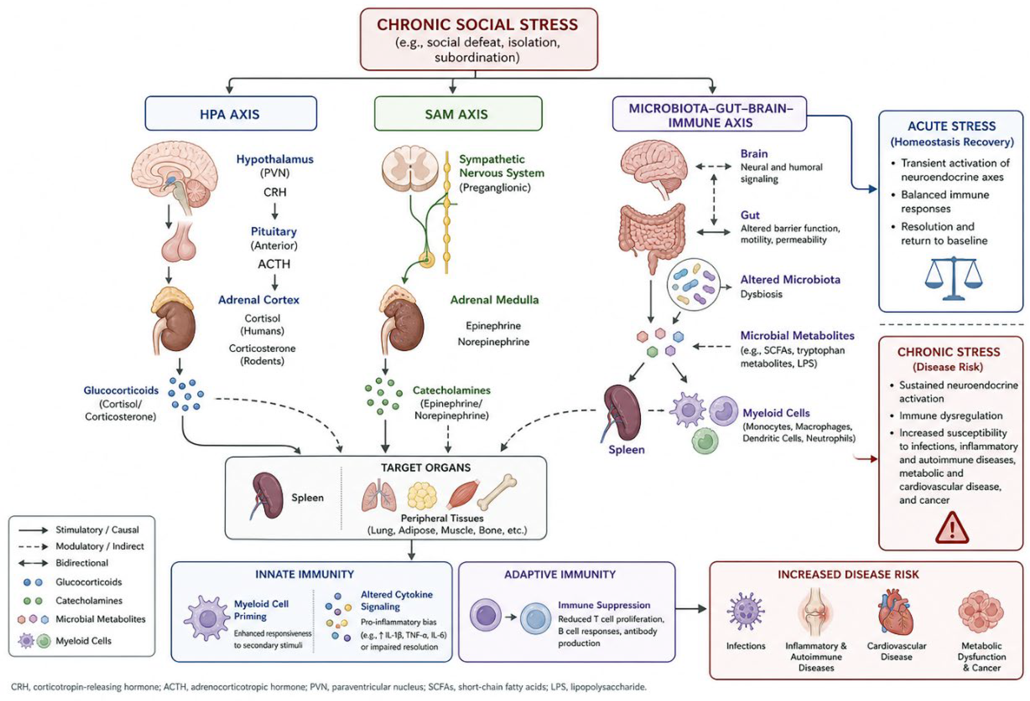
Figure 2.
SNS innervation of lymphoid tissues. Sympathetic nervous system (SNS) activation by brain/SCN/stress signals leads to norepinephrine release in primary and secondary lymphoid organs. In bone marrow, β3-adrenergic signaling on stromal cells lowers CXCL12 and promotes HSC/HSPC egress and myelopoiesis. In the thymus, adrenergic fibers, and β/α1-adrenergic receptors on thymocytes and epithelial cells regulate thymocyte differentiation and maturation. In the spleen, splenic nerve input controls monocyte reservoir release, splenic myelopoiesis, and germinal center support. In lymph nodes, β2-adrenergic receptors on lymphocytes enhance CCR7/CXCR4-dependent retention and reduce lymphocyte egress. Blue/teal arrows and symbols indicate SNS-driven activation or promotion; red/orange arrows and symbols indicate inhibition or reduction of the indicated process.
Figure 2.
SNS innervation of lymphoid tissues. Sympathetic nervous system (SNS) activation by brain/SCN/stress signals leads to norepinephrine release in primary and secondary lymphoid organs. In bone marrow, β3-adrenergic signaling on stromal cells lowers CXCL12 and promotes HSC/HSPC egress and myelopoiesis. In the thymus, adrenergic fibers, and β/α1-adrenergic receptors on thymocytes and epithelial cells regulate thymocyte differentiation and maturation. In the spleen, splenic nerve input controls monocyte reservoir release, splenic myelopoiesis, and germinal center support. In lymph nodes, β2-adrenergic receptors on lymphocytes enhance CCR7/CXCR4-dependent retention and reduce lymphocyte egress. Blue/teal arrows and symbols indicate SNS-driven activation or promotion; red/orange arrows and symbols indicate inhibition or reduction of the indicated process.
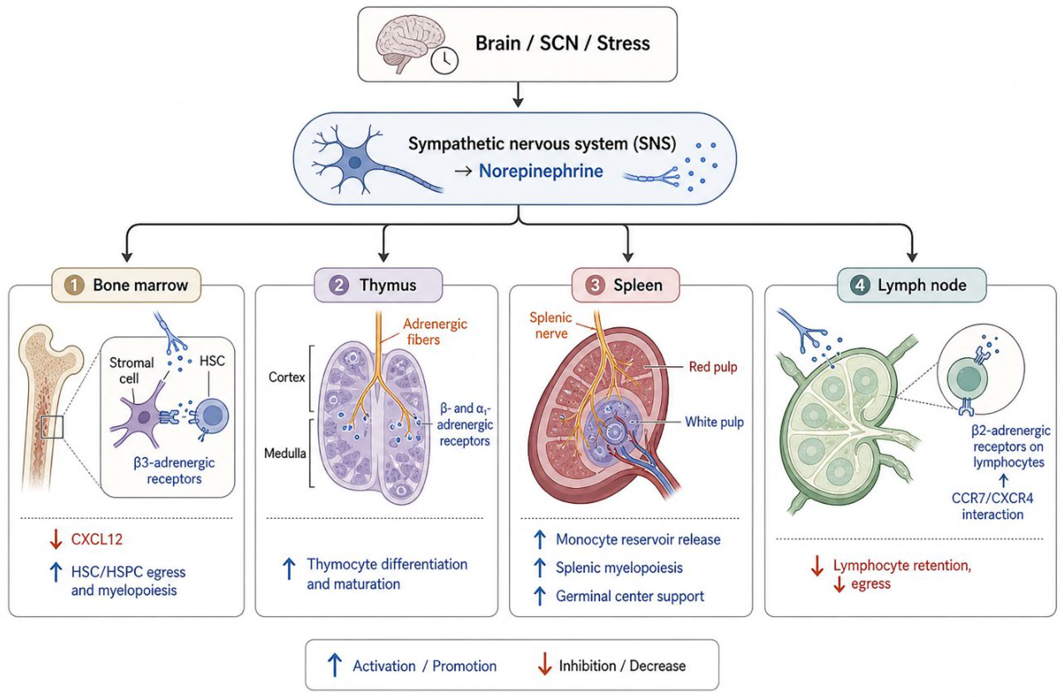
Figure 3.
Adrenergic receptor signaling in immune cells. Epinephrine and norepinephrine signal through distinct adrenergic receptor subtypes on immune cells, leading to cell type– and context-specific outcomes. β2-adrenergic receptors, broadly expressed on macrophages, dendritic cells, CD4 T cells, CD8 T cells, and B cells, couple to Gs to increase cAMP and activate PKA, which inhibits NF-κB/AP-1 signaling, promotes IL-10 production, and reduces IL-12 and TNFα in myeloid cells, shifts CD4 T cell responses away from Th1 toward Th17, dampens IFN-γ/TNFα secretion and cytolytic activity in CD8 T cells, and enhances IgG1 and IgE class switching and antibody production in B cells. α2-adrenergic receptors, expressed on macrophages and dendritic cells, couple to Gi to reduce cAMP and modulate p38 MAPK, supporting nitric oxide–dependent antimicrobial and pro-inflammatory responses at low catecholamine concentrations but exerting anti-inflammatory effects in other contexts by limiting myeloid accumulation and NF-κB-driven cytokine production. β1-adrenergic receptors, upregulated on terminally exhausted CD8 T cells in chronic viral infection and cancer, promote T cell exhaustion and loss of effector function in the presence of sustained catecholamine signaling, whereas β-blockade can restore CD8 T cell activity and improve responses to immunotherapy. Blue/teal symbols and arrows indicate activation or promotion; red/orange symbols and arrows indicate inhibition or suppression; symbols next to receptor icons denote Gs (stimulatory) versus Gi (inhibitory) G-protein coupling.
Figure 3.
Adrenergic receptor signaling in immune cells. Epinephrine and norepinephrine signal through distinct adrenergic receptor subtypes on immune cells, leading to cell type– and context-specific outcomes. β2-adrenergic receptors, broadly expressed on macrophages, dendritic cells, CD4 T cells, CD8 T cells, and B cells, couple to Gs to increase cAMP and activate PKA, which inhibits NF-κB/AP-1 signaling, promotes IL-10 production, and reduces IL-12 and TNFα in myeloid cells, shifts CD4 T cell responses away from Th1 toward Th17, dampens IFN-γ/TNFα secretion and cytolytic activity in CD8 T cells, and enhances IgG1 and IgE class switching and antibody production in B cells. α2-adrenergic receptors, expressed on macrophages and dendritic cells, couple to Gi to reduce cAMP and modulate p38 MAPK, supporting nitric oxide–dependent antimicrobial and pro-inflammatory responses at low catecholamine concentrations but exerting anti-inflammatory effects in other contexts by limiting myeloid accumulation and NF-κB-driven cytokine production. β1-adrenergic receptors, upregulated on terminally exhausted CD8 T cells in chronic viral infection and cancer, promote T cell exhaustion and loss of effector function in the presence of sustained catecholamine signaling, whereas β-blockade can restore CD8 T cell activity and improve responses to immunotherapy. Blue/teal symbols and arrows indicate activation or promotion; red/orange symbols and arrows indicate inhibition or suppression; symbols next to receptor icons denote Gs (stimulatory) versus Gi (inhibitory) G-protein coupling.
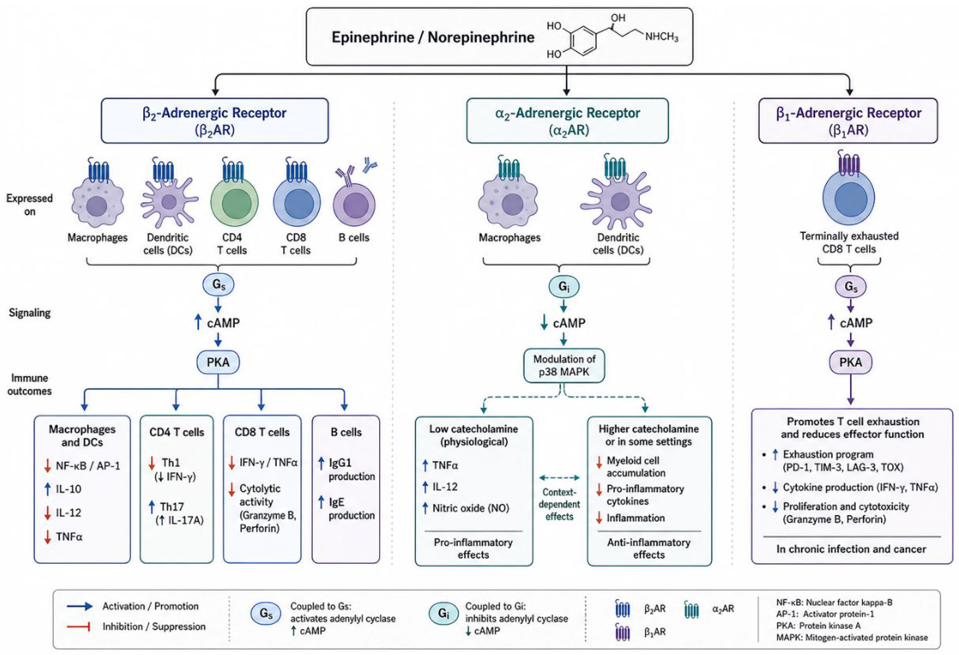
Figure 4.
SDR systemic effects. Chronic social disruption stress activates the HPA axis and sympathetic nervous system, driving linked changes in spleen/bone marrow, gut, and brain. In hematopoietic tissues, β-adrenergic signaling increases myelopoiesis, expands glucocorticoid-resistant CD11b+Ly6Chigh monocytes and neutrophils, and, together with translocation of commensal Lactobacillus, primes myeloid cells to produce high IL-1β, IL-6, TNFα, and IL-23 while hepcidin-mediated iron restriction impairs erythropoiesis. In the gut, SDR causes dysbiosis (reduced Lactobacillus), oxidative/pro-inflammatory epithelial responses, barrier thinning, and microbial/metabolite leakage. In the brain, IL-6-primed, IL-1β-producing monocytes enter stress-sensitive regions via ICAM-1/VCAM-1–upregulated vessels and interact with IL-1R1+ endothelium and microglia to generate neuroinflammation, anxiety-like behavior, social withdrawal, and cognitive deficits.
Figure 4.
SDR systemic effects. Chronic social disruption stress activates the HPA axis and sympathetic nervous system, driving linked changes in spleen/bone marrow, gut, and brain. In hematopoietic tissues, β-adrenergic signaling increases myelopoiesis, expands glucocorticoid-resistant CD11b+Ly6Chigh monocytes and neutrophils, and, together with translocation of commensal Lactobacillus, primes myeloid cells to produce high IL-1β, IL-6, TNFα, and IL-23 while hepcidin-mediated iron restriction impairs erythropoiesis. In the gut, SDR causes dysbiosis (reduced Lactobacillus), oxidative/pro-inflammatory epithelial responses, barrier thinning, and microbial/metabolite leakage. In the brain, IL-6-primed, IL-1β-producing monocytes enter stress-sensitive regions via ICAM-1/VCAM-1–upregulated vessels and interact with IL-1R1+ endothelium and microglia to generate neuroinflammation, anxiety-like behavior, social withdrawal, and cognitive deficits.
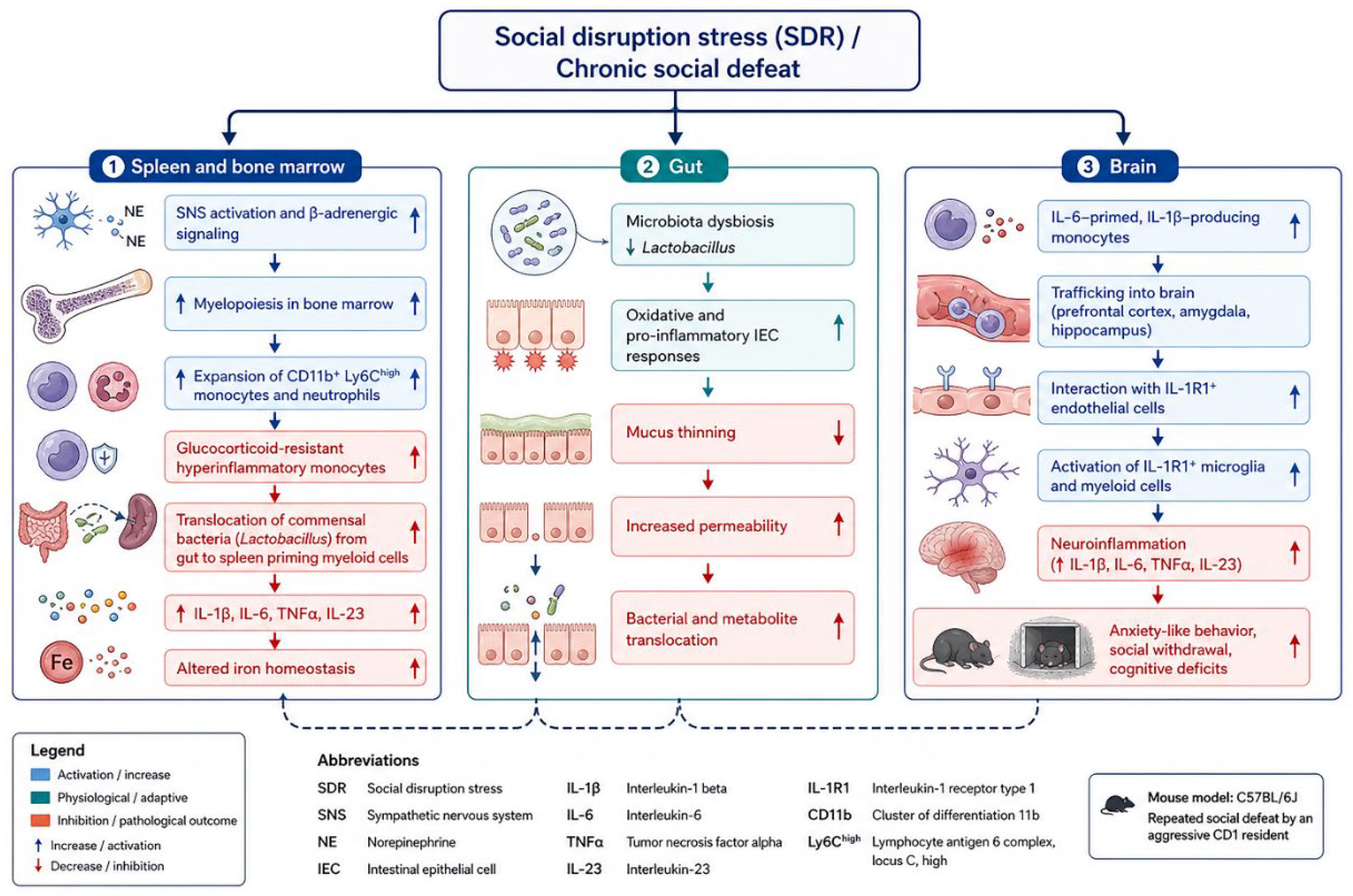
Figure 5.
Chronic social disruption stress (SDR) can enhance or worsen antiviral defenses depending on the pathogen and timing. Before influenza A virus infection, SDR increases dendritic cell immunogenicity and IAV-specific CD8+ T cell responses, boosting IFN-γ/IFN-α production and hastening viral clearance, including during memory responses. Before HSV-1 infection, SDR augments CD11b+ macrophage influx into cornea and trigeminal ganglia, elevates IFN-α and TNF-α, and reduces viral protein levels, consistent with enhanced innate viral control. In contrast, SDR prior to Theiler’s murine encephalomyelitis virus (TMEV) infection raises IL-6, increases CNS inflammation, sickness behavior, and viral titers, and impairs virus-specific CD4+/CD8+ T cells in the CNS, whereas SDR concurrent with infection modestly reduces disease severity. Blue/teal elements indicate enhanced antiviral responses; red/orange elements indicate exacerbated pathology or impaired adaptive immunity.
Figure 5.
Chronic social disruption stress (SDR) can enhance or worsen antiviral defenses depending on the pathogen and timing. Before influenza A virus infection, SDR increases dendritic cell immunogenicity and IAV-specific CD8+ T cell responses, boosting IFN-γ/IFN-α production and hastening viral clearance, including during memory responses. Before HSV-1 infection, SDR augments CD11b+ macrophage influx into cornea and trigeminal ganglia, elevates IFN-α and TNF-α, and reduces viral protein levels, consistent with enhanced innate viral control. In contrast, SDR prior to Theiler’s murine encephalomyelitis virus (TMEV) infection raises IL-6, increases CNS inflammation, sickness behavior, and viral titers, and impairs virus-specific CD4+/CD8+ T cells in the CNS, whereas SDR concurrent with infection modestly reduces disease severity. Blue/teal elements indicate enhanced antiviral responses; red/orange elements indicate exacerbated pathology or impaired adaptive immunity.
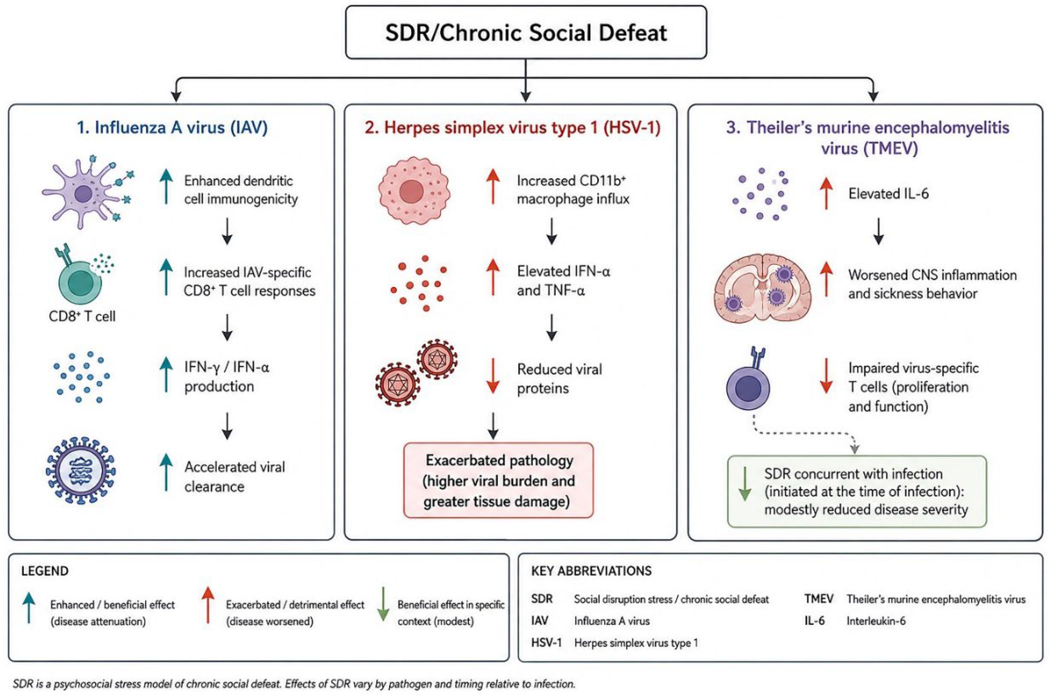
Disclaimer/Publisher’s Note: The statements, opinions and data contained in all publications are solely those of the individual author(s) and contributor(s) and not of MDPI and/or the editor(s). MDPI and/or the editor(s) disclaim responsibility for any injury to people or property resulting from any ideas, methods, instructions or products referred to in the content. |
© 2026 by the authors. Licensee MDPI, Basel, Switzerland. This article is an open access article distributed under the terms and conditions of the Creative Commons Attribution (CC BY) license.
Copyright: This open access article is published under a Creative Commons CC BY 4.0 license, which permit the free download, distribution, and reuse, provided that the author and preprint are cited in any reuse.


