
Submitted:
20 June 2026
Posted:
24 June 2026
You are already at the latest version
Abstract
Quercetin is a promising nutraceutical flavonoid, but its application in aqueous formulations is limited by poor water solubility and chemical instability. In this study, renatured β-1,3-1,6-glucan nanoparticles (r-glucan NPs) were evaluated as a food-compatible carrier for quercetin and compared with β-cyclodextrin (β-CD). UV–visible absorption and circular dichroism analyses indicated that quercetin was incorporated into hydrophobic, chiral, cavity-rich domains of r-glucan NPs, where it formed a partially stacked arrangement. r-Glucan NPs showed a higher quercetin loading than β-CD and markedly improved the apparent aqueous solubility/dispersibility of quercetin, reaching 1,620 ± 15.3 µM compared with 347 ± 16.4 µM for the β-CD complex and 7.11 ± 3.97 µM for free quercetin. Although encapsulation reduced the apparent radical-scavenging activity at an equal quercetin concentration, r-glucan NPs enabled a much higher formulation-level ORAC value in water. r-Glucan NPs also showed slower apparent loss of carrier-associated quercetin and improved photostability and pH-dependent stability. In Caco-2 cells, quercetin-loaded r-glucan NPs exhibited cellular antioxidant activity without additional carrier-derived cytotoxicity. These results suggest that r-glucan NPs are promising polysaccharide-based carriers for water-dispersible and stable quercetin delivery.
Keywords:
quercetin
; renatured β-1
; 3-1
; 6-glucan nanoparticles
; aqueous dispersibility
; antioxidant activity
; photostability
; pH-dependent stability
; Caco-2 cells
; nutraceutical delivery
1. Introduction
Quercetin is a representative dietary flavonoid widely distributed in fruits, vegetables, and grains, and has attracted considerable interest as a functional food and nutraceutical ingredient because of its antioxidant, anti-inflammatory, and other health-promoting properties [1,2]. These biological activities make quercetin a promising candidate for use in beverages, supplements, and health-related formulations. However, its practical application in aqueous and oral formulations remains limited by its extremely low water solubility and poor stability under gastrointestinal conditions. The aqueous solubility of quercetin has been reported to be approximately 0.01 mg mL-1 [3], which severely restricts its homogeneous dispersion in water-based products. In addition, quercetin is susceptible to degradation and/or chemical transformation under gastrointestinal conditions, resulting in low oral bioavailability, reportedly less than 2% [4]. Therefore, an effective delivery system that improves the aqueous dispersibility, stability, and functional availability of quercetin is required for its broader use as a nutraceutical ingredient.
Various formulation strategies have been investigated to improve the poor water compatibility of quercetin, including cyclodextrin-based inclusion complexes, emulsions, microemulsions, liposomes, and polymeric nanoparticles [5,6,7,8,9]. Among these systems, β-cyclodextrin (β-CD) is one of the most widely studied carriers because its hydrophobic cavity can partially accommodate hydrophobic guest molecules and improve their apparent aqueous solubility through host–guest complexation [10]. However, the capacity of native β-CD to solubilize quercetin is limited, and chemical modification of cyclodextrins is often required to further enhance complexation efficiency or formulation performance [3,11]. Although such modifications can improve binding affinity and aqueous solubility, they may also increase structural complexity and raise additional considerations for food and nutraceutical applications, where simple composition, safety, and regulatory acceptability are important. Emulsion-based systems can also improve quercetin dispersion [5,11,12], but their practical use may be constrained by formulation complexity, physical instability, and the need for surfactants or oil phases. Thus, there remains a need for food-compatible carrier materials that can maintain quercetin in aqueous media while preserving its functional activity.
Naturally derived polysaccharides are attractive candidates for this purpose because of their biocompatibility, structural diversity, and potential applicability in food-related systems [5,13,14]. β-1,3-1,6-Glucan is a particularly interesting polysaccharide, as it is derived from natural sources such as yeast, fungi, and seaweed and is associated with beneficial physiological functions, including immune-modulating activity [15]. Conventional glucan-based carrier systems, however, have often relied on top-down particulate approaches, in which bioactive compounds are adsorbed onto micronized glucan particles [16,17]. Such systems can suffer from sedimentation in water and relatively rapid release of loaded compounds, limiting their usefulness as stable aqueous delivery platforms for poorly water-soluble nutraceutical ingredients.
To address these limitations, we previously developed renatured β-1,3-1,6-glucan nanoparticles (r-glucan NPs) using a bottom-up self-assembly approach [18]. In this method, the native triple-helical structure of β-1,3-1,6-glucan is first dissociated under alkaline conditions and then partially renatured during neutralization to form nanoparticles. During this renaturation process, hydrophobic cavity-like microenvironments are generated within the glucan assemblies, enabling the incorporation of hydrophobic guest molecules. Importantly, these nanoparticles are formed without chemical crosslinkers and have been shown to exhibit high aqueous dispersibility and sustained-retention/release properties [18,19,20]. These characteristics suggest that r-glucan NPs may serve as a simple, polysaccharide-based, and food-compatible delivery platform for poorly water-soluble functional compounds. Thus, in the present study, we investigated r-glucan NPs as a delivery carrier for quercetin and evaluated their effects on the aqueous dispersibility, stability, release behavior, and antioxidant performance of quercetin. β-CD was used as a representative benchmark carrier to clarify the advantages and limitations of the r-glucan NP system. The encapsulation state of quercetin was characterized by UV–visible absorption spectroscopy, circular dichroism spectroscopy, and dynamic light scattering. We further compared the apparent aqueous dispersibility, ORAC-based antioxidant capacity, photostability, pH-dependent stability, cytocompatibility, and cellular antioxidant activity of free quercetin, quercetin/β-CD complexes, and quercetin-loaded r-glucan NPs. Through these evaluations, this study aims to demonstrate the potential of r-glucan NPs as a food-compatible delivery system capable of improving the practical functionality of quercetin in aqueous nutraceutical formulations.
2. Materials and Methods
2.1. Materials
Quercetin, acetone, methanol, 99.5% ethanol, ethyl acetate, dipotassium hydrogen phosphate, potassium dihydrogen phosphate, acetic acid, hydrochloric acid, fluorescein, Trolox, 2,2′-azobis(2-amidinopropane) dihydrochloride (AAPH), sodium hydroxide, phenol, sulfuric acid, 3-(4,5-Dimethyl-2-thiazolyl)-2,5-diphenyl-2H-tetrazolium bromide (MTT), Hank’s balanced salt solution (HBSS) (-) without phenol red, Dulbecco’s modified Eagle’s medium (DMEM), fetal bovine serum (FBS), penicillin–streptomycin, phosphate-buffered saline (PBS), and trypsin–EDTA were purchased from Fujifilm Wako Chemicals Co., Ltd. (Osaka, Japan). β-Cyclodextrin (β-CD) was purchased from Tokyo Chemical Industry Co., Ltd. (Tokyo, Japan). 2′,7′-Dichlorodihydrofluorescein diacetate (DCFH-DA) was purchased from Sigma-Aldrich (St. Louis, MO, USA). All reagents were used without further purification unless otherwise stated.
2.2. β-1,3-1,6-glucan
β-1,3-1,6-Glucan was obtained by fermentation of the K-1 strain of Aureobasidium pullulans according to a previously reported method [18]. The A. pullulans K-1 strain was kindly provided by Prof. T. Suzuki. The molecular-weight parameters of the obtained β-1,3-1,6-glucan were estimated to be Mw = 1,250,000, Mn = 58,800, and Mw/Mn = 21.2, as determined by gel permeation chromatography (GPC) [18,19].
2.3. Preparation of Renatured β-1,3-1,6-glucan Nanoparticles (r-glucan NPs)
Renatured β-1,3-1,6-glucan nanoparticles (r-glucan NPs) were prepared according to a previously reported method [18]. β-1,3-1,6-Glucan was dissolved in 1.0 M aqueous NaOH to dissociate its native triple-helical structure into single chains. The insoluble fraction was removed by centrifugation at 16,000 × g for 20 min at 25 °C. The resulting alkaline solution was neutralized by adding an equivalent volume of 1.0 M aqueous HCl, and the mixture was stirred at room temperature for 1 day to allow renaturation and nanoparticle formation. The obtained suspension was dialyzed against distilled water for 1 day using a dialysis membrane with a molecular weight cut-off of 14,000 Da. Gel-like glucan aggregates formed during neutralization and dialysis were then removed by centrifugation at 16,000 × g for 20 min at 25 °C, yielding a stock dispersion of r-glucan NPs. The concentration of r-glucan NPs in the stock dispersion was determined as the glucose unit concentration using the phenol–sulfuric acid method [21].
2.4. Preparation of Quercetin-Loaded r-glucan NPs
An aqueous dispersion of r-glucan NPs was prepared at a glucose unit concentration of 1.0 mM in a volume of 900 µL. To this dispersion, 100 µL of a 50 mM quercetin solution in acetone was added, giving a final acetone content of 10 vol%. The mixture was vortexed and then incubated at room temperature for 3 h under light-protected conditions, with vortex mixing every 60 min. After incubation, the dispersion was centrifuged at 11,000 × g for 10 min at 25 °C to remove precipitated unencapsulated quercetin. The supernatant was collected, and 800 µL of the supernatant was freeze-dried. Before each experiment, the freeze-dried sample was redispersed in 800 µL of distilled water and centrifuged again at 11,000 × g for 10 min at 25 °C to remove quercetin that was not stably retained in the r-glucan NP system after removal of water and acetone. The resulting supernatant was used as the quercetin-loaded r-glucan NP sample [19].
2.5. Quantification of Quercetin Encapsulated in r-glucan NPs
The amount of quercetin encapsulated in r-glucan NPs was quantified using an alkaline dissociation method [18]. The freeze-dried quercetin-loaded r-glucan NP powder was redispersed in distilled water at half of the volume used before freeze-drying. The dispersion was mixed with an equal volume of 2.0 M aqueous NaOH and allowed to stand for 3 h at room temperature under light-protected conditions to dissociate the r-glucan NP structure and release encapsulated quercetin. The UV–visible absorption spectrum was then recorded using a UV-1800 spectrophotometer (Shimadzu, Kyoto, Japan). The quercetin concentration was calculated from a calibration curve prepared using quercetin in 1.0 M aqueous NaOH. Measurements were performed in triplicate. Quercetin loading was expressed as the mole fraction of quercetin, where the molar amount of r-glucan NPs was defined on the basis of glucose units quantified by the phenol–sulfuric acid method.
2.6. Preparation of the Quercetin/β-CD Complex
The quercetin/β-CD complex was prepared by a solid–liquid extraction method assisted by ultrasonic treatment. β-CD was dissolved in distilled water, and the total glucose unit concentration of the β-CD solution was determined using the phenol–sulfuric acid method. The solution was then diluted with distilled water to a glucose unit concentration of 1.0 mM. An excess amount of quercetin was added to the β-CD solution until undissolved quercetin remained. The mixture was vortexed for 10 s and sonicated for 1 h under ice-cooling using an ultrasonic homogenizer (UH-50, SMT Co., Ltd., Tokyo, Japan). After sonication, the suspension was centrifuged at 11,000 × g for 10 min to remove uncomplexed quercetin. The resulting supernatant was collected and freeze-dried using a freeze dryer (FDU-1200, EYELA, Tokyo, Japan). The obtained freeze-dried powder was used as the quercetin/β-CD complex in subsequent experiments.
2.7. Quantification of Quercetin Incorporated into β-CD
The amount of quercetin incorporated into β-CD was quantified by UV–visible absorption spectroscopy using an ethanol-based calibration curve as indicated in the previous report with minor modification [22,23]. To determine the total amount of quercetin present in the freeze-dried quercetin/β-CD sample, the powder prepared at a total glucose unit concentration of 1.0 mM was dissolved in ethanol. The UV–visible absorption spectrum was measured using a 1.0 cm quartz cell, and the total amount of quercetin was calculated from the calibration curve. Separately, the amount of water-insoluble, uncomplexed quercetin was determined. The freeze-dried quercetin/β-CD sample was redissolved in distilled water and centrifuged at 11,000 × g for 10 min. After removal of the supernatant, the remaining precipitate was dissolved in ethanol. The UV–visible absorption spectrum was measured, and the amount of uncomplexed quercetin was calculated from the ethanol calibration curve. The amount of quercetin incorporated into β-CD was calculated by subtracting the amount of water-insoluble quercetin from the total amount of quercetin present in the freeze-dried sample. For β-CD, the amount of incorporated quercetin was also expressed as the mole fraction of quercetin, with the β-CD amount expressed as glucose-unit equivalents based on the total glucose-unit concentration determined by the phenol–sulfuric acid method, in the same manner as for r-glucan NPs.
2.8. UV–Visible Absorption and Circular Dichroism Spectroscopy
UV–visible absorption and circular dichroism (CD) spectra of free quercetin, the quercetin/β-CD complex, and quercetin-loaded r-glucan NPs were measured to evaluate the encapsulation state of quercetin in each carrier system. UV–visible absorption spectra were recorded using a UV-1800 spectrophotometer (Shimadzu, Kyoto, Japan) with a quartz cell of 1.0 mm path length. CD spectra were recorded using a JASCO J-820 spectropolarimeter (JASCO, Tokyo, Japan) equipped with a temperature controller and a quartz cell of 1.0 cm path length. All measurements were performed at 25 °C. The concentration of free quercetin was adjusted to 3.47 µM, whereas those of the quercetin/β-CD complex and quercetin-loaded r-glucan NPs were adjusted to 150 µM on a quercetin basis. Spectra were recorded over the wavelength range of 230–500 nm. For CD measurements, each spectrum was obtained by averaging three scans. Distilled water was used as the blank for baseline correction.
2.9. Dynamic Light Scattering Measurements
Dynamic light scattering (DLS) measurements were performed to evaluate the hydrodynamic size distributions of blank r-glucan NPs, the quercetin/β-CD complex, and quercetin-loaded r-glucan NPs. Measurements were carried out using a Zetasizer Nano ZS instrument (Malvern Panalytical Ltd., Worcestershire, UK) equipped with a temperature controller. Samples were dispersed in distilled water and measured at 25 °C using disposable polystyrene cuvettes with a 1 cm path length. Before measurement, the samples were equilibrated at 25 °C for 2 min. The supernatants obtained after centrifugation at 11,000 × g for 10 min at 25 °C were used for DLS measurements without further filtration. The concentrations of blank r-glucan NPs and quercetin-loaded r-glucan NPs were adjusted to 4.0% (w/v) on a glucan basis. The quercetin/β-CD complex was measured as a saturated aqueous solution prepared at a β-CD concentration of 1.0% (w/v). The hydrodynamic diameter and polydispersity index (PDI) were obtained from cumulants analysis using the instrument software. Size distributions are presented as number-weighted hydrodynamic diameter distributions. Each sample was measured 30 times, and the results are presented as the mean ± SD of three independently prepared samples.
2.10. Apparent Aqueous Solubility/Dispersibility Test
The apparent aqueous solubility or dispersibility of free quercetin, the quercetin/β-CD complex, and quercetin-loaded r-glucan NPs was evaluated by a stepwise addition method. Distilled water (1.0 mL) was added to a 1.5 mL plastic tube, followed by the addition of 10 mg of each sample. The mixture was vortexed to dissolve or disperse the sample. When the sample was completely dissolved or dispersed, an additional 10 mg portion was added, and the same procedure was repeated until undissolved material remained. The resulting mixture was centrifuged at 11,000 × g for 10 min at 25 °C, and the supernatant was collected. The collected supernatant was centrifuged again under the same conditions to remove any remaining insoluble material. For free quercetin and the quercetin/β-CD complex, a 100 µL aliquot of the final supernatant was mixed with 900 µL of ethanol, and the mixture was vortexed. The UV–visible absorption spectrum was recorded, and the apparent saturated quercetin concentration was determined from the absorbance at the absorption maximum of quercetin using a calibration curve prepared in ethanol. For quercetin-loaded r-glucan NPs, a 100 µL aliquot of the final supernatant was mixed with 100 µL of 2.0 M aqueous NaOH. The mixture was allowed to stand for 3 h at room temperature under light-protected conditions to dissociate the r-glucan NP structure and release encapsulated quercetin. After incubation, the UV–visible absorption spectrum was recorded. The apparent saturated quercetin concentration was determined using a calibration curve prepared in 1.0 M aqueous NaOH.
2.11. ORAC Assay
The oxygen radical absorbance capacity (ORAC) assay was conducted according to the hydrophilic ORAC method reported by Watanabe et al. with minor modifications [24]. ORAC values were determined using Trolox as the standard antioxidant.
First, the appropriate measurement concentration range of each sample was determined. Then, net area under the curve (net AUC) values within the appropriate concentration range were used to calculate the ORAC values. Samples were diluted with assay buffer consisting of 75 mM phosphate buffer (pH 7.4). Trolox standard solutions were prepared at concentrations of 6.25, 12.5, 25, and 50 µM in the same assay buffer. A 35 µL aliquot of the diluted sample solution or Trolox standard solution was added to each well of a 96-well plate. Subsequently, 115 µL of fluorescein solution (110.7 nM) and 50 µL of AAPH solution (31.7 mM) were added to each well. The plate was covered with a plate seal to prevent evaporation and incubated at 37 °C. Fluorescence intensity was monitored every 2 min for 90 min using a microplate reader (Thermo Fisher Scientific Inc., Waltham, MA, USA) at an excitation wavelength of 485 nm and an emission wavelength of 528 nm. The AUC of the fluorescence decay curve from 8 to 90 min after addition of AAPH was calculated for each well. The net AUC was calculated by subtracting the AUC of the blank from that of the sample or Trolox standard. A calibration curve was prepared from the net AUC values of the Trolox standard solutions, and the antioxidant capacity of each sample was expressed as Trolox equivalents. Carrier-only controls of blank r-glucan NPs and β-CD were also measured at the same carrier concentrations as those used in the corresponding quercetin inclusion complexes.
2.12. Apparent Release/Retention Assay of Quercetin from Carrier Systems
The apparent release or retention behavior of quercetin from the quercetin/β-CD complex and quercetin-loaded r-glucan NPs was evaluated in aqueous solution containing 1% ethanol. The quercetin concentration of each sample was adjusted to 25 µM on a quercetin basis. Each sample solution was added to a 96-well glass plate at 250 µL per well and incubated at 37 °C under light-protected conditions. UV–visible absorption spectra were recorded at 1 h intervals using a Varioskan LUX microplate reader (Thermo Fisher Scientific Inc., Waltham, MA, USA). The residual fraction of carrier-associated quercetin was calculated from the absorbance at the absorption maximum of each sample at each time point relative to the initial absorbance at 0 h. The apparent release/retention behavior was analyzed by plotting the residual fraction of carrier-associated quercetin as a function of incubation time. When appropriate, the apparent rate constant for the decrease in carrier-associated quercetin was determined from the slope of the linear plot of ln(At/A0) versus incubation time, where At and A0 represent the absorbance values of quercetin at time t and 0 h, respectively. All measurements were performed in triplicate, and the data are presented as mean ± SD.
2.13. Photodegradation Assay of Free and Encapsulated Quercetin
The photostability of free quercetin, the quercetin/β-CD complex, and quercetin-loaded r-glucan NPs was evaluated under fluorescent light irradiation. The quercetin concentration of each sample was adjusted to 25 µM on a quercetin basis in aqueous solution containing 1% ethanol. Each sample solution was added to a 96-well plate at 300 µL per well. The plate was placed 10 cm below a fluorescent lamp (Panasonic SQ655 equipped with a 26 W fluorescent lamp) and irradiated at room temperature (25 °C). UV–visible absorption spectra were recorded at 1 h intervals over 12 h using a Varioskan LUX microplate reader (Thermo Fisher Scientific Inc., Waltham, MA, USA). The residual fraction of quercetin was calculated from the absorbance at the absorption maximum of each sample at each time point relative to the initial absorbance at 0 h. The apparent photodegradation rate constant was determined from the slope of the linear plot of ln(At/A0) versus irradiation time, where At and A0 represent the absorbance values of quercetin at time t and 0 h, respectively. All measurements were performed in triplicate, and the data are presented as mean ± SD.
2.14. pH-Dependent Stability Assay of Free and Encapsulated Quercetin
The pH-dependent stability of free quercetin, the quercetin/β-CD complex, and quercetin-loaded r-glucan NPs was evaluated under acidic and intestinal pH conditions. Solutions at pH 1.2 and 6.8 were prepared to mimic gastric and intestinal environments, respectively. The pH 1.2 solution was prepared by dissolving NaCl (1.0 g) in approximately 400 mL of distilled water, adjusting the pH with HCl, and then diluting the solution to 500 mL. The pH 6.8 solution was prepared by dissolving KH2PO4 (3.40 g) in approximately 400 mL of distilled water, adjusting the pH with NaOH, and then diluting the solution to 500 mL. The pH values of the samples were measured using a LAQUA twin compact pH meter (HORIBA, Kyoto, Japan), calibrated with standard buffer solutions before use. Each sample was diluted to a final quercetin concentration of 25 µM on a quercetin basis. The final solvent composition was pH solution/water/ethanol = 89/10/1 (v/v/v). Each sample solution was added to a 96-well glass plate at 250 µL per well. To minimize evaporation during incubation at 37 °C, each well was overlaid with liquid paraffin. UV–visible absorption spectra were recorded at 1 h intervals over 12 h using a Varioskan LUX microplate reader (Thermo Fisher Scientific Inc., Waltham, MA, USA). The residual fraction of quercetin was calculated from the absorbance at the absorption maximum of each sample at each time point relative to the initial absorbance at 0 h. The apparent degradation rate constant was determined from the slope of the linear plot of ln(At/A0) versus incubation time, where At and A0 represent the absorbance values of quercetin at time t and 0 h, respectively. All measurements were performed in triplicate, and the data are presented as mean ± SD.
2.15. Evaluation of AAPH-Induced Degradation of Glucan Chains by GPC
To evaluate whether peroxyl radicals cleave the glucan chains constituting r-glucan NPs, AAPH-induced degradation of r-glucan was analyzed by GPC. An r-glucan NP stock dispersion was prepared in distilled water, and its concentration was determined as the glucose unit concentration using the phenol–sulfuric acid method. The obtained stock dispersion had a glucose unit concentration of 95 mM and was diluted with distilled water to 40 mM. The r-glucan solution (40 mM, 25 mL) was transferred to a 50 mL plastic tube. Separately, a 40 mM AAPH solution was freshly prepared using distilled water prewarmed to 37 °C. The AAPH solution (25 mL) was added to the r-glucan solution and mixed, giving final concentrations of 20 mM r-glucan, on a glucose unit basis, and 20 mM AAPH. The mixture was incubated at 37 °C to generate peroxyl radicals from AAPH. At predetermined time points of 3, 5.5, 23, and 48 h, 5 mL aliquots were collected from the reaction mixture and immediately frozen to stop further reaction. After all samples were collected, the frozen aliquots were lyophilized. Each freeze-dried sample was redissolved in 5 mL of 0.7 M aqueous NaOH, and the molecular-weight (Mw) of the glucan chains was analyzed by GPC as reported previously [18].
2.16. Cell Culture
Caco-2 cells were obtained from ATCC and cultured in DMEM supplemented with 10% FBS and 1% penicillin–streptomycin. Cells were maintained at 37 °C in a humidified atmosphere containing 5% CO2. The culture medium was replaced every 2–3 days, and the cells were subcultured before reaching full confluence using trypsin–EDTA.
2.17. Cytotoxicity Assay in Caco-2 Cells
The cytotoxicity of free quercetin, the quercetin/β-CD complex, and quercetin-loaded r-glucan NPs toward Caco-2 cells was evaluated using an MTT assay. Caco-2 cells were seeded in 96-well plates at a density of 5.0 × 10³ cells per well and cultured for 24 h before sample treatment. Free quercetin was added as a DMSO stock solution, whereas the quercetin/β-CD complex and quercetin-loaded r-glucan NPs were added as aqueous solutions. The final quercetin concentrations were adjusted to the indicated concentrations. The final DMSO concentration in the culture medium was kept constant at 0.1% in all relevant wells, including the vehicle control. After exposure to each sample for 24 h at 37 °C under 5% CO₂, cell viability was evaluated according to the manufacturer’s instructions for the assay reagent. Absorbance was measured at 535 nm using a microplate reader. Cell viability was calculated relative to untreated control cells using the following equation (1):
Cell viability (%) = [(Asample – Ablank)/(Acontrol – Ablank)] × 100
where Asample, Acontrol, and Ablank represent the absorbance values of sample-treated wells, untreated control wells, and blank wells without cells, respectively. Measurements were performed in triplicate, and the data are presented as mean ± SD.
2.18. Cellular Antioxidant Activity (CAA) Assay
Cellular antioxidant activity (CAA) was evaluated in Caco-2 cells using an AAPH-induced oxidative stress model. Caco-2 cells were seeded in black 96-well plates at a density of 5.0 × 10⁴ cells per well in 100 µL of culture medium and precultured for 24 h at 37 °C under a humidified atmosphere containing 5% CO₂. After preculture, the medium was removed, and the cells were washed once with PBS. Then, 100 µL of serum-free DMEM containing 60 µM DCFH-DA and each sample, namely free quercetin, the quercetin/β-CD complex, or quercetin-loaded r-glucan NPs, was added to each well at the indicated quercetin concentrations. The cells were incubated for 20 min at 37 °C. After incubation, the reaction solution was removed, and the cells were washed again with PBS. Oxidative stress was induced by adding 100 µL of HBSS containing 500 µM AAPH to each well. For blank wells, HBSS without AAPH was added instead. Fluorescence intensity was immediately monitored using a fluorescence microplate reader at an excitation wavelength of 485 nm and an emission wavelength of 535 nm every 5 min for 90 min at 37 °C. The area under the fluorescence intensity–time curve (AUC) was calculated, and cellular antioxidant activity was evaluated using the following equation (2):
where AUCsample is the area under the fluorescence curve of cells treated with each quercetin formulation in the presence of AAPH, AUCAAPH control is that of cells treated with AAPH in the absence of quercetin, and AUCblank is that of cells treated with HBSS without AAPH. Measurements were performed in triplicate, and the data are presented as mean ± SD.
CAA (%) = [1 – (AUCsample – AUCblank)/(AUCAAPH control – AUCblank)] × 100
2.19. Statistical Analysis
All experiments were performed in triplicate, and data are presented as mean ± SD. Mean values and standard deviations were calculated using Microsoft Excel. Statistical significance between two groups was evaluated using pairwise Student’s t-tests in Microsoft Excel. A value of p < 0.05 was considered statistically significant.
3. Results and discussion
3.1. Preparation and Characterization of Quercetin-Loaded r-glucan NPs
Quercetin-loaded r-glucan NPs were prepared by adding an acetone stock solution of quercetin to an aqueous dispersion of r-glucan NPs at a final acetone content of 10 vol%. Under these conditions, poorly water-soluble quercetin was expected to partition into hydrophobic cavity-like microenvironments formed within the r-glucan NPs. After incubation, precipitated unencapsulated quercetin was removed by centrifugation, and the supernatant was freeze-dried to remove water and acetone. The dried material was then redispersed in distilled water and centrifuged again to remove quercetin that was solubilized only in the initial acetone-containing medium but was not stably retained after acetone removal. Therefore, the quercetin detected in the final aqueous dispersion was regarded as the fraction stably associated with r-glucan NPs.
The amount of quercetin incorporated into r-glucan NPs was quantified after alkaline dissociation of the glucan nanoparticle structure, which released the encapsulated quercetin into solution. For comparison, the quercetin/β-CD complex was prepared as a representative low-molecular-weight host system, and the incorporated quercetin amount was evaluated from the difference between the total quercetin content and the water-insoluble fraction after redispersion. The mole fraction of quercetin was 2.42 ± 0.41 mol% for quercetin-loaded r-glucan NPs. This value was approximately 3.1-fold higher than that for the quercetin/β-CD complex (0.77 ± 0.03 mol%). This result indicates that r-glucan NPs provided a higher effective incorporation capacity for quercetin than β-CD on a glucose-unit basis. The higher loading capacity of r-glucan NPs is likely attributable to their polymeric and supramolecular architecture, in which multiple hydrophobic cavity-like regions are generated during glucan renaturation, providing broader and more flexible microenvironments than the discrete cavity of β-CD.
The encapsulation state of quercetin was first examined by UV–visible absorption spectroscopy (Figure 1a). Free quercetin showed an absorption maximum at 368 nm in the long-wavelength absorption band, while the quercetin/β-CD complex exhibited an almost identical absorption maximum at 367 nm. This negligible difference suggests that β-CD complexation did not substantially alter the electronic environment of quercetin. In contrast, quercetin-loaded r-glucan NPs showed a clearer bathochromic shift of the absorption maximum to 372 nm. This red shift suggests that the electronic state of quercetin was more substantially altered in the r-glucan NP system than in the β-CD system. A similar bathochromic shift was previously observed for curcumin incorporated into r-glucan NPs [18].
Further information on the encapsulation mode was obtained from CD spectroscopy (Figure 1b). Free quercetin showed no appreciable CD signal, as expected for an achiral chromophore in solution. The quercetin/β-CD complex exhibited an induced CD (ICD) signal mainly around 270 nm, whereas no clear ICD signal was observed in the long-wavelength absorption region around 370 nm. This spectral feature suggests that the chromophoric region responsible for the short-wavelength absorption band was affected by the chiral environment of β-CD, whereas the chromophore associated with the long-wavelength absorption band was not strongly influenced by the β-CD cavity. In other words, the absence of an ICD signal in the long-wavelength region suggests that this part of the quercetin molecule was not deeply included in the chiral β-CD cavity. This interpretation is also consistent with the UV–visible absorption spectrum, in which the absorption maximum of the quercetin/β-CD complex was almost unchanged compared with that of free quercetin. Therefore, β-CD is likely to interact with quercetin in a partial inclusion mode rather than fully surrounding the entire aromatic framework. In contrast, the CD spectrum of quercetin-loaded r-glucan NPs was markedly different from that of the quercetin/β-CD complex. Clear ICD signals were observed not only around 270 nm but also in the long-wavelength absorption region around 370 nm. This indicates that a broader portion of the quercetin chromophore was influenced by the chiral environment of the β-1,3-1,6-glucan framework. Notably, the ICD signal around 370 nm showed a split Cotton effect, suggesting exciton coupling between neighboring quercetin chromophores arranged in a chiral geometry. This spectral feature is consistent with the idea that quercetin molecules are not simply isolated in individual cavities, but are organized in a chiral and partially stacked arrangement within the hydrophobic cavity-rich domains of r-glucan NPs. A similar split Cotton effect was previously observed for curcumin incorporated into r-glucan NPs and was attributed to twisted stacking of the guest molecules along the glucan helices [18,20].
DLS measurements were then performed to evaluate how quercetin incorporation affected the dispersion state of the carrier systems (Figure S1). Blank r-glucan NPs showed an apparent hydrodynamic diameter of 11.5 ± 2.6 nm with a PDI of 0.80 ± 0.04. This result is consistent with the formation of small glucan-based nanoparticles, although the high PDI value indicates that the dispersion contained a broad distribution of glucan species. After quercetin loading, the apparent hydrodynamic diameter increased to 163 ± 113 nm, with a PDI of 0.393 ± 0.036. This size increase indicates that quercetin incorporation substantially changed the dispersion state of r-glucan NPs. A similar increase in particle size upon guest incorporation was observed in our previous study on curcumin-loaded r-glucan NPs, in which the hydrodynamic diameter increased from 17.8 ± 5.0 nm to 69.6 ± 31.6 nm after curcumin incorporation [20]. This previous result suggests that r-glucan NPs can expand or swell when hydrophobic guest molecules are incorporated into their cavity-rich domains. Therefore, the increase in hydrodynamic diameter observed in the present quercetin-loaded system is consistent with the guest-induced structural expansion of r-glucan NPs. However, the apparent hydrodynamic diameter of quercetin-loaded r-glucan NPs was larger than that previously observed for curcumin-loaded r-glucan NPs. Thus, the present quercetin-loaded system is unlikely to consist solely of individually swollen nanoparticles. Rather, quercetin incorporation may promote both swelling of the glucan nanoparticle domains and secondary association among quercetin-loaded domains. This interpretation is consistent with the UV–visible and CD results. The bathochromic shift in the absorption spectrum and the split Cotton effect in the CD spectrum suggest that quercetin molecules are located in hydrophobic, chiral, and partially stacked microenvironments. Such intermolecular organization of quercetin within r-glucan NP domains may strengthen hydrophobic interactions between guest-loaded domains and promote secondary association of nanoparticles. Importantly, however, the quercetin-loaded r-glucan NP sample remained dispersible in water after freeze-drying and redispersion, indicating that these larger assemblies were colloidally dispersible rather than forming macroscopic precipitates. In contrast, the quercetin/β-CD complex showed a much larger apparent hydrodynamic diameter of 850 ± 103 nm with a PDI of 0.457 ± 0.067 (Figure S1). This result indicates that the quercetin/β-CD complex did not exist only as isolated molecular inclusion complexes in water, but also formed larger secondary aggregates. This behavior may be related to the partial inclusion mode suggested by the CD spectrum. If only part of the quercetin molecule is included in the β-CD cavity, the remaining exposed aromatic region may still participate in intermolecular hydrophobic and π–π interactions, thereby driving aggregation of the quercetin/β-CD complexes.
3.2. Enhanced Apparent Aqueous Solubility/Dispersibility Of Quercetin
Based on the distinct encapsulation state of quercetin in r-glucan NPs described above, we next examined whether this carrier system improved the ability of quercetin to remain in the aqueous phase. Because quercetin-loaded r-glucan NPs are dispersed as carrier-associated nanoparticle assemblies rather than dissolved as individual quercetin molecules, the obtained values are described here as apparent aqueous solubility/dispersibility. The apparent aqueous solubility/dispersibility of quercetin was evaluated by measuring the quercetin concentration in the supernatant after centrifugation following stepwise addition of each formulation to water (Figure 2). Free quercetin was also examined as a reference because of its intrinsically poor water solubility.
As shown in Figure 2, the quercetin concentration in the aqueous supernatant increased with increasing amounts of both carrier systems. However, the quercetin-loaded r-glucan NP system showed a much steeper increase than the quercetin/β-CD complex and maintained substantially higher quercetin concentrations over the entire examined range. The apparent saturated quercetin concentration reached 1,620 ± 15.3 µM for quercetin-loaded r-glucan NPs, whereas that of the quercetin/β-CD complex was 347 ± 16.4 µM. In contrast, free quercetin showed an apparent aqueous solubility of only 7.11 ± 3.97 µM under the same conditions. Thus, encapsulation in r-glucan NPs increased the apparent aqueous solubility/dispersibility of quercetin by approximately 228-fold relative to free quercetin and by approximately 4.7-fold relative to the quercetin/β-CD complex. The superior performance of r-glucan NPs is consistent with the encapsulation model proposed above. The hydrophobic cavity-rich domains generated during glucan renaturation provide broader and more flexible microenvironments than the discrete cavity of β-CD, allowing a larger amount of quercetin to be retained in a water-dispersible state. In addition, the chiral and partially stacked arrangement of quercetin suggested by the CD spectra may contribute to the stabilization of quercetin within the r-glucan NP assemblies. By contrast, the lower aqueous quercetin concentration in the β-CD system is likely related to partial inclusion and secondary aggregation, as suggested by the spectroscopic and DLS analyses. These features probably limited the amount of quercetin that could be stably maintained in the aqueous phase. Representative photographs of the saturated aqueous supernatants are shown in Figure S2. The quercetin-loaded r-glucan NP sample remained visually homogeneous and showed a more intense yellow color than the free quercetin and quercetin/β-CD samples, indicating that a larger amount of quercetin was retained in the aqueous phase. These visual observations support the quantitative results shown in Figure 2 and suggest that the enhanced quercetin concentration in the r-glucan NP system reflects stable aqueous dispersion rather than transient solubilization.
These results demonstrate that r-glucan NPs are highly effective in improving the apparent aqueous solubility/dispersibility of quercetin. This property is particularly advantageous for the use of quercetin in aqueous food, nutraceutical, and health-related formulations, where homogeneous dispersion and high functional ingredient loading are essential.
3.3. Antioxidant Capacity of Quercetin after Inclusion
3.3.1. Antioxidant Activity at a Fixed Quercetin Concentration
The results described above demonstrated that r-glucan NPs markedly increased the amount of quercetin that could be maintained in the aqueous phase. However, increased aqueous dispersibility does not necessarily mean that the antioxidant activity of quercetin is fully preserved after encapsulation, because incorporation into carrier domains may restrict the accessibility of quercetin to radical species. Therefore, we first compared the antioxidant activity of free and encapsulated quercetin at the same quercetin concentration using the ORAC assay. This assay evaluates the ability of antioxidants to suppress fluorescence decay induced by AAPH-derived peroxyl radicals and is suitable for comparing radical-scavenging activity in aqueous systems.
Figure 3 shows the ORAC values of free quercetin, the quercetin/β-CD complex, and quercetin-loaded r-glucan NPs at a fixed quercetin concentration of 5.0 µM. Free quercetin showed the highest ORAC value among the three samples, indicating that molecularly available quercetin can efficiently scavenge peroxyl radicals in the aqueous assay medium. In contrast, the ORAC value of quercetin-loaded r-glucan NPs decreased to 51.5% of that of free quercetin. This decrease suggests that encapsulation partially restricted the direct accessibility of quercetin to peroxyl radicals. However, the quercetin/β-CD complex showed a much lower ORAC value, corresponding to only 16.5% of that of free quercetin and approximately 32% of that of quercetin-loaded r-glucan NPs. Thus, although encapsulation reduced the apparent radical-scavenging activity of quercetin in both carrier systems, r-glucan NPs retained the antioxidant activity of quercetin much more effectively than β-CD. These ORAC values should be interpreted as the apparent antioxidant capacities of the whole carrier–quercetin systems rather than as the intrinsic activity of quercetin alone. Under the corresponding carrier concentration used for the quercetin-loaded r-glucan NP sample, blank r-glucan NPs showed an ORAC value of 10.9 µmol TE L-1, whereas β-CD alone was below the detectable level. Therefore, in the r-glucan NP system, both carrier-associated quercetin and the glucan-based carrier matrix may contribute to the measured ORAC response. However, because the radical-responsive behavior of r-glucan NPs is examined later, the present comparison focuses on the total apparent antioxidant capacity of each quercetin formulation at the same quercetin concentration.
The difference between the two car”Ier ’ystems is consistent with the encapsulation states discussed above. In the quercetin/β-CD complex, partial inclusion and secondary aggregation may reduce the effective exposure of individual quercetin molecules to the aqueous phase and limit the accessibility of peroxyl radicals to the phenolic hydroxyl groups responsible for antioxidant activity. By contrast, quercetin-loaded r-glucan NPs retained more than half of the ORAC activity of free quercetin, despite the incorporation of quercetin into the glucan nanoparticles. This result suggests that a substantial fraction of quercetin remained accessible to peroxyl radicals or was gradually released from the r-glucan NPs during the assay. The ORAC value of quercetin-loaded r-glucan NPs may therefore reflect the combined contributions of several quercetin populations: quercetin released from the nanoparticles into the surrounding medium, quercetin retained within the hydrophobic cavity-rich domains but still accessible to radical species, and quercetin located near the surface or interfacial regions of the nanoparticles. These results indicate that, although encapsulation in r-glucan NPs partially reduces the intrinsic radical-scavenging activity of quercetin on an equal-concentration basis, it preserves a substantially larger fraction of antioxidant activity than β-CD complexation.
3.3.2. Superior maximal Antioxidant Capacity Enabled by Enhanced Aqueous Dispersibility
The fixed-concentration ORAC assay demonstrated that the apparent radical-scavenging activity of quercetin decreased after encapsulation. However, in aqueous food and nutraceutical formulations, practical antioxidant performance is determined not only by the activity per quercetin molecule but also by the maximum amount of quercetin that can be maintained in the aqueous phase. Therefore, the concentration-dependent ORAC values of free quercetin, the quercetin/β-CD complex, and quercetin-loaded r-glucan NPs were evaluated over the concentration ranges accessible for each formulation (Figure 4). In the low-concentration region, where all three formulations could be directly compared, free quercetin showed the highest ORAC value at a given quercetin concentration. Quercetin-loaded r-glucan NPs showed a lower but still substantial ORAC response, whereas the quercetin/β-CD complex showed the lowest activity. This tendency is consistent with the fixed-concentration comparison described above, indicating that encapsulation partially reduces the accessibility of quercetin to peroxyl radicals, but that r-glucan NPs preserve the antioxidant activity of quercetin more effectively than β-CD.
In contrast, comparison over the full accessible concentration range revealed a different and practically important feature. Because of its poor aqueous solubility, free quercetin could be evaluated only over a limited concentration range and reached a maximum ORAC value of 368 ± 38.2 µmol TE L-1 under the present aqueous conditions. The quercetin/β-CD complex increased the attainable ORAC value to 815 ± 235 µmol TE L-1, corresponding to an approximately 2.2-fold improvement over free quercetin. However, this enhancement remained limited by the modest aqueous loading capacity and reduced apparent antioxidant activity of the β-CD complex. By contrast, quercetin-loaded r-glucan NPs exhibited a marked concentration-dependent increase in ORAC value over a much broader concentration range and reached a maximum ORAC value of 11,600 ± 1200 µmol TE L-1. This value was approximately 32-fold higher than that of free quercetin and approximately 14-fold higher than that of the quercetin/β-CD complex. Thus, although the apparent ORAC activity of quercetin in r-glucan NPs was lower than that of free quercetin at the same quercetin concentration, the greatly enhanced aqueous dispersibility of the r-glucan NP system more than compensated for this reduction. As a result, r-glucan NPs enabled a substantially higher total antioxidant capacity in water.
These results highlight the importance of distinguishing between molecular-level antioxidant activity and formulation-level antioxidant capacity. Free quercetin is highly active when molecularly available, but its poor aqueous solubility severely limits the maximum antioxidant capacity achievable in water. Β-CD partially improves aqueous delivery, but its effect is constrained by limited incorporation capacity and reduced radical accessibility. In contrast, r-glucan NPs combine high aqueous dispersibility with retention of a substantial fraction of quercetin’s radical-scavenging activity. Therefore, r-glucan NPs provide a more effective water-compatible delivery platform for maximizing the antioxidant potential of quercetin in aqueous food and nutraceutical formulations.
3.4. Sustained Retention/Release Behavior of Quercetin from r-glucan NPs
The results described above showed that quercetin-loaded r-glucan NPs provided a much higher formulation-level antioxidant capacity in water than free quercetin and the quercetin/β-CD complex. For aqueous food and nutraceutical formulations, however, it is also important that quercetin remains in a carrier-associated and water-dispersible state over time. Therefore, we next compared the time-dependent retention of quercetin in quercetin-loaded r-glucan NPs and the quercetin/β-CD complex under aqueous conditions.
The apparent retention/release behavior was evaluated by monitoring the residual fraction of carrier-associated quercetin in aqueous solution containing 1% ethanol. Because quercetin has very low aqueous solubility, quercetin dissociated from the carrier-associated state is expected to precipitate or form poorly dispersed aggregates in the aqueous medium. Thus, in this assay, the decrease in the optically detectable quercetin signal was interpreted as the apparent loss of carrier-associated quercetin, rather than as a direct quantification of freely dissolved quercetin.
As shown in Figure 5, the residual fraction of carrier-associated quercetin gradually decreased with incubation time in both carrier systems. However, the decrease was clearly slower for quercetin-loaded r-glucan NPs than for the quercetin/β-CD complex, indicating that r-glucan NPs retained quercetin more stably under the present aqueous conditions. This result suggests that the r-glucan NP system suppresses rapid dissociation of quercetin from the carrier-associated state. The apparent decrease in carrier-associated quercetin was further analyzed using a pseudo-first-order kinetic model by plotting ln(At/A0) against incubation time (Figure S3). The apparent rate constant for quercetin-loaded r-glucan NPs was 0.0227 h-1, whereas that for the quercetin/β-CD complex was 0.0597 h-1. Thus, the apparent loss of carrier-associated quercetin proceeded approximately 2.6 times faster in the β-CD complex than in the r-glucan NP system. This kinetic analysis quantitatively supports the superior retention capability of r-glucan NPs.
The difference in retention behavior is likely attributable to the distinct host environments of the two carrier systems. In the β-CD complex, partial inclusion may allow relatively facile dissociation of quercetin from the β-CD cavity, and quercetin released into the aqueous phase may subsequently precipitate or form poorly dispersed aggregates because of its low water solubility. In contrast, r-glucan NPs provide hydrophobic cavity-rich microenvironments generated by the partial renaturation of β-1,3-1,6-glucan chains. Quercetin incorporated into these domains may be stabilized through multiple interactions, including hydrophobic interactions, possible hydrogen-bonding interactions, and supramolecular organization within the glucan nanoparticle assemblies. These features likely contribute to the slower apparent loss of carrier-associated quercetin from the r-glucan NP system.
These results indicate that r-glucan NPs function not only as an aqueous dispersing carrier for quercetin but also as a retention-controlling matrix. The ability to maintain quercetin in a carrier-associated and water-dispersible state over time is expected to be advantageous for preserving quercetin functionality in aqueous food and nutraceutical formulations. However, for practical use in such formulations, it is also necessary to protect quercetin from chemical degradation during storage, processing, and exposure to gastrointestinal environments. Therefore, we next evaluated whether encapsulation in r-glucan NPs improved the stability of quercetin under light exposure and under acidic and intestinal pH conditions.
3.5. Encapsulation in r-glucan NPs Improves the Photostability and pH-Dependent Stability of Quercetin
In addition to aqueous dispersibility and sustained retention, the chemical stability of quercetin is an important factor for its practical use in food and nutraceutical formulations. Quercetin is susceptible to degradation under light exposure and under different pH conditions [25,26]. Therefore, we evaluated the effects of encapsulation in r-glucan NPs on the photostability and pH-dependent stability of quercetin and compared the results with those of free quercetin and the quercetin/β-CD complex.
The photodegradation behavior of free and encapsulated quercetin was first examined under fluorescent light irradiation (Figure 6). In all samples, the residual quercetin fraction gradually decreased with irradiation time, indicating progressive photodegradation. However, the quercetin-loaded r-glucan NP system retained a larger fraction of quercetin than free quercetin and the quercetin/β-CD complex throughout the measurement period. In contrast, the quercetin/β-CD complex showed only limited improvement compared with free quercetin under the present conditions. These results indicate that encapsulation in r-glucan NPs effectively suppressed the photodegradation of quercetin.
The photodegradation profiles were further analyzed using a pseudo-first-order kinetic model by plotting ln(At/A0) against irradiation time (Figure S4). The apparent photodegradation rate constant of quercetin-loaded r-glucan NPs was 0.0935 h-1, which was lower than those of free quercetin and the quercetin/β-CD complex, 0.160 and 0.153 h-1, respectively. Thus, the photodegradation of quercetin proceeded more slowly in the r-glucan NP system than in the other two systems. This kinetic analysis quantitatively supports the enhanced photostability provided by r-glucan NPs.
The pH-dependent stability of quercetin was then evaluated under acidic and intestinal pH conditions, corresponding to pH 1.2 and 6.8, respectively (Figure 7). At pH 1.2, which mimics gastric conditions, free quercetin gradually degraded during incubation. Encapsulation suppressed this decrease, and the quercetin-loaded r-glucan NP system showed the highest residual quercetin fraction among the three samples. The quercetin/β-CD complex also showed a certain protective effect under acidic conditions, but its effect was weaker than that of r-glucan NPs. These results suggest that r-glucan NPs can protect quercetin against degradation under strongly acidic conditions. At pH 6.8, corresponding to intestinal conditions, quercetin degradation was also observed in all samples. However, quercetin-loaded r-glucan NPs again retained a larger fraction of quercetin than free quercetin and the quercetin/β-CD complex throughout the incubation period. In contrast, the quercetin/β-CD complex did not show a clear advantage over free quercetin under this near-neutral condition. This result indicates that the protective effect of β-CD was limited under the present conditions, whereas r-glucan NPs provided more robust stabilization of quercetin under both acidic and intestinal pH conditions.
The pH-dependent degradation profiles were further analyzed using a pseudo-first-order kinetic model (Figure S5). The apparent degradation rate constants increased at pH 6.8 compared with those at pH 1.2 for all samples. This tendency is consistent with the generally reported pH-dependent instability of quercetin, in which quercetin degradation is accelerated under near-neutral to weakly alkaline conditions compared with strongly acidic conditions [27]. In the present study, the apparent degradation rate constant of free quercetin increased from 0.0268 h⁻¹ at pH 1.2 to 0.0742 h⁻¹ at pH 6.8, indicating that quercetin degradation was markedly accelerated under intestinal pH conditions. Encapsulation influenced this pH-dependent degradation behavior. At pH 1.2, the rate constants of quercetin-loaded r-glucan NPs and the quercetin/β-CD complex were 0.0166 and 0.0142 h-1, respectively, both of which were 38-47% lower than that of free quercetin. This result indicates that both carrier systems suppressed quercetin degradation under acidic conditions. However, the difference between r-glucan NPs and β-CD was small at pH 1.2, suggesting that both encapsulation environments provided comparable protection under strongly acidic conditions.
At pH 6.8, clearer differences among the formulations were observed. The apparent degradation rate constant of quercetin-loaded r-glucan NPs was 0.0544 h⁻¹, whereas those of free quercetin and the quercetin/β-CD complex were 0.0742 h⁻¹ and 0.0837 h⁻¹, respectively. Thus, r-glucan NPs reduced the degradation rate by approximately 27% compared with free quercetin and by approximately 35% compared with the quercetin/β-CD complex. In contrast, the quercetin/β-CD complex showed a slightly higher degradation rate than free quercetin at pH 6.8, suggesting that β-CD did not effectively protect quercetin under intestinal pH conditions. The limited protective effect of β-CD at pH 6.8 may also be related to the ionization state of quercetin. The first acid dissociation constant (pKa1) of quercetin has been reported to be in the range of approximately 7.2 [28]. Therefore, at pH 6.8, a fraction of quercetin molecules can exist as deprotonated phenolate species, whereas quercetin is expected to remain predominantly protonated at pH 1.2. Formation of phenolate anions increases the polarity and hydration of quercetin and may weaken its affinity for the hydrophobic cavity of β-CD. Consequently, partial deprotonation of quercetin at pH 6.8 may facilitate dissociation from the β-CD cavity, increasing its exposure to the aqueous environment and accelerating degradation. This interpretation is consistent with the observation that the quercetin/β-CD complex did not effectively suppress quercetin degradation under intestinal pH conditions. In contrast, quercetin-loaded r-glucan NPs showed improved stability at pH 6.8. This difference may arise from the polymeric and multivalent nature of the r-glucan NP carrier. The hydrophobic cavity-rich domains and partially stacked arrangement of quercetin within the glucan nanoparticles may provide multiple stabilizing interactions, allowing quercetin to remain associated with the carrier even under conditions where partial ionization occurs. Thus, r-glucan NPs may better protect quercetin from pH-induced degradation than β-CD under near-neutral intestinal conditions.
These results demonstrate that r-glucan NPs act not only as an aqueous dispersing and retention-controlling carrier but also as a protective matrix for quercetin. Encapsulation in r-glucan NPs improved the photostability and pH-dependent stability of quercetin, particularly under intestinal pH conditions where quercetin degradation was accelerated. This stabilizing effect, together with the enhanced aqueous dispersibility and sustained retention described above, supports the potential of r-glucan NPs as a food-compatible delivery platform for quercetin.
3.6. Cellular Compatibility, Cellular Antioxidant Activity, and Possible Oxidative-Stress-Responsive Behavior of Quercetin-Loaded r-glucan NPs
We demonstrated that r-glucan NPs enhanced the apparent aqueous solubility/dispersibility, formulation-level antioxidant capacity, sustained retention, photostability, and pH-dependent stability of quercetin in cell-free systems. These results suggest that r-glucan NPs can maintain quercetin in a water-dispersible and protected state under conditions relevant to food and nutraceutical formulations. To determine whether these physicochemical advantages translated into cellular functionality, we next evaluated the cytocompatibility and cellular antioxidant activity of quercetin-loaded r-glucan NPs using Caco-2 cells as an intestinal epithelial cell model [29]. In addition, AAPH-induced degradation of glucan chains was examined to explore whether the r-glucan NP carrier could respond to oxidative conditions.
3.6.1. Cytotoxicity Evaluation in Caco-2 Cells
Before evaluating cellular antioxidant activity, the cytotoxicity of each quercetin formulation toward Caco-2 cells was examined using an MTT assay. Caco-2 cells were exposed to free quercetin, the quercetin/β-CD complex, or quercetin-loaded r-glucan NPs for 24 h, and cell viability was then evaluated. As shown in Figure 8, the viability profiles of the three formulations were broadly similar at the same quercetin concentrations. Quercetin-loaded r-glucan NPs did not show higher cytotoxicity than free quercetin or the quercetin/β-CD complex. These results indicate that encapsulation in r-glucan NPs did not introduce additional carrier-derived cytotoxicity under the present experimental conditions. Therefore, the following cellular antioxidant activity assay was performed under conditions where the observed differences were not primarily attributable to carrier-induced cytotoxicity.
3.6.2. Cellular Antioxidant Activity in Caco-2 Cells
The cellular antioxidant activity of each formulation was then evaluated in Caco-2 cells under AAPH-induced oxidative stress conditions. Caco-2 cells are widely used as an intestinal epithelial cell model and are therefore suitable for evaluating the cellular antioxidant potential of orally relevant bioactive compounds [30,31]. In this assay, intracellular oxidative stress was induced by AAPH-derived peroxyl radicals, and the ability of each quercetin formulation to suppress oxidative stress was compared.
As shown in Figure 9, all quercetin formulations exhibited concentration-dependent cellular antioxidant activity. However, the magnitude and concentration dependence of the response differed among the formulations. Quercetin-loaded r-glucan NPs showed a rapid increase in cellular antioxidant activity even at relatively low quercetin concentrations and reached higher activity over the examined concentration range. In contrast, free quercetin showed a more gradual increase, and its cellular antioxidant activity remained lower than that of the r-glucan NP system, particularly in the low- to middle-concentration range. The quercetin/β-CD complex exhibited intermediate behavior, indicating that β-CD complexation improved the cellular antioxidant performance of quercetin to some extent, but less effectively than r-glucan NPs. This trend differs from the ORAC results obtained at a fixed quercetin concentration in the cell-free system, where free quercetin showed the highest apparent radical-scavenging activity. This discrepancy indicates that cellular antioxidant performance is not governed solely by the intrinsic radical-scavenging activity of quercetin molecules. Instead, aqueous dispersibility, stability during incubation, cellular availability, and the release or exposure behavior of quercetin from the carrier system likely play important roles.
Free quercetin is highly active when molecularly available, but its poor aqueous solubility and tendency to precipitate or aggregate can limit the amount of quercetin that remains accessible to cells. Similarly, although β-CD improved the apparent aqueous solubility/dispersibility of quercetin, partial inclusion, secondary aggregation, and limited protection under near-neutral pH conditions may restrict the effective availability of quercetin in the cellular environment. In contrast, r-glucan NPs maintained quercetin in a water-dispersible and carrier-associated state while providing protection against pH-dependent degradation. As shown in the preceding sections, r-glucan NPs enhanced aqueous dispersibility, preserved a substantial fraction of antioxidant activity, slowed the apparent loss of carrier-associated quercetin, and improved quercetin stability under gastrointestinal pH conditions. These combined properties likely contributed to the higher cellular antioxidant activity observed for quercetin-loaded r-glucan NPs.
The strong cellular antioxidant response of the r-glucan NP system at low quercetin concentrations is particularly important from a nutraceutical delivery perspective. It suggests that r-glucan NPs can present quercetin to intestinal epithelial cells in an antioxidant-active form more efficiently than free quercetin or the quercetin/β-CD complex. Therefore, r-glucan NPs enhanced not only the apparent aqueous dispersibility and stability of quercetin but also its effective antioxidant performance in an intestinal epithelial cell model.
3.6.3. AAPH-Induced Degradation of Glucan Chains and Possible Oxidative-Stress-Responsive Behavior
The enhanced cellular antioxidant activity of quercetin-loaded r-glucan NPs may be related not only to their improved aqueous dispersibility, sustained retention, and protective effect, but also to the responsiveness of the glucan carrier to oxidative conditions. In the CAA assay, AAPH is used as a peroxyl radical generator to induce oxidative stress. Therefore, we examined whether AAPH-generated peroxyl radicals could degrade the glucan chains constituting r-glucan NPs.
The molecular-weight distribution of glucan chains after incubation with AAPH was analyzed by GPC (Figure 10). With increasing incubation time, the molecular-weight distribution shifted toward the lower-molecular-weight region. In addition, the weight-average molecular weight (Mw) decreased over time. These results indicate that AAPH-generated peroxyl radicals cleaved the β-1,3-1,6-glucan chains constituting r-glucan NPs under the present oxidative conditions.
This oxidative degradation of glucan chains may be relevant to the release or exposure behavior of encapsulated quercetin. Quercetin molecules are considered to be retained within or around hydrophobic cavity-like and cavity-rich microenvironments formed by partially renatured glucan chains. If peroxyl radicals partially cleave these glucan chains, the nanoparticle structure may become loosened or reorganized, which could increase the accessibility of encapsulated quercetin to radical species and/or facilitate quercetin release from the carrier-associated state. In this sense, r-glucan NPs may behave as a possible oxidative-stress-responsive carrier that retains quercetin under normal aqueous conditions but promotes quercetin exposure or release under oxidative stress.
This interpretation is supported by our previous study on r-glucan NPs, in which the molecular weight of the constituent β-1,3-1,6-glucan chains was shown to affect the release rate of incorporated curcumin. In that study, lower-molecular-weight glucan chains formed less developed cavity structures and resulted in faster guest release, whereas higher-molecular-weight glucan chains provided more developed cavity environments and slower release [20]. Therefore, the AAPH-induced decrease in glucan molecular weight observed in the present study may facilitate the release or exposure of quercetin from r-glucan NPs under radical-generating conditions.
This possible oxidative-stress-responsive behavior is consistent with the CAA results described above. In the cell-free ORAC assay at a fixed quercetin concentration, quercetin-loaded r-glucan NPs showed lower apparent antioxidant activity than free quercetin, likely because part of the quercetin was retained within the nanoparticle structure and was less directly accessible to peroxyl radicals. In contrast, in Caco-2 cells exposed to AAPH-induced oxidative stress, quercetin-loaded r-glucan NPs exhibited higher cellular antioxidant activity than free quercetin and the quercetin/β-CD complex. The AAPH-induced cleavage of glucan chains may increase the effective availability of quercetin in the oxidative cellular environment, thereby contributing to the strong cellular antioxidant effect of the r-glucan NP system.
It should be noted that the present GPC experiment demonstrates oxidative degradation of the glucan carrier but does not directly quantify quercetin release in the presence of AAPH. Therefore, the oxidative-stress-responsive release mechanism should be regarded as a plausible interpretation rather than direct proof. Nevertheless, the combination of enhanced cellular antioxidant activity and AAPH-induced glucan chain degradation supports the possibility that r-glucan NPs are not simply passive solubilizing carriers. Rather, they may function as a food-compatible antioxidant delivery system that combines high aqueous dispersibility, sustained retention under normal conditions, protective stabilization, and oxidative-stress-responsive exposure or release of quercetin.
4. Conclusions
In this study, r-glucan NPs were evaluated as a food-compatible delivery system for quercetin. Compared with β-CD, r-glucan NPs incorporated a larger amount of quercetin and provided a distinct hydrophobic and chiral microenvironment, as indicated by UV–visible absorption and CD spectral analyses. The bathochromic shift and split Cotton effect observed for quercetin-loaded r-glucan NPs suggested that quercetin molecules were organized in a partially stacked arrangement within the hydrophobic cavity-rich domains of the glucan nanoparticle assemblies. Encapsulation in r-glucan NPs markedly improved the apparent aqueous solubility/dispersibility of quercetin. Although the apparent radical-scavenging activity of quercetin decreased after encapsulation on an equal-concentration basis, r-glucan NPs retained a substantial fraction of its antioxidant activity and enabled a much higher formulation-level ORAC value in water because of their superior aqueous dispersibility. In addition, r-glucan NPs showed slower apparent loss of carrier-associated quercetin than β-CD, indicating that the glucan nanoparticle assemblies functioned as a retention-controlling matrix. The r-glucan NP system also improved the photostability and pH-dependent stability of quercetin. In particular, r-glucan NPs suppressed quercetin degradation under both acidic and intestinal pH conditions, whereas the protective effect of β-CD was limited, especially at pH 6.8. These results suggest that the polymeric and multivalent cavity-rich structure of r-glucan NPs provides a more effective protective environment for quercetin than the discrete cavity of β-CD. In Caco-2 cells, quercetin-loaded r-glucan NPs did not show additional carrier-derived cytotoxicity and exhibited stronger cellular antioxidant activity than free quercetin and the quercetin/β-CD complex. Furthermore, AAPH-induced degradation of glucan chains suggested that r-glucan NPs may undergo structural changes under oxidative stress conditions. Considering the previously reported relationship between glucan chain length and guest release behavior, this radical-induced degradation may facilitate the exposure or release of encapsulated quercetin in oxidative environments.
These results demonstrate that r-glucan NPs are not merely solubilizing carriers, but multifunctional polysaccharide-based delivery platforms that combine high aqueous dispersibility, sustained retention, improved stability, preserved antioxidant capacity, and possible oxidative-stress-responsive behavior. Therefore, r-glucan NPs may serve as promising food-compatible carriers for quercetin and other poorly water-soluble nutraceutical ingredients.
Supplementary Materials
The following supporting information can be downloaded at the website of this paper posted on Preprints.org, Figure S1: Number-weighted hydrodynamic size distributions of blank r-glucan NPs, quercetin-loaded r-glucan NPs, and the quercetin/β-CD complex measured by dynamic light scattering; Figure S2: Representative photographs of saturated aqueous supernatants of free quercetin, quercetin-loaded r-glucan NPs, and the quercetin/β-CD complex; Figure S3: Apparent pseudo-first-order analysis of the loss of carrier-associated quercetin from quercetin-loaded r-glucan NPs and the quercetin/β-CD complex; Figure S4: Apparent pseudo-first-order analysis of quercetin photodegradation under fluorescent light irradiation; Figure S5: Apparent pseudo-first-order analysis of pH-dependent quercetin degradation at pH 1.2 and pH 6.8.
Author Contributions
Conceptualization, K.K.; methodology, K.K.; validation, S.K. and N.D.; formal analysis, S.K.; investigation, S.K. and N.D.; resources, T.S.; data curation, S.K.; writing—original draft preparation, K.K.; writing—review and editing, K.K.; supervision, K.K.; project administration, K.K.; funding acquisition, K.K. All authors have read and agreed to the published version of the manuscript.
Funding
This work was supported by the Hirao Taro Foundation of Konan Gakuen for Academic Research, Japan.
Institutional Review Board Statement
Not applicable.
Informed Consent Statement
Not applicable.
Data Availability Statement
The data supporting the findings of this study are included in this article and its Supplementary Materials. Additional data are available from the corresponding author upon reasonable request.
Acknowledgments
Not applicable.
Conflicts of Interest
Kazuya Koumoto is a professor at Konan University and the CEO of B-Lab Co., Ltd., which is involved in the development and commercialization of β-glucan-based nanomaterials related to the materials described in this study. This relationship may be perceived as a potential conflict of interest. The study was conducted as part of a master’s thesis research project at Konan University. B-Lab Co., Ltd. and the funder had no role in the design of the study; in the collection, analyses, or interpretation of data; in the writing of the manuscript; or in the decision to publish the results. The remaining authors declare no conflicts of interest.
Abbreviations
The following abbreviations are used in this manuscript:
| AAPH | 2,2′-azobis(2-amidinopropane) dihydrochloride |
| AUC | area under the curve |
| β-CD | β-cyclodextrin |
| CAA | cellular antioxidant activity |
| CD | circular dichroism |
| DCFH-DA | 2′,7′-dichlorodihydrofluorescein diacetate |
| DLS | dynamic light scattering |
| DMEM | Dulbecco’s modified Eagle’s medium |
| FBS | fetal bovine serum |
| GPC | gel permeation chromatography |
| ICD | induced circular dichroism |
| MTT | 3-(4,5-Dimethyl-2-thiazolyl)-2,5-diphenyl-2H-tetrazolium bromide |
| Mn | number-average molecular weight |
| Mw | weight-average molecular weight |
| ORAC | oxygen radical absorbance capacity |
| PBS | phosphate-buffered saline |
| PDI | polydispersity index |
| r-glucan NPs | renatured β-1,3-1,6-glucan nanoparticles |
References
- Aghababaei, F.; Hadidi, M. Recent Advances in Potential Health Benefits of Quercetin. Pharmaceuticals 2023, 16, 1020. [Google Scholar] [CrossRef] [PubMed]
- Frenț, O.-D.; Ștefan, L.; Morgovan, C.M.; Duțeanu, N.; Dejeu, I.L.; Marian, E.; Vicaș, L.; Manole, F. A Systematic Review: Quercetin—Secondary Metabolite of the Flavonol Class, with Multiple Health Benefits and Low Bioavailability. Int. J. Mol. Sci. 2024, 25, 12091. [Google Scholar] [CrossRef] [PubMed]
- Manta, K.; Papakyriakopoulou, P.; Chountoulesi, M.; Diamantis, D.A.; Spaneas, D.; Vakali, V.; Naziris, N.; Chatziathanasiadou, M.V.; Andreadelis, I.; Moschovou, K.; et al. Preparation and Biophysical Characterization of Quercetin Inclusion Complexes with β-Cyclodextrin Derivatives to Be Formulated as Possible Nose-to-Brain Quercetin Delivery Systems. Mol. Pharm. 2020, 17, 4241–4255. [Google Scholar] [CrossRef] [PubMed]
- Wang, H.; Di, W.; Gao, X.; Guo, Y.; Tang, T.; Bai, X.; Cao, H. Materials, Syntheses and Biomedical Applications of Nano-Quercetin Formulations. Int. J. Nanomed. 2025, 20, 8729–8764. [Google Scholar] [CrossRef] [PubMed]
- Bai, C.; Hu, X.; Lu, S.; Xu, B.; Ma, T.; Song, Y. Recent Advances and Application Challenges of Quercetin-Loaded Delivery Systems: From Material Selection to Bioavailability. J. Future Foods 2025, in press. [Google Scholar] [CrossRef]
- Osojnik Črnivec, I.G.; Skrt, M.; Polak, T.; Šeremet, D.; Mrak, P.; Komes, D.; Vrhovšek, U.; Poklar Ulrih, N. Aspects of Quercetin Stability and Its Liposomal Enhancement in Yellow Onion Skin Extracts. Food Chem. 2024, 459, 140347. [Google Scholar] [CrossRef] [PubMed]
- Liu, S.; Han, Y.; Liu, X.; Wang, R.; Sun, B.; Nakabayashi, A.; Chai, J.; Li, D. Improving the Solubilization, Stability, Dialysis Performance and Antioxidant Properties of Quercetin in Surfactant-Free Microemulsion. J. Mol. Liq. 2024, 409, 125531. [Google Scholar] [CrossRef]
- Kim, E.S.; Kim, D.Y.; Lee, J.-S.; Lee, H.G. Quercetin Delivery Characteristics of Chitosan Nanoparticles Prepared with Different Molecular Weight Polyanion Cross-Linkers. Carbohydr. Polym. 2021, 267, 118157. [Google Scholar] [CrossRef] [PubMed]
- Liao, L.; McClements, D.J.; Chen, X.; Zhu, Y.; Liu, Y.; Liang, R.; Zou, L.; Liu, W. Dietary proteins as excipient ingredients for improving the solubility, stability, and bioaccessibility of quercetin: Role of intermolecular interactions. Food Res. Int. 2022, 161, 111806. [Google Scholar] [CrossRef] [PubMed]
- Nagaraj, K.; Karuppiah, C.; Wadaan, M.A.; Maity, P.; Kaliyaperumal, R.; Vaishnavi, E.; Rajaraman, D.; Abhijith, S.M.; Ramaraj, S.K.; Mathivanan, I. Synthesis, Characterization, Molecular Modeling, Binding Energies of β-Cyclodextrin-Inclusion Complexes of Quercetin: Modification of Photo Physical Behavior upon β-CD Complexation. Spectrochim. Acta A Mol. Biomol. Spectrosc. 2024, 313, 124091. [Google Scholar] [CrossRef] [PubMed]
- Azzi, J.; Jraij, A.; Auezova, L.; Fourmentin, S.; Greige-Gerges, H. Novel Findings for Quercetin Encapsulation and Preservation with Cyclodextrins, Liposomes, and Drug-in-Cyclodextrin-in-Liposomes. Food Hydrocoll. 2018, 81, 328–340. [Google Scholar] [CrossRef]
- Chen, X.; Chen, Y.; Huang, Y.; Zou, L.; Liu, C.; McClements, D.J.; Liu, W. Hybrid bionanoparticle-stabilized Pickering emulsions for quercetin delivery: Effect of interfacial composition on release, lipolysis, and bioaccessibility. ACS Appl. Nano Mater. 2019, 2, 6462–6472. [Google Scholar] [CrossRef]
- Moon, H.; Lertpatipanpong, P.; Hong, Y.; Kim, C.T.; Baek, S.J. Nano-Encapsulated Quercetin by Soluble Soybean Polysaccharide/Chitosan Enhances Anti-Cancer, Anti-Inflammation, and Anti-Oxidant Activities. J. Funct. Foods 2021, 87, 104756. [Google Scholar] [CrossRef]
- Mochizuki, S.; Sakurai, K. A Novel Polysaccharide/Polynucleotide Complex and Its Application to Bio-Functional DNA Delivery System. Polym. J. 2009, 41, 343–353. [Google Scholar] [CrossRef]
- Suzuki, T.; Kusano, K.; Kondo, N.; Nishikawa, K.; Nakano, M.; Ohno, N. Biological Activity of High-Purity β-1,3-1,6-Glucan Derived from Aureobasidium pullulans: A Literature Review. Nutrients 2021, 13, 242. [Google Scholar] [CrossRef] [PubMed]
- Ruphuy, G.; Saloň, I.; Tomas, J.; Šalamúnová, P.; Hanuš, J.; Štěpánek, F. Encapsulation of Poorly Soluble Drugs in Yeast Glucan Particles by Spray Drying Improves Dispersion and Dissolution Properties. Int. J. Pharm. 2020, 576, 118990. [Google Scholar] [CrossRef] [PubMed]
- Šalamúnová, P.; Saloň, I.; Ruphuy, G.; Kroupová, J.; Balouch, M.; Hanuš, J.; Štěpánek, F. Evaluation of β-Glucan Particles as Dual-Function Carriers for Poorly Soluble Drugs. Eur. J. Pharm. Biopharm. 2021, 168, 15–25. [Google Scholar] [CrossRef] [PubMed]
- Kodama, A.; Nakagawa, A.; Nonoguchi, Y.; Sakurai, H.; Yano, C.; Suzuki, T.; Koumoto, K. Solubilization of Poorly Water-Soluble Bioactive Molecules in Neutral Aqueous Media by Complexation with Renatured β-1,3-1,6-Glucan Nanoparticles. Biopolymers 2020, 111, e23349. [Google Scholar] [CrossRef] [PubMed]
- Kodama, A.; Nishida, Y.; Doi, N.; Motoji, R.; Suzuki, T.; Koumoto, K. Bottom-up fabrication of redispersible β-1,3-1,6-glucan nanoparticle powders with enhanced aqueous solubility and improved macrophage accessibility. J. Biotechnol. 2025. submitted. [Google Scholar] [CrossRef]
- Nonoguchi, Y.; Kodama, A.; Unesaki, R.; Doi, N.; Suzuki, T.; Koumoto, K. Modification of the Release Rate of Curcumin from the Cavity of Renatured β-1,3-1,6-Glucan Nanoparticles by Changing the Molecular Weight of the Constituent Polysaccharide Chains. Chem. Lett. 2020, 49, 1150–1153. [Google Scholar] [CrossRef]
- Dubois, M.; Gilles, K.A.; Hamilton, J.K.; Rebers, P.A.; Smith, F. Colorimetric Method for Determination of Sugars and Related Substances. Anal. Chem. 1956, 28, 350–356. [Google Scholar] [CrossRef]
- Koontz, J.L.; Marcy, J.E.; O’Keefe, S.F.; Duncan, S.E. Cyclodextrin Inclusion Complex Formation and Solid-State Characterization of the Natural Antioxidants α-Tocopherol and Quercetin. J. Agric. Food Chem. 2009, 57, 1162–1171. [Google Scholar] [CrossRef] [PubMed]
- Li, X.; Liu, Y.; Zheng, J.; Zhu, X.; Fang, W.; Lei, S.; Zhuang, W.; Wu, J.; Hao, T.; You, S.; Wei, X.; Qin, W.; Liu, Y.; Chen, M. β-Cyclodextrin/Thymol Microcapsule-Embedded Starch Coatings for Synchronized Antimicrobial Release and Shelf-Life Extension in Blueberries. Foods 2025, 14, 3132. [Google Scholar] [CrossRef] [PubMed]
- Watanabe, J.; Oki, T.; Takebayashi, J.; Yamasaki, K.; Takano-Ishikawa, Y.; Hino, A.; Yasui, A. Method Validation by Interlaboratory Studies of Improved Hydrophilic Oxygen Radical Absorbance Capacity Methods for the Determination of Antioxidant Capacities of Antioxidant Solutions and Food Extracts. Anal. Sci. 2012, 28, 159–165. [Google Scholar] [CrossRef] [PubMed]
- Dall’Acqua, S.; Miolo, G.; Innocenti, G.; Caffieri, S. The Photodegradation of Quercetin: Relation to Oxidation. Molecules 2012, 17, 8898–8907. [Google Scholar] [CrossRef] [PubMed]
- Jurasekova, Z.; Domingo, C.; García-Ramos, J.V.; Sánchez-Cortés, S. Effect of pH on the Chemical Modification of Quercetin and Structurally Related Flavonoids Characterized by Optical (UV-Visible and Raman) Spectroscopy. Phys. Chem. Chem. Phys. 2014, 16, 12802–12811. [Google Scholar] [CrossRef] [PubMed]
- Kellil, A.; Grigorakis, S.; Loupassaki, S.; Makris, D.P. Empirical Kinetic Modelling and Mechanisms of Quercetin Thermal Degradation in Aqueous Model Systems: Effect of pH and Addition of Antioxidants. Appl. Sci. 2021, 11, 2579. [Google Scholar] [CrossRef]
- Herrero-Martínez, J.M.; Sanmartin, M.; Rosés, M.; Bosch, E.; Ràfols, C. Determination of Dissociation Constants of Flavonoids by Capillary Electrophoresis. Electrophoresis 2005, 26, 1886–1895. [Google Scholar] [CrossRef] [PubMed]
- Sambuy, Y.; De Angelis, I.; Ranaldi, G.; Scarino, M.L.; Stammati, A.; Zucco, F. The Caco-2 Cell Line as a Model of the Intestinal Barrier: Influence of Cell and Culture-Related Factors on Caco-2 Cell Functional Characteristics. Cell Biol. Toxicol. 2005, 21, 1–26. [Google Scholar] [CrossRef] [PubMed]
- Wolfe, K.L.; Liu, R.H. Cellular Antioxidant Activity (CAA) Assay for Assessing Antioxidants, Foods, and Dietary Supplements. J. Agric. Food Chem. 2007, 55, 8896–8907. [Google Scholar] [CrossRef] [PubMed]
- Kellett, M.E.; Greenspan, P.; Pegg, R.B. Modification of the Cellular Antioxidant Activity (CAA) Assay to Study Phenolic Antioxidants in a Caco-2 Cell Line. Food Chem. 2018, 244, 359–363. [Google Scholar] [CrossRef] [PubMed]
Figure 1.
UV–visible absorption and CD spectra of free and encapsulated quercetin. (a) UV–visible absorption spectra and (b) CD spectra of free quercetin, quercetin encapsulated in renatured β-1,3-1,6-glucan nanoparticles (r-glucan NPs), and quercetin encapsulated in β-CD. Free quercetin, quercetin-loaded r-glucan NPs, and quercetin/β-CD complex are shown in green, red, and blue, respectively. The quercetin concentrations were 3.47 µM for free quercetin and 150 µM, on a quercetin basis, for both encapsulated systems. Owing to the limited aqueous solubility of free quercetin, different quercetin concentrations were used; therefore, the spectra are intended mainly for qualitative comparison of spectral features, including absorption shifts and induced CD signals.
Figure 1.
UV–visible absorption and CD spectra of free and encapsulated quercetin. (a) UV–visible absorption spectra and (b) CD spectra of free quercetin, quercetin encapsulated in renatured β-1,3-1,6-glucan nanoparticles (r-glucan NPs), and quercetin encapsulated in β-CD. Free quercetin, quercetin-loaded r-glucan NPs, and quercetin/β-CD complex are shown in green, red, and blue, respectively. The quercetin concentrations were 3.47 µM for free quercetin and 150 µM, on a quercetin basis, for both encapsulated systems. Owing to the limited aqueous solubility of free quercetin, different quercetin concentrations were used; therefore, the spectra are intended mainly for qualitative comparison of spectral features, including absorption shifts and induced CD signals.
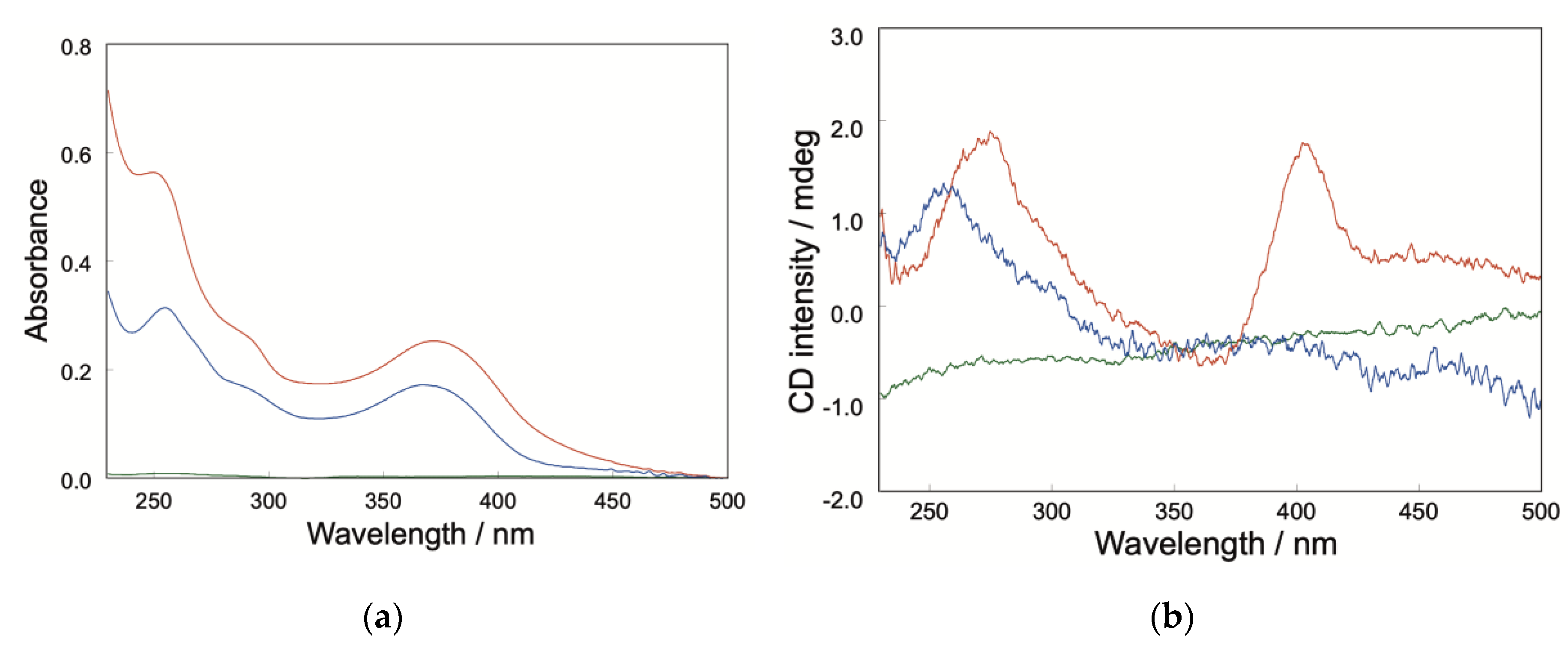
Figure 2.
Apparent aqueous solubility/dispersibility of quercetin-loaded r-glucan NPs and the quercetin/β-CD complex. The quercetin concentration in the supernatant after centrifugation was plotted as a function of the amount of added inclusion complex. Quercetin-loaded r-glucan NPs and the quercetin/β-CD complex are shown as red circles and blue squares, respectively.
Figure 2.
Apparent aqueous solubility/dispersibility of quercetin-loaded r-glucan NPs and the quercetin/β-CD complex. The quercetin concentration in the supernatant after centrifugation was plotted as a function of the amount of added inclusion complex. Quercetin-loaded r-glucan NPs and the quercetin/β-CD complex are shown as red circles and blue squares, respectively.
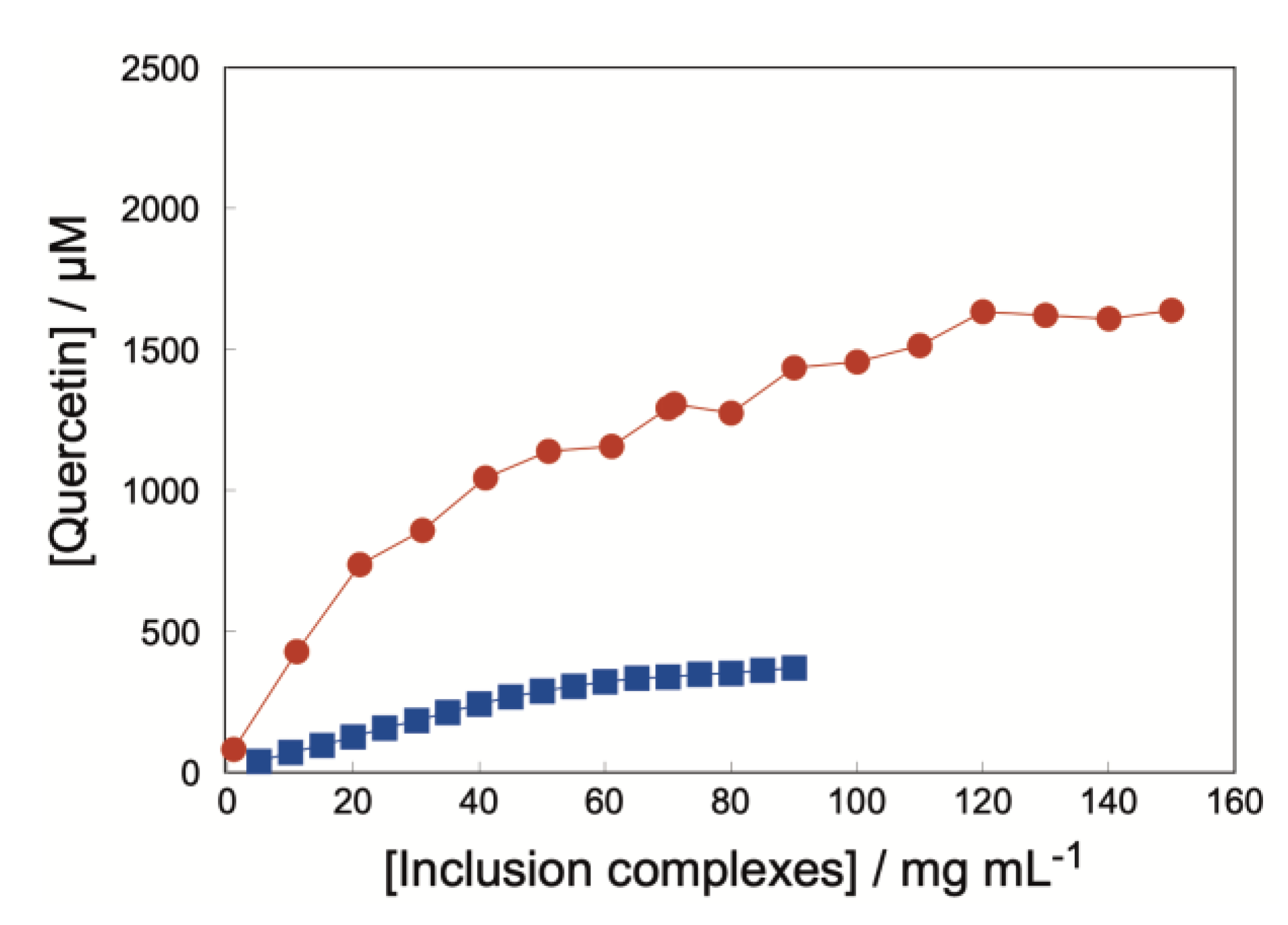
Figure 3.
Antioxidant activity of free and encapsulated quercetin at a fixed quercetin concentration. Antioxidant activities of free quercetin, the quercetin/β-CD complex, and the quercetin-loaded r-glucan NPs were compared at a fixed quercetin concentration of 5.0 µM, on a quercetin basis. Antioxidant capacity was evaluated using the ORAC assay and expressed as Trolox equivalents. Data are presented as mean ± SD (n = 3). Statistical significance is indicated as *p < 0.05, **p < 0.01, and ***p < 0.001.
Figure 3.
Antioxidant activity of free and encapsulated quercetin at a fixed quercetin concentration. Antioxidant activities of free quercetin, the quercetin/β-CD complex, and the quercetin-loaded r-glucan NPs were compared at a fixed quercetin concentration of 5.0 µM, on a quercetin basis. Antioxidant capacity was evaluated using the ORAC assay and expressed as Trolox equivalents. Data are presented as mean ± SD (n = 3). Statistical significance is indicated as *p < 0.05, **p < 0.01, and ***p < 0.001.
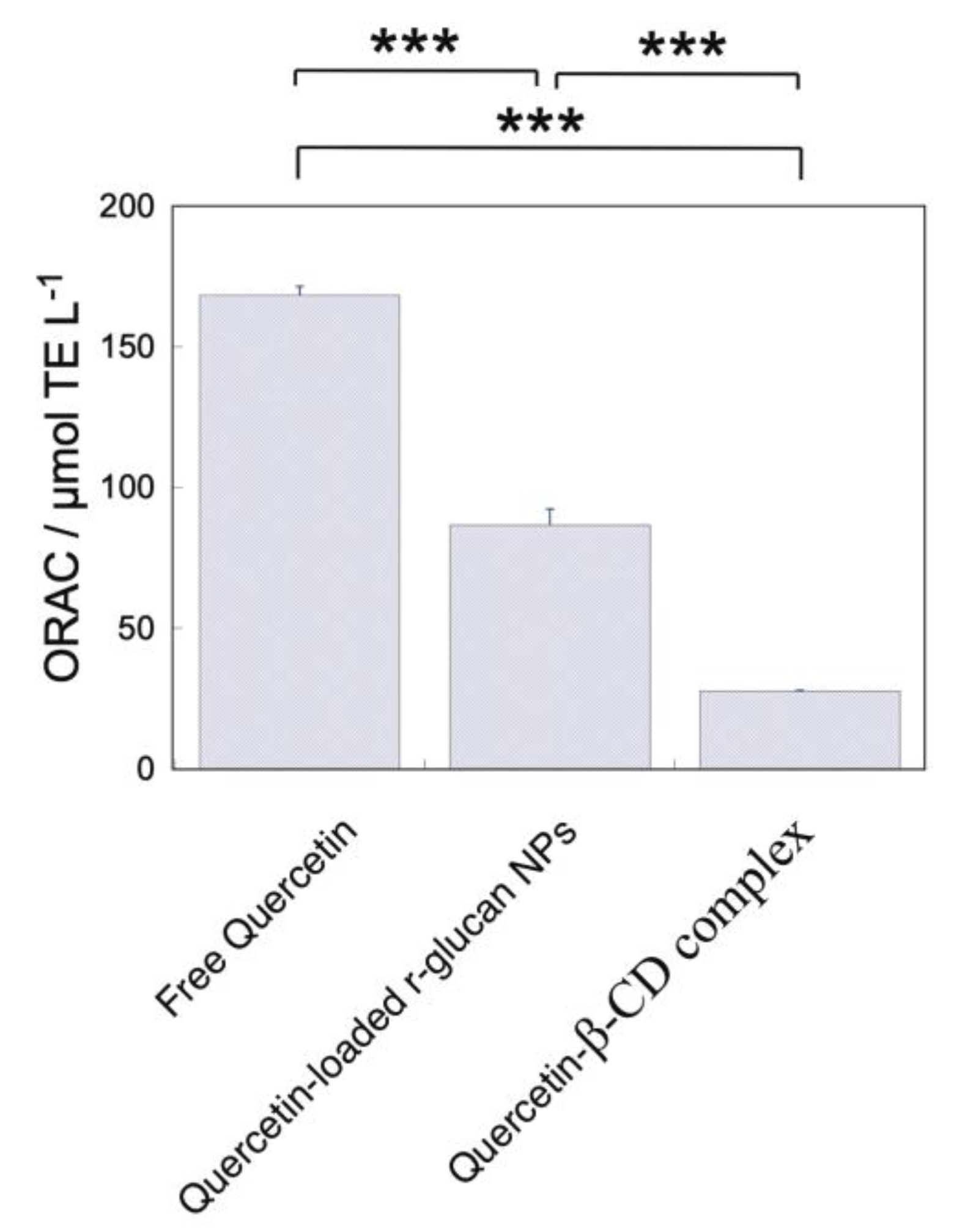
Figure 4.
Concentration-dependent antioxidant activity of free and encapsulated quercetin. ORAC values of free quercetin, the quercetin/β-CD complex, and quercetin-loaded r-glucan NPs were plotted as a function of quercetin concentration, on a quercetin basis. Free quercetin, the quercetin/β-CD complex, and quercetin-loaded r-glucan NPs are shown as green triangles, blue squares, and red circles, respectively. The inset shows a magnified view of the low-concentration range, enabling direct comparison of the three formulations at concentrations where all samples could be evaluated. Antioxidant capacity was evaluated using the ORAC assay and expressed as Trolox equivalents. Data are presented as mean ± SD (n = 3).
Figure 4.
Concentration-dependent antioxidant activity of free and encapsulated quercetin. ORAC values of free quercetin, the quercetin/β-CD complex, and quercetin-loaded r-glucan NPs were plotted as a function of quercetin concentration, on a quercetin basis. Free quercetin, the quercetin/β-CD complex, and quercetin-loaded r-glucan NPs are shown as green triangles, blue squares, and red circles, respectively. The inset shows a magnified view of the low-concentration range, enabling direct comparison of the three formulations at concentrations where all samples could be evaluated. Antioxidant capacity was evaluated using the ORAC assay and expressed as Trolox equivalents. Data are presented as mean ± SD (n = 3).
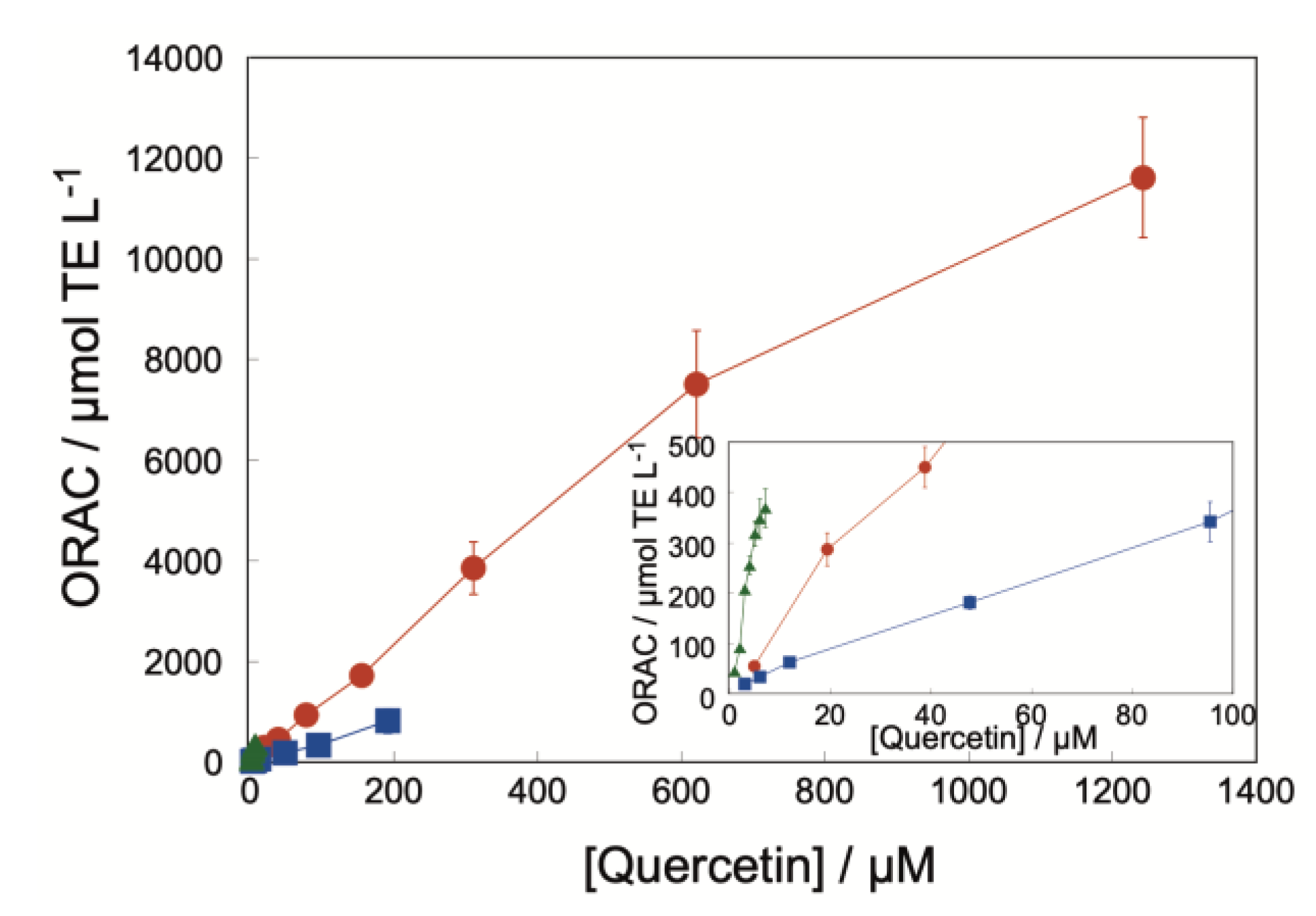
Figure 5.
Release behavior of quercetin from carrier systems. Time-dependent changes in the residual fraction of carrier-associated quercetin in quercetin-loaded r-glucan NPs and the quercetin/β-CD complex. Quercetin-loaded r-glucan NPs and the quercetin/β-CD complex are shown as red circles and blue squares, respectively. The residual fraction was calculated as At/A0, where At and A0 represent the absorbance values of quercetin at time t and 0 h, respectively.
Figure 5.
Release behavior of quercetin from carrier systems. Time-dependent changes in the residual fraction of carrier-associated quercetin in quercetin-loaded r-glucan NPs and the quercetin/β-CD complex. Quercetin-loaded r-glucan NPs and the quercetin/β-CD complex are shown as red circles and blue squares, respectively. The residual fraction was calculated as At/A0, where At and A0 represent the absorbance values of quercetin at time t and 0 h, respectively.
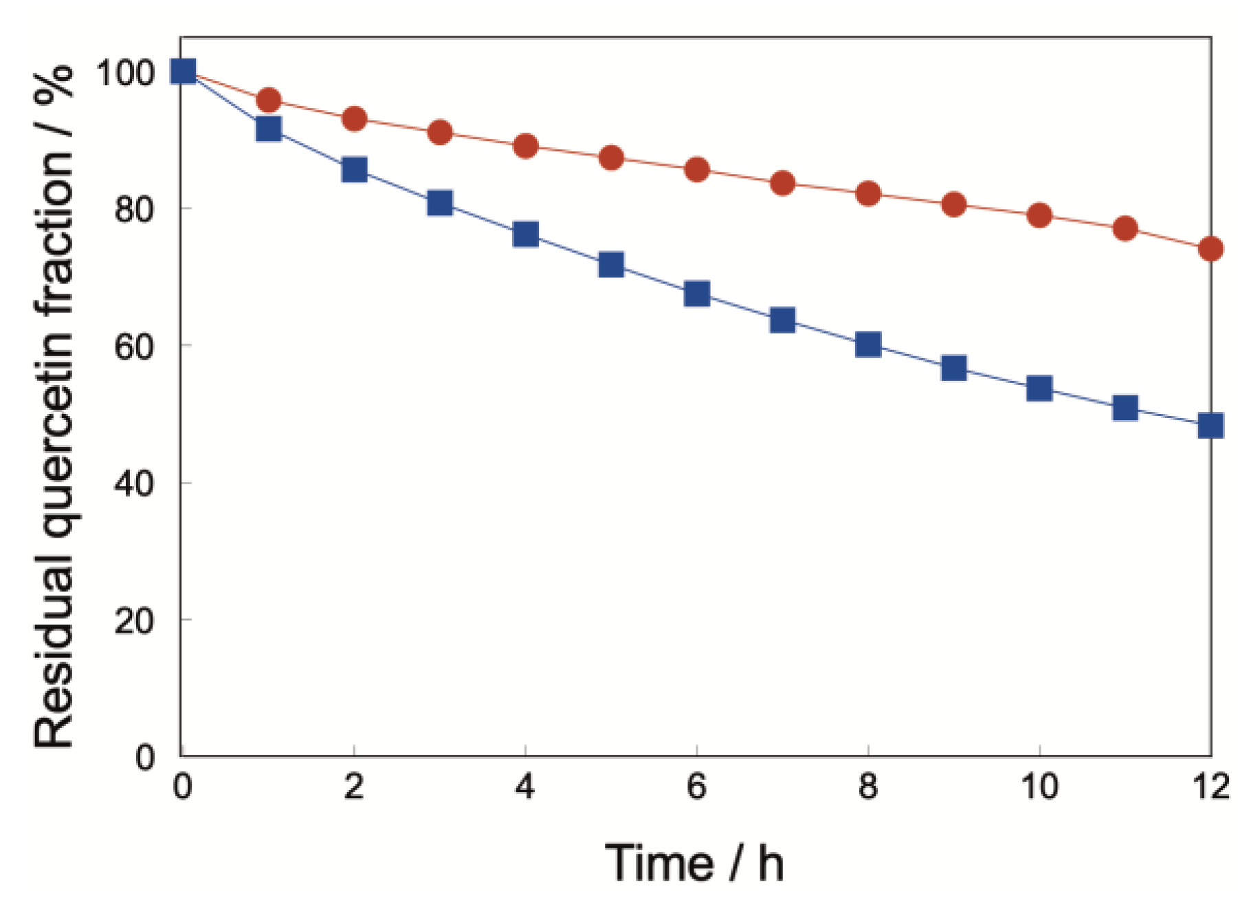
Figure 6.
Photodegradation behavior of free quercetin (green triangles), the quercetin-loaded r-glucan NPs (red circles), and the quercetin/β-CD complex (blue squares) under light exposure. The residual quercetin fraction decreased over time in all samples, whereas the quercetin-loaded r-glucan NPs showed improved photostability compared with free quercetin and the quercetin/β-CD complex. Data are presented as mean ± SD (n = 3).
Figure 6.
Photodegradation behavior of free quercetin (green triangles), the quercetin-loaded r-glucan NPs (red circles), and the quercetin/β-CD complex (blue squares) under light exposure. The residual quercetin fraction decreased over time in all samples, whereas the quercetin-loaded r-glucan NPs showed improved photostability compared with free quercetin and the quercetin/β-CD complex. Data are presented as mean ± SD (n = 3).
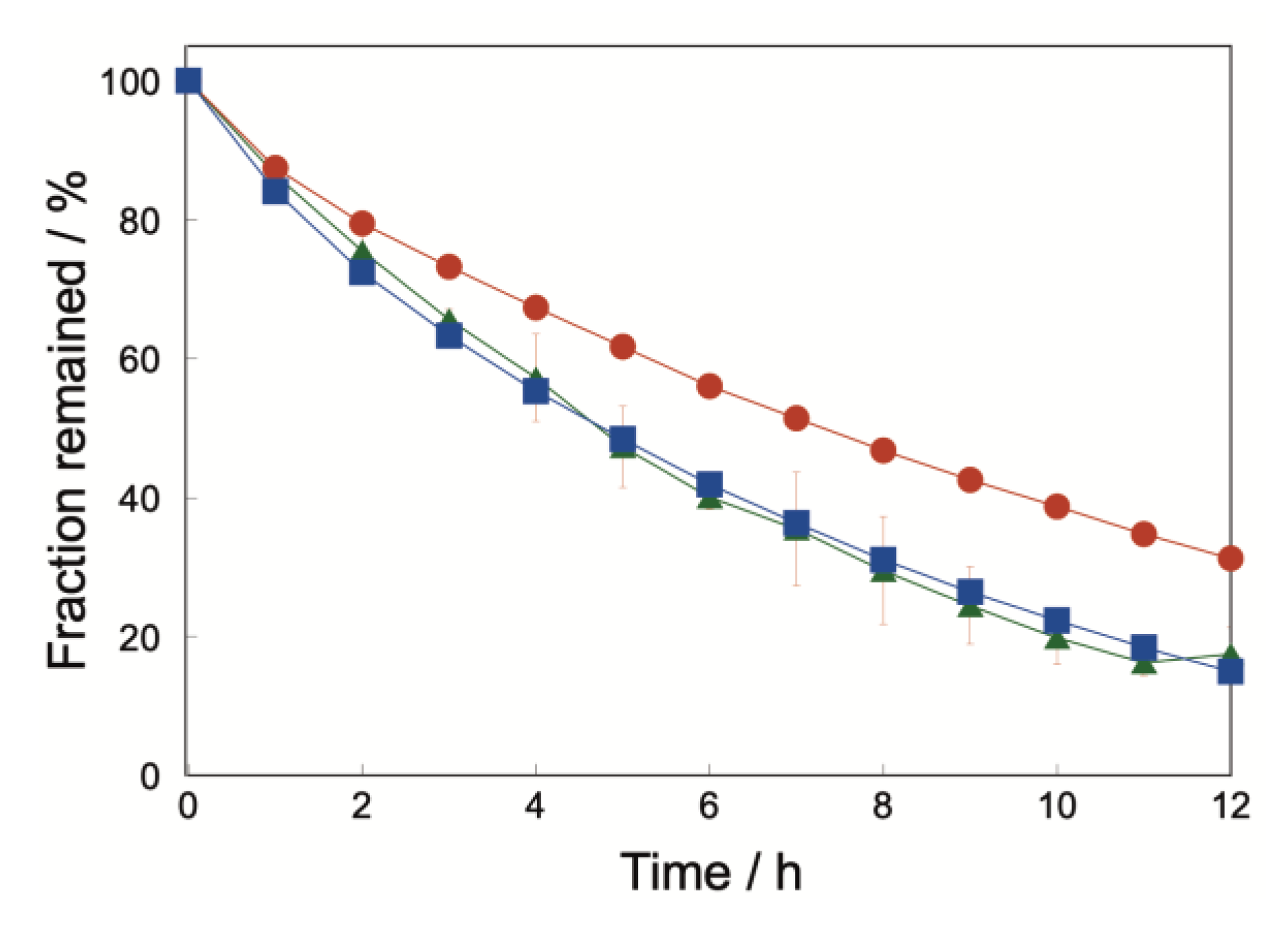
Figure 7.
pH-dependent stability of free and encapsulated quercetin. Time-dependent changes in the residual quercetin fraction of free quercetin, quercetin-loaded r-glucan NPs, and the quercetin/β-CD complex at (a) pH 1.2 and (b) pH 6.8. Free quercetin, quercetin-loaded r-glucan NPs, and the quercetin/β-CD complex are shown as green triangles, red circles, and blue squares, respectively. The residual fraction was calculated as At/A0, where At and A0 represent the absorbance values of quercetin at time t and 0 h, respectively. Data are presented as mean ± SD (n = 3).
Figure 7.
pH-dependent stability of free and encapsulated quercetin. Time-dependent changes in the residual quercetin fraction of free quercetin, quercetin-loaded r-glucan NPs, and the quercetin/β-CD complex at (a) pH 1.2 and (b) pH 6.8. Free quercetin, quercetin-loaded r-glucan NPs, and the quercetin/β-CD complex are shown as green triangles, red circles, and blue squares, respectively. The residual fraction was calculated as At/A0, where At and A0 represent the absorbance values of quercetin at time t and 0 h, respectively. Data are presented as mean ± SD (n = 3).
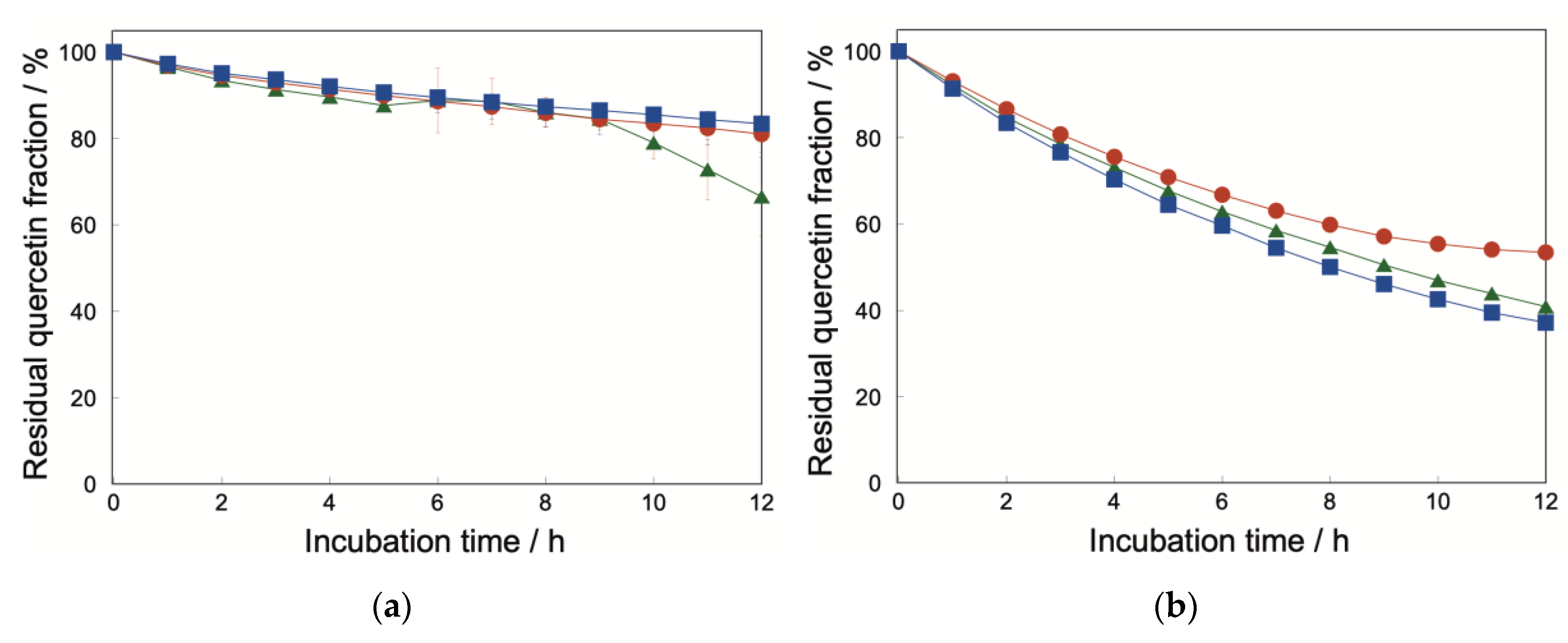
Figure 8.
Cytotoxicity of free and encapsulated quercetin in Caco-2 cells. Caco-2 cells were exposed to free quercetin, quercetin-loaded r-glucan NPs, or the quercetin/β-CD complex at the indicated quercetin concentrations for 24 h at 37 °C under 5% CO₂. Cell viability was then evaluated and expressed relative to the untreated control. Free quercetin, quercetin-loaded r-glucan NPs, and the quercetin/β-CD complex are shown in green, red, and blue, respectively. Data are presented as mean ± SD (n = 3).
Figure 8.
Cytotoxicity of free and encapsulated quercetin in Caco-2 cells. Caco-2 cells were exposed to free quercetin, quercetin-loaded r-glucan NPs, or the quercetin/β-CD complex at the indicated quercetin concentrations for 24 h at 37 °C under 5% CO₂. Cell viability was then evaluated and expressed relative to the untreated control. Free quercetin, quercetin-loaded r-glucan NPs, and the quercetin/β-CD complex are shown in green, red, and blue, respectively. Data are presented as mean ± SD (n = 3).
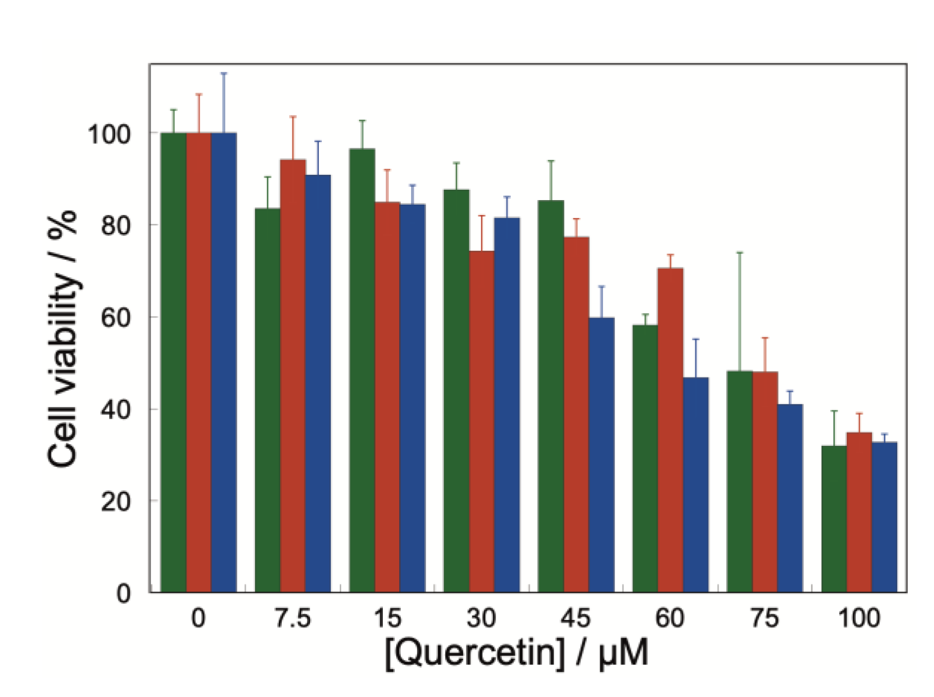
Figure 9.
Cellular antioxidant activity of free and encapsulated quercetin in Caco-2 cells. Cellular antioxidant activity (CAA) of free quercetin, the quercetin/β-CD complex, and quercetin-loaded r-glucan NPs was evaluated in Caco-2 cells at the indicated quercetin concentrations under AAPH-induced oxidative stress conditions. Free quercetin, quercetin-loaded r-glucan NPs, and the quercetin/β-CD complex are shown in green, red, and blue, respectively. Data are presented as mean ± SD (n = 3).
Figure 9.
Cellular antioxidant activity of free and encapsulated quercetin in Caco-2 cells. Cellular antioxidant activity (CAA) of free quercetin, the quercetin/β-CD complex, and quercetin-loaded r-glucan NPs was evaluated in Caco-2 cells at the indicated quercetin concentrations under AAPH-induced oxidative stress conditions. Free quercetin, quercetin-loaded r-glucan NPs, and the quercetin/β-CD complex are shown in green, red, and blue, respectively. Data are presented as mean ± SD (n = 3).
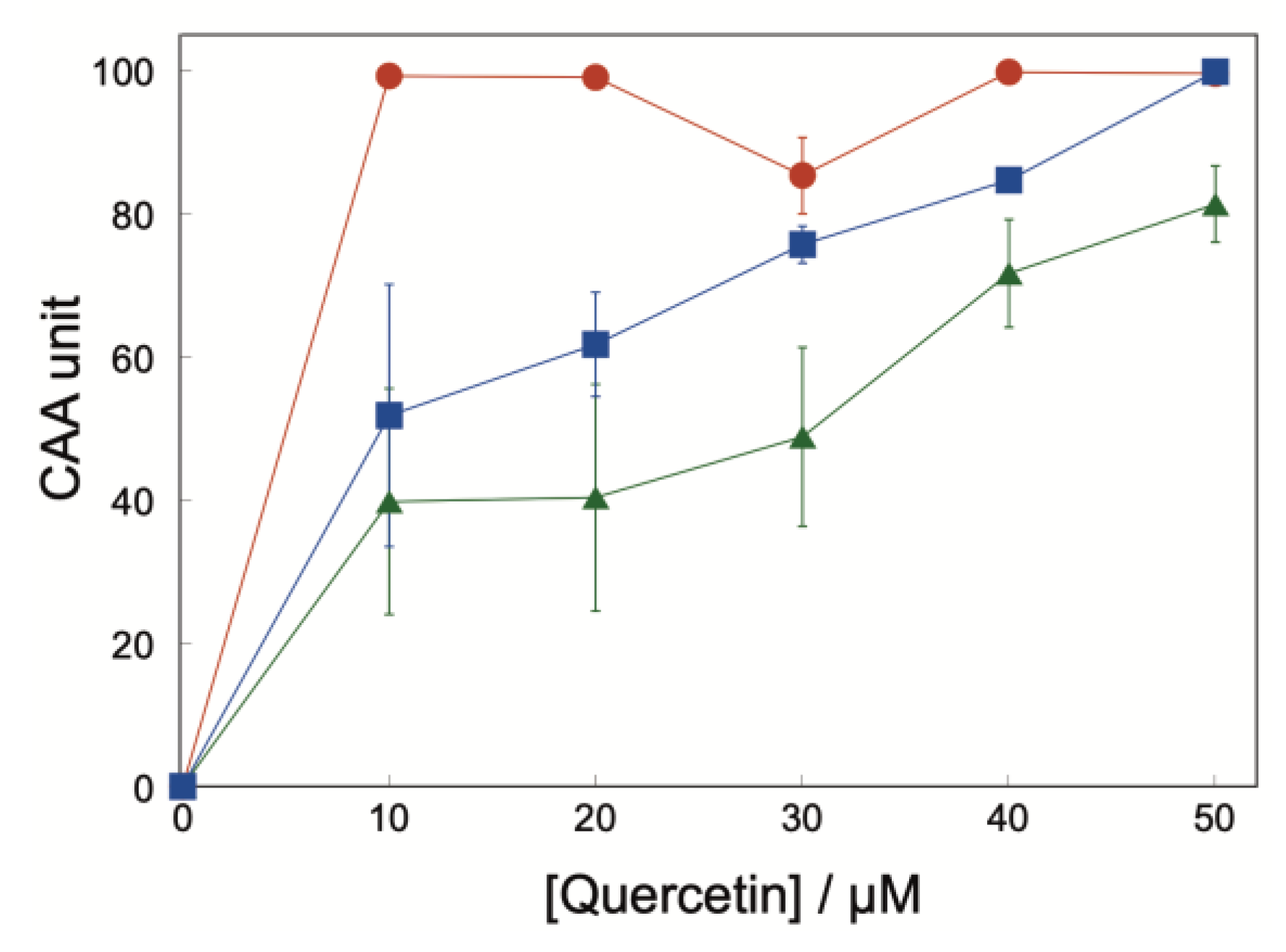
Figure 10.
AAPH-induced degradation of glucan chains constituting r-glucan NPs. Time-dependent changes in the Mw of glucan chains after incubation of r-glucan NPs with AAPH at 37 °C.
Figure 10.
AAPH-induced degradation of glucan chains constituting r-glucan NPs. Time-dependent changes in the Mw of glucan chains after incubation of r-glucan NPs with AAPH at 37 °C.
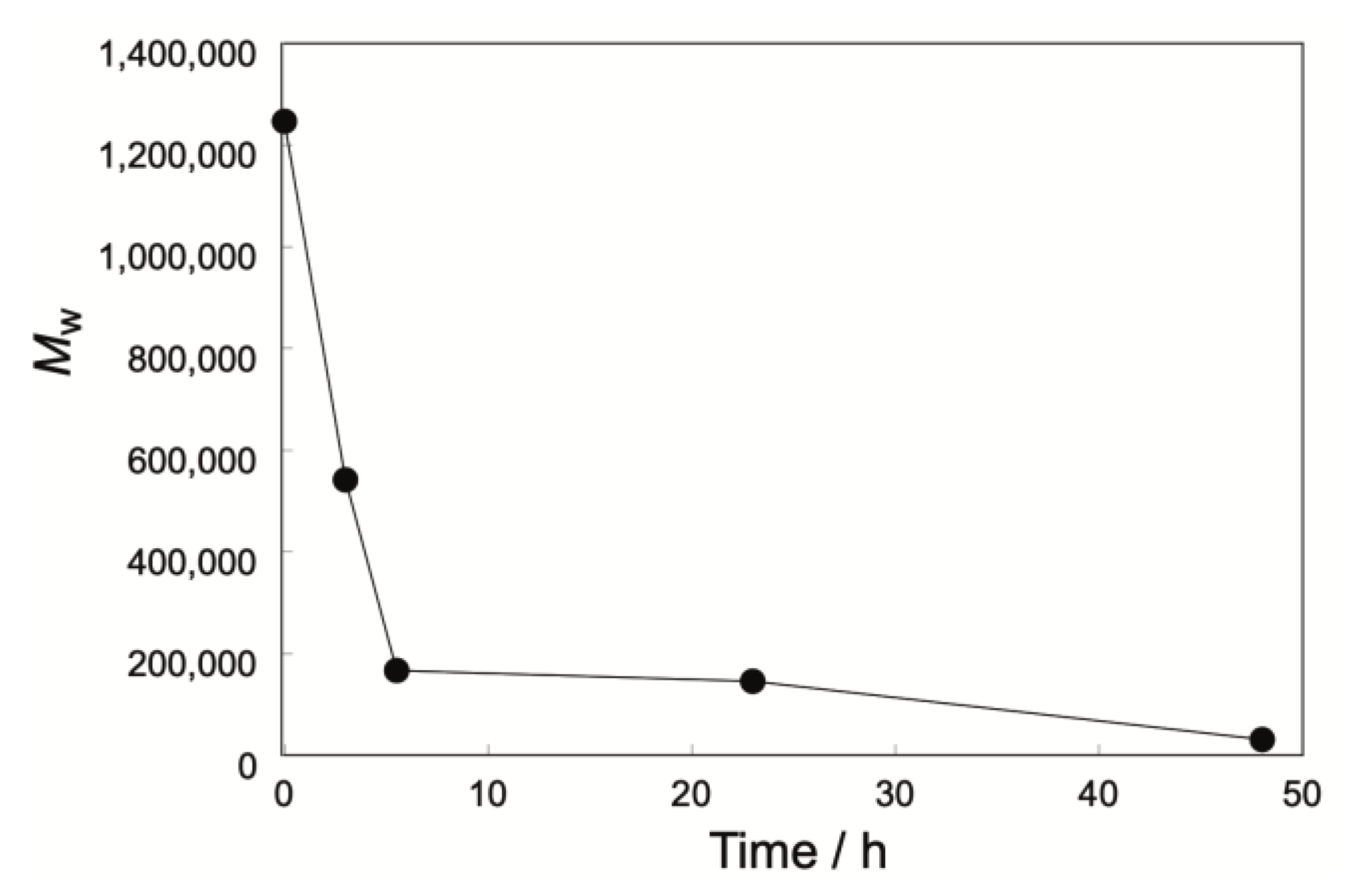
Disclaimer/Publisher’s Note: The statements, opinions and data contained in all publications are solely those of the individual author(s) and contributor(s) and not of MDPI and/or the editor(s). MDPI and/or the editor(s) disclaim responsibility for any injury to people or property resulting from any ideas, methods, instructions or products referred to in the content. |
© 2026 by the authors. Licensee MDPI, Basel, Switzerland. This article is an open access article distributed under the terms and conditions of the Creative Commons Attribution (CC BY) license.
Copyright: This open access article is published under a Creative Commons CC BY 4.0 license, which permit the free download, distribution, and reuse, provided that the author and preprint are cited in any reuse.


