

Submitted:
20 June 2026
Posted:
23 June 2026
You are already at the latest version
Abstract
Hepatitis E virus (HEV) is a major etiological agent of acute viral hepatitis globally. Although HEV infection is mostly self-limiting in immunocompetent individuals, immunocompromised individuals particularly pregnant women are at high risk for progression to chronic hepatitis and related complications. The virus comprises eight genotypes from HEV-1 to HEV-8 with distinct host ranges: HEV-1 and HEV-2 are limited to humans; HEV-3, HEV-4, HEV-7 and Rat-HEV are zoonotic; whereas HEV-5, HEV-6, and HEV-8 are limited to animal. In addition to hepatic disease, HEV is recognized for its extrahepatic manifestations, including neurological and renal disorders. Due to continued burden of transmission and the absence of globally implemented vaccination strategy the preventive measures based on improved water quality, hygiene, and targeted immunization remain critical for controlling HEV infection. This review Summarizes current knowledge on HEV genome, epidemiology, pathogenesis, clinical features, diagnosis, prevention, and highlights emerging research priorities as future perspective.
Keywords:
HEV
; Genotype
; immunocompromised
; immunocompetent
; hepatic
; extrahepatic
1. Introduction
Hepatitis E virus (HEV) is a single-stranded positive-sense RNA virus (ssRNA+) classified in the species Paslahepevirus balayani, genus Paslahepevirus, subfamily Orthohepevirinae, and family Hepeviridae [1,2,3]. HEV was first discovered during 1978 outbreak in the Kashmir Valley. About 1,700 people died as a result of this outbreak, which afflicted about 52,000 people with icteric hepatitis. It was spread via contaminated water. About 20% of patients experienced cholestasis.In addition, bile plugs in the canaliculi and the development of pseudo-ductules by hepatocytes surrounding these plugs were among the distinctive liver histology seen in certain patients. Despite these findings, every patient who survived had a self-limiting course of illness[4,5,6].
HEV causes a significant burden of liver disease and is one of the major causes of viral hepatitis globally. While HEV infection in infants and young children is generally mild or asymptomatic, pregnant women, immunosuppressed individuals, older adults, and those with underlying liver disease are at higher risk for severe disease and complications. An estimated 20 million HEV infections occur each year, resulting in approximately 3.4 million clinical cases and 70,000 HEV-related deaths globally. Although hepatitis E typically self-limiting, with a 0.5–3% case fatality rate in adults, under certain predisposing condition including pregnancy it can result a serious clinical consequence including fulminant hepatic failure, which has high death rate of 20% to 30% [1,7,8,9,10]. Although its zoonotic transmission is primarily known to occur in developed nations, HEV is mostly linked to significant outbreaks in developing countries. The virus exists as a heterogeneous group of strains with distinct epidemiological and biological characteristics, contributing to substantial clinical and public health impact [11,12,13].
Despite HEV global impact, it has been remained neglected relative to hepatitis A, B, C, and D. There are eight genotypes of HEV, designated HEV-1 to HEV-8. HEV-1 and HEV-2 are causes of widespread epidemics commonly in developing countries and they spread through fecal-oral route. In contrast, HEV-3 and HEV-4 are zoonotic and primarily found in developed countries and infect humans via consumption of contaminated meat or direct contact. More recently HEV-7 is discovered as zoonotic HEV with documented human infection linked to the consumption of camel products. Genotypes 5, 6, and 8 are only infecting animals [14,15,16,17,18]. Furthermore, emerging HEV associated with rodents, specifically rat HEV, have been identified as possible zoonotic infections. Rat HEV is categorized under genus Rocahepevirus and differs genetically from strains of the HEV that infect humans. [19,20,21,22]. Over the past 20 years, there has been an increase in the number of reported cases of the HEV. Studies indicates that this increase is not only due to a rise in the actual incidence of infection but also to improved case assessment, improved diagnostic capabilities, and increased awareness about the disease[23]. In this review relevant updated articles including original studies and literature reviews on HEV was identified and were considered through searches of PubMed, Scopus, and Web of Science using keywords including “HEV genome, HEV epidemiology, HEV diagnosis, HEV treatment, and HEV prevention.
2. Genome Organization and Molecular Structure of HEV
2.1. Virion Structure
The hepatitis E virus possesses a ssRNA genome of approximately 7.2kb. There are two variants of this virus: The first one is environmentally stable non-enveloped virion that increases viral persistence in water and promotes fecal oral transmission. This non-enveloped form is generated when virus particles that were initially released from hepatocytes as quasi-enveloped virions enter the biliary tract and are exposed to bile salts, which act like detergents and remove the host-derived lipid membrane, resulting a capsid without its lipid envelope. The second one is HEV, which is released as a quasi-enveloped particle that acquires host-derived lipid membranes circulating in the bloodstream. While the non-enveloped form is environmentally stable and promotes transmission, this quasi-envelope protects the virus from neutralizing antibodies and aids in immune evasion. This structural character allows HEV to circulate in blood while maintaining its infectivity. The complex transmission dynamics, pathogenicity, and survival of HEV both inside and outside the host are attributed to this structural duality, which is thought to be a unique biological characteristic of the virus [24,25,26].
2.2. Genome Organization
The non-enveloped HEV has a diameter of 27–34 nm. Open reading frames (ORFs) are discontinuous sections of the HEV genome which contains ORF 1, 2,3 and 4. ORF-1 encodes for a polyprotein which is important in viral replication including RNA-dependent RNA polymerase, papain-like cysteine protease, RNA helicase, methyltransferase and hypervariable region (HVR) or proline-rich region (PRR)[2,27,28]. RNA helicase unwinds RNA secondary structures during replication, methyltransferase is essential in 5′ RNA capping and genome integrity, and an RNA-dependent RNA polymerase used for synthesizing viral RNA genomes and transcripts. Furthermore, the HVR within ORF-1 exhibits high sequence diversity and it facilitates the interactions between host and viral factors, thereby regulating replication processes. Post-translational modifications, including phosphorylation of serine residues, likely further modulate its structural dynamics and function. The HVR frequently undergoes insertions, deletions, and duplications, with host-derived insertions shown to enhance replication[27,29,30,31]. Although ORF-1 was first predicted to have a papain-like cysteine protease (PCP)-like domain based on sequence analysis, its role in proteolysis has not been clearly establish[32]. ORF-2 encodes the viral capsid protein, it is a structural protein that surrounds and protects the viral genetic material, it is highly immunogenic, and antibodies against this protein have neutralizing and protective features. ORF-3 which partially overlaps ORF-2 encodes a small multifunctional phosphoprotein with viroporin-like activity, it is involved in the morphogenesis and pathogenesis of virion contributing to viral egress and modulation of host pathways. The recently identified ORF-4 is exclusive to HEV-1 and it is essential for HEV RNA polymerase to function properly [33,34,35,36].
2.3. Replication Cycle
Hepatitis E virus entrance into hepatocytes is mostly mediated by endocytic pathways. According to recent studies, a number of host cell surface receptor are involved in HEV invasion. Epidermal growth factor receptor (EGFR) and T-cell immunoglobulin and mucin domain-1 (TIM-1) are two of the receptor that have been suggested as possible facilitators of attachment and entrance of HEV [37]. Following attachment and internalization, the viral capsid is uncoated and enabling the positive-sense RNA genome to act directly as messenger RNA for translation of the nonstructural polyprotein encoded by ORF-1. The ORF-1 proteins form replication complexes associated with intracellular membranes, where synthesis of negative-sense RNA intermediates occurs. These intermediates serve as templates for the production of both genomic and sub genomic RNAs required for viral protein expression and genome amplification. Virion assembly takes place in the cytoplasm, and newly formed particles are released predominantly through the host cell secretory pathway, often acquiring a host-derived lipid membrane that generates quasi-enveloped virions, the ORF-3 protein uses Annexin II to facilitate the formation and release of quasi-enveloped virions [26,38,39]. Y-Box Binding Protein 1 (YBX1) and eukaryotic translation initiation factor 4H (EIF4H) are important host factors for HEV pathogenesis and replication. In addition, Rab5A host factor regulates early endosome-mediated replication[40].
3. Manifestation
HEV infections have diverse clinical manifestations including acute and self-limiting hepatitis, chronic hepatitis, cirrhosis, and liver failure[41]. Acute HEV infection is asymptomatic or mildly symptomatic, infections caused by HEV-1 and HEV-2 are more severe than those caused by HEV-3 and HEV-4 [42]. Clinically, acute HEV infection may show symptoms including malaise, fever, body aches, nausea, vomiting, dark-colored urine and jaundice. While most of acute HEV infections in immunocompetent individuals do not require particular treatment, it has been repeatedly demonstrated that substantial rates of morbidity and mortality are associated with HEV infection in pregnant women and immunocompromised individuals. Eclampsia, hemorrhagic complications, and liver failure are the main causes of HEV-1 infection-related death, which can reach maternal mortality up to 20%. Additionally, jaundice, hepatitis, and hepatomegaly are signs of newborn infection that raise the risk of neonatal mortality [41,42,43]. Furthermore, chronic HEV infections can cause fibrotic remodeling, nodules, and cirrhosis, among other structural damage to the liver[44].About 10% of patients with a chronic HEV infection develop cirrhosis within two to five years[45].
4. Pathogenesis
4.1. Hepatic Pathogenesis
The main processes in the pathogenesis of HEV infection include viral entrance, replication within hepatocyte, and host immune-mediated liver damage. In HEV infection, host immunological responses are mostly responsible for liver damage rather than direct viral cytopathic effect. Hepatocyte apoptosis and necrosis are encouraged by inflammatory cytokines such as tumor necrosis factor-α, interleukin-6, and interferon-γ, which are released when innate immune mechanisms, such as Kupffer cells, natural killer cells, and interferon-mediated antiviral pathways are activated. Furthermore, by targeting infected hepatocytes, adaptive immune responses specifically, cytotoxic CD8 T cell activity further contribute to the death of hepatic tissue. Lobular hepatitis, hepatocyte degeneration, localized necrosis, and cholestasis are among the pathological alterations that occur. In addition, due to insufficient T-cell-mediated viral clearance. Hormonal and immunological changes make the pregnant women more vulnerable to serious illness including fulminant hepatic failure, viral genotype, viral load, and host immunological state determine the severity of the disease [43,46,47]. HEV-1 causes severe disease in pregnancy. High hormonal level change may enhance viral replication and weaken antiviral defenses, increasing viral load. Simultaneously, the pregnancy-associated Th2 immune bias suppresses Th1-mediated viral clearance. Furthermore, HEV-1 particularly can infect and replicate in placental trophoblasts, causing placental injury, inflammation and possible placental transmission. Severe disease is also associated with elevated pro-inflammatory cytokines such as TNF-α and IL-6, which may worsen hepatocellular damage and contribute to fulminant liver failure[48,49,50].
4.2. Extrahepatic Pathogenesis
A combination of direct viral invasion of extrahepatic organs, immune-mediated damage and systemic inflammatory responses are likely to be responsible for the increasingly recognized extrahepatic symptoms of HEV infection. HEV has been found in a number of non-hepatic locations, indicating potential viral spread outside of hepatocytes. Neurological involvement is among the most clinically severe extrahepatic consequences. HEV infection has been linked to conditions such as Guillain-Barré syndrome, neuralgic amyotrophy, and encephalitis, which may be caused by immune-mediated neuronal damage [51,52]. According to studies the mechanism by which HEV causes neurological disorder is associated with the ability of HEV virions cross the blood-brain barrier (BBB). The virus is capable of infecting human brain microvascular endothelial cells, which are important components of the BBB, thereby supporting productive viral infection in vitro. Furthermore, an experimental infection study in pigs shows that HEV RNA was present in the brain and spinal cord, indicating neuroinvasion. Notably, histological abnormalities and increased levels of proinflammatory cytokines, such as TNF-α and interleukin 18, were observed in pigs with detectable HEV RNA in CNS tissues[53].
Another significant category of extrahepatic symptoms is hematological disorders including immune complex deposition, cytokine-mediated suppression of hematopoiesis, or systemic inflammatory activation may cause thrombocytopenia, hemolytic anemia, bone marrow suppression and anemia due to glucose-6-phospate dehydrogonase deficiency. There have also been reports of renal involvement, including glomerulonephritis and acute kidney injury, which are thought to be caused by cytokine-induced renal injury and immune-mediated glomerular damage. Furthermore, rare cases of thyroid dysfunction, myositis, and pancreatic damage have been reported, indicating the systemic effects of HEV infection [54,55,56,57]. According to recent HEV studies the male reproductive system can also serve as a habitat for persistent infection, studies has verified that the ejaculate of patients with persistent infections contains infectious HEV particles for extended periods of time [58].
5. Epidemiology
The epidemiology of HEV reflects a substantial disease burden globally, with an estimated one-third of the world’s population living in endemic regions[7,41,59]. A global seroprevalence estimation is performed by a large systematic review based on 8,153 records, of which 419 studies met the inclusion criteria. Among these, 287 studies were included in a pooled analysis to estimate global seroprevalence, with 302 studies focusing on the general population. The pooled global anti-HEV IgG seroprevalence, based on 1,099,717 individuals, was 12.47%, indicating that approximately one in eight people worldwide have been previously exposed to HEV. In contrast, anti-HEV IgM seroprevalence reflecting recent was 1.47%, based on 98 studies including 479,001 participants. Significant geographic variance was found on six continents. The highest anti-HEV IgG seroprevalence has been reported in Africa (21.76%), followed by Asia (15.80%), Europe (9.31%), North America (8.05%), South America (7.28%), and Oceania (5.99%). Similarly, recent infection rates (IgM) are highest in Africa (3.09%) and Asia (1.86%), with lower rates observed in South America (2.43%), Europe (0.79%), and North America (0.22%). These regional differences likely reflect disparities in sanitation, water quality, socioeconomic conditions, and exposure to zoonotic reservoirs. Furthermore, the prevalence of active infection, as indicated by HEV RNA positivity, was substantially lower at 0.20% among 3,444,752 individuals [11,60].
The Paslahepevirus balayani species has eight different HEV genotypes, of which HEV-1, HEV-2, HEV-3, HEV-4 and HEV-7 can infect humans. The primary reservoir for HEV-1 and HEV-2 is humans and epidemics emerge in various parts of Asia, Africa, Mexico, and the Middle East[61]. Infections with HEV-3 and HEV-4 are mostly contracted through zoonotic transmission in developed nations. HEV-3 is the most common genotype that causes HEV infections. The incidence of HEV infections varies throughout Europe; in southwest France, HEV3 is hyperendemic and has a very high seroprevalence rate (> 50%). Additionally, it is endemic in Germany, Belgium, the Netherlands, and northern France. In these nations, up to 30% of people have already been exposed to HEVs. According to a survey carried out in 30 European nations, the annual number of HEV infection cases risen from 514 in 2005 to 5617 in 2015 [62,63,64,65,66,67]. Recently, rat HEV has been increasingly recognized as a potential cause of human infection, particularly in immunocompromised individuals. Human cases have been documented in Hong Kong, central Africa, and Spain. Rats, which closely and frequently come into contact with humans and domestic animals, have become a natural reservoir of HEV. Similarly, rare human infections have been linked to HEV-7, which has been found in camels suggesting a broader host range and zoonotic potential. These emerging strains warrant further epidemiological investigation to better understand their public health significance [68,69,70,71,72].
6. Transmission Dynamics
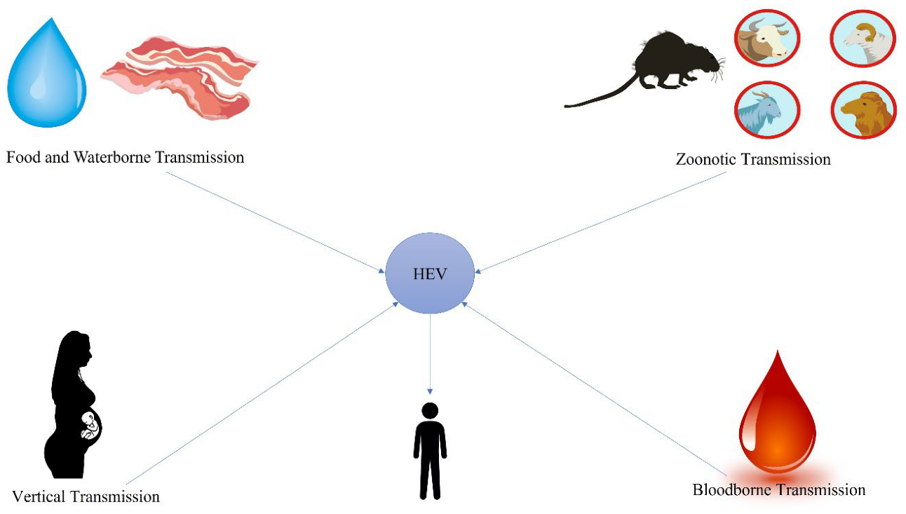
Hepatitis E infection is one of the prevalent hepatitis viruses globally. Transmission of hepatitis E virus occurs through feco-oral route, zoonotic transmission, Food and water borne transmission, vertical transmission, bloodborne transmission, person-to-person and nosocomial transmission. The transmission dynamics mainly depend on environmental sanitation, host immunity, viral genotype, absence of blood screening and human–animal interaction. Understanding these pathways is important for outbreak control and disease prevention[73,74,75,76,77]. (Figure 1) shows summarized transmission ways of HEV.
6.1. Food and Waterborne Transmission
The most important route for HEV infection worldwide is waterborne transmission. When sewage disposal facilities are insufficient, the virus is released from the feces of infected people contaminates drinking water supplies. The most vulnerable populations are those who reside in places with inadequate sanitation, dense populations, and restricted access to clean water. Outbreaks commonly occur following natural disasters such as floods or during rainy seasons when surface water mixes with sewage [78,79]. Additionally, foodborne transmission happens when contaminated water, soil, or hands contaminate consumable foods. If fresh fruits, vegetables, and seafood are irrigated or cleaned with contaminated water, they may harbor virus particles. Therefore, Proper food hygiene are essential preventive measures[80,81,82].
6.2. Zoonotic Transmission
Zoonotic transmission is becoming more widely acknowledged, especially in developed nations. The virus can infect a variety of animals, such as, pigs, deer, camels, wild boars and rats. Although the seroprevalence in these animals varies, pigs are thought to be the primary reservoir for HEV since they continuously have the highest seroprevalence, ranging from 8% to 93%[76,83,84].
The most common way that humans get infected is by direct animal contact by occupational groups including farmers, veterinarians, slaughterhouse workers and consuming raw animal products. Renou et al. have documented the transmission of HEV through processed meat products, showing that eating raw or undercooked processed meat products carries a significant risk for HEV infections. Furthermore, large-scale meat pooling or cross-contamination through surfaces during the food product chain may also be a means of transmission. In addition to meat, milk from infected goats and cows infected with HEV was found to contain HEV RNA. Zoonotic transmission is primarily caused by genotypes 3, 4, and 7. In addition, rat HEV have been recognized as potential zoonotic diseases, the routes of transmission are involve environmental exposure to contaminated surfaces, food, or water, as well as contact with rodent excreta [16,41,84,85,86].
6.3. Bloodborne Transmission
In healthcare systems, blood transfusion transmission is a growing concern. Blood donation from infected individuals is a potential source of infection since individuals can be viremic even if they do not exhibit symptoms. During the incubation phase, the virus can remain in blood, and many countries neglect to regularly screen for it. Immunocompromised patients, such as organ transplant recipients or patients receiving chemotherapy, are particularly susceptible to transfusion-transmitted infection[87,88]. The quasi-enveloped form of HEV circulates in the blood of infected individuals while cloaked by host membranes. There have been numerous documented examples that offer concrete proof of HEV transmission through blood products. HEV-4 and HEV-3 infections represent a significant risk for organ and blood donation recipients due to their immunosuppressed and poor health status, which can result in a fulminant or chronic HEV infection, even though HEV transmission during solid organ transplantation has seldom been observed[89,90,91].
6.4. Vertical Transmission
The virus can spread vertically from an infected mother to the fetus, usually in the later stages of pregnancy, the virus may infect fetal tissues after passing through the placental barrier. Due to immunological and hormonal changes that occur during pregnancy, pregnant women may experience more severe symptoms including high risks of maternal liver failure, preterm delivery, and high fetal mortality [92,93]. Because of the spatial coherence and lack of severe cases in pregnant women infected with HEV-3 and HEV-4, the severe HEV disease progression in the second and third trimesters of pregnancy is attributed to HEV-1 and HEV-2 infections. According to reports, HEV can infect a fetus in as many as 73.3–100% of pregnant women[46,92,94].
6.5. Person-to-Person and Nosocomial Transmission
Direct person-to-person transmission of HEV is comparatively rare compared to hepatitis A. However, Household transmission can happen in places with shared sanitary facilities and inadequate hygiene standards. Although hospital-acquired infections are not commonly reported, they can occur as a result of contaminated medical equipment, improper handling of blood products, or contact with bodily fluids. Therefore, Healthcare facilities must strictly adhere to infection control measures for HEV [12,77].
7. Laboratory Diagnosis of HEV
The incubation time for HEV ranges from roughly 15 to 60 days. Serological or molecular techniques are used in the laboratory to diagnose HEV infection. Serological testing is the most widely used diagnostic approach. Anti-HEV IgM and IgG serologic assays are frequently utilized in diagnosis. Anti-HEV IgM antibodies appear during the acute phase of infection, usually around the onset of symptoms, and indicate recent or ongoing infection. In contrast, anti-HEV IgG antibodies appear shortly after IgM and can persist for years, serving as markers of past exposure or immunity. Rapid immunochromatographic tests are commonly used serological method it detects anti-HEV IgM. Furthermore, enzyme-Linked Immunosorbent Assay is the other serological test for diagnosing Hepatitis E by detecting HEV-specific IgM for recent infection and IgG for past exposure, often using recombinant ORF-2 capsid proteins. while the immunoperoxidase monolayer assay (IPMA) is another antibody detection method[95,96].
Furthermore, molecular detection methods, particularly reverse transcription polymerase chain reaction (RT-PCR) are considered the gold standard for confirming HEV infection during the early phase of infection, HEV RNA can be detected in blood, stool, and urine. Viraemia generally persists for about 3–6 weeks, while viral shedding in feces may last 4–6 weeks. Notably, HEV RNA has also been detected in urine, sometimes persisting longer than in serum. Because urine is continuously produced, viral RNA can remain detectable even after serum viraemia declines, potentially extending the diagnostic window[97,98,99,100,101]. RT-PCR is especially valuable for direct detection of HEV RNA during the early phase of infection before antibody development[41,101,102]. Recently, alternative nucleic acid amplification methods such as loop-mediated isothermal amplification (LAMP) have been developed for rapid and cost-effective detection of HEV[103,104].In addition, digital PCR is another emerging technique that offers high sensitivity and precise quantification of viral RNA, although its use remains largely limited to research settings. Furthermore, sequencing-based approaches, including next-generation sequencing (NGS), are increasingly used for genotyping and molecular epidemiology studies. Overall, an integrated diagnostic approach combining clinical evaluation, serological testing, and molecular methods is recommended to enhance diagnostic accuracy. The selection of appropriate specimens including serum, stool, and urine should be guided by the stage of infection and available laboratory capacity[105,106].
8. HEV Treatment
Antiviral treatment options for patients with chronic hepatitis E include dose reduction of immunosuppression medications, particularly those targeting T lymphocytes. This approach provides viral clearance in up to one-third of patients and administration of ribavirin monotherapy or in combination with pegylated interferon alpha (ribavirin, alpha-interferon) for at least three months shows significant efficacy in the treatment of chronic hepatitis E [107,108]. For instance, ribavirin monotherapy was used to treat a patient with HEV genotype 3 who experienced severe acute hepatitis and reduced liver function. In response, the patient's bilirubin level and international normalized ratio improved, and their liver transaminases quickly decreased. Furthermore, reduction of immunosuppression in 16 recipients of solid organ transplants with chronic hepatitis E led to clearance of HEV in 4 cases (25%) [109,110,111,112]. Another option for treatment of HEV is pegylated interferon. Treatment duration lasts from three to twelve months; four out of five patients responded well to pegylated interferon alpha, which resulted in long-term HEV RNA eradication. However, interferon therapy can cause significant side effects and organ rejection in transplant recipients. In addition, for patients with fulminate hepatic failure, liver transplantation is the only treatment option. Since most transplant recipients cannot utilize interferon-alpha and ribavirin is not recommended for pregnant patients, there is an urgent need for new antiviral medications that are both safe and effective [109,113,114,115].
9. In Vitro and In Vivo Model for HEV Studies
In vitro platforms that support the entire life cycle of HEV genotypes have been constructed by researchers using induced pluripotent stem cell (iPSC)-derived multilineage organoids, including liver, intestine, and brain models. These models demonstrate HEV's capacity for pan-tissue infection and reveal specific pathological impacts across different organ systems[116,117]. The virus infects many cell types in liver organoids, resulting in decreased albumin output and increased liver enzymes and IL-6, which are signs of severe hepatocellular damage. When epithelial and mesenchymal cells in intestinal organoids become infected, the intestinal barrier breaks down and pro-inflammatory cytokines significantly increase. Furthermore, brain organoids exhibit strong neural tropism, with HEV infecting various neurons and glial cells, specifically altering the number of dopaminergic neurons. These models have been used for drug screening by demonstrating that treatment with the antiviral ribavirin partially reverses the virus's damaging effects across all three tissues, thereby establishing a powerful and versatile tool for studying HEV pathogenesis and developing targeted antiviral therapies[116,118,119].
In addition, the mongolian gerbil is highly valued as in vivo model due to its widespread susceptibility to different wild-type HEV genotypes and it closely resembles human clinical characteristics, including increased liver enzymes, persistent viral shedding, and severe liver damage caused by mitochondrial apoptosis[117,120,121,122,123]. In addition to gerbils, scientists use humanized mice to investigate medication efficacy and human-specific HEV replication, since pigs are natural reservoirs for HEV-3 and HEV-4, they are also considered the canonical model for understanding zoonotic transmission dynamics. Rabbits are also being used to study HEV infections [124,125,126].
10. Prevention Strategies for Hepatitis E Virus
10.1. Food, Water, and Sanitation-Based Prevention
Prevention of HEV transmission depends on proper food handling, safe water, and improved sanitation practices. Foodborne transmission can be minimized by thoroughly cooking meat from common animal reservoirs associated with zoonotic HEV genotypes and avoiding the consumption of undercooked products, minimizing exposure to contact with rodent and maintaining strict hygiene during food preparation further reduces the risk of infection. In addition, improving water quality and sanitation is essential to prevent fecal–oral transmission. Furthermore, individuals in high-risk occupational groups, such as farmers, veterinarians, and slaughterhouse workers, should follow appropriate protective measures to reduce exposure to infected animals and contaminated materials. Community-level prevention includes rapid detection and response to outbreaks, health education on hygiene and food safety, and long-term improvements in sanitation infrastructure measures helps to reduce HEV transmission in both endemic and non-endemic settings[127,128,129]. Furthermore, Immunocompromised individuals, transplant recipients, and pregnant women should avoid high-risk foods and adhere strictly to safe food and water practices. Pregnant women should also avoid travel to areas experiencing HEV outbreaks due to the high risk of severe disease [130,131,132].
10.2. Blood and Organ Safety
Routine screening of blood products and organ donors to reduce the risk of HEV transmission. This is particularly critical for high-risk populations, including immunocompromised individuals, transplant recipients, and patients with underlying liver disease. In the context of organ transplantation, careful donor screening and risk assessment are essential, as HEV can be transmitted through infected organs and may lead to persistent infection in recipients. Strengthening surveillance systems and adopting standardized screening protocols can further improve blood and organ safety, especially in endemic regions[133,134,135].
10.3. Vaccination for HEV
A recombinant vaccine developed by the Walter Reed Army Institute and GlaxoSmithKline showed 96% effectiveness after three doses in a Phase II study of 2000 Nepalese soldiers for protective immunity. [136,137]. Furthermore, A recombinant HEV vaccine (HEV 239) prevented acute hepatitis E with 94%–100% effectiveness on Phase III research involving more than 100,000 chinese adults. This vaccine contains 30 g of pure HEV antigen adsorbed on 0.8 mg of aluminum hydroxide. It is based on HEV genotype 1 and is made in bacterial cells. It was safe for pregnant women and did not have any noticeable, unexpected adverse effects. However, additional studies is needed before the vaccine can be used in certain risk populations, such as immunocompromised people or those with end-stage liver disease. The vaccine has been approved in China, India and Pakistan [136,138,139,140]. Furthermore, neutralizing antibodies that can be detected in the sera of individuals exposed to HEV, indicating prior infection and contributing to protection against reinfection. In addition to humoral immunity, HEV-specific CD4⁺ and CD8⁺ T-cell responses are observed during acute infection and can persist long-term in recovered individuals, immunodominant epitopes located within ORF 2 and 3 play a key role in eliciting these cellular immune responses[141,142,143].
11. Conclusions and Future Perspectives
HEV is a complex and evolving pathogen with substantial global health implications. Its dual modes of transmission, diverse genotypes, and variable clinical manifestations present unique challenges for surveillance, diagnosis, and management. Advances in molecular diagnostics, deeper understanding of viral biology, and the development of effective vaccines offer promising opportunities for improved control. However, significant gaps remain, particularly in global vaccine deployment, standardized diagnostics, and characterization of extrahepatic disease. Expanding global access to diagnosis and prevention strategies for HEV remain critical priorities as enhanced diagnostic platforms with higher sensitivity, rapid detection, and genotype specific resolution are urgently needed to identify acute and chronic infections. At the therapeutic level, the development of targeted antivirals, optimization of ribavirin regimens, and evaluation of combination therapies represent essential steps toward effective treatment of chronic HEV infection. A deeper investigation into viral pathogenesis, host–virus interactions, and immunological determinants of severe disease will be instrumental in identifying biomarkers for early prognostication and guiding personalized therapy. Given HEV’s zoonotic transmission dynamics, the adoption of integrated one Health surveillance systems incorporating human, animal, and environmental monitoring will be vital for tracking viral evolution, preventing foodborne transmission, and controlling outbreaks. Advances in structural virology, genomics, and reverse genetics are expected to accelerate the discovery of novel vaccine candidates and antiviral targets. Collectively, these future developments aim to reduce global HEV burden through improved prevention, early detection, and evidence-based clinical management. A coordinated, multidisciplinary approach integrating molecular biology, virology, clinical medicine, public health, and one Health strategies are essential for addressing the continued burden of HEV infection worldwide.
Author Contributions
All authors consent to publication of this manuscript.
Funding
The authors received no specific funding for this work.
Institutional Review Board Statement
Not applicable.:
Informed Consent Statement
Not applicable.
Clinical trial number: Not applicable.
Data Availability Statement
This Manuscript doesn’t generate data. Further inquiries can be directed to the corresponding author(s).
Acknowledgments
The authors would like to thank biotechnology research center, Addis Ababa university.
Conflicts of Interest
The authors declare no conflict of interest.
References
- Prpić, J.; Baymakova, M. Hepatitis E virus (HEV) infection among humans and animals: epidemiology, clinical characteristics, treatment, and prevention. MDPI 2023, 931. [Google Scholar] [CrossRef]
- Wang, B.; Meng, X.-J. Structural and molecular biology of hepatitis E virus. Comput. Struct. Biotechnol. J. 2021, 19, 1907–1916. [Google Scholar] [CrossRef] [PubMed]
- Kim, D.-H.; et al. Utility of hypervariable region in hepatitis E virus for genetic evolution analysis and epidemiological studies. J. General. Virol. 2025, 106(2), 002080. [Google Scholar] [CrossRef]
- Khuroo, M.S. Discovery of hepatitis E: the epidemic non-A, non-B hepatitis 30 years down the memory lane. Virus Res. 2011, 161(1), 3–14. [Google Scholar] [PubMed]
- Khuroo, M.S. Discovery of hepatitis E and its impact on global health: a journey of 44 years about an incredible human-interest story. Viruses 2023, 15(8), 1745. [Google Scholar] [CrossRef] [PubMed]
- Khuroo, M.S. Hepatitis E and pregnancy: an unholy alliance unmasked from Kashmir, India. Viruses 2021, 13(7), 1329. [Google Scholar] [CrossRef] [PubMed]
- Pallerla, S.R.; et al. Hepatitis E virus infection: circulation, molecular epidemiology, and impact on global health. Pathogens 2020, 9(10), 856. [Google Scholar] [CrossRef] [PubMed]
- Rein, D.B.; et al. The global burden of hepatitis E virus genotypes 1 and 2 in 2005. Hepatology 2012, 55(4), 988–997. [Google Scholar] [CrossRef] [PubMed]
- Nimgaonkar, I.; et al. Hepatitis E virus: advances and challenges. Nat. Rev. Gastroenterol. Hepatol. 2018, 15(2), 96–110. [Google Scholar] [PubMed]
- Khuroo, M.; Kamili, S. Aetiology, clinical course and outcome of sporadic acute viral hepatitis in pregnancy. J. Viral Hepat. 2003, 10(1), 61–69. [Google Scholar] [CrossRef] [PubMed]
- Li, P.; et al. The global epidemiology of hepatitis E virus infection: a systematic review and meta-analysis. Liver Int. 2020, 40(7), 1516–1528. [Google Scholar] [PubMed]
- Khuroo, M.S.; Khuroo, M.S.; Khuroo, N.S. Transmission of hepatitis E virus in developing countries. Viruses 2016, 8(9), 253. [Google Scholar] [CrossRef] [PubMed]
- Kasem, A.; et al. Epidemiology of hepatitis E virus infection. Epidemiol. Mikrobiol. Imunol. 2019, 68(4), 176–82. [Google Scholar] [PubMed]
- Teshale, E.H.; Hu, D.J. Hepatitis E: Epidemiology and prevention. World J. Hepatol. 2011, 3(12), 285. [Google Scholar] [CrossRef] [PubMed]
- Minosse, C.; et al. Clinical characteristics of acute hepatitis E and their correlation with HEV genotype 3 subtypes in Italy. Pathogens 2020, 9(10), 832. [Google Scholar] [CrossRef] [PubMed]
- Sridhar, S.; et al. Hepatitis E virus genotypes and evolution: emergence of camel hepatitis E variants. Int. J. Mol. Sci. 2017, 18(4), 869. [Google Scholar] [CrossRef] [PubMed]
- Modiyinji, A.F.; et al. Epidemiology of hepatitis E virus infection in animals in Africa: a systematic review and meta-analysis. BMC Vet. Res. 2021, 17(1), 50. [Google Scholar] [CrossRef] [PubMed]
- Wang, B.; Meng, X.J. Hepatitis E virus: host tropism and zoonotic infection. Curr. Opin. Microbiol. 2021, 59, 8–15. [Google Scholar] [PubMed]
- Yang, Z.; et al. Prevalence, Risk Factors, and Genetic Evolution of Rat Hepatitis E Virus in Small Mammals from Southwestern Yunnan, China. Biology 2025, 14(12), 1685. [Google Scholar] [CrossRef] [PubMed]
- Ding, Q.; et al. Prevalence and molecular characterization of hepatitis E virus (HEV) from wild rodents in Hubei Province, China. Infect. Genet. Evol. 2024, 121, 105602. [Google Scholar] [CrossRef] [PubMed]
- Erdin, M.; et al. Genetic characterization of rat hepatitis E virus (Rocahepevirus ratti) in urban brown rats (Rattus norvegicus) in Helsinki, Finland. Arch. Virol. 2025, 170(10), 215. [Google Scholar] [CrossRef] [PubMed]
- Tian, D.; et al. Antiviral resistance and barrier integrity at the maternal-fetal interface restrict hepatitis E virus from crossing the placental barrier. Proc. Natl. Acad. Sci. 2025, 122(18), e2501128122. [Google Scholar] [CrossRef] [PubMed]
- Capai, L.; Charrel, R.; Falchi, A. Hepatitis E in high-income countries: what do we know? And what are the knowledge gaps? Viruses 2018, 10(6), 285. [Google Scholar] [CrossRef] [PubMed]
- Gremmel, N. Hepatitis E virus: isolation of porcine virus strains and characterization of serum antibodies. Dissertation, Tierärztliche Hochschule Hannover, Hannover, 2023. [Google Scholar]
- Reyes, G.; et al. Molecular organization and replication of hepatitis E virus (HEV). In Unconventional Agents and Unclassified Viruses: Recent Advances in Biology and Epidemiology; 1993; pp. 15–25. [Google Scholar]
- LeDesma, R.; Nimgaonkar, I.; Ploss, A. Hepatitis E virus replication. Viruses 2019, 11(8), 719. [Google Scholar] [CrossRef] [PubMed]
- Cancela, F.; et al. Structural aspects of hepatitis E virus. Arch. Virol. 2022, 167(12), 2457–2481. [Google Scholar] [CrossRef] [PubMed]
- Yang, Y.-L.; Nan, Y.-C. Open reading frame 3 protein of hepatitis E virus: Multi-function protein with endless potential. World J. Gastroenterol. 2021, 27(20), 2458. [Google Scholar] [CrossRef] [PubMed]
- Parvez, M.K. Molecular characterization of hepatitis E virus ORF1 gene supports a papain-like cysteine protease (PCP)-domain activity. Virus Res. 2013, 178(2), 553–6. [Google Scholar] [PubMed]
- Yin, X.; Li, X.; Feng, Z. Role of envelopment in the HEV life cycle. Viruses 2016, 8(8), 229. [Google Scholar] [CrossRef] [PubMed]
- Wang, B.; et al. Phosphorylation of Ser711 residue in the hypervariable region of zoonotic genotype 3 hepatitis E virus is important for virus replication. MBio 2024, 15(11), e02635–24. [Google Scholar] [CrossRef] [PubMed]
- Pierce, D.M.; et al. Thrombin cleavage of the hepatitis E virus polyprotein at multiple conserved locations is required for genome replication. PLoS Pathog. 2023, 19(7), e1011529. [Google Scholar] [CrossRef] [PubMed]
- Ding, Q.; et al. Hepatitis E virus ORF3 is a functional ion channel required for release of infectious particles. Proc. Natl. Acad. Sci. 2017, 114(5), 1147–1152. [Google Scholar] [CrossRef] [PubMed]
- Nair, V.P.; et al. Endoplasmic reticulum stress induced synthesis of a novel viral factor mediates efficient replication of genotype-1 hepatitis E virus. PLoS Pathog. 2016, 12(4), e1005521. [Google Scholar] [CrossRef] [PubMed]
- Saraswat, S.; Chaudhary, M.; Sehgal, D. Hepatitis E virus cysteine protease has papain like properties validated by in silico modeling and cell-free inhibition assays. Front. Cell. Infect. Microbiol. 2020, 9, 478. [Google Scholar] [CrossRef] [PubMed]
- Wang, B.; Meng, X.J. Structural and molecular biology of hepatitis E virus. Comput Struct. Biotechnol. J. 2021, 19, 1907–1916. [Google Scholar] [CrossRef] [PubMed]
- Brüggemann, Y.; et al. How hepatitis E virus invades hepatocytes: the mystery of viral entry. Trends Microbiol. 2025. [Google Scholar] [CrossRef] [PubMed]
- Kenney, S.P.; Meng, X.-J. Hepatitis E virus genome structure and replication strategy. Cold Spring Harb. Perspect. Med. 2019, 9(1), a031724. [Google Scholar] [PubMed]
- Liu, X.; et al. Palmitoylation-dependent association with Annexin II directs hepatitis E virus ORF3 sorting into vesicles and quasi-enveloped virions. Proc. Natl. Acad. Sci. U S A 2025, 122(1), e2418751122. [Google Scholar] [PubMed]
- Ju, X.; et al. EIF4H and YBX1 are essential host factors for hepatitis E virus replication and pathogenesis. Proc. Natl. Acad. Sci. 2026, 123(10), e2529289123. [Google Scholar] [CrossRef] [PubMed]
- Aslan, A.T.; Balaban, H.Y. Hepatitis E virus: Epidemiology, diagnosis, clinical manifestations, and treatment. World J. Gastroenterol. 2020, 26(37), 5543. [Google Scholar] [CrossRef] [PubMed]
- Pischke, S.; Wedemeyer, H. Hepatitis E virus infection: multiple faces of an underestimated problem. J. Hepatol. 2013, 58(5), 1045–1046. [Google Scholar] [CrossRef] [PubMed]
- Lhomme, S.; et al. Clinical manifestations, pathogenesis and treatment of hepatitis E virus infections. J. Clin. Med. 2020, 9(2), 331. [Google Scholar] [CrossRef] [PubMed]
- Gérolami, R.; Moal, V.; Colson, P. Chronic hepatitis E with cirrhosis in a kidney-transplant recipient. N. Engl. J. Med. 2008, 358(8), 859–860. [Google Scholar] [CrossRef] [PubMed]
- Kamar, N.; et al. Hepatitis E virus-related cirrhosis in kidneyand kidney-pancreas-transplant recipients. Am. J. Transplant. 2008, 8(8), 1744–1748. [Google Scholar] [PubMed]
- Yadav, K.K.; Kenney, S.P. Hepatitis E virus immunopathogenesis. Pathogens 2021, 10(9), 1180. [Google Scholar] [CrossRef]
- Lin, S.; Zhang, Y.-J. Advances in hepatitis E virus biology and pathogenesis. Viruses 2021, 13(2), 267. [Google Scholar] [CrossRef] [PubMed]
- Wu, C.; Wu, X.; Xia, J. Hepatitis E virus infection during pregnancy. Virol. J. 2020, 17(1), 73. [Google Scholar] [CrossRef] [PubMed]
- Tian, D.; et al. Antiviral resistance and barrier integrity at the maternal-fetal interface restrict hepatitis E virus from crossing the placental barrier. Proc. Natl. Acad. Sci. U S A 2025, 122(18), e2501128122. [Google Scholar] [CrossRef] [PubMed]
- Gouilly, J.; et al. Genotype specific pathogenicity of hepatitis E virus at the human maternal-fetal interface. Nat. Commun. 2018, 9(1), 4748. [Google Scholar] [CrossRef] [PubMed]
- El-Mokhtar, M.A.; Sayed, I.M. Model systems for studying extrahepatic pathogenesis of hepatitis E virus. Current knowledge and future directions. Rev. Med. Virol. 2021, 31(6), e2218. [Google Scholar] [CrossRef] [PubMed]
- Liu, H.; Ma, Y. Hepatitis E virus-associated Guillain–Barre syndrome: Revision of the literature. Brain Behav. 2020, 10(1), e01496. [Google Scholar] [PubMed]
- Tian, D.; et al. Hepatitis E virus infects brain microvascular endothelial cells, crosses the blood-brain barrier, and invades the central nervous system. Proc. Natl. Acad. Sci. U S A 2022, 119(24), e2201862119. [Google Scholar] [PubMed]
- Fousekis, F.S.; Mitselos, I.V.; Christodoulou, D.K. Extrahepatic manifestations of hepatitis E virus: An overview. Clin. Mol. Hepatol. 2019, 26(1), 16. [Google Scholar] [PubMed]
- Cao, X.; et al. Hematologic system damage is common and related to severity in patients with acute hepatitis E. 2024. [Google Scholar] [CrossRef] [PubMed]
- Fan, H.; et al. Prognostic significance of end-stage liver diseases, respiratory tract infection, and chronic kidney diseases in symptomatic acute hepatitis E. Front. Cell. Infect. Microbiol. 2021, 10, 593674. [Google Scholar] [CrossRef] [PubMed]
- Kovvuru, K.; et al. Renal manifestations of hepatitis E among immunocompetent and solid organ transplant recipients. World J. Hepatol. 2022, 14(3), 516. [Google Scholar] [CrossRef] [PubMed]
- Horvatits, T.; et al. Hepatitis E virus persists in the ejaculate of chronically infected men. J. Hepatol. 2021, 75(1), 55–63. [Google Scholar] [CrossRef] [PubMed]
- Murrison, L.B.; Sherman, K.E. The enigma of hepatitis E virus. Gastroenterol. Hepatol. 2017, 13(8), 484. [Google Scholar]
- Letafati, A.; et al. From discovery to treatment: tracing the path of hepatitis E virus. Virol. J. 2024, 21(1), 194. [Google Scholar] [CrossRef] [PubMed]
- Aggarwal, R. Hepatitis E: Historical, contemporary and future perspectives. J. Gastroenterol. Hepatol. 2011, 26, 72–82. [Google Scholar] [CrossRef] [PubMed]
- Aspinall, E.J.; et al. Hepatitis E virus infection in Europe: surveillance and descriptive epidemiology of confirmed cases, 2005 to 2015. Eurosurveillance 2017, 22(26), 30561. [Google Scholar] [CrossRef] [PubMed]
- Kamar, N.; et al. Hepatitis e. The Lancet 2012, 379(9835), 2477–2488. [Google Scholar] [CrossRef]
- Mansuy, J.-M.; et al. Hepatitis E virus antibodies in blood donors, France. Emerg. Infect. Dis. 2011, 17(12), 2309. [Google Scholar] [CrossRef] [PubMed]
- Lu, L.; Li, C.; Hagedorn, C.H. Phylogenetic analysis of global hepatitis E virus sequences: genetic diversity, subtypes and zoonosis. Rev. Med. Virol. 2006, 16(1), 5–36. [Google Scholar] [PubMed]
- Yan, B.; et al. Hepatitis E virus in yellow cattle, Shandong, Eastern China. Emerg. Infect. Dis. 2016, 22(12), 2211. [Google Scholar] [CrossRef] [PubMed]
- Dencs, Á.; et al. Molecular Epidemiology of Hepatitis E Virus in Hungary (2018–2025): Emergence of Rare Subtypes and First Detection of HEV-4 in Central Europe. Viruses 2025, 17(10), 1389. [Google Scholar] [CrossRef] [PubMed]
- Reuter, G.; Boros, Á.; Pankovics, P. Review of hepatitis E virus in rats: evident risk of species Orthohepevirus C to human zoonotic infection and disease. Viruses 2020, 12(10), 1148. [Google Scholar] [CrossRef] [PubMed]
- Santos-Silva, S.; et al. A systematic review of hepatitis E virus detection in camels. Vet. Sci. 2023, 10(5), 323. [Google Scholar] [CrossRef] [PubMed]
- Sridhar, S.; et al. Rat hepatitis E virus as cause of persistent hepatitis after liver transplant. Emerg. Infect. Dis. 2018, 24(12), 2241. [Google Scholar] [CrossRef] [PubMed]
- Andonov, A.; et al. Rat hepatitis E virus linked to severe acute hepatitis in an immunocompetent patient. J. Infect. Dis. 2019, 220(6), 951–955. [Google Scholar] [CrossRef] [PubMed]
- Rivero-Juarez, A.; et al. Orthohepevirus C infection as an emerging cause of acute hepatitis in Spain: First report in Europe. J. Hepatol. 2022, 77(2), 326–331. [Google Scholar] [CrossRef] [PubMed]
- Kardena, I.M.; et al. Zoonotic Hepatitis E Virus: Epidemiology, Animal Reservoirs, and Control Strategies. World 2025, 15(1), 182–193. [Google Scholar] [CrossRef]
- Ji, C.; et al. When HEV infection meets organ transplantation. Int. J. Surg. 2025, 10.1097. [Google Scholar]
- Kmush, B.; et al. Epidemiology of hepatitis E in low-and middle-income countries of Asia and Africa. In Seminars in liver disease; Thieme Medical Publishers, 2013. [Google Scholar]
- Bode, I.R.; Surakaram, A.; Gowtham, M. Hepatitis E virus (HEV) unveiled: A comprehensive examination from Genotypes, epidemiology and zoonotic challenges to public health strategies. In Emerging zoonotic threats from swine: A public health perspective; Springer, 2025; pp. 175–190. [Google Scholar]
- Lampejo, T.; et al. Nosocomial transmission of hepatitis E virus and development of chronic infection: the wider impact of COVID-19. J. Clin. Virol. 2022, 148, 105083. [Google Scholar] [CrossRef] [PubMed]
- Nelson, K.E.; Labrique, A.B.; Kmush, B.L. Epidemiology of genotype 1 and 2 hepatitis E virus infections. Cold Spring Harb. Perspect. Med. 2019, 9(6), a031732. [Google Scholar] [PubMed]
- Lanrewaju, A.A.; et al. Global public health implications of human exposure to viral contaminated water. Front. Microbiol. 2022, 13, 981896. [Google Scholar] [CrossRef] [PubMed]
- Nemes, K.; Persson, S.; Simonsson, M. Hepatitis A virus and hepatitis E virus as food-and waterborne pathogens—transmission routes and methods for detection in food. Viruses 2023, 15(8), 1725. [Google Scholar] [PubMed]
- Hazards, E.P.o.B.; et al. Public health risks associated with hepatitis E virus (HEV) as a food-borne pathogen. Efsa J. 2017, 15(7), e04886. [Google Scholar]
- Treagus, S.; et al. The foodborne transmission of hepatitis E virus to humans. Food Environ. Virol. 2021, 13(2), 127–145. [Google Scholar] [CrossRef] [PubMed]
- Sooryanarain, H.; Meng, X.-J. Swine hepatitis E virus: Cross-species infection, pork safety and chronic infection. Virus Res. 2020, 284, 197985. [Google Scholar] [CrossRef] [PubMed]
- Sato, Y.; et al. A nationwide survey of hepatitis E virus (HEV) infection in wild boars in Japan: identification of boar HEV strains of genotypes 3 and 4 and unrecognized genotypes. Arch. Virol. 2011, 156(8), 1345–1358. [Google Scholar] [CrossRef] [PubMed]
- Khalafalla, A.I., Zoonotic diseases transmitted from the camels. Front. Vet. Sci. 2023, 10, 1244833. [PubMed]
- Huang, F.; et al. Excretion of infectious hepatitis E virus into milk in cows imposes high risks of zoonosis. Hepatology 2016, 64(2), 350–359. [Google Scholar] [CrossRef] [PubMed]
- Lopes, T. Prevalence and risks of Hepatitis E virus infection in blood donors from the Western Cape, South Africa; Stellenbosch University: Stellenbosch, 2016. [Google Scholar]
- Haase, J.A.; et al. Transmission patterns of hepatitis E virus. Curr. Opin. Virol. 2025. 70, 101451. [Google Scholar]
- Harvala, H.; et al. Fulminant transfusion-associated hepatitis E virus infection despite screening, England, 2016–2020. Emerg. Infect. Dis. 2022, 28(9), 1805. [Google Scholar] [CrossRef] [PubMed]
- Gallian, P.; et al. Transfusion-transmitted hepatitis E virus infection in France. Transfus. Med. Rev. 2019, 33(3), 146–153. [Google Scholar] [CrossRef] [PubMed]
- Kamar, N.; et al. Factors associated with chronic hepatitis in patients with hepatitis E virus infection who have received solid organ transplants. Gastroenterology 2011, 140(5), 1481–1489. [Google Scholar] [CrossRef] [PubMed]
- Sharma, S.; et al. Risk factors for vertical transmission of hepatitis E virus infection. J. Viral Hepat. 2017, 24(11), 1067–1075. [Google Scholar] [CrossRef] [PubMed]
- Yu, W.; et al. Vertical transmission of hepatitis E virus in pregnant rhesus macaques. Sci. Rep. 2020, 10(1), 17517. [Google Scholar] [CrossRef] [PubMed]
- Kumar, R.M.; et al. Sero-prevalence and mother-to-infant transmission of hepatitis E virus among pregnant women in the United Arab Emirates. Eur. J. Obstet. Gynecol. Reprod. Biol. 2001, 100(1), 9–15. [Google Scholar] [PubMed]
- Duan, B.-f.; Feng, Y. Current knowledge on the epidemiology and detection methods of hepatitis E virus in China. Virol. J. 2024, 21(1), 307. [Google Scholar] [CrossRef] [PubMed]
- Hu, W.P.; et al. Double-antigen enzyme-linked immunosorbent assay for detection of hepatitis E virus-specific antibodies in human or swine sera. Clin. Vaccine Immunol. 2008, 15(8), 1151–7. [Google Scholar] [PubMed]
- Liver, E.A. f.T.S.o.T., EASL Clinical Practice Guidelines on hepatitis E virus infection. J. Hepatol. 2018, 68(6), 1256–1271. [Google Scholar]
- Dalton, H.R.; et al. EASL Clinical Practice Guidelines on hepatitis E virus infection. J. Hepatol. 2018, 68(6), 1256–1271. [Google Scholar] [CrossRef]
- Pandeya, A.; et al. Evaluation of liver enzymes and calculation of AST to ALT ratio in patients with acute viral hepatitis. Eur. J. Med. Sci. 2021, 5(2). [Google Scholar]
- Kamar, N.; et al. Hepatitis E virus infection. Nat. Rev. Dis. Prim. 2017, 3(1), 1–16. [Google Scholar] [CrossRef]
- Ying, D.; et al. Urine is a viral antigen reservoir in hepatitis E virus infection. Hepatology 2023, 77(5), 1722–1734. [Google Scholar] [CrossRef] [PubMed]
- Zhang, X.; et al. Validation of an internally controlled multiplex real time RT-PCR for detection and typing of HEV genotype 3 and 4. J. Virol. Methods 2013, 193(2), 432–438. [Google Scholar] [CrossRef] [PubMed]
- Wei, B.; et al. Rapid visual detection of hepatitis E virus combining reverse transcription recombinase-aided amplification with lateral flow dipstick and real-time fluorescence. J. Clin. Microbiol. 2025, 63(2), e01064–24. [Google Scholar] [PubMed]
- Amdiyee, A.A.; Tessema, T.S. Isothermal nucleic acid amplification techniques: A comprehensive overview on their principle & applications as alternative to PCR. Pract. Lab. Med. 2026, e00523. [Google Scholar]
- La Bella, G.; et al. Duplex droplet digital PCR assay for quantification of hepatitis E virus in food. Viruses 2024, 16(3), 413. [Google Scholar] [CrossRef] [PubMed]
- Alavian, S.E.; Heiat, M.; Ranjbar, R. A Comprehensive Overview of Hepatitis Virus Genotyping Methods. Nov. Biomed. 2023, 11(2). [Google Scholar]
- Kamar, N.; et al. Influence of immunosuppressive therapy on the natural history of genotype 3 hepatitis-E virus infection after organ transplantation. Transplantation 2010, 89(3), 353–360. [Google Scholar] [CrossRef] [PubMed]
- Péron, J.-M.; et al. Prolonged hepatitis E in an immunocompromised patient. J. Gastroenterol. Hepatol. 2006, 21(7), 1223–1224. [Google Scholar] [CrossRef] [PubMed]
- Kamar, N.; et al. Pegylated interferon-α for treating chronic hepatitis E virus infection after liver transplantation. Clin. Infect. Dis. 2010, 50(5), e30–e33. [Google Scholar] [PubMed]
- Pischke, S.; et al. 1255 CHRONIC HEPATITIS E IN IMMUNOSUPPRESSED PATIENTS BEYOND LIVER-AND KIDNEY TRANSPLANTATION. J. Hepatol. 2011, 54, S495. [Google Scholar]
- Mallet, V.; et al. Brief communication: case reports of ribavirin treatment for chronic hepatitis E. Ann. Intern. Med. 2010, 153(2), 85–89. [Google Scholar] [CrossRef] [PubMed]
- Kamar, N.; et al. Ribavirin therapy inhibits viral replication on patients with chronic hepatitis e virus infection. Gastroenterology 2010, 139(5), 1612–1618. [Google Scholar] [CrossRef] [PubMed]
- Dalton, H.R.; et al. Hepatitis E virus and neurological injury. Nat. Rev. Neurol. 2016, 12(2), 77. [Google Scholar] [PubMed]
- Haagsma, E.B.; et al. Treatment of chronic hepatitis E in liver transplant recipients with pegylated interferon alpha-2b. Liver Transplant. 2010, 16(4), 474–477. [Google Scholar] [CrossRef]
- Bose, P.D.; et al. High viral load and deregulation of the progesterone receptor signaling pathway: association with hepatitis E-related poor pregnancy outcome. J. Hepatol. 2011, 54(6), 1107–1113. [Google Scholar] [CrossRef] [PubMed]
- Liu, F.; et al. iPSC-induced multilineage liver organoids, small intestinal organoids and brain organoids sustain pangenotype hepatitis E virus propagation. Gut 2026, 75(4), 787–800. [Google Scholar] [PubMed]
- Liu, T.; et al. An immunocompetent Mongolian gerbil model for hepatitis E virus genotype 1 infection. Gastroenterology 2024, 167(4), 750–763. e10. [Google Scholar] [CrossRef] [PubMed]
- Xiang, K.; Zhuang, H. Liver organoid potential application for hepatitis e virus infection. In Hepatitis E Virus; Springer, 2023; pp. 133–139. [Google Scholar]
- Cui, B.; et al. The Application and Challenges of Brain Organoids in Exploring the Mechanism of Arbovirus Infection. Microorganisms 2025, 13(6), 1281. [Google Scholar] [CrossRef] [PubMed]
- Xu, L.-D.; et al. Revisiting the mongolian gerbil model for hepatitis E virus by reverse genetics. Microbiol. Spectr. 2022, 10(2), e02193–21. [Google Scholar] [CrossRef] [PubMed]
- Gouttenoire, J.; Neyts, J. A Hepatitis E Virus Infection Model in the Mongolian Gerbil: Ready for Antiviral and Vaccine Studies. Gastroenterology 2024, 167(4), 652–653. [Google Scholar] [CrossRef] [PubMed]
- Liu, T.; et al. Prior HEV genotype 1 infection is not protective against rat HEV infection in Mongolian gerbils. J. Hepatol. 2025, 83(3), e143–e145. [Google Scholar] [CrossRef] [PubMed]
- Yang, Y.; et al. Mitochondrial dysfunction and mitophagy pathway activation in hepatitis E virus-infected livers of Mongolian gerbils. Virus Res. 2021, 302, 198369. [Google Scholar] [CrossRef] [PubMed]
- Li, Y.; et al. BALB/c mouse is a potential animal model system for studying acute and chronic genotype 4 hepatitis E virus infection. Front. Microbiol. 2020, 11, 1156. [Google Scholar] [CrossRef] [PubMed]
- He, Q.; et al. Immunocompromised rabbit model of chronic HEV reveals liver fibrosis and distinct efficacy of different vaccination strategies. Hepatology 2022, 76(3), 788–802. [Google Scholar] [CrossRef] [PubMed]
- León-Janampa, N.; et al. A pig model of chronic hepatitis E displaying persistent viremia and a downregulation of innate immune responses in the liver. Hepatol. Commun. 2023, 7(11), e0274. [Google Scholar] [CrossRef] [PubMed]
- Kirkwood, C.D.; Dobscha, K.R.; Steele, A.D. Hepatitis E should be a global public health priority: recommendations for improving surveillance and prevention. Expert Rev. Vaccines 2020, 19(12), 1129–1140. [Google Scholar] [CrossRef] [PubMed]
- Takuissu, G.; et al. Hepatitis E virus in water environments: a systematic review and meta-analysis. Food Environ. Virol. 2022, 14(3), 223–235. [Google Scholar] [CrossRef] [PubMed]
- Ferri, G.; Vergara, A. Hepatitis E virus in the food of animal origin: a review. Foodborne Pathog. Dis. 2021, 18(6), 368–377. [Google Scholar] [CrossRef] [PubMed]
- Wang, M.; et al. Vaccination Strategies and Research Gaps in Hepatitis E Virus for Special Populations. Vaccines 2025, 13(6), 621. [Google Scholar] [CrossRef] [PubMed]
- Alexandrova, R.; et al. Hepatitis E virus (HEV) infection among immunocompromised individuals: a brief narrative review. Infect. Drug Resist. 2024, 1021–1040. [Google Scholar] [CrossRef] [PubMed]
- Damiris, K.; et al. Hepatitis E in immunocompromised individuals. World J. Hepatol. 2022, 14(3), 482. [Google Scholar] [CrossRef] [PubMed]
- Bi, H.; et al. Hepatitis E virus and blood transfusion safety. Epidemiol. Infect. 2020, 148, e158. [Google Scholar] [CrossRef] [PubMed]
- Janahi, E.M.; et al. Implications of hepatitis E virus in blood transfusions, hemodialysis, and solid organ transplants. Medicina 2020, 56(5), 206. [Google Scholar] [CrossRef] [PubMed]
- Denner, J.; et al. Why all blood donations should be tested for hepatitis E virus (HEV). BMC Infect. Dis. 2019, 19(1), 541. [Google Scholar] [CrossRef] [PubMed]
- Shrestha, M.P.; et al. Safety and efficacy of a recombinant hepatitis E vaccine. N. Engl. J. Med. 2007, 356(9), 895–903. [Google Scholar] [CrossRef] [PubMed]
- Purcell, R.H.; et al. Pre-clinical immunogenicity and efficacy trial of a recombinant hepatitis E vaccine. Vaccine 2003, 21(19-20), 2607–2615. [Google Scholar] [CrossRef] [PubMed]
- Zhu, F.-C.; et al. Efficacy and safety of a recombinant hepatitis E vaccine in healthy adults: a large-scale, randomised, double-blind placebo-controlled, phase 3 trial. The Lancet 2010, 376(9744), 895–902. [Google Scholar]
- Wedemeyer, H.; Pischke, S. Hepatitis E vaccination—is HEV 239 the breakthrough? Nat. Rev. Gastroenterol. Hepatol. 2011, 8(1), 8–10. [Google Scholar] [CrossRef] [PubMed]
- Hartley, C.; et al. Hepatitis E vaccines updates. Vaccines 2024, 12(7), 722. [Google Scholar] [CrossRef] [PubMed]
- Zhou, Y.-H.; Purcell, R.H.; Emerson, S.U. A truncated ORF2 protein contains the most immunogenic site on ORF2: antibody responses to non-vaccine sequences following challenge of vaccinated and non-vaccinated macaques with hepatitis E virus. Vaccine 2005, 23(24), 3157–3165. [Google Scholar] [PubMed]
- Husain, M.; et al. Effector T cells immune reactivity among patients with acute hepatitis E. J. Viral Hepat. 2011, 18(10), e603–e608. [Google Scholar] [CrossRef] [PubMed]
- Aggarwal, R.; et al. T-cell epitope mapping of ORF2 and ORF3 proteins of human hepatitis E virus. J. Viral Hepat. 2007, 14(4), 283–292. [Google Scholar] [PubMed]
Figure 1.
Common Transmission pathways of Hepatitis E virus (HEV).
Disclaimer/Publisher’s Note: The statements, opinions and data contained in all publications are solely those of the individual author(s) and contributor(s) and not of MDPI and/or the editor(s). MDPI and/or the editor(s) disclaim responsibility for any injury to people or property resulting from any ideas, methods, instructions or products referred to in the content. |
© 2026 by the authors. Licensee MDPI, Basel, Switzerland. This article is an open access article distributed under the terms and conditions of the Creative Commons Attribution (CC BY) license (http://creativecommons.org/licenses/by/4.0/).
Copyright: This open access article is published under a Creative Commons CC BY 4.0 license, which permit the free download, distribution, and reuse, provided that the author and preprint are cited in any reuse.


