

Submitted:
15 June 2026
Posted:
17 June 2026
You are already at the latest version
Abstract
Hydrogen sulfide (H₂S) is a vital gasotransmitter essential for maintaining redox homeostasis and modulating inflammatory responses. Herein, we examined a collection of Drosophila melanogaster knockout (KO) lines lacking key transsulfuration (cbs, cse) and sulfide metabolism (tst1) genes, generated in our laboratory, to elucidate the role of this adaptive system in immunity. Genetic ablation of these pathways results in profound H₂S deficiency and hyperhomocysteinemia, leading to chronic oxidative stress. This leads to constitutive activation of major immune pathways—including IMD, Toll, and JAK-STAT—even in the absence of infection, a hallmark of chronic inflammation. Indeed, transcriptomic analysis and qRT-PCR studies revealed significant upregulation of these pathways in double (cbs;cse) and triple (cbs;cse;tst1) KO flies under control conditions. Following septic injury with Bacillus subtilis, double and triple KO flies exhibited increased expression of antimicrobial peptides (AMPs) and pattern recognition receptors compared to control and TST1-/- single KO lines. Notably, double- and triple-KO lines showed more prolonged immune activation. The triple KO flies exhibited the worst survival rate following septic injury. These results demonstrate that H₂S deficiency causes chronic inflammation through the sustained activation of the immune pathways, highlighting sulfur metabolism as a crucial regulator of homeostasis and innate immunity in Drosophila.
Keywords:
Drosophila
; sulfur metabolism
; inflammation
; immune pathways
1. Introduction
Innate immunity represents the first and most evolutionarily conserved line of defense against invading pathogens [1]. In multicellular organisms, this system provides reliable and rapid protection against various infections, while avoiding significant collateral damage from chronic or excessive inflammation [2]. This balance is tightly regulated by a complex network of signaling pathways, cellular responses, and, increasingly recognized, metabolic processes. Metabolism is no longer viewed merely as a housekeeping function for energy production but as a dynamic source of signaling molecules that directly instruct immune cell function and inflammatory outcomes [3]. Furthermore, the metabolic state of both the host and the invading pathogen is now understood to play a decisive role in the nature and outcome of an infection [4].
Among the many metabolic immunomodulators, hydrogen sulfide (H₂S) has recently emerged as a key gaseous transmitter with potent anti-inflammatory and cytoprotective properties [5].
In mammals, H2S is endogenously produced primarily via the transsulfuration pathway, catalyzed by the enzymes cystathionine β-synthase (CBS), cystathionine γ-lyase (CSE), and 3-mercaptopyruvate sulfurtransferase (3-MST) [6], and via a separate pathway involving thiosulfate sulfurtransferase (TST/TSTD1) [7,8,9], which is crucial for cyanide detoxification and mitochondrial sulfide oxidation. H2S influences a myriad of physiological processes, including vasodilation, neuromodulation, and mitochondrial bioenergetics [10]. Crucially, in the context of immunity, H2S has been shown to attenuate the production of pro-inflammatory cytokines, inhibit leukocyte adhesion, and scavenge reactive oxygen species (ROS), thereby protecting tissues from inflammatory damage. Conversely, dysregulation of H2S production is implicated in the pathogenesis of many chronic inflammatory diseases in humans [11].
Insects, diverse and ecologically dominant organisms, rely exclusively on innate immunity to defend against a wide array of microbial threats [12]. The fruit fly Drosophila melanogaster possesses an innate immune system that shares certain fundamental principles with mammalian immunity, including the use of conserved NF-κB signaling pathways (i.e., IMD and Toll pathways) and the production of antimicrobial peptides (AMPs) [13,14,15].
Drosophila also possesses orthologs of key sulfur metabolism enzymes—CBS and CSE—involved in the canonical transsulfuration pathway, as well as the tst1 gene, which we recently described [16] and which encodes a cytoplasmic sulfurtransferase homologous to the human protein (TSTD1) [7,8], involved in cyanide detoxification and sulfide metabolism. In Drosophila, deletions in the genes encoding key enzymes of the H₂S metabolic pathway (cbs, cse, and tst1) lead to H₂S deficiency and hyperhomocysteinemia. These abnormalities induce oxidative stress and predispose the flies to chronic inflammation [16]. Knockouts of these three genes and their combinations allow us to analyze their distinct and overlapping roles in H₂S metabolism across various aspects of the fly’s life, including basal metabolism, detoxification, and immune regulation.
Previous studies have shown that hemocyte activation and ROS production at wound sites protect flies against subsequent infection, a phenomenon interpreted as innate immune "training" [17]. Beyond the immediate immune response, a growing body of research has established that various adaptive cellular systems actively modulate immune function. For example, heat shock proteins (Hsps) are known to regulate inflammation and protect against proteotoxic stress during infection [18]. The JNK stress pathway, which integrates signals from ROS and inflammatory cues, controls the expression of detoxification enzymes and immune effectors [19]. Additionally, metabolic pathways such as insulin/IGF signaling play a critical role in redirecting energy resources toward immune defense in the case of infection [20].
However, the role of the H₂S-producing system in these adaptive immune processes in Drosophila has not been investigated. Given the well-documented anti-inflammatory and cytoprotective properties of H₂S, we hypothesized that genes involved in sulfur metabolism within the transsulfuration pathway play a key role in regulating innate immunity.
In this study, we investigated the immunological consequences of single, double, and triple knockouts of H₂S-producing genes following sterile thoracic injury and after septic injury with Bacillus subtilis (B. subtilis).
The key question is whether immune dysregulation in the KO lines arises solely due to impaired H2S production and the associated accumulation of homocysteine, or whether it is also a consequence of the accumulation of toxic metabolites, e.g., cyanide, resulting from the tst1 gene knockout.
In this investigation, we employ a multi-faceted approach, combining transcriptomics analysis, survival assays following sterile and septic injury of adult flies, and dynamic qRT-PCR profiling to corroborate our transcriptomic data. Our results show that H2S deficiency strongly activates the fly's innate immune system. While KO of tst1 per se does not significantly affect flies' survival after septic injury, triple KO of the pertinent genes resulted in complex dysregulation of the innate immune system, significantly reducing fly survival after B. subtilis infection.
2. Materials and Methods
2.1. Drosophila Stocks
Transgenic knockout (KO) lines with deletions of the following genes: cystathionine β-synthase (cbs), cystathionine γ-lyase (cse), thiosulfate sulfurtransferase (tst1), as well as their combinations (double KO cbs; cse and triple KO cbs; cse; tst1) have been previously described [16]. Experiments investigating the systemic immune response were conducted using 5-day-old virgin females of D. melanogaster from the control line 58492 (BDSC 58492, Bloomington, Iowa, USA) and transgenic lines with knockouts of sulfur metabolism genes. All flies were reared on standard sugar-yeast agar medium in a temperature- and humidity-controlled incubator at a constant temperature of 25 °C under a 12-hour light/12-hour dark (LD) cycle.
2.2. Flies Survival Upon Bacterial Infection
To activate the systemic immune response, flies were infected with bacteria by the needle pricking method [21,22]. The Gram-positive bacterium Bacillus subtilis 168 with a DAP-type peptidoglycan on its cell surface was used. One day before the experiment, a single bacterial colony was cultured overnight in a sterile 10-mL tube containing 2 mL of LB medium in a thermoshaker at a temperature of 35°C and tube rotation speed of 200 revolutions per minute. The optical density of the bacterial solution was measured on a spectrophotometer at a wavelength of 600 nm. The bacteria, pelleted by centrifugation, were resuspended in PBS at a concentration of 5 OD. 5-day-old D. melanogaster females from the tested lines were briefly anesthetized with CO2 using a Benchtop Flowbuddy Fly Station (Genesee Scientific, Cat. No. 59-122BC, USA). A 0.1-mm-diameter stainless steel needle (Fine Science Tools, catalog no. 26002-10) was dipped into a bacterial suspension with a concentration of 5 OD and used to make a small puncture in the cuticle in the thorax region of a fly. The 20 – 25 flies were then transferred to new test tubes containing standard food. Individual flies that died on the day of the puncture due to severe injuries were not included in the subsequent analysis. For each infection, three to five replicate survival experiments were conducted. Eighty to ninety flies were used for each replicate. Survivors were counted daily, and flies were transferred to new vials with food every two days.
A clean puncture was performed using a needle sterilized with ethanol and washed with PBS. The median survival time was calculated over a period of 7 or 10 days, depending on the experimental conditions. Kaplan–Meier survival curves are presented for all experiments. The statistical significance of differences in survival time was assessed using the log-rank test with the Bonferroni-corrections. * p < 0.05, **p < 0.001.
2.3. Quantification of Microbial Load for Growth Kinetics
Five-day-old female flies were infected with B subtilis. At defined time points after infection, flies were anesthetized, and three individuals were pooled per time point. Each fly pool was homogenized in 300 μL of LB medium (equivalent to 100 μL per fly) using an Eppendorf pestle driven by a cordless motor mixer (KIMBLE Pellet Pestle, USA). The homogenates were serially diluted (10-, 100-, and 1000-times), and 50 – 100 μL aliquots were plated onto LB agar Petri dishes, followed by overnight incubation at 37 °C. At least three replicates of pooled flies were performed for each infection experiment, and a minimum of four independent biological replicates were performed in total. Colony-forming units (CFU) were counted manually to ensure accuracy. To verify proper bacterial growth, single-colony PCR was performed on randomly selected colonies using B. subtilis-specific primers [23]. To monitor the number of bacteria introduced during puncture, pooled fly samples were plated immediately after injection, allowing estimation of the initial dose of B. subtilis entering the fly during injection and to exclude variability associated with the injection technique. Statistical differences in bacterial load between groups were assessed using one-way analysis of variance (ANOVA). Fourth-order polynomial regression was used to fit bacterial load kinetic curves. Shaded areas in the graphs represent the 95% confidence intervals of the regression model, calculated based on the t-distribution.
2.4. Quantification of Pathogen Load Upon Death (PLUD)
From a population of flies infected with B. subtilis, a single fly exhibiting pronounced symptoms of infectious pathology was selected: loss of locomotor activity, inability to move vertically, and lack of response to mechanical stimulation. Sample preparation and homogenate plating were performed in accordance with the protocol in the “Quantification of microbial load for growth kinetics” method. Results were expressed as CFU per fly [21,24]. For PLUD data, the median and interquartile range (Q1–Q3) are shown, as the distribution of values was characterized by marked asymmetry.
2.5. RNA Extraction
Total RNA was isolated using RNAzol RT reagent (Molecular Research Center, USA). Briefly, 10–20 flies were homogenized in 200 μL of RNAzol reagent, after which 80 μL of dH₂O was added, and the mixture was incubated on ice for 10 minutes. The homogenate was then centrifuged at 15,000×g for 10 minutes at 4°C. The resulting supernatant was collected and transferred to a new sterile 1.5-mL tube. An additional purification step was performed by adding 1 μL of 4-bromoanisole (MRC, BN 191). The mixture was incubated at room temperature for 3 minutes and centrifuged again at 15,000×g for 10 minutes at 4°C. The supernatant was transferred to a new sterile tube, and 200 μL of isopropyl alcohol was added to precipitate the RNA. The mixture was incubated on ice for 30 min and then centrifuged at 20,000×g for 20 min at 4°C. The RNA pellet was washed twice with chilled 75% ethanol and dissolved in RNase-free water.
RNA concentration was measured using a Qubit Fluorometer (Invitrogen, USA). RNA quality was assessed using the RNA Nano Chips kit (cat#5067-1511, Agilent Technologies, USA) on an Agilent BioAnalyzer 2100.
2.6. RNA-Seq Library Preparation and Transcriptomic Analysis
RNA-seq libraries were prepared using the VAHTS Universal V8 RNA-seq Library Prep Kit for Illumina (cat# NR605-01, Vazyme Biotech, China) according to the manufacturer’s protocol. Sequencing was performed on an Illumina NextSeq 2000 platform. Raw reads obtained with the Illumina NextSeq 2000 device were assessed on passing quality thresholds with fastqc, subsequently trimmed of adaptor sequences and low-quality reads with Trimmomatic software [25], and aligned on a reference genome of D. melanogaster (BDGP v. 6.54) using the STAR aligner [26]. SAM files were converted to BAM, sorted and indexed with samtools [27], and BAM files were subjected to the feature Counts utility, to count exon-spanning reads on each gene [28]. Differentially expressed gene (DEG) lists were statistically determined with various functions implemented in the edgeR R package [29]. Gene selection was based primarily on passing of false discovery rate-corrected p-value < 0.05. Gene set enrichment analysis was conducted with the clusterProfiler R package, and all gene expression-related visualisation tasks data were done with the help of the ggplot2 R package [30]. RNA sequencing was performed using the equipment of the Engelhardt Institute of Molecular Biology RAS ‘Genome’ Centre (http://www.eimb.ru/rus/ckp/ccu_genome_c.php (accessed on 2 June 2025)). The sequence data were deposited in the NCBI GEO database under accession number GSE333980.
2.7. Gene Expression via Real-Time QPCR
Total RNA was extracted from 10-15 flies per sample using RNAzol RT reagent (see section 5, Methods). Three independent biological replicates were analyzed. Reverse transcription was performed using MMLV RT kit (Evrogen, Russia) with Random(dN)10-primer. Quantitative real-time PCR was carried out on an Applied Biosystems 7500/7500 Fast Real-Time PCR System (Applied Biosystems) in 96-well plates using qPCRmix-HSSYBR+LowROX (Evrogen, Russia). Data points represent the average of three independent experiments, and error bars indicate standard deviation. To compare mRNA levels between the studied groups, analysis of variance (ANOVA) followed by Tukey’s HSD test was used. P-values ≤0.05 were considered statistically significant. The primers used in the qRT-PCR experiments are listed in Table S1.
3. Results
3.1. Knockout of Transsulfuration Pathway Genes Alters Survival and Bacterial Clearance After Septic Injury
To assess the role of genes involved in the transsulfuration pathway in flies’ innate immunity, survival experiments were conducted using the control 58492 line, as well as lines with single, double, and triple knockouts in the transsulfuration pathway genes.
At the first stage, we analyzed the survival of flies after sterile injury (Fig 1. A). Sterile injury under non-sterile conditions mimics the damage and contamination sustained by flies in their natural habitat and activates the immune response in Drosophila. Such injury may lead to improved immune protection for the fly, suggesting a form of innate immune "training" [17]. However, in flies with KOs of genes involved in the transsulfuration pathway, injury itself may lead to chronic inflammation. Our experiments monitoring the survival of flies after sterile injury failed to reveal significant differences between the lines compared. We can only speak of a trend toward slightly better survival rates in the control 58492 and TST1 -/- lines compared to the other KOs (Figure 1 A).
Sterile injury led to increased expression of key transcription factors involved in the IMD and Toll pathways (e.g., Relish and Dif). (Figure 1B, C), which expression varies among KO lines. Notably, the highest induction was observed in double and triple KO lines, whereas the lowest induction was evident in the control line (58492) and the line with a tst1 gene KO. qRT-PCR analysis of major AMPs revealed similar differences in expression patterns in the lines studied (Figure 1D–G).
At the next step, we analyzed fly survival following septic injury with B. subtilis.
Triple KO flies exhibited the lowest survival rate in these experiments, while the control (58492) and TST1-/- lines had the best survival rates (Figure 1 H). Interestingly, the double-KO line showed the greatest variation in survival rates, whereas the lines with knockouts of the cse and tst1 genes were characterized by a longer period of mortality monitored among individual flies.
We also used real-time PCR to analyze the induction kinetics of key transcription factors and AMPs. (Figure 1 I-N). Septic damage causes the greatest induction of the transcription factors Relish, Dif, and AMPs, including diptericin A, drosomycin, and bomanin Bc1 in double and triple KO flies. Interestingly, drosomycin is maximally induced only in the double KO, while showing similar expression levels in all other lines.
In parallel to our survival experiments, we examined the growth kinetics of the B. subtilis load in all studied lines. In the control line, the infection follows a classic kinetic curve (proliferation-peak-clearance) (Figure 1 O): a phase of exponential B. subtilis proliferation is observed, reaching peak values (10⁵–10⁶ CFU) after 48–96 hours. The mean value is 6 × 10⁴ CFU. The peak is followed by a phase of active clearance. Thus, after 120 hours, the bacterial load decreases significantly, and a significant proportion of replicates falls below 100 CFU. This observation apparently indicates effective elimination of the pathogen and restoration of microbial homeostasis in the flies.
In line with single KOs of the transsulfuration pathway genes, more rapid bacterial proliferation is observed during the first 24 hours. Thus, in line 58492, 24 hours after septic injury, the median bacterial load reaches 6 × 10⁴ CFU, whereas in lines with single knockouts, it ranges from 6 × 10⁴ to 5 × 10⁵ CFU. The main differences between the lines are observed in 120 hours after injection, when rapid bacterial elimination begins in the control line, accomplished by 172 hours. In line with the cbs gene knockout, bacterial elimination occurs more slowly and is completed only by 240 hours. In the CSE-/- KO line, while the peak of bacterial proliferation is slightly shifted to the left, elimination slows down after 144 hours, and even 240 hours after infection, infected flies with a bacterial load of up to 10³ CFU remain at certain points. The TST1-/- line is characterized by the highest bacterial load (maximum 4×10⁶) and the greatest range of CFU values. In this line, infected flies with CFU counts ranging from 100 to 500 also remain 240 hours after septic injury. It should be noted that for the cse and tst1 genes KO lines, which demonstrate delayed clearance of infection, a prolonged mortality trend is also observed, judging by the survival curve (Figure 1. H).
Notably, the bacterial load curve for the double- and triple-knockout lines differs significantly from that of the control line. After 8 and 16 hours, the curve is slightly shifted to the left; the peak for the triple- and double-knockout lines occurs at 48 hours. Although the maximum KOE level reaches 10⁶ at certain points, the average KOE value is 10⁴ (Figure 1. O, S. Fig 1). In these lines, the active elimination phase begins after 72–96 hours, which coincides with the peak in fly mortality (Figure 1H). Thus, on the one hand, an effective immune response prevents bacterial proliferation to the level observed in the control line (7 × 10⁵ KOE), while on the other hand, it leads to fly death at lower KOE values. Analysis of the bacterial load at the time of death (PLUD, Figure 1P) shows that the average lethal threshold for flies with single knockouts is 10⁶ KOE, which is comparable to the control. However, lines with combined KOs exhibit marked variability: some flies die at significantly lower values (10⁴–10⁵ KOE). This indicates that disruption of the transsulfuration pathway in KO flies leads to pronounced instability in the immune response: many individuals die at the peak of bacterial proliferation, before they can enter the persistence or clearance phase.
3.2. Analysis of Transcriptomic Libraries: A Hydrogen Sulfide Deficiency Promotes the Development of a Pro-Inflammatory Transcriptional Signature
Knockouts of sulfur metabolism genes leading to reduced H2S levels and hyperhomocystenemia (primarily in CBS-/-, double, and triple KO lines) apparently cause oxidative stress in Drosophila [16], which may lead to chronic inflammatory processes.
To further investigate the consequences of these gene knockouts, we analyzed transcriptomic libraries obtained from 5-day-old females of the control 58492, TST1-/-, double, and triple KO lines.
Herein, we focused on the TST1⁻/⁻, double, and triple KO lines, excluding single CBS-/- and CSE-/- KOs. This choice was based on two observations from the survival and bacterial load experiments. First, single CBS-/- and CSE-/- KOs exhibited only mild phenotypes: they showed delayed bacterial clearance and prolonged mortality, but their survival rates and lethal bacterial loads (PLUD ~10⁶ CFU) remained comparable to the control line (Figure 1H, P). Consistent with this, qRT-PCR analysis revealed only moderate induction of immune genes in single CBS-/- and CSE-/- KOs compared to double and triple KOs (Figure 1B–G, I–N). Second, the most pronounced and distinct phenotypes—including high survival variability, death at low bacterial loads (10⁴–10⁵ CFU), and the greatest immune dysregulation—were observed in double and triple KOs (Figure 1 H, O, P). The TST1⁻/⁻, single KO, while showing a mild phenotype, served as an essential control to reveal the contribution of tst1 loss in the double KO background (triple KO). Thus, this combination of genotypes allowed us to capture the full spectrum from mild (TST1⁻/⁻) to severe (double KO) and decompensated (triple KO) immune-metabolic dysregulation. The flies were analyzed without injury and after septic injury with B. subtilis (Figure 2A and B). The analysis was conducted to identify differences in the expression of genes involved in key immune response pathways (IMD and Toll), as well as in related processes such as protease regulation, opsonization, phagocytosis, digestion, and stress response.
Multi-dimensional scaling of log-scaled CPM expression values of all samples studied revealed clear separation between intact and B. subtilis infected samples (Figure 2A), which is indicated on the Dimension 1 X-axis. Visible clustering of control non-infected flies with double and triple KOs is also evident, while the difference between intact control flies and TST1⁻/⁻ KO flies is relatively low. B. subtilis -infected samples are separated into two major groups: one includes control 58492 flies and flies with tst1 deletion, and another - double and triple KO flies.
A series of comparisons was conducted to determine the role of genes involved in sulfur metabolism in the immune response of Drosophila: Gene expression levels in KO lines were compared to the control line under normal conditions without any infection. Gene expression levels following septic injury were compared to the corresponding intact lines. Gene expression levels in KO lines following septic injury were compared to the control line after the same septic injury.
GSEA analysis (Figure 2B) shows that double and triple knockouts have a significant impact on all vital processes. Interestingly, when comparing infected flies (with double and triple KOs) to the infected control line 58492, a similar pattern of changes is observed, which differs significantly from the changes revealed in the infected TST1-/- line. It is also evident that B. subtilis infection induces expression of gene clusters involved in DNA repair, replication, and translation, as well as in the immune response, heat response, and glutathione metabolism. Notably, all other vital processes, including the development and functioning of the nervous system, as well as cell division and differentiation, are suppressed. Thus, when fighting infection, the organism apparently sacrifices growth and reproduction in favor of survival.
Interestingly, under basal conditions, lines with double and triple KOs exhibited increased expression of several key genes involved in immune response. In particular, we detected elevated levels of transcripts encoding the peptidoglycan-recognizing proteins (PGRPs) PGRP-LC, PGRP-LA, and PGRP-LF (Figure 3 A). Notably, PGRP-LC and PGRP-LA are primary sensors of bacterial infection [31], whereas PGRP-LF acts as a negative regulator of the IMD pathway [32]. In addition, increased expression of PGRP-LB, an amidase that degrades peptidoglycan and limits activation of the IMD pathway, was observed predominantly in the triple KO line. Furthermore, the triple KO line exhibited elevated basal expression of pirk (PGRP-LC-interacting protein), a well-characterized negative feedback regulator of the IMD pathway. Pirk functions by binding to PGRP-LC and preventing its interaction with downstream signaling components, thereby attenuating NF-κB activation [33]. Its upregulation, in particular, in the triple KO flies suggests an enhanced compensatory attempt to counterbalance chronic immune activation in this genotype.
Despite the transcriptional activation of both stimulatory and inhibitory components, no change in the expression of most AMPs (except def) was observed (Figure 3B), probably indicating the presence of compensatory mechanisms that prevent the development of uncontrolled autoimmune inflammation in control conditions.
Following septic injury, all lines, particularly those with double and triple KOs, exhibited an active immune profile (Figure 3, Supplemental Figure 2). Thus, in this series of experiments, a strong Relish induction is observed primarily in double and triple-KO lines (Figure 1B). In contrast, the intracellular receptor PGRP-LA was induced mainly in control and TST1-/- lines. Notably, the intracellular receptor PGRP-LE was induced in all lines. This receptor, together with PGRP-LC, increases sensitivity to bacterial signals [34]. Additionally, PGRP-SD and PGRP-SB1 were induced in all lines, with the strongest upregulation observed in KO lines, especially in double and triple KOs. PGRP-SD transmits a signal to the PGRP-LC/SA receptors [35], whereas PGRP-SB1 cleaves DAP-containing peptides due to its amidase activity but does not inhibit the IMD pathway [36].
At the same time, negative regulators were induced primarily in double KO flies, including the zinc-dependent amidase PGRP-SC2 and the immunomodulatory cytokine Diedel (See box plots in Supplemental Figure 2) [37,38,39].
Furthermore, in double- and triple-KO lines under control conditions, we observed basal activation of the stress JNK pathway. This conclusion is based on increased transcript levels of the key JNK pathway components: TAK1 (a central mediator activating both NF-κB and JNK), the transcription factor kayak [40,41], and the downstream JNK target Mmp [42].
As for the Toll pathway (Figure 2), the expression levels of the transcription factors Dif/Dorsal and the inhibitor cactus in intact KOs flies remained largely unchanged. At the same time, in double, and to a lesser extent, in triple KO lines, increased expression of several genes involved in the Toll pathway was observed, including PGRP-SA, GNBP2, and Toll cascade serine proteases (psh, Grass, SPE, Sphe, spirit). In contrast, the expression of the serine proteases gastrulation defective (gd) and snake (snk) was reduced both under the control conditions and in infected double and triple KOs (Figure 3 and Supplemental Figure 2.) These proteases, originally characterized as participants in the dorso-ventral organization of the embryo, are also involved in the adult immune response within the Toll pathway [15]. The observed decrease in their expression indicates that normal functioning of the transsulfuration pathway is necessary to maintain basal expression and/or stability of this specific proteolytic branch of the Toll pathway.
Negative regulator of the Toll pathway, a serine protease Necrotic (nec), was expressed at higher levels in double KO flies compared to the control line. Necrotic functions by inhibiting the proteolytic cascade, which activates Spätzle, thereby limiting the intensity and duration of Toll-dependent immune response [43]. Its upregulation in double KO flies likely represents a compensatory feedback mechanism to restrain excessive Toll pathway activation caused by H₂S deficiency. Notably, after septic injury, nec expression was increased in all lines, more strongly in KOs, suggesting a general stress-induced enhancement of this negative regulatory loop (Supplemental Figure 2)
Following septic injury, all lines showed induction of Toll pathway components; however, this induction was stronger in KO flies compared to the control line. Specifically, the highest levels were observed for the following genes: the transcription factor Dif (Figure 1), GNBP-like3 [43], and the serine protease Hayan. This protease is a key regulator of innate immunity, which mediates the pro-phenoloxidase (PPO) activation cascade and the Toll signaling pathway [44]. The levels of serine proteases: spirit, Ser7 [45,46], and the protease Sp7, which is involved in bacterial clearance [47], were also upregulated (Figure 3 and Supplemental Figure 2). In addition, SPE (Spätzle-processing enzyme) expression was elevated, particularly in the tst1⁻/⁻ and double KO lines, to a lesser extent in triple KO flies.
When infected KO lines were compared to the infected control line, triple KOs showed the most pronounced differences in expression levels of IMD and Toll pathway genes, affecting both positive and negative regulators. In double KOs, these differences were less pronounced, with PGRP-SC2 being the only negative regulator consistently altered. It is of note that the TST1-/- single KO exhibited only minimal changes (Figure 3.A). This graded response—triple > double > tst1 to control—demonstrates that the extent of transcriptional immune dysregulation correlates with the degree of H₂S pathway disruption. The triple KO flies, lacking both transsulfuration and sulfide metabolism genes, display the most pronounced alterations in the expression of both positive and negative regulators, consistent with their poorest survival outcome.
3.3. Elevated Baseline Levels of Bomanins and Marked Induction of AMPs Expression Following Septic Injury in H₂S-Deficient Flies
To evaluate the functional consequences of the observed immune pathway activation, we analyzed the expression levels of major AMPs and host defense factors - Bomanins under normal conditions and after septic injury with B. subtilis. (Figure 3 B, C). Under control conditions, double and triple KO flies exhibited increased basal expression of the AMP Defensin and most Bomanin-encoding genes, including BomS1, BomS2, BomS3, BomS6, BomT2, and BomBc2 (Figure 3C, 3D). All these cysteine-rich peptides contain a specific pattern of cysteine residues that form internal disulfide bridges [48,49,50]. This structural feature enables their interaction with sphingolipids such as glucosylceramide, which are critical components of fungal membranes and host lipid rafts. Bomanins have been shown to play a role in toxin neutralisation. Notably, BomS6—which is critically important for survival upon exposure to the neurotoxin verruculogen [48]—and BomT2 demonstrated increased expression levels in double- and triple-KOs under control conditions (Figure 3D). We speculate that elevated basal levels of Defensin and Bomanins serve as a specific biomarker of a neuroprotective response against endogenous intoxication caused by oxidative stress in KO flies lacking H₂S-producing enzymes.
Following septic injury, a strong induction of antimicrobial peptides (AMPs) was observed, with the highest degree of induction noted in flies with double and triple knockouts, and the lowest in flies of the TST1-/- line. Only Attacin D (AttD) was virtually not induced in the control and TST1-/- lines. AttD is an atypical effector of the IMD pathway, and recent studies revealed its unique role in mediating inflammatory damage to Malpighian tubules, where it remains inside tubular cells and causes damage due to its aggregation and oligomerization [51]. The greatest post-infection induction in double and triple KOs was observed for Edin, Drosocin, Diptericin A, Diptericin B, Cecropin A2, and Cecropin B, Attacin B, and Attacin C. Interestingly, Drsl3 was induced only in the double KO line.
As expected, Bomanins levels were upregulated after septic injury in all lines, with particularly strong induction detected in double and triple KO flies. The most significantly up-regulated genes in this category were Bombardier, BomBc1, BomBc3, BomS1, BomT1, Dso1(Im4), and Dso2 (Im14). Figure 3 C, D.
3.4. Compensatory Activation of Serpins and Reprogramming of Lectin and Phagocytic Receptor Patterns in KO Lines
Serine proteases and their inhibitors (serpins) are involved in innate immunity and morphogenesis [45], controlling proteolytic cascades and rapid physiological responses (Figure 4). To this end, several Drosophila serpins function in immune responses (i.e., Spn43Ac, Spn27A, Spn28Dc, Spn42Dc, Spn77Ba [45,52]. Moreover, Spn47C, Spn27A, Spn28Dc, Spn77Bc, and Spn55B are strongly upregulated in response to toxic α-synuclein accumulation [53] and are induced in starved larvae [54], indicating their activation under stress.
Under control conditions, we observed elevated expression of several serpins, i.e., Spn47C, Spn77Ba, and Spn28D, in double and triple KOs. Spn77Ba is the main inhibitor of proteases that trigger the melanization process, protecting tracheal and intestinal epithelia from damage and systemic inflammation [13,55]. After septic injury in all lines, Spn28DC, Spn42Da, and Spn88Eb were upregulated (Figure 4A, Supplemental Figure3). In infected double and triple KOs, the elevated level for most of the analyzed serpins (except Spn43Ad) [45,56] was also observed. The increased expression of these serpins in knockout lines, which exhibit heightened pro-inflammatory responses, likely indicates a compensatory mechanism aimed at reducing the intensity of the immune response.
Besides the pattern recognition receptors (PRR), insect genomes encode secreted recognition molecules such as thioester-containing proteins (TEPs). These proteins play an important role in the host's innate immune response by recognizing and promoting the elimination of various invading microbes [57]. TEPs represent secreted recognition molecules similar to mammalian complement C3/α2-macroglobulin [58]. In Drosophila, TEP2 and TEP4 promote phagocytosis of Gram-negative bacteria and fungi [59]. It is of note that Tep genes are induced by bacterial, fungal, and parasitic challenges [60,61].
Under control conditions, Tep2 and Tep4 expression levels were slightly elevated in double and triple KO lines (Figure 4B). This may reflect regulation of basal TEP expression by the JNK pathway, which is activated by oxidative stress in double and triple KOs [62,63]. After septic injury, Tep1 was upregulated only in double and triple KO, Tep4 in TST1-/- and double KO, and Tep2 in all lines (more strongly in double KO) (Supplemental. Figure4). Tep1 and Tep4 are among genes activated immediately after tissue damage [64], and Tep4 modulates the immune response after infection [64] under JNK cascade control. The observed interline differences suggest an important immunomodulatory role of H2S in regulating the JNK stress response pathway.
Nimrod B proteins (NimB1–B5) represent secreted opsonins produced by hemocytes that bind bacteria [65] and apoptotic cells, enhancing efferocytosis [66]. We observed increased expression of the NimB1 gene under control conditions in the double KO. Following septic injury, NimB1 was induced in all lines, NimB2 only in the double KO, while NimB3 was upregulated in all KO lines.
Insects recognize invading pathogens by a variety of pattern-recognition receptors (PRRs) that identify pathogen-associated molecular patterns (PAMPs) on the surface of the microbes [67]. Scavenger receptors (SCRs) and C-type lectins (CTLs) belong to the PRR family. We observed complex regulation of lectins and scavenger receptors depending on the status of the transsulfuration pathway genes (cbs, cse, tst1). Class C scavenger receptor (dSR-CI) and Dscam act as pattern recognition molecules that facilitate the phagocytosis of both Escherichia coli and Staphylococcus aureus but not yeast [68]. Notably, Dscam may function as either a phagocytic receptor or as an opsonin [69]. In our libraries, under control conditions, the expression of dSR-CI and Dscam—as well as in infected flies—was highest in flies with triple and double KOs. The observed constitutive upregulation of these receptors suggests that H2S deficiency mimics a state of "chronic infection" (Figure 4).
Proteins of the Nimrod family (Eater and NimC1) are transmembrane pattern recognition receptors. They are the primary phagocytic receptors for Gram-positive bacteria. Both proteins synergistically promote bacterial phagocytosis [70]. However, phagocytosis of microorganisms occurs even in the absence of NimC1, but not in the absence of Eater. Furthermore, Eater functions as a cell adhesion molecule [70]. Eater and NimC1 were differentially expressed in the lines studied. Thus, NimC1 expression was higher in the control line and the TST1-/- line, whereas Eater expression was elevated in the double and triple knockout lines under control conditions. Characteristically, following septic injury, the expression of both genes decreases. (Supplemental Figure 4).
Closely related secreted lectins 37Da and 37Db exhibited diametrically opposite expression patterns in the studied lines. This phenomenon mirrors the inverse regulation of the phagocytic receptors Eater and NimC1 in response to H2S deficiency. Thus, in control and TST1-/- flies, Lectin-37Da is expressed, while in double and triple KOs, it is suppressed, and Lectin-37Db becomes strongly upregulated. This isoform switch suggests a reprogramming of both humoral pattern recognition and phagocytic receptor repertoires under conditions of H2S deficiency characteristic of KO lines.
3.5. Changes in the Transcription of Genes Involved in Digestion and Metabolism in Knockout Lines
Since the insect gut serves not only as a digestive organ but also as a key component of the innate immune system, acting as the first line of defense against foodborne pathogens [71], we investigated whether there are differences in the expression levels of key enzymes involved in digestion among the lines studied.
Under control conditions, double and triple KO lines showed increased expression of trypsin-encoding genes (alpha-zeta) active in different regions of the midgut [72] (Figure 5). A similar but distinct expression pattern was observed for the Jonah gene family, which encodes chymotrypsin-like serine proteases. These genes are normally expressed in the midgut and participate in the digestion, but they are also known to be involved in immune responses [73,74]. In double and triple KO lines, we detected increased expression of several Jonah genes, including Jon66Cii, Jon44E, and Jon74E. In the triple knockout line, additional members of the Jonah family were also upregulated, namely Jon99Ci, Jon99Cii, Jon99Ciii, Jon99Fi, Jon25Bi, Jon25Bii, Jon65Ai, Jon65Aii, Jon65Aiii and Jon65Aiv (Figure 5) After septic injury, the greatest reduction in the expression levels of both trypsin-encoding genes and Jonah proteases was observed in the control line, and to a lesser extent in TST1 -/- and triple KO lines. Notably, in the double KO line, no reduction in trypsin or Jonah expression was detected after septic injury— their levels remained similar to basal conditions (Figure 5). At the same time, the relative expression levels of these genes in the infected double and triple-knockout lines, become similar upregulated in comparison with the infected 58492 line (Figure 5), suggesting that there is a certain balance in the expression levels of digestive proteases during the immune response.
It has been shown that septic injury triggers a robust innate immune response associated with a significant energy expenditure and physiological reorganization [75]. Under control conditions, double KO flies exhibited elevated expression of several maltases (Mal-A1, Mal-A2, Mal-A4, Mal-A6, Mal-A8) and the gene tobi (target of brain insulin) (Figure 5). In contrast, triple KO flies showed the lowest tobi expression levels, and only a modest increase in Mal-A2, Mal-A4, and Mal-A8 expression. Gene tobi encodes α-glucosidase - a key component of insulin and glucagon signaling in the brain, playing an important role in glucose metabolism and energy balance [76].
Basal levels of ilp2 and ilp6 were also elevated in double and, to a lesser extent, in triple KOs, while ilp7 (required for reproduction) were reduced. This suggests that oxidative stress and associated energy deficit in double KO trigger several adaptive mechanisms [77] involving activation of specific genes for carbohydrate mobilization. Thus, double KO flies show a clear transcriptional signature of carbohydrate mobilization, whereas triple KO flies largely lack this response, further highlighting the distinct metabolic phenotypes of these genotypes.
Notably, after septic injury, maltase expression was increased in all lines, with the highest induction observed in the TST1⁻/⁻ KO line. Although the expression of MalA2, A6, A8, and A7 was induced in the control and TST1-/- lines, in the infected KO lines maltase expression levels remained higher than in the control infected line, except MalB1 and MalB2, whose expression was reduced in both TST1-/- and triple KO lines. Notably, after septic injury all lines showed increased expression of the tobi gene, which promotes the release of sugar reserves to support the immune response. However, the brain’s insulin signal (ilp2) responded oppositely: its level remained unchanged in control flies and flies with the TST1-/- KO, but decreased in flies with double and triple KOs (Figure 5B). Consequently, ilp2 levels became similarly low in all infected lines. This suggests that infection overrides genotype-specific differences, pushing all flies toward a common 'low insulin' state. Characteristically, in all lines ilp8 level was reduced after septic injury. This downregulation is typically associated with Toll pathway activation, which counteracts insulin signaling by redirecting nutrients (lipids and sugars) toward AMPs synthesis [78]. In contrast, ilp6 levels remained comparatively high in double and triple KOs after septic injury, suggesting persistent insulin-like signaling from the fat body in H2S-deficient flies.
5.6. Chronic Oxidative Stress Activates Detoxification Systems and Stress Response in Double and Triple KOS
The performed transcriptomic analysis indicates a state of chronic systemic stress caused by disrupted H2S metabolism in double and triple KO flies. This is evidenced by activation of various detoxification systems: increased expression of Cytochrome P450 genes, Glutathione S-transferases (GSTs), UDP-glycosyltransferases (UGTs), and ecdysteroid 22-kinases [79] (Supplementary Figure 5).
After septic injury, several of these genes were further upregulated, including Cyp6a8, Cyp6w, Cyp4ac3, Cyp6a18, Cyp309a2, Cyp12d1-p, Cyp6a23, Cyp4p3, Cyp309a1, and Cyp6a20. In contrast, other genes were induced mainly in the control and TST1-/- lines: Cyp4e3, CG14245, CG11878, CG6908, GstD2, GstD3, Ugt36A1, Ugt37C2 (Supplementary Figure 5). Notably, Cyp6a8 and GstD5 were induced only in double KO; GstD8 in double and triple KOs. Their upregulation due to bacterial infection suggests that they are necessary for neutralizing toxins, alongside the standard production of antimicrobial peptides (AMPs). In addition to detoxification enzymes, double and triple KOs under control conditions exhibited elevated expression of genes important for stress response, including hsp67Bc, hsp68, totm, totx, Gadd45, Fst, MtnD, MtnE, Grik, stv, Dh44, ZnT41F, Zip71B, and (only in double KO) totA and totC. Among other stress-inducible genes, Frost expression was elevated under control conditions and induced by septic injury in all lines. Metallothioneins and Diuretic hormone 44 were induced predominantly in double KOs.
Taken together, these data demonstrate that H₂S-deficient flies, particularly double and triple KOs, exhibit a pronounced transcriptional stress response involving elevated levels of detoxification enzymes, heat shock proteins, and metallothioneins, indicating a state of chronic oxidative stress and metabolic adaptation.
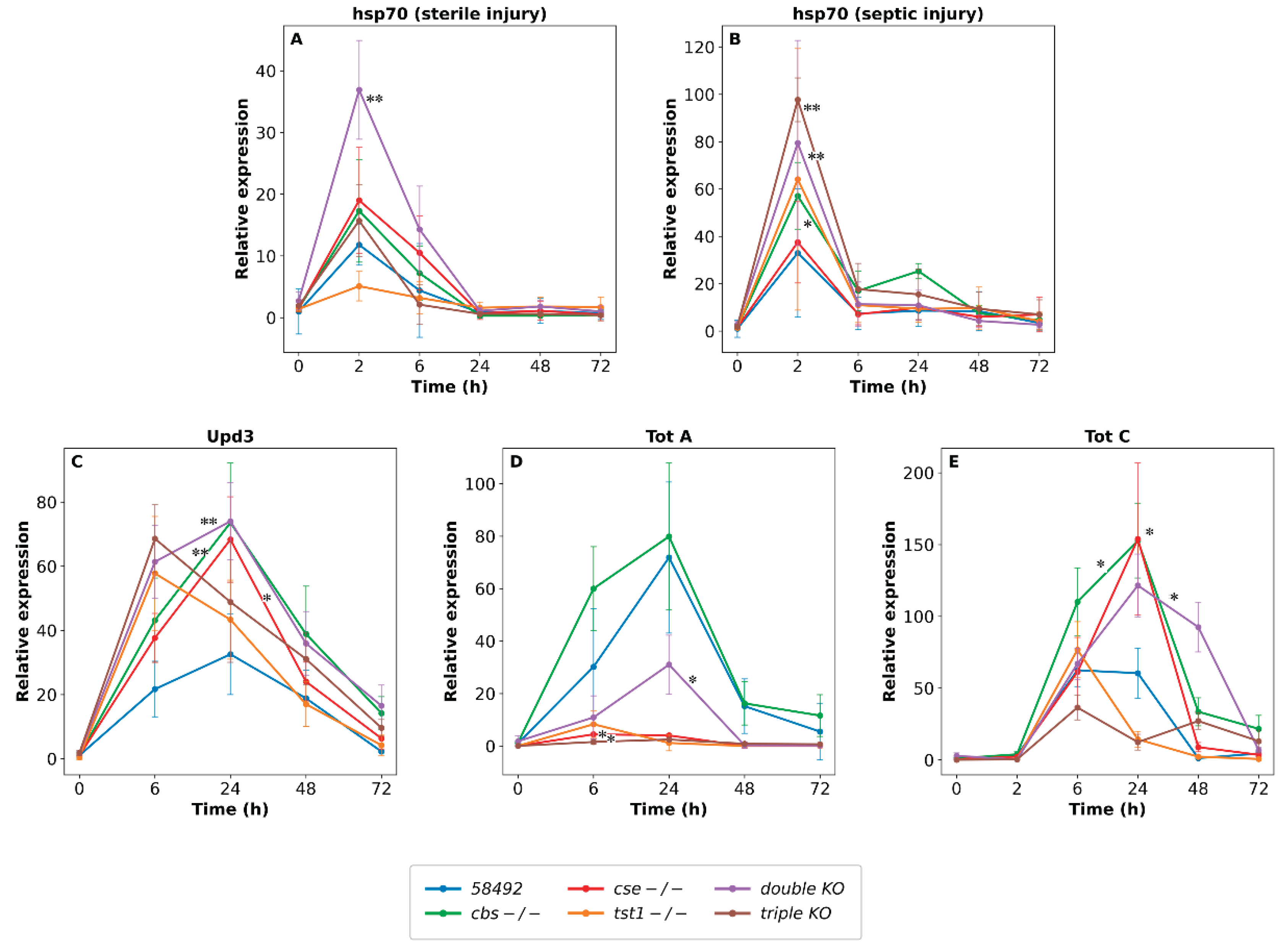
After septic injury, hsp23 and hsp68 were induced in all lines. However, in transcriptomic libraries at 6 h post-infection, we did not observe significant induction of the major heat shock protein hsp70. Previously, we demonstrated complex interactions between the Hsps system and genes involved in H2S synthesis and metabolism in flies [80]. Since even sterile injury induces ROS production [17], we monitored hsp70 expression by qRT-PCR in KO lines after both sterile and septic injuries.
Our experiments demonstrated that cuticle damage per se induced hsp70 transcription in all studied lines. Notably, a significant increase in hsp70 induction after septic injury compared to sterile injection occurred preferentially in KO lines. In both cases, induction peaked at 2 h post-injury. Sterile injury caused maximal hsp70 induction in the double KO line, whereas septic injury caused maximal hsp70 induction in both double and triple KOs. These data suggest that basic proteostasis is severely disrupted in KO flies. Even minor damage to the cuticle leads to the formation of a greater number of damaged or aggregated proteins, which appears to trigger a stronger signal for the activation of HSF1—the primary regulator of proteotoxic stress and an inducer of Hsps. During infection, oxidative stress intensifies, leading to even stronger hsp70 induction in double/triple KOs. Among other stress-inducible genes regulated by the JAK/STAT pathway are genes of the Turandot family. In Drosophila this family consists of eight genes whose expression is altered under various stress conditions, including infection [81]. Turandots protect the epithelial cells of the tracheal system from the damaging effects of AMPs [79]. From our transcriptomic data at 6 h post-infection point, totA was induced only in the control line, while totC was induced in all lines, maximally in control and double KO (Figure 6). Because Turandot genes typically show maximal expression at 16–24 h after infection [81,82], we performed qPCR to examine the dynamics of totA and totC expression in our experiments.
The analysis demonstrated that totA was induced mainly in control, CBS-/- flies, and to a lesser extent in double KO flies. On the other hand, totC was induced in all lines, peaking at 24 h in CBS-/-, CSE-/-, and double KO lines, but was minimally expressed in triple KO.
After septic injury, totM was induced in control and TST1-/- lines, totX in control, TST1-/-, and double KO (Figure 6). Thus, in the triple KO, we did not observe a significant increase in the expression levels of any genes belonging to the Turandot family after septic injury. This may account for the maximal compensatory hsp70 induction seen in this line [82].
Turandot genes (TotA, TotC, TotM) are controlled by the JAK/STAT pathway and are activated by the cytokine-like ligand Unpaired 3 (Upd3), as well as by the IMD pathway [83]. Upd3 is normally expressed at very low levels and was not detected in our transcriptomic analysis; therefore, we examined its expression dynamics by qRT-PCR. The experiments demonstrated that Upd3 was maximally induced in double and triple KOs and thus cannot explain the differential totA/totC expression observed in the studied lines. Taken together, these data indicate that triple KO flies fail to mount a proper Turandot response upon infection, which probably contributes to their severe proteostatic imbalance, judging by the highest hsp70 induction. This observation highlights the critical role of the JAK/STAT-Turandot axis in mitigating stress-induced damage under conditions of H₂S deficiency.
Figure 7.
Kinetics of the relative expression of hsp70, Upd3, TotA, and TotC genes measured by qRT-PCR following injury, * p<0.05, ** p<0.001.
Figure 7.
Kinetics of the relative expression of hsp70, Upd3, TotA, and TotC genes measured by qRT-PCR following injury, * p<0.05, ** p<0.001.
4. Discussion
This study demonstrates that a genetic disruption of sulfur metabolism—specifically, combined deletions of the cbs, cse, and tst1 genes in D. melanogaster significantly alters the homeostasis of the whole innate immune system, leading to chronic inflammation, immune dysregulation, and altered survival rates following septic injury. The accumulated results indicate that the transsulfuration pathway and endogenous H2S production are key modulators of immune signaling, metabolic reprogramming, and stress responses in the intact organism.
4.1. Survival and Bacterial load: Genotype-Specific Outcomes
Survival experiments following septic injury with B. subtilis revealed genotype-dependent differences. The control line (58492) and the TST1-/- single KO exhibited the best survival rates, whereas the triple KO showed the poorest survival. The double KO displayed the greatest variation in survival outcomes, suggesting an unstable immune–metabolic equilibrium.
Analysis of bacterial load kinetics provided mechanistic insights into these survival patterns. The control line exhibited a classic infectious curve: exponential bacterial proliferation peaking at 48–96 hours (median ~6×10⁴ CFU), followed by active clearance and restoration of microbial homeostasis by 172 hours. Triple KO displayed an atypical phenotype: bacterial proliferation was partially suppressed (peak median ~10⁴ CFU, lower than controls), yet flies died at significantly lower bacterial loads (10⁴–10⁵ CFU compared to ~10⁶ CFU in controls). This suggests that mortality in organisms with a triple knockout is not caused by an excessive bacterial load, but rather by immune-mediated damage.
The double KO showed intermediate characteristics: bacterial load peaked at 48 hours, and active elimination began after 72–96 hours, coinciding with peak fly mortality. Analysis of bacterial load at the time of death (PLUD) confirmed that single KOs die at bacterial loads comparable to controls (~10⁶ CFU), whereas double and triple KOs exhibit high variability, with many individuals dying at significantly lower CFU values (10⁴–10⁵). This supports the conclusion that combined loss of H₂S-producing enzymes leads to death from immune-mediated pathology due to excessive inflammatory tissue damage, rather than from uncontrolled infection.
4.2. Chronic Immune Activation Under Basal Conditions
H2S deficiency disrupts standard metabolic housekeeping, generating a state of chronic oxidative stress and hyperhomocysteinemia, which acts as a continuous, sterile danger signal. Our transcriptomic data revealed that this redox imbalance establishes a new, primed immune threshold under basal conditions. To counteract spontaneous autoinflammatory pathology, the organism upregulates a set of compensatory defense mechanisms. Thus, we observed significant basal induction of detoxification networks—such as Cytochrome P450 and Glutathione S-transferases (GSTs) (Supplemental Figure 5)—and specific protective proteins, including serpins and Bomanins (Figure 3 and Figure 4).
Our transcriptomic analysis of KO lines shows that H₂S deficiency radically alters the transcriptional landscape of the entire innate immune system, stress responses, and several key metabolic pathways in Drosophila.
Under control conditions, double and triple KO flies exhibited increased expression of pattern recognition receptors (e.g., PGRP-LC, PGRP-LA), negative regulators (PGRP-LF, PGRP-LB), and stress-related genes (TAK1, kayak, Mmp1), but did not show an increase of most AMPs. This mismatch suggests that H2S deficiency drives the immune system of KO flies to a state of readiness. However, compensatory mechanisms (including increased amidase activity and serpin expression) prevent the development of spontaneous autoinflammatory pathology. Thus, the increased expression of such serpins as Spn77Ba and Spn47C probably represents a protective mechanism against systemic inflammation and epithelial damage in these lines.
It is of note that Spn77Ba is the main inhibitor of melanization proteases, protecting tracheal and intestinal epithelia [45,55]; Spn47C, an ortholog of human SERPINH1/Hsp47 [84], may bind IRE1 under proteotoxic stress to prevent cell death.
The upregulation of cysteine-rich Bomanins (e.g., BomBc1, BomS6) and Defensin under sterile conditions is of particular interest. We hypothesize that these peptides function as a systemic "molecular shield". By interacting with lipid rafts, they probably stabilize host cell membranes against the deleterious effects of reactive oxygen species (ROS) and lipid peroxidation [49,85], acting as a direct compensatory mechanism in KO lines for the loss of H2S-mediated antioxidant capacity.
4.3. Double and Triple Knockout: Immune Hyperactivation Caused by H2S Deficiency
The double knockout (cbs/cse-/-) results in immune hyperactivation caused by H2S deficiency. These flies show the highest AMPs induction, a strong stress response (high hsp70 levels, high TotC levels). This phenotype is consistent with a model in which H₂S acts as an anti-inflammatory mediator [5] and its removal can lead to an uncontrolled immune response [11]. After septic injury, double KOs displayed a strong induction of IMD and Toll pathway components, including Relish, Dif, multiple serine proteases (Hayan, Sp7, SPE), and a broad array of AMPs (Diptericins, Cecropins, Edin, Attacin C/B/D). Notably, Attacin D—an atypical IMD effector that causes damage by aggregating within Malpighian tubule cells—was induced only in double and triple KOs, suggesting that renal pathology may contribute to the poor survival of these lines. This transcriptional hyper-reactivity was balanced by co-induction of negative regulators (PGRP-SC2, Diedel, Necrotic), indicating that feedback inhibition remains operational but may be insufficient to restore homeostasis.
The triple KO flies are of particular interest. Although in this line H2S level is similar to that of double KO flies (Shilova et al., 2025), these flies exhibited poorer survival after bacterial infection and less pronounced AMPs induction in comparison with the double KO line. This paradox may be partially explained by our transcriptomic data. Thus, triple KO flies exhibit a significant upregulation of negative immune regulators, including pirk, PGRP-LB, and PGRP-SC1/2. This indicates that the immune system of this line has probably reached a state of exhaustion or active suppression. However, this suppression appears to reduce flies' ability to mount effective antimicrobial defenses when needed, leading to low survival rates in the case of septic infections. Besides, the KO of the tst1 gene apparently exacerbates intoxication. The thing is that since TST1 is a sulfurtransferase involved in cyanide detoxification [86], its removal may result in the accumulation of cyanide or other reactive forms of sulfur. In the context of an already compromised transsulfuration pathway (e.g., in double KO), this causes additional metabolic stress, which impairs the viability and resistance of the host organism as well as immune dysregulation.
This interpretation is supported by the peculiar characteristics of the TST1-/- KO flies. This line, deprived of the enzyme involved in detoxification processes, exhibits a surprisingly mild immune phenotype: in particular, its survival rate following septic injury was comparable to that of the control line, as was its basal transcriptome. However, septic injury induces multiple differences in the expression of immune-related genes in comparison with the control flies. This indicates that loss of tst1, while insufficient to cause chronic inflammation, does modulate the dynamics of the immune response upon infection, and its importance is unmasked under stress conditions.
It has also recently been shown that cyanide, produced endogenously in mammalian cells and tissues, performs various functions and provides cytoprotection at physiological concentrations [87]. Thus, its primary role in immunity may be indirect, by maintaining general metabolic health, but it becomes critically important when the transsulfuration pathway (cbs/cse) is already compromised.
4.4. Digestive Proteases: Transcriptomic Evidence for Levelling of Expression in Knockout Lines After Septic Injury
The insect gut serves not only as a digestive organ but also as a key component of the innate immune system [71,88], acting as the first line of defense against foodborne pathogens. Our data reveal that disruption of sulfur metabolism profoundly alters the transcriptional regulation of several digestive enzymes, with distinct genotype-specific patterns.
Under control conditions, double and triple knockout lines showed increased expression of trypsin-encoding genes and several Jonah family chymotrypsin-like serine proteases (Jon66Cii, Jon44E, Jon74E). However, in the triple KO, increased expression levels were observed for many other members of the Jonah family (including the Jon99, Jon25, and Jon65 subfamilies). This expanded Jonah response may reflect more severe gut dysbiosis or a broader compensatory metabolic adaptation in triple KOs, likely linked to tst1 loss and cyanide toxicity. After septic injury, the control and TST1-/- KO lines exhibited a pronounced reduction in expression levels of both trypsin-encoding genes and Jonah proteases, reflecting the classical shift of energy expenditure from digestion to immunity [75]. A similar, though less pronounced, reduction was observed in the triple KO line. In contrast, no such decrease was observed in double KOs. It is important to note that when comparing the relative expression levels of these genes in infected double and triple KOs with those in the infected control line (58492), their expression profiles continued to show elevated expression (Figure 5). This demonstrates that KO lines with low H2S maintain a stable homeostatic balance in digestive protease expression during the immune response.
Notably, double KO flies display elevated basal transcripts of tobi, maltases, and ilp2, suggesting that these flies experience chronic energy stress, likely due to oxidative damage and metabolic inefficiency. Following infection, all flies exhibit increased expression levels of tobi and maltases. The level of ilp2 transcripts decreases in double- and triple-KO, but not in control line, resulting in equally low expression levels across all lines. This observation suggests that the Toll-mediated suppression of insulin signaling in all lines redirects nutrients toward the immune defense.
4.5. Stress Pathway Activation.
Basal elevation of TAK1, kayak, and Mmp1 in double, triple KOs suggests that loss of transsulfuration metabolites primes stress-related signaling even without infection. The activation of JNK likely stimulates the expression of detoxification enzymes (e.g., CYP, GST, UGT) and TEP opsonins [16,62], contributing to the observed state of immune readiness. In double and triple KO lines, a more pronounced induction of hsp70 is observed following a septic injury, indicating a greater degree of disruption in cellular proteostasis. Furthermore, the triple KO flies, which exhibited the lowest levels of Turandot induction (totA, totC, totM, totX), also showed the greatest increase in hsp70 levels following septic injury, which is consistent with reports that Turandot gene knockdown increases hsp70 expression. This inverse correlation supports the model postulating that Turandot proteins act in the hemolymph to mitigate extracellular damage, analogous to the intracellular function of Hsps [82]. The failure of the triple KO to mount a proper Turandot response may contribute to its extreme sensitivity to infection.
5. Conclusions
H₂S acts as a critical metabolic checkpoint that inhibits the basal activation of immune, stress, and metabolic pathways. Its deficiency leads to the sustained activation of these pathways in non-infectious conditions, resulting in the development of chronic inflammation. This state of readiness leads to an overly active immune response during infection, while simultaneously triggering compensatory defense mechanisms (Bomanins, Serpins, Turandots) to limit collateral damage. These results establish a new link between sulfur metabolism, immune homeostasis, proteostasis, and metabolic resilience in vivo.
This work establishes that sulfur metabolism in Drosophila is not a monolithic regulator but a modular network whose disruption leads to distinct immunopathological states. H₂S produced by CBS/CSE is a key anti-inflammatory signal. Importantly, the observed immune dysregulation is primarily driven by H₂S deficiency rather than by homocysteine accumulation. This conclusion is supported by the fact that the single CBS⁻/⁻ line, despite exhibiting hyperhomocysteinemia comparable to the double KO, retains higher H₂S levels and shows a significantly milder phenotype after infection. The TST1 enzyme plays a key role in metabolic detoxification, the importance of which becomes evident when the transsulfuration pathway is disrupted. These findings provide a conceptual framework for a deeper understanding of human conditions such as severe hyperhomocysteinemia or combined metabolic disorders, which may be accompanied by immune dysfunction and systemic toxicity. The accumulated results argue for therapeutic strategies that not only modulate H₂S levels but also support broader sulfur metabolic flux and detoxification pathways to restore holistic immune-metabolic homeostasis.
6. Limitations
6.1. Transcriptional Level Only, One 6h Time Point in Transcriptomic library: Our Conclusions Are Primarily Based on the Transcriptomic Data. Protein Abundance, Enzymatic Activity, and Functional Outcomes Were not Directly Measured.
6.2. Single Pathogen. Responses to other Pathogens may Differ.
6.3. Suggested TST1 Toxicity Mechanism Is Speculative: Suggested Cyanide Accumulation in Triple KO Flies Requires Direct Biochemical Validation.
6.4. Gut Microbiota not Profiled: Changes in Jonah Proteases Suggest Dysbiosis, but this Was not Directly Tested.
Despite these limitations, our data for the first time provide a comprehensive transcriptional map of how H2S deficiency remodels innate immunity, stress responses, carbohydrate metabolism, and insulin like peptide expression in Drosophila. This study provides a framework for understanding human conditions where sulfur metabolism is disrupted, such as hyperhomocysteinemia or combined metabolic disorders, where immune dysfunction and systemic toxicity coexist.
Supplementary Materials
The following supporting information can be downloaded at the website of this paper posted on Preprints.org: Table S1. List of primers used for qRT-PCR experiments. Figure S1. Growth kinetics of Bacillus subtilis 168 in wild-type (58492), single-deletion mutants (cbs -/-, cse -/-, tst1 -/-), and multiple-deletion mutants (double/triple KO). Figure S2. Box plots illustrating differences in the expression of genes involved in the immune response. Figure S3. Box plots showing differences in Serpin expression under control conditions and following septic injury. Figure S4. Box plots showing differences in the expression of opsonins and lectins under control conditions and following septic injury. Figure S5. Heat map of detoxification system components with varying expression levels.
Author Contributions
O.Z., V.S., M.E., designed research, V.S., O.G., V.A., A.A., K.P., A.D., performed experiments; A.R. software, V.S., A.R.,V.A., O.Z., D.G. analyzed data; O.Z., V.S., and M.E. wrote the paper. All authors have read and agreed to the published version of the manuscript.
Funding
All the experiments reported in this study were supported by a grant from the Russian Science Foundation (grant № 24-14-00216 (OG)).
Institutional Review Board Statement
Not applicable.
Informed Consent Statement
Not applicable.
Data Availability Statement
The original contributions presented in this study are included in the article and Supplementary Materials.
Acknowledgments
We are grateful to E. Zelentsova, L. Chuvakova for their assistance in the experiments and discussion of the results.
Conflicts of Interest
The authors declare no conflicts of interest.
References
- Akira, S.; Uematsu, S.; Takeuchi, O. Pathogen Recognition and Innate Immunity. Cell 2006, 124, 783–801. [Google Scholar] [CrossRef] [PubMed]
- Alemán, M. “The Good, the Bad and the Ugly”: Interplay of Innate Immunity and Inflammation. Cell. Microbiol. 2022, 2022, 1–11. [Google Scholar] [CrossRef]
- Chi, H. Immunometabolism at the Intersection of Metabolic Signaling, Cell Fate, and Systems Immunology. Cell Mol. Immunol. 2022, 19, 299–302. [Google Scholar] [CrossRef] [PubMed]
- Mironov, A.; Seregina, T.; Nagornykh, M.; Luhachack, L.G.; Korolkova, N.; Lopes, L.E.; Kotova, V.; Zavilgelsky, G.; Shakulov, R.; Shatalin, K.; et al. Mechanism of H2 S-Mediated Protection against Oxidative Stress in Escherichia Coli. Proc. Natl. Acad. Sci. U.S.A. 2017, 114, 6022–6027. [Google Scholar] [CrossRef] [PubMed]
- Wallace, J.L.; Blackler, R.W.; Chan, M.V.; Da Silva, G.J.; Elsheikh, W.; Flannigan, K.L.; Gamaniek, I.; Manko, A.; Wang, L.; Motta, J.-P.; et al. Anti-Inflammatory and Cytoprotective Actions of Hydrogen Sulfide: Translation to Therapeutics. Antioxid. Redox Signal. 2015, 22, 398–410. [Google Scholar] [CrossRef] [PubMed]
- Kimura, H. Hydrogen Sulfide and Polysulfides as Signaling Molecules. Proc. Jpn. Acad. Ser. B Phys. Biol. Sci. 2015, 91, 131–159. [Google Scholar] [CrossRef] [PubMed]
- Libiad, M.; Motl, N.; Akey, D.L.; Sakamoto, N.; Fearon, E.R.; Smith, J.L.; Banerjee, R. Thiosulfate Sulfurtransferase-like Domain–Containing 1 Protein Interacts with Thioredoxin. J. Biol. Chem. 2018, 293, 2675–2686. [Google Scholar] [CrossRef] [PubMed]
- Melideo, S.L.; Jackson, M.R.; Jorns, M.S. Biosynthesis of a Central Intermediate in Hydrogen Sulfide Metabolism by a Novel Human Sulfurtransferase and Its Yeast Ortholog. Biochemistry 2014, 53, 4739–4753. [Google Scholar] [CrossRef] [PubMed]
- Nagahara, N.; Tanaka, M.; Tanaka, Y.; Ito, T. Novel Characterization of Antioxidant Enzyme, 3-Mercaptopyruvate Sulfurtransferase-Knockout Mice: Overexpression of the Evolutionarily-Related Enzyme Rhodanese. Antioxidants 2019, 8, 116. [Google Scholar] [CrossRef] [PubMed]
- Cirino, G.; Szabo, C.; Papapetropoulos, A. Physiological Roles of Hydrogen Sulfide in Mammalian Cells, Tissues, and Organs. Physiol. Rev. 2023, 103, 31–276. [Google Scholar] [CrossRef] [PubMed]
- Dilek, N.; Papapetropoulos, A.; Toliver-Kinsky, T.; Szabo, C. Hydrogen Sulfide: An Endogenous Regulator of the Immune System. Pharmacol. Res. 2020, 161, 105119. [Google Scholar] [CrossRef] [PubMed]
- Makkar, P.; Singh, C.K.; V, N.; Narang, P.K.; Sodhi, K.K. Insect Immunity Unveiled: Exploring the Molecular and Cellular Defenses against Microbial Threats. Arch. Microbiol. 2026, 208, 379. [Google Scholar] [CrossRef] [PubMed]
- De Gregorio, E.; Spellman, P.T.; Tzou, P.; Rubin, G.M.; Lemaitre, B. The Toll and Imd Pathways Are the Major Regulators of the Immune Response in Drosophila. EMBO J. 2002, 21, 2568–2579. [Google Scholar] [CrossRef] [PubMed]
- Lemaitre, B.; Hoffmann, J. The Host Defense of Drosophila Melanogaster. Annu. Rev. Immunol. 2007, 25, 697–743. [Google Scholar] [CrossRef] [PubMed]
- Valanne, S.; Wang, J.-H.; Rämet, M. The Drosophila Toll Signaling Pathway. J. Immunol. 2011, 186, 649–656. [Google Scholar] [CrossRef] [PubMed]
- Shilova, V.Y.; Garbuz, D.G.; Chuvakova, L.N.; Rezvykh, A.P.; Funikov, S.Y.; Davletshin, A.I.; Sorokina, S.Y.; Nikitina, E.A.; Gorenskaya, O.; Evgen’ev, M.B.; et al. The Biological Consequences of the Knockout of Genes Involved in the Synthesis and Metabolism of H2S in Drosophila Melanogaster. Antioxidants 2025, 14, 693. [Google Scholar] [CrossRef] [PubMed]
- Chakrabarti, S.; Visweswariah, S.S. Intramacrophage ROS Primes the Innate Immune System via JAK/STAT and Toll Activation. Cell Rep. 2020, 33, 108368. [Google Scholar] [CrossRef] [PubMed]
- Bolhassani, A.; Agi, E. Heat Shock Proteins in Infection. Clin. Chim. Acta 2019, 498, 90–100. [Google Scholar] [CrossRef] [PubMed]
- Gan, T.; Fan, L.; Zhao, L.; Misra, M.; Liu, M.; Zhang, M.; Su, Y. JNK Signaling in Drosophila Aging and Longevity. IJMS 2021, 22, 9649. [Google Scholar] [CrossRef] [PubMed]
- Darby, A.M.; Lazzaro, B.P. Interactions between Innate Immunity and Insulin Signaling Affect Resistance to Infection in Insects. Front. Immunol. 2023, 14, 1276357. [Google Scholar] [CrossRef] [PubMed]
- Duneau, D.; Ferdy, J.-B.; Revah, J.; Kondolf, H.; Ortiz, G.A.; Lazzaro, B.P.; Buchon, N. Stochastic Variation in the Initial Phase of Bacterial Infection Predicts the Probability of Survival in D. Melanogaster. eLife 2017, 6, e28298. [Google Scholar] [CrossRef] [PubMed]
- Khalil, S.; Jacobson, E.; Chambers, M.C.; Lazzaro, B.P. Systemic Bacterial Infection and Immune Defense Phenotypes in Drosophila Melanogaster. JoVE 2015, 52613. [Google Scholar] [CrossRef] [PubMed]
- Ashe, S.; Maji, U.J.; Sen, R.; Mohanty, S.; Maiti, N.K. Specific Oligonucleotide Primers for Detection of Endoglucanase Positive Bacillus Subtilis by PCR. 3 Biotech. 2014, 4, 461–465. [Google Scholar] [CrossRef] [PubMed]
- Ryckebusch, F.; Tian, Y.; Rapin, M.; Schüpfer, F.; Hanson, M.A.; Lemaitre, B. Layers of Immunity: Deconstructing the Drosophila Effector Response. eLife 2025, 14, RP107030. [Google Scholar] [CrossRef]
- Bolger, A.M.; Lohse, M.; Usadel, B. Trimmomatic: A Flexible Trimmer for Illumina Sequence Data. Bioinformatics 2014, 30, 2114–2120. [Google Scholar] [CrossRef] [PubMed]
- Dobin, A.; Davis, C.A.; Schlesinger, F.; Drenkow, J.; Zaleski, C.; Jha, S.; Batut, P.; Chaisson, M.; Gingeras, T.R. STAR: Ultrafast Universal RNA-Seq Aligner. Bioinformatics 2013, 29, 15–21. [Google Scholar] [CrossRef] [PubMed]
- Li, H.; Handsaker, B.; Wysoker, A.; Fennell, T.; Ruan, J.; Homer, N.; Marth, G.; Abecasis, G.; Durbin, R. 1000 Genome Project Data Processing Subgroup. The Sequence Alignment/Map Format and SAMtools. Bioinformatics 2009, 25, 2078–2079. [Google Scholar] [CrossRef] [PubMed]
- Liao, Y.; Smyth, G.K.; Shi, W. featureCounts: An Efficient General Purpose Program for Assigning Sequence Reads to Genomic Features. Bioinformatics 2014, 30, 923–930. [Google Scholar] [CrossRef] [PubMed]
- Robinson, M.D.; McCarthy, D.J.; Smyth, G.K. edgeR : A Bioconductor Package for Differential Expression Analysis of Digital Gene Expression Data. Bioinformatics 2010, 26, 139–140. [Google Scholar] [CrossRef] [PubMed]
- Yu, G.; Wang, L.-G.; Han, Y.; He, Q.-Y. clusterProfiler: An R Package for Comparing Biological Themes Among Gene Clusters. OMICS A J. Integr. Biol. 2012, 16, 284–287. [Google Scholar] [CrossRef] [PubMed]
- Kurata, S. Peptidoglycan Recognition Proteins in Drosophila Immunity. Dev. Comp. Immunol. 2014, 42, 36–41. [Google Scholar] [CrossRef] [PubMed]
- Maillet, F.; Bischoff, V.; Vignal, C.; Hoffmann, J.; Royet, J. The Drosophila Peptidoglycan Recognition Protein PGRP-LF Blocks PGRP-LC and IMD/JNK Pathway Activation. Cell Host Microbe 2008, 3, 293–303. [Google Scholar] [CrossRef] [PubMed]
- Kleino, A.; Myllymäki, H.; Kallio, J.; Vanha-aho, L.-M.; Oksanen, K.; Ulvila, J.; Hultmark, D.; Valanne, S.; Rämet, M. Pirk Is a Negative Regulator of the Drosophila Imd Pathway. J. Immunol. 2008, 180, 5413–5422. [Google Scholar] [CrossRef] [PubMed]
- Takehana, A.; Yano, T.; Mita, S.; Kotani, A.; Oshima, Y.; Kurata, S. Peptidoglycan Recognition Protein (PGRP)-LE and PGRP-LC Act Synergistically in Drosophila Immunity. EMBO J. 2004, 23, 4690–4700. [Google Scholar] [CrossRef] [PubMed]
- Iatsenko, I.; Kondo, S.; Mengin-Lecreulx, D.; Lemaitre, B. PGRP-SD, an Extracellular Pattern-Recognition Receptor, Enhances Peptidoglycan-Mediated Activation of the Drosophila Imd Pathway. Immunity 2016, 45, 1013–1023. [Google Scholar] [CrossRef] [PubMed]
- Zaidman-Rémy, A.; Poidevin, M.; Hervé, M.; Welchman, D.P.; Paredes, J.C.; Fahlander, C.; Steiner, H.; Mengin-Lecreulx, D.; Lemaitre, B. Drosophila Immunity: Analysis of PGRP-SB1 Expression, Enzymatic Activity and Function. PLoS ONE 2011, 6, e17231. [Google Scholar] [CrossRef] [PubMed]
- Lamiable, O.; Kellenberger, C.; Kemp, C.; Troxler, L.; Pelte, N.; Boutros, M.; Marques, J.T.; Daeffler, L.; Hoffmann, J.A.; Roussel, A.; et al. Cytokine Diedel and a Viral Homologue Suppress the IMD Pathway in Drosophila. Proc. Natl. Acad. Sci. U.S.A. 2016, 113, 698–703. [Google Scholar] [CrossRef] [PubMed]
- Zaidman-Rémy, A.; Hervé, M.; Poidevin, M.; Pili-Floury, S.; Kim, M.-S.; Blanot, D.; Oh, B.-H.; Ueda, R.; Mengin-Lecreulx, D.; Lemaitre, B. The Drosophila Amidase PGRP-LB Modulates the Immune Response to Bacterial Infection. Immunity 2006, 24, 463–473. [Google Scholar] [CrossRef] [PubMed]
- Bischoff, V.; Vignal, C.; Duvic, B.; Boneca, I.G.; Hoffmann, J.A.; Royet, J. Downregulation of the Drosophila Immune Response by Peptidoglycan-Recognition Proteins SC1 and SC2. PLoS Pathog. 2006, 2, e14. [Google Scholar] [CrossRef] [PubMed]
- Chen, L.; Paquette, N.; Mamoor, S.; Rus, F.; Nandy, A.; Leszyk, J.; Shaffer, S.A.; Silverman, N. Innate Immune Signaling in Drosophila Is Regulated by Transforming Growth Factor β (TGFβ)-Activated Kinase (Tak1)-Triggered Ubiquitin Editing. J. Biol. Chem. 2017, 292, 8738–8749. [Google Scholar] [CrossRef] [PubMed]
- Kallio, J.; Leinonen, A.; Ulvila, J.; Valanne, S.; Ezekowitz, R.A.; Rämet, M. Functional Analysis of Immune Response Genes in Drosophila Identifies JNK Pathway as a Regulator of Antimicrobial Peptide Gene Expression in S2 Cells. Microbes Infect. 2005, 7, 811–819. [Google Scholar] [CrossRef] [PubMed]
- Uhlirova, M.; Bohmann, D. JNK- and Fos-regulated Mmp1 Expression Cooperates with Ras to Induce Invasive Tumors in Drosophila. EMBO J. 2006, 25, 5294–5304. [Google Scholar] [CrossRef] [PubMed]
- Pelte, N.; Robertson, A.S.; Zou, Z.; Belorgey, D.; Dafforn, T.R.; Jiang, H.; Lomas, D.; Reichhart, J.-M.; Gubb, D. Immune Challenge Induces N-Terminal Cleavage of the Drosophila Serpin Necrotic. Insect Biochem. Mol. Biol. 2006, 36, 37–46. [Google Scholar] [CrossRef] [PubMed]
- Nam, H.; Jang, I.; You, H.; Lee, K.; Lee, W. Genetic Evidence of a Redox-dependent Systemic Wound Response via Hayan Protease-Phenoloxidase System in Drosophila. EMBO J. 2012, 31, 1253–1265. [Google Scholar] [CrossRef] [PubMed]
- Reichhart, J.-M.; Gubb, D.; Leclerc, V. The Drosophila Serpins. In Methods in Enzymology; Elsevier, 2011; Vol. 499, pp. 205–225. ISBN 978-0-12-386471-0. [Google Scholar]
- Veillard, F.; Troxler, L.; Reichhart, J.-M. Drosophila Melanogaster Clip-Domain Serine Proteases: Structure, Function and Regulation. Biochimie 2016, 122, 255–269. [Google Scholar] [CrossRef] [PubMed]
- Shan, T.; Wang, Y.; Bhattarai, K.; Jiang, H. An Evolutionarily Conserved Serine Protease Network Mediates Melanization and Toll Activation in Drosophila. Sci. Adv. 2023, 9, eadk2756. [Google Scholar] [CrossRef] [PubMed]
- Xu, R.; Lou, Y.; Tidu, A.; Bulet, P.; Heinekamp, T.; Martin, F.; Brakhage, A.; Li, Z.; Liégeois, S.; Ferrandon, D. The Toll Pathway Mediates Drosophila Resilience to Aspergillus Mycotoxins through Specific Bomanins. EMBO Rep. 2023, 24, EMBR202256036. [Google Scholar] [CrossRef]
- Lou, Y.; Zhang, B.; Zhang, Z.; Pan, Y.; Yang, J.; Li, L.; Huang, J.; Yuan, Z.; Liegeois, S.; Bulet, P.; et al. Distinct Bomanins at the Drosophila 55C Locus Function in Resistance and Resilience to Infections. EMBO Rep. 2026, 27, 629–653. [Google Scholar] [CrossRef] [PubMed]
- Lindsay, S.A.; Lin, S.J.H.; Wasserman, S.A. Short-Form Bomanins Mediate Humoral Immunity in Drosophila. J. Innate Immun. 2018, 10, 306–314. [Google Scholar] [CrossRef] [PubMed]
- Oi, A.; Shinoda, N.; Nagashima, S.; Miura, M.; Obata, F. A Nonsecretory Antimicrobial Peptide Mediates Inflammatory Organ Damage in Drosophila Renal Tubules. Cell Rep. 2025, 44, 115082. [Google Scholar] [CrossRef] [PubMed]
- Reichhart, J.-M. Tip of Another Iceberg: Drosophila Serpins. Trends Cell Biol. 2005, 15, 659–665. [Google Scholar] [CrossRef] [PubMed]
- Moore, J.; Wu, T.; Dhindsa, J.; El Fadel, O.; Le, A.; Perez, A.; Amoh, B.; Tarkunde, A.; Zhu, K.F.; Avalos, M.; et al. Longitudinal Multi-Omics in Alpha-Synuclein Drosophila Model Discriminates Disease- from Age-Associated Pathologies in Parkinson’s Disease. npj Park. Dis. 2025, 11, 46. [Google Scholar] [CrossRef] [PubMed]
- Handke, B.; Poernbacher, I.; Goetze, S.; Ahrens, C.H.; Omasits, U.; Marty, F.; Simigdala, N.; Meyer, I.; Wollscheid, B.; Brunner, E.; et al. The Hemolymph Proteome of Fed and Starved Drosophila Larvae. PLoS ONE 2013, 8, e67208. [Google Scholar] [CrossRef] [PubMed]
- Tang, H.; Kambris, Z.; Lemaitre, B.; Hashimoto, C. A Serpin That Regulates Immune Melanization in the Respiratory System of Drosophila. Dev. Cell 2008, 15, 617–626. [Google Scholar] [CrossRef] [PubMed]
- Scherfer, C.; Tang, H.; Kambris, Z.; Lhocine, N.; Hashimoto, C.; Lemaitre, B. Drosophila Serpin-28D Regulates Hemolymph Phenoloxidase Activity and Adult Pigmentation. Dev. Biol. 2008, 323, 189–196. [Google Scholar] [CrossRef] [PubMed]
- Shokal, U.; Eleftherianos, I. Evolution and Function of Thioester-Containing Proteins and the Complement System in the Innate Immune Response. Front. Immunol. 2017, 8, 759. [Google Scholar] [CrossRef] [PubMed]
- Lagueux, M.; Perrodou, E.; Levashina, E.A.; Capovilla, M.; Hoffmann, J.A. Constitutive Expression of a Complement-like Protein in Toll and JAK Gain-of-Function Mutants of Drosophila. Proc. Natl. Acad. Sci. U.S.A. 2000, 97, 11427–11432. [Google Scholar] [CrossRef] [PubMed]
- Dostálová, A.; Rommelaere, S.; Poidevin, M.; Lemaitre, B. Thioester-Containing Proteins Regulate the Toll Pathway and Play a Role in Drosophila Defence against Microbial Pathogens and Parasitoid Wasps. BMC Biol. 2017, 15, 79. [Google Scholar] [CrossRef] [PubMed]
- Garver, L.S.; De Almeida Oliveira, G.; Barillas-Mury, C. The JNK Pathway Is a Key Mediator of Anopheles Gambiae Antiplasmodial Immunity. PLoS Pathog. 2013, 9, e1003622. [Google Scholar] [CrossRef] [PubMed]
- Bou Aoun, R.; Hetru, C.; Troxler, L.; Doucet, D.; Ferrandon, D.; Matt, N. Analysis of Thioester-Containing Proteins during the Innate Immune Response of Drosophila Melanogaster. J. Innate Immun. 2011, 3, 52–64. [Google Scholar] [CrossRef]
- Tafesh-Edwards, G.; Eleftherianos, I. JNK Signaling in Drosophila Immunity and Homeostasis. Immunol. Lett. 2020, 226, 7–11. [Google Scholar] [CrossRef] [PubMed]
- Peirce, M.J.; Mitchell, S.N.; Kakani, E.G.; Scarpelli, P.; South, A.; Shaw, W.R.; Werling, K.L.; Gabrieli, P.; Marcenac, P.; Bordoni, M.; et al. JNK Signaling Regulates Oviposition in the Malaria Vector Anopheles Gambiae. Sci. Rep. 2020, 10, 14344. [Google Scholar] [CrossRef] [PubMed]
- Shokal, U.; Eleftherianos, I. Thioester-Containing Protein-4 Regulates the Drosophila Immune Signaling and Function against the Pathogen Photorhabdus. J. Innate Immun. 2017, 9, 83–93. [Google Scholar] [CrossRef] [PubMed]
- Petrignani, B.; Rommelaere, S.; Hakim-Mishnaevski, K.; Masson, F.; Ramond, E.; Hilu-Dadia, R.; Poidevin, M.; Kondo, S.; Kurant, E.; Lemaitre, B. A Secreted Factor NimrodB4 Promotes the Elimination of Apoptotic Corpses by Phagocytes in Drosophila. EMBO Rep. 2021, 22, EMBR202052262. [Google Scholar] [CrossRef] [PubMed]
- Ramond, E.; Petrignani, B.; Dudzic, J.P.; Boquete, J.; Poidevin, M.; Kondo, S.; Lemaitre, B. The Adipokine NimrodB5 Regulates Peripheral Hematopoiesis in Drosophila. FEBS J. 2020, 287, 3399–3426. [Google Scholar] [CrossRef] [PubMed]
- Lu, Y.; Su, F.; Li, Q.; Zhang, J.; Li, Y.; Tang, T.; Hu, Q.; Yu, X.-Q. Pattern Recognition Receptors in Drosophila Immune Responses. Dev. Comp. Immunol. 2020, 102, 103468. [Google Scholar] [CrossRef] [PubMed]
- Rämet, M.; Pearson, A.; Manfruelli, P.; Li, X.; Koziel, H.; Göbel, V.; Chung, E.; Krieger, M.; Ezekowitz, R.A.B. Drosophila Scavenger Receptor CI Is a Pattern Recognition Receptor for Bacteria. Immunity 2001, 15, 1027–1038. [Google Scholar] [CrossRef] [PubMed]
- Li, X.-J.; Yang, L.; Li, D.; Zhu, Y.-T.; Wang, Q.; Li, W.-W. Pathogen-Specific Binding Soluble Down Syndrome Cell Adhesion Molecule (Dscam) Regulates Phagocytosis via Membrane-Bound Dscam in Crab. Front. Immunol. 2018, 9, 801. [Google Scholar] [CrossRef] [PubMed]
- Kocks, C.; Cho, J.H.; Nehme, N.; Ulvila, J.; Pearson, A.M.; Meister, M.; Strom, C.; Conto, S.L.; Hetru, C.; Stuart, L.M.; et al. Eater, a Transmembrane Protein Mediating Phagocytosis of Bacterial Pathogens in Drosophila. Cell 2005, 123, 335–346. [Google Scholar] [CrossRef] [PubMed]
- Khan, S.A.; Kojour, M.A.M.; Han, Y.S. Recent Trends in Insect Gut Immunity. Front. Immunol. 2023, 14, 1272143. [Google Scholar] [CrossRef] [PubMed]
- Lemaitre, B.; Miguel-Aliaga, I. The Digestive Tract of Drosophila Melanogaster. Annu. Rev. Genet. 2013, 47, 377–404. [Google Scholar] [CrossRef] [PubMed]
- Yadav, S.; Eleftherianos, I. Participation of the Serine Protease Jonah66Ci in the Drosophila Antinematode Immune Response. Infect. Immun. 2019, 87, e00094-19. [Google Scholar] [CrossRef] [PubMed]
- Chtarbanova, S.; Lamiable, O.; Lee, K.-Z.; Galiana, D.; Troxler, L.; Meignin, C.; Hetru, C.; Hoffmann, J.A.; Daeffler, L.; Imler, J.-L. Drosophila C Virus Systemic Infection Leads to Intestinal Obstruction. J. Virol. 2014, 88, 14057–14069. [Google Scholar] [CrossRef] [PubMed]
- Bland, M.L. Regulating Metabolism to Shape Immune Function: Lessons from Drosophila. Semin. Cell Dev. Biol. 2023, 138, 128–141. [Google Scholar] [CrossRef] [PubMed]
- Buch, S.; Melcher, C.; Bauer, M.; Katzenberger, J.; Pankratz, M.J. Opposing Effects of Dietary Protein and Sugar Regulate a Transcriptional Target of Drosophila Insulin-like Peptide Signaling. Cell Metab. 2008, 7, 321–332. [Google Scholar] [CrossRef] [PubMed]
- Kodrík, D.; Bednářová, A.; Zemanová, M.; Krishnan, N. Hormonal Regulation of Response to Oxidative Stress in Insects—An Update. IJMS 2015, 16, 25788–25816. [Google Scholar] [CrossRef] [PubMed]
- Roth, S.W.; Bitterman, M.D.; Birnbaum, M.J.; Bland, M.L. Innate Immune Signaling in Drosophila Blocks Insulin Signaling by Uncoupling PI(3,4,5)P3 Production and Akt Activation. Cell Rep. 2018, 22, 2550–2556. [Google Scholar] [CrossRef] [PubMed]
- Rommelaere, S.; Carboni, A.; Bada Juarez, J.F.; Boquete, J.-P.; Abriata, L.A.; Teixeira Pinto Meireles, F.; Rukes, V.; Vincent, C.; Kondo, S.; Dionne, M.S.; et al. A Humoral Stress Response Protects Drosophila Tissues from Antimicrobial Peptides. Curr. Biol. 2024, 34, 1426–1437.e6. [Google Scholar] [CrossRef] [PubMed]
- Shilova, V.; Zatsepina, O.; Zakluta, A.; Karpov, D.; Chuvakova, L.; Garbuz, D.; Evgen’ev, M. Age-Dependent Expression Profiles of Two Adaptogenic Systems and Thermotolerance in Drosophila Melanogaster. Cell Stress Chaperones 2020, 25, 305–315. [Google Scholar] [CrossRef] [PubMed]
- Ekengren, S.; Hultmark, D. A Family of Turandot-Related Genes in the Humoral Stress Response of Drosophila. Biochem. Biophys. Res. Commun. 2001, 284, 998–1003. [Google Scholar] [CrossRef] [PubMed]
- Amstrup, A.B.; Bæk, I.; Loeschcke, V.; Givskov Sørensen, J. A Functional Study of the Role of Turandot Genes in Drosophila Melanogaster: An Emerging Candidate Mechanism for Inducible Heat Tolerance. J. Insect Physiol. 2022, 143, 104456. [Google Scholar] [CrossRef] [PubMed]
- Agaisse, H.; Petersen, U.-M.; Boutros, M.; Mathey-Prevot, B.; Perrimon, N. Signaling Role of Hemocytes in Drosophila JAK/STAT-Dependent Response to Septic Injury. Dev. Cell 2003, 5, 441–450. [Google Scholar] [CrossRef] [PubMed]
- Kumar, A.; Bhandari, A.; Sarde, S.J.; Goswami, C. Ancestry & Molecular Evolutionary Analyses of Heat Shock Protein 47 kDa (HSP47/SERPINH1). Sci. Rep. 2017, 7, 10394. [Google Scholar] [CrossRef] [PubMed]
- Munteanu, C.; Turnea, M.A.; Rotariu, M. Hydrogen Sulfide: An Emerging Regulator of Oxidative Stress and Cellular Homeostasis—A Comprehensive One-Year Review. Antioxidants 2023, 12, 1737. [Google Scholar] [CrossRef] [PubMed]
- Luo, Y.; Melhem, S.; Feelisch, M.; Chatre, L.; Morton, N.M.; Dolga, A.M.; Van Goor, H. Thiosulphate Sulfurtransferase: Biological Roles and Therapeutic Potential. Redox Biol. 2025, 82, 103595. [Google Scholar] [CrossRef] [PubMed]
- Zuhra, K.; Petrosino, M.; Janickova, L.; Petric, J.; Ascenção, K.; Vignane, T.; Khalaf, M.; Philipp, T.M.; Ravani, S.; Anand, A.; et al. Regulation of Mammalian Cellular Metabolism by Endogenous Cyanide Production. Nat. Metab. 2025, 7, 531–555. [Google Scholar] [CrossRef] [PubMed]
- Zeng, T.; Jaffar, S.; Xu, Y.; Qi, Y. The Intestinal Immune Defense System in Insects. IJMS 2022, 23, 15132. [Google Scholar] [CrossRef] [PubMed]
Figure 1.
Analysis of the immune response and survival in Drosophila lines with knockouts of the transsulfuration pathway genes. A. Survival kinetics following sterile injury. H. Survival kinetics following septic injury with B. subtilis. The median survival time was calculated. To compare survival curves the log-rank test was used. The statistical significance of differences in survival time was assessed using the Bonferroni-corrections. * p < 0.05, **p < 0.001 (KOs were compared with 58492 line. (B–G). Kinetics of relative expression of immune response genes, measured by qRT-PCR following sterile and septic (I–N) injury. Data represents the mean ± standard deviation from three biological replicates.* p< 0.05, ** p<0.001, t-test. O. Kinetics of B. subtilis load in the control line (58492) and lines with single and combined gene KOs (cbs, cse, tst1). Solid lines indicate a fourth-order polynomial regression. Shaded areas represent the 95% confidence intervals of the polynomial approximation. All experiments were performed with n ≥ 3 independent biological replicates. There are differences among all six groups. Statistical significance was estimated using one-way ANOVA (p<0.05). P. Measurement of pathogen load upon death (PLUD) following B. subtilis infection. Each data point represents a pooled sample from three flies. The differences are not statistically significant.
Figure 1.
Analysis of the immune response and survival in Drosophila lines with knockouts of the transsulfuration pathway genes. A. Survival kinetics following sterile injury. H. Survival kinetics following septic injury with B. subtilis. The median survival time was calculated. To compare survival curves the log-rank test was used. The statistical significance of differences in survival time was assessed using the Bonferroni-corrections. * p < 0.05, **p < 0.001 (KOs were compared with 58492 line. (B–G). Kinetics of relative expression of immune response genes, measured by qRT-PCR following sterile and septic (I–N) injury. Data represents the mean ± standard deviation from three biological replicates.* p< 0.05, ** p<0.001, t-test. O. Kinetics of B. subtilis load in the control line (58492) and lines with single and combined gene KOs (cbs, cse, tst1). Solid lines indicate a fourth-order polynomial regression. Shaded areas represent the 95% confidence intervals of the polynomial approximation. All experiments were performed with n ≥ 3 independent biological replicates. There are differences among all six groups. Statistical significance was estimated using one-way ANOVA (p<0.05). P. Measurement of pathogen load upon death (PLUD) following B. subtilis infection. Each data point represents a pooled sample from three flies. The differences are not statistically significant.
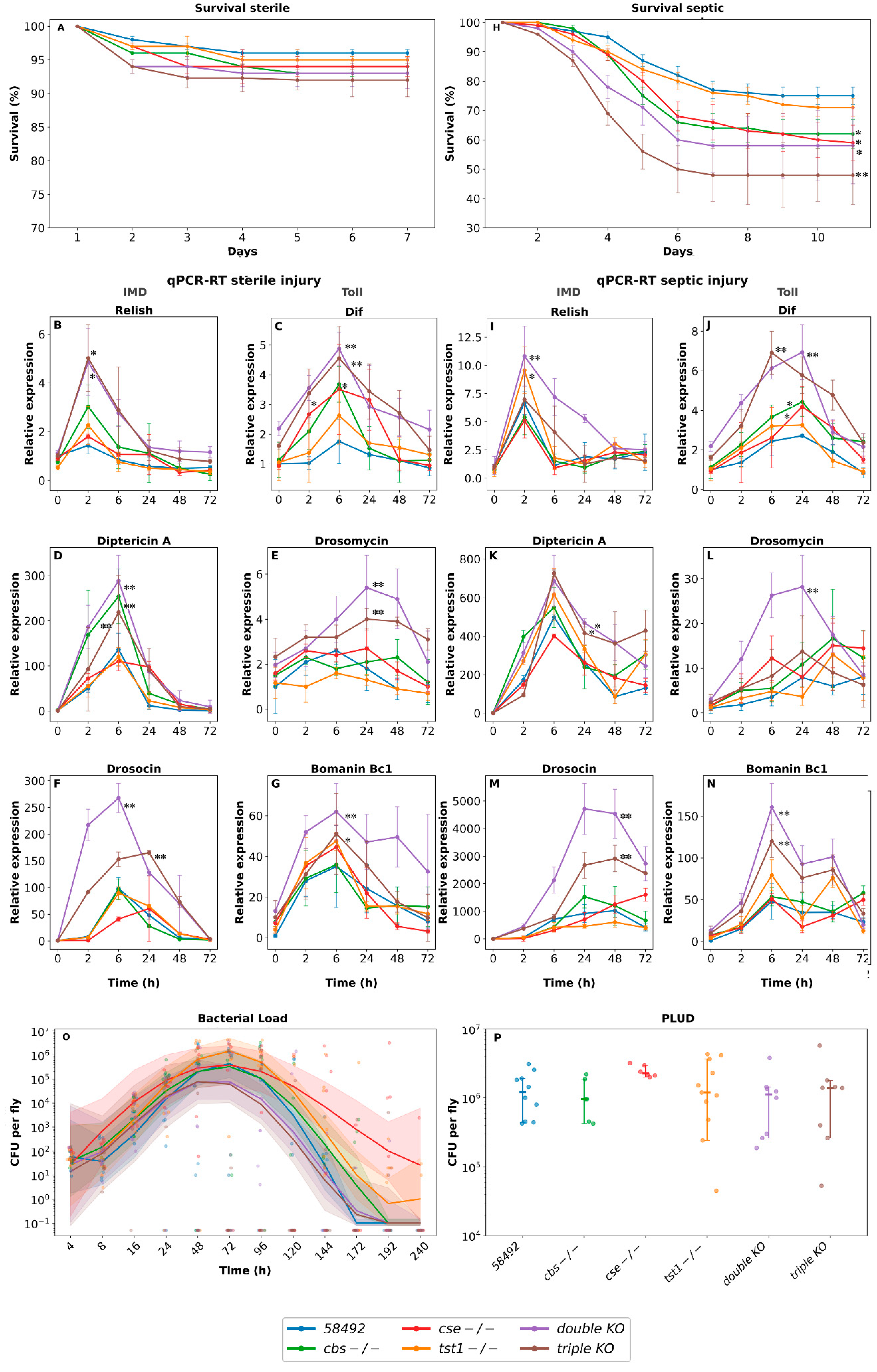
Figure 2.
A. Multi-dimensional scaling of log-scaled CPM expression. B. GSEA analysis of differentially expressed genes in females with single TST1-/-, double, and triple KO. Three pairwise comparisons were done: 1. Intact (healthy) KO flies to control 58492 intact flies. 2. Infected with B. subtilis (B.S.) KO flies to infected 58492 flies. 3. B. subtilis infected flies to intact flies of the same genotype. Gene ontology (GO) categories were used as terms. The color of each cell represents the averaged LogFC value of genes enriched in the category (color code shown on the middle right side of the plot), which is also depicted by the number of genes in each cell. Grey border thickness represents the proportion of enriched genes, divided by the number of all genes in a category.
Figure 2.
A. Multi-dimensional scaling of log-scaled CPM expression. B. GSEA analysis of differentially expressed genes in females with single TST1-/-, double, and triple KO. Three pairwise comparisons were done: 1. Intact (healthy) KO flies to control 58492 intact flies. 2. Infected with B. subtilis (B.S.) KO flies to infected 58492 flies. 3. B. subtilis infected flies to intact flies of the same genotype. Gene ontology (GO) categories were used as terms. The color of each cell represents the averaged LogFC value of genes enriched in the category (color code shown on the middle right side of the plot), which is also depicted by the number of genes in each cell. Grey border thickness represents the proportion of enriched genes, divided by the number of all genes in a category.
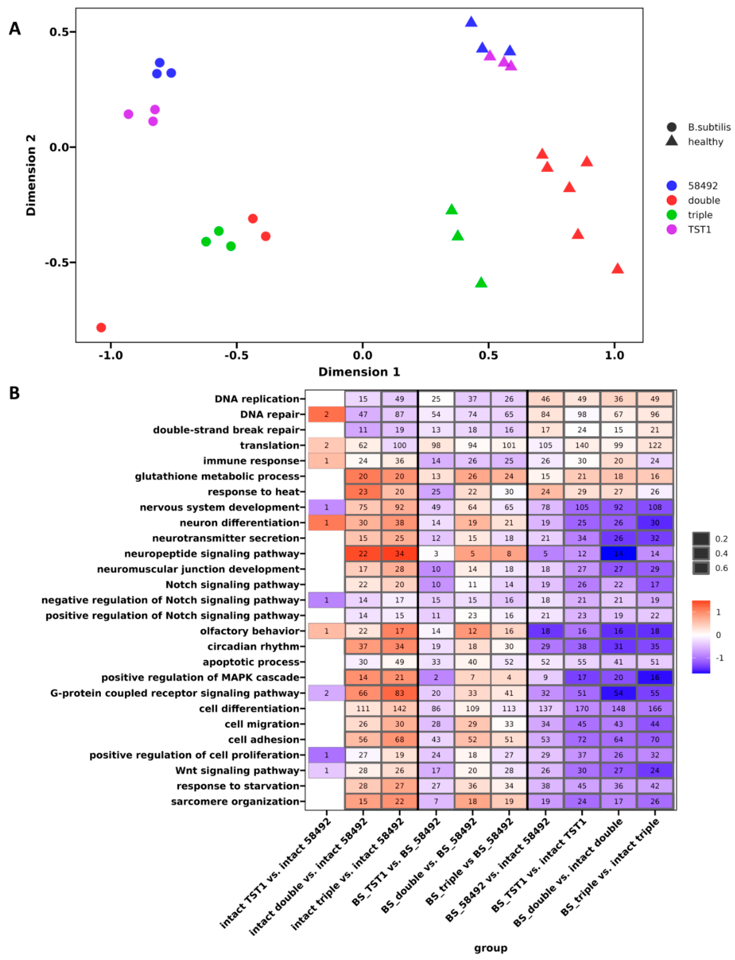
Figure 3.
Differential expression of genes involved in immune response: Gene selection was based on passing of false discovery rate-corrected p-value < 0.05. A - components of IMD, Toll, JAK-STAT pathways, B - AMPs, C - Bomanins in intact KO flies and after septic injury with B. subtilis. Three pairwise comparisons were made: 1. Intact (uninfected) KO flies to control 58492 intact flies. 2. Infected KO flies to infected 58492 flies. 3. Infected flies to intact flies of the same genotype. D. Box plots demonstrating differential expression of the Bomanins under control conditions and after septic injury.
Figure 3.
Differential expression of genes involved in immune response: Gene selection was based on passing of false discovery rate-corrected p-value < 0.05. A - components of IMD, Toll, JAK-STAT pathways, B - AMPs, C - Bomanins in intact KO flies and after septic injury with B. subtilis. Three pairwise comparisons were made: 1. Intact (uninfected) KO flies to control 58492 intact flies. 2. Infected KO flies to infected 58492 flies. 3. Infected flies to intact flies of the same genotype. D. Box plots demonstrating differential expression of the Bomanins under control conditions and after septic injury.
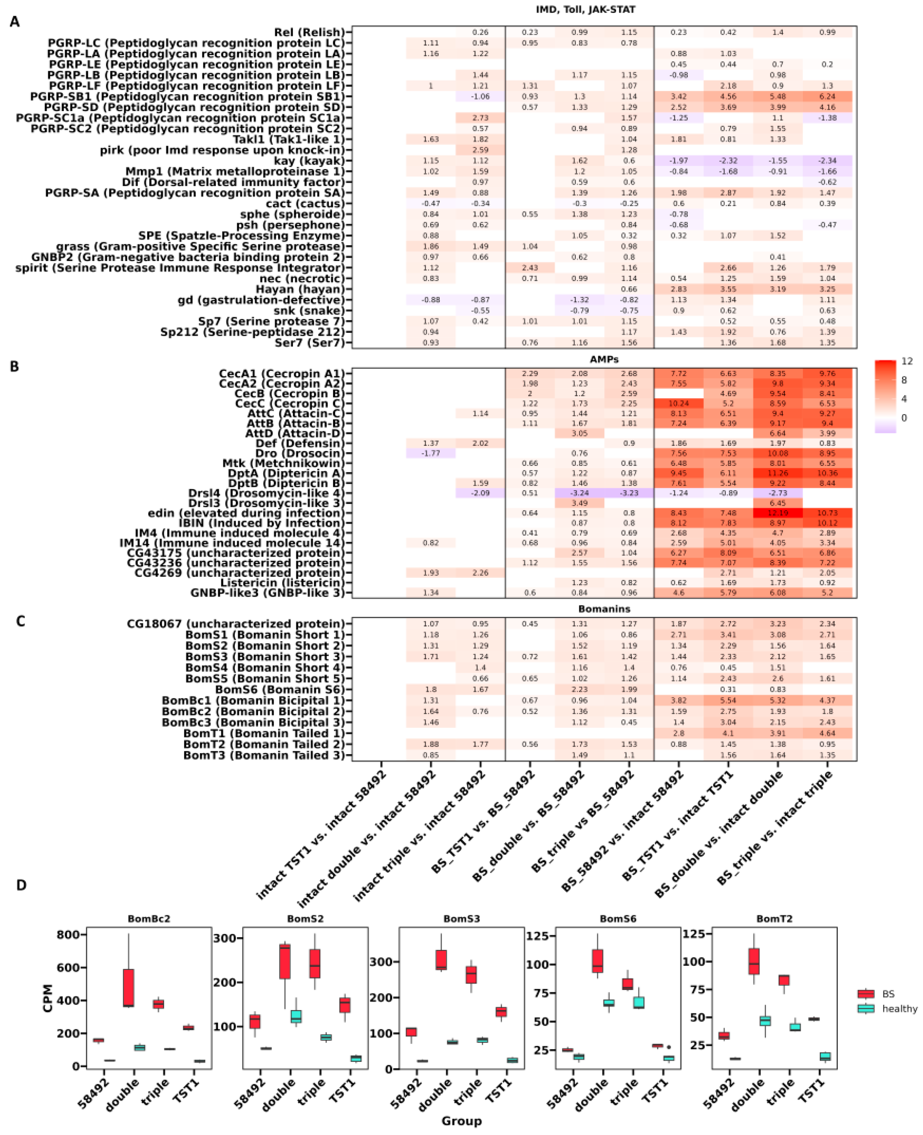
Figure 4.
Differential expression of genes encoding: A serpins, B opsonin&lectins- in intact KO flies and after septic injury with B. subtilis. Three pairwise comparisons were done: 1. Intact (healthy) KO flies to control 58492 intact flies. 2. Infected B. subtilis (BS) KO flies to infected B. subtilis 58492 flies. 3. B. subtilis infected flies to intact flies of the same genotype.
Figure 4.
Differential expression of genes encoding: A serpins, B opsonin&lectins- in intact KO flies and after septic injury with B. subtilis. Three pairwise comparisons were done: 1. Intact (healthy) KO flies to control 58492 intact flies. 2. Infected B. subtilis (BS) KO flies to infected B. subtilis 58492 flies. 3. B. subtilis infected flies to intact flies of the same genotype.
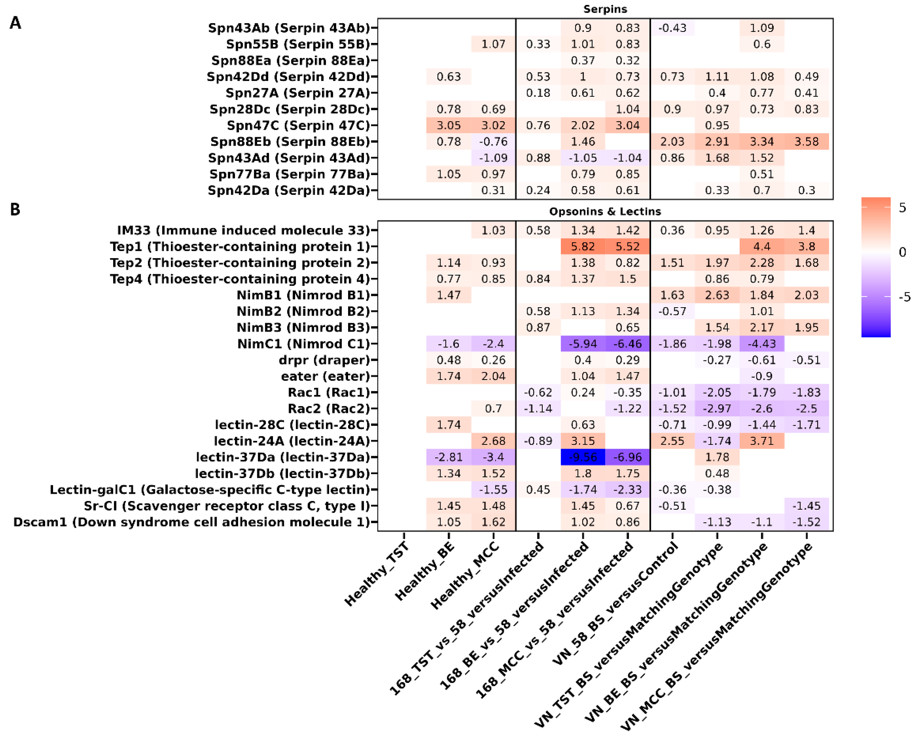
Figure 5.
Differential expression of genes encoding A. Trypsins, Jonahs, Maltases. B. Box plots illustrating differences in ilps expression.
Figure 5.
Differential expression of genes encoding A. Trypsins, Jonahs, Maltases. B. Box plots illustrating differences in ilps expression.
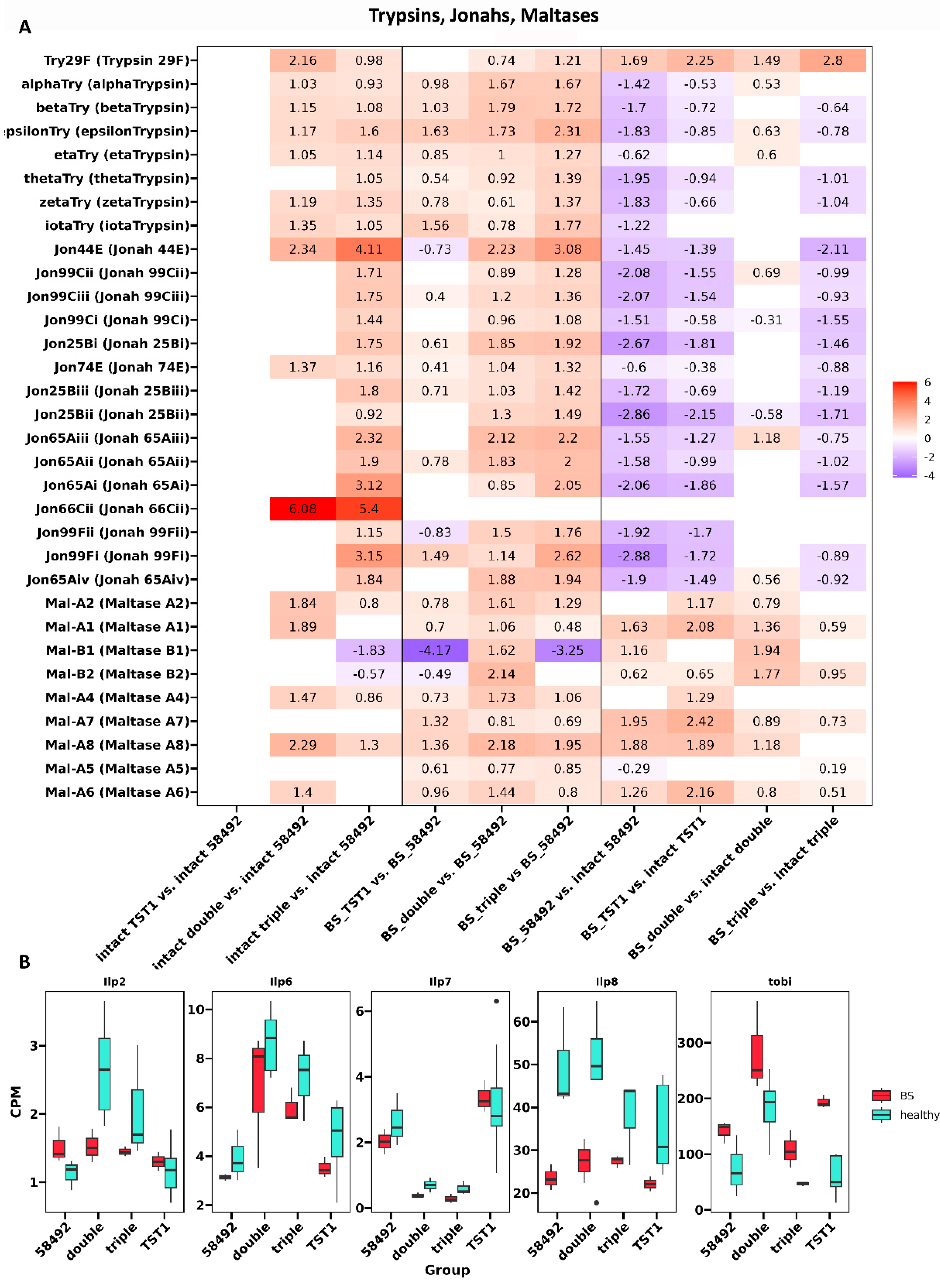
Figure 6.
Differential expression of stress response genes.
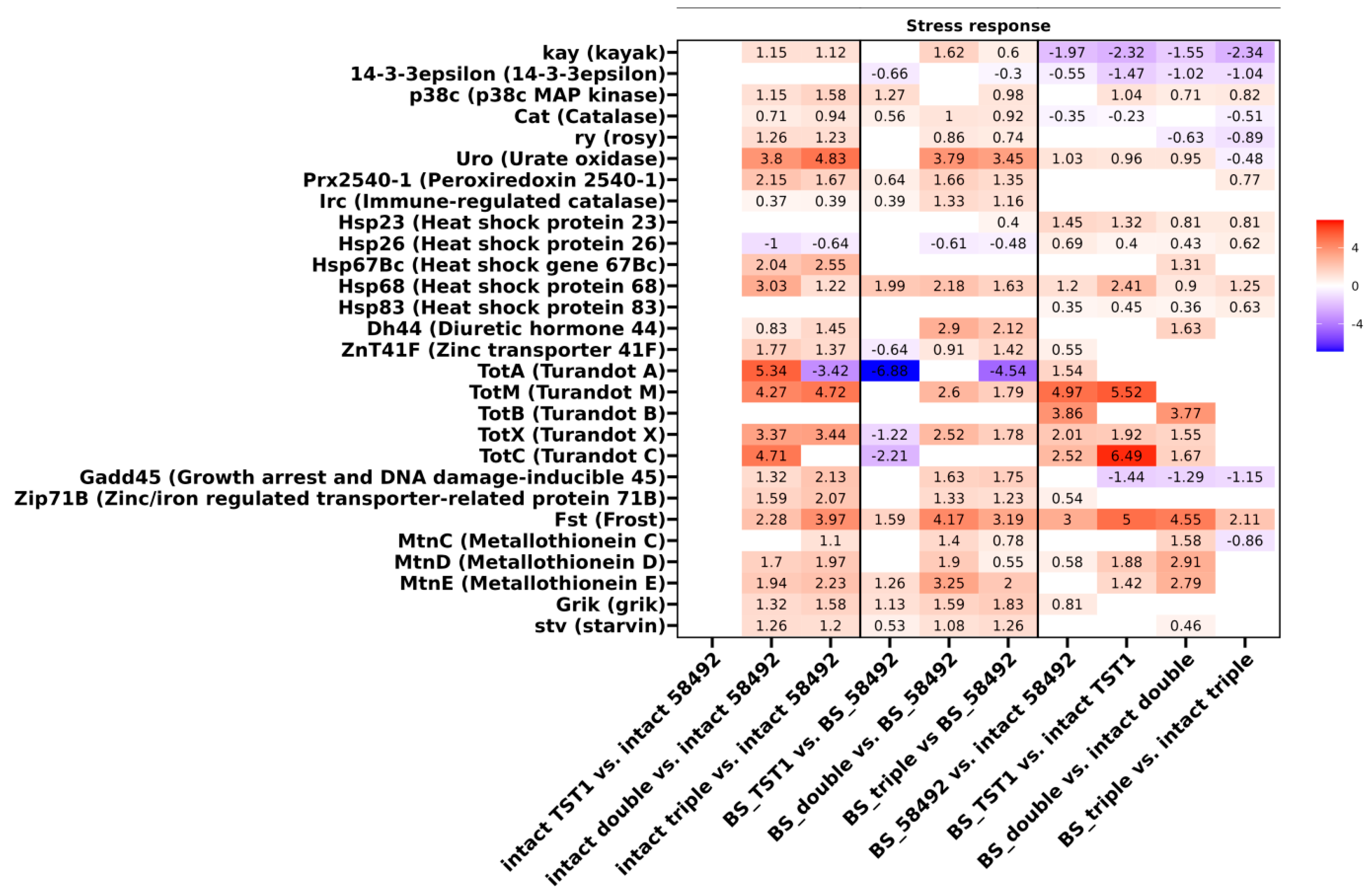
Disclaimer/Publisher’s Note: The statements, opinions and data contained in all publications are solely those of the individual author(s) and contributor(s) and not of MDPI and/or the editor(s). MDPI and/or the editor(s) disclaim responsibility for any injury to people or property resulting from any ideas, methods, instructions or products referred to in the content. |
© 2026 by the authors. Licensee MDPI, Basel, Switzerland. This article is an open access article distributed under the terms and conditions of the Creative Commons Attribution (CC BY) license (http://creativecommons.org/licenses/by/4.0/).
Copyright: This open access article is published under a Creative Commons CC BY 4.0 license, which permit the free download, distribution, and reuse, provided that the author and preprint are cited in any reuse.