Submitted:
15 June 2026
Posted:
16 June 2026
You are already at the latest version
Abstract
Soybean dregs are one of the main by-products during the processing of traditional soybean products, which are rich in insoluble dietary fiber. Insoluble dietary fiber has high mechanical strength and stability. After a certain treatment, it can have the po-tential to stabilize Pickering emulsions. In this research, high-purity soybean dregs insoluble dietary fibers (HPSIDF) were modified by alkaline H₂O₂, with oleic acid (OA) modified in a green synergistic modification. The surface of HPSIDF was etched to in-crease the reaction sites for hydrophobic modification. The hydrophobic modification was then used to enhance the emulsification properties, and finally, the performance of the particles in Pickering emulsions and the emulsification mechanism were analyzed. The absolute value of the ζ-potential of the hydrophobically modified high-purity soybean dregs insoluble dietary fiber (O-HPSIDF) increased from 15.87 mV to 37.27 mV. The stability of the Pickering emulsion stabilized by O-HPSIDF was greatly en-hanced, the droplet size of the emulsion decreased, the distribution was more uniform, and the emulsifier particles were able to form a network structure at the interface, which formed a certain mechanical strength for the emulsion.
Keywords:
insoluble dietary fiber
; etching modification
; hydrophobic modification
; Pickering emulsion
; rheological behavior
1. Introduction
Soybean dregs, a major byproduct generated during traditional soybean product processing, is rich in dietary fiber with a content reaching 84.47%. Notably, insoluble dietary fiber (IDF) constitutes 83.23% of this total. Soybean dregs insoluble dietary fiber (SIDF) mainly consists of cellulose, hemicellulose, and lignin, and is a class of dietary fiber that cannot be dissolved in water. These fiber components cannot be digested by human digestive enzymes and are subsequently partially fermented in the large intestine or excreted directly from the body, and have a wide range of applications in the food field [1]. It was shown that SIDF could hold water up to 9.32 g/g, oil up to 6.41 g/g, and solubility up to 2.50 mL/g [2], and could exhibit good emulsification in the Pickering emulsion system [3,4,5]. SIDF can be used as green, natural, low-cost emulsifiers, thickeners, or food packaging materials, thereby reducing the food industry's dependence on synthetic additives and providing a new pathway for the resourcing of agricultural by-products.
Pickering emulsion is an oil-in-water or water-in-oil emulsion that uses solid particles or colloidal particles that are insoluble in water and have certain hydrophilic and lipophilic properties as emulsifiers. Unlike conventional emulsifiers, the emulsifiers in Pickering emulsions are difficult to resolve at the interface like surfactants in conventional emulsions, and therefore have higher stability, anti-coalescence, and good elastic response [6]. In addition, the emulsifiers of Pickering emulsions are mostly made of natural, biodegradable materials (e.g., cellulose, starch, etc.), which are less toxic [7] and more biocompatible than surfactants [7]. It also reduces production costs and minimizes waste, making it more environmentally friendly and economical. IDF is a good emulsifier for polysaccharide-based Pickering emulsions. Studies have shown that IDF from bamboo shoots can be used as emulsifiers to prepare stable Pickering emulsions [8], and IDF from enoki mushrooms can stabilize oil-in-water emulsions [9], indicating that insoluble dietary fibers have certain development potential in the field of emulsifiers for Pickering emulsions.
However, most insoluble dietary fibers have larger particles and a more compact structure [10], and these characteristics will weaken the emulsification of the particles, so they need to be modified in some way. The emulsifiability of IDF can be enhanced by modification treatments, such as reducing the droplet size [11], increasing the specific surface area, increasing the complexity of the surface spatial structure, regulating the type and number of chemical groups exposed on the surface, etc., and so on. At present, the modification of insoluble dietary fibers can be divided into four types according to the modification mechanism: physical modification, chemical modification, biological modification, and joint modification. The emulsifiability of grapefruit pomelo peel insoluble dietary fibers was significantly elevated after hydrophobicity by octenyl succinic anhydride [12]. Citrus insoluble dietary fibers modified by ultrahigh pressure can form a more uniform interfacial adsorption network in Pickering emulsions and greatly improve the stability of the emulsion system [13]. For example, by modifying the SIDF at the molecular level, the balance between hydrophilicity and hydrophobicity of the particles can be precisely regulated, and the emulsifying property of the particles can be further improved.
In this study, high-purity soybean dregs insoluble dietary fiber (HPSIDF) was prepared from SIDF and further processed by etching and hydrophobic modification. Stabilizing Pickering emulsions with etch-hydrophobic synergistically modified HPSIDF (O-HPSIDF) as a solid emulsifier and analyzing the role of O-HPSIDF in enhancing the stability of Pickering emulsions, exploring the emulsification mechanism of the particles in the emulsion system, and broadening the way of high-value utilization of HPSIDF in the field of emulsifiers.
2. Materials and Methods
2.1. Materials and Reagents
The defatted soybean dregs used in the experiment were provided by Shandong Jiahua Health Products Co. α-Amylase (4.0×10⁴u/g, 0.25 g/mL), neutral protease (6.0×10⁴ u/g, 0.005 g/mL), glucosidase (10.×104u/g, 0.02 g/mL) were purchased from Beijing Solarbio Science Technology Co. H2O2 solution, OA and other reagents were purchased from Tianjin Xinbote Chemical Co.
2.2. Preparation of HPSIDF, H-HPSIDF and O-HPSIDF
2.2.1. Preparation of HPSIDF
HPSIDF was prepared using the method of Lyu et al [14]. In brief, defatted soybean dregs 15 g, 750 mL of distilled water, and 7.5 mL of α-amylase were mixed homogeneously and stirred at 95 °C for 35 min. The reaction was cooled to 60 °C. An additional 4.5 mL of neutral protease was added, and stirring was continued for 30 min at 60 °C. Cool the reactants to room temperature again, and 150 mL of acetic acid solution (3 mol/L) was added, and the pH of the liquid in the conical flask was adjusted to 4.5±0.02. 6 mL of glucoamylase was added, then stirred at 60 ℃ for 30 min. Then distilled water was added at 70 ℃ and then stood for 2 h. Continued to add 95% food-grade ethanol in a volume of three times the total liquid volume, stirred well, and then stood for 45 min. Filtered the precipitate, air-dried it at room temperature, milled it, and sieved it through a 200-mesh sieve to obtain the HPSIDF with purity greater than 90%.
2.2.2. Preparation of H-HPSIDF
HPSIDF was etched using H2O2 (3%) solution. 2 g of HPSIDF and 40 mL of H2O2 (3%) solution were mixed well. The pH of the reactants was adjusted to 9.0 and then stirred at 55 °C for 4 h (600 r/min). After the pH of the reactants was adjusted to neutral, distilled water was added to 300 mL. Sediment is separated by centrifugation. After repeating the centrifugation step three times, the sediment was vacuum lyophilized, ground, and finally, alkaline H2O2 etch-modified H-HPSIDF was obtained.
2.2.3. Preparation of O-HPSIDF
O-HPSIDF was mixed with OA at a material-liquid ratio of 1:40, and 150 μL of 6 mol/L HCl solution was added. The reactants were dispersed by sonication at 800 W for 15 min and heated at 80 °C for 11 h. The precipitate was added to 40 mL of 95% ethanol and mixed well. The precipitate was centrifuged, and the step of adding ethanol and centrifugation was repeated three times. Then add distilled water to the precipitate and mix well, centrifuge the precipitate, and repeat the steps of adding distilled water and centrifugation twice. Finally, the final precipitate was vacuum lyophilized and ground to a fine white powder to obtain O-HPSIDF.
2.2.4. Preparation of the Pickering Emulsions
Referring to the method used by Liu et al. with slight modification [15], HPSIDF, H-HPSIDF, and O-HPSIDF were used as emulsifiers to prepare Pickering emulsions, respectively. The amount of emulsifier added was 1.1% and the ratio of oil phase was 15%. Primary emulsification of the mixture was carried out using a high-speed shear dispersing emulsifier (10,000 r/min) before transferring it to an ultrasonic cell crusher and sonicating it for 14 min at 600 W. The different Pickering emulsions finally obtained were stabilized by each of the three different emulsifiers.
2.3. Chemical Composition and Physicochemical Properties of the HPSIDF, H-HPSIDF and O-HPSIDF
2.3.1. Water Holding Capacity (WHC)
WHC was determined according to the method reported previously [16]. 0.5 g of each of HPSIDF, H-HPSIDF, and O-HPSIDF was taken in a centrifuge tube, and 40 mL of distilled water was added. The centrifuge tubes were placed in a vertical position with the pointed bottom facing downwards for 24 h, and centrifuged for 25 min (4500 r/min). The supernatant was poured off, and the mass of the remaining wet sediment was weighed and recorded. Repeat the above test 3 times and take the average value to calculate the water holding capacity according to the following formula:
2.3.2. Oil Holding Capacity (OHC)
OHC was determined according to the method reported previously [16]. 0.5 g of each of HPSIDF, H-HPSIDF, and O-HPSIDF was taken in a centrifuge tube, respectively, and 35 mL of edible oil was added. The centrifuge tubes were placed in a vertical stand for 24 h with the pointed bottom facing downwards and centrifuged for 25 min (4500 r/min). The supernatant was poured off, and the mass of the remaining wet sediment was weighed and recorded. Repeat the above test 3 times and take the average value to calculate the water holding capacity according to the following formula:
2.3.3. Water Swelling Capacity (WSC)
WSC was determined according to the method reported previously [16]. 0.5 g of each of HPSIDF, H-HPSIDF, and O-HPSIDF was taken in a 50 mL graduated cylinder, and the initial volume was recorded.
30 mL of distilled water was added to the graduated cylinder and mixed well, and left to stand for 24 h. The volume of the sample after swelling at this point was recorded. The solubility was calculated according to the following formula:
2.4. Droplet Sizes and ζ-Potential Measurements
Zetasizer Advance (Malvern Panalytical, UK) was used to evaluate the droplet size and ζ-potential measurements. The HPSIDF, H-HPSIDF, and O-HPSIDF samples were taken as 0.05 g, respectively, and mixed well with 10 mL of distilled water. 1 mL of the mixture was taken in the cuvette of the droplet size analyzer. Each sample was preheated in the machine at 25 ℃ for 2 min, and then its droplet size value was measured. Then take 1 mL of the mixture in the potential cuvette of the droplet size analyzer and measure its potential value after preheating at 25 ℃ for 2 min.
2.5. Fourier-Transform Infrared (FTIR) Analysis
1 g each of dried HPSIDF, H-HPSIDF, and O-HPSIDF was taken and mixed well with 0.2 g of KBr, which was dried to constant weight. The mixture was ground for 10 min into a uniform fine powder using an agate mortar and pressed into uniform transparent discs of the size of a dime coin using a tablet press. The samples were detected by Fourier transform infrared spectroscopy.
2.6. X-Ray Diffraction (XRD)
X-ray diffraction was performed using a PANalytical X'pert3 (Malvern Panalytical instruments Ltd, UK). The crystal structures of HPSIDF, H-HPSIDF, and O-HPSIDF were observed under the measurement conditions of 5-900 and scanning speed of 20/2 min. The crystallinity of HPSIDF and H-HPSIDF was calculated using the following equation
where: Dc - degree of crystallinity, I002- diffraction intensity in the crystalline region (2θ = 20.780); Iam- diffraction intensity in the amorphous region (2θ = 18.00)
2.7. Scanning Electron Microscopy (SEM)
The microscopic morphology of HPSIDF, H-HPSIDF, and O-HPSIDF was observed using a S4800 (Hitachi, JP). The dried HPSIDF and H-HPSIDF were gently fixed on conductive adhesive tape, lightly blown off the unadhered powder on the surface, and placed in an ion sputtering apparatus for gold spraying. Then the sample stage was transferred to a scanning electron microscope, and the surface microforms of the samples were observed under an accelerating voltage of 10 kV, and the images were taken at 500, 1 k, 2 k, 5 k, and 20 k magnifications.
2.8. Thermogravimetry-Differential Scanning Calorimetry (TG-DSC)
Thermogravimetric analysis of HPSIDF, H-HPSIDF, and O-HPSIDF was performed using a Q600 (TA, USA) simultaneous thermal analyzer. The samples were taken as 3 mg each, and the ambient gas flow rate of nitrogen was set at 50 mL/min, and the test temperature was 10 ℃-600 ℃. The samples were tested for 60 min at a temperature increase rate of 10 ℃/min, and the changes of mass versus temperature and time were recorded and plotted as curves.
2.9. Pickering Emulsion Stability Test
2.9.1. Storage Treatment
Pickering emulsions of HPSIDF, H-HPSIDF, and O-HPSIDF were prepared according to the preparation process obtained in 2.2.4. The emulsions were left at room temperature and were observed at 3, 7, 10, and 15 days. The changes in the state of the three emulsions were compared.
2.9.2. Centrifuge Treatment
Pickering emulsions of HPSIDF, H-HPSIDF, and O-HPSIDF were prepared according to the preparation process obtained in 2.2.4. The emulsions were centrifuged (6000 r/min, 8000 r/min, 10,000 r/min) for 15 min at room temperature, and then the emulsion states were observed. The changes in the three emulsion states were compared.
2.9.3. Freeze-Thaw Treatment
Pickering emulsions of HPSIDF, H-HPSIDF, and O-HPSIDF were prepared according to the preparation process obtained in 2.2.4. The emulsions were first placed in -80 °C for 12 h and then thawed at room temperature. The state of the treated emulsions was observed, and the changes in the state of the three emulsions were compared.
2.9.4. Heat Treatment
Pickering emulsions of HPSIDF, H-HPSIDF, and O-HPSIDF were prepared according to the preparation process obtained in 2.2.4. The emulsions were heated in a water bath at 65 ℃ and 85 ℃ for 25 min, respectively, to observe the state of the treated emulsions and compare the changes in the state of the different emulsions.
2.9.5. Storage Treatments at Different pH Values
Pickering emulsions of HPSIDF, H-HPSIDF, and O-HPSIDF were prepared according to the preparation process obtained in 2.2.4. The pH of the emulsions was adjusted to 3, 4, 5, 6, 7, 8, 9. The state of the emulsions after being treated and after standing for 15 days was observed and compared
2.10. ζ-Potential Measurements of Pickering Emulsions
ζ-potential of the emulsion samples was measured using Zetasizer Advance (Malvern Panalytical, UK). The same volume of HPSIDF, H-HPSIDF, and O-HPSIDF stabilized emulsions was taken, diluted, and 1 mL was taken in the potentiometric cuvette, and the potential values were measured.
2.11. Microstructure of Pickering Emulsion
2.11.1. Inverted Fluorescence Microscope
The way emulsions were detected by an Inverted fluorescence microscope was determined by the method reported previously [12]. Droplet size, aggregation, and flocculation of the emulsions were observed using Leica DMILLED (Leica, GER). The emulsions prepared from HPSIDF, H-HPSIDF, and O-HPSIDF were taken separately and diluted in a ratio of 1:10. The diluted solution was dropped on a slide, and the droplet size, aggregation, and flocculation of the emulsions were observed using an inverted fluorescence microscope.
2.11.2. Laser Confocal Microscope
The way emulsions were detected by the Laser confocal microscope was determined by the method reported previously [3]. The microstructure of the emulsions was observed using a Leica Stellaris 5 (Leica, GER). The Pickering emulsions were stained using three dyes, Nile Red, Calcofluor white, and Nile Blue, respectively, and microstructural photographs of the emulsions were obtained at a scale of 50 μm.
2.12. Rheological Measurements
The emulsion samples prepared by HPSIDF, H-HPSIDF, and O-HPSIDF were placed on the rheometer operating table, set to a 1.0 mm gap, with a shear rate of 0.01-100 s-1 at 25 ℃. The variation of apparent viscosity of the emulsions with shear rate was determined and plotted as static rheological curves.
The frequencies within the linear viscoelastic region were determined for emulsions prepared from HPSIDF, H-HPSIDF, and O-HPSIDF to analyze the dynamic rheological properties of the samples. The value of oscillatory strain was fixed at 1%, and the oscillation frequency range value was 0.1-100 rad/s. The variation of the oscillation frequency of the emulsions concerning the energy storage modulus, G′, and loss modulus, G′′, respectively, was determined and plotted as a dynamic rheological curve.
2.13. Statistical Analysis
Data was analyzed using IBM SPSS Statistics 23 software, and images were drawn using Origin 2021. Three parallel experiments were set up for each experiment, and the experimental data were presented in the form of mean ± standard deviation (n=3, independently repeated experiments). The significance of differences between groups was determined by one-way analysis of variance (ANOVA) combined with Tukey's post hoc test, and the significance level was set at α = 0.05. According to the statistical labeling convention, when the comparison between groups showed p < 0.05, the difference was identified by using a different lowercase letter (e.g., a, b, and c); if the two groups shared at least one of the same letter (e.g., group ab versus group a), it indicated that the level of statistically significant difference between the comparison groups was not reached.
3. Results and Discussion
3.1. The Microstructural Properties of the Prepared O-HPSIDF
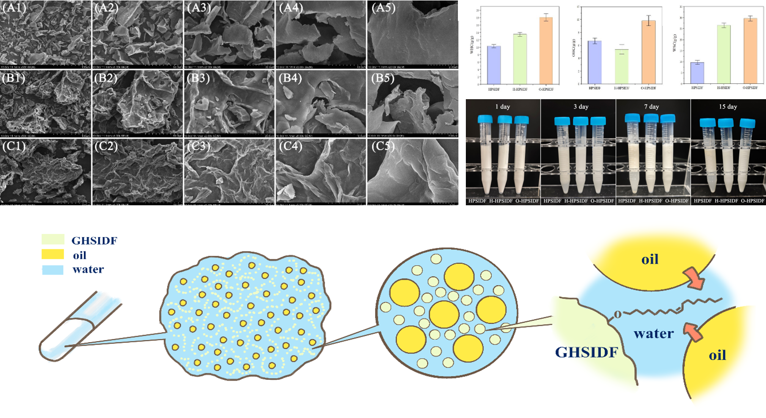
The surface micro-morphology of HPSIDF, H-HPSIDF, and O-HPSIDF is shown in Figure 1. The untreated HPSIDF is in the form of a curled sheet with a relatively smooth surface and a dense structure. The surface of H-HPSIDF after H2O2 etching treatment showed a large number of holes and grooves, which changed the original smooth surface into a spongy structure with a complex spatial structure, and the internal structure was exposed through the holes and gaps. The hydrophobically treated O-HPSIDF still has a more complex spatial structure than the HPSIDF, but with shallower grooves and fewer holes than the H-HPSIDF.
The reason for the formation of the complex structure of H-HPSIDF may be that alkaline H2O2 disrupted the structure of H-HPSIDF to some extent. This caused the formation of more pores and richer folds on the particle surface, exposing more internal structures and thus providing more modification sites for the next hydrophobic modification. The Structural changes of O-HPSIDF may be due to the newly formed modified layer during hydrophobic modification of oleic acid, clogging up the original hole. In addition, the treatment process leads to a certain degree of collapse of the spatial structure of the particles. However, O-HPSIDF still showed a complex morphology with multiple folds, and the complexity of the surface space structure was higher than that of HPSIDF, and such a surface structure could enhance the emulsification of O-HPSIDF.
As shown in Figure 2A, the XRD patterns of HPSIDF, H-HPSIDF, and O-HPSIDF show typical cellulose type I characteristics at 2θ. This indicates that the main components of HPSIDF, H-HPSIDF, and O-HPSIDF are natural cellulose type I structures. The 2θ angles of the two are relatively close to each other, indicating that they have similar crystalline types, and the modification treatment will not change the crystalline types of HPSIDF. However, the crystallinity of H-HPSIDF and O-HPSIDF was slightly decreased, probably due to the etching treatment and hydrophobic modification process, which had a certain destructive or modifying effect on some amorphous zones and the edge of crystalline zones in the dietary fibers, but due to the relative stability of the internal structure of the crystalline zones, the overall crystallinity did not change much, and the diffraction peak positions and shapes did not undergo any obvious changes.
A comparison of the Fourier transform infrared spectral characteristics of HPSIDF, H-HPSIDF, and O-HPSIDF samples is shown in Figure 2B. It can be observed that in the range of 3200-3600 cm−1 is a typical feature of hydroxyl groups, and the telescopic vibration of hydroxyl (-OH) absorption peaks were observed, suggesting that hydroxyl groups are present in all samples, which may be originated from the strong absorption generated by the hydrogen-bonding network of the primary hydroxyl group (C6-OH) and the secondary hydroxyl group (C2/C3-OH) in the molecular chain of cellulose, the acetylated hydroxyl group in the side chains of hemicellulose and the telescopic vibration of the phenol hydroxyl (Ar-OH) of lignin, etc. [17,18]. The double peaks located near 2920 cm−1 and 2850 cm−1 correspond to the antisymmetric and symmetric telescopic vibrational modes of methyl (-CH3) and methylene (-CH2-)[19], which may originate from smidgen residual protein fractions in the fibers of the soybean dregs. The characteristic absorption peaks in the 1650-1750 cm-1 region correspond to carbonyl (C=O) stretching vibration modes, suggesting the presence of carbonyl structures in the sample, and that such groups may originate from lignin fractions [18,20,21]. The absorption peak near 1740 cm-1 may originate from the vibration of the ester group, and the absorption peak in the range of 1450-1600 cm−1 is closely related to the backbone vibration of the lignin benzene ring (C=C aryl ring stretching), suggesting that the lignin component is preserved [22].
As shown in Figure 2C-2E, the thermal properties of the materials can be evaluated by thermogravimetric analysis, and the thermogravimetric curves (TG) and first-order differential thermogravimetric curves (DTG) are shown in Fig. The weight-loss peaks of HPSIDF, H-HPSIDF, and O-HPSIDF near 60 °C are attributed to the evaporation of water from the surfaces, which is fully evaporated at 150 °C. Continued temperature increase resulted in a double peak near 250 °C. The appearance of double peaks may be due to the volatilization of cellulose, hemicellulose, and lignin pyrolysis. The maximum decomposition rate of H-HPSIDF increased significantly after the etching treatment, and this situation may be related to the disruption of chemical bonds, which makes the originally tightly bound components easier to separate and volatilize. The maximum weight loss peak temperature of O-HPSIDF after hydrophobic modification was significantly shifted to the right and the maximum decomposition rate was significantly reduced, which indicated that the introduction of a modification layer after oleic acid modification led to an increase in the thermal stability of O-HPSIDF, which was corroborated by an increase in the peak intensity of the O-HPSIDF curve in the latter half of the DSC plot.
3.2. The Processing Characteristics of the Prepared O-HPSIDF
HPSIDF, H-HPSIDF, and O-HPSIDF were tested and analyzed for WHC, OHC, and WSC, as in Figure 3A-C. The results showed that after being treated by the etching of H2O2, the WHC and WSC of H-HPSIDF increased, and the OHC slightly decreased. After being modified by hydrophobization, the OHC of O-HPSIDF was significantly elevated. The increase in WHC and WSC of H-HPSIDF could be attributed to the fact that the etching of HPSIDF surface by H2O2 produced more pores on the surface and exposed more hydrophilic groups, such as active hydroxyls, and the sites at which water could bind were increased. The decrease in OHC may be due to the destruction of lipophilic groups on the surface and inside of HPSIDF and the decrease in oil binding sites. The increase in OHC of OHPSIDF may be due to the modification of the surface of O-HPSIDF by oleic acid, which resulted in an increase in lipophilic groups and consequently an increase in OHC.
As shown in Table 1. The droplet size of soya bean residue insoluble dietary fiber (HPSIDF) before modification was 82.43±0.27 μm, and the droplet size of H-HPSIDF after the etching treatment with H2O2 was reduced to 7.04±0.18 μm, and that of O-HPSIDF was reduced to 4.99±0.22 μm. This result may be attributed to the fact that it may be possible since the alkaline environment of H2O2 destroys the covalent bonds between cellulose and covalent bonds between cellulose and hemicellulose as well as hemicellulose, and the lignin was partially solubilized and oxidized by the alkaline H2O2[23,24], providing more hydrophobic reaction sites for the next step of oleic acid modification. The oleic acid molecules affected the state of dietary fiber molecules during the modification of O-HPSIDF, which disrupted the large-scale particle state and aggregation morphology of the particles, resulting in certain rupture of the particles, which led to the reduction of droplet size.
The ζ-potential of HPSIDF was -15.87±0.32 mV, that of H-HPSIDF was -17.21±0.45 mV, and that of O-HPSIDF was -37.27 mV±1.52 mV, which indicated that the surface charge density of the fiber particles increased after modification. The increase in the absolute value of the ζ-potential of H-HPSIDF may be since the etching treatment destroys the surface of the particles, resulting in the exposure of more negatively charged groups such as hydroxyl and carboxyl groups. The increase in the absolute value of the ζ-potential of O-HPSIDF may be since the oleic acid esterifies the surface of the O-HPSIDF, which introduces new charged groups and increases the charge density on the surface of the O-HPSIDF, increasing the absolute value of the ζ-potential. The absolute value increases. The higher absolute value of ζ-potential can inhibit the agglomeration between particles, improve the stability of the liquid system, and enhance the emulsification of O-HPSIDF in Pickering emulsion.
3.3. Evaluation of the Interfacial Activity and Stability of the Prepared O-HPSIDF
To determine the interfacial activity of the prepared O-HPSIDF, it was dispersed in water-oil two phases to form a Pickering emulsion. As shown in Figure 4A1-A3, different flow properties can be observed for different Pickering emulsions expressed in containers with the same tilt angle. The thinnest emulsion in the figure is the Pickering emulsion of HPSIDF, followed by the Pickering emulsion of H-HPSIDF. The most viscous emulsion state is the Pickering emulsion of O-HPSIDF, which indicates that the O-HPSIDF emulsion has a strong resistance to external deformation, probably due to the increased affinity of O-HPSIDF with the oil phase after hydrophobic modification, resulting in a better grip on the oil-water interface and the formation of a more robust network to better stabilize the emulsion system.
The Pickering emulsion, stabilized by O-HPSIDF, was subjected to centrifuge, freeze-thaw, and heat treatment to determine the stability. Centrifugal stability simulates the ability of emulsions to withstand extreme external forces in everyday life due to transportation, violent shaking, etc. It also accelerates the evaluation of stability for very long storage times. As shown in Figure 4B1-B4, the three emulsions showed varying degrees of stratification after centrifugation. The HPSIDF emulsion showed the most severe stratification, with a large amount of turbid aqueous phase precipitating in the middle layer. The H-HPSIDF emulsion retained more of the emulsified layer, with some of the turbid aqueous phase also appreciated in the middle layer, and the O-HPSIDF emulsion retained most of the emulsified layer. This phenomenon may be attributed to the hydrophobic modification of O-HPSIDF, which greatly enhanced the binding ability of O-HPSIDF to biphases, forming a strong spatial structure to resist the strong irritating external force caused by centrifugation.
Freeze-thaw treatment can examine the adsorption effect of the emulsifier particles and the stability of the emulsions when the ambient temperature difference is large. As shown in Figure 4C1-C2, HPSIDF emulsions showed large oil droplets, turbidity, and delamination after freeze-thaw, and the emulsions had undergone demulsification. H-HPSIDF emulsions showed significant delamination after Freeze-thaw, and the emulsions became cloudy. O-HPSIDF showed only slight turbidity at the bottom after Freeze-thaw, and there was no obvious delamination. The results show that the O-HPSIDF emulsion has excellent freeze-thaw stability, indicating that the emulsifying property of O-HPSIDF modified by oleic acid hydrophobicity is greatly enhanced.
As shown in Figure 4D1-D3, when the three emulsions were heated at 65℃, the HPSIDF emulsion showed obvious delamination, and the H-HPSIDF emulsion and O-HPSIDF emulsion did not change significantly. When the three emulsions were heated at 85℃, a large amount of delamination appeared in the HPSIDF emulsion, the H-HPSIDF emulsion became inhomogeneous, and the O-HPSIDF emulsion still maintained a uniform milky white color. It indicates that the hydrophobically modified O-HPSIDF particles can still stabilize the Pickering emulsion system at higher temperatures and show excellent emulsification ability, and the Pickering emulsions prepared by this emulsifier have good thermal stability, which confirms that the hydrophobic modification can enhance its emulsification properties.
The storage stability of the Pickering emulsion, to some extent, reflects the emulsification performance of the emulsifier; therefore, the storage stability of the emulsion was tested over a period of 15 days. As shown in Figure 5A1-A4, the condition of the Pickering emulsions of HPSIDF, H-HPSIDF, and O-HPSIDF remained good on the first day, with all three emulsions being homogeneous white emulsions with no visible precipitation or layering. On the third day, the bottom of the HPSIDF emulsion showed some delamination, and a turbid water phase precipitated below, while the H-HPSIDF and O-HPSIDF emulsions did not show delamination. On the seventh day, the turbid water phase precipitated from the emulsion of HPSIDF increased further, and the emulsion of H-HPSIDF and O-HPSIDF still did not show delamination. On the 15th day, the HPSIDF emulsion maintained the original layering phenomenon, the H-HPSIDF emulsion was turbid but not layered, and the O-HPSIDF emulsion was still a uniform milky white color and did not produce the phenomenon of layering or turbidity, indicating that the emulsifying property of O-HPSIDF was enhanced after modification. The modified O-HPSIDF may have enhanced the role of grasping and attaching the two phases at the interface, which greatly improved the stability of this Pickering emulsion.
The pH values have a certain degree of influence on the storage stability of the Pickering emulsions. As shown in Figure 5B1-B3 and C1-C3, HPSIDF, H-HPSIDF, and O-HPSIDF were not in the state of precipitation and delamination at different pH values on the day of emulsion preparation. With the increase of storage time up to 15 days, delamination of HPSIDF gradually occurred, and the severity of delamination increased with the increase of pH, indicating that Pickering emulsions prepared with HPSIDF were less stable under alkaline conditions. The Pickering emulsion prepared by H-HPSIDF showed less delamination only at the bottom under a higher alkaline environment. The Pickering emulsions prepared by O-HPSIDF showed no delamination and precipitation at all pH values, indicating that the Pickering emulsions stabilized by O-HPSIDF possessed strong tolerance to environmental pH.
3.4. Microstructural Characterization and Physicochemical Properties of Pickering Emulsions Stabilized by O-HPSIDF
In the HPSIDF emulsion, the size of the droplets was not uniform. There were large oil droplets with extremely large sizes, some smaller droplets were aggregated near the large droplets, some of the large droplets tended to be aggregated, and there were fibrous HPSIDF with large sizes observed in the emulsion (Figure 6.A1-A3). H-HPSIDF emulsion droplet size is relatively more uniform, and the size of the droplets is greatly reduced; there are only a few large droplets that exist between droplets. The spacing between droplets is relatively more uniform, and the distribution state is more regular. The droplet size of the Pickering emulsion prepared by O-HPSIDF is the most uniform; there are almost no obvious larger droplets, the distribution of droplets is dense and uniform, and the spacing between droplets is more consistent, indicating that O-HPSIDF can best stabilize the Pickering emulsion.
The microstructure of the emulsion was observed by laser confocal microscopy, in which the blue part was the emulsifier, and the red part was the oil droplets, as shown in Figure 6. The Pickering emulsion prepared by HPSIDF contained oil droplets of large size, and the size was very uneven. The distribution of the emulsifier is also not uniform. The combination of emulsifier and oil droplets is relatively loose, both have the phenomenon of similar aggregation phenomenon, and the overall emulsion structure is rough. The size of the oil droplets of the emulsions prepared by H-HPSIDF was smaller and more homogeneous than that of the HPSIDF emulsions, and the emulsifier was more uniformly distributed around the oil droplets, which may be since the smaller droplet size, more surface-active groups, and more complex spatial structure of the H-HPSIDF enhanced its original emulsifying properties. Both emulsifier and oil droplets in the O-HPSIDF emulsion showed highly uniform dispersion, the oil droplets with the smallest size and the most uniform distribution, the fibers showed highly homogeneous distribution, and the emulsion structure was fine. This may be since the hydrophobic-treated O-HPSIDF retains a part of the emulsifying property of H-HPSIDF, and the subsequent hydrophobic modification further enhances its emulsifying ability so that it can better adsorb at the oil-water interface, reduce the interfacial energy, and form a dense and stable interfacial membrane, which can effectively hinder the oil droplets from approaching each other to achieve highly stable emulsification effect.
Figure 7 showed the rheological measurement images of the emulsions prepared using HPSIDF, H-HPSIDF, and O-HPSIDF as emulsifiers, respectively. It can be observed from Figure 7A that in the shear stress versus shear rate plot, the O-HPSIDF emulsion corresponds to the highest position of the curve, having the highest shear stress at the same shear rate. This can indicate that the O-HPSIDF emulsion has the strongest resistance to flow deformation, while the Pickering emulsion stabilized with H-HPSIDF as emulsifier is the second highest, and the HPSIDF emulsion has the worst resistance to flow deformation.
In Figure 7B, with the increase of shear rate, all the three emulsions show the characteristic of “shear thinning” with the increase of shear rate and the gradual decrease of viscosity, which is a typical characteristic of non-Newtonian fluids [25,26]. O-HPSIDF emulsion has the highest initial viscosity, and the decrease of viscosity with shear rate is relatively small. H-HPSIDF emulsion has medium initial viscosity and a medium decrease in viscosity with shear rate. HPSIDF emulsion has the lowest initial viscosity and the greatest decrease in viscosity with shear rate. The reason for the pseudoplastic fluid is that the fluid contains intertwined long chains or macromolecules, which can form a certain structure to hinder the flow of the object in the stationary state [27,28]. With the application of the shear force and the increase of the shear rate, the molecular chains are arranged directionally along the direction of fluid flow, which leads to the reduction of the entanglement phenomenon between molecules, and thus reduces the resistance within the fluid, and the viscosity decreases accordingly [10,29]. The results show that a high degree of entanglement structure exists within the O-HPSIDF emulsion.
As seen in Figure 7C, the energy storage modulus of the three emulsions is greater than the loss modulus, indicating that the three emulsions are elastic-based emulsions with a certain solid-like characteristic and better stability. When the frequency increases, the energy storage modulus of all three emulsions rises significantly, indicating that the ability of the emulsions to resist deformation at high frequencies is enhanced, and the elastic properties are more prominent. O-HPSIDF emulsion has the largest energy storage modulus, and its stabilized emulsion has the strongest elasticity, optimal structural stability, and high deformation resistance. H-HPSIDF emulsion occupies second place, indicating that the etching place can change the interfacial properties of particles to some extent. The results show that the hydrophobic modification further optimizes the adsorption and arrangement of the particles at the interface, strengthens the firmness of the adsorption of O-HPSIDF at the interface, and forms a high-stability Pickering emulsion, which is capable of counteracting a certain amount of external forces, which verifies the positive effect of hydrophobic modification on the improvement of the rheological properties and stability of emulsions.
In summary, the Pickering emulsion stabilized by O-HPSIDF has a high consistency and structural strength, and its internal structure is not easily destroyed when subjected to external forces, which can better maintain the stability of the system. This may be since the particles of untreated HPSIDF are more compact and larger, and the number of hydrophilic and lipophilic groups exposed on the surface is lower, which stabilizes the emulsion droplets less. The surface of the etch-treated H-HPSIDF became loosely porous and reduced in size, exposing more hydrophilic groups and improving its interaction with the emulsion droplets, which led to an increase in its ability to stabilize the emulsion. O-HPSIDF was hydrophobized to introduce new hydrophobic groups on the surface, making it more amphiphilic. O-HPSIDF is efficiently adsorbed on the surface of emulsion droplets and forms a denser interfacial film, which forms a certain mesh structure in the three-dimensional structure, thus preventing the escape of droplets [30]. This structure also enhances the interaction between the emulsion droplets, which improves the viscosity and deformation resistance of the emulsion [31], resulting in better stability and structural strength of the emulsion.
4. Conclusions
In conclusion, the etching-hydrophobicity synergistic modification could optimize the emulsifying capacity of insoluble dietary fiber particles. Etching increases the surface roughness and reactive sites, thus providing conditions for hydrophobic modification. The hydrophobic modification further optimizes the emulsifying capacity of the fibers by introducing new hydrophobic groups, which results in a Pickering emulsion with high stability. The O-HPSIDF-stabilized Pickering emulsions with the smallest and uniformly distributed droplet sizes had the highest stability of the emulsions. The analysis shows that hydrophobic treatment can enhance the adsorption ability of the particles at the interface of the two phases, and O-HPSIDF can form a layer of compact interfacial film, which further forms a three-dimensional network structure. This plays a good role in immobilizing the oil and water phases, and at the same time, can effectively buffer the impact of external forces, so that the emulsion stability is greatly improved. The etch-hydrophobic modified O-HPSIDF can be used as a natural, green, and safe solid emulsifier to replace chemical synthetic emulsifiers for future applications in the field of plant-based and clean-label food products, and its good deformation resistance can make it a unique stabilization advantage in long-distance transportation. This study deepens the understanding of the surface modification mechanism of dietary fibers and provides ideas for the development of new, efficient, and controllable surface modification processes for dietary fibers and the design of Pickering emulsifiers.
Author Contributions
writing—original draft preparation, Shuhan Ge and Wendan Jing; Validation, Haoyuan Li and Lingchao Wu; data curation, Shuhan Ge; funding acquisition, Wendan Jing and Hansong Yu; All authors have read and agreed to the published version of the manuscript.
Funding
This research was supported by the China Agriculture Research System of MOF and MARA (CARS-04), and Jilin Province Science and Technology Development Plan Item (20250602014RC).
Informed Consent Statement
Not applicable.
Data Availability Statement
Raw data can be provided by the corresponding author on request.
Conflicts of Interest
The authors declare no conflicts of interest.
Abbreviations
| HPSIDF | High-purity soybean dregs insoluble dietary fibers |
| H-HPSIDF | Alkaline H2O2 etch-modified high-purity soybean dregs insoluble dietary fiber |
| O-HPSIDF | Oleic acid modified high-purity soybean dregs insoluble dietary fiber |
References
- Lupton, J.R.; Betteridge, V.A.; Pijls, L.T.J. Codex final definition of dietary fibre: issues of implementation. 2009, 1, 206–212. [Google Scholar] [CrossRef]
- Tang, L.; Hu, M.; Bai, S.; Wang, B.; Fan, B.; Zhang, L.; Wang, F. Extraction of insoluble soybean fiber by alternating ultrasonic/alkali and its improved superior physicochemical and functional properties. Int. J. Biol. Macromol. 2024, 263, 130505. [Google Scholar] [CrossRef] [PubMed]
- Yuan, Z.; Zhu, D.; Xu, X.; Xu, J.; Yang, L.; Song, H.; Wang, S.; Liu, J.; Liu. HJIJoBM: Homogenized soybean hull suspension as an emulsifier for oil/water emulsions: Synergistic effect of the insoluble fiber and soluble polysaccharide. 2023, 237, 123950. [Google Scholar] [CrossRef] [PubMed]
- Cai, Y.; Huang, L.; Zhou, F.; Zhao, Q.; Zhao, M.; Van der Meeren. PJFC: Characteristics of insoluble soybean fiber (ISF) concentrated emulsions: Effects of pretreatment on ISF and freeze–thaw stability of emulsions. 2023, 427, 136738. [Google Scholar] [CrossRef] [PubMed]
- Yue, B.; Hanyu, X.; Yang, Y.; Xiujuan, W.; Hansong, Y.; Chunhong, P. Preparation and Characterization of Pickering Emulsions with Modified Okara Insoluble Dietary Fiber. Foods 2021. [Google Scholar] [CrossRef] [PubMed]
- Jiahao, P.; Jianping, C.; Xuejiao, W.; Ying, W.; Jun-Bing, F. Pickering emulsion: From controllable fabrication to biomedical application. In Interdisciplinary Medicine; 2023. [Google Scholar]
- Jinze, P.; Ning, W.; Congcong, C.; Guangshan, X. Preparation of Pickering Emulsion for Antibacterial, Anti-Inflammatory and Wound Healing. J. Biosci. Med. 2022. [Google Scholar] [CrossRef]
- He, K.; Li, Q.; Li, Y.; Li, B.; Liu, S. Water-insoluble dietary fibers from bamboo shoot used as plant food particles for the stabilization of O/W Pickering emulsion. Food Chem. 2020, 310, 125925. [Google Scholar] [CrossRef] [PubMed]
- He, K.; Zhang, X.; Li, Y.; Li, B.; Liu. SJFH: Water-insoluble dietary-fibers from Flammulina velutiper used as edible stabilizers for oil-in-water Pickering emulsions. 2020, 101, 105519. [Google Scholar] [CrossRef]
- Ziming, G.U.O.; Xueying, F.; Xiaoyun, X.U.; Lufeng, W. Construction and characterization of insoluble citrus fiber-corn oil Pickering emulsion. J. Huazhong Agric. Univ. 2023. [Google Scholar] [CrossRef]
- Ben, Y.; Qianqian, C.; Joe, M.R.; Changwen, Y.; Lufeng, W. The lipid digestion behavior of oil-in-water emulsions stabilized by different particle-sized insoluble dietary fiber from citrus peel. In Food Chemistry: X; 2023. [Google Scholar]
- Gao, K.; Liu, Y.; Liu, T.; Song, X.; Ruan, R.; Feng, S.; Wang, X.; Cui. XJFH: OSA improved the stability and applicability of emulsions prepared with enzymatically hydrolyzed pomelo peel insoluble fiber. 2022, 132, 107806. [Google Scholar] [CrossRef]
- Yang, X.; Mao, K.; Sang, Y.; Tian, G.; Liu, X.; Mao, N.; Huo, M.; Yan, S. Citrus derived Pickering emulsion stabilized by insoluble citrus dietary fiber modified by ultra-high pressure. LWT 2023, 184, 115112. [Google Scholar] [CrossRef]
- Lyu, B.; Wang, H.; Swallah, M.S.; Fu, H.; Shen, Y.; Guo, Z.; Tong, X.; Li, Y.; Yu, H.; Jiang, L. Structure, properties and potential bioactivities of high-purity insoluble fibre from soybean dregs (Okara). Food Chem. 2021, 364, 130402. [Google Scholar] [CrossRef] [PubMed]
- Liu, M.; Liang, J.; Jing, C.; Yue, Y.; Xia, Y.; Yuan, Y.; Yue, T.J.F.C. Preparation and characterization of Lycium Barbarum seed oil Pickering emulsions and evaluation of antioxidant activity. 2023, 405, 134906. [Google Scholar] [CrossRef]
- Huang, Y.; Li, C.; Zheng, S.; Fu, X.; Huang, Q.; Liu, G.; Chen. QJM: Influence of Three Modification Methods on the Structure, Physicochemical, and Functional Properties of Insoluble Dietary Fiber from Rosa roxburghii Tratt Pomace. 2024, 29, 2111. [Google Scholar] [CrossRef] [PubMed]
- Sang, J.; Li, L.; Wen, J.; Liu, H.; Wu, J.; Yu, Y.; Xu, Y.; Gu, Q.; Fu, M.; Lin. XJL: Chemical composition, structural and functional properties of insoluble dietary fiber obtained from the Shatian pomelo peel sponge layer using different modification methods. 2022, 165, 113737. [Google Scholar] [CrossRef]
- Zhang, X.; Zeng, Y.; Liu, J.; Men, Y.; Sun. YJFCA: Effects of three extraction methods on the structural and functional properties of insoluble dietary fibers from mycoprotein. 2023, 2, 100299. [Google Scholar] [CrossRef]
- Zheng, Y.; Li, Y.; Tian, H. Effects of carboxymethylation, acidic treatment, hydroxypropylation and heating combined with enzymatic hydrolysis on structural and physicochemical properties of palm kernel expeller dietary fiber. Lwt 2020, 133, 100023–106438. [Google Scholar] [CrossRef]
- Wang, L.; Tian, Y.; Chen, Y.; Chen. JJCc: Effects of acid treatment on the physicochemical and functional properties of wheat bran insoluble dietary fiber. 2022, 99, 343–354. [Google Scholar]
- Wang, S.; Fang, Y.; Xu, Y.; Zhu, B.; Piao, J.; Zhu, L.; Yao, L.; Liu, K.; Wang, S.; Zhang, Q. The effects of different extraction methods on physicochemical, functional and physiological properties of soluble and insoluble dietary fiber from Rubus chingii Hu. fruits. J. Funct. Foods 2022, 93, 105081 %@ 101756–104646. [Google Scholar] [CrossRef]
- Zhang, M.-Y.; Liao, A.-M.; Thakur, K.; Huang, J.-H.; Zhang, J.-G.; Wei, Z.-J. Modification of wheat bran insoluble dietary fiber with carboxymethylation, complex enzymatic hydrolysis and ultrafine comminution. Food Chem. 2019, 297, 124983. [Google Scholar] [CrossRef] [PubMed]
- Meng, X.; Liu, F.; Xiao, Y.; Cao, J.; Wang, M.; Duan, X. Alterations in physicochemical and functional properties of buckwheat straw insoluble dietary fiber by alkaline hydrogen peroxide treatment. Food Chem. X 2019, 3, 100029. [Google Scholar] [CrossRef] [PubMed]
- Jiang, G.; Bai, X.; Wu, Z.; Li, S.; Zhao, C.; Ramachandraiah, K. Modification of ginseng insoluble dietary fiber through alkaline hydrogen peroxide treatment and its impact on structure, physicochemical and functional properties. LWT 2021, 150, 111956. [Google Scholar] [CrossRef]
- Boutin, C.; Giroux, H.J.; Paquin, P.; Britten. MJIDJ: Characterization and acid-induced gelation of butter oil emulsions produced from heated whey protein dispersions. 2007, 17, 696–703. [Google Scholar] [CrossRef]
- Chhabra, R.P.; Richardson, J.F. Non-Newtonian Fluid Behaviour. Non Newton. Flow Appl. Rheol. 2008. [Google Scholar] [CrossRef]
- Li, Q.; Xie, B.; Wang, Y.; Wang, Y.; Peng, L.; Li, Y.; Li, B.; Liu, S.J.F. h: Cellulose nanofibrils from Miscanthus floridulus straw as green particle emulsifier for O/W Pickering emulsion. 2019, 97, 105214. [Google Scholar] [CrossRef]
- Sheng, G.; Zhaojing, J.; Hanjun, M.; Pei, P.; Benguo, L.; Guizhao, L. Fabrication and characterization of novel edible Pickering emulsion gels stabilized by dihydromyricetin. Food Chem. 2021. [Google Scholar] [CrossRef] [PubMed]
- Marco D: Inertial theories for dilute viscoelastic polymer blends with a volume preserving microstructure. J. Non Newton. Fluid Mech. 2012. [CrossRef]
- Feilin, L.; Xinyu, Z.; Shujuan, Y.; Furui, H.; Wenqi, Q.; Houkui, G.; Gaobo, Y.; Yuhong, F.; Jiacheng, L. Interfacial regulation and visualization of Pickering emulsion stabilized by Ca2+-triggered amphiphilic alginate-based fluorescent aggregates. Food Hydrocoll. 2021. [Google Scholar] [CrossRef]
- Chao, W.; Zhe, L.; Lanyi, Z.; Bo, J.; Hui, H.; Xiaojie, M.; Jinjin, Z.; Marc, P.; Qiang, W.; Aimin, S. Effect of oil content and protein particles concentration on non-dairy whip topping based on Pickering emulsion system and their 3D printing properties. Food Hydrocoll. 2023. [Google Scholar] [CrossRef]
Figure 1.
(A1, A2, A3, A4, A5) SEM image of HPSIDF. (B1, B2, B3, B4, B5) SEM image of H-HPSIDF. (C1, C2, C3, C4, C5) SEM image of O-HPSIDF(magnification: 500 × A1, B1 and C1; 1000 × A2, B2 and C2; 2000 × A3, B3 and C3; 5000 × A4, B4 and C4; 20,000 × A5, B5 and C5).
Figure 1.
(A1, A2, A3, A4, A5) SEM image of HPSIDF. (B1, B2, B3, B4, B5) SEM image of H-HPSIDF. (C1, C2, C3, C4, C5) SEM image of O-HPSIDF(magnification: 500 × A1, B1 and C1; 1000 × A2, B2 and C2; 2000 × A3, B3 and C3; 5000 × A4, B4 and C4; 20,000 × A5, B5 and C5).

Figure 2.
(A) X-ray diffraction of HPSIDF, H-HPSIDF, and O-HPSIDF. (B) FT-IR spectra of HPSIDF, H-HPSIDF, and O-HPSIDF. (C,D, E) TG-DSC image of HPSIDF, H-HPSIDF, and O-HPSIDF. (C) Thermogravimetric curve (TG). (D) First-order differential thermogravimetric curve (DTG). (E) Heat flow versus temperature curve (DSC).

Figure 3.
(A) WHC of HPSIDF, H-HPSIDF and O-HPSIDF. (B) OHC of HPSIDF, H-HPSIDF and O-HPSIDF. (C) WSC of HPSIDF, H-HPSIDF and O-HPSIDF.
Figure 3.
(A) WHC of HPSIDF, H-HPSIDF and O-HPSIDF. (B) OHC of HPSIDF, H-HPSIDF and O-HPSIDF. (C) WSC of HPSIDF, H-HPSIDF and O-HPSIDF.

Figure 4.
(A1-A3) Comparison of the fluidity of HPSIDF, H-HPSIDF, and O-HPSIDF emulsions. (B1-B4) Changes in centrifugal treatment of HPSIDF, H-HPSIDF, and O-HPSIDF emulsions. (C1, C2) Changes in HPSIDF, H-HPSIDF & O-HPSIDF emulsion freeze-thaw treatments. (C1) Freeze-thaw before treatment. (C2) Freeze-thaw after treatment. (D1-D3) Changes in heat treatment of HPSIDF, H-HPSIDF, and O-HPSIDF emulsions.

Figure 5.
(A1-A4) Changes in HPSIDF, H-HPSIDF, and O-HPSIDF emulsions after storage. (B1-B3, C1-C3) Changes of HPSIDF, H-HPSIDF, and O-HPSIDF emulsions after storage at different pH values. (B1-B3) Day 1. (C1-C3) Day 15.
Figure 5.
(A1-A4) Changes in HPSIDF, H-HPSIDF, and O-HPSIDF emulsions after storage. (B1-B3, C1-C3) Changes of HPSIDF, H-HPSIDF, and O-HPSIDF emulsions after storage at different pH values. (B1-B3) Day 1. (C1-C3) Day 15.

Figure 6.
(A1-A3) Inverted fluorescence microscope image of HPSIDF emulsion, H-HPSIDF emulsion, and O-HPSIDF emulsion at 40x magnification. (A1) HPSIDF emulsion. (A2) H-HPSIDF Pickering emulsion. (A3) O-HPSIDF emulsion). (B1-D3) Laser confocal microscope images of HPSIDF emulsion, H-HPSIDF emulsion, and O-HPSIDF emulsion (scale bar: 50 μm). (B1-B3) HPSIDF emulsion. (C1-C3) H-HPSIDF emulsion. (D1-D3) O-HPSIDF emulsion.

Figure 7.
Rheological analysis graphs for HPSIDF emulsions, H-HPSIDF emulsions, and O-HPSIDF emulsions. (A) Plot of shear stress versus shear rate. (B) Plot of viscosity versus shear rate. (C) Plot of storage modulus and loss modulus versus frequency.
Figure 7.
Rheological analysis graphs for HPSIDF emulsions, H-HPSIDF emulsions, and O-HPSIDF emulsions. (A) Plot of shear stress versus shear rate. (B) Plot of viscosity versus shear rate. (C) Plot of storage modulus and loss modulus versus frequency.


Table 1.
Particle size and zeta potential of HPSIDF, H-HPSIDF, and O-HPSIDF.
| Sample | Droplet sizes(μm) | ζ-potential(mV) |
| HPSIDF | 82.43±0.27 a | -15.87±0.32 a |
| H-HPSIDF | 7.04±0.18 b | -17.21±0.45 b |
| O-HPSIDF | 4.99 ±0.22 c | -37.27±1.52 c |
Note: Values = mean ± standard deviation (n=3). Within the same column, different lowercase letters indicate a significant difference (p < 0.05), while the same letter indicates a non-significant difference (p > 0.05).
Disclaimer/Publisher’s Note: The statements, opinions and data contained in all publications are solely those of the individual author(s) and contributor(s) and not of MDPI and/or the editor(s). MDPI and/or the editor(s) disclaim responsibility for any injury to people or property resulting from any ideas, methods, instructions or products referred to in the content. |
© 2026 by the authors. Licensee MDPI, Basel, Switzerland. This article is an open access article distributed under the terms and conditions of the Creative Commons Attribution (CC BY) license (http://creativecommons.org/licenses/by/4.0/).
Copyright: This open access article is published under a Creative Commons CC BY 4.0 license, which permit the free download, distribution, and reuse, provided that the author and preprint are cited in any reuse.


