

Submitted:
12 June 2026
Posted:
15 June 2026
You are already at the latest version
Abstract
Scaffold architecture profoundly influences tissue regeneration through the mechanical, topographical, and biochemical cues it presents to cells. Yet the relationship between architectural complexity and regenerative performance remains difficult to interpret: geometrically elaborate scaffolds do not necessarily produce more organized tissues, while comparatively simple architectures can exert strong organizational effects. We propose that scaffold-guided tissue organization is better understood through a distinction between geometric complexity and informational dimensionality. We argue that scaffold performance depends less on geometric complexity than on the number of stable, biologically interpretable dimensions available for cellular mechanotransduction. Drawing heuristically on kernel methods in machine learning, we suggest that scaffold architectures can transform poorly structured mechanosensory environments into conditions where alternative organizational trajectories become more distinguishable. Mechanotransduction provides the biological basis for this process, integrating scaffold-derived cues through focal adhesions, cytoskeletal tension, nuclear deformation, and YAP/TAZ signaling. This perspective suggests that scaffold design should be evaluated not only by architectural complexity or biomimetic resemblance, but by its capacity to generate stable and interpretable mechanosensory environments. More broadly, this shifts the design question from how complex a scaffold is to whether its architecture generates stable, cell-readable mechanosensory information.
Keywords:
tissue engineering
; support vector machine
; kernel transformation
; mechanotransduction
; scaffold topography
1. The Problem of Organization in Tissue Engineering
The central challenge of tissue engineering is not simply keeping cells alive, but guiding cellular collectives toward stable, functional, and architecturally coherent forms [1]. Although cells possess intrinsic capacities for adhesion, migration, differentiation, and extracellular matrix deposition [2], these processes do not spontaneously converge toward organized tissue architectures under arbitrary physical conditions [3]. Cells cultured on smooth or weakly structured substrates frequently generate disordered, metastable organizations despite remaining viable and metabolically active [4,5]. The fundamental problem is organizational: physical environments must somehow bias cells toward coherent architectures rather than the disordered, weakly coordinated states they default to in vitro.
Over the past decades, biomaterial scaffolds have evolved from passive supports into active microenvironments capable of influencing cellular behavior through biochemical, mechanical, and topographical cues [6,7,8,9,10,11,12]. The native extracellular matrix (ECM) represents a dynamically structured and multiscale instructive environment integrating fibrillar architecture, anisotropy, curvature, stiffness gradients, ligand accessibility, viscoelasticity, and spatial heterogeneity across multiple scales to regulate tissue homeostasis, mechanotransduction, and regenerative morphogenesis [13,14,15]. Importantly, biological organization emerges not from isolated geometric features alone, but from the coordinated interaction of partially independent mechanical, spatial, and biochemical constraints acting across scales [16,17,18,19].
Contemporary scaffold fabrication has consequently emphasized biomimicry, employing electrospinning, lithography, additive manufacturing, and related techniques to reproduce aspects of ECM geometry and hierarchy [20,21,22,23]. Yet for all the empirical progress, scaffold design has remained largely heuristic [24]. Although contemporary mechanobiology increasingly recognizes that scaffold geometry regulates collective cellular organization through mechanotransduction [25], many studies still focus on individual cue-response relationships. What remains difficult to explain is why certain scaffold architectures reliably stabilize coherent organization, whereas others, despite comparable geometric richness, generate heterogeneous, metastable, or weakly coordinated outcomes [26].
Existing mechanobiological models successfully describe many of the signaling pathways through which cells respond to physical stimuli, including integrin engagement, focal adhesion dynamics, cytoskeletal tension, and mechanosensitive transcriptional regulators such as YAP/TAZ [27,28]. Recent studies further demonstrate that scaffold curvature, pore geometry, and nanoscale topographical accessibility can directly regulate cytoskeletal organization, YAP/TAZ signaling, stemness, and lineage specification [29,30,31], suggesting that scaffold architecture does not merely modulate isolated signaling pathways but restructures the organizational conditions under which cellular decisions unfold [17]. Scaffold architecture, in other words, does not merely present physical cues — it shapes whether those cues can be coherently integrated at all [32].
The relevant question is whether scaffold architecture provides cues that cells can reliably integrate into coordinated behavior. Adding structural complexity does not reliably improve organization: disordered three-dimensional environments can amplify spatial heterogeneity while leaving the number of mechanically interpretable variables the cell can actually use unchanged [33]. A geometrically heterogeneous environment is not necessarily informationally rich from the perspective of the cell. Environments may exhibit extensive geometric heterogeneity while still failing to provide stable and biologically interpretable mechanotransductive cues capable of guiding coherent tissue organization [34].
Here we propose an informational interpretation of scaffold-guided tissue organization based on the concepts of effective dimensionality and morphogenetic separability. The central claim is that scaffold architecture can influence regeneration not by adding complexity per se, but by increasing the number of stable, partially independent, and mechanotransductively interpretable variables available to cells. Isotropic or weakly structured environments can be understood as informationally low-dimensional systems in which organized and disorganized cellular states remain poorly distinguishable and dynamically unstable [4]. Engineered scaffold topographies, by contrast, may introduce additional interpretable variables — including anisotropy, curvature, pore orientation, stiffness gradients, and hierarchical spatial organization — thereby expanding the effective informational dimensionality of the cellular microenvironment [35]. Through this transformation, previously unstable organizational patterns may become more reproducible and dynamically stable.
Mechanotransduction provides the biological mechanism through which this interpretation becomes meaningful. Cells do not construct an explicit representation of the surrounding environment; instead, they continuously integrate complex spatial and mechanical inputs into states of cytoskeletal tension, nuclear deformation, and downstream signaling activity [27,36]. These integrated signals bias cellular trajectories toward specific organizational outcomes, while feedback mechanisms convert graded mechanical responses into stabilized morphological states such as alignment, differentiation, or coordinated tissue assembly [37,38].
The aim of this paper is deliberately narrower than a general theory of morphogenesis, biological possibility, or biological computation, where such a theoretical approach is commoner. The focus is scaffold-guided tissue organization at the cell–material interface: how engineered architectures alter the dimensional structure of mechanosensory environments and thereby influence the separability and stabilization of cellular organizational states. Likewise, the term morphogenesis is used here primarily in the context of scaffold-guided tissue regeneration and engineered tissue organization rather than as a universal theory of embryonic development. Scaffold fabrication can thus be reframed not primarily as the imitation of extracellular morphology, but as the design of mechanotransductively interpretable constraints capable of stabilizing desired tissue architectures [39].
2. Geometric Complexity and Effective Informational Dimensionality
Scaffold architecture is widely recognized as a major determinant of tissue organization, but the relationship between architectural sophistication and regenerative performance remains difficult to interpret. More elaborate architectures are often expected to provide richer biological guidance because they more closely approximate the structural complexity of native extracellular matrices. Yet this relationship is neither straightforward nor necessarily monotonic. Geometrically complex environments do not always generate coherent tissue organization, whereas comparatively simple architectures may exert strong organizational effects when they provide stable directional, mechanical, or topographical cues [39,40].
These observations point to a distinction between geometric complexity and informational dimensionality. Scaffold performance depends less on the number of structural features than on the number of mechanotransductively meaningful variables available to the cell.
The local environment sensed by a cell can be understood as a multidimensional collection of biophysical and biochemical variables, including adhesion density, curvature, stiffness, stiffness gradients, topographical roughness, fiber orientation, anisotropy, ligand presentation, degradation dynamics, diffusible signaling gradients, and scaffold-mediated delivery of proliferative or differentiation cues [6,36]. Formally, these variables can be represented schematically as a mechanosensory feature vector:
where denotes adhesion-related variables, curvature, stiffness, stiffness gradients, topographical organization, anisotropy and orientation, porosity or connectivity, biochemical signaling variables including ligand or growth-factor presentation, degradation-dependent environmental remodeling, etc. The specific composition of this vector may vary across experimental systems; its purpose here is to make explicit that cells operate within environments defined by multiple potentially informative dimensions.
Not all measurable scaffold variables contribute equally to cellular organization. Some environmental features may provide overlapping information, vary across spatial and temporal scales, or couple only indirectly to mechanotransductive pathways [27,41,42]. What matters for informational dimensionality is not how many features a scaffold presents, but how many of them are stable, mutually distinguishable, and coupled to mechanotransductive pathways. This relationship can be expressed qualitatively as:
where denotes the effective informational dimensionality of the mechanosensory landscape. In this sense, two scaffold features contribute independently to informational dimensionality only insofar as they produce distinguishable and biologically interpretable mechanotransductive consequences. Environments in which many cues are redundant, dynamically unstable, or poorly interpretable may therefore remain informationally low-dimensional even when geometrically complex [4,33].
Historically, the standard substrate in cell biology has been the flat culture dish, selected for optical clarity and experimental reproducibility rather than physiological relevance [4]. Although such surfaces are suitable for maintaining viability, they provide limited organizational information [43]. Their mechanical and topographical uniformity offers no privileged axis for orientation, migration, or coordinated force transmission. Fibroblasts cultured on smooth glass surfaces may survive and proliferate, yet frequently exhibit weak orientational coherence, disordered extracellular matrix deposition, and metastable colonies lacking large-scale architectural organization [44,45]. Likewise, myoblasts fused under isotropic conditions can produce structurally viable myotube networks that nevertheless fail to achieve coordinated alignment or synchronized contraction [46]. A cell that is alive is not necessarily a cell that is doing anything useful architecturally.
Importantly, increasing spatial complexity does not automatically solve this problem. Transitioning from two-dimensional to three-dimensional culture is therefore not, by itself, sufficient to ensure coherent tissue organization [47,48]. Disordered collagen or fibrin gels may contain extensive geometric heterogeneity while still failing to provide stable and interpretable mechanical constraints [49]. Fibers may be randomly oriented, local adhesion geometries may fluctuate unpredictably, and stiffness gradients may emerge without coherent spatial correlation [50]. Cells then encounter a noisy mechanical landscape in which local directional information changes across space and time. Under these conditions, three-dimensionality may amplify variability rather than stabilize organization [51].
This distinction between spatial complexity and informational dimensionality is critical. A highly heterogeneous environment may contain substantial geometric richness while still failing to generate independent and biologically meaningful constraints capable of guiding collective organization [40]. Isotropic and weakly structured environments are therefore organizationally underconstrained: desirable morphogenetic states remain poorly resolved within a continuum of competing alternatives. Cells survive, proliferate, and migrate — yet without coherent directional constraints, those activities do not add up to tissue [33].
The analogy with kernel methods in machine learning helps clarify this point, but only in a restricted heuristic sense. In machine learning, certain datasets cannot be cleanly separated within a low-dimensional representation because the available features are insufficient to distinguish the relevant classes. Kernel methods address this limitation by transforming the data into a higher-dimensional feature space in which latent structure becomes accessible and previously entangled states become separable [41,42]. The relevance of this principle to tissue engineering lies not in a literal equivalence between cells and algorithms, but in an analogous organizational problem: biological environments may fail to provide enough stable and interpretable dimensions for cellular collectives to resolve coherent organizational trajectories [40]. Successful scaffold design depends less on maximizing geometric complexity per se than on selectively amplifying mechanotransductively interpretable constraints capable of stabilizing coherent regenerative trajectories. Ordered tissue formation requires environments that introduce stable and partially independent informational variables into the cellular sensing landscape [40]. Scaffold topography becomes important precisely because it can provide such variables through anisotropy, curvature, alignment, hierarchical organization, and spatial gradients [52]. In this sense, scaffold architecture functions not simply as structural support, but as an informational transformation of the cellular microenvironment: it increases effective dimensionality by introducing additional mechanosensory variables that cells can detect, integrate, and use to stabilize organized tissue states.
The following section examines how engineered scaffold architectures can produce this dimensional expansion and introduces the notation used to describe scaffold-induced transformations of the mechanosensory environment.
3. Scaffold Architecture as Informational Expansion
If weakly structured environments provide only a limited set of mechanotransductively meaningful constraints, then one important function of scaffold engineering is to introduce additional biologically interpretable dimensions into the cellular microenvironment (Fig. 1).
Formally, this transformation can be represented schematically as:
where represents the initial low-dimensional mechanosensory environment and represents the expanded informational landscape generated by scaffold architecture. Importantly, Φ is not intended as a literal computational operator, but as a compact formal representation of how scaffold architectures restructure the cellular sensing landscape through coupled mechanical, spatial, topographical, and biochemical constraints. Scaffold fabrication can therefore be viewed as a process of dimensional expansion. By introducing additional mechanotransductively relevant variables, scaffold architectures reshape the informational structure of the cellular environment and modify the conditions under which tissue organization emerges.
Dimensional expansion differs from geometric complexity. The relevant question is not how many structural features a scaffold contains, but whether those features introduce stable and biologically interpretable mechanotransductive variables [40]. A surface populated with random irregularities may be geometrically rich while contributing little useful information to tissue organization [33]. Conversely, relatively simple architectures can exert strong organizational effects when they introduce coherent directional, mechanical, or spatial constraints [53].
Engineered scaffold architectures progressively structure the cellular microenvironment through anisotropy [54], curvature [55], pore geometry [56], stiffness gradients [57], and hierarchical spatial organization In doing so, they do not simply increase geometric complexity, but generate more differentiated mechanotransductive conditions under which previously unstable morphogenetic trajectories may become reproducible and dynamically stable [58]. The scaffold therefore acts as an active mechanosensory environment that reshapes the range of organizational trajectories available to cellular collectives.
The native extracellular matrix (ECM) exemplifies this principle. Biological tissues are not organized through isolated cues, but through coordinated constellations of spatial and mechanical constraints distributed across scales ranging from nanometric fibrillar assemblies to macroscopic anatomical organization [3]. The ECM achieves robustness not through maximal complexity, but through structured and interpretable complexity [59]. Scaffold engineering attempts to reproduce aspects of this organizational logic by constructing environments in which cells encounter coherent rather than contradictory mechanical information [60].
Figure 1.
Scaffold architecture as dimensional expansion of the mechanosensory environment. Weakly structured environments provide limited and often redundant mechanosensory information, resulting in low effective informational dimensionality. Engineered scaffold architectures introduce additional interpretable variables, including anisotropy, curvature, alignment, stiffness gradients, and hierarchical organization. This dimensional expansion increases the number of biologically meaningful constraints available to mechanotransduction, thereby promoting the emergence and stabilization of organized tissue architectures.
Figure 1.
Scaffold architecture as dimensional expansion of the mechanosensory environment. Weakly structured environments provide limited and often redundant mechanosensory information, resulting in low effective informational dimensionality. Engineered scaffold architectures introduce additional interpretable variables, including anisotropy, curvature, alignment, stiffness gradients, and hierarchical organization. This dimensional expansion increases the number of biologically meaningful constraints available to mechanotransduction, thereby promoting the emergence and stabilization of organized tissue architectures.
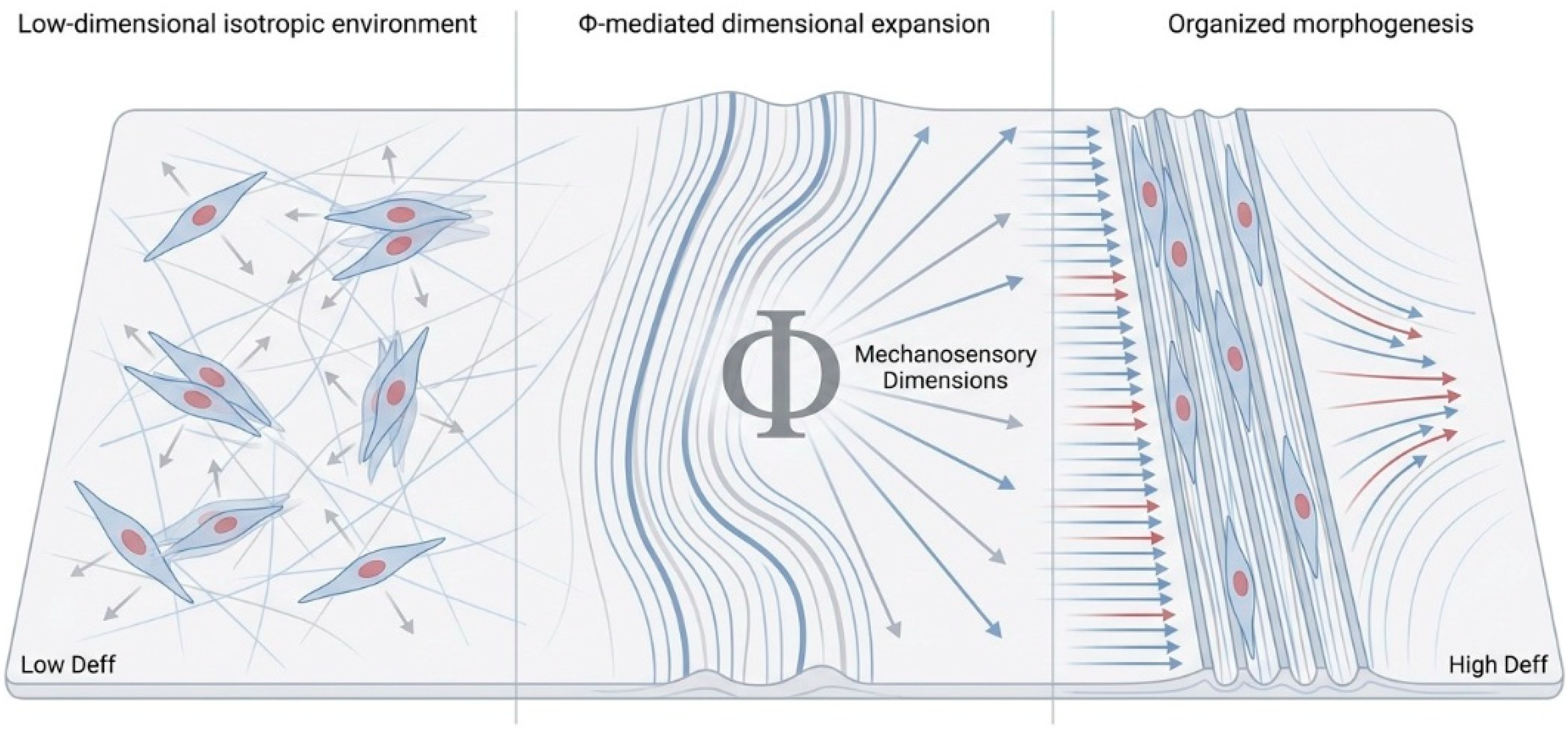
Different fabrication techniques can be interpreted as different strategies for introducing informational dimensionality into the cellular environment. Electrospinning, for example, generates fibrous architectures whose orientation can range from random to highly aligned depending on collector geometry and processing conditions [21,61]. Highly aligned fibers introduce strong directional constraints that bias migration, elongation, and cytoskeletal tension along privileged axes [54]. In contrast, randomly deposited fibers provide substantially weaker orientational information even when overall material composition remains identical [46]. Beyond electrospinning, other fabrication strategies introduce distinct classes of interpretable constraints. Freeze-drying and directional solidification impose anisotropic channel geometries that structure infiltration trajectories and force transmission along coherent axes [62]. Thermally induced phase separation generates interconnected pore networks whose morphological tuning controls the spatial distribution of adhesion and confinement cues [63]. Soft lithography and nanoimprinting operate at the submicrometric scale, where groove and ridge periodicity directly governs focal adhesion organization and nuclear deformation [64]. Additive manufacturing extends this logic to larger spatial scales, enabling precise regulation of pore orientation, curvature, and hierarchical connectivity across the full construct architecture [65]. Despite their mechanical and chemical diversity, these approaches share a common organizational function: each introduces a distinct category of stable, partially independent mechanosensory variable that cells can detect, integrate, and respond to collectively.
The critical issue, however, is not the fabrication method itself, but the type of informational variable introduced into the cellular environment. Each architectural modification changes the set of mechanical relationships available to mechanosensory interpretation. In this sense, fabrication parameters act indirectly: they do not determine cellular behavior directly but reshape the informational geometry through which cellular decisions emerge. Different scaffold architectures therefore correspond to different Φ-transformations acting on the mechanosensory landscape available to the cell.
Experimental observations support this interpretation. Rabel et al. demonstrated that osteoblast responses correlate more strongly with the developed interfacial area ratio (Sdr) than with mean roughness alone, suggesting that the organization of available adhesion states is more important than simple geometric irregularity. Increasing Sdr promoted focal adhesion maturation, cytoskeletal tension, and elongation, consistent with a Φ-mediated expansion of interpretable adhesion configurations. Yet excessive surface complexity impaired proliferation, indicating that dimensional expansion is beneficial only when it remains biologically coherent rather than informationally noisy [66].
Similarly, Dalby and colleagues showed that nanoscale disorder could induce osteogenic differentiation even in the absence of biochemical induction factors, implying that specific forms of topographical heterogeneity enrich the set of mechanically interpretable states available to the cell [67]. Subsequent studies demonstrated that nanoscale architecture influences cytoskeletal tension, nuclear deformation, and gene expression through coordinated alterations in adhesion geometry and force distribution [36].
Together, these findings support the view that scaffold architecture reorganizes the mechanosensory landscape available to cells, thereby influencing tissue organization. The next section examines how cells interpret these expanded informational landscapes through mechanotransduction, converting distributed geometric and mechanical inputs into integrated organizational responses.
4. Mechanotransduction as Integrative Evaluation
The functional significance of scaffold architecture depends not only on the presence of organizational constraints, but on the existence of cellular systems capable of detecting and integrating them. Mechanotransduction performs this integrative role. Cells do not construct explicit geometric representations of their environment; rather, they continuously translate distributed mechanical, topographical, and biochemical cues into coherent internal states that guide adhesion, migration, proliferation, differentiation, and extracellular matrix production [68]. Mechanotransduction can therefore be understood as the biological process through which scaffold-derived information becomes functionally meaningful for cellular organization. The distributed-to-integrated logic of this process is illustrated schematically in Figure 2.
This process begins at the cell–material interface, where transmembrane integrins bind extracellular ligands and organize into focal adhesions (FAs), dynamic multiprotein complexes containing talin, paxillin, vinculin, and focal adhesion kinase (FAK) [69]. These structures function as mechanosensory hubs, coupling extracellular geometry to intracellular force generation [70]. Their formation and maturation depend not simply on the presence of adhesive sites, but on their spatial organization, local curvature, stiffness, and capacity to support cytoskeletal tension [28]. Scaffold architecture therefore shapes cellular behavior indirectly by restructuring the mechanical relationships available to focal adhesion assembly and force transmission [71].
Importantly, cells do not interpret each geometric feature independently. Rather than tracking every ridge, pore, or fiber orientation separately, the mechanotransductive apparatus continuously collapses distributed environmental information into integrated mechanical states [68]. Cytoskeletal tension emerges from the collective interaction of adhesion geometry, substrate mechanics, actomyosin contractility, and spatial confinement [27]. Mechanotransduction effectively integrates distributed environmental cues into a single mechanical state [72].
This compatibility can be represented schematically by introducing a biological compatibility function, , intended to capture the degree to which two organizational states become similar after transformation by the scaffold environment:
where and denote cellular or environmental organizational states and represents the scaffold-mediated transformation of the mechanosensory environment. The expression is intended only as a heuristic representation of mechanotransductive compatibility. It does not imply that cells perform machine-learning algorithms. Rather, it captures the idea that scaffold architecture influences how effectively cellular organization can be coordinated with the mechanical and spatial structure of the environment.
This integration is mediated primarily through the actin cytoskeleton and associated contractile machinery. Actomyosin tension couples local adhesion events into a global mechanical network capable of transmitting forces across the entire cell body [73]. Environments that support stable force propagation promote focal adhesion maturation, stress-fiber assembly, and coherent cytoskeletal polarization [74]. Conversely, isotropic or mechanically incoherent environments generate fragmented force distributions and unstable cytoskeletal organization [75]. The resulting mechanical state can be viewed as an integrated summary of multiple environmental influences, including adhesion geometry, substrate mechanics, force transmission, and spatial confinement.
Mechanical information is subsequently transmitted inward through direct physical and biochemical pathways [76]. Cytoskeletal forces propagate to the nucleus through the LINC complex (linker of nucleoskeleton and cytoskeleton), composed primarily of nesprins and SUN-domain proteins that mechanically couple the cytoskeleton to the nuclear envelope [77]. Simultaneously, force-sensitive proteins within FAs undergo conformational changes that activate downstream signaling pathways including FAK, RhoA/ROCK, and YAP/TAZ [78,79]. These pathways convert distributed mechanical states into transcriptional responses controlling proliferation, migration, lineage commitment, and extracellular matrix production [80].
Among these systems, YAP/TAZ signaling provides a particularly important example of mechanotransductive integration [81]. Increased cytoskeletal tension promotes nuclear localization of YAP/TAZ, whereas mechanically unstable or weakly adhesive environments favor cytoplasmic sequestration and reduced transcriptional activity [82]. Crucially, YAP/TAZ activation does not reflect a single isolated environmental parameter, but the integrated outcome of multiple interacting variables including substrate stiffness, adhesion geometry, spreading area, anisotropy, and force transmission efficiency [83]. Nuclear YAP/TAZ localization can therefore be interpreted as an integrated readout of how cells have collectively processed multiple mechanical and spatial features of their environment.
This compatibility evaluation defines a continuous organizational coordinate along which cellular behaviors become progressively biased. In mechanically coherent environments, cells preferentially stabilize elongated morphologies, aligned migration patterns, reinforced cytoskeletal tension, and lineage-specific transcriptional programs. Cells encountering incoherent or noisy environments instead display fluctuating morphologies, unstable force distributions, and heterogeneous phenotypic outcomes [6]. Mechanotransduction thus transforms distributed geometric complexity into integrated organizational tendencies.
Importantly, many biological responses remain initially graded and continuous. Cytoskeletal tension, focal adhesion maturation, and YAP/TAZ activation often vary progressively across environmental conditions [84]. Yet tissue organization frequently requires discrete and stable outcomes: a cell commits to differentiation, aligns with a tissue axis, or enters a persistent morphogenetic program [85]. These transitions emerge through nonlinear intracellular feedback mechanisms capable of converting continuous mechanical inputs into stabilized organizational states. Positive feedback loops, bistable regulatory circuits, and mechanically reinforced cytoskeletal architectures amplify small differences in mechanotransductive state until distinct phenotypes become robustly maintained despite molecular or environmental noise [37,38].
From this perspective, mechanotransduction does not merely transmit forces; it integrates distributed environmental information into coherent biological responses. Scaffold architecture influences the set of cues available for interpretation, while mechanotransduction determines how those cues are combined into organizational states capable of guiding migration, differentiation, extracellular matrix deposition, and collective tissue assembly.
This interpretation provides a mechanistic bridge between scaffold architecture and developmental robustness [86]. If Φ-transformed topographies expand the informational dimensionality of the environment, mechanotransduction determines whether that expanded landscape can be coherently interpreted and stabilized biologically.
The next section examines how these integrated evaluations generate attractor-like organizational states and how scaffold design influences their robustness against perturbation.
5. Organizational Robustness Through Attractor Stabilization
If mechanotransduction integrates distributed environmental information into coherent organizational tendencies, a further question arises: how do these tendencies become stable tissue architectures? Cells continuously experience molecular noise, fluctuating adhesion dynamics, stochastic gene expression, and changing mechanical conditions [38]. Yet developing and regenerating tissues often converge toward reproducible forms despite this variability [87]. Scaffold-guided regeneration must therefore be understood not only in terms of organizational guidance, but also in terms of organizational stabilization.
The distinction introduced in the previous sections between geometric complexity and informational dimensionality becomes important here. Increasing the number of mechanotransductively interpretable dimensions may make alternative organizational trajectories more distinguishable, but distinguishability alone is not sufficient for robust tissue formation. Regenerative outcomes must remain stable in the presence of biological variability, mechanical perturbation, and environmental noise. The central challenge is therefore not simply the generation of organizational tendencies, but their maintenance through time.
Biological stabilization is more naturally described using the language of dynamical systems. Cells and tissues can be conceived as occupying continuously evolving state spaces shaped by mechanical constraints, cytoskeletal organization, signaling activity, extracellular matrix deposition, and reciprocal cell-cell interactions [88]. Within these spaces, stable organizational states can be understood as attractor-like configurations toward which cellular trajectories preferentially converge [89]. Scaffold architecture does not prescribe a unique outcome; rather, it alters the structure of the organizational landscape, increasing the likelihood that specific trajectories will emerge and persist. The relationship between scaffold architecture, organizational landscapes, and developmental robustness is illustrated schematically in Figure 3.
This principle can be represented schematically through a potential-landscape formalism in which denotes the organizational potential associated with a cellular state . Lower values of correspond to more stable and robust organizational configurations, whereas higher values correspond to less stable states that are more susceptible to perturbation. Within this representation, cellular states tend to evolve toward locally stable minima:
Here, represents the organizational potential associated with the current cellular state, V0 represents the intrinsic energetic baseline of the system, ΔV reflects the energetic cost associated with mechanical or organizational mismatch, and Kbio represents the degree of mechanotransductive compatibility between the current cellular state and a mechanically favorable organizational configuration . Importantly, this equation is not intended as a predictive quantitative model, but as a compact representation of the organizational logic underlying scaffold-guided tissue regeneration [82,89].
Under this interpretation, tissue organization corresponds to convergence toward relatively stable organizational states. Scaffold architecture influences this convergence by restructuring the mechanical and informational relationships available to cellular collectives. Environments providing coherent anisotropic constraints deepen specific attractor basins, making organized morphologies dynamically favored and increasingly resistant to perturbation. By contrast, isotropic or informationally incoherent environments generate shallow and weakly defined basins in which cellular trajectories fluctuate among competing configurations without stable convergence.
Experimental findings from several tissue-engineering systems are consistent with this view. In peripheral nerve regeneration, aligned electrospun fibers convert stochastic Schwann-cell migration into coherent directional organization, promoting axonal bridging and functional reconnection [90]. The scaffold does not explicitly determine a single cellular path; rather, anisotropic architecture stabilizes aligned trajectories while suppressing disordered alternatives. Similarly, patterned substrates and naturally anisotropic matrices used in skeletal muscle engineering promote the emergence of parallel myotube arrays capable of coordinated contraction [46]. Under isotropic conditions, myotubes remain viable but fail to maintain stable collective alignment. The distinction again lies not in viability, but in organizational stabilization.
Gradient-based scaffolds provide an additional example. In tendon-to-bone interfaces, mineralization and stiffness vary continuously across space [91]. Stem cells cultured within such environments preferentially stabilize region-specific phenotypes corresponding to local mechanical conditions, producing transitions between tenogenic and osteogenic organizational states [92]. These outcomes emerge not from isolated biochemical instructions alone, but from the coordinated structuring of the mechanical and spatial landscape across multiple dimensions.
Developmental robustness can be interpreted as the stability of these attractor basins against perturbation. Within the landscape formalism introduced above, the depth and sharpness of an attractor basin can be characterized by the local curvature of the potential. Intuitively, a steep basin is difficult to escape: perturbations that displace the system from the organized state encounter strong restoring forces that drive it back toward the attractor. Shallow basins offer weaker resistance, allowing fluctuations to redirect organizational trajectories more easily [93]. Formally, this curvature is proportional to the second spatial derivative of the potential:
where large values of R indicate strongly stabilized organizational states and small values indicate configurations vulnerable to noise and perturbation. Scaffold architectures that introduce coherent mechanosensory constraints through anisotropy, curvature, stiffness gradients, pore orientation, or hierarchical organization increase the stability of these attractors by making organizational trajectories more reproducible and less sensitive to local fluctuations.
This perspective suggests that scaffold optimization should focus less on reproducing morphological complexity and more on stabilizing desired organizational states. Architectural features become important not because they increase complexity per se, but because they reshape the organizational landscape in ways that enhance robustness and reproducibility.
Scaffold-guided regeneration links dimensional expansion, mechanotransductive integration, and organizational stabilization. The following section examines the limits of this interpretation and explores its implications for scaffold design and computational biomaterial optimization.
6. Designing Informationally Structured Regenerative Environments
The distinction between geometric complexity and effective informational dimensionality has important implications for biomaterial design, including which architectural features create environmental conditions that cells can reliably detect, integrate, and exploit during tissue formation.
This perspective shifts attention away from complexity as an objective in itself and toward the informational structure of the cellular microenvironment. A growing body of evidence suggests that regenerative outcomes emerge from the interaction of multiple mechanical, geometric, topographical, transport, and temporal variables rather than from isolated parameters [94]. Regenerative outcomes emerge from the interaction of multiple environmental variables rather than from isolated parameters [95,96].
An important implication is that the properties measured by engineers are not always identical to those experienced by cells. Experimental studies have shown that the local mechanical environment perceived at cellular length scales may diverge substantially from bulk material properties [97], while transport accessibility and pore interconnection can exert stronger biological effects than nominal pore size alone [98]. Likewise, hierarchical pore architectures frequently outperform simpler structures not because any individual pore dimension is optimal, but because multiple scales cooperate to support transport, infiltration, mechanical support, and extracellular matrix deposition simultaneously [94]. These observations suggest that the biologically relevant variables are often those that remain locally accessible and interpretable to cellular mechanosensing rather than those most easily quantified at the material scale.
From this perspective, architectural features such as anisotropy, curvature, pore orientation, hierarchical organization, stiffness gradients, transport pathways, and degradation dynamics become important because they introduce distinct classes of mechanosensory and organizational information. Anisotropy establishes preferred directions for migration and matrix deposition [95]. Curvature modifies force distribution and nuclear deformation [99]. Pore architecture structures infiltration pathways and transport accessibility [98]. Stiffness gradients generate spatially organized mechanical information [100]. Degradation kinetics determine the persistence of organizational cues through time. Hierarchical architectures coordinate these dimensions across multiple scales. Their value lies not primarily in reproducing native morphology, but in expanding the repertoire of biologically meaningful variables available for mechanotransductive integration.
This interpretation also helps explain why regenerative performance is often poorly predicted by geometric sophistication alone. Geometrically elaborate scaffolds may contain numerous structural features while providing relatively little additional organizational information if those features are redundant, unstable, or mechanotransductively incoherent [101]. Conversely, comparatively simple architectures may exert strong organizational effects when they introduce robust directional, mechanical, transport, or biochemical constraints [102]. Evidence from aligned fibrous scaffolds, anisotropic architectures, and hierarchically interconnected porous systems suggests that coherent organizational cues can be more influential than geometric richness per se [95,98,103]. The relevant design criterion is therefore not the amount of structural complexity, but the extent to which complexity generates stable and biologically interpretable dimensions for cellular organization.
A further implication is that scaffold design cannot be treated as a purely static optimization problem. Cell–scaffold systems continuously remodel themselves through matrix deposition, degradation, force generation, and local restructuring of the microenvironment [104]. Mechanical properties perceived by cells may evolve during regeneration, while the organization of the developing tissue progressively modifies the very environment from which it emerged [105]. Effective scaffold architectures must therefore do more than provide useful initial cues: they must preserve sufficient informational coherence as regeneration unfolds. The challenge is not simply to create an initial template, but to maintain organizational accessibility throughout the regenerative process.
The analogy with kernel methods remains useful at a limited heuristic level. Scaffold architectures may be viewed as transformations that increase the distinguishability of alternative organizational trajectories by introducing additional interpretable dimensions into the cellular environment. However, biological systems are not deterministic classifiers. Cellular outcomes remain influenced by stochastic gene expression, local matrix heterogeneity, variable adhesion dynamics, and continuous cell-mediated remodeling [106]. Scaffold architecture therefore does not determine a unique outcome; rather, it biases cellular populations toward organizationally coherent regions of state space while reducing the accessibility of unstable alternatives.
This perspective generates a series of experimentally testable hypotheses. First, scaffolds with high geometric complexity but low mechanotransductive coherence should produce heterogeneous or unstable organizational outcomes despite their structural sophistication [101]. Second, architectures combining multiple partially independent dimensions—such as anisotropy, stiffness gradients, hierarchical porosity, transport accessibility, and controlled biochemical presentation—should stabilize tissue organization more effectively than architectures manipulating only a single parameter [94]. Third, the organizational effects of these architectures should be reflected in integrated mechanotransductive readouts, including focal adhesion maturation, cytoskeletal alignment, nuclear deformation, extracellular matrix organization, and YAP/TAZ localization [107]. Fourth, cellular responses should correlate more strongly with local and cell-accessible descriptors of the microenvironment than with bulk material properties alone [97]. Finally, organizational states generated in informationally structured environments should exhibit greater robustness to perturbation and remodeling than states generated under informationally poor conditions [108].
These considerations also suggest a role for computational and AI-assisted scaffold design. Current machine-learning approaches increasingly predict material properties, degradation kinetics, mechanical behavior, manufacturability, and, in some cases, proxies of cellular growth [109]. The perspective developed here suggests an additional objective: identifying architectural configurations that generate stable and biologically interpretable mechanosensory dimensions. Rather than searching exclusively for biomimetic geometries, future computational approaches could seek scaffold architectures that maximize mechanotransductive coherence while minimizing redundancy and informational noise. Although such approaches remain largely prospective, they point toward a shift from optimizing scaffold geometry alone to optimizing the informational structure of the cellular microenvironment.
More broadly, the perspective developed here suggests that successful regenerative biomaterials do not function because they reproduce the extracellular matrix in exhaustive structural detail. Rather, they appear to create sufficiently structured environments within which organized regeneration becomes more accessible. Scaffold architecture contributes to this process by shaping the mechanical, spatial, transport, and temporal dimensions available to cellular collectives. Tissue engineering can therefore be understood not simply as the fabrication of materials, but as the design of environments capable of supporting robust biological self-organization.
7. Conclusion: From Complexity to Informational Dimensionality
The central challenge of tissue engineering is not simply to maintain cellular viability, but to guide cellular collectives toward stable and functional tissue organization. Although scaffold architecture is widely recognized as a major determinant of regenerative outcomes, the relationship between structural complexity and organizational performance remains difficult to interpret. Geometrically elaborate scaffolds do not necessarily generate coherent tissue architectures, while comparatively simple designs can sometimes exert remarkably strong organizational effects.
This paper has argued that understanding this apparent discrepancy requires distinguishing geometric complexity from effective informational dimensionality. Scaffold architectures influence regeneration not merely by increasing structural richness, but by introducing stable, partially independent, and biologically interpretable dimensions that cells can detect and integrate through mechanotransduction. Anisotropy, curvature, pore orientation, stiffness gradients, hierarchical organization, transport accessibility, and biochemical presentation contribute to tissue organization insofar as they generate coherent mechanosensory information rather than complexity alone.
Mechanotransduction provides the biological basis for this framework by integrating distributed environmental cues into coherent organizational states that can subsequently be stabilized through cellular and tissue-level feedback.
Under this interpretation, successful scaffold design depends less on reproducing the visible complexity of native tissues than on generating environments that provide coherent and mechanotransductively interpretable constraints. Scaffold architecture can therefore be viewed as a means of structuring the informational conditions under which cellular self-organization unfolds. The practical implication is that scaffold performance should be evaluated not only by material composition, geometric sophistication, or biomimetic resemblance, but by the extent to which scaffold architectures introduce stable and biologically meaningful dimensions capable of supporting robust tissue regeneration.
Author Contributions
Conceptualization, C.G. and M.T.C.; methodology, C.G. and M.T.C.; formal analysis, C.G.; writing—original draft preparation, C.G. and M.T.C.; writing—review and editing, C.G., M.T.C., M.M. and S.G.; visualization, C.G.; supervision, S.G.All authors have read and agreed to the published version of the manuscript.
Funding
This research received no external funding.
Institutional Review Board Statement
Not applicable.
Informed Consent Statement
Not applicable.
Data Availability Statement
No new data were created or analyzed in this study. Data sharing is not applicable to this article.
Acknowledgments
The authors would like to thank dr. Silvana Belletti for her insights.
Conflicts of Interest
The authors declare no conflicts of interest.
References
- Miklosic, G.; Ferguson, S.J.; D’Este, M. Engineering Complex Tissue-like Microenvironments with Biomaterials and Biofabrication. Trends Biotechnol. 2024, 42, 1241–1257. [Google Scholar] [CrossRef] [PubMed]
- Bandzerewicz, A.; Gadomska-Gajadhur, A. Into the Tissues: Extracellular Matrix and Its Artificial Substitutes: Cell Signalling Mechanisms. Cells 2022, 11, 914. [Google Scholar] [CrossRef] [PubMed]
- Crossley, R.M.; Johnson, S.; Tsingos, E.; Bell, Z.; Berardi, M.; Botticelli, M.; Braat, Q.J.S.; Metzcar, J.; Ruscone, M.; Yin, Y.; et al. Modeling the Extracellular Matrix in Cell Migration and Morphogenesis: A Guide for the Curious Biologist. Front. Cell Dev. Biol. 2024, 12. [Google Scholar] [CrossRef] [PubMed]
- Bril, M.; Saberi, A.; Jorba, I.; van Turnhout, M.C.; Sahlgren, C.M.; Bouten, C.V.C.; Schenning, A.P.H.J.; Kurniawan, N.A. Shape-Morphing Photoresponsive Hydrogels Reveal Dynamic Topographical Conditioning of Fibroblasts. Adv. Sci. 2023, 10. [Google Scholar] [CrossRef] [PubMed]
- Luo, Y.; Gu, M.; Park, M.; Fang, X.; Kwon, Y.; Urueña, J.M.; Read de Alaniz, J.; Helgeson, M.E.; Marchetti, C.M.; Valentine, M.T. Molecular-Scale Substrate Anisotropy, Crowding and Division Drive Collective Behaviours in Cell Monolayers. J. R. Soc. Interface 2023, 20. [Google Scholar] [CrossRef] [PubMed]
- Ong, H.T.; Sriram, M.; Susapto, H.H.; Li, Y.; Jiang, Y.; Voelcker, N.H.; Young, J.L.; Holle, A.W.; Elnathan, R. The Rise of Mechanobiology for Advanced Cell Engineering and Manufacturing. Adv. Mater. 2025, 37. [Google Scholar] [CrossRef] [PubMed]
- Ramos-Díez, S.; Camarero-Espinosa, S. Topography as a Cue for Driving Osteochondral Unit Regeneration. Biofabrication 2026, 18, 012003. [Google Scholar] [CrossRef]
- Gao, J.; Xiong, F.; Yang, Z.; Ye, X.; Ma, K.; Shi, B.; Li, Z.; Long, K.; You, Z.; Zhang, L.; et al. Bridging the Gap: Gradient Scaffolds as Bioinstructive Platforms for Peripheral Nerve Repair. Chem. Soc. Rev. 2026, 55, 4376–4408. [Google Scholar] [CrossRef] [PubMed]
- Yang, T.; Fang, Z.; Zhang, J.; Zheng, S. Physical Cues in Biomaterials Modulate Macrophage Polarization for Bone Regeneration: A Review. Front. Bioeng. Biotechnol. 2025, 13. [Google Scholar] [CrossRef] [PubMed]
- Maksoud, F.J.; de la Paz, M.F.V.; Hann, A.J.; Thanarak, J.; Reilly, G.C.; Claeyssens, F.; Green, N.H.; Zhang, Y.S. Porous Biomaterials for Tissue Engineering: A Review. J. Mater. Chem. B 2022, 10, 8111–8165. [Google Scholar] [CrossRef] [PubMed]
- Jesus, D.; Pinho, A.R.; Gomes, M.C.; Oliveira, C.S.; Mano, J.F. Emerging Modulators for Osteogenic Differentiation: A Combination of Chemical and Topographical Cues for Bone Microenvironment Engineering. Soft Matter 2022, 18, 3107–3119. [Google Scholar] [CrossRef] [PubMed]
- Zhang, W.; Zeng, X.; Deng, X.; Yang, F.; Ma, X.; Gao, W. Smart Biomaterials: As Active Immune Modulators to Shape pro-Regenerative Microenvironments. Front. Cell Dev. Biol. 2025, 13. [Google Scholar] [CrossRef] [PubMed]
- Berdiaki, A.; Neagu, M.; Tzanakakis, P.; Spyridaki, I.; Pérez, S.; Nikitovic, D. Extracellular Matrix Components and Mechanosensing Pathways in Health and Disease. Biomolecules 2024, 14, 1186. [Google Scholar] [CrossRef] [PubMed]
- Saraswathibhatla, A.; Indana, D.; Chaudhuri, O. Cell–Extracellular Matrix Mechanotransduction in 3D. Nat. Rev. Mol. Cell Biol. 2023, 24, 495–516. [Google Scholar] [CrossRef] [PubMed]
- Eslami, H.; Darvishi, A. Extracellular Matrix Viscoelasticity: A Dynamic Regulator of Cellular Behavior. Ann. Biomed. Eng. 2025, 53, 2029–2046. [Google Scholar] [CrossRef] [PubMed]
- Jang, J.-H. Extracellular Matrix in Human Disease and Therapy: From Pathogenic Remodeling to Biomaterial Platforms and Precision Diagnostics. Biomedicines 2026, 14, 247. [Google Scholar] [CrossRef] [PubMed]
- Hong, H.; Kim, D.; Jung, H.; Kim, S.; Min, S.; Kim, C.; Kim, K.; Rha, H.; Kang, H. Biomimetic Dynamics of Nanoscale Groove and Ridge Topography for Stem Cell Regulation. Adv. Mater. 2025, 37. [Google Scholar] [CrossRef] [PubMed]
- Iturriaga, L.; Van Gordon, K.D.; Larrañaga-Jaurrieta, G.; Camarero-Espinosa, S. Strategies to Introduce Topographical and Structural Cues in 3D-Printed Scaffolds and Implications in Tissue Regeneration. Adv. Nanobiomed Res. 2021, 1. [Google Scholar] [CrossRef]
- Cun, X.; Hosta-Rigau, L. Topography: A Biophysical Approach to Direct the Fate of Mesenchymal Stem Cells in Tissue Engineering Applications. Nanomaterials 2020, 10, 1–41. [Google Scholar] [CrossRef] [PubMed]
- Zarei, M.; Pourmostafa, A.; Moghanian, A.; Miri, A.K. Integration of Electrospinning and 3D Bioprinting for Fibrous Tissue Manufacturing. Bioprinting 2026, 54, e00470. [Google Scholar] [CrossRef]
- Dai, X.; Nie, W.; Shen, H.; Machens, H.-G.; Böker, K.; Taheri, S.; Lehmann, W.; Shen, Y.; Schilling, A.F. Electrospinning Based Biomaterials for Biomimetic Fabrication, Bioactive Protein Delivery and Wound Regenerative Repair. Regen. Biomater. 2025, 12. [Google Scholar] [CrossRef] [PubMed]
- Carotenuto, F.; Politi, S.; Ul Haq, A.; De Matteis, F.; Tamburri, E.; Terranova, M.L.; Teodori, L.; Pasquo, A.; Di Nardo, P. From Soft to Hard Biomimetic Materials: Tuning Micro/Nano-Architecture of Scaffolds for Tissue Regeneration. Micromachines . 2022, 13, 780. [Google Scholar] [CrossRef] [PubMed]
- Ciulla, M.G.; Massironi, A.; Sugni, M.; Ensign, M.A.; Marzorati, S.; Forouharshad, M. Recent Advances in the Development of Biomimetic Materials. Gels 2023, 9, 833. [Google Scholar] [CrossRef] [PubMed]
- Sonnenberg, M.; Petrik, D.; Miehe, R.; Bauernhansl, T. Foundations of Design for Biointelligence: Expert Insights on Engineering Challenges and Design Heuristics. Res. Eng. Des. 2026, 37, 18. [Google Scholar] [CrossRef]
- Saraswathibhatla, A.; Indana, D.; Chaudhuri, O. Cell–Extracellular Matrix Mechanotransduction in 3D. Nat. Rev. Mol. Cell Biol. 2023, 24, 495–516. [Google Scholar] [CrossRef] [PubMed]
- Herrera, A.; Molina, M.S.; Günther, R.; Orellano, I.; Brauer, E.; Diederich, S.; Serrano, A.; Behncke, R.; Korus, G.; Leemhuis, H.; et al. Cell Type-Specific Response to Curvature Controls Tissue Growth Dynamics in Biomaterial Pores. Bioact. Mater. 2026, 61, 482–497. [Google Scholar] [CrossRef] [PubMed]
- Kalukula, Y.; Ciccone, G.; Mohammed, D.; Procès, A.; Versaevel, M.; Deridoux, A.; Ergot, L.; Barbier, Z.; Mansy, M.; Aucouturier, R.; et al. Unlocking the Therapeutic Potential of Cellular Mechanobiology. Sci. Adv. 2025, 11. [Google Scholar] [CrossRef] [PubMed]
- Jafarinia, H.; Shi, L.; Wolfenson, H.; Carlier, A. YAP Phosphorylation within Integrin Adhesions: Insights from a Computational Model. Biophys. J. 2024, 123, 3658–3668. [Google Scholar] [CrossRef] [PubMed]
- Qiao, P.; Zhao, H.; Ma, Z.; Hasani, M.; Song, Y. Cellular Mechano-Memory Informs Biomaterials and Mechano-Medicine. Cell Rep. Phys. Sci. 2026, 7. [Google Scholar] [CrossRef]
- Chen, A.; Harris, C.; Yoo, E.; Qu, C.-K. Biomechanical Regulation of Stem Cells: Implications for Cell Therapeutics. iScience 2026, 29. [Google Scholar] [CrossRef] [PubMed]
- Cao, S.; Yuan, Q. An Update of Nanotopographical Surfaces in Modulating Stem Cell Fate: A Narrative Review. Biomater. Transl. 2022, 3, 55–64. [Google Scholar] [CrossRef] [PubMed]
- Tortorici, M.; Petersen, A.; Ehrhart, K.; Duda, G.N.; Checa, S. Scaffold-Dependent Mechanical and Architectural Cues Guide Osteochondral Defect Healing in Silico. Front. Bioeng. Biotechnol. 2021, 9. [Google Scholar] [CrossRef] [PubMed]
- Gonthier, A.R.; Botvinick, E.L.; Grosberg, A.; Mohraz, A. Effect of Porous Substrate Topographies on Cell Dynamics: A Computational Study. ACS Biomater. Sci. Eng. 2023, 9, 5666–5678. [Google Scholar] [CrossRef] [PubMed]
- Rajendran, A.K.; Sankar, D.; Amirthalingam, S.; Kim, H.D.; Rangasamy, J.; Hwang, N.S. Trends in Mechanobiology Guided Tissue Engineering and Tools to Study Cell-Substrate Interactions: A Brief Review. Biomater. Res. 2023, 27. [Google Scholar] [CrossRef] [PubMed]
- Callens, S.J.P.; Fan, D.; van Hengel, I.A.J.; Minneboo, M.; Díaz-Payno, P.J.; Stevens, M.M.; Fratila-Apachitei, L.E.; Zadpoor, A.A. Emergent Collective Organization of Bone Cells in Complex Curvature Fields. Nat. Commun. 2023, 14, 855. [Google Scholar] [CrossRef] [PubMed]
- Lee, J.-H.; Yun, Y.G.; Kim, H.-W. Matrix-Induced Nuclear Remodeling and Mechano-Therapeutics. Cell Rep. 2025, 44. [Google Scholar] [CrossRef] [PubMed]
- Niu, L.; Cheng, B.; Huang, G.; Nan, K.; Han, S.; Ren, H.; Liu, N.; Li, Y.; Genin, G.M.; Xu, F. A Positive Mechanobiological Feedback Loop Controls Bistable Switching of Cardiac Fibroblast Phenotype. Cell Discov. 2022, 8, 84. [Google Scholar] [CrossRef] [PubMed]
- Villeneuve, C.; Hashmi, A.; Ylivinkka, I.; Lawson-Keister, E.; Miroshnikova, Y.A.; Pérez-González, C.; Myllymäki, S.-M.; Bertillot, F.; Yadav, B.; Zhang, T.; et al. Mechanical Forces across Compartments Coordinate Cell Shape and Fate Transitions to Generate Tissue Architecture. Nat. Cell Biol. 2024, 26, 207–218. [Google Scholar] [CrossRef] [PubMed]
- Galli, C.; Colangelo, M.T.; Meleti, M.; Guizzardi, S. Morphospace Engineering: Morphological Computation in Scaffold Design. BioSystems 2025, 105676. [Google Scholar] [CrossRef] [PubMed]
- Werner, M.; Kurniawan, N.A.; Bouten, C.V.C. Cellular Geometry Sensing at Different Length Scales and Its Implications for Scaffold Design. Materials 2020, 13, 963. [Google Scholar] [CrossRef] [PubMed]
- Smola, A.J.; Schölkopf, B. Learning with Kernels; GMD-Forschungszentrum Informationstechnik Berlin, Germany, 1998; Vol. 4. [Google Scholar]
- Hofmann, T.; Schölkopf, B.; Smola, A.J. Kernel Methods in Machine Learning. Ann. Stat. 2008, 36. [Google Scholar] [CrossRef]
- Cortesi, M.; Warton, K.; Ford, C.E. Beyond 2D Cell Cultures: How 3D Models Are Changing the in Vitro Study of Ovarian Cancer and How to Make the Most of Them. PeerJ 2024, 12, e17603. [Google Scholar] [CrossRef] [PubMed]
- Zhao, F.; Zhang, M.; Nizamoglu, M.; Kaper, H.J.; Brouwer, L.A.; Borghuis, T.; Burgess, J.K.; Harmsen, M.C.; Sharma, P.K. Fibroblast Alignment and Matrix Remodeling Induced by a Stiffness Gradient in a Skin-Derived Extracellular Matrix Hydrogel. Acta Biomater. 2024, 182, 67–80. [Google Scholar] [CrossRef] [PubMed]
- Keijzer, K.A.E.; Tsingos, E.; Merks, R.M.H. How Cells Align to Structured Collagen Fibrils: A Hybrid Cellular Potts and Molecular Dynamics Model with Dynamic Mechanosensitive Focal Adhesions. Front. Cell Dev. Biol. 2025, 12. [Google Scholar] [CrossRef] [PubMed]
- Skillin, N.P.; Kirkpatrick, B.E.; Herbert, K.M.; Nelson, B.R.; Hach, G.K.; Günay, K.A.; Khan, R.M.; DelRio, F.W.; White, T.J.; Anseth, K.S. Stiffness Anisotropy Coordinates Supracellular Contractility Driving Long-Range Myotube-ECM Alignment. Sci. Adv. 2024, 10. [Google Scholar] [CrossRef] [PubMed]
- Joshi, I.M.; Mansouri, M.; Ahmed, A.; De Silva, D.; Simon, R.A.; Esmaili, P.; Desa, D.E.; Elias, T.M.; Brown, E.B.; Abhyankar, V. V. Microengineering 3D Collagen Matrices with Tumor-Mimetic Gradients in Fiber Alignment. Adv. Funct. Mater. 2024, 34. [Google Scholar] [CrossRef] [PubMed]
- Doha, U.; Aydin, O.; Joy, M.S.H.; Emon, B.; Drennan, W.; Saif, M.T.A. Disorder to Order Transition in Cell-ECM Systems Mediated by Cell-Cell Collective Interactions. Acta Biomater. 2022, 154, 290–301. [Google Scholar] [CrossRef] [PubMed]
- Sapudom, J.; Karaman, S.; Quartey, B.C.; Mohamed, W.K.E.; Mahtani, N.; Garcia-Sabaté, A.; Teo, J. Collagen Fibril Orientation Instructs Fibroblast Differentiation Via Cell Contractility. Adv. Sci. 2023, 10. [Google Scholar] [CrossRef] [PubMed]
- Böhringer, D.; Bauer, A.; Moravec, I.; Bischof, L.; Kah, D.; Mark, C.; Grundy, T.J.; Görlach, E.; O’Neill, G.M.; Budday, S.; et al. Fiber Alignment in 3D Collagen Networks as a Biophysical Marker for Cell Contractility. Matrix Biol. 2023, 124, 39–48. [Google Scholar] [CrossRef] [PubMed]
- Liu, K.; Wiendels, M.; Yuan, H.; Ruan, C.; Kouwer, P.H.J. Cell-Matrix Reciprocity in 3D Culture Models with Nonlinear Elasticity. Bioact. Mater. 2022, 9, 316–331. [Google Scholar] [CrossRef] [PubMed]
- Chi, J.; Wang, M.; Chen, J.; Hu, L.; Chen, Z.; Backman, L.J.; Zhang, W. Topographic Orientation of Scaffolds for Tissue Regeneration: Recent Advances in Biomaterial Design and Applications. Biomimetics 2022, 7, 131. [Google Scholar] [CrossRef] [PubMed]
- Grelier, M.; Martin, C.U.; Milas, M.; Bruskin-Pevzner, L.; Segal, Y.; Ramani, M.; Nuić, L.; Le Saux, G.; Smith, A.-S.; Schvartzman, M. Heterogeneity in Environmental Stiffness Alone Can Guide Cells and Shape Tissues. 2025. [Google Scholar] [CrossRef]
- Bagchi, A.; Sarker, B.; Zhang, J.; Foston, M.; Pathak, A. Fast yet Force-Effective Mode of Supracellular Collective Cell Migration Due to Extracellular Force Transmission. PLoS Comput. Biol. 2025, 21, e1012664. [Google Scholar] [CrossRef] [PubMed]
- Arnold, C.; Tahmaz, I.; Chapon, M.-L.; Maayouf, H.; Luchnikov, V.; Milan, J.-L.; Borghi, F.; Pieuchot, L. Bending the Rules: Curvature’s Impact on Cell Biology. BMC Biol. 2025, 23, 296. [Google Scholar] [CrossRef] [PubMed]
- Mukasheva, F.; Adilova, L.; Dyussenbinov, A.; Yernaimanova, B.; Abilev, M.; Akilbekova, D. Optimizing Scaffold Pore Size for Tissue Engineering: Insights across Various Tissue Types. Front. Bioeng. Biotechnol. 2024, 12. [Google Scholar] [CrossRef] [PubMed]
- Hadden, W.J.; Young, J.L.; Holle, A.W.; McFetridge, M.L.; Kim, D.Y.; Wijesinghe, P.; Taylor-Weiner, H.; Wen, J.H.; Lee, A.R.; Bieback, K.; et al. Stem Cell Migration and Mechanotransduction on Linear Stiffness Gradient Hydrogels. Proc. Natl. Acad. Sci. 2017, 114, 5647–5652. [Google Scholar] [CrossRef] [PubMed]
- Fu, R.; Jones, E.; Chen, N.; Sun, B.; Si, B.; Wei, Z.A.; Ameer, G.; Sun, C.; Ding, Y. Multiscale 3D Printing of Nanoporous Scaffolds with Surface Topography for Guiding 3D Cell Alignment. Adv. Healthc. Mater. 2026, 15. [Google Scholar] [CrossRef]
- Walters, N.J.; Gentleman, E. Evolving Insights in Cell–Matrix Interactions: Elucidating How Non-Soluble Properties of the Extracellular Niche Direct Stem Cell Fate. Acta Biomater. 2015, 11, 3–16. [Google Scholar] [CrossRef] [PubMed]
- Causa, F.; Netti, P.A.; Ambrosio, L. A Multi-Functional Scaffold for Tissue Regeneration: The Need to Engineer a Tissue Analogue. Biomaterials 2007, 28, 5093–5099. [Google Scholar] [CrossRef] [PubMed]
- Ghosal, K.; Agatemor, C.; Špitálsky, Z.; Thomas, S.; Kny, E. Electrospinning Tissue Engineering and Wound Dressing Scaffolds from Polymer-Titanium Dioxide Nanocomposites. Chem. Eng. J. 2019, 358, 1262–1278. [Google Scholar] [CrossRef]
- Shahbazi, M.-A.; Ghalkhani, M.; Maleki, H. Directional Freeze-Casting: A Bioinspired Method to Assemble Multifunctional Aligned Porous Structures for Advanced Applications. Adv. Eng. Mater. 2020, 22. [Google Scholar] [CrossRef]
- Wang, A.; Ma, Y.; Zhao, D. Pore Engineering of Porous Materials: Effects and Applications. ACS Nano 2024, 18, 22829–22854. [Google Scholar] [CrossRef] [PubMed]
- Del Campo, A.; Arzt, E. Fabrication Approaches for Generating Complex Micro-and Nanopatterns on Polymeric Surfaces. Chem. Rev. 2008, 108, 911–945. [Google Scholar] [CrossRef] [PubMed]
- Liu, T.; Wang, Y.; Kuang, T. Oriented Porous Polymer Scaffolds in Tissue Engineering: A Comprehensive Review of Preparation Strategies and Applications. Macromol. Mater. Eng. 2024, 309. [Google Scholar] [CrossRef]
- Rabel, K.; Kohal, R.-J.; Steinberg, T.; Tomakidi, P.; Rolauffs, B.; Adolfsson, E.; Palmero, P.; Fürderer, T.; Altmann, B. Controlling Osteoblast Morphology and Proliferation via Surface Micro-Topographies of Implant Biomaterials. Sci. Rep. 2020, 10, 12810. [Google Scholar] [CrossRef] [PubMed]
- Dalby, M.J.; Gadegaard, N.; Tare, R.; Andar, A.; Riehle, M.O.; Herzyk, P.; Wilkinson, C.D.W.; Oreffo, R.O.C. The Control of Human Mesenchymal Cell Differentiation Using Nanoscale Symmetry and Disorder. Nat. Mater. 2007, 6, 997–1003. [Google Scholar] [CrossRef] [PubMed]
- Cao, R.; Tian, H.; Tian, Y.; Fu, X. A Hierarchical Mechanotransduction System: From Macro to Micro. Adv. Sci. 2024, 11, 2302327. [Google Scholar]
- Yokoyama, Y.; Domkam, N.; Kabir, H.; Mansour, A.; Tsukamoto, S.; Yerima, G.; Adachi, T.; Mofrad, M.R.K. Emerging Mechanomedicines Informed by Mechanotransduction along the Integrin–Cytoskeleton–Nucleus Axis. APL Bioeng. 2025, 9. [Google Scholar] [CrossRef] [PubMed]
- Le Coq, J.; Acebrón, I.; Rodrigo Martin, B.; Lopez Navajas, P.; Lietha, D. New Insights into FAK Structure and Function in Focal Adhesions. J. Cell Sci. 2022, 135, jcs259089. [Google Scholar] [CrossRef] [PubMed]
- Ahn, Y.; Chang, H.; Baek, J. 3D Scaffolds-Specific Cellular Mechanoresponse as a Pivotal Regulating Factor in Tissue Engineering. JMST Adv. 2024, 6, 121–134. [Google Scholar] [CrossRef]
- Dupont, S.; Wickström, S.A. Mechanical Regulation of Chromatin and Transcription. Nat. Rev. Genet. 2022, 23, 624–643. [Google Scholar] [CrossRef] [PubMed]
- Katsuta, H.; Sokabe, M.; Hirata, H. From Stress Fiber to Focal Adhesion: A Role of Actin Crosslinkers in Force Transmission. Front. Cell Dev. Biol. 2024, 12. [Google Scholar] [CrossRef] [PubMed]
- Weißenbruch, K.; Mayor, R. Actomyosin Forces in Cell Migration: Moving beyond Cell Body Retraction. BioEssays 2024, 46. [Google Scholar] [CrossRef] [PubMed]
- Doss, B.L.; Pan, M.; Gupta, M.; Grenci, G.; Mège, R.-M.; Lim, C.T.; Sheetz, M.P.; Voituriez, R.; Ladoux, B. Cell Response to Substrate Rigidity Is Regulated by Active and Passive Cytoskeletal Stress. Proc. Natl. Acad. Sci. 2020, 117, 12817–12825. [Google Scholar] [CrossRef] [PubMed]
- De Belly, H.; Paluch, E.K.; Chalut, K.J. Interplay between Mechanics and Signalling in Regulating Cell Fate. Nat. Rev. Mol. Cell Biol. 2022, 23, 465–480. [Google Scholar] [CrossRef] [PubMed]
- Bougaran, P.; Bautch, V.L. Life at the Crossroads: The Nuclear LINC Complex and Vascular Mechanotransduction. Front. Physiol. 2024, 15, 1411995. [Google Scholar] [CrossRef] [PubMed]
- Piccolo, S.; Dupont, S.; Cordenonsi, M. The Biology of YAP/TAZ: Hippo Signaling and Beyond. Physiol. Rev. 2014, 94, 1287–1312. [Google Scholar] [CrossRef] [PubMed]
- Battilana, G.; Zanconato, F.; Piccolo, S. Mechanisms of YAP/TAZ Transcriptional Control. Cell Stress 2021, 5, 167. [Google Scholar] [CrossRef] [PubMed]
- Mierke, C.T. Extracellular Matrix Cues Regulate Mechanosensing and Mechanotransduction of Cancer Cells. Cells 2024, 13, 96. [Google Scholar] [CrossRef] [PubMed]
- Heng, B.C.; Zhang, X.; Aubel, D.; Bai, Y.; Li, X.; Wei, Y.; Fussenegger, M.; Deng, X. An Overview of Signaling Pathways Regulating YAP/TAZ Activity. Cell. Mol. Life Sci. 2020, 78, 497. [Google Scholar] [CrossRef] [PubMed]
- Scott, K.E.; Fraley, S.I.; Rangamani, P. A Spatial Model of YAP/TAZ Signaling Reveals How Stiffness, Dimensionality, and Shape Contribute to Emergent Outcomes. Proc. Natl. Acad. Sci. 2021, 118, e2021571118. [Google Scholar] [CrossRef] [PubMed]
- Francis, E.A.; Sarikhani, E.; Patel, V.; Meganathan, D.P.; Sadr, L.; Tahir, A.; Jahed, Z.; Rangamani, P. Nanoscale Curvature Regulates YAP/TAZ Nuclear Localization Through Nuclear Deformation and Rupture. Adv. Sci. 2025, 12. [Google Scholar] [CrossRef] [PubMed]
- Brusatin, G.; Panciera, T.; Gandin, A.; Citron, A.; Piccolo, S. Biomaterials and Engineered Microenvironments to Control YAP/TAZ-Dependent Cell Behaviour. Nat. Mater. 2018, 17, 1063–1075. [Google Scholar] [CrossRef] [PubMed]
- Han, P.; Gomez, G.A.; Duda, G.N.; Ivanovski, S.; Poh, P.S.P. Scaffold Geometry Modulation of Mechanotransduction and Its Influence on Epigenetics. Acta Biomater. 2023, 163, 259–274. [Google Scholar] [CrossRef] [PubMed]
- Xie, W.; Wei, X.; Kang, H.; Jiang, H.; Chu, Z.; Lin, Y.; Hou, Y.; Wei, Q. Static and Dynamic: Evolving Biomaterial Mechanical Properties to Control Cellular Mechanotransduction. Adv. Sci. 2023, 10, 2204594. [Google Scholar] [CrossRef] [PubMed]
- Ameer, G.A. Understanding and Harnessing Variability in Regenerative Engineering. Regen. Eng. Transl. Med. 2020, 6, 429–432. [Google Scholar] [CrossRef]
- Goody, M.F.; Henry, C.A. Dynamic Interactions between Cells and Their Extracellular Matrix Mediate Embryonic Development. Mol. Reprod. Dev. Inc. Gamete Res. 2010, 77, 475–488. [Google Scholar] [CrossRef] [PubMed]
- Takeda, H.; Kameo, Y.; Inoue, Y.; Adachi, T. An Energy Landscape Approach to Understanding Variety and Robustness in Tissue Morphogenesis. Biomech. Model. Mechanobiol. 2019, 19, 471. [Google Scholar] [CrossRef] [PubMed]
- Fan, Z.; Yu, W.; Wen, X.; Ding, X.; Li, X. Aligned Conductive Magnetic Nanofibers with Directional Magnetic Field Stimulation Promotes Peripheral Nerve Regeneration. Adv. Sci. 2025, 12, e01665. [Google Scholar] [CrossRef] [PubMed]
- Deymier, A.C.; An, Y.; Boyle, J.J.; Schwartz, A.G.; Birman, V.; Genin, G.M.; Thomopoulos, S.; Barber, A.H. Micro-Mechanical Properties of the Tendon-to-Bone Attachment. Acta Biomater. 2017, 56, 25–35. [Google Scholar] [CrossRef] [PubMed]
- Chen, Y.; Hao, M.; Bousso, I.; Thomopoulos, S.; Xia, Y. Reliable Fabrication of Mineral-Graded Scaffolds by Spin-Coating and Laser Machining for Use in Tendon-to-Bone Insertion Repair. Adv. Healthc. Mater. 2024, 13. [Google Scholar] [CrossRef] [PubMed]
- Heino, M.T.J.; Proverbio, D.; Marchand, G.; Resnicow, K.; Hankonen, N. Attractor Landscapes: A Unifying Conceptual Model for Understanding Behaviour Change across Scales of Observation. Health Psychol. Rev. 2023, 17, 655–672. [Google Scholar] [PubMed]
- Gómez-Cerezo, M.N.; Peña, J.; Ivanovski, S.; Arcos, D.; Vallet-Regí, M.; Vaquette, C. Multiscale Porosity in Mesoporous Bioglass 3D-Printed Scaffolds for Bone Regeneration. Mater. Sci. Eng. C 2021, 120, 111706. [Google Scholar] [CrossRef] [PubMed]
- Pomp, W.; Schakenraad, K.; Balcıoğlu, H.E.; van Hoorn, H.; Danen, E.H.J.; Merks, R.M.H.; Schmidt, T.; Giomi, L. Cytoskeletal Anisotropy Controls Geometry and Forces of Adherent Cells. Phys. Rev. Lett. 2018, 121, 178101. [Google Scholar] [CrossRef] [PubMed]
- Sun, M.; Spill, F.; Zaman, M.H. A Computational Model of YAP/TAZ Mechanosensing. Biophys. J. 2016, 110, 2540–2550. [Google Scholar] [CrossRef] [PubMed]
- Ciccone, G.; Dobre, O.; Gibson, G.M.; Rey, J.M.; Gonzalez–Garcia, C.; Vassalli, M.; Salmeron–Sanchez, M.; Tassieri, M. What Caging Force Cells Feel in 3D Hydrogels: A Rheological Perspective. Adv. Healthc. Mater. 2020, 9. [Google Scholar] [CrossRef] [PubMed]
- Watson, N.J.; Johal, R.K.; Glover, Z.; Reinwald, Y.; White, L.J.; Ghaemmaghami, A.M.; Morgan, S.P.; Rose, F.R.A.J.; Povey, M.J.W.; Parker, N.G. Post-Processing of Polymer Foam Tissue Scaffolds with High Power Ultrasound: A Route to Increased Pore Interconnectivity, Pore Size and Fluid Transport. Mater. Sci. Eng. C 2013, 33, 4825–4832. [Google Scholar] [CrossRef] [PubMed]
- Luciano, M.; Tomba, C.; Roux, A.; Gabriele, S. How Multiscale Curvature Couples Forces to Cellular Functions. Nat. Rev. Phys. 2024, 6, 246–268. [Google Scholar] [CrossRef]
- Yang, H.; Berthier, E.; Li, C.; Ronceray, P.; Han, Y.L.; Broedersz, C.P.; Cai, S.; Guo, M. Local Nonlinear Elastic Response of Extracellular Matrices. arXiv 2022, arXiv:2208.06386. [Google Scholar]
- Casarrubios, L.; Gómez-Cerezo, N.; Sánchez-Salcedo, S.; Feito, M.J.; Serrano, M.C.; Saiz-Pardo, M.; Ortega, L.; de Pablo, D.; Díaz-Güemes, I.; Fernández-Tomé, B.; et al. Silicon Substituted Hydroxyapatite/VEGF Scaffolds Stimulate Bone Regeneration in Osteoporotic Sheep. Acta Biomater. 2020, 101, 544–553. [Google Scholar] [CrossRef] [PubMed]
- McMurtrey, R.J. Patterned and Functionalized Nanofiber Scaffolds in Three-Dimensional Hydrogel Constructs Enhance Neurite Outgrowth and Directional Control. J. Neural Eng. 2014, 11, 066009. [Google Scholar] [CrossRef] [PubMed]
- Sprio, S.; Panseri, S.; Montesi, M.; Dapporto, M.; Ruffini, A.; Dozio, S.M.; Cavuoto, R.; Misseroni, D.; Paggi, M.; Bigoni, D.; et al. Hierarchical Porosity Inherited by Natural Sources Affects the Mechanical and Biological Behaviour of Bone Scaffolds. J. Eur. Ceram. Soc. 2020, 40, 1717–1727. [Google Scholar] [CrossRef]
- Xing, F.; Li, L.; Zhou, C.; Long, C.; Wu, L.; Lei, H.; Kong, Q.; Fan, Y.; Xiang, Z.; Zhang, X. Regulation and Directing Stem Cell Fate by Tissue Engineering Functional Microenvironments: Scaffold Physical and Chemical Cues. Stem Cells Int. 2019, 2019, 2180925. [Google Scholar] [CrossRef] [PubMed]
- Han, Y.L.; Ronceray, P.; Xu, G.; Malandrino, A.; Kamm, R.D.; Lenz, M.; Broedersz, C.P.; Guo, M. Cell Contraction Induces Long-Ranged Stress Stiffening in the Extracellular Matrix. Proc. Natl. Acad. Sci. 2018, 115, 4075–4080. [Google Scholar] [CrossRef] [PubMed]
- Chen, X.; Sheng, Y.; Chen, L.; Tang, M.; Jiao, F. Quantifying Cell Fate Change under Different Stochastic Gene Activation Frameworks. Quant. Biol. 2025, 13. [Google Scholar] [CrossRef] [PubMed]
- Swanson, W.B.; Omi, M.; Woodbury, S.M.; Douglas, L.M.; Eberle, M.; Ma, P.X.; Hatch, N.E.; Mishina, Y. Scaffold Pore Curvature Influences ΜSC Fate through Differential Cellular Organization and YAP/TAZ Activity. Int. J. Mol. Sci. 2022, 23, 4499. [Google Scholar] [CrossRef] [PubMed]
- Hermans, L.H.L.; Van Kelle, M.A.J.; Oomen, P.J.A.; Lopata, R. G.P.; Loerakker, S.; Bouten, C.V.C. Scaffold Geometry-Imposed Anisotropic Mechanical Loading Guides the Evolution of the Mechanical State of Engineered Cardiovascular Tissues in Vitro. Front. Bioeng. Biotechnol. 2022, 10. [Google Scholar] [CrossRef] [PubMed]
- Wu, Y.; Ding, X.; Wang, Y.; Ouyang, D. Harnessing the Power of Machine Learning into Tissue Engineering: Current Progress and Future Prospects. Burn. Trauma 2024, 12. [Google Scholar] [CrossRef] [PubMed]
Figure 2.
Mechanotransductive integration of distributed scaffold-derived cues. Engineered scaffold architectures introduce multiple mechanical, topographical, and spatial cues including anisotropy, curvature, adhesion geometry, and stiffness gradients. Rather than interpreting each feature independently, cells integrate these cues collectively through focal adhesions, cytoskeletal tension, and mechanosensitive signaling pathways. Mechanotransduction therefore functions as an implicit evaluative system that compresses distributed environmental information into coherent internal organizational states without requiring explicit reconstruction of the surrounding geometry. Integrated mechanical states subsequently influence nuclear deformation, YAP/TAZ signaling, cytoskeletal polarization, migration, differentiation, and extracellular matrix organization, thereby biasing morphogenetic trajectories toward stabilized tissue architectures.
Figure 2.
Mechanotransductive integration of distributed scaffold-derived cues. Engineered scaffold architectures introduce multiple mechanical, topographical, and spatial cues including anisotropy, curvature, adhesion geometry, and stiffness gradients. Rather than interpreting each feature independently, cells integrate these cues collectively through focal adhesions, cytoskeletal tension, and mechanosensitive signaling pathways. Mechanotransduction therefore functions as an implicit evaluative system that compresses distributed environmental information into coherent internal organizational states without requiring explicit reconstruction of the surrounding geometry. Integrated mechanical states subsequently influence nuclear deformation, YAP/TAZ signaling, cytoskeletal polarization, migration, differentiation, and extracellular matrix organization, thereby biasing morphogenetic trajectories toward stabilized tissue architectures.
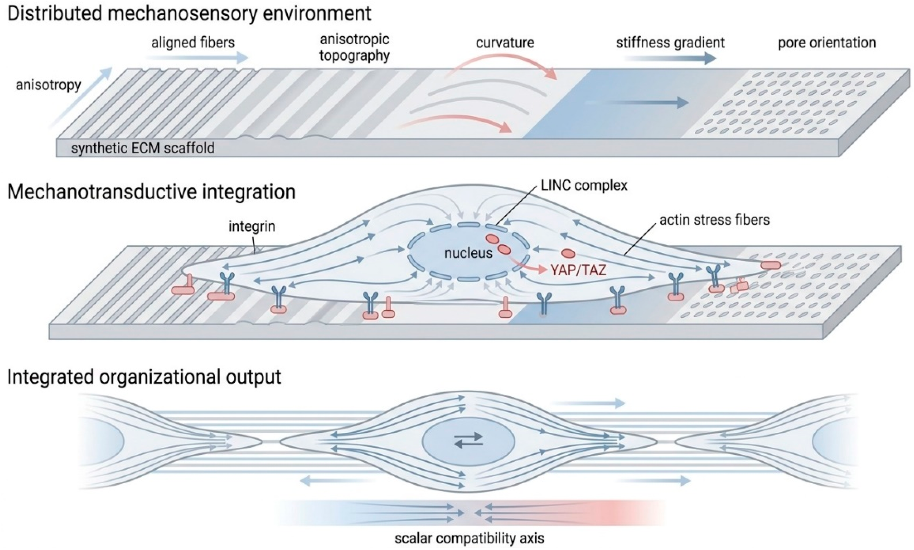
Figure 3.
Scaffold-guided stabilization of tissue-organizational attractors. Cellular organization unfolds within a dynamic organizational landscape shaped by mechanotransduction, extracellular architecture, and intracellular feedback. Weakly structured environments generate shallow and poorly defined attractor basins, producing heterogeneous or unstable organizational outcomes. Scaffold architectures introducing coherent mechanosensory constraints deepen specific attractor basins and suppress competing trajectories. Robust tissue organization therefore emerges through the stabilization of dynamically favored organizational states rather than through deterministic programming. Deep attractor basins confer resistance to molecular noise, adhesion variability, and mechanical perturbation, thereby increasing organizational robustness.
Figure 3.
Scaffold-guided stabilization of tissue-organizational attractors. Cellular organization unfolds within a dynamic organizational landscape shaped by mechanotransduction, extracellular architecture, and intracellular feedback. Weakly structured environments generate shallow and poorly defined attractor basins, producing heterogeneous or unstable organizational outcomes. Scaffold architectures introducing coherent mechanosensory constraints deepen specific attractor basins and suppress competing trajectories. Robust tissue organization therefore emerges through the stabilization of dynamically favored organizational states rather than through deterministic programming. Deep attractor basins confer resistance to molecular noise, adhesion variability, and mechanical perturbation, thereby increasing organizational robustness.
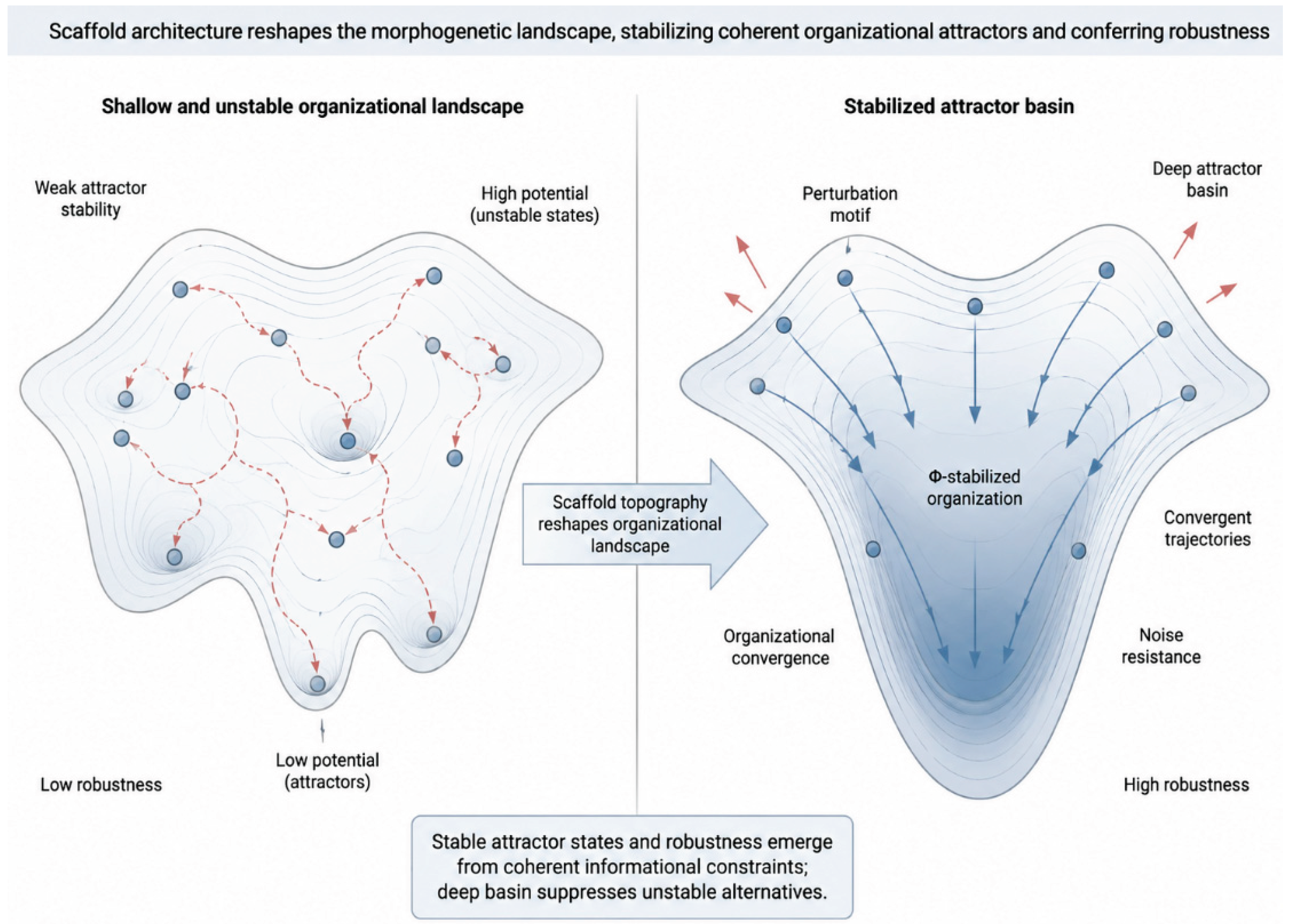
Disclaimer/Publisher’s Note: The statements, opinions and data contained in all publications are solely those of the individual author(s) and contributor(s) and not of MDPI and/or the editor(s). MDPI and/or the editor(s) disclaim responsibility for any injury to people or property resulting from any ideas, methods, instructions or products referred to in the content. |
© 2026 by the authors. Licensee MDPI, Basel, Switzerland. This article is an open access article distributed under the terms and conditions of the Creative Commons Attribution (CC BY) license (http://creativecommons.org/licenses/by/4.0/).
Copyright: This open access article is published under a Creative Commons CC BY 4.0 license, which permit the free download, distribution, and reuse, provided that the author and preprint are cited in any reuse.


