

Submitted:
10 June 2026
Posted:
11 June 2026
You are already at the latest version
Abstract
Cadmium (Cd) is a widespread environmental pollutant that threatens pollinators. This study examined the effects of Cd on survival, tissue accumulation, gut structure, microbiota, and gene expression in larval and adult worker honey bees (Apis mellifera). Larvae received diets with 0–1.25 mg/kg Cd, and adults were fed 0–4 mg/kg Cd in sucrose solution. Survival, Cd accumulation, gut histology, microbiota composition, and transcriptomic responses were assessed. Cd exposure reduced larval survival, pupation, and eclosion in a dose-dependent manner and decreased adult survival at the highest concentration. Cd accumulated in both stages, including brain tissue, and caused gut tissue damage. Adult gut microbiota showed increased Gilliamella abundance at high Cd levels. Transcriptomic profiling revealed upregulation of detoxification and immune genes in larvae and stress-response genes in adults. Larvae were more sensitive than adults, and Cd exposure impaired survival, gut integrity, microbiota balance, and gene expression, offering insights for ecological risk assessment of heavy metal pollution in pollinators.
Keywords:
Apis mellifera
; cadmium
; sublethal dose
; gut histology
; gut microbiota
; 16S rRNA sequencing
; transcriptome sequencing
1. Introduction
Honey bees (Apis mellifera) are essential pollinators in natural and agricultural ecosystems, contributing to biodiversity, crop productivity, and global food security[1]. Accelerating industrialization and intensive agriculture have increased environmental contamination by heavy metals, which poses risks to pollinator health. Among these pollutants, cadmium (Cd) is particularly concerning due to its high toxicity, persistence, and bioaccumulative properties. Cd enters agroecosystems through mining, industrial emissions, phosphate fertilizers, and wastewater irrigation, subsequently transferring to honey bee colonies via nectar and pollen. Chronic exposure can impair individual fitness, colony development, and pollination services[2].
Previous studies have documented adverse effects of Cd at multiple biological levels. Chronic exposure to CdO nanoparticles reduces survival and disrupts physiological functions[3]. Cd and other metals, such as copper and lead, suppress larval development and decrease adult survival[4]. Prolonged exposure also impairs olfactory learning and memory, likely through oxidative stress and dysregulation of brain odorant perception genes[5]. Histopathological analyses reveal chromatin condensation, mitochondrial swelling, and endoplasmic reticulum disruption in midgut epithelial cells[6]. Cd alters gut microbiota composition, increasing opportunistic bacteria such as Serratia and reducing host adaptability[7]. Colony-level studies show Cd accumulation in hive matrices, negatively affecting brood production and overall colony health[8].
Despite these findings, key gaps remain. Tissue-specific Cd accumulation across developmental stages is poorly characterized. The relationship between Cd-induced gut histopathology and microbial community changes in adults is unclear. Additionally, stage-specific intestinal transcriptomic responses to Cd are largely unknown. To address these gaps, we exposed larval and adult worker bees to sublethal Cd concentrations, integrating survival analysis, Cd bioaccumulation measurement, gut histology, 16S rRNA-based microbiota profiling, and transcriptomic sequencing. Emphasis was placed on comparing larval and adult responses to reveal stage-specific susceptibility and underlying mechanisms. These results provide mechanistic insights into Cd toxicity in honey bees and inform strategies for pollinator protection and sustainable agricultural management in contaminated environments.
2. Materials and Methods
2.1. Test Materials
2.1.1. Western Honey Bee Workers
Experimental western honey bees (Apis mellifera) were obtained from an apiary confirmed to be free of heavy metal contamination. Colonies were selected based on synchronized oviposition by the queen and a Varroa destructor infestation rate of ≤ 1%. Only healthy colonies without visible signs of disease were used.
For larval experiments, a queen excluder was used to confine the queen to an empty comb for 12 hours to obtain age-synchronized eggs. After 72 hours, the eggs hatched into 1-day-old larvae. Larvae were carefully examined to ensure they were free of disease and parasitic infection before being transferred for in vitro rearing.
For adult experiments, sealed brood combs containing mature pupae were collected from the same colony and incubated at 34.5 °C and 70% relative humidity. Newly emerged worker bees (≤ 24-hour old) were marked on the thorax and returned to their original colony for 7 days to allow gut microbiota stabilization. After this acclimation period, bees were collected and used for subsequent cage exposure experiments.
2.1.2. Reagents and Instruments
Cadmium chloride (CdCl₂, purity > 99%) was purchased from Xilong Scientific (Guangzhou, China). The RNAprep Pure Animal Total RNA Extraction Kit was purchased from Tiangen Biotech (Beijing, China), the PrimeScript RT Reagent Kit (with gDNA Eraser) from Takara Bio (Shiga, Japan), and the VAHTS 16S Amplicon PCR Master Mix for Illumina together with the VAHTS Universal V5 RNA-seq Library Preparation Kit from Vazyme Biotech (Nanjing, China). All molecular procedures were performed according to the manufacturers’ instructions. Cd concentrations were determined via inductively coupled plasma mass spectrometry (ICP-MS; Thermo Fisher Scientific, Waltham, MA, USA). For histological analysis, sections were prepared using a Leica RM2235 rotary microtome (Leica Microsystems, Wetzlar, Germany) and digitized with a Pannoramic Midi II slide scanner (3DHISTECH, Budapest, Hungary). High-throughput sequencing was executed on the Illumina NovaSeq 6000 platform (Illumina, San Diego, CA, USA). Supplemental instrumentation included a microplate reader (Bio-Rad Laboratories, Hercules, CA, USA) and an ultrapure water system (MilliporeSigma, Burlington, MA, USA).
2.2. Cadmium Treatment Protocol
2.2.1. Preparation of Cadmium Stock Solution
CdCl₂ was accurately weighed and dissolved in sterile ultrapure water to prepare a 1 g/L stock solution. The stock solution was stored at 4°C in the dark until use. For experimental treatments, the stock solution was diluted with artificial diet or 50% (w/v) sucrose solution to obtain the desired final concentrations.
2.2.2. Larval Exposure Treatment
In vitro larval rearing was conducted in accordance with the OECD (2016) Test No.239: Guidelines for Guidance Document on Honey Bee Larval Toxicity Test following Repeated Exposure[9]. One-day-old larvae were grafted into plastic queen cell cups placed in 48-well culture plates, with one larva per well, and maintained in an incubator at 34.5 °C and 95% relative humidity.
Larvae were fed according to a standardized diet schedule. On day 1, each larva received 20 μL of diet A (50% royal jelly, 6% glucose, 6% fructose, 1% yeast extract, and 37% sterile water). On day 3, 20 μL of diet B (50% royal jelly, 7.5% glucose, 7.5% fructose, 1.5% yeast extract, and 33.5% sterile water) was provided. On days 4-6, larvae were fed 30, 40, and 50 μL, respectively, of diet C (50% royal jelly, 9% glucose, 9% fructose, 2% yeast extract, and 30% sterile water). On day 7, larvae were transferred to 24-well plates lined with sterile lens paper for pupation. On day 15, pupae were placed in emergence cages supplied with pollen and 50% (w/v) sucrose solution. During the pupal stage, temperature and relative humidity were maintained at 35 °C and 75%, respectively, until adult emergence on day 21.
For Cd exposure, the Cd stock solution was added to diets B and C in place of sterile water to achieve final concentrations of 0, 0.25, and 1.25 mg/kg. The treatment concentrations of Cd were determined with reference to previous studies[4] and slightly modified for the present experimental system.Cd exposure levels were selected as 20% of the reported 48-hour LC₅₀ and a five-fold increased concentration. Each treatment consisted of five independent replicates, with 48 larvae per replicate.
2.2.3. Adult Exposure Treatment
After a 7-day gut microbiota stabilization period in their original colonies, marked worker bees were randomly assigned to experimental cages (50 bees per cage). Cages were maintained in a controlled-environment chamber at 30 °C and 70% relative humidity. Bees were provided ad libitum access to 50% (w/v) sucrose solution containing Cd at final concentrations of 0, 0.4, or 4 mg/kg. These exposure concentrations were selected based on the 48-hour LC₅₀ value reported by Di et al.[4], corresponding to approximately 5% and one-tenth of the LC₅₀. The sucrose solutions were freshly prepared and replaced daily throughout the 21-day exposure period. Each treatment group consisted of five independent replicates, with 50 worker bees per replicate.
2.3. Measurement Indicators and Methods
2.3.1. Assessment of Survival and Development
For larvae, survival and developmental parameters were recorded at key stages. The larval survival rate was determined on day 7 and calculated as the number of live larvae on day 7 divided by the number of live larvae on day 3, expressed as a percentage. Pupation rate was assessed on day 15 and calculated as the number of pupae on day 15 divided by the number of live larvae on day 8, expressed as a percentage. Eclosion rate was calculated on day 21 as the number of emerged adults divided by the number of pupae on day 15, also expressed as a percentage.
For adult workers, daily mortality was recorded throughout the exposure period. Survival curves were generated using the Kaplan-Meier method, and differences between treatment groups were evaluated using the log-rank test, following the approach described by Tackenberg et al.[10]
2.3.2. Cadmium Accumulation Analysis
Samples were collected on day 7 of larval development and day 21 of adult exposure. Whole larvae were used for analysis, whereas adult bees were dissected to remove legs, wings, and gut prior to separating the head, thorax, and abdomen. Cd concentrations in all samples were determined using ICP-MS, following the protocol described by Voica et al.[11] Each treatment included five biological replicates.
2.3.3. Histopathological Analysis of Intestinal Tissues
Intestinal tissue samples were collected on day 7 for larvae and day 21 for adult workers. For larvae, the epidermis and fat body were carefully removed to isolate the gut, whereas for adults, the midgut and hindgut were excised. Tissues were fixed in 4% paraformaldehyde for 12 hours. Following a slightly modified version of Yi Yao’s paraffin sectioning protocol[12], samples were dehydrated through a graded ethanol series, cleared with xylene, and embedded in paraffin. Serial sections of 6 μm thickness were prepared, stained with hematoxylin and eosin, and mounted for microscopic examination. Intestinal morphology, including wall architecture, cellular organization, and microvilli structure, was assessed using a Pannoramic MIDI II digital slide scanner. Five biological replicates were analyzed for each Cd concentration.
2.3.4. Adult Intestinal Microbiota Analysis
Intestinal samples were collected from adult worker bees in the control and 4 mg/kg cadmium treatment groups. For each biological replicate, six intestines were pooled, with three replicates per group. Total DNA was extracted using the MagPure Soil DNA LQ Kit according to the manufacturer’s instructions. The V3-V4 hypervariable regions of the 16S rRNA gene were amplified using universal primers 343F and 798R, as described by Nossa et al.[13], and sequencing was performed on the Illumina NovaSeq 6000 platform. Raw sequences were processed for quality control and chimera removal using QIIME2[14], and taxonomy was assigned against the Silva 138 database. Genus-level microbial composition was analyzed, and α-diversity indices (Shannon, Simpson, Chao1, and ACE) were calculated. β-Diversity was evaluated using principal coordinates analysis (PCoA) based on Bray-Curtis distances.
2.3.5. Intestinal Transcriptome Sequencing and Analysis
Intestines were collected from larvae (day 7; control and 0.25 mg/kg Cd groups) and adults (day 21; control and 4 mg/kg Cd groups). Six intestines were pooled to form one biological sample, with three replicates per group. Total RNA was extracted using the TRIzol method and assessed for quality with a NanoDrop 2000 spectrophotometer and Agilent 2100 Bioanalyzer. RNA-seq libraries were prepared using the VAHTS Universal V5 RNA-seq Library Prep Kit and sequenced on an Illumina NovaSeq 6000 platform (150 bp paired-end reads). Low-quality reads were filtered with fastp[15], and clean reads were aligned to the honeybee reference genome (Amel_HAv3.1) using HISAT2[16]. Differentially expressed genes (DEGs) were identified with DESeq2[17], using a significance threshold of P < 0.05 and |log2 FC| > 1. GO and KEGG pathway enrichment analyses were subsequently performed to explore functional implications.
2.3.6. Primer Design and qPCR Validation
Key DEGs from larvae and adults were selected for qPCR validation. Primers were designed based on published references[18,19], with Glyceraldehyde-3-phosphate dehydrogenase (GAPDH) as the reference gene (sequences listed in Table S1). Each 20 μL qPCR reaction contained 10 μL master mix, 0.8 μL of each primer, 2 μL cDNA template, and 7.2 μL nuclease-free water. The thermal cycling program included an initial denaturation at 95 °C for 3 min, followed by 40 cycles of 95 °C for 10 s and annealing at the gene-specific temperature (Table 1) for 30 s, with a final melting curve analysis to confirm primer specificity. Relative gene expression was calculated using the 2⁻ΔΔCt method[20].
2.4. Data Analysis
Data were analyzed using SPSS 26.0. Survival curves were generated using the Kaplan-Meier method and compared with the log-rank test. Quantitative data, including heavy metal concentrations and microbial abundances, are presented as mean ± standard error (Mean ± SE). Intergroup differences were assessed by one-way ANOVA, followed by Fisher’s LSD post hoc test when P < 0.05. Figures were generated using GraphPad Prism 10, and R v3.2.0 was used for hierarchical clustering and enrichment analyses of DEGs in the transcriptome.
3. Results
3.1. Effects of Cadmium on the Survival of Apis mellifera Worker Larvae and Adults
Cd exposure significantly inhibited larval survival and development in a dose-dependent manner(Table S2). Compared with the control group (0 mg/kg), larvae exposed to 0.25 mg/kg Cd showed a 9.36% reduction in survival rate, a 42.43% decrease in pupation rate, and a 40.93% decline in eclosion rate. At 1.25 mg/kg, survival decreased to 78.08%, pupation rate to 40.72%, and eclosion rate to 24.43%, all significantly lower than those of the control group (P < 0.05). Kaplan-Meier survival analysis of adult bees (Figure 1) revealed no significant difference between the 0.4 mg/kg group and the control (P > 0.05), whereas the 4 mg/kg group showed a significantly reduced survival rate (P < 0.05), with mortality increasing markedly from day 11 onward. Overall, larvae exhibited substantially greater sensitivity to Cd than adults.
3.2. Cadmium Accumulation in Bees
Cd concentrations in larvae increased significantly with rising exposure levels (Figure 2A). In the 0.25 mg/kg group, Cd content was 42 times that of the control, while in the 1.25 mg/kg group, it reached 151 times the control level and 2.57 times higher than in the 0.25 mg/kg group. A similar trend was observed in newly emerged adults on day 21, although Cd levels were slightly lower than those in day-7 larvae, without statistical significance. This suggests that bees may partially eliminate Cd through molting or shedding of midgut epithelial cells[21].
Cd accumulation in adult bees exhibited clear tissue-specific patterns (Figure 2B and Figure 2C). In the control group, Cd levels were near the detection limit. In the 0.4 mg/kg group, Cd concentrations in the abdomen were significantly higher than those in the head or thorax, with no significant difference between the latter two. At 4 mg/kg, abdominal Cd accumulation increased further, reaching 5.5 times that of the head and 10.8 times that of the thorax. All three body regions showed significantly higher Cd levels than in the 0.4 mg/kg group. Notably, Cd was also detected in brain tissue, indicating its ability to cross the blood-brain barrier[22].
3.3. Cadmium-Induced Damage to Honeybee Intestinal Tissues
Histological examination of larval midgut sections (Figure 3A - 3C) showed that, in the control group, intestinal epithelial cells were regularly arranged with intact brush borders (bb). In the 0.25 mg/kg group, epithelial cells appeared loosely and irregularly arranged, accompanied by microvillar shedding and epithelial thinning. In the 1.25 mg/kg group, cells were elongated and distorted, with increased vacuolization and evidence of cellular disintegration.
Hindgut tissue sections of adult bees (Figure 3D - 3F) showed that, in the control group, epithelial cells were neatly arranged with intact microvilli. In the 0.4 mg/kg group, partial microvillar detachment was observed, although overall cellular organization remained relatively regular. In the 4 mg/kg group, microvillar loss was more extensive, and intercellular spaces were markedly widened. The severity of intestinal damage increased with Cd concentration. These findings are consistent with Dabour et al.[6], who reported Cd nanoparticle-induced injury in worker bee midgut cells, further indicating that the gut is a primary target organ of Cd toxicity.
3.4. Effects of Cadmium on the Gut Microbiota of Adult Bees
Alpha diversity analysis showed slight increases in the Shannon and Simpson indices and minor decreases in the Chao1 and ACE indices in the Cd-treated groups; however, none of these differences were statistically significant (P > 0.05) (Figure 4A). Beta diversity analysis based on Bray-Curtis distances revealed partial separation between control and Cd-treated groups in the PCoA plots, but the Adonis test yielded P = 0.1, indicating no significant intergroup difference (Figure 4B).
16S rRNA sequencing of adult bee guts following Cd exposure showed that, at the genus level, the dominant bacteria were Commensalibacter, Snodgrassella, Gilliamella, Lactobacillus, Enterobacter, and Frischella (Figure 4C). Compared with the control group, the relative abundance of Gilliamella increased significantly from 6.80% to 22.80% in the 4 mg/kg Cd group (P = 0.02). In contrast, the abundances of Enterobacter and Frischella slightly decreased, although these changes were not statistically significant (P > 0.05) .
3.4. Transcriptomic Responses of Honeybee Gut to Cadmium Exposure
We identified 367 significantly DEGs in larval guts, of which 298 were upregulated and 69 downregulated (Figure 4A). In adult guts, 288 DEGs were detected, including 213 upregulated and 75 downregulated (Figure 4A), indicating distinct molecular responses to Cd exposure between larvae and adults. In larval guts, detoxification-related genes (Cyp4g11, Cyp6as5, LOC413908, GstS4), antimicrobial peptide gene (Def1), and heat shock protein gene (HSP90) were significantly upregulated (2- to 113-fold), whereas the α-glucosidase gene (AGLU2) was downregulated by 9.2-fold (Table 2; Figure 5B).
In adult guts, heat shock protein genes (HSP90, LOC411700) and cytochrome gene (LOC552679) were significantly upregulated (2.2- to 8.7-fold), while the α-glucosidase gene (Hbg2) was downregulated by 2-fold (Table 2; Figure 5C). Heat shock proteins stabilize protein structures and repair damaged proteins, thereby providing stress protection. Their upregulation suggests that adults primarily rely on stress-response mechanisms to counteract Cd toxicity. qPCR validation confirmed that the expression trends of key DEGs aligned with transcriptomic data (Figure 5D), supporting the reliability of the sequencing results.
Upregulated genes in larval guts were significantly enriched in nine pathways, including drug metabolism (cytochrome P450), xenobiotic metabolism (cytochrome P450), and unsaturated fatty acid biosynthesis (Figure 5B). Downregulated genes were enriched in seven pathways, such as the pentose phosphate pathway, other glycan degradation, and lysosome.
In adult guts, upregulated genes were significantly enriched in seven pathways, including actin cytoskeleton regulation, protein processing in the endoplasmic reticulum, and tyrosine metabolism (Figure 5C). Downregulated genes were enriched in four pathways, including lysosome, sphingolipid metabolism, and neuroactive ligand–receptor interaction.
This divergence is consistent with Liu et al.[23], who reported developmental stage-specific gene expression profiles, and Evans et al.[24], who observed metabolic pathway differences between honeybee larvae and adults, reflecting distinct adaptive strategies to Cd toxicity across developmental stages.
4. Discussion
Cd, a pervasive environmental pollutant, can enter honey bee bodies through contaminated nectar and pollen, posing significant threats to individual survival, colony reproduction, and pollination services[25]. The present study demonstrates that Cd exposure suppresses the survival and development of both larval and adult Apis mellifera in a dose-dependent manner, with larvae exhibiting markedly greater sensitivity than adults. These findings are consistent with those of Di et al.[4], who reported that Cd exposure significantly increases mortality in both larvae and adult bees, further supporting the heightened vulnerability of immature individuals to heavy metal stress.
Specifically, the eclosion rate of larvae exposed to 1.25 mg/kg Cd decreased to 34.61% of that in the control group (Table 2). In contrast, adult bees showed significant survival suppression only at the highest exposure level (4 mg/kg) (Figure 1B). This stage-dependent difference in sensitivity is likely associated with developmental and physiological characteristics. Larvae are in a phase of rapid growth and active cell proliferation, yet their detoxification metabolism and immune defense systems remain underdeveloped, rendering them more susceptible to Cd toxicity. By comparison, adult bees possess structurally and functionally mature excretory systems, an intact gut barrier, and more efficient detoxification pathways, which collectively contribute to a greater capacity to mitigate Cd-induced toxic burden[22].
The tissue-specific accumulation of Cd in bees provides critical insights into its toxicological targets and modes of action. In larvae, Cd levels increased exponentially with rising exposure concentrations, indicating a strong capacity for metal accumulation during early developmental stages. In contrast, in adult bees, Cd predominantly accumulated in the abdomen, corresponding to the primary location of the digestive tract, while markedly lower levels were detected in the head and thorax. This distribution pattern is highly consistent with those reported in other insects, such as the Chinese rice grasshopper[23] and the yellow poplar borer[26], further supporting the gut as the principal site for Cd absorption, sequestration, and metabolism.
Notably, significant Cd deposition was detected in the brain tissue of adult bees, providing direct evidence that Cd can penetrate the blood-brain barrier and accumulate in neural tissues. This finding offers tissue-level mechanistic support for previous observations by Li et al.[5], who reported that chronic Cd exposure impairs olfactory learning and homing ability in honey bees, and further substantiates the potential of Cd to disrupt neural and behavioral functions.
Furthermore, no significant difference in Cd content was observed between day-7 larvae and newly emerged day-21 adults, although Cd levels were slightly higher in larvae. This pattern suggests that bees may partially eliminate accumulated Cd during metamorphosis, potentially through processes such as molting during the pupal stage and the shedding and renewal of midgut epithelial cells, thereby reducing the internal burden of heavy metal toxicity[27].
As a central organ for nutrient digestion, absorption, innate immune defense, and xenobiotic metabolism, the structural integrity and barrier stability of the bee gut are fundamental to maintaining individual health. In this study, H&E staining revealed that Cd exposure induced pronounced, dose-dependent pathological damage in the larval midgut and adult hindgut. These alterations were characterized by thinning of the epithelial mucosa, disorganized and loosely arranged cells, extensive loss of microvilli, widened intercellular spaces, and, in severe cases, cellular vacuolation and disintegration. These findings are consistent with those of Dabour et al.[6], who reported ultrastructural damage in the midgut epithelial cells of worker bees following Cd nanoparticle exposure.
Such pathological changes are likely to directly compromise the mechanical barrier function of the gut. On one hand, damage to the epithelium and microvilli can markedly impair digestive and absorptive efficiency, leading to metabolic disruption and insufficient energy supply for larval development and adult physiological activities. On the other hand, disruption of gut barrier integrity may facilitate the translocation of commensal microbiota and increase susceptibility to invasion by exogenous pathogens. Collectively, these effects provide a critical pathological basis for the downstream toxic consequences of Cd exposure.
The bee gut microbiota forms a highly host-specific core community that plays essential roles in nutrient metabolism, immune regulation, and detoxification of xenobiotics. In the present study, exposure to 4 mg/kg Cd significantly increased the relative abundance of the genus Gilliamella in adult guts, while overall α- and β-diversity of the gut microbiota remained unchanged. Gilliamella, a core gut symbiont, normally contributes to the degradation of complex polysaccharides, short-chain fatty acid production, and maintenance of the gut anaerobic microenvironment.
The observed increase in Gilliamella abundance is not a random fluctuation but rather a specific compensatory adjustment of the gut microbiota in response to Cd stress, which may operate via two mechanisms. First, genomic analyses have shown that Gilliamella strains commonly harbor genes conferring heavy metal resistance, including those encoding cation efflux pumps (czcA/czcD families), metallothioneins, and metal-chelating proteins[28]. These genes can actively extrude or chelate Cd ions, thereby reducing free Cd concentrations in the gut lumen and mitigating toxicity to epithelial cells, providing Gilliamella with a survival advantage under Cd exposure. Second, Cd-induced epithelial damage alters the nutritional composition and oxygen levels in the gut microenvironment. Gilliamella exhibits greater tolerance to such environmental stress than other core symbionts, such as Snodgrassella and Lactobacillus, enabling it to dominate microbial competition and become a prevalent genus under Cd stress.
However, this compensatory increase is not entirely beneficial. The spatial interactions between Gilliamella and Snodgrassella are critical for maintaining the anaerobic gut environment and barrier function. Overproliferation of Gilliamella may disrupt the functional balance of the core microbiota, further exacerbating gut dysbiosis and barrier damage.
Transcriptomic analysis further revealed the molecular mechanisms underlying developmental stage-specific responses of bees to Cd stress. In larval guts, 367 significantly differentially expressed genes (DEGs) were identified, while 288 DEGs were detected in adult guts, with clear differences in expression profiles and enriched pathways between the two stages, reflecting distinct adaptive strategies to Cd exposure.
In larvae, genes of the cytochrome P450 family, Cyp4g11 and Cyp6as5, were significantly upregulated by 4.72- and 5.13-fold, respectively, while the glutathione S-transferase gene GstS4 increased 4.26-fold. The Cyp4 and Cyp6 families are core components of xenobiotic detoxification in bees. Cyp6as5 has been reported to metabolize neonicotinoids and pyrethroids via hydroxylation, reducing their bioavailability and toxicity, whereas Cyp4g11 also participates in hydrocarbon synthesis and mitigation of oxidative damage. The coordinated upregulation of these genes indicates that Cd, as a non-biological stressor, activates a broad-spectrum chemical defense system in larvae, originally evolved to handle plant secondary metabolites and agricultural pesticides, thereby mitigating Cd toxicity. Notably, sustained P450 activity consumes considerable energy and NADPH, which may divert resources from larval growth and development, providing a molecular explanation for the observed reduction in pupation and eclosion rates under Cd exposure.
Additionally, the antimicrobial peptide gene Def1 was upregulated 3.57-fold in larvae. Def1 is a key effector of humoral innate immunity, exerting broad-spectrum antibacterial activity by disrupting bacterial cell membranes[29,30]. Given the observed structural damage to the larval gut epithelium, including disorganized cell arrangement, microvilli loss, and widened intercellular spaces, we hypothesize that Cd-induced gut barrier disruption triggers a “leaky gut” effect. This allows commensal bacterial components (e.g., peptidoglycan, lipopolysaccharides) or viable bacteria to translocate into the hemolymph, activating systemic innate immune responses and inducing strong upregulation of antimicrobial peptides such as Def1. While this immune activation helps limit secondary bacterial invasion, persistent immune stimulation can cause chronic gut inflammation and consume substantial host energy, further impairing larval growth and development, representing a key immune-mediated mechanism of Cd toxicity.
In adult guts, heat shock protein genes HSP90 and LOC411700 were upregulated more than twofold, indicating that adults primarily rely on cellular stress-response pathways to cope with Cd. As molecular chaperones, heat shock proteins stabilize protein conformations, repair denatured proteins, and inhibit apoptosis, mitigating oxidative and structural damage induced by Cd, thereby preserving gut cellular homeostasis.
Finally, α-glucosidase genes were significantly downregulated in both larvae and adults, suggesting that Cd exposure impairs carbohydrate metabolism and energy supply, further contributing to reduced survival. These findings are consistent with previous studies on heavy metal toxicity in insects[31].
This study has several limitations. First, it focused primarily on the acute effects of sublethal Cd exposure, whereas in natural environments, bees are typically exposed to low-level chronic Cd, often in combination with other heavy metals or pesticides. Future studies should design chronic exposure experiments that more closely reflect field conditions to assess long-term accumulation and the combined toxic effects of multiple contaminants. Second, the mechanistic analyses in this study were largely based on multi-omics correlations and lack direct functional validation. Subsequent research could employ RNAi-mediated gene knockdown, in vitro bacterial culture, and metagenomic sequencing to verify the detoxification role of Cyp family genes, the heavy metal resistance functions of Gilliamella, and the causal relationships between Cd-induced gut barrier disruption and immune activation. Third, this study only investigated the toxicity of Cd at the individual worker bee level; future work should expand to the colony level to examine how Cd contamination affects colony reproduction, division of labor, and pollination performance, thereby refining ecological risk assessments of environmental Cd pollution.
5. Conclusions
This study systematically elucidated the multidimensional toxic effects of sublethal Cd exposure on both larval and adult worker bees. Cd suppressed larval survival, pupation, and eclosion, as well as adult survival, in a dose-dependent manner, with larvae exhibiting markedly higher sensitivity than adults. Cd also accumulated significantly within bee tissues, displaying tissue-specific distribution and the ability to cross the blood-brain barrier. Exposure induced pronounced pathological damage in the gut, compromised intestinal barrier integrity, and triggered significant enrichment of the core gut symbiont Gilliamella in adults, resulting in gut microbiota dysbiosis. At the molecular level, Cd markedly activated detoxification and immune-related genes in larvae and stress-response genes in adults, highlighting distinct strategies employed by different developmental stages to cope with Cd stress. Overall, this study clarifies the toxic effects and underlying mechanisms of Cd across bee developmental stages, providing multidimensional scientific evidence for ecological risk assessment of environmental Cd pollution, bee resource conservation, and healthy apiculture management.
Supplementary Materials
The following supporting information can be downloaded at the website of this paper posted on Preprints.org. Table S1: Primer sequences for qPCR validarion; Table S2: Effects of dietary cadmium supplemental level on survival of honeybee larvae.
Author Contributions
Conceptualization, Z.Z. and L.Z.; methodology, L.Z.; software, D.Z.; validation, D.Z.; formal analysis, D.Z.; investigation, D.Z.; resources, Z.Z.; data curation, D.Z.; writing—original draft preparation, D.Z.; writing—review and editing, D.Z.; visualization, D.Z.; supervision, L.Z.; project administration, Z.Z.; funding acquisition, Z.Z. All authors have read and agreed to the published version of the manuscript.
Funding
This work was supported by the Earmarked Fund for China Agriculture Research System (CARS-44-KXJ15).
Data Availability Statement
The data presented in this study are available in this article.
Acknowledgments
The authors are thankful to the Earmarked Fund for China Agriculture Research System (CARS-44-KXJ15). All authors have read and agreed to the published version of the manuscript.
Conflicts of Interest
The authors declare no conflicts of interest.
Abbreviations
The following abbreviations are used in this manuscript:
| Cd | Cadmium |
| ICP-MS | Inductively Coupled Plasma Mass Spectrometry |
| H.E. | Hematoxylin-Eosin Staining |
| OECD | Organisation for Economic Co-operation and Development |
References
- Papa, G.; Maier, R.; Durazzo, A.; Lucarini, M.; Karabagias, I.K.; Plutino, M.; Bianchetto, E.; Aromolo, R.; Pignatti, G.; Ambrogio, A.; et al. The Honey Bee Apis mellifera: An Insect at the Interface between Human and Ecosystem Health. Biology 2022, 11, 233. [Google Scholar] [CrossRef]
- Knoll, S.; Cappai, M.G. Foraging activity of honey bees (Apis mellifera L., 1758) and exposure to cadmium: A review. Biol. Trace Elem. Res. 2024, 202, 5733–5742. [Google Scholar] [CrossRef] [PubMed]
- Al Naggar, Y.; Dabour, K.; Masry, S.; Sadek, A.; Naiem, E.; Giesy, J.P. Sublethal effects of chronic exposure to CdO or PbO nanoparticles or their binary mixture on the honey bee (Apis millefera L.). Env. Sci. Pollut. Res. Int. 2020, 27, 19004–19015. [Google Scholar] [CrossRef] [PubMed]
- Di, N.; Hladun, K.R.; Zhang, K.; Liu, T.X.; Trumble, J.T. Laboratory bioassays on the impact of cadmium, copper and lead on the development and survival of honeybee (Apis mellifera L.) larvae and foragers. Chemosphere 2016, 152, 530–538. [Google Scholar] [CrossRef]
- Li, Z.; Qiu, Y.; Li, J.; Wan, K.; Nie, H.; Su, S. Chronic Cadmium Exposure Induces Impaired Olfactory Learning and Altered Brain Gene Expression in Honey Bees (Apis mellifera). Insects 2022, 13. [Google Scholar] [CrossRef]
- Dabour, K.; Al Naggar, Y.; Masry, S.; Naiem, E.; Giesy, J.P. Cellular alterations in midgut cells of honey bee workers (Apis millefera L.) exposed to sublethal concentrations of CdO or PbO nanoparticles or their binary mixture. Sci. Total Env. 2019, 651, 1356–1367. [Google Scholar] [CrossRef]
- Rothman, J.A.; Leger, L.; Kirkwood, J.S.; McFrederick, Q.S. Cadmium and Selenate Exposure Affects the Honey Bee Microbiome and Metabolome, and Bee-Associated Bacteria Show Potential for Bioaccumulation. Appl. Env. Microbiol. 2019, 85. [Google Scholar] [CrossRef]
- Hladun, K.R.; Di, N.; Liu, T.X.; Trumble, J.T. Metal contaminant accumulation in the hive: Consequences for whole-colony health and brood production in the honey bee (Apis mellifera L.). Env. Toxicol. Chem. 2016, 35, 322–329. [Google Scholar] [CrossRef]
- OECD. Guidance Document on Honey Bee Larval Toxicity Test following Repeated Exposure; OECD Publishing: Paris, 2016; Volume No.239, pp. 6–7. [Google Scholar]
- Tackenberg, M.C.; Giannoni-Guzmán, M.A.; Sanchez-Perez, E.; Doll, C.A.; Agosto-Rivera, J.L.; Broadie, K.; Moore, D.; McMahon, D.G. Neonicotinoids disrupt circadian rhythms and sleep in honey bees. Sci. Rep. 2020, 10, 17929. [Google Scholar] [CrossRef]
- Voica, C.; Iordache, A.M.; Ionete, R.E. Multielemental characterization of honey using inductively coupled plasma mass spectrometry fused with chemometrics. J. Mass Spectrom. 2020, 55, e4512. [Google Scholar] [CrossRef] [PubMed]
- Yi, Y.; Liu, Y.-B.; Barron, A.B.; Zeng, Z.-J. Effects of commercial queen rearing methods on queen fecundity and genome methylation. Apidologie 2020, 52, 282–291. [Google Scholar] [CrossRef]
- Nossa, C.W.; Oberdorf, W.E.; Yang, L.; Aas, J.A.; Paster, B.J.; DeSantis, T.Z.; Brodie, E.L.; Malamud, D.; Poles, M.A.; Pei, Z. Design of 16S rRNA gene primers for 454 pyrosequencing of the human foregut microbiome. World J. Gastroenterol. WJG 2010, 16, 4135. [Google Scholar] [CrossRef]
- Bolyen, E.; Rideout, J.R.; Dillon, M.R.; Bokulich, N.A.; Abnet, C.C.; Al-Ghalith, G.A.; Alexander, H.; Alm, E.J.; Arumugam, M.; Asnicar, F. Reproducible, interactive, scalable and extensible microbiome data science using QIIME 2. Nat. Biotechnol. 2019, 37, 852–857. [Google Scholar] [CrossRef]
- Chen, S.; Zhou, Y.; Chen, Y.; Gu, J. fastp: an ultra-fast all-in-one FASTQ preprocessor. Bioinformatics 2018, 34, i884–i890. [Google Scholar] [CrossRef]
- Kim, D.; Langmead, B.; Salzberg, S.L. HISAT: a fast spliced aligner with low memory requirements. Nat. Methods 2015, 12, 357–360. [Google Scholar] [CrossRef]
- Love, M.I.; Huber, W.; Anders, S. Moderated estimation of fold change and dispersion for RNA-seq data with DESeq2. Genome Biol. 2014, 15, 550. [Google Scholar] [CrossRef]
- Ratnieks, F.L.; Carreck, N.L. Clarity on honey bee collapse? Science 2010, 327, 152–153. [Google Scholar] [CrossRef]
- Boncristiani, H.; Underwood, R.; Schwarz, R.; Evans, J.D.; Pettis, J. Direct effect of acaricides on pathogen loads and gene expression levels in honey bees Apis mellifera. J. Insect Physiol. 2012, 58, 613–620. [Google Scholar] [CrossRef] [PubMed]
- Schmittgen, T.D.; Livak, K.J. Analyzing real-time PCR data by the comparative CT method. Nat. Protoc. 2008, 3, 1101–1108. [Google Scholar] [CrossRef] [PubMed]
- Polykretis, P.; Delfino, G.; Petrocelli, I.; Cervo, R.; Tanteri, G.; Montori, G.; Perito, B.; Branca, J.J.V.; Morucci, G.; Gulisano, M. Evidence of immunocompetence reduction induced by cadmium exposure in honey bees (Apis mellifera). Environ. Pollut. 2016, 218, 826–834. [Google Scholar] [CrossRef] [PubMed]
- Quigley, T.; Amdam, G. Ultrastructural Organization of the Honeybee Blood-Brain Barrier and Comparison with Age. bioRxiv 2024, 2024, 2009, 2027.615080. [Google Scholar]
- Liu, X.; Li, L.; Guo, Y.; Xi, Y.; Ma, E. Cadmium accumulation in Oxya Chinensis in the ecosystem of soil-plant-insect. J. Agro-Environ. Sci. 2006, 25, 301–304. [Google Scholar]
- Evans, J.D.; Wheeler, D.E. Differential gene expression between developing queens and workers in the honey bee, Apis mellifera. Proc. Natl. Acad. Sci. 1999, 96, 5575–5580. [Google Scholar] [CrossRef]
- Hladun, K.R.; Di, N.; Liu, T.X.; Trumble, J.T. Metal contaminant accumulation in the hive: Consequences for whole-colony health and brood production in the honey bee (Apis mellifera L.). Environ. Toxicol. Chem. 2016, 35, 322–329. [Google Scholar] [CrossRef]
- Esenin, A.; Ma, W. Heavy metals (Cd, Cu, Zn) in wood and wood-feeding insects and other invertebrates associated with decaying pine trees. Bull. Environ. Contam. Toxicol. 2000, 64. [Google Scholar] [CrossRef]
- Martins, G.F.; Neves, C.A.; Campos, L.A.O.; Serrão, J.E. The regenerative cells during the metamorphosis in the midgut of bees. Micron 2006, 37, 161–168. [Google Scholar] [CrossRef] [PubMed]
- Ellegaard, K.M.; Engel, P. New reference genome sequences for 17 bacterial strains of the honey bee gut microbiota. Microbiol. Resour. Announc. 2018, 7. [Google Scholar] [CrossRef]
- Danihlík, J.; Aronstein, K.; Petřivalský, M. Antimicrobial peptides: A key component of honey bee innate immunity: Physiology, biochemistry, and chemical ecology. J. Apic. Res. 2015, 54, 123–136. [Google Scholar] [CrossRef]
- Gilbert, M.; Wilkinson, C. Microsomal oxidases in the honey bee, Apis mellifera (L.). Pestic. Biochem. Physiol. 1974, 4, 56–66. [Google Scholar] [CrossRef]
- Wang, S.; Wan, S.; Shen, Q.; Yue, L.; Wang, J.; Zhou, M.; Li, Y.; Tang, B. Cadmium disrupts energy metabolism in the Harmonia axyridis via mediating trehalose metabolism pathway: A multi-omics analysis. J. Hazard. Mater. 2026, 505, 141412. [Google Scholar] [CrossRef]
Figure 1.
Kaplan-Meier survival curves of adult worker bees exposed to different concentrations of cadmium. An asterisk (*) indicates a significant difference compared with the control group (P < 0.05).
Figure 1.
Kaplan-Meier survival curves of adult worker bees exposed to different concentrations of cadmium. An asterisk (*) indicates a significant difference compared with the control group (P < 0.05).
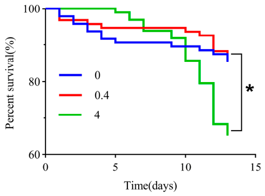
Figure 2.
Cadmium accumulation in honey bee larvae and adults.(A) Cadmium levels in 7-day-old larvae and 21-day-old newly emerged adults under different exposure concentrations. (B) Tissue-specific cadmium accumulation in the head, thorax, and abdomen of adult bees at different concentrations. (C) Cadmium concentration in adult bees (excluding the gut) in relation to exposure level. Note: Asterisks (*) indicate statistically significant differences (P < 0.05).
Figure 2.
Cadmium accumulation in honey bee larvae and adults.(A) Cadmium levels in 7-day-old larvae and 21-day-old newly emerged adults under different exposure concentrations. (B) Tissue-specific cadmium accumulation in the head, thorax, and abdomen of adult bees at different concentrations. (C) Cadmium concentration in adult bees (excluding the gut) in relation to exposure level. Note: Asterisks (*) indicate statistically significant differences (P < 0.05).
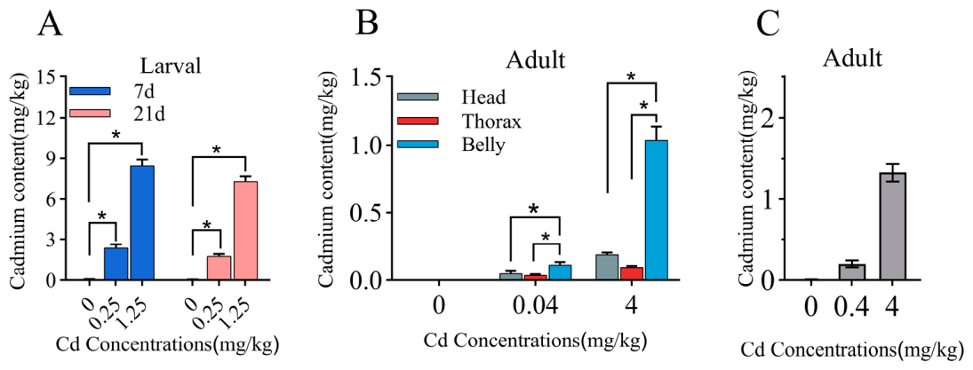
Figure 3.
Gut histological sections of bees under different Cd treatments. (A-C) Larval midgut sections: control, 0.25 mg/kg Cd, and 1.25 mg/kg Cd groups, respectively. (D-F) Adult hindgut sections: control, 0.4 mg/kg Cd, and 4 mg/kg Cd groups, respectively. L, gut lumen; n, nuclei of gut epithelial cells; bb, brush border (microvilli); cm, longitudinal muscle.
Figure 3.
Gut histological sections of bees under different Cd treatments. (A-C) Larval midgut sections: control, 0.25 mg/kg Cd, and 1.25 mg/kg Cd groups, respectively. (D-F) Adult hindgut sections: control, 0.4 mg/kg Cd, and 4 mg/kg Cd groups, respectively. L, gut lumen; n, nuclei of gut epithelial cells; bb, brush border (microvilli); cm, longitudinal muscle.
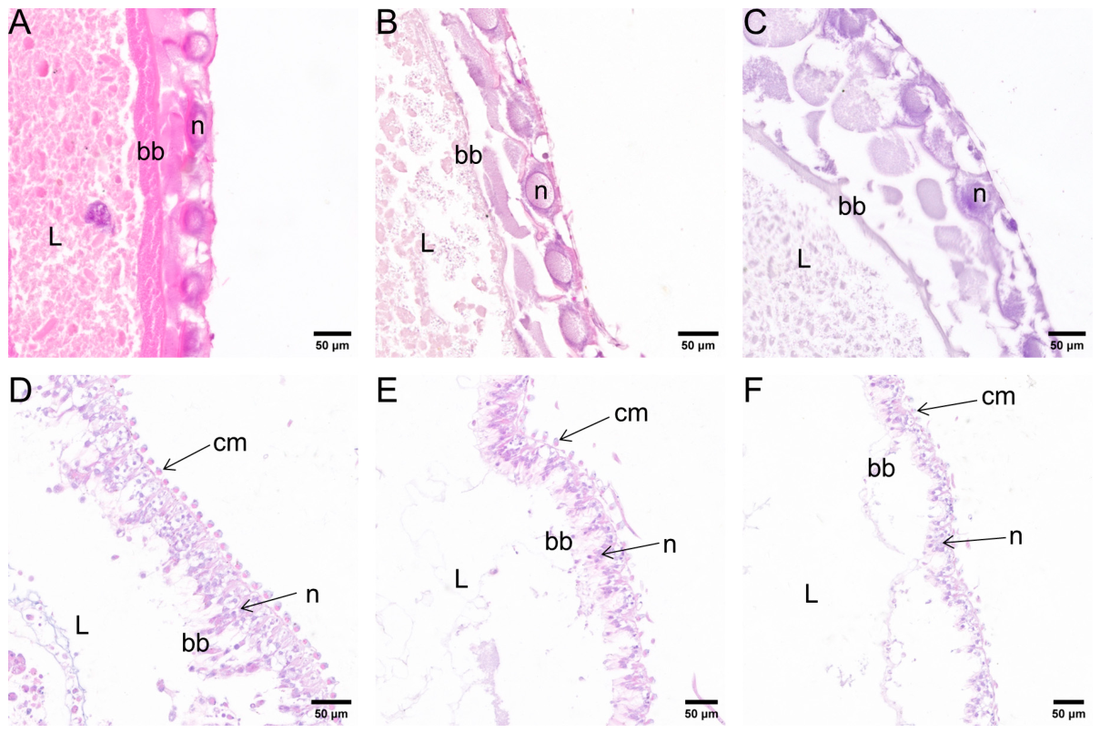
Figure 4.
Changes in adult gut microbiota composition following Cd exposure. (A) Alpha diversity analysis of gut microbiota. (B) Beta diversity analysis of gut microbiota. ns indicates no significant difference, and * indicates a significant difference (P < 0.05). (C) Relative abundance of gut bacteria at the genus level.
Figure 4.
Changes in adult gut microbiota composition following Cd exposure. (A) Alpha diversity analysis of gut microbiota. (B) Beta diversity analysis of gut microbiota. ns indicates no significant difference, and * indicates a significant difference (P < 0.05). (C) Relative abundance of gut bacteria at the genus level.
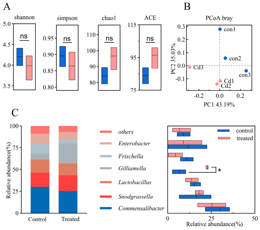
Figure 5.
Transcriptomic analysis of honeybee larval and adult gut tissues following Cd exposure. (A) Number of upregulated and downregulated differentially expressed genes (DEGs). (B) Volcano plot of DEGs in larval gut tissues. (C) Volcano plot of DEGs in adult gut tissues. Note: Upregulated and downregulated genes were identified using a threshold of |log₂ fold change| > 1 and P < 0.05.
Figure 5.
Transcriptomic analysis of honeybee larval and adult gut tissues following Cd exposure. (A) Number of upregulated and downregulated differentially expressed genes (DEGs). (B) Volcano plot of DEGs in larval gut tissues. (C) Volcano plot of DEGs in adult gut tissues. Note: Upregulated and downregulated genes were identified using a threshold of |log₂ fold change| > 1 and P < 0.05.
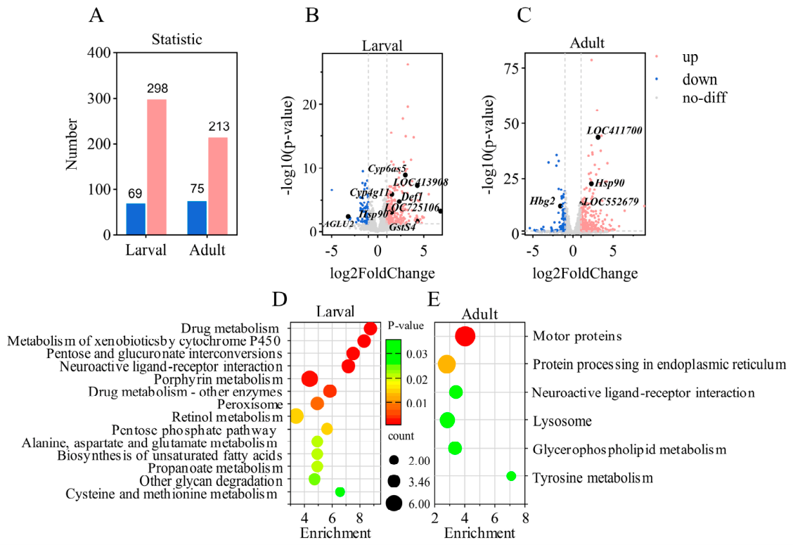

Table 1.
Key Differentially Expressed Genes in Honeybee Gut Following Cadmium Exposure
| Gene Name | Larvae (0.25 mg/kg Cd v.s. Control) | Adults (4 mg/kg Cd v.s. Control) |
| CYP4G11 (cytochrome P450) | 4.72±0.15a | 1.85±0.07b |
| CYP6AS5 (cytochrome P450) | 5.13±0.18a | 1.62±0.06b |
| LOC413908 (cytochrome P450) | 3.89±0.12a | 1.47±0.05b |
| GSTS4 (glutathione S-transferase) | 4.26±0.14a | 1.93±0.08b |
| def1 (defensin-1) | 3.57±0.11a | 1.23±0.04b |
| HSP90 (heat shock protein 90) | 3.12±0.09a | 2.14±0.09a |
| LOC411700 (heat shock protein 80) | - | 2.07±0.08a |
| LOC552679 (cytochrome 4C3) | - | 2.11±0.09a |
| AGLU2 (α-glucosidase) | -3.17±0.10a | - |
| HBG2 (α-glucosidase) | - | -1.02±0.03a |
Experimental data are presented as mean ± SEM. Differences were considered statistically significant at P < 0.05, highly significant at P < 0.01, and not significant at P ≥ 0.05.
Disclaimer/Publisher’s Note: The statements, opinions and data contained in all publications are solely those of the individual author(s) and contributor(s) and not of MDPI and/or the editor(s). MDPI and/or the editor(s) disclaim responsibility for any injury to people or property resulting from any ideas, methods, instructions or products referred to in the content. |
© 2026 by the authors. Licensee MDPI, Basel, Switzerland. This article is an open access article distributed under the terms and conditions of the Creative Commons Attribution (CC BY) license (http://creativecommons.org/licenses/by/4.0/).
Copyright: This open access article is published under a Creative Commons CC BY 4.0 license, which permit the free download, distribution, and reuse, provided that the author and preprint are cited in any reuse.


