

Submitted:
09 June 2026
Posted:
10 June 2026
You are already at the latest version
Abstract
Laser-driven accelerated particle beams have revolutionized the field of cancer treatment by enabling the delivery of extremely high dose-rates of radiation to solid tumors in a matter of femto- to picoseconds. In this study, radiobiological effectiveness of ultrashort pulsed electron beams generated by Advanced Research Electron Accelerator Laboratory (AREAL) accelerator and conventional X-rays were compared on two non-small cell lung cancer (NSCLC) cell lines, A549 (wild-type p53) and H1299 (p53-deficient). NSCLC cells were irradiated at using a AREAL accelerator (a peak dose rate of 1.6 × 10¹⁰ Gy/s, pulse duration of 4.5 × 10⁻¹³ s, repetition rate of 20 Hz) or X-ray unit at an absorbed dose rate of 0.85 Gy/min. Clonogenic survival analysis, γH2AX foci enumeration and genome-wide transcriptome analysis were performed. Clonogenic survival curves showed increased ra-diosensitivity of A549 cells after AREAL exposure compared to X-rays (RBEs=1.2), while H1299 radiosensitivity has not changed. AREAL exposure caused higher linear dose–dependent increase in the number of residual γH2AX foci in both A549 and H1299 cells compared to X-rays. The DNA double-strand breaks in H1299 cells were mainly repaired through homologous recombination following AREAL exposure. However, the gene ex-pression analysis of pathway activation levels revealed a down-regulation of the same pathways in A549 cells. This resulted in activation of Integrin-linked kinase-dependent apoptosis, G2 and proliferation arrest of A549 cells.
Keywords:
laser-driven accelerators
; ionizing radiation
; radiotherapy
; ultrashort pulsed electron beam
; DNA double-strand breaks
; γH2AX
; non-small cell lung cancer
; transcriptomics
1. Introduction
Lung cancer is the leading cause of cancer-related mortality worldwide. Based on the histological subtype about 85% of all lung cancer cases diagnosed as non-small cell lung cancer (NSCLC) [1]. Radiotherapy is a treatment modality for locally advanced unresectable tumors or given concomitantly with chemotherapy. Despite the recent advances in image-guided technologies the efficacy of radiotherapy is still limited by the necessity to take into account the therapeutic window, which is determined as the balance between the dose that causes damage to tumor cells and the tolerance of normal tissues [2].
High-dose rate radiotherapy techniques, including laser-driven particle accelerators have emerged as a promising strategy for cancer treatment. Using ultrashort duration (from femtoseconds to picoseconds) ultra-high instantaneous dose rate (up to tens of GGy/s during a pulse) radiation reduces the normal tissue toxicities commonly associated with conventional radiotherapy, while still maintaining local tumor control [3,4,5]. Based on this premise, the underlying effect relies on the fact that the duration of irradiation by a single ultrashort pulse is significantly shorter than the half-life (t1/2) of free radicals (e.g., hydroxyl radicals with a half-life of approximately 1 ns) responsible for DNA damage in normal tissues [6]. From the other hand, ultra-high dose rate of laser-driven electron beams rely on dose-rate effectiveness factor (DREF), when the high energy depositions to cancer cells occur concomitantly with primary radical reactions, multiple biomolecular damage (membrane and DNA lesions), and repair. Nevertheless, studies into ultrashort pulsed radiotherapy are currently sparse and need further elucidation before implementing in clinics.
The aim of this study was to analyze the impact of ultrashort pulsed electron beams, generated by the state-of-the-art Advanced Research Electron Accelerator Laboratory (AREAL) accelerator, which operates at a frequency of 20 Hz. The primary goal was to conduct a comprehensive investigation and comparison of these beams with conventional X-rays, focusing on their effects on two distinct human NSCLC cell lines: A549 (with wild-type p53) and H1299 (p53-deficient). The investigation explored various effects, such as clonogenic survival, the quantity of residual γH2AX foci, gene expression patterns, and the activation levels of crucial cellular processes like DNA repair, cell cycle arrest, and cell death.
2. Results
2.1. Radiosensitivity of NSCLC Cell Lines After Treatment by Laser-Driven Ultrashort Electron Beams and X-Rays
Clonogenic assay is a “gold standard” for assessing cancer cell radiosensitivity [7]. It is based on the ability of a single cell to undergo “unlimited” division growing into a colony consisting of at least 50 cells. Thus, clonogenic assay is the method of choice to determine cell reproductive death after treatment with ionizing radiation and is widely used to compare biological effectiveness of different types of irradiation. Survival curves were fitted using the following linear–quadratic (LQ) model: SF = exp (−αD − βD2), where SF is the surviving fraction and D is the physical dose. Based on the SF of the clonogenic assay (Figure 1a,b) the relative biological effectiveness (RBE) was calculated. RBE is the ratio of the dose of test beam required to obtain the same level of biological effect compared to the reference radiation, X-ray. The RBE of ultrashort electron beams was calculated based on the SF=0.1. For 10% cell survival of A549 cells irradiated with AREAL was 1.2 times more effective (RBEs= 1.2) compared to X-rays, while there was no difference in RBE between tested and reference radiation (RBEs= 1.0) for H1299 cells. However, H1299 cells showed overall lower radiosensitivity compared to A549 cells after both AREAL and X-rays exposure (Figure 1c,d) (no statistical significant difference was found). While X-ray survival curves show the typical shoulder, cell survival after ultrashort electron beams exploits lower cell survival per dose and fitted with a linear model. For A549 cells, the average values for α and β parameters were respectively 0.31 Gy-1 and 0.03 Gy-2 for X-rays and the α parameter was equal to 0.58 Gy-1 for ultrashort electron beams. The radiosensitivity parameters α and β for X-rays for H1299 were estimated to 0.38 Gy-1 and 0.01 Gy-2 respectively and the α parameter was equal to 0.47 Gy-1 for ultrashort electron beams.
2.2. Dose-Responses for Residual γH2AX Foci
Both cell lines showed a noticeable difference in the dose response curves for residual γH2AX foci 24 hours after irradiation, serving as the second radiobiological endpoint. In line with the findings of the clonogenic assay, the ultrashort beam (AREAL) irradiation exhibited a remarkable and statistically significant dose-response effect, resulting in a higher number of residual γH2AX foci in both A549 (Z=2.4, p=0.016) and H1299 cells (Z=12.2, p<0.0001) compared to X-rays (Figure 2a,b). A dose-dependent relationships are described by a linear equation (y = a + b × x), where the slope coefficient b reflects the increase in the effect per dose unit. Comparing the slope coefficients of dose–response curves obtained under different exposure types quantifies the fold-difference in response per unit dose.
The changes in the residual foci number are described by linear equations: equations (1), (2) for A549 cells after AREAL and X-rays exposure, respectively, and equations (3), (4) for H1299 cells after AREAL and X-rays exposure, respectively.
where “y” is a mean number of foci per cell nucleus and “x” is irradiation dose (Gy).
y = (1.274±0.1434)*x + (4.700±0.6795), R2 = 0.9404
y = (0.8577±0.09675)*x+ (2.796±0.4585), R2 = 0.9402
y = (3.064±0.2236)*x + (6.517±1.060), R2 = 0.9741
y = (1.272±0.06459)*x + (4.981±0.3062), R2 = 0.9873,
It is noteworthy that H1299 cells, despite being less sensitive to radiation overall, displayed nearly double the amount of residual γH2AX foci compared to A549 cells after both AREAL exposure (Z=6.7, p<0.0001) and X-ray exposure (Z=3.5, p=0.0004) (Figure 2c, d). The results unequivocally indicate that NSCLC cells undergo a delayed degradation of DSB repair proteins following exposure to ultrashort beam irradiation (AREAL), in contrast to X-rays. This delayed degradation of DSB repair proteins contributes to the accumulation of residual γH2AX foci, indicating a compromised DNA repair mechanism in response to AREAL irradiation. Our observation further highlights the potential of AREAL as an effective radiotherapy modality for NSCLC treatment, especially in cases where conventional X-ray irradiation may be less effective.
2.3. Differential Gene Expression of NSCLC Cells After Ultrashort Beam (AREAL) and X-Rays Irradiation
To evaluate gene expression changes between two NCSLC cell lines the RNA sequencing was performed 24 hours after irradiation. Both AREAL and X-rays significantly increased differential gene expression (DEG) (log10(control)>1, |log2FC|>1) in both A549 and H1299 cell lines (Table 1). Among them 198 genes were commonly up-regulated and 318 genes were down-regulated in both A549 and H1299 cells 24 hours after AREAL exposure (Figure 3). X-rays irradiation caused up-regulation of only 23 common genes and down-regulation of 73 common genes in two NSCLC cell lines (Figure 3).
Within the obtained gene sets, Gene Ontology (GO)-based functional analysis provides statistically enriched GO terms that show gene relationships according to three ontology categories including biological process, molecular function and cellular component [8]. Significantly enriched GO terms involved in protein hidroxylation, glucose metabolism, protein and histone modification were common for both cell lines after AREAL exposure (Figure 3b), while X-rays irradiation was associated with enrichment of GO terms related to muscle development, neurological development/mechanosensory behaviour, meiosis, and neutrophil activation, reflecting cytoskeletal remodeling, mechanotransduction, DNA repair/recombination, and pro-inflammatory signalling, respectively. (Figure 3c). Ribosomal RNA biosynthesis was mostly down-regulated for both cell lines after AREAL exposure (Figure 3d). X-rays significantly reduced Toll-like receptor 4 (TLR4) pathway (Figure 3e).
2.4. Radiation-Induced Transcriptome Alteration in NSCLC Cells Through Pathway Activation Level (PAL) Analysis
Pathway Activation Level (PAL) is an integral parameter, which serves as an accurate qualitative measure of pathway activation [9]. PALs are logarithmic fold-change expression levels (case compared to control samples) for the gene products involved in a certain molecular pathway. PAL analysis was performed for functional analysis of radioresponsive gene sets acquired from the RNA-seq of irradiated NSCLC cells relative to their control (unexposed) cells using data from Reactome, NCI Pathway Interaction, Biocarta, KEGG and Primary databases. Here we present the top 10 up-regulated (PAL > 0) and down-regulated (PAL < 0) pathways associated with the transcriptome alteration in A549 and H1299 cells after ultrashort electron beam (AREAL) (Figure 4a,c) and X-rays irradiation (Figure 4b,d).
Through our analysis of the PAL results, we have discovered various crucial factors related to the generation of anti-inflammatory mediators. These factors include the degradation of anandamide, glucose import facilitated by the AKT pathway, and the significant role played by the TLR pathway in regulating the production of interleukin 10 and the formation of granulocyte colony stimulation. These findings suggest that there is a meaningful association between the metabolic, immunological, and inflammatory responses and the radiation reaction of A549 cells exposed to AREAL. Figure 4a reveals that A549 reaction to AREAL exposure involves several crucial pathways, such as thyroid hormone metabolism, cell division, chromosome segregation, and DNA transcription, replication, and unwinding.
Additionally, it highlights the significant down-regulation of pathways related to apoptosis and Type II diabetes mellitus, which in lung cancer lines reflects changes in insulin/energy-metabolism signaling remarkably, these are among the top 10 pathways with the lowest PAL-score. In contrast, X-ray irradiation induced significant changes in DNA repair pathways, cell cycle arrest, cytoskeletal reorganization, and cell motility, while also leading to the activation of FOXA2 and FOXA3 and inducing apoptosis in A549 cells (Figure 4b). The biosynthesis of resolvins and lipoxins, as well as the production of granulocyte colony-stimulating factors, were found to be significantly downregulated. These findings indicate that ultrashort beam and X-ray radiation exposure have the opposite effect on A549 cells.
Exposure to AREAL led to a remarkable increase in the activity of the Androgen receptor, which was mediated by the transcription factor GATA-binding protein 2 (GATA2). This activation resulted in several important biological effects, such as the regulation of ATP sensitive K+ channels, sulfite oxidation, D mannose degradation, and the negative regulation of apoptosis. Furthermore, it also played a significant role in the regulation of the immune system in the H1299 cell line. On the other hand, exposure to AREAL had a profound impact on the down-regulation of various pathways, including mitochondrial transcription, cell cycle progression (specifically the STAT3 G1/S progression, PLK2, and PLK4 events), c-Kit pathways, and FGFR1b pathway, as shown in Figure 4c. These findings indicate the complex and extensive effects of AREAL exposure on cellular processes. At the same time, pathway up-regulation of X-ray irradiated H1299 cells was mostly associated with mineral absorption (such as inositol polyphosphate) and DNA repair. Homologous recombination (HR) DNA repair pathways, such as RAD51 assembly, BRCA1/BRCA2 pathway activation, which in turn could be possible due to S, G2/M cell cycle arrest (PTC1 receptor and ATM activity) (Figure 4d). SLIT/ROBO pathway and Notch signaling pathways were among the most downregulated following X-ray exposure of H1299 cells. In these cells, crucial interactions between cells (such as integrins angiogenesis and electric transmission across gap junctions), as well as important signaling pathways (such as ILK signaling, ERK signaling, and Stat3 signal transduction pathway), were predominately suppressed. Additionally, the regulation of NO biosynthesis by ceramids and the degradation of NFKBIA also exhibited down-regulation in these cells (Figure 4d).
2.5. DNA Repair Pathway Activation Levels of NSCLC Cells After Ultrashort Beam Irradiation (AREAL) and X-Rays
PALs were calculated to determine the variations in the activity of DNA repair pathways between A549 and H1299 cell lines after exposure to AREAL and X-rays. Initially, 38 molecular pathways associated with DNA repair were used for analysis. Many pathways didn’t show significant differences between cell lines and exposure types. That’s why a threshold was set: for each pathway the difference between the largest and the smallest value should be greater than 10. After that, there were 19 repair pathways mainly associated with the repair of single-stranded DNA lesions (base-excision repair, nucleotide excision repair) and homologous recombination pathway (Figure 5).
Both A549 and H1299 cells showed slight increase in DNA repair activity by 24 hours after X-rays exposure (PAL score ≤ 10). The activation of BRCA1 and Fanconi anemia pathways in H1299 cells, following exposure to X-rays, indicates a 10-fold increase in the DNA double-strand break repair activity of these cells through homologous recombination. When compared to X-rays as a reference irradiation type, AREAL exposed H1299 cells exhibited a slight decrease in DNA repair activity through the evaluated pathways (Figure 5). The decrease in DNA repair PALs was even more pronounced in A549 cells compared to H1299 cells. The pathways that showed the highest down-regulation included Homologous Recombination through the Fanconi Anemia pathway, ATR Signaling pathway, ATM pathway, and Mismatch Repair (Figure 5).
2.6. Cell Cycle and Cell Death Pathway Activation Levels of NSCLC Cells After Ultrashort Beam Irradiation (AREAL) and X-Rays
To further elucidate the transcriptome alteration between AREAL and X-rays exposed NSCLC cells, DNA repair pathways connected to cell cycle and cell death were analyzed. The 54 cell cycle pathways containing “G1”, “G2”, “S phase” or “Cell Cycle” in the title and 150 pathways associated with cell death (containing in the title: “Apoptosis” or “Cell Death”) were chosen.
More than 30 times up-regulation of Integrin-linked kinase (ILK) apoptosis, Cell cycle proliferation and G2 phase arrest pathway were observed in AREAL exposed A549 cells (Figure 5a) compared to X-rays. Antiapoptotic regulation in these cells was achieved through 3-phosphoinositide-dependent protein kinase (PDK)-1/Akt mediated inactivation of Bad and caspase 3/9, while apoptosis was induced through ILK/ARA55 pathway (Figure 5b). ILK/GSK-3beta interaction was involved in modulations of the cell cycle and proliferation after AREAL irradiation of A549 cells (Figure 6c,d). The same pathways but less pronounced were activated following X-rays compared to unirradiated control (Figure 6a).
Figure 6a shows a fascinating contrast in the impact of X-ray irradiation on apoptosis and cell cycle PALs in H1299 cells, in comparison to the effects observed in A549 cells after AREAL exposure. The G2/M transition PALs in H1299 cells were significantly increased after 24 hours of AREAL exposure, specifically through the “reactome Regulation of PLK1 activity at G2/M transition main pathway.” On the other hand, the “KEGG Cell cycle main pathway” showed a remarkable 10-fold down-regulation in comparison to the unirradiated control.
We further investigated the involvement of crucial signaling pathways associated with cancer radioresistance, including Akt, NF-kB, WNT, p53, beta-catenin, etc. (16 pathways in total) (Figure 6e). X-rays exposure resulted in decrease of Akt and beta-integrin signalling for both NSCLC cell lines. As anticipated, the AREAL exposed A549 cells showed an increase in the levels of Akt, beta-integrin, and WNT pathway associated with ILK signaling. In contrast, only a minimal activation of the WNT pathway was observed in H1299 cells. NF-kB, beta-catenin, and Cyclin D1 exhibited minimal to no alteration when comparing AREAL exposed cell lines to X-ray exposed cell lines.
3. Discussion
Laser-driven particle accelerators has emerged from FLASH radiotherapy approach as a promising strategy for cancer treatment after publication of Favaudon V. et al., where high dose-rates (>40 Gy s−1) of electron beams produced less pulmonary lesions in C57 black mice than conventional dose-rate exposure [10]. Although the use of ultra high pulse dose rates (up to tens of GGy/s) and ultrashort (from femto- to picoseconds) high peak dose delivery is a promising strategy, there is still conflicting evidence on whether laser-driven electron beams have a significant impact on tumor cells [11].
We conducted an investigation to assess the relative biological effectiveness (RBE) of the groundbreaking laser acceleration technology. Our study focused on the clonogenic analysis of two NSCLC cell lines, where we examined the impact of ultra-high peak dose rate resulting from short pulse duration and very high pulse dose. An enhanced RBE on cell survival for pulsed laser electrons compared to X-rays was observed exclusively in A549 cells. In case of H1299 cells no significant difference was observed between survival fractions after laser accelerated electron beam and X-rays. After analyzing the two NSCLC cell lines, we consistently observed that H1299 (p53-deficient) exhibited a lower overall radiosensitivity compared to the A549 (p53-wild-type) cell line, regardless of the type of irradiation.
Although A549 cells, in general, exhibited higher radiosensitivity, they demonstrated lower levels of residual γH2AX foci following both AREAL and X-ray exposure when compared to H1299 cells. A possible reason behind the contrasting numbers of foci and clonogenic survival could be that the remaining foci might not always signify unrepaired double-strand breaks (DSBs); instead, they could indicate that the γH2AX markers have not yet been dephosphorylated. Moreover, this disparity might also reflect areas of chromatin decondensation, which can trigger a p53-mediated senescence-like growth arrest [12,13]. It may be created as secondary foci post-IR by attempted repair of clustered damage sites by DNA glycosylases [14,15]. Furthermore, HR can lead to the formation of even more γH2AX foci in H1299 cells.
Considering the differences in radiosensitivity and residual focus numbers between p53 wild type (A549) and p53-deficient (H1299) NSCLC cell lines, one could propose that the response of cancer cells to ultrashort pulsed electron beams is specific to their genotype. To shed light on this idea, we conducted a transcriptomic analysis of two NSCLC cell lines after exposure to 2 Gy of both AREAL and X-rays for 24 hours.
The pathways selected for analysis are crucial in understanding the impacts of radiation exposure. These pathways encompass various mechanisms such as DNA repair and cell death, as well as the crucial pathways involved in sensing and signaling DNA damage. Additionally, the regulation of cell cycle checkpoints plays a pivotal role in ensuring proper cellular response. Within just 24 hours of exposure, noticeable alterations in transcription levels were witnessed in both cell types, regardless of the radiation exposure method.
The up-regulation of the HR-associated pathway (BRCA1, Fanconi anemia) PALs in H1299 cells, 24 hours after exposure to AREAL, indicates a delay in dephosphorylation and the formation of additional γH2AX foci. Moreover, these pathways were down-regulated in A549 cells, particularly when compared to X-rays. After 24 hours of being exposed to AREAL, A549 cells displayed a remarkable activation of multiple pathways related to cell cycle proliferation, G2 arrest, and apoptosis through the Integrin-linked kinase (ILK) pathway. This observation coincided with a down-regulation of DNA repair processes. On the other hand, H1299 cells that were exposed to X-rays showed a completely contrasting trend in the activation of these pathways, known as PALs. ILK is a major serine-threonine kinase of integrins and widely expressed in a broad range of human tissues [16]. Targeting of ILK results in radioresistance in head and neck and lung cancer cells and in radiosensitization of GBM cells [17,18,19]. ILK interacts with the cytoplasmic tails of β1-, β2-, and β3-integrins and is also involved in the regulation of focal adhesion formation, adhesion, and spreading as well as subcellular aggregation and dynamics of actin stress fibers [20]. Previous research has uncovered the extraordinary dual function of ILK in governing the biology of cancer cells. This versatile protein acts as both a proto-oncogene, promoting uncontrolled cell growth, and a tumor suppressor, inhibiting tumor formation [21]. Its impact extends beyond one aspect of cellular activity, encompassing a wide array of vital biological functions. The mentioned functions play a crucial role in controlling cell growth, division, and facilitating invasion and migration. Moreover, they amplify their mobility and stimulate the formation of new blood vessels. Furthermore, they play a crucial role in precisely controlling cell growth, differentiation, and programmed cell death. When stably, or transiently overexpressed in cells, ILK can inhibit GSK-3β activity [22], which in turn, leads to G2 phase arrest and cell cycle proliferation (Figure 5c,d), while ILK-mediated Akt signalling increases (Figure 6e). GSK-3β is a negative regulator of Wnt and growth factor receptor signaling. Inhibiting GSK-3β activity by ILK leads to the increase in Wnt pathway activation. Akt, in its turn, promotes cell survival by mediating the cellular growth factors and blocking apoptosis by the inactivation of pro-apoptotic proteins (Figure 6b). Thus, both Akt-mediated anti-apoptosis and ILK/ARA55 pro-apoptotic pathways were simultaneously induced in A549 cells following AREAL exposure (Figure 6b) [23].
Apart from “classic” DNA damage response pathways such as DNA repair, cell cycle arrest and cell death, other metabolic pathways can mediate cell response to irradiation and induce radioresistance. In our study, Androgen receptor (AR) activity pathway was identified as the most up-regulated in H1299 cells following AREAL exposure (Figure 4c). The importance of AR gene pathway in the development of resistance to radiotherapy is well established [24,25]. AR expression is directly promoted by GATA2 [26]. Our findings provide additional evidence that, when it comes to battling resistance against existing cancer treatments, AR signaling poses a challenge that demands innovative strategies for its suppression.
Although it is commonly believed that laser-driven accelerated particle beams can deliver high doses in a very short time (ranging from femto- to picoseconds), thereby minimizing the production of reactive oxygen species (ROS), our findings from PALs experiments conducted on A549 cells indicate a significant enhancement in the anti-inflammatory response. There is a strong relationship between chronic inflammation and oxidative damage after exposure to IR [27]. In A549 cells resolvin and lipoxin biosynthesis pathways emerge as a possible radioprotective mechanism triggered by AREAL irradiation (Figure 4a). Resolution of inflammation is an active process, regulated by biochemical mediators and receptor-signaling pathways, which is driven by specialized proresolving lipid mediators (SPMs), such as lipoxins, E-series resolvins, D-series resolvins, protectins/neuroprotectins, and maresins [28]. Lipoxins are arachidonic acid (ARA) derivatives and anti-inflammatory lipid mediators that are produced endogenously by the organism [29,30]. Both lipoxins and resolvins were previously shown to inhibit UV radiation-induced skin inflammation and oxidative stress and irradiation-induced damage to the inner ear [31,32,33]. There is a lack of available literature regarding the impact of resolvins and lipoxins on radioprotection, specifically in the context of exposure to laser-driven accelerated particle beams. This scarcity of information highlights the need for additional research and exploration in this field.
Laser-Driven Ultrashort Electron Beam radiotherapy can also play a major role in activation pathways involved in immune response [34]. Both A549 and H1299 cell lines significantly activated “Endogeneous TLR signalling pathway” involved in regulation of interleikin 10 (IL-10) and granulocite colony stimulating factor production following AREAL exposure. Toll-like receptors (TLRs) initiate intracellular signalling pathways leading to the synthesis and secretion of various cytokines and chemokines by cells of the innate immune system including IL-10 [35]. IL-10 promotes activation of tumor-resident CD8+ T cells, which aids tumor rejection and suppression of cancer-associated inflammation [36].
In H1299 cells alone, the activation of immune system-associated pathways induced by AREAL was even more remarkable. This includes a boost in positive T cell selection, enhanced regulation of dendritic cell cytokine production, and improved D-mannose degradation (Figure 4c). Previously, the role of D-mannose in facilitating immunotherapy and radiotherapy of triple-negative breast cancer was elucidated [37]. D-mannose can lead to abnormal glycosylation and proteasomal degradation of PD-L1, which in turn promotes T cell activation and T cell killing of tumor cells. Moreover, D-mannose–induced PD-L1 degradation also results in messenger RNA destabilization of DNA damage repair–related genes, thereby sensitizing breast cancer cells to ionizing radiation (IR) treatment and facilitating radiotherapy of TNBC in mice [38]. Interestingly, metformin, a drug widely used for the treatment of type 2 diabetes, was reported to induce abnormal glycosylation and degradation of PD-L1 [39]. Further investigation is necessary to fully understand the impact of AREAL exposure on the immune system’s response to cancer cells. Additionally, exploring its effects in the tumor microenvironment holds great potential.
4. Materials and Methods
4.1. Cell Lines
Cell lines H1299 (p53-deficient, ATCC # CRL-5803) and A549 (p53-wild-type, ATCC # CCL-185) were used in this experiment. Cells were maintained in DMEM (Gibco, Thermo Fisher Scientific, Waltham, MA, USA) containing 10% FBS (Thermo Fisher Scientific, Waltham, MA, USA), 2.5 mM L-Glutamine (Thermo Fisher Scientific, Waltham, MA, USA), 100 IU/mL penicillin (Sigma Aldrich, Darmstadt, Germany), and 100 μg/mL streptomycin (Sigma Aldrich, Darmstadt, Germany). Both cell cultures were maintained at 37 °C in 5% CO2. The cells were in logarithmic growth state at the radiation exposure.
4.2. X-Ray and Ultrashort Beam Irradiation
Conventional X-ray irradiation using a 200 kV X-ray RUB RUST-M1 X-irradiator facility (JSC “Ruselectronics”, Moscow, Russia) with the dose rate of 0.85 Gy/min (2.5 mA, 1.5 mm Al filter) at room temperature was performed.
The electron beam generated by a laser-driven radiofrequency gun-based linear AREAL accelerator [40] as a source of ultrashort beam irradiation was carried out. The electron beem parameters are presented in Table 1.
The dosimetric measurements estimating the integral dose over the pulse were performed with a Faraday cup (commercially available). Cells were irradiated at doses range of 0.25–10 Gy (~140 electron pulses per 1 Gy, a repetition rate of 20 Hz). A peak dose rate of 1.6 × 10¹⁰ Gy/s was estimated from the electron pulse duration of 4.5 × 10⁻¹³ s. The mean absorbed dose-rate of 11.70 ± 0.98 Gy/min was calculated over the period of irradiation taking into account the repetition rate of 20 Hz and sample mass of 4.2 g, 1% charge fluctuation and 1% beam energy fluctuation.

Table 2.
Characterization of the AREAL laser-generated electron beam.
| AREAL Beam Parameters | UV Laser Parameters | ||
|---|---|---|---|
| Beam charge (pC) | 30 | Wavelength (nm) | 258 |
| Electron energy (MeV) | 3.6 | Pulse energy (µJ) | 200 |
| Pulse duration (fs) | 450 | Repetition rate (Hz) | 1–50 |
| Pulse repetition rate (Hz) | 1–50 | Energy stability | <1% |
| Beam spot size (mm) | 15 | Beam divergence (mrad) | <0.3 |
| Norm. emittance (mm-mrad) | <0.5 | Beam diameter (mm) | 2.0 |
| RMS energy spread | <1.15% | - | - |
| Online dose information | Faraday cup | - | - |
4.3. Clonogenic Assay
Cells were seeded into 25 cm2 tissue culture flasks at 70–80% confluence and exposed to a single dose of 0 Gy, 2 Gy, 4 Gy, and 6 Gy of either X-rays or ultrashort beam irradiation (AREAL). Immediately after irradiation cells were collected by trypsinization and seeded on 60 mm Petri dishes at a density of 500 (for 0 Gy, 2 Gy) and 2000 (for 4 Gy, 6 Gy) cells/well, correspondingly. After two weeks of incubation at 37◦C in a humidified atmosphere with 5% CO2, cells were fixed with 100% methanol for 15 min at room temperature with subsequent staining with 0.5% methylene blue solution for 25 min. Colonies containing more than 50 individual cells were counted. Plating efficiency (PE) and survival fraction (SF) were calculated using the following equations:
4.4. Calculation of RBE
The relative biological effectiveness (RBE) is defined as the ratio between the absorbed dose of radiation under investigation (AREAL Beam, D) needed to obtain the same biological effect for the reference radiation (X-rays, Dref) (Equation (7)).
The observed dose-response relationships in the dataset of clonogenic survival were described with the LQM (Equation (8)):
where the surviving fraction S is a function of the absorbed dose D, α and β are LQM fitting parameters for the linear and quadratic term respectively.
S = exp (−αD – βD2),
Thus, the RBE was evaluated as a function of the surviving fraction S (RBES) using Equation (5):
where α, β, αref, and βref are the linear and quadratic terms of the LQM for the radiation under investigation and the reference photon exposure, respectively.
Immunofluorescence Analysis of γH2AX Foci
According the previously described protocol of γH2AX immunocytochemical staining of cells [41] was performed. Primary antibody against γH2AX (dilution 1:200, clone EP854(2)Y, MerckMillipore, Burlington, VT, USA) and secondary IgG (H + L) goat anti-mouse (Alexa Fluor 488 conjugated, dilution 1:600; Merck-Millipore, Burlington, VT, USA) were used. Cells were imaged using a Nikon Eclipse Ni-U microscope (Nikon, Tokyo, Japan) equipped with a ProgRes MFcool high-resolution camera (Jenoptik AG, Jena, Germany) using filter sets UV-2E/C and B-2E/C. For each data point, a total of 300–400 nuclei were analyzed. Foci were calculated using DARFI software (http://github.com/varnivey/darfi; accessed on 19 September 2016)
4.5. Transcriptomic Analysis
Exponentially growing A549 and H1299 cells were exposed to 2 Gy either of AREAL or X-rays. Subsequent to irradiation, cells were seeded into 6-well plates in DMEM supplemented with 10% fetal bovine serum 24 h. After incubation, cells were transferred into centrifuge tubes, washed with PBS and centrifuged at 1500 rpm for 10 min at 4oC. Cells were transferred into PCR tubes, cell concentration was counted, and centrifuged at 1500 rpm for 10 min at 4oC. Then the supernatant was discarded as maximum as possible. Cells were frozen at -80oC in the freezer and transported with dry ice. RNA libraries were created, sequenced, and initially analyzed according to the protocols previously used to create the Atlas of RNA Sequencing Profiles of Normal Human Tissues [42]. RNA isolation was performed using the QIAGEN RNeasy Kit (Qiagen), and the length of the isolated RNA (RIN) was measured using an Agilent 2100 bioanalyzer (Agilent, USA). RNA concentration was measured using a Qubit RNA Assay Kit (Thermo Fisher Scientific, USA). Ribosomal RNA was removed using the KAPA RNA Hyper Kit with RiboErase (Roche, Switzerland). Library concentrations and length distribution of amplified cDNA were measured using the Qubit dsDNA HS Assay kit (Life Technologies, USA) and Agilent TapeStation (Agilent), respectively. Samples were sequenced using an Illumina NextSeq 550 instrument using a protocol for single-ended reads with an average length of 75 bp. with a reading depth of about 30 million per sample. Primary quality control of data from sequenced libraries was performed using the Illumina SAV software. Demultiplexing was performed using the Illumina Bcl2fastq2 v2.17 program according to the publication [42].
4.6. Bioinformatics Analysis
FASTQ read files were analyzed using the STAR software [43] in “GeneCounts” mode using transcriptome annotation from Ensembl (GRCh38 genome assembly and GRCh38.89 transcriptome annotation). The data was normalized using DESeq2 [44].
Changes in the activation of intracellular molecular pathways of irradiated cells compared to control cells were quantified using the Oncobox bioinformatics [45].
Pathway activation level (PAL) for each molecular pathway was calculated using the formula:
where PALp is the level of activation of the molecular pathway p; CNRn (case-to-normal ratio) - the ratio of gene n expression level in a tumor sample under study to an average level for the control group; ln is the natural logarithm; the discrete ARRnp value (role of activator/repressor) of gene n product in the p pathway is determined as follows: ARRnp is −1 if gene product n inhibits pathway p; 1 if n activates pathway p; 0 if n has ambiguous or unclear role in a pathway p; 0.5 or −0.5, if n is rather activator of a pathway or its inhibitor, respectively.
4.7. Statistics
Statistical and mathematical analyses of the data were conducted using the Statistica 8.0 software (StatSoft, Tulsa, OK, USA). The results are presented as the means of three independent experiments ± standard error. Statistical significance was tested using the Student t-test. The slope coefficients of dose–response curves were compared using the Z-test.
5. Conclusions
In this study, we demonstrate p53-dependent effects of ultrashort pulsed irradiation in NSCLC cells, where higher radiosensitivity was observed in p53-wild-type cells (A549) compared to X-ray irradiation. It may be attributed to the down-regulated DNА repair processes revealed by transcriptome analysis. Additionally, the G2 arrest and Integrin-linked kinase (ILK) dependent apoptosis are associated with the mechanism of ultrashort pulsed irradiation-induced death of A549 cells. Nevertheless, the activation of anti-inflammatory response pathways after AREAL exposure may lead to a reduction in the intended cell-killing effect. The most activated signaling pathways discovered in this study have the potential to be the prime candidates for inhibition in further research on the combined impact of chemotherapy and radiation. Our findings revealed a significant increase in the number of residual γH2AX foci after exposure to ultrashort electron beam in comparison to X-ray irradiation. This effect was particularly pronounced in p53-deficient cells (H1299). The reason for the significant presence of residual γH2AX foci may be attributed to the delay in dephosphorylation and the increased formation of additional γH2AX foci in H1299 cells due to the up-regulation of HR-related pathways.
Author Contributions
Conceptualization, A.N.O. and R.A.; methodology, N.V. and B.G.; software, P.P.; validation, N.V. and A.N.O.; formal analysis, P.P. and A.O.; investigation, N.V., N.B., A.C., A.O., A.S., G.T., R.G., N.S., Y.F., А.M., A.T., D.G., A.V., and G.H.; resources, B.G and A.N.O.; data curation, M.Р., P.P. and A.N.O.; writing—original draft preparation, M.P.; writing—review and editing, S.L., N.B., A.N.O.; visualization, M.P., P.P. and A.O.; supervision, A.N.O.; project administration, A.N.O., R.A. and B.G. All authors have read and agreed to the published version of the manuscript.
Funding
The work was supported by the Russian Science Foundation (project no. 19-14-00151), the Ministry of Science and Higher Education of the Russian Federation (FFZE-2025-0030, project no. 1024122500008-3) and the Committee of Science MES of the Republic of Armenia (project no. 24FP-3A057).
Institutional Review Board Statement
Not applicable.
Informed Consent Statement
Not applicable.
Data Availability Statement
The data used to support the findings of this study are available from the corresponding author upon request.
Conflicts of Interest
The authors declare no conflicts of interest.
References
- Herbst, R.S.; Morgensztern, D.; Boshoff, C. The biology and management of non-small cell lung cancer. Nature 2018, 553, 446–454. [Google Scholar] [CrossRef]
- Reda, M.; Bagley, A.F.; Zaidan, H.Y.; Yantasee, W. Augmenting the therapeutic window of radiotherapy: A perspective on molecularly targeted therapies and nanomaterials. Radiother. Oncol. 2020, 150, 225–235. [Google Scholar] [CrossRef]
- Andreassi, M.G.; Borghini, A.; Pulignani, S.; Baffigi, F.; Fulgentini, L.; Koester, P.; Cresci, M.; Vecoli, C.; Lamia, D.; Russo, G.; et al. Radiobiological Effectiveness of Ultrashort Laser-Driven Electron Bunches: Micronucleus Frequency, Telomere Shortening and Cell Viability. Radiat. Res. 2016, 186, 245–253. [Google Scholar] [CrossRef]
- Fuchs, T.; Szymanowski, H.; Oelfke, U.; Glinec, Y.; Rechatin, C.; Faure, J.; Malka, V. Treatment planning for laser-accelerated very-high energy electrons. Phys. Med. Biol. 2009, 54, 3315–3328. [Google Scholar] [CrossRef] [PubMed]
- Esarey, E.; Labate, L.; Schroeder, C.B.; Andreassi, M.G.; Baffigi, F.; Leemans, W.P.; Ledingham, K.W.D.; Basta, G.; Bizzarri, R.; Jaroszynski, D.A.; et al. Small-scale laser based electron accelerators for biology and medicine: a comparative study of the biological effectiveness. 2013, 8779, 87790O. [Google Scholar] [CrossRef]
- Manti, L.; Perozziello, F.M.; Borghesi, M.; Candiano, G.; Chaudhary, P.; Cirrone, G.A.P.; Doria, D.; Gwynne, D.; Leanza, R.; Prise, K.M.; et al. The radiobiology of laser-driven particle beams: focus on sub-lethal responses of normal human cells. J. Instrum. 2017, 12, C03084–C03084. [Google Scholar] [CrossRef]
- Franken, N.A.; Rodermond, H.M.; Stap, J.; Haveman, J.; van Bree, C. Clonogenic assay of cells in vitro. Nat. Protoc. 2006, 1, 2315–2319. [Google Scholar] [CrossRef] [PubMed]
- Zheng, Q.; Wang, X.J. GOEAST: a web-based software toolkit for Gene Ontology enrichment analysis. Nucleic Acids Res. 2008, 36, W358–363. [Google Scholar] [CrossRef]
- Borisov, N.; Sorokin, M.; Garazha, A.; Buzdin, A. Quantitation of Molecular Pathway Activation Using RNA Sequencing Data. Methods Mol. Biol. 2020, 2063, 189–206. [Google Scholar] [CrossRef]
- Favaudon, V.; Caplier, L.; Monceau, V.; Pouzoulet, F.; Sayarath, M.; Fouillade, C.; Poupon, M.F.; Brito, I.; Hupe, P.; Bourhis, J.; et al. Ultrahigh dose-rate FLASH irradiation increases the differential response between normal and tumor tissue in mice. Sci. Transl. Med. 2014, 6, 245ra293. [Google Scholar] [CrossRef]
- Laschinsky, L.; Baumann, M.; Beyreuther, E.; Enghardt, W.; Kaluza, M.; Karsch, L.; Lessmann, E.; Naumburger, D.; Nicolai, M.; Richter, C.; et al. Radiobiological Effectiveness of Laser Accelerated Electrons in Comparison to Electron Beams from a Conventional Linear Accelerator. J. Radiat. Res. 2012, 53, 395–403. [Google Scholar] [CrossRef]
- Chiolo, I.; Minoda, A.; Colmenares, S.U.; Polyzos, A.; Costes, S.V.; Karpen, G.H. Double-strand breaks in heterochromatin move outside of a dynamic HP1a domain to complete recombinational repair. Cell 2011, 144, 732–744. [Google Scholar] [CrossRef] [PubMed]
- Suzuki, M.; Suzuki, K.; Kodama, S.; Watanabe, M. Interstitial chromatin alteration causes persistent p53 activation involved in the radiation-induced senescence-like growth arrest. Biochem Biophys. Res. Commun. 2006, 340, 145–150. [Google Scholar] [CrossRef] [PubMed]
- Georgakilas, A.G.; Bennett, P.V.; Wilson, D.M., 3rd; Sutherland, B.M. Processing of bistranded abasic DNA clusters in gamma-irradiated human hematopoietic cells. Nucleic Acids Res. 2004, 32, 5609–5620. [Google Scholar] [CrossRef] [PubMed]
- Yang, N.; Galick, H.; Wallace, S.S. Attempted base excision repair of ionizing radiation damage in human lymphoblastoid cells produces lethal and mutagenic double strand breaks. DNA Repair (Amst) 2004, 3, 1323–1334. [Google Scholar] [CrossRef]
- Górska, A.; Mazur, A.J. Integrin-linked kinase (ILK): the known vs. the unknown and perspectives. Cell. Mol. Life Sci. 2022, 79. [Google Scholar] [CrossRef]
- Eke, I.; Hehlgans, S.; Cordes, N. There’s something about ILK. Int. J. Radiat. Biol. 2009, 85, 929–936. [Google Scholar] [CrossRef]
- Monferran, S.; Skuli, N.; Delmas, C.; Favre, G.; Bonnet, J.; Cohen-Jonathan-Moyal, E.; Toulas, C. Alphavbeta3 and alphavbeta5 integrins control glioma cell response to ionising radiation through ILK and RhoB. Int. J. Cancer 2008, 123, 357–364. [Google Scholar] [CrossRef]
- Cordes, N. Overexpression of hyperactive integrin-linked kinase leads to increased cellular radiosensitivity. Cancer Res. 2004, 64, 5683–5692. [Google Scholar] [CrossRef]
- Hannigan, G.E.; Leung-Hagesteijn, C.; Fitz-Gibbon, L.; Coppolino, M.G.; Radeva, G.; Filmus, J.; Bell, J.C.; Dedhar, S. Regulation of cell adhesion and anchorage-dependent growth by a new β1-integrin-linked protein kinase. Nature 1996, 379, 91–96. [Google Scholar] [CrossRef]
- Yen, C.-F.; Wang, H.-S.; Lee, C.-L.; Liao, S.-K. Roles of integrin-linked kinase in cell signaling and its perspectives as a therapeutic target. Gynecol. Minim. Invasive Ther. 2014, 3, 67–72. [Google Scholar] [CrossRef]
- Delcommenne, M.; Tan, C.; Gray, V.; Rue, L.; Woodgett, J.; Dedhar, S. Phosphoinositide-3-OH kinase-dependent regulation of glycogen synthase kinase 3 and protein kinase B/AKT by the integrin-linked kinase. Proc. Natl. Acad. Sci. 1998, 95, 11211–11216. [Google Scholar] [CrossRef]
- Montecillo-Aguado, M.; Tirado-Rodriguez, B.; Huerta-Yepez, S. The Involvement of Polyunsaturated Fatty Acids in Apoptosis Mechanisms and Their Implications in Cancer. Int. J. Mol. Sci. 2023, 24. [Google Scholar] [CrossRef]
- Yin, Y.; Li, R.; Xu, K.; Ding, S.; Li, J.; Baek, G.; Ramanand, S.G.; Ding, S.; Liu, Z.; Gao, Y.; et al. Androgen Receptor Variants Mediate DNA Repair after Prostate Cancer Irradiation. Cancer Res. 2017, 77, 4745–4754. [Google Scholar] [CrossRef]
- Saxby, H.; Boussios, S.; Mikropoulos, C. Androgen Receptor Gene Pathway Upregulation and Radiation Resistance in Oligometastatic Prostate Cancer. Int. J. Mol. Sci. 2022, 23. [Google Scholar] [CrossRef]
- He, B.; Lanz, R.B.; Fiskus, W.; Geng, C.; Yi, P.; Hartig, S.M.; Rajapakshe, K.; Shou, J.; Wei, L.; Shah, S.S.; et al. GATA2 facilitates steroid receptor coactivator recruitment to the androgen receptor complex. Proc. Natl. Acad. Sci. 2014, 111, 18261–18266. [Google Scholar] [CrossRef]
- Yahyapour, R.; Amini, P.; Rezapour, S.; Cheki, M.; Rezaeyan, A.; Farhood, B.; Shabeeb, D.; Musa, A.E.; Fallah, H.; Najafi, M. Radiation-induced inflammation and autoimmune diseases. Mil. Med. Res. 2018, 5. [Google Scholar] [CrossRef] [PubMed]
- Serhan, C.N.; Petasis, N.A. Resolvins and protectins in inflammation resolution. Chem. Rev. 2011, 111, 5922–5943. [Google Scholar] [CrossRef]
- Rood, K.M.; Patel, N.; DeVengencie, I.M.; Quinn, J.P.; Gowdy, K.M.; Costantine, M.M.; Kniss, D.A. Aspirin modulates production of pro-inflammatory and pro-resolving mediators in endothelial cells. PLoS ONE 2023, 18, e0283163. [Google Scholar] [CrossRef] [PubMed]
- Chi, J.; Cheng, J.; Wang, S.; Li, C.; Chen, M. Promising Anti-Inflammatory Tools: Biomedical Efficacy of Lipoxins and Their Synthetic Pathways. Int. J. Mol. Sci. 2023, 24. [Google Scholar] [CrossRef] [PubMed]
- Martinez, R.M.; Fattori, V.; Saito, P.; Melo, C.B.P.; Borghi, S.M.; Pinto, I.C.; Bussmann, A.J.C.; Baracat, M.M.; Georgetti, S.R.; Verri, W.A., Jr.; et al. Lipoxin A4 inhibits UV radiation-induced skin inflammation and oxidative stress in mice. J. Dermatol. Sci. 2018. [Google Scholar] [CrossRef]
- Melo, C.P.B.; Saito, P.; Martinez, R.M.; Staurengo-Ferrari, L.; Pinto, I.C.; Rodrigues, C.C.A.; Badaro-Garcia, S.; Vignoli, J.A.; Baracat, M.M.; Bussmann, A.J.C.; et al. Aspirin-Triggered Resolvin D1 (AT-RvD1) Protects Mouse Skin against UVB-Induced Inflammation and Oxidative Stress. Molecules 2023, 28. [Google Scholar] [CrossRef] [PubMed]
- Zhang, J.; Xu, A.; Niu, T.; Liu, C.; Zhang, Y.; Li, T.; Wang, J.; Wang, Y.; Sun, D. A unique radioprotective effect of resolvin E1 reduces irradiation-induced damage to the inner ear by inhibiting the inflammatory response. Radiat. Oncol. 2020, 15. [Google Scholar] [CrossRef]
- Atkinson, J.; Bezak, E.; Le, H.; Kempson, I. The current status of FLASH particle therapy: a systematic review. Phys. Eng. Sci. Med. 2023, 46, 529–560. [Google Scholar] [CrossRef] [PubMed]
- Yu, L.; Wang, L.; Chen, S. Endogenous toll-like receptor ligands and their biological significance. J. Cell Mol. Med. 2010, 14, 2592–2603. [Google Scholar] [CrossRef]
- Dennis, K.L.; Blatner, N.R.; Gounari, F.; Khazaie, K. Current status of interleukin-10 and regulatory T-cells in cancer. Curr. Opin. Oncol. 2013, 25, 637–645. [Google Scholar] [CrossRef]
- Zhang, R.; Yang, Y.; Dong, W.; Lin, M.; He, J.; Zhang, X.; Tian, T.; Yang, Y.; Chen, K.; Lei, Q.Y.; et al. D-mannose facilitates immunotherapy and radiotherapy of triple-negative breast cancer via degradation of PD-L1. Proc. Natl. Acad. Sci. U S A 2022, 119. [Google Scholar] [CrossRef]
- Zhang, R.; Yang, Y.; Dong, W.; Lin, M.; He, J.; Zhang, X.; Tian, T.; Yang, Y.; Chen, K.; Lei, Q.-Y.; et al. D-mannose facilitates immunotherapy and radiotherapy of triple-negative breast cancer via degradation of PD-L1. Proc. Natl. Acad. Sci. 2022, 119. [Google Scholar] [CrossRef]
- Cha, J.-H.; Yang, W.-H.; Xia, W.; Wei, Y.; Chan, L.-C.; Lim, S.-O.; Li, C.-W.; Kim, T.; Chang, S.-S.; Lee, H.-H.; et al. Metformin Promotes Antitumor Immunity via Endoplasmic-Reticulum-Associated Degradation of PD-L1. Mol. Cell 2018, 71, 606–620.e607. [Google Scholar] [CrossRef]
- Tsakanov, V.M.; Aroutiounian, R.M.; Amatuni, G.A.; Aloyan, L.R.; Aslanyan, L.G.; Avagyan, V.S.; Babayan, N.S.; Buniatyan, V.V.; Dalyan, Y.B.; Davtyan, H.D.; et al. AREAL low energy electron beam applications in life and materials sciences. Nuclear Instruments and Methods in Physics Research Section A: Accelerators, Spectrometers, Detectors and Associated Equipment 2016, 829, 248–253. [Google Scholar] [CrossRef]
- Osipov, A.; Chigasova, A.; Yashkina, E.; Ignatov, M.; Fedotov, Y.; Molodtsova, D.; Vorobyeva, N.; Osipov, A.N. Residual Foci of DNA Damage Response Proteins in Relation to Cellular Senescence and Autophagy in X-Ray Irradiated Fibroblasts. Cells 2023, 12. [Google Scholar] [CrossRef] [PubMed]
- Suntsova, M.; Gaifullin, N.; Allina, D.; Reshetun, A.; Li, X.; Mendeleeva, L.; Surin, V.; Sergeeva, A.; Spirin, P.; Prassolov, V.; et al. Atlas of RNA sequencing profiles for normal human tissues. Sci. Data 2019, 6, 36. [Google Scholar] [CrossRef]
- Dobin, A.; Davis, C.A.; Schlesinger, F.; Drenkow, J.; Zaleski, C.; Jha, S.; Batut, P.; Chaisson, M.; Gingeras, T.R. STAR: ultrafast universal RNA-seq aligner. Bioinformatics 2013, 29, 15–21. [Google Scholar] [CrossRef] [PubMed]
- Love, M.I.; Huber, W.; Anders, S. Moderated estimation of fold change and dispersion for RNA-seq data with DESeq2. Genome Biol. 2014, 15. [Google Scholar] [CrossRef] [PubMed]
- Sorokin, M.; Kholodenko, R.; Suntsova, M.; Malakhova, G.; Garazha, A.; Kholodenko, I.; Poddubskaya, E.; Lantsov, D.; Stilidi, I.; Arhiri, P.; et al. Oncobox Bioinformatical Platform for Selecting Potentially Effective Combinations of Target Cancer Drugs Using High-Throughput Gene Expression Data. Cancers 2018, 10. [Google Scholar] [CrossRef]
Figure 1.
Radiosensitivity of A549 (a) and H1299 (b) cell lines after exposure to ultrashort beam (AREAL) irradiation and X-rays. The same survival fraction curves for two NSCLC cell lines are given in comparison after AREAL exposure (c) and X-rays (d).
Figure 1.
Radiosensitivity of A549 (a) and H1299 (b) cell lines after exposure to ultrashort beam (AREAL) irradiation and X-rays. The same survival fraction curves for two NSCLC cell lines are given in comparison after AREAL exposure (c) and X-rays (d).
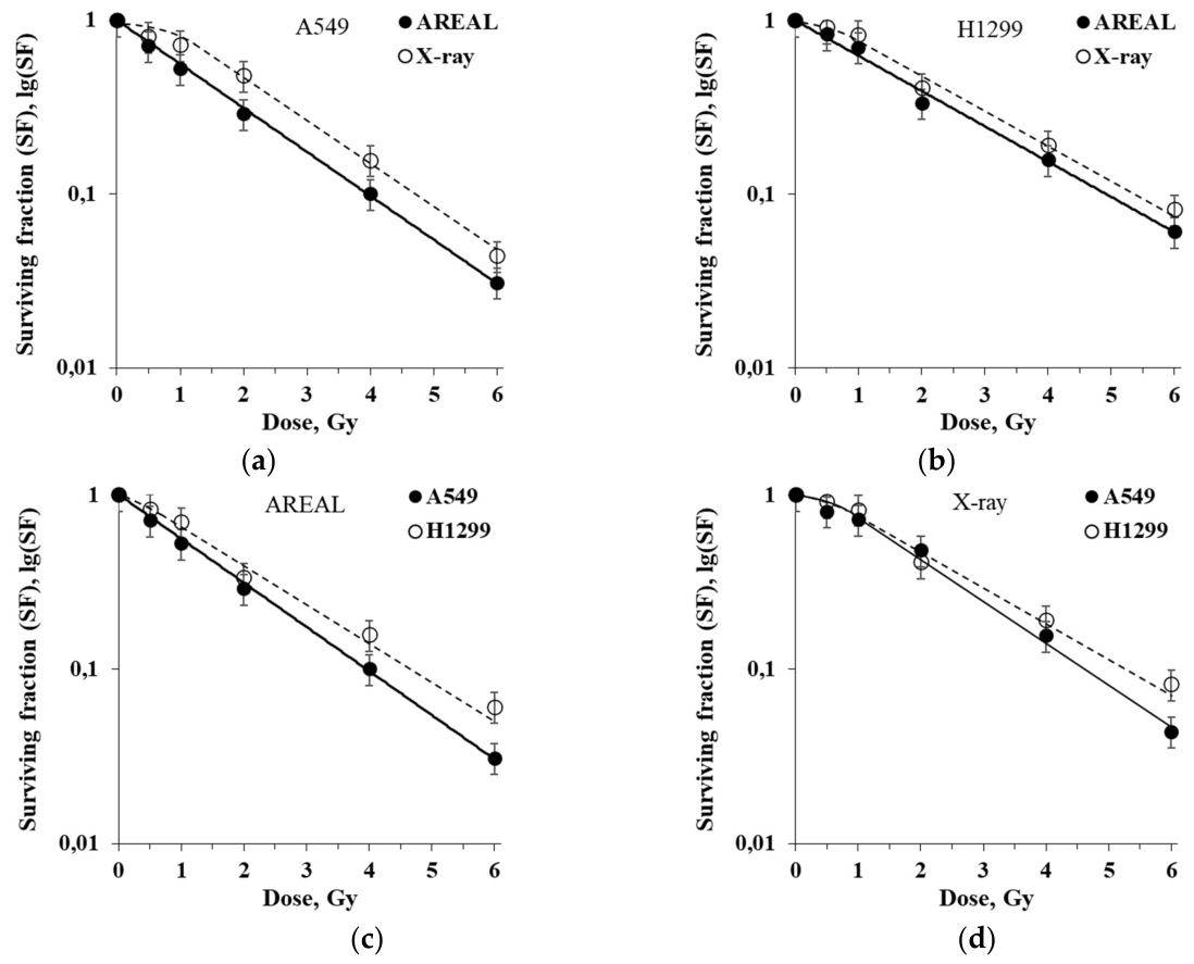
Figure 2.
Dose-dependent changes in the residual γH2AX foci number in A549 (a) and H1299 (b) NSCLC cell lines after exposure to ultrashort beam (AREAL) and X-rays irradiation.The same curves for two NSCLC cell lines are given in comparison after AREAL exposure (c) and X-rays (d).
Figure 2.
Dose-dependent changes in the residual γH2AX foci number in A549 (a) and H1299 (b) NSCLC cell lines after exposure to ultrashort beam (AREAL) and X-rays irradiation.The same curves for two NSCLC cell lines are given in comparison after AREAL exposure (c) and X-rays (d).
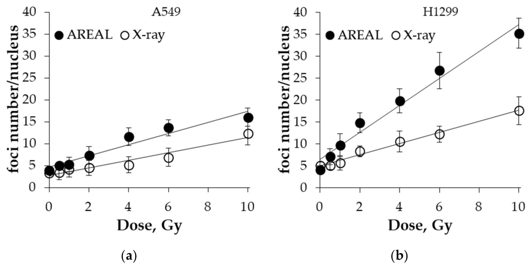
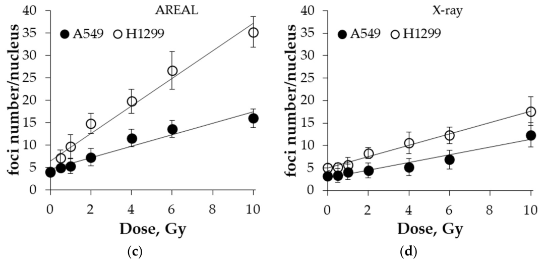
Figure 3.
Analysis of differential gene intersection showing the number of up-regulated and down-regulated genes in A549 cells ∩ H1299 cells after AREAL and X-rays exposure (a). Differential gene intersection for biological process ontology for common up-regulated and down-regulated genes in A549 cells ∩ H1299 cells 24 hours after exposure to AREAL and X-rays. Common up-regulated genes after AREAL (b) and X-rays (c). Common down-regulated genes after AREAL (d) and X-rays (e) exposure; adjusted p-value<0.05.
Figure 3.
Analysis of differential gene intersection showing the number of up-regulated and down-regulated genes in A549 cells ∩ H1299 cells after AREAL and X-rays exposure (a). Differential gene intersection for biological process ontology for common up-regulated and down-regulated genes in A549 cells ∩ H1299 cells 24 hours after exposure to AREAL and X-rays. Common up-regulated genes after AREAL (b) and X-rays (c). Common down-regulated genes after AREAL (d) and X-rays (e) exposure; adjusted p-value<0.05.
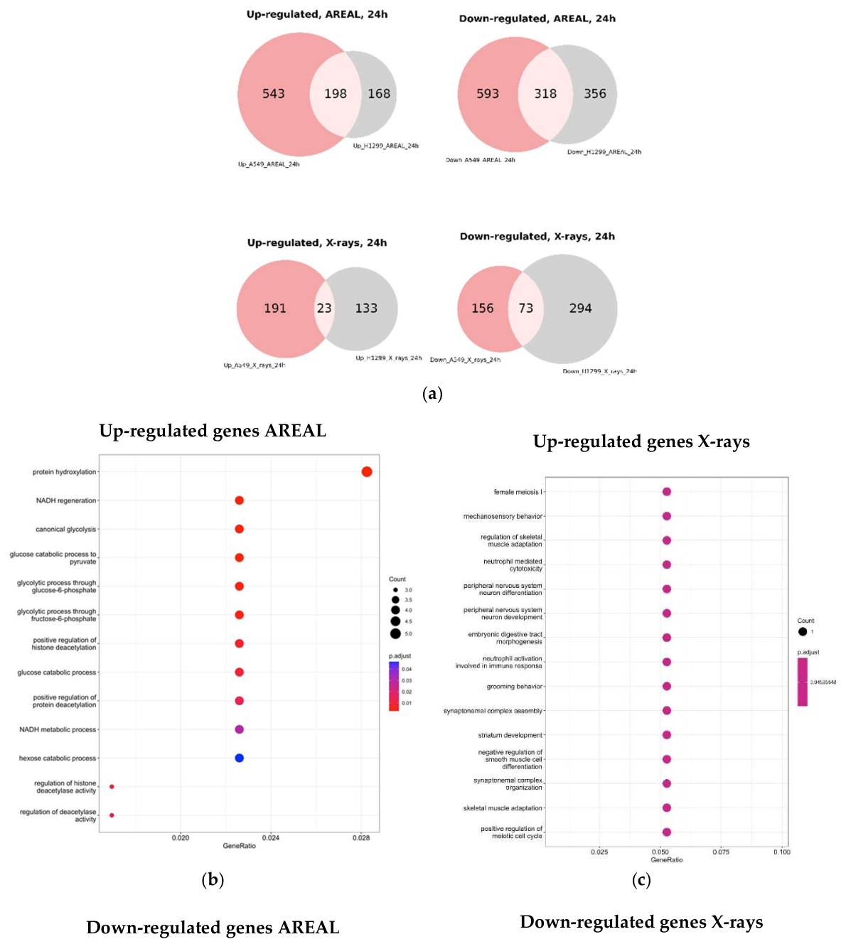
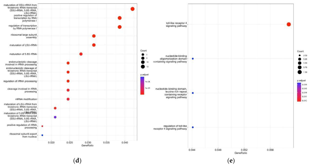
Figure 4.
PAL chart of A549 and H1299 cells: Top 10 up- and down-regulated pathways in A549 cells 24 hours after exposure to ultrashort beam irradiation (AREAL) (a) and X-rays (b). Top 10 up- and down-regulated pathways in H1299 cells 24 hours after exposure to ultrashort beam irradiation (AREAL) (c) and X-rays (d). Benjamini Hochberg adjusted p-value <0.05 (only pathways containing 10 and more genes).
Figure 4.
PAL chart of A549 and H1299 cells: Top 10 up- and down-regulated pathways in A549 cells 24 hours after exposure to ultrashort beam irradiation (AREAL) (a) and X-rays (b). Top 10 up- and down-regulated pathways in H1299 cells 24 hours after exposure to ultrashort beam irradiation (AREAL) (c) and X-rays (d). Benjamini Hochberg adjusted p-value <0.05 (only pathways containing 10 and more genes).
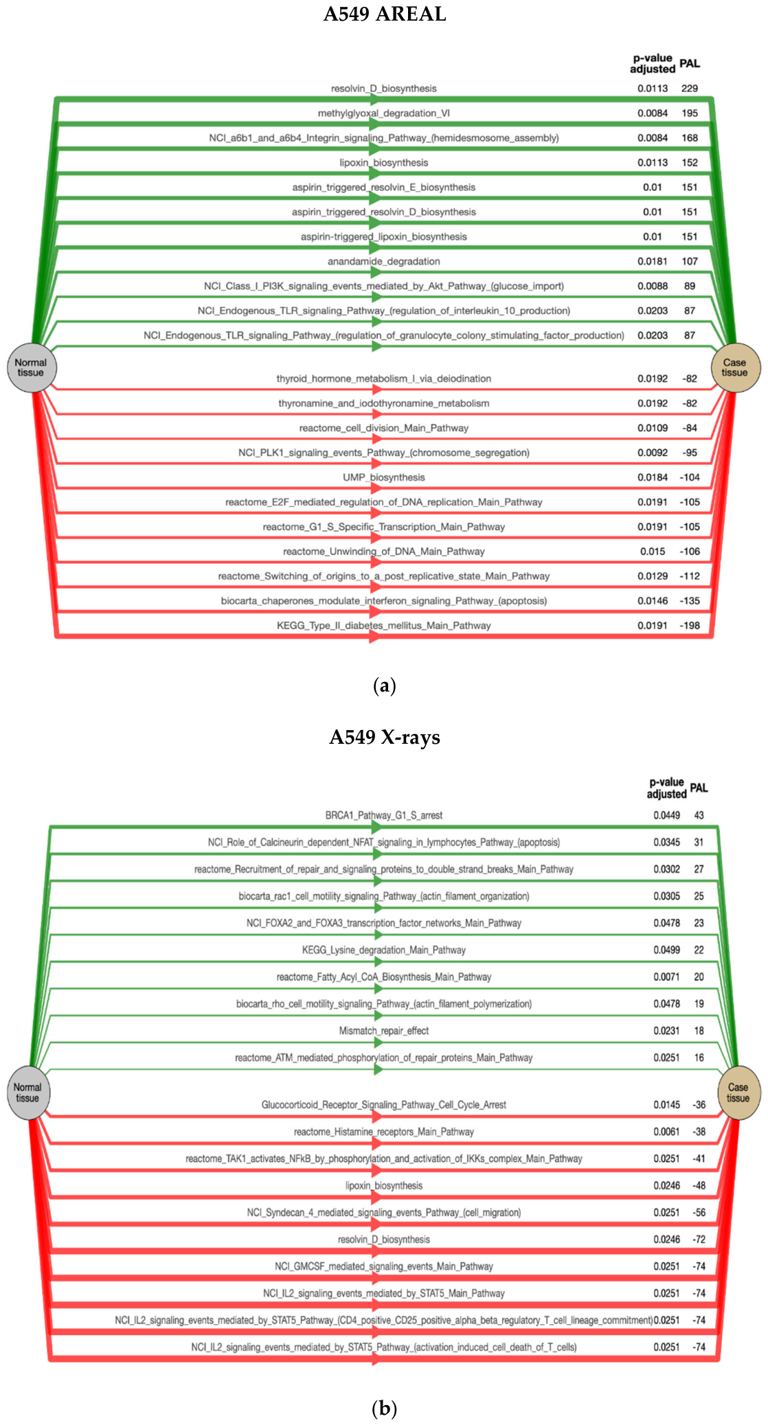
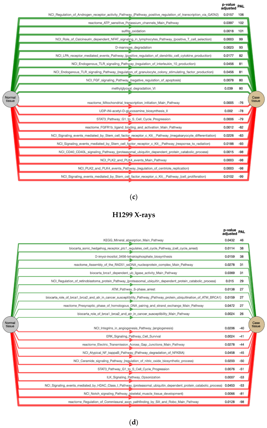
Figure 5.
Heatmap of selected DNA repair pathways of A549 and H1299 cell lines 24 hours after exposure to ultrashort beam irradiation (AREAL) and X-rays. AREAL and X-rays PALs are adjusted to unirradiated control and AREAL PALs are normalized to X-rays (X-rays irradiated cells used as control).
Figure 5.
Heatmap of selected DNA repair pathways of A549 and H1299 cell lines 24 hours after exposure to ultrashort beam irradiation (AREAL) and X-rays. AREAL and X-rays PALs are adjusted to unirradiated control and AREAL PALs are normalized to X-rays (X-rays irradiated cells used as control).
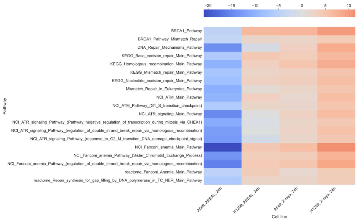
Figure 6.
Heatmap of pathways connected to cell cycle and cell death of A549 and H1299 cell lines 24 hours after exposure to AREAL and X-rays (a). Integrin-linked kinase (ILK) apoptosis (b), ILK pathway G2 phase arrest (c) and ILK pathway Cell cycle proliferation (d). Heat map of RNA-Seq transcriptome analysis for 16 selected pathways in A549 and H1299 cells (e). PALs are adjusted to unirradiated control and AREAL PALs are normalized to X-rays (X-rays irradiated cells used as control).
Figure 6.
Heatmap of pathways connected to cell cycle and cell death of A549 and H1299 cell lines 24 hours after exposure to AREAL and X-rays (a). Integrin-linked kinase (ILK) apoptosis (b), ILK pathway G2 phase arrest (c) and ILK pathway Cell cycle proliferation (d). Heat map of RNA-Seq transcriptome analysis for 16 selected pathways in A549 and H1299 cells (e). PALs are adjusted to unirradiated control and AREAL PALs are normalized to X-rays (X-rays irradiated cells used as control).
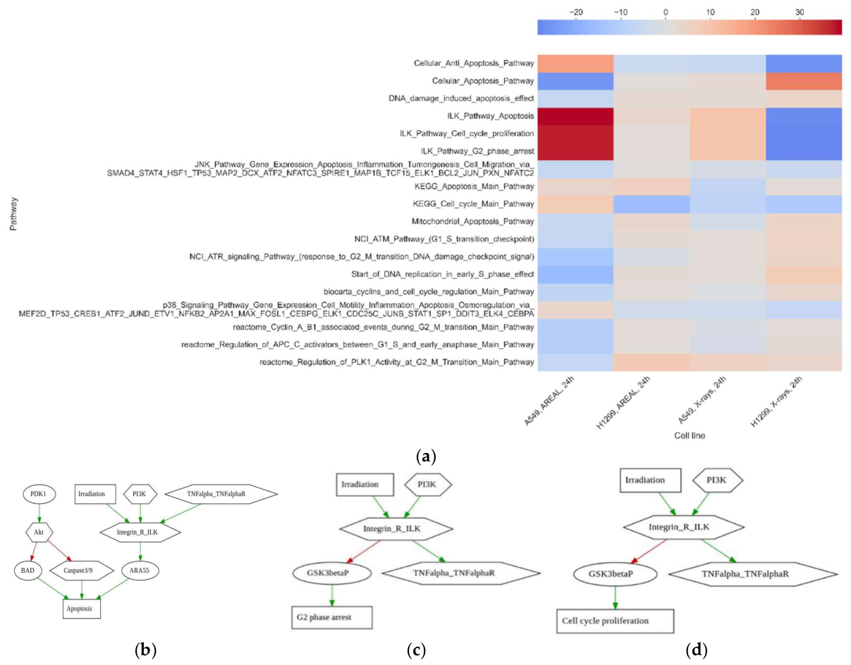
Table 1.
Differential Gene Expression of A549 and H1299 Cells Exposed to AREAL and X-rays.
| DEG, 24h | A549_AREAL | A549_X_rays | H1299_AREAL | H1299_X_rays |
|---|---|---|---|---|
| All | 1652 | 443 | 1040 | 523 |
| Up-regulated | 741 | 214 | 366 | 156 |
| Down-regulated | 911 | 229 | 674 | 367 |
Disclaimer/Publisher’s Note: The statements, opinions and data contained in all publications are solely those of the individual author(s) and contributor(s) and not of MDPI and/or the editor(s). MDPI and/or the editor(s) disclaim responsibility for any injury to people or property resulting from any ideas, methods, instructions or products referred to in the content. |
© 2026 by the authors. Licensee MDPI, Basel, Switzerland. This article is an open access article distributed under the terms and conditions of the Creative Commons Attribution (CC BY) license (http://creativecommons.org/licenses/by/4.0/).
Copyright: This open access article is published under a Creative Commons CC BY 4.0 license, which permit the free download, distribution, and reuse, provided that the author and preprint are cited in any reuse.