Submitted:
08 June 2026
Posted:
09 June 2026
You are already at the latest version
Abstract
This study successfully fabricated a graphene nanoplatelets-polythiophene (GNP-PTh) nanocomposites containing varying graphene concentrations (10-50 wt.%) through oxidative polymerization technique. The resulting nanocomposites revealed effective degradation of bromophenol blue and anticancer activity against U-87 glioma cells. The nanomaterials were characterized using FTIR and SEM that confirmed the integration of graphene nanoplatelets within the polythiophene matrix. UV-Vis spectroscopy and Tauc plot measurements reported the improved optical behavior and gradual decrease in band gap energy from 2.28 eV for pure PTh to 1.76 eV for GNP-PTh(50), representing better electronic connection and charge carrier transfer characteristics. The photocatalytic degradation of the synthesized nanocomposites was systematically assessed under different pH (5, 7 & 11) and temperature conditions (25-70 °C). The effects of pH and temperature exposed that alkaline conditions and optimized temperature favorably enhances the photocatalytic degradation kinetics and decrease the activation energy. In addition to wastewater treatment, the prepared nanocomposites explored remarkable concentration-dependent anticancer action against U-87 glioma cells. The GNP-PTh(50) showed considerably lesser IC₅₀ values and higher cytotoxic effects compared to pure PTh NPs. Conclusively, the synergistic effects of GNPs with PTh improved the structural, optical, photocatalytic, and biomedical features of the nanocomposites. These findings highlighted the potential of GNP-PTh nanocomposites, explicitly GNP-PTh(50), as a promising multifunctional nanomaterials for wastewater remediation and anticancer effects.
Keywords:
graphene nanoplatelets
; photocatalysis
; bromophenol blue degradation
; anticancer activity
; temperature and pH effects
1. Introduction
The day by day increasing toxic wastes has polluted the water badly which is now demanding safe and clean water for human utilization. Recently, the textile industries enhances the risk of water contamination due to release of organic dyes [1]. Among different dyes synthetic dyes almost 90% are highly used as a colorants in plastic, printing ink, cosmetic, food, textile and paper industries for the colorization of substances and consequently the resulting-colored seepage is dumped into sewerage system [2,3,4]. Bromophenol blue (BPB) is a water-soluble dye and mostly employed in different industries like textile, biological staining, and leather zones, consist of toxic compounds which are harmful to the environment and human health. [5]. The BPB is famous for causing the cancer and induces skin & eyes irritation in addition also damages the neurological, reproductive, respiratory systems in human beings upon internal/external exposure [6,7]. Therefore, the removal of BPB is very important before its liberation to the water bases.
In literature, different methods such as coagulation, photocatalysis, ion exchange, adsorption and ozonation are used to remove these toxic organic compounds from water, [8,9]. Among these, photocatalytic degradation is one of the most frequently utilized technique due to cost-effective and eco-friendly nature, and it can easily eradicate most of dyes [10,11,12]. Though, an appropriate photocatalysts with higher stability and efficiency is a critical challenging in environmental and ecological sciences [13,14]. So in order to overcome this challenge, production of cost-effective recyclable, and effective photocatalyst is needed for real-world applications. Graphene-based nanomaterials have been widely employed in photocatalytic and biomedical applications because of their better chemical and physical characteristics.
Graphene nanoplatelets (GNPs) are considered as a best materials for different applications like photocatalyst, automotive, construction, aerospace, electronic industries and biomedical [15]. Basically, GNPs are the derivative of the graphene and possess sp2-hybridized carbon atoms with stacked atomic layers. Furthermore, GNPs showed better mechanical and thermal strength to polymer matrix composites and polymer nanocomposites, making it suitable in various field such as sensors, fuel cells, energy storage and conversion devices [17,18,19,20,21]. These features opened new corridors for collecting a range of organic and inorganic nanoscale components with GNPs, to tailor the joint characteristics in a new manner to obtain the advance level of requirements in nanotechnology [22].
The structural properties of graphene can be enhanced by compositing with Polymer in the form of nanocomposites. Literature revealed that conducting polymers (CPs) have attracted the scientists’ attention because of their easy synthesis methods and intrinsic electrical conductivity. Few number of CPs are reported with advance functional properties [23] such as polypyrrole (PPy), polythiophene (PTh) and polyaniline (PANI). Among these CPs, polythiophene (PTh) is a well-known conducting or semiconducting polymer that has recently attracted considerable interest owing to its π-conjugated electronic network that help in facilitation of charge separation and photon absorption in the process of photocatalytic degradation [24,25]. In addition, PTh is widely used sensors, in solar cells, biocompatibility and environmental remediation technologies [26].
The nanocomposites of graphene-polythiophene showed higher electrical conductivity and larger surface area of graphene with the light-harvesting complex and controlling optical features of PTh. These properties drives a new generation of smart nanomaterials for dual-purpose of environmental purification (photocatalysis) and precision oncology (anticancer therapy) [27].
Thus in this study we choose GNPs based PTh nanoparticles (NPs) nanocomposites for dual-function photocatalytic degradation and cytotoxicity study due to the remarkable physicochemical and surface-related advance features of graphene-based materials, and we mainly focused on varying graphene concentrations (10–50 wt.%) to keenly study the impact of graphene intercalation with PTh NPs. The synthesized nanocomposites were investigated for the photocatalytic degradation of bromophenol blue (BPB) under varying pH and temperature conditions, as well as for in vitro anticancer activity against U-87 glioma cells. The incorporation of GNPs significantly improved reactive oxygen species generation, charge transfer, and surface-mediated interactions, resulting in enhanced photocatalytic efficacy and better anticancer action.
2. Materials and Methods
2.1. Materials and Chemicals
Graphene nanoplatelets (99.9%, KNano), hexadecyltrimethylammonium bromide (CTAB) (≥ 98%, Aldrich), anhydrous ferric chloride (98%, Aldrich), thiophene (98%, Aldrich), and were used as received.
2.2. Preparation of PTh NPs
PTh NPs were prepared via in-situ chemical oxidative polymerization method [28]. In brief, 2.0 mL of thiophene monomer and 0.0034 M CTAB surfactant mixture was made in 30 mL distilled water with 15 min of continuous stirring. Afterward, 0.055 M FeCl3 solution as an oxidant was dropwise added to the mixture of monomer & surfactant, the polymerization reaction started and which was identified by the change in color of reaction mixture and it turned to brown. The polymerization process was accomplished in 24 h at 30 °C. Lastly, the dark-brown precipitates of PTh was washed with methanol and distilled water, and then a colorless end-products was obtained that was finally dehydrated in oven.
2.3. Preparation of GNP-PTh Nanocomposites
The nanocomposites of GNP-PTh with different weight percentages of GNPs (10%, 30% and 50%) were prepared through in-situ chemical oxidative polymerization technique [29]. In the process, anhydrous FeCl3 was utilized as oxidizing agent. 1.0 mL of thiophene monomer and 0.0034 M CTAB surfactant were added in distilled water (30 mL) for 15 min continuously stirring. After that, GNPs in a specific concentration were sonicated with thiophene suspension for 45 min.
Scheme 1.
Schematic illustration of the synthesis of GNP-PTh(10-50%) nanocomposites.

At that time, 0.055 M solution of FeCl3 added dropwise to the reaction mixture of monomer-surfactant. In the process of polymerization, the color of solution changed to brown and the reaction was finished in 24 h at30 °C. The GNP-PTh nanocomposites in the form of dark-brown precipitates were collected as an end-product. Finally, after washing with methanol and distilled water the colorless product was attained. The resultant nanocomposites were dried at 80 °C in oven.
2.4. Characterization
Different characterization methods such as Fourier Transform Infrared Spectroscopy (FTIR), UV-Vis- NIR spectroscopy and Scanning Electron Microscopy (SEM) were employed for pure PTh NPs and GNP-PTh nanocomposites to measure their applicability in photocatalytic degradation and bioactivity.
2.5. Photocatalytic Degradation
The photocatalytic activity of pure PTh NPs and GNP-PTh (10-50%) nanocomposites against BPB degradation were carried out by using a UV-Vis spectrophotometer. The experiments were employed at different temperature, and pH. Firstly, the photocatalyst (0.05 g) was mixed with 20 mg L−1 BPB solution at various temperature ranges (25-50 °C), and at different pH (5, 7 & 11). Prior to irradiation, the solutions were magnetically stirred in the dark for 1 h in order to confirm the adsorption/desorption equilibrium among BPB and nanocomposites. After that, the absorption spectrum of the centrifuged reaction mixture was observed via UV–Vis spectrophotometer. The change in concentration of BPB was assessed by observing the optical intensity of absorption spectra.
2.6. Biocompatibility Test: Cytotoxicity Assay
2.6.1. Cell Culture
The anticancer action of pure PTh NPs and GNP-PTh(10-50%) nanocomposites were estimated on the glioma cell line U-87 (commercially available). The U-87 cell line of glioma origin were gifted by Cancer Cell Culture and Precision Onco-medicine Lab, Khyber Medical University Peshawar, Pakistan. The cell lines were developed in DMEM/F12 medium supplemented with 10% FBS and 1% pen strep and incubated in humidified environment with 5% CO2 supply. The cells were daily checked for infections and confluency. After getting the confluency, cells were separated by trypsinization method and some portion of cells were well-preserved for future usage while the remaining cells were sub-cultured in 96 well plate for investigation. Cells (1 x 10³) were seeded per well in the 96 well plate with the addition of fresh media and then the plate was incubated for 24-48 hrs.
2.6.2. Preparation of Nanocomposites for Anticancer Activity
The parent stock was prepared by solubilizing the nanocomposites in DMSO according to literature reported method. After that, working stock solution was made from parent stock solution.

| Samples | Solubility | Parent stock |
|---|---|---|
| PTh NPs | 0.2-0.3 mg/ml | 1 mg/5ml |
| GNP-PTh(10) | 0.2-0.3 mg/ml | 1 mg/5ml |
| GNP-PTh(30) | 0.2-0.3 mg/ml | 1 mg/5ml |
| GNP-PTh(50) | 0.2-0.3 mg/ml | 1 mg/5ml |
2.7. Drug Inhibition Assay
The IC50 value of each nanocomposite on u87 cell lines was evaluated by using the drug assay in triplicates through serial dilution process. Original U-87 were utilized as a control. The as-prepared nanocomposites were taken in different doses i.e., 400µg, 200µg, 100µg, 50µg, 25µg, 12.5µg, 6.25µg and 0µg. The cells were incubated and subsequently monitored after 24 hr, and then 48 hr. After incubation, the crystal violet assay was implemented and absorbance were measured via ELISA reader. Results were calculated by the Graphpad Prism 8 software.
3. Results and Discussion
3.1. Characterization
The FTIR spectra of pure PTh NPs and GNP-PTh (10-50%) nanocomposites are given in Figure 1(a-d). The FTIR results confirms that thiophene successfully polymerized and GNPs are effectively incorporated into the polymer matrix. Distinctive absorption bands linked with the thiophene backbone and graphene/polymer interfacial connections are noticeably detected in all samples as reported in our previous study [5].
The FTIR spectrum of pure PTh (Figure 1(a)) displays distinctive peaks which corresponds to the conjugated structure of thiophene. The absorption bands detected in the region of 1676 cm-1 and 1421 cm-1 represents the C=C stretching vibrations of thiophene ring that confirms the development of conjugated polymer network. The bands appeared nearby 617 cm-1 and 535 cm-1 are connected with C-S-C and C-S stretching vibrations of aromatic thiophene ring respectrively, while the peaks appears in lower wavenumber region 780 cm-1 attributed to C–H out-of-plane bending vibrations of the thiophene units.
After incorporation of graphene nanoplatelets, noticeable changes in peak intensity, broadening, and slight shifts in characteristic absorption bands are observed in the spectra of GNP-PTh nanocomposites as shown in Figure 1(b-d). The band around 2931–2938 cm-1 designates the stretching vibrations of C-H. The bands raised in the region of 1406 cm-1 and 1657 cm-1 ascribed the C=C vibration of GNPs. The peaks appeared at 1050–1059 cm-1 are allocated to the in-plane bending of C-H, and the band observed at 694 cm-1 are related to the stretching vibration of C-S in thiophene ring, whereas peaks in the region of 1119 cm-1 are associated with the stretching vibrations of C-O. The signal displacements in these spectra indicates that PTh nanostructures are finely anchored on the surface of GNPs. These spectral variations also designate strong interfacial contact among GNPs and PTh matrix, which is predominantly accredited to the π–π interactions amid the aromatic graphene surface and the conjugated thiophene backbone, which improves structural stability and electronic connection in the prepared nanocomposite system [30].
The gradual variation are observed in the FTIR peak intensity by increasing the content of graphene in the nanocomposites further confirms the successful integration of GNPs in the polymer matrix. Specifically, the spectra of GNP-PTh(50) reveal more prominent spectral modifications, signifying higher interfacial contact and better dispersion of GNPs in the conducting polymer network. Such kind of interactions improve the charge transfer and lower electron-hole recombination, which are important for well photocatalytic degradation efficacy and anticancer action [31].
The gradual variation are observed in the FTIR peak intensity by increasing the content of graphene in the nanocomposites further confirms the successful integration of GNPs in the polymer matrix. Specifically, the spectra of GNP-PTh(50) reveal more prominent spectral modifications, signifying higher interfacial contact and better dispersion of GNPs in the conducting polymer network. Such kind of interactions improve the charge transfer and lower electron-hole recombination, which are important for well photocatalytic degradation efficacy and anticancer action [31].
The surface morphology and microstructural features of pure PTh NPs and GNP-PTh (10-50%) nanocomposites were studied by scanning electron microscopy (SEM), as presented in Figure 2(a-d). The SEM images deliver clear evidence for the efficacious development of PTh NPs and the effective integration of GNPs in the polymer network [32].
The SEM micrograph of pure PTh nanoparticles (Figure 2(a)) exposes the production of densely agglomerated granular nanoparticles with relatively irregular surface morphology. The particles are dispersed irregularly and make clustered configurations because of stronger intermolecular interactions between PTh chains. The inset image further endorses the nanoscale morphology of the PTh NPs. Particle size analysis shows that average particle size is found in nanometer range, specify the successful formation of nanoscale polymer particles.
At higher magnification (Figure 2(b-d)), the PTh NPs reveal solid and aggregated morphology with interconnected grainy domains. Such accretion behavior is usually detected in conducting polymers because of the π–π interactions and strong van der Waals forces in the polymer chains. The particles size was lie in the range of 0.45-1.46 nm for PTh NPs. The rough and porous surface morphology of PTh is significant for interfacial adsorption processes and offers more active sites for photocatalysis. After the addition of graphene nanoplatelets, important morphological modifications are detected in the GNP-PTh nanocomposites (Figure 2(d)). The SEM image clearly shows the existence of sheet-like graphene nanoplatelets and PTh NPs dispersed on the surface of graphene. The graphene sheets provide a layered and wrinkled morphology with higher surface roughness and bigger surface area. The incorporation of PTh NPs on the surface of graphene endorses strong interfacial interaction among graphene and the PTh matrix. Inclusively, the SEM analysis confirms the successful production of GNPs-loaded PTh nanocomposites with intersected porous architecture, improved interfacial coarseness, and greater graphene–polymer surface contact. These structural characteristics can significantly enhance the photocatalytic and biomedical performance of the as-prepared nanocomposites.
The optical studies and electronic transitions of pure PTh NPs and GNP-PTh(10-50%) nanocomposites were examined by UV–Vis spectroscopy, as shown in Figure 3(a–d). The spectra gives significant evidences about the interconnection among GNPs and the PTh matrix, in addition to this the effect of graphene integration on the optical behavior of the nanocomposites is also highlighted. The UV–Vis spectrum of pure PTh (Figure 3(a)) displays distinctive absorption band at 393 nm which is related with the π–π* electronic transitions of the conjugated thiophene backbone. This broad absorption band confirms the existence of aromatic/conjugated system within the conducting polymer units. The absorption in the visible region is linked to delocalized charge carriers and conjugated electron transport in the PTh system.
After incorporation of graphene nanoplatelets, noticeable modifications in absorption intensity and spectral profile are observed in the GNP-PTh nanocomposites (Figure 3(b–d)). The gradual variation in absorbance behavior with increasing graphene content confirms successful interaction between graphene nanoplatelets and the PTh matrix. The higher absorption of graphene-containing nanocomposites may be attributed to improved electronic interaction and charge-transfer processes between graphene sheets and the conjugated polymer chains. Conversely, GNP-PTh nanocomposites shows an absorption band at 230 nm which is attributed to the π to π* transition of aromatic carbon-carbon (C=C) bonds, and a shoulder appeared around 340 nm is due to n-π* transition of the carbon-oxygen (C=O) bond [33]. The band at 260–270 nm also corresponds to π to π* transition between PTh and GNPs. A red shift in π-π* transition is observed by increasing content of graphene [34]. The spectral modifications detected in GNP-PTh nanocomposites recommended the improved π-electron delocalization and enhanced surface electronic connection in the composite morphology.
The optical band gap energies of pure PTh NPs and GNP-PTh nanocomposites were measured by Tauc plots estimated from UV–Vis absorption data, as presented in Figure 4(a-d). The plots of (αhυ)2 versus photon energy (hυ) were used to determine the direct optical band gap energies by extrapolating the linear region to the energy axis. The measured band gap energy values of pure PTh NPs was 2.28 eV, which indicates the semiconducting nature of PTh [35]. After integration of GNPs, a steady reduction in band gap energy was noticed when graphene content was increased. The calculated band gap energies for GNP-PTh(10), GNP-PTh(30), and GNP-PTh(50) nanocomposites were around 2.00 eV, 1.85 eV, and 1.76 eV, respectively. The progressive decrease in band gap energy suggests stronger electronic contact between GNPs and the aromatic PTh system.
The loading of graphene gives more electronic states and increases π-electron delocalization in the nanocomposite configuration, thus enabling easy electronic transitions and enhancing charge carrier motion. The lessening of optical band gap is significant for photocatalytic activity as low band gap energies endorse higher visible-light absorption and enable effective formation of electron-hole pairs when exposed to irradiation. Among all nanomaterials, GNP-PTh(50) showed lowermost band gap energy (1.76 eV), which indicates greater electronic conductivity and better light-harvesting ability. This result is fully consistent with the higher photocatalytic degradation efficacy, lesser activation energy, and improved anticancer performance detected for graphene-rich nanocomposites [36].
3.2. Photocatalytic Degradation
Photocatalytic degradation of pure PTh NPs and GNP-PTh nanocomposites with varying graphene content (10%, 30%, and 50%) against bromophenol blue (BPB) were carried out by studying the effect of temperature and pH.
The effect of temperature on the photocatalytic degradation of pure PTh NPs and GNP-PTh (10-50%) nanocomposites was analytically examined at various temperatures ranges from 25-70 °C as shown in Figure 5(a-d). Spectra showed that absorbance intensity gradually decreased near the distinguishing absorption maximum of BPB (~620 nm) with enhancing the temperature, which confirmed the successive degradation of dye. Amongst all photocatalysts, GNP-PTh(50) revealed the most distinct reduction in absorbance intensity, signifying higher photocatalytic action than pure PTh, GNP-PTh(10) and GNP-PTh(30) as presented in Figure 5(d). While, pure PTh NPs gives comparatively lesser decrease in absorbance, imitating reduced degradation behavior. The higher degradation efficiency of GNP-PTh nanocomposites at higher temperatures is accredited to higher surface-mediated adsorption, faster kinetic reactions and interfacial charge transfer. Higher temperature indorses better contact among catalyst and dye molecules active sites whereas instantaneously augmenting the movement of photogenerated charge carriers. The GNPs presence further assists electron transport and overpowers charge recombination, as a result generates more reactive oxygen species for degradation of dye molecules. In addition, greater graphene content ominously increases the thermal response of the photocatalytic system, with GNP-PTh (50%) sustaining the maximum degradation efficacy during the selected and studies temperature range. This performance approves the part of graphene in improving both the surface reactivity and interfacial photocatalytic processes within the nanocomposite system [37].
Kinetic studies revealed that dye photocatalytic activity noticeably enhanced by elevated the temperature for all prepared-samples, as proved by the progressive rise in degradation proficiency and consistent reduction in Ct/C0 values as given in Figure 6(a-c). The Ct/C0 values further approve this tendency, lessening from 0.819 to 0.637 for pure PTh, and importantly from 0.442 to 0.295 for GNP-PTh(50), signifying greater degradation performance at higher temperatures shown in Figure 6(a). This trend is consistent with increase in ln(C0/Ct) values that rises from 0.199 to 0.451 for pure PTh NPs and 0.816 to 1.221 for GNP-PTh(50, evidencing that process of degradation follows pseudo-first-order kinetics at elevated temperatures as presented in Figure 6(b). The percentage % degradation efficiency in case of pure PTh NPs, the degradation efficacy upsurges from 18.06% at 25 °C to 36.27% at 70 °C, representing a moderate activity. On the other hand, GNP-PTh nanocomposites displayed extensively high degradation efficacies, while GNP-PTh(50) percent degradation increases from 55.78% to 70.52% over the same temperature range which can be seen in Figure 6(c). So, the enhancement in photocatalytic performance with temperature is attributed to various factors like a rise in temperature boosts the kinetic energy of reactant molecules, which as a result increases the frequency of active collisions among dye molecules and active sites on the surface of catalyst. Furthermore, the higher temperatures also assists quicker diffusion and mass transfer, letting more effective collaboration amongst BPB molecules and photocatalyst.
Comparatively, GNP-PTh(50) steadily displays the maximum degradation efficacy and faster kinetics, followed by GNP-PTh(30), GNP-PTh(10), and pure PTh NPs at all temperatures. This tendency indicated the effective role of graphene loading in improving both surface reactivity and electron transport properties. The better activity of larger graphene content is corresponds to its high surface area, robust π–π connections with BPB molecules, and greater electrical conductivity, all of which predominantly enhanced adsorption and augmented photocatalytic reactions. Overall, the temperature and graphene loading synergistically enhanced the photocatalytic efficiency, with greater temperatures intensifying the significance of graphene integration in the PTh matrix. This specifies that optimizing both material composition and operational conditions is important for attaining efficient photocatalytic system [38].
The effect of pH on the photocatalytic degradation performance of pure PTh NPs and GNP-PTh nanocomposites was systematically explored at different pH (5, 7 & 11). Since pH strongly influences the surface charge of photocatalysts, adsorption behavior of dye molecules, and generation of reactive oxygen species, it plays a critical role in determining photocatalytic efficiency. To evaluate the pH-dependent degradation behavior, UV-Vis absorbance spectra and kinetic parameters were analyzed for pure PTh and GNP-PTh nanocomposites containing varying graphene concentrations (10-50%). The results demonstrate that both graphene loading and pH significantly affect the degradation efficiency of bromophenol blue, with enhanced photocatalytic activity observed under basic conditions. The UV-Vis absorbance spectra recorded under acidic and basic conditions (Figure 7(a-d)) demonstrate the significant influence of pH on the photocatalytic degradation behavior of bromophenol blue. A noticeable reduction in absorbance intensity near the characteristic absorption maximum (~620 nm) is observed under basic conditions, indicating enhanced degradation efficiency compared to acidic conditions. Among all photocatalysts, GNP-PTh (50%) exhibits the most pronounced decrease in absorbance intensity, confirming its superior photocatalytic activity, as shown in Figure 7(d). The enhanced degradation under alkaline conditions can be attributed to increased formation of hydroxyl radicals and improved surface reactivity, which accelerate oxidative degradation pathways. The incorporation of graphene nanoplatelets further enhances photocatalytic performance by improving surface adsorption and facilitating interfacial charge transfer. The high surface area and electrical conductivity of graphene promote efficient separation of photogenerated charge carriers, thereby increasing the generation of reactive oxygen species responsible for dye degradation [39]. Overall, the spectra presented in Figure 7(a-d) confirm that both pH and graphene loading strongly influence the surface-mediated photocatalytic behavior of GNP-PTh nanocomposites.
To evaluate the photocatalytic degradation efficiency and reaction kinetics of bromophenol blue (BPB), normalized concentration (Ct/C0), logarithmic kinetic behavior ln Co/Ct and percentage degradation were systematically analyzed for pure PTh and GNP-PTh nanocomposites containing varying graphene concentrations (10-50%). These kinetic parameters provide important insight into the degradation behavior, reaction rate, and influence of graphene incorporation on the photocatalytic process under different pH conditions. Figure 8(a) illustrates the variation in normalized concentration (Ct/C0) of bromophenol blue after photocatalytic treatment under acidic (pH 5) and basic (pH 11) conditions A progressive decrease in Ct/C0 along with a corresponding increase in ln Co/Ct and percentage degradation is observed with increasing graphene content, confirming a significant enhancement in photocatalytic efficiency upon incorporation of graphene nanoplatelets. Among all samples, GNP–PTh(50) exhibited the lowest Ct/C0 values, decreasing from 0.232 at pH 5 to 0.059 at pH 11, corresponding to the highest degradation efficiency. In contrast, pure PTh retained comparatively higher Ct/C0 values, decreasing from 0.833 at pH 5 to 0.277 at pH 11, reflecting its limited photocatalytic activity. This trend clearly demonstrated the synergistic effect between GNPs and PTh NPs, where increasing graphene loading improves photocatalytic performance. The enhanced activity of GNP-PTh nanocomposites can be attributed to several surface and interfacial phenomena. Graphene nanoplatelets possess high specific surface area, electron mobility, and excellent adsorption capacity, which facilitate the adsorption of dye molecules and improve light-driven charge separation. The intimate interfacial contact between graphene and PTh promotes efficient electron transfer, thereby suppressing the recombination of photogenerated electron–hole pairs [39,40].
Furthermore, graphene acts as an electron acceptor and transporter, enabling rapid migration of photogenerated electrons from PTh to graphene sheets. This process enhances the generation of reactive oxygen species (ROS), such as •OH and O2− radicals, which are primarily responsible for the degradation of organic dyes. Consequently, the superior performance of GNP-PTh(50) confirms that higher graphene loading strengthens interfacial charge transfer and surface reactivity, leading to enhanced photocatalytic degradation [41].
Additionally, slightly improved performance under basic conditions compared to acidic conditions suggests that alkaline media favor the generation of hydroxyl radicals, further accelerating dye degradation. Figure 8(b) presents the pseudo-first-order kinetic behavior of BPB degradation, expressed as ln(C0/Ct), for all photocatalysts under different pH conditions. The increasing trend in ln(C0/Ct) values with higher graphene content confirms that the photocatalytic degradation follows pseudo-first-order reaction kinetics, consistent with the Langmuir-Hinshelwood model commonly applied to heterogeneous photocatalysis. At pH 11, GNP–PTh(50) displayed the maximum ln(C0/Ct) value of 2.57, while pure PTh presented the less value of 0.321, designating substantially improved degradation kinetics on graphene addition to the nanocomposites. The linear fitting plots revealed a positive correlation among pH and degradation kinetics, endorsing that alkaline conditions are favorable for photocatalytic performance of as-prepared nanocomposites. This kinetic enhancement further validates the critical role of graphene in accelerating the photocatalytic process. The improved kinetic performance of GNP-PTh nanocomposites can be explained by the efficient interfacial charge transfer and extended π–π conjugation between GNPs and PTh NPs. The presence of graphene enhances the conductivity of the composite system, facilitating rapid electron transport and minimizing charge recombination losses. Moreover, graphene provides additional active sites for adsorption and reaction, thereby increasing the effective collision frequency between reactive species and dye molecules. The observed increase in reaction rate with graphene loading also indicates a strong correlation between surface properties and photocatalytic kinetics. The results clearly demonstrate that interfacial engineering of nanocomposites plays a pivotal role in determining both efficiency and kinetics of photocatalytic systems [42].
Figure 8(c) demonstrations a systematic rise in degradation efficacy with higher graphene loading. The highest degradation efficiency of 94.1% was achieved by GNP–PTh(50) at pH 11, while pure PTh exhibited a comparatively lower degradation efficiency of 62.3% which clearly indicated that graphene incorporation significantly enhances the photocatalytic activity, with the highest performance achieved at 50% graphene loading. The high degradation performance of GNP-PTh nanocomposites is accredited to the synergistic interconnection among graphene nanoplatelets and PTh at the interface level. GNPs offers a huge specific surface area, which improves the adsorption of BPB molecules through π–π bonds in the aromatic dye and graphene sheets. This augmented adsorption increases the accessibility of BPB molecules on active
For instance, GNP-PTh(50) increases degradation from 76.8% (acidic) to 83.3% (basic), so this increase corresponds to the larger accessibility of hydroxyl ions (OH-) in alkaline medium that endorses the generation of hydroxyl radicals which is a main oxidizing species in photocatalytic processes [43]. The maximum degradation of GNP-PTh(50) endorses that optimized graphene content substantially improves both adsorption capability and charge transfer efficacy, leading to higher BPB degradation.
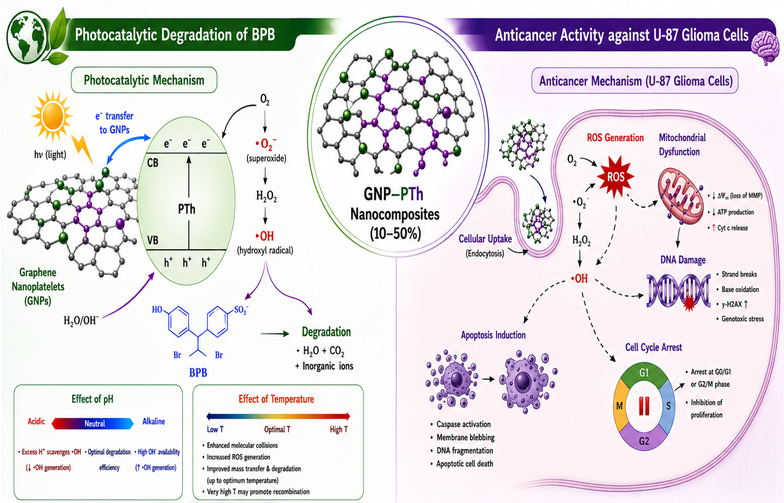
Proposed Photocatalytic Degradation Mechanism
The proposed photocatalytic degradation mechanism of BPB using GNP-PTh nanocomposites is illustrated in Figure 9. The higher photocatalytic degradation is mostly endorsed to the synergistic collaboration among GNPs and PTh NPs, which enhances light absorption, surface adsorption, charge separation, and generation of reactive oxygen species. Upon irradiation, PTh NPs absorbs energy and as a result produces electron-hole pairs. Electrons get excited from the valence band to the conduction band, leave the holes in the valence band. In pure PTh, these photogenerated electrons and holes can recombine quickly, decreasing photocatalytic efficacy. Though, in GNP-PTh nanocomposites, GNPs act as effective electron acceptors and transport pathways. The photogenerated electrons travel from PTh to GNPs, which overwhelms electron-hole recombination and extends the charge carriers lifetime. The transferred electrons rejoin with dissolved oxygen molecules adsorbed on the surface of catalyst to produce superoxide radicals. In the meantime, photogenerated holes react with water molecules or hydroxyl ions and generates hydroxyl radicals. These reactive oxygen species, particularly •OH and •O2−, are extremely oxidative and outbreak BPB molecules adsorbed on the surface of nanocomposite, leading to progressive degradation and ultimate mineralization into H2O, inorganic ions and CO2 [44]. The pH of the reaction medium are imperative in adjusting radical formation and degradation efficacy. Under acidic conditions, higher H+ ions reduce the accessibility of hydroxyl radicals by scavenging reactive species, leading to moderately lesser degradation. Under neutral to mildly alkaline conditions, the OH− ions availability rises, endorsing hydroxyl radical generation and enhanced the degradation performance [44,45].
Furthermore, temperature also effects the degradation system by affecting the motion of molecules, surface reaction kinetics, diffusion and adsorption/desorption equilibrium. Enhancing the temperature increases collisions of molecules among BPB molecules and active sites, which grows mass transfer, and accelerates ROS-mediated oxidation. The better GNP-PTh(50) performance is therefore connected with its greater graphene loading, which offers a higher active surface area, strong π–π interface with BPB, enhanced electron transport, and lesser charge recombination. Inclusively, degradation of BPB over GNP-PTh nanocomposites happens through a surface-mediated linkage involving adsorption of dye molecules, charge separation, ROS formation, oxidative bond cleavage, and final end-products [46].
3.3. Anticancer Activity Against U-87 Glioma Cells
The in vitro cytotoxic activity PTh NPs and GNP-PTh (10-50%) nanocomposites were estimated against glioma cell line U-87 at different concentrations (400µg, 200µg, 100µg, 50µg, 25µg, 12.5µg, 6.25µg and 0µg) using MTT assay. The prepared nanocomposites showed a noticeable growth-inhibitory effect on U-87 cell lines in a dose dependent manner as presented in Figure 10 and Figure 11.
A clear concentration-dependent decrease in cell viability was observed for all samples, indicating enhanced cytotoxic effects with increasing nanocomposite concentration as given in Figure 10. Among all the samples, GNP-PTh(50) displayed the highest anticancer action, presentation the lowermost cell viability at high concentrations, while pure PTh NPs showed relatively weak cytotoxic activity. The improved anticancer action of graphene-loaded nanocomposites is accredited to the synergistic contact among GNPs and PTh Nps, which increases surface action and endorses oxidative stress inside the cancer cells.
The IC50 values obtained from the dose–response curves (Figure 11) further clarified the better anticancer action of GNP-PTh nanocomposites against U-87 glioma cells. A progressive reduction in IC50 values was noted with increasing graphene content, which indicates that graphene integration substantially increases the cytotoxic efficacy of nanocomposites. Pure PTh NPs demonstrated the maximum IC50 value (~50 µM), which is comparatively lesser anticancer activity. On the other hand, GNP-PTh(10), , GNP-PTh(30), and , GNP-PTh(50) nanocomposites progressively showed smaller IC50 values of approximately 30 µM, 18 µM, and 10 µM, respectively. Among all prepared nanomaterials, GNP-PTh(50) revealed the least IC50 value, indorsing stronger inhibitory effect towards glioma cell viability. The decrease in IC50 with enhancing GNPs loading is accredited to higher surface action, better connection with cellular membranes, and greater formation of reactive oxygen species (ROS). The large surface area and improved electron transport characteristics of GNPs assist oxidative stress within cancerous cells, causing the membrane damage, apoptosis and mitochondrial dysfunction. The IC50 tendency is in consistency with photocatalytic degradation behavior noted for BPB, where larger GNPs content leads to the higher generation of reactive species and enhanced surface-mediated reactivity. These results proved that interfacial networking through GNPs integration not only improves photocatalytic behavior but also significantly enhances the biomedical applications of nanocomposites. The findings finally revealed that GNP-PTh nanocomposites, mainly GNP-PTh(50), possess favorable anticancer capability towards U-87 glioma cells, emphasizing their prospective for multifunctional environmental and biomedical applications.
Mechanistic Overview
The proposed mechanism for anticancer activity of GNP-PTh nanocomposites towards U-87 glioma cells is shown in Figure 12. The highest cytotoxic activity of nanocomposites is predominantly endorsed to the synergistic effect of GNPs and PTh NPs combination, which significantly stimulates oxidative stress, cellular interaction and apoptosis-induced cell death. On exposure to U-87 glioma cells, the GNP-PTh nanocomposites interact with cellular membrane and are internalized by endocytic networks. The higher surface area and layered morphology of GNPs simplify effective cellular uptake and stronger connection with intracellular components. Once internalized, the nanocomposites mediate formation of excessive ROS, comprising hydroxyl radicals and superoxide radicals, causing the oxidative stress in cancer cells. The raised ROS levels interrupt mitochondrial membrane and damage mitochondrial function, which causes ATP diminution and discharge of cytochrome c into the cytoplasm. Instantaneously, oxidative stress damages DNA membrane, and protein oxidation, which communally inhibit the important cellular functions and stimulate genotoxic stress. The oxidative imbalance further mediate cell cycle detention and destroys cellular propagation, eventually stimulating apoptosis-related signaling pathways. Activation of caspase-dependent apoptotic mechanisms causes membrane blebbing, chromatin condensation and programmed cell expiration, thus decreasing cell viability. The higher anticancer action noted for graphene-rich nanocomposites, specifically GNP-PTh(50) is due to the higher surface reactivity, strong cell–nanocomposite connection, and bigger ROS-induced oxidative impairment. The dose-dependent decrease in cell viability and lesser IC50 values further indorse that higher graphene content increases the cytotoxic effectiveness of the nanocomposites against U-87 glioma cells. Overall, the anticancer action of GNP-PTh nanocomposites is governed by a synergistic mechanism comprising cellular uptake, ROS formation, mitochondrial impairment, oxidative stress, cell cycle detention, and apoptosis stimulation.
4. Conclusions
In this project, GNP-PTh nanocomposites with different graphene loading (10–50 wt.%) were successfully fabricated through osidative polymerization technique. Morphological and structural characterization successfully revealed the integration of GNPs with PTh NPs and exposed stronger interfacial connection among GNPs and PTh NPs. The UV-Vis spectra and band gap studies confirmed that graphene combination highly enhanced the optical and electronic characteristics of nanocomposites by decreasing the band gap energy and improving charge transfer actions. The prepared nanocomposites showed significant photocatalytic degradation proficiency against BPB, highly influenced by temperature and pH conditions. Among all the prepared nanomaterials, GNP-PTh(50) revealed the maximum photocatalytic action, lower activation energy, and better degradation effectiveness in optimized conditions. In addition to environmental remediation, the GNP-PTh nanocomposites also displayed imperative anticancer action towards U-87 glioma cells. A dose-dependent decrease in cell viability was noted for all nanocomposites, while GNP-PTh(50) exhibited higher cytotoxic properties and lesser IC50 values. Conclusively, the synergistic effect GNPs with PTh NPs noticeably improved the optical, structural, morphological, photocatalytic, and biomedical features of nanocomposites. The findings revealed that GNP-PTh nanocomposites, specifically GNP-PTh (50) are favorable multifunctional nanomaterials for wastewater treatment and anticancer studies, which highlights their efficacy for future environmental and biomedical technologies.
Author Contributions
H.N. conceived and designed the study, performed data analysis and validation, and prepared the original manuscript draft. U.A. contributed to data analysis, data validation, and manuscript editing. R.K. contributed to data analysis and manuscript review and editing. All authors have read and agreed to the published version of the manuscript.
Funding
This research received no specific grant from any funding agency in the public, commercial, or not-for-profit sectors.
Data Availability Statement
Upon reasonable request, data can be provided by the corresponding author.
Acknowledgments
The author gratefully acknowledges the support of the TWAS-CNPq Postgraduate Fellowship (Sandwich PhD Program, Grant No. 315724/2018-8), which provided fellowship support during the course of the doctoral studies. The authors also acknowledge the facilities and technical support provided by the Institute of Chemical Sciences, University of Peshawar, Pakistan, University of Campinas (UNICAMP), Brazil and the University of Pisa, Italy.
Conflicts of Interest
The authors declare no conflict of interest.
References
- Kamenická, B. Chemical degradation of azo dyes using different reducing agents: a review. J. Water Process. Eng. 2024. [Google Scholar]
- Singh, V.; et al. Degradation of food dyes via biological methods: a state-of-the-art review. Bioresour. Technol. Rep. 2024. [Google Scholar] [CrossRef]
- Samarasinghe, L.V.; et al. Recent advances in visible light-activated photocatalysts for degradation of dyes: a comprehensive review. Chemosphere 2024. [Google Scholar] [CrossRef] [PubMed]
- Rasool, et al. Biogenic synthesis and characterization of ZnO nanoparticles for degradation of synthetic dyes: a sustainable environmental cleaner approach. J. Clean. Prod. 2023. [Google Scholar] [CrossRef]
- Noreen, H.; et al. Synthesis of graphene nanoplatelets/polythiophene nanocomposites with enhanced photocatalytic degradation of bromophenol blue and antibacterial properties. Mater. Res. Bull. 2021. [Google Scholar] [CrossRef]
- Katubi, K. MohammedSaleh; et al. Synthesis of rGO-supported NiCo2O4/NiS nanocomposite for effective degradation of diclofenac and bromophenol blue Ceram. Int. 2024. [Google Scholar]
- Moussaid, D.; et al. High photocatalytic activity and stability of MnV2O6 and Mn2V2O7 synthesized by simple low temperature method for bromophenol blue degradation. J. Photochem. Photobiol. A Chem. 2023. [Google Scholar] [CrossRef]
- Haqmal, E.; et al. Uncovering the influence of Ni-doping in CuBi2O4 photocatalyst on its visible-light-responsiveness for the efficient removal of toxic alizarin yellow R dye from wastewater Colloids Surf. A Physicochem. Eng. Asp. 2024. [Google Scholar] [CrossRef]
- Ahmad, et al. A comprehensive review on the advancement of transition metals incorporated on functional magnetic nanocomposites for the catalytic reduction and photocatalytic degradation of organic pollutants. Coord. Chem. Rev. 2024. [Google Scholar] [CrossRef]
- Bashir, B.; et al. CuxNi1-xO nanostructures and their nanocomposites with reduced graphene oxide: synthesis, characterization, and photocatalytic applications Ceram. Int. 2021. [Google Scholar] [CrossRef]
- Chandra, M.R.; Rao, T.S.; Pammi, S.V.N.; Sreedhar, B. An enhanced visible light active rutile titania-copper/polythiophene nanohybrid material for the degradation of rhodamine B dye. Mater. Sci. Semicond. Process. 2015, 30, 672–681. [Google Scholar] [CrossRef]
- Mustafa, M.; Bashir, S.; Moosvi, S.K.; Najar, M.H.; Masoodi, M.H.; Rizvi, M.A. Hybrid Polymer Composite of Prussian Red Doped Polythiophene for Adsorptive Wastewater Treatment Application. Acta Chim. Slov. 2022, 69, 848–862. [Google Scholar] [CrossRef] [PubMed]
- Ramirez, J.H.; Maldonaldo-Hodar, F.J.; Perez-Cadenas, A.F.; Moreno-Casilla, C.; Costa, C.A.; Madeira, L.M. Azo-dye Orange II degradation by heterogeneous Fenton-like reaction using carbon-Fe catalysts. Appl. Catal. B-Environ. Energy 2007, 75, 312–323. [Google Scholar] [CrossRef]
- Naguib, M.; Kurtoglu, M.; Presser, V.; Lu, J.; Niu, J.; Heon, M.; Hultman, L.; Gogotsi, Y.; Barsoum, M.W. Two-dimensional Nanocrystals Produced by Exfloliation of Ti3AlC2. Adv. Mater. 2011, 23, 4248–4253. [Google Scholar] [CrossRef]
- Noreen, Hamsa; Iqbal, Javed; Arshad, Aqsa; Faryal, Rani; Khattak, Rozina. Sunlight induced catalytic degradation of bromophenol blue and antibacterial performance of graphene nanoplatelets/polypyrrole nanocomposites. J. Solid State Chem. 2019, 275, 141–148. [Google Scholar] [CrossRef]
- ZhuZhu, Y.; Murali, S.; Cai, W.; Li, X.; Suk, J.W.; Potts, J.R.; Ruoff, R.S. Adv. Mater. 2010, 35, 3906. [CrossRef] [PubMed]
- Geim, A.K.; Novoselov, K.S. Nat. Mater. 2007, 6 183.
- Compton; Nguyen, S.T.; Small. 2010, 6, 711. [PubMed]
- Fan, X.; Chang, D.W.; Chen, X.; Baek, J.B.; Dai, L. Curr. Opin. Chem. Eng. 2016, 11 52.
- Wu, S.; He, Q.; Tan, C.; Wang, Y.; Zhang, H.; Small. 2013, 8, 1160.
- Dreyer, D.R.; Park, S.; Bielawski, C.W.; Ruoff, R.S. Chem. Soc. Rev. 2010, 39, 228. [CrossRef]
- Winey, K.I.; Vaia, R.A. MRS Bull. 2007, 32 314.
- Ahmad, S.; Mujahid, M.; Mohammad, F. Graphene/Nickel Oxide-Based Nanocomposite of Polyaniline with Special Reference to Ammonia Sensing. ACS Omega 2018, 3, 9378–9387. [Google Scholar] [CrossRef]
- Dutta, K.; Rana, D. Polythiophenes: An emerging class of promising water purifying materials. Eur. Polym. J. 2019, 119, 370–385. [Google Scholar] [CrossRef]
- Tran, N.M.; Ta, Q.T.H.; Sreedhar, A.; Noh, J.S. Ti3C2TX MXene playing as a strong methylene blue adsorbent in wastewater. Appl. Surf. Sci. 2021, 537, 148006. [Google Scholar] [CrossRef]
- Bassaid, S.; Benhaoua, C.; Taleb, M.; Sahli, M.; Dehbi, A. Physical and Chemical Properties of Composites Based on Polythiophene and Titanium Dioxide Nanoparticles for Photocatalysis. Polym. Sci. Ser. B 2021, 63, 291–303. [Google Scholar] [CrossRef]
- Liang, Junhan; Wu, Yang; Zhang, Changyuan; Yi, Ran; Zheng, Jing; Zhao, Ruifen; Shan, Dan; Wang, Baiqi. Graphene-based nanomaterials in photodynamic therapy: synthesis strategies, functional roles, and clinical translation for tumor treatment. Int. J. Nanomed. 2025, 8359–8392. [Google Scholar] [CrossRef] [PubMed]
- Li, Y.N.; Vamyounis, G.; Holdcroft, S. Tuning optical properties and enhancingsolid-state emission of poly(thiophene)s by molecular control a post functionalization approach. Macromolecules 2002, 35, 6900–6906. [Google Scholar] [CrossRef]
- Mahmoudi, E.; Ng, L.Y.; Ba-Abbad, M.M.; Mohammad, A.W. Novel nanohybrid polysulfone membrane embedded with silver nanoparticles on graphene oxide nanoplates. Chem. Eng. J. 2015, 277, 110. [Google Scholar] [CrossRef]
- Husain, Ahmad; Ahmad, Sharique; Mohammad, Faiz. Synthesis, characterisation and ethanol sensing application of polythiophene/graphene nanocomposite. Mater. Chem. Phys. 2020, 239, 122324. [Google Scholar] [CrossRef]
- Husain, Ahmad; Ahmad, Sharique; Mohammad, Faiz. Polythiophene/graphene/zinc tungstate nanocomposite: Synthesis, characterization, DC electrical conductivity and cigarette smoke sensing application. Polym. Polym. Compos. 2021, 29(no. 6), 605–616. [Google Scholar] [CrossRef]
- Bora, Chandramika; Pegu, Rupa; Saikia, Bhaskar J.; Dolui, Swapan K. Synthesis of polythiophene/graphene oxide composites by interfacial polymerization and evaluation of their electrical and electrochemical properties. Polym. Int. 2014, 63(no. 12), 2061–2067. [Google Scholar] [CrossRef]
- Patil, B. H.; Patil, S. J.; Lokhande, C. D. Electrochemical characterization of chemically synthesized polythiophene thin films: performance of asymmetric supercapacitor device. Electroanalysis 2014, 26(no. 9), 2023–2032. [Google Scholar] [CrossRef]
- Bora, Chandramika; Pegu, Rupa; Saikia, Bhaskar J.; Dolui, Swapan K. Synthesis of polythiophene/graphene oxide composites by interfacial polymerization and evaluation of their electrical and electrochemical properties. Polym. Int. 2014, 63(no. 12), 2061–2067. [Google Scholar] [CrossRef]
- Najar; Hanief, Mohd; Majid, Kowsar. Synthesis, characterization, electrical and thermal properties of nanocomposite of polythiophene with nanophotoadduct: a potent composite for electronic use. J. Mater. Sci. Mater. Electron. 2013, 24(no. 11), 4332–4339. [Google Scholar] [CrossRef]
- Husain, Ahmad; Ahmad, Sharique; Mohammad, Faiz. Synthesis, characterisation and ethanol sensing application of polythiophene/graphene nanocomposite. Mater. Chem. Phys. 2020, 239, 122324. [Google Scholar] [CrossRef]
- Haghgir, Ali; Hosseini, Seyyed Hossein; Tanzifi, Marjan; Yaraki, Mohammad Tavakkoli; Bayati, Behrouz; Saemian, Tahoura; Koohi, Maedeh. Synthesis of polythiophene/zeolite/iron nanocomposite for adsorptive remediation of azo dye: Optimized by Taguchi method. Chem. Eng. Res. Des. 2022, 183, 525–537. [Google Scholar] [CrossRef]
- Li, Danyi; Wang, Fangzheng; Huang, Yaqiong; Li, Linfan; Li, Jihao. The temperature dependence of 4-NP degradation by graphene-enhanced polyacrylamide hydrogels doped with gold nanoparticles. Mater. Lett. 2025, 391, 138463. [Google Scholar] [CrossRef]
- Saint, Uttama Kumar, and Sasmal D. BARAL SC. Effect of Ph on Photocatalytic Degradation of Methylene Blue in Water by Facile Hydrothermally Grown Tio2 Nanoparticles Under Natural Sunlight. arXiv Published online. 2024. [Google Scholar] [CrossRef]
- Zulmajdi, S. L. N.; Ajak, S. N. F. H.; Hobley, J.; Duraman, N.; Harunsani, M. H.; Yasin, H. M.; Nur, M.; Usman, A. Kinetics of photocatalytic degradation of methylene blue in aqueous dispersions of TiO2 nanoparticles under UV-LED irradiation. Am. J. Nanomater 2017, 5(no. 2017), 1–6. [Google Scholar]
- Enesca, Alexandru; Cazan, Cristina. Polymer composite-based materials with photocatalytic applications in wastewater organic pollutant removal: a mini review. Polymers 2022, 14(no. 16), 3291. [Google Scholar] [CrossRef] [PubMed]
- Bae, Young-Hwan; Hong, Seongin; Noh, Jin-Seo. Polythiophene/Ti3C2TX MXene Composites for Effective Removal of Diverse Organic Dyes via Complementary Activity of Adsorption and Photodegradation. Molecules 2025, 30(no. 6), 1393. [Google Scholar] [CrossRef]
- Gaur, J.; Kumar, S.; Pal, M.; Kaur, H.; Supreet; Badru, R.; Momoh, J.; Pal, R.; Kumar, S. Bio-engineered, phyto-decorated, multi-form P. betle/ZnO as a potential photocatalytic agent. Adv. Nat. Sci. Nanosci. Nanotechnol. 2023, 14, 035014. [Google Scholar] [CrossRef]
- Wong, Y.C.; Szeto, Y.S.; Cheung, W.H.; McKay, G. Pseudo-first-order kinetic studies of the sorption of acid dyes onto chitosan. J. Appl. Polym. Sci. 2004, 92, 1633–1645. [Google Scholar] [CrossRef]
- Ho, Y.S.; McKay, G. Pseudo-second order model for sorption processes. Process Biochem. 1999, 34, 451–465. [Google Scholar] [CrossRef]
- Ngullie, R.C.; Alaswad, S.O.; Bhuvaneswari, K.; Shanmugum, P.; Pazhanivel, T.; Arunachalam, P. Synthesis and Characterization of Efficient ZnO/g-C3N4 Nanocomposites Photocatalyst for Photocatalytic Degradation of Methylene Blue. Coatings 2020, 10, 500. [Google Scholar] [CrossRef]
Figure 1.
FTIR spectra of (a) pure PTh NPs, (b) GNP-PTh(10), (c) GNP-PTh(30), and (d) GNP-PTh(50) nanocomposites.
Figure 1.
FTIR spectra of (a) pure PTh NPs, (b) GNP-PTh(10), (c) GNP-PTh(30), and (d) GNP-PTh(50) nanocomposites.

Figure 2.
SEM micrographs of (a) pure PTh NPs, (b) GNP-PTh(10), (c) GNP-PTh(30), and (d) GNP-PTh(50) nanocomposites.
Figure 2.
SEM micrographs of (a) pure PTh NPs, (b) GNP-PTh(10), (c) GNP-PTh(30), and (d) GNP-PTh(50) nanocomposites.

Figure 3.
UV–Vis absorption spectra of (a) pure PTh NPs, (b) GNP-PTh(10), (c) GNP-PTh(30), and (d) GNP-PTh(50) nanocomposites.
Figure 3.
UV–Vis absorption spectra of (a) pure PTh NPs, (b) GNP-PTh(10), (c) GNP-PTh(30), and (d) GNP-PTh(50) nanocomposites.

Figure 4.
Tauc plots of (a) pure PTh NPs, (b) GNP-PTh(10), (c) GNP-PTh(30), and (d) GNP-PTh(50) nanocomposites.
Figure 4.
Tauc plots of (a) pure PTh NPs, (b) GNP-PTh(10), (c) GNP-PTh(30), and (d) GNP-PTh(50) nanocomposites.

Figure 5.
UV-Vis absorbance spectra of photocatalytic degradation at different temperatures using (a) pure PTh NPs, (b) GNP-PTh(10), (c) GNP-PTh(30), and (d) GNP-PTh(50) nanocomposites.
Figure 5.
UV-Vis absorbance spectra of photocatalytic degradation at different temperatures using (a) pure PTh NPs, (b) GNP-PTh(10), (c) GNP-PTh(30), and (d) GNP-PTh(50) nanocomposites.

Figure 6.
Temperature-dependent photocatalytic activity of pure PTh and GNP-PTh (10–50%) nanocomposites: (a) Ct/C0, (b) ln(C0/Ct), and (c) percentage degradation.
Figure 6.
Temperature-dependent photocatalytic activity of pure PTh and GNP-PTh (10–50%) nanocomposites: (a) Ct/C0, (b) ln(C0/Ct), and (c) percentage degradation.

Figure 7.
UV-Vis absorbance spectra of bromophenol blue under acidic and basic conditions using (a) pure PTh NPs, (b) GNP-PTh(10), (c) GNP-PTh(30), and (d) GNP-PTh(50) nanocomposites.
Figure 7.
UV-Vis absorbance spectra of bromophenol blue under acidic and basic conditions using (a) pure PTh NPs, (b) GNP-PTh(10), (c) GNP-PTh(30), and (d) GNP-PTh(50) nanocomposites.

Figure 8.
Effect of pH on the photocatalytic degradation performance of pure PTh and GNP-PTh (10–50%) nanocomposites: (a) normalized dye concentration (Ct/C0), (b) degradation kinetics expressed as ln(C0/Ct), and (c) degradation efficiency (%).
Figure 8.
Effect of pH on the photocatalytic degradation performance of pure PTh and GNP-PTh (10–50%) nanocomposites: (a) normalized dye concentration (Ct/C0), (b) degradation kinetics expressed as ln(C0/Ct), and (c) degradation efficiency (%).

Figure 9.
Schematic illustration of the proposed photocatalytic degradation mechanism of BPB over GNP-PTh nanocomposites at different pH and temperature conditions.
Figure 9.
Schematic illustration of the proposed photocatalytic degradation mechanism of BPB over GNP-PTh nanocomposites at different pH and temperature conditions.

Figure 10.
In vitro cytotoxic activity of pure PTh NPs and GNP-PTh (10-50%) nanocomposites against U-87 glioma cells at different concentrations.
Figure 10.
In vitro cytotoxic activity of pure PTh NPs and GNP-PTh (10-50%) nanocomposites against U-87 glioma cells at different concentrations.

Figure 11.
The dose-response curves of pure PTh NPs and GNP-PTh (10-50%) nanocomposites against U-87 glioma cells.
Figure 11.
The dose-response curves of pure PTh NPs and GNP-PTh (10-50%) nanocomposites against U-87 glioma cells.

Figure 12.
Schematic illustration of the proposed anticancer mechanism of GNP-PTh nanocomposites against U-87 glioma cells.
Figure 12.
Schematic illustration of the proposed anticancer mechanism of GNP-PTh nanocomposites against U-87 glioma cells.

Disclaimer/Publisher’s Note: The statements, opinions and data contained in all publications are solely those of the individual author(s) and contributor(s) and not of MDPI and/or the editor(s). MDPI and/or the editor(s) disclaim responsibility for any injury to people or property resulting from any ideas, methods, instructions or products referred to in the content. |
© 2026 by the authors. Licensee MDPI, Basel, Switzerland. This article is an open access article distributed under the terms and conditions of the Creative Commons Attribution (CC BY) license (http://creativecommons.org/licenses/by/4.0/).
Copyright: This open access article is published under a Creative Commons CC BY 4.0 license, which permit the free download, distribution, and reuse, provided that the author and preprint are cited in any reuse.


