

Submitted:
13 July 2026
Posted:
23 July 2026
You are already at the latest version
Abstract
Lactopontin (LPN) is a bioactive milk protein of increasing interest for nutritional applications. While traditionally sourced from bovine milk, precision fermentation now provides an alternative means of producing recombinant human lactopontin (rhLPN). This study compared the gastrointestinal digestion and intestinal interaction characteristics of rhLPN and commercially available bmLPN within a representative nutritional matrix using complementary in vitro gastrointestinal models. Protein digestion was initially characterised using the INFOGEST static digestion protocol, followed by evaluation in the MuCo-Absorb+ model, which combines simulated gastrointestinal digestion with a mucus-covered differentiated Caco-2 epithelial interface to assess post-digestive epithelial interaction and transepithelial transport. Digestion products were characterised by Western blotting, SDS-PAGE, size-exclusion HPLC and LC-MS/MS, while epithelial compatibility was assessed by transepithelial electrical resistance (TEER) and cell viability. Within both models, rhLPN and bmLPN exhibited highly comparable digestion behaviour and peptide molecular weight distributions across all concentrations and manufacturing scales evaluated. Differences in peptide molecular weight distributions were observed between the INFOGEST and MuCo-Absorb+ models, demonstrating that digestion model configuration had a greater influence on digestion outcome than protein source, concentration or manufacturing scale. No intact lactopontin was detected in the basolateral compartment by Western blotting, RP-HPLC or LC-MS/MS. However, low-molecular-weight peptides derived from the nutritional matrix were detected following transepithelial transport. This pattern is consistent with loss of the intact integrin-binding region during intestinal digestion. Digestion products from both protein sources maintained epithelial barrier integrity and cell viability throughout the transport experiment. Collectively, these findings demonstrate that precision-fermented rhLPN undergoes gastrointestinal processing and intestinal interaction comparable to commercially available bmLPN, supporting its application as an alternative source of dietary lactopontin while highlighting the importance of model selection when evaluating the digestive fate of bioactive food proteins.
Keywords:
lactopontin
; osteopontin
; precision fermentation
; bioactive milk proteins
; gastrointestinal digestion
; intestinal barrier model
; peptide transport
; nutritional ingredients
1. Introduction
The dietary form of osteopontin, known as lactopontin (LPN; also termed secreted phosphoprotein 1, SPP1) is consumed across the lifespan, primarily via human breast milk during infancy and bovine milk and dairy-derived products in later life. Milk-derived LPN has been reported to modulate immune function, support intestinal development and influence epithelial responses in in vitro, animal and clinical studies, making it an increasingly important bioactive ingredient for nutritional applications [1,2,3,4,5].
While human milk represents the natural source of human lactopontin, its use as an ingredient is inherently limited. Consequently, bovine milk-derived lactopontin has become the only commercially available source of purified lactopontin for nutritional applications. Human and bovine milk lactopontin share a high degree of sequence and structural conservation, including approximately 61% amino acid sequence identity and preservation of key functional features such as the canonical arginine-glycine-aspartate (RGD) integrin-binding motif, phosphorylation and glycosylation sites, and regulatory proteolytic cleavage regions [6,7]. Consistent with this conservation, previous studies have reported broadly similar gastrointestinal digestion behaviour and intestinal cell responses for human and bovine milk lactopontin [8,9,10]. Increasing interest in the reported health benefits of lactopontin has led to its incorporation into functional dairy ingredients, including whey-derived products enriched in LPN, such as Lacprodan® OPN-10 (Arla Food Ingredients), and has stimulated interest in alternative production platforms capable of delivering scalable, consistent, and potentially human-relevant sources of lactopontin.
Precision fermentation offers such an approach, enabling production of recombinant human lactopontin (rhLPN) with defined human primary sequence. As with other recombinant proteins, post-translational modification (PTM) profiles may be influenced by the expression host and manufacturing process [11,12]. Our recent characterisation of rhLPN produced in Kluyveromyces lactis demonstrated stable and reproducible phosphorylation and glycosylation profiles across pilot-scale manufacturing batches, with PTM patterns closely resembling those reported for human milk lactopontin [13]. Given the influence of PTMs on enzyme accessibility and therefore digestion, it is important to further determine whether rhLPN exhibits gastrointestinal digestion and intestinal interaction characteristics comparable to those of commercially available bovine milk lactopontin (bmLPN).
To characterise both the generation of bioaccessible digestion products and their subsequent intestinal fate, rhLPN and bmLPN were evaluated using complementary in vitro gastrointestinal models (Figure 1). Initial assessment was performed using the INFOGEST static digestion protocol, the widely adopted method for investigating protein digestion and peptide generation under simulated gastrointestinal conditions [14,15]. The adoption of this protocol has substantially improved inter-laboratory reproducibility and comparability of digestion studies; however, its scope is inherently limited to luminal digestion processes. For dietary proteins, gastrointestinal digestion releases and solubilises proteins and peptides from the food matrix, generating the bioaccessible fraction available for subsequent interaction with the intestinal epithelium [16]. Characterising protein stability together with the molecular weight distribution of these digestion products is therefore an important step in understanding the nutritional fate of bioactive proteins following oral consumption. Consequently, while INFOGEST enables robust evaluation of protein stability, proteolysis, and the generation of bioaccessible digestion products, it does not address the subsequent fate of these species at the intestinal surface, including their interaction with mucus, effects on epithelial barrier function, or potential transepithelial transfer [17].
Understanding nutritional fate requires consideration of both the generation of bioaccessible digestion products and their subsequent interaction with the intestinal epithelium. Although systemic absorption of intact dietary proteins is generally limited, peptides generated during digestion may influence epithelial biology through local interactions with the intestinal surface as well as, in some cases, transepithelial transport [18,19]. Accordingly, advanced in vitro intestinal models have emerged as complementary approaches to conventional digestion systems, enabling mechanistic investigation of epithelial exposure and transport within controlled human-relevant experimental platforms while reducing reliance on animal studies. To investigate these aspects, intestinal models must capture not only epithelial transport processes but also the mucus layer, an important physiological component that influences epithelial exposure and molecular diffusion [20].
To address these considerations, rhLPN and bmLPN were further evaluated using the MuCo-Absorb+ model, an in vitro platform that combines simulated gastrointestinal digestion with a physiologically relevant intestinal interface, enabling sequential assessment of bioaccessible digestion products and their subsequent interaction with a physiologically relevant intestinal interface within a single experimental workflow [21,22]. The intestinal component comprises a differentiated Caco-2 epithelial monolayer, a mucus layer, and a permeable membrane separating the luminal and basolateral compartments. Differentiated Caco-2 cells express a range of transport systems involved in the uptake of dietary amino acids and small peptides, including the proton-coupled oligopeptide transporter PepT1 and multiple amino acid transporters [19]. While Caco-2 monocultures do not fully recapitulate the cellular complexity of the intestinal epithelium, they remain a widely used model for investigating epithelial barrier function and transepithelial transport [23].
By integrating digestion with a mucus-covered intestinal epithelial barrier, the MuCo-Absorb+ model enables assessment of several aspects of the post-digestive fate of dietary proteins. Analysis of the basolateral compartment allows evaluation of whether intact lactopontin or digestion-derived peptides become available for transepithelial passage following gastrointestinal processing. Monitoring transepithelial electrical resistance (TEER) provides an indicator of epithelial barrier integrity during exposure to digestion products, while cell viability measurements assess epithelial compatibility under representative nutritional exposure conditions. Collectively, these measurements complement digestion-only analyses by providing insight into the intestinal interaction and transport of digestion-derived protein products.
Given that protein digestion is influenced by both the formulation matrix and the exposure level [24], the study was designed to reflect representative nutritional use conditions. Lactopontin was evaluated within a representative nutritional matrix and across a range of concentrations corresponding to intended-use and elevated exposure scenarios. This design enabled assessment of whether protein source or exposure level influenced digestion behaviour, peptide generation, or intestinal interaction under conditions relevant to nutritional applications.
The present study builds on our prior physicochemical characterisation of rhLPN [13] and evaluates its gastrointestinal digestion and intestinal interaction in comparison with bmLPN. Using complementary digestion and intestinal models, we investigated whether recombinant and bovine milk-derived lactopontin exhibit comparable digestive behaviour and generate similar peptide populations across representative nutritional exposure levels. The study further examined epithelial compatibility, the transepithelial availability of digestion-derived peptides, and the persistence of intact lactopontin following gastrointestinal processing. Together, these analyses provide insight into the gastrointestinal processing, peptide generation, and intestinal availability of lactopontin-derived peptides within a representative nutritional matrix and enable evaluation of precision-fermented lactopontin in the context of nutritional and food science applications.
2. Materials and Methods
2.1. Materials
Recombinant human lactopontin (rhLPN) was produced by Better Dairy Ltd. (London, UK) using a precision fermentation platform. Production employed an engineered Kluyveromyces lactis strain (BD-LPN60) designed to co-express the human kinase Fam20C, enabling phosphorylation of the secreted rhLPN during fermentation. Initial laboratory-scale production (rhLPNLab) was carried out in shake-flask batch cultures, while pilot-scale manufacture (rhLPNPilot) was performed across five independent 750 L fed-batch fermentation runs. Following fermentation, cell biomass was removed from the culture broth by centrifugation and filtration before concentration of the soluble protein fraction. rhLPN was subsequently purified using anion-exchange chromatography, after which buffer exchange and desalting were performed by dialysis. For pilot-scale batches, the purified material was additionally lyophilised to yield a stable powdered product suitable for downstream analysis and formulation studies.
Commercial LPN from milk (bmLPN), in the form of Lacprodan® OPN-10, was purchased from Arla Food Ingredients (Viby J, Denmark). All the other reagents were purchased from Sigma unless otherwise stated.
2.1.1. Preparation of the Adult Model Food Matrix
To evaluate the impact of a complex food matrix on digestion kinetics, bovine milk (bmLPN) and recombinant human lactopontin (rhLPN) were incorporated into a standardized adult nutritional model. The proteins were dissolved at two consumer-relevant doses (D1: 0.0864, D2: 0.7344 mg/mL) and one high-exposure dose (D3: 3.672 mg/mL) in a matrix designed to simulate the macronutrient and ionic profile of a representative commercial formula. This matrix consisted of 100 mg/mL whey protein isolate (90% purity; Volactive® UltraWhey 90 Instant, Bacarel Express), 60 mg/mL maltodextrin (Maltodextrin Premium Quality, Special Ingredients Ltd.), 0.15 mM MgCl₂·6H₂O, and 1.5 mM CaCl₂·2H₂O. The formulation was prepared as a 2X stock to ensure a consistent chemical environment. Components were initially dissolved in 70% of the required deionized water by vortexing, then incubated at 37 °C under continuous agitation for 30 minutes to achieve a uniform suspension. The final pH was adjusted to 6.8 using 1 M NaOH or HCl and the solution was brought to its final volume with deionized water.
2.2. INFOGEST Digestion
An in vitro simulation of adult digestion was performed on rhLPNLab, rhLPNPilot and bmLPN at D2 in the adult model food matrix. Additional digests of rhLPNPilot and bmLPN were conducted in the same matrix at concentrations of D1 and D3. Matrix-only controls were included as negative controls. Digestions were performed in triplicate according to the INFOGEST static digestion protocol [15], with the following modifications:
(i) As the test product was formulated as a liquid beverage with an expected short oral residence time, the oral phase was omitted, consistent with previous recommendations [14]. The digestion model, therefore, consisted only of sequential gastric and intestinal phases, employing pepsin (2000 U/mL, P7012) and pancreatin (100 TAME U trypsin/mL, P7545) as the respective proteolytic enzymes.
(ii) Pepsin activity was determined using a validated high-throughput microplate assay rather than the original single-cuvette method [25]. Gastric lipase was omitted because protein digestion was the primary outcome of the study and lipid digestion was not evaluated, consistent with common practice when fat hydrolysis is not under investigation [26].
(iii) As a pH-stat apparatus was not available, reagent addition and pH adjustment were performed manually in the following sequence: simulated digestive fluid addition, CaCl2 addition, pH adjustment, volume normalisation with water, and final addition of enzymes and crude bile extract (10 mM total bile acids, B3883) [26].
2.3. MuCo-Absorb+ Model
The MuCo-Absorb+ model, developed by Aelius Biotech Ltd. (Newcastle, UK) was used to assess the digestion and interaction of lactopontin digestion products with the intestinal epithelium. This proprietary platform integrates simulated gastrointestinal digestion with an absorptive epithelial interface incorporating a mucus layer, thereby extending conventional two-dimensional cell culture into a more physiologically representative three-dimensional intestinal model.
2.3.1. Cell Culture
The human intestinal Caco-2 cell line was originally obtained from ATCC. Cells were prepared, maintained, and subcultured according to established best practices [27]. Monolayers were seeded onto 0.4 µm-pore polyethylene terephthalate (PET) membrane inserts at a density of 150,000 cells/cm2 and maintained in complete medium for 21 days to allow differentiation. Monolayer differentiation and barrier formation were confirmed by measurement of transepithelial electrical resistance (TEER).
2.3.2. Digestion
LPN matrix samples were subjected to fed-state digestion in triplicate using a miniaturised gastrointestinal system (MGS), essentially as previously described with minor modifications [28]. In addition to the enzymes listed in [28], whole porcine bile was used in the intestinal phase of the model. Aliquots were collected at T0 (salivary phase), T120 (gastric phase), and T240 (intestinal phase). Samples were heat-inactivated at 100 °C for 10 min, and intestinal phase samples were additionally treated with Pefabloc SC to a final concentration of 5 mM.
2.3.3. Intestinal Interaction, Barrier Integrity, and Cellular Viability
T240 intestinal digesta generated from rhLPN, bmLPN, and matrix-only control samples were applied directly to the apical compartment of differentiated Caco-2 monolayers containing a mucus layer, in duplicate. A four-hour absorption phase was chosen in the MuCo-Absorb+ model to reflect physiologically relevant adult intestinal transit times (3–5 h) [29]. Following this incubation, apical and basolateral fractions were collected, heat-inactivated at 100 °C for 10 min, and supplemented with Pefabloc SC to a final concentration of 5 mM.
Epithelial barrier integrity was assessed by measurement of TEER using an EVOM Manual epithelial volt/ohm meter (WPI Inc., USA) fitted with Ag–AgCl electrodes, according to the manufacturer’s instructions. TEER measurements were recorded immediately before application of T240 intestinal digesta (T0) and following the 4 h incubation period (T240). TEER values were calculated as TEER = (R − Rb) × A, where R represents the resistance of the membrane insert containing cells; Rb represents the resistance of the membrane insert alone; and A corresponds to the membrane growth area (cm²). Additional TEER controls included: (i) “live” control, consisting of cells exposed to Hank’s Balanced Salt Solution (HBSS) only; (ii) “MGS blank” control, consisting of cells with a mucus layer exposed to T240 intestinal digest with no matrix or LPN; and (iii) “mucin control”, consisting of cells with the mucus layer exposed to HBSS only.
Cell viability following the 4 h exposure to MGS digesta and controls was assessed using the CellTiter-Blue® Cell Viability Assay (Promega, UK), according to the manufacturer’s instructions. An additional viability control consisted of a “dead” control, in which cells were exposed to 70% ethanol following incubation in HBSS.
2.4. Analysis Methods
All samples were snap frozen and shipped to Better Dairy Ltd. (London, UK) for analysis. Samples were stored at -80 °C and analysed within 4 months.
2.4.1. SDS-PAGE and Western Blotting
SDS-PAGE was routinely performed in parallel with Western blotting to confirm the separation and digestion of total protein content. For brevity, representative SDS-PAGE images are shown in the Supplementary Material for D2 doses in INFOGEST (Figure S1) and MuCo-Absorb+ models (Figure S4).
SDS-PAGE used a 12% Bis-Tris gel and 1X MES buffer under denaturing conditions for 35 min at a constant voltage of 200 V. Sample concentrations were normalised using the dilution factor at each stage of the model, and between 0.26 and 1.2 μg LPN protein/LPN protein equivalent was loaded per lane as listed in the individual figure captions. The negative control for Western blots contained deionised water and loading buffer; positive controls contained the LPN of interest in loading buffer (no matrix). SDS-PAGE was followed by Coomassie staining (ISB1L, Abcam) or Western blotting using Kementech synthetic blocking buffer (4650A, 2B Scientific) and Monoclonal Mouse Antibody for hLPN (MAB222P, BBI Solutions) at 1/15,000 (rhLPN) or 1/400 (bmLPN) dilution. Secondary antibody was Goat anti-mouse IgG, HRP-conjugated (31430, Fisher) at a 1/10,000 dilution, and imaged using ECL reagent (ab133406, Abcam) with chemiluminescent detection for a 10x, 500 ms exposure (ChemiLITE, Thistle Scientific).
2.4.2. Digestion Profile by Size Exclusion Chromatography (SE-HPLC)
Samples were centrifuged at 10,000 × g for 20 min at 4 °C to precipitate the matrix, and the supernatant was filtered through 0.22 μm filters before injection (8161, Costar SpinX, 5,000 × g, 10 min, 4 °C). A 1 μL injection was carried out in duplicate to an Agilent 1100 HPLC equipped with an AdvanceBio SEC 130 Å, 2.7 µm, 4.6 × 300 mm column (PL1580-5350, Agilent). Isocratic elution at 30 °C using 25 mM phosphate buffer, 200 mM NaCl, pH 7, at 0.2 mL/min for 35 min was followed by detection at 214 nm. Method suitability was confirmed by analysis of the AdvanceBio SEC 130 Å Protein Standard before use (5190-9416, Agilent). The linear regression of log Mr of the standard peptides vs gel phase distribution coefficient, Kav, was plotted. The apparent molecular weight distribution of peptides, expressed as the percentage of the total area under the curve (% of total AUC), was calculated. Due to the omission of brush-border enzymes in the INFOGEST model, it is generally accepted that the molecular weight cut-off (MWCO) for bioaccessible peptides in vitro digestion products should be higher than the in vivo MWCO of 0.5 kDa [30]. For this study, 1 kDa was considered as the bioaccessible peptide MWCO. Size ranges were defined as previously described for bmLPN: >5 kDa, 1-5 kDa, and <1 kDa [31].
2.4.3. Quantification of Lactopontin by RP-HPLC
A reversed-phase (RP-) HPLC method was used to analyse basal samples for the presence of intact LPN on an Agilent 1100 HPLC with UV detection at 214 nm and a RRHD Eclipse Plus C18 column, 2.1 x 50 mm, 1.8 μm (959757-902, Agilent) with guard column (821725-901, Agilent). Injections of 10 μL were eluted using 0.35 mL/min total flow and gradient elution (mobile phase A: deionised water + 0.1% TFA and mobile phase B: acetonitrile + 0.1% TFA). Gradient elution program: 0-2 min 20% B, 8-10 min 53% B, 10.5-12 min 100% B. A calibration curve was generated using rhLPN in the basal matrix prior to experiments. Separate calibration curves were generated for rhLPN and bmLPN. The resulting limits of detection (LoD) were 19 μg/mL and 48 μg/mL, respectively.
2.4.4. Intact LPN Quantification in Basolateral Compartment (HPLC-ESI-qTOF-MS/MS)
Sample preparation for detecting intact LPN in basolateral samples was essentially as described [26]. Modifications included the use of smaller-pore-size filters (30 kDa MWCO), from which the retentate was collected for tryptic digestion, thereby reducing the possibility that intact LPN would be lost during sample processing. A 200 μL aliquot of each sample was centrifuged at 12,000 x g at 4 °C for 30 min and rinsed with 200 μL of ultrapure water three times. The retentate was mixed with ultrapure water to a final volume of 100 μL. Sample recovery during filtration was validated before sample analysis using known amounts of rhLPN and bmLPN, yielding 74.3% and 69.3%, respectively. Trypsin (V5111, Promega) digestion was performed at 100:1 protein: trypsin overnight at 37 °C, quenched with 17.5% formic acid, and centrifuged to remove particulates. Peptides were analyzed on an Agilent 6545-XT Q-TOF LC-MS/MS system using an AdvanceBio Peptide Map 2.1 x 150 mm, 2.7 µm column (653750-902, Agilent). In parallel, calibration curves were generated using synthetic peptides GDSVVYGLR (rhLPN) and GDSVAYGLK (bmLPN), purity >95% (PeptideSynthetics, Cambridge, UK). LoDs were 117.19 ng/mL for rhLPN and 937.5 ng/mL for bmLPN. LoDs were defined by all triplicate values at a calibration level meeting the following criteria: S/N >3, mass match score >70% and mass accuracy of ± 20 ppm. Samples with mass match score <70% and mass accuracy higher than 20 ppm were considered outliers. Limits of quantification (LoQs) were defined by all triplicate values at a calibration level meeting the following criteria: S/N >10, mass match score >70%, mass accuracy ± 20 ppm, ± 20% accuracy and CV <20%. The same unique peptides were analyzed in the samples to quantify each LPN by using Mass Hunter Quantitative Analysis for TOF 11.0 (Agilent).
2.4.5. Peptide Identification in the Basolateral Compartment by HPLC-ESI-qTOF-MS/MS
LC-MS/MS was performed to identify peptides from individual proteins to assess if the transfer of bioavailable peptides had occurred across the intestinal barrier of the MuCo-Absorb+ model. Samples were subjected to clean-up using C18 SPE (52603-U, Merck) based on [32], with some modifications. Briefly, columns were wetted with 99% ACN + 0.1% FA, equilibrated with 1% ACN + 0.1% FA, 200 μL of sample was adsorbed, impurities washed away, and peptides eluted stepwise with increasing proportion of ACN + 0.1% FA to a final concentration of 80% ACN + 0.1% FA in a total of one column volume. The solvent was evaporated, and the samples were resuspended in 20 μL of LC-MS water. Data acquisition was as described in Section 2.4.4. Data was processed by two methods: i) Spectrum Mill MS Proteomics Software Rev BI.07.11.216 (Agilent) and ii) Mass Hunter Bioconfirm rev 11.0 (Agilent).
Spectrum Mill extraction settings: Charge 1-7, precursor MH+ of 275 to 5000 Da. MS/MS search included oxidised methionine (M), deamidated N/G (N), and phosphorylation (S/T) as variable modifications, with tolerances of 10 ppm and 50 ppm for precursor and product ions respectively and “no enzyme” as suggested by [33]. Extracted results were screened against a subset of the SwissProt Sep2018 reviewed database consisting of the following species: Homo sapiens, Bos taurus, Sus scrofa and Saccheromyces spp. proteomes due to the origin of the materials used in each model. The custom database also included the recombinant sequences in the production strain BD-LPN60. Screening included the application of a peptide-level FDR filter of 1.2%, a generally accepted criterion in the literature [34]. Protein ID was accepted, and peptide intensities were reported if two or more unique peptides matched the corresponding database entry.
The peptide identification was performed by Bioconfirm with the following settings: non-reduced, deamidation, oxidation (M), phosphorylation (S/T) and phosphorylation (Y) modifications with non-specific digestion. Only peptides with a score of 85.0 and above were reported.
2.4.6. Statistical Analysis
Molecular weight distribution data obtained by SE-HPLC and β-lactoglobulin (BLG) peptide counts and abundances determined by LC-MS/MS were analysed using Brown–Forsythe and Welch one-way ANOVA followed by Dunnett’s T3 multiple-comparisons test in GraphPad Prism 11.0. Molecular weight distribution data obtained by SE-HPLC at the end of the intestinal phase of INFOGEST and the MuCo-Absorb+ model were analysed by Welch’s unpaired two-tailed t-test in GraphPad Prism 11.0. Cell viability and transepithelial electrical resistance (TEER) data were analysed using mixed-effects models to account for the hierarchical experimental design. For cell viability, treatment was fitted as a fixed effect and plate (digest) as a random effect. For TEER, data were expressed as the percentage of the initial TEER value (T240/T0 × 100), with protein source, dose, and their interaction fitted as fixed effects and plate (digest) included as a random effect. Studentised residuals were inspected to identify potential outliers, with values exceeding ±3 considered for exclusion. Where significant treatment effects were identified, pairwise comparisons were examined to determine the source of the differences. Statistical significance was assessed at α = 0.05 for all analyses.
3. Results
3.1. INFOGEST Digestion Profiles Are Comparable Across LPN Source, Dose, and Manufacturing Scale
Digestion of all samples under INFOGEST conditions proceeded as expected, with progressive proteolysis reflected by a reduction in apparent molecular weight throughout the gastric and intestinal phases, as shown by SDS-PAGE analysis (Supplementary Figure S1). Western blot analysis of samples containing lactopontin at the intended-use concentration D2 (0.7344 mg/mL) identified multiple lactopontin-reactive species during the gastric phase, with apparent molecular weights ranging from approximately 10–40 kDa (Figure 2). The monoclonal antibody used for Western blot analysis (MAB222P) recognises an epitope within the conserved integrin-binding region of lactopontin, encompassing the RGD-containing domain. Previous studies have demonstrated that N-terminal lactopontin fragments containing this region remain detectable following gastric digestion, likely due to local structural features and post-translational modifications (PTMs) that reduce susceptibility to proteolysis [8].
Minor differences in gastric phase digestion behaviour were observed between recombinant human lactopontin (rhLPN) and bovine milk-derived lactopontin (bmLPN), with rhLPN exhibiting modestly greater resistance to gastric proteolysis. This observation is consistent with previous reports demonstrating that differences in PTM complexity can influence susceptibility to pepsin cleavage, particularly in regions proximal to the integrin-binding domain [7]. As previously characterised, the PTM profile of rhLPN more closely resembles that of human lactopontin, which may contribute to the slightly increased gastric stability observed here [13].
Despite these minor gastric phase differences, lactopontin was no longer detectable by Western blot following transition to the intestinal phase for either rhLPN or bmLPN. Loss of detectable signal is consistent with extensive intestinal proteolysis of the integrin-binding region and/or generation of peptides below the effective detection range of the assay, rather than definitive absence of lactopontin-derived material.
To assess whether digestive outcomes were influenced by exposure level, rhLPNPilot and bmLPN were additionally evaluated across the three doses described previously: D1 (0.0864 mg/mL), D2 (0.7344 mg/mL), and D3 (3.672 mg/mL). Lactopontin at D1 was below the limit of detection of the Western blot assay and therefore did not generate a detectable signal. Comparable digestion profiles were observed under the high-exposure D3 condition, in which lactopontin accounted for 3.6% (w/w) of the total protein in the matrix (Supplementary Figure S2). These findings indicate that increasing lactopontin exposure from the intended-use concentration (D2) to the high-exposure condition (D3) did not measurably alter the overall pattern or extent of proteolysis under INFOGEST conditions.
Monitoring lactopontin digestion within a complex nutritional matrix presents an analytical challenge because the presence of multiple dietary proteins complicates tracking digestion of an individual protein species. To address this, complementary analytical approaches were employed. Western blotting provided lactopontin-specific detection of intact and partially digested species within the matrix. At the same time, size-exclusion HPLC (SE-HPLC) enabled quantitative assessment of the total peptide molecular-weight distribution [35]. Comparison of peptide molecular weight distributions at the end of the intestinal phase demonstrated no significant differences between bmLPN, rhLPNLab and rhLPNPilot preparations at D2 (p > 0.05; Supplementary Figure S3). Similarly, no significant differences in final peptide distribution were observed across the D1, D2, and D3 exposure conditions (p > 0.05; Supplementary Figure S3). These results demonstrate convergence of digestion profiles following intestinal proteolysis, irrespective of protein source, manufacturing scale, or exposure level.
Together, the Western blot and SE-HPLC data demonstrate comparable digestive behaviour for rhLPN and bmLPN in a complex nutritional matrix under INFOGEST conditions. Final peptide molecular-weight distributions were unaffected by production scale or exposure level, indicating consistent gastrointestinal processing across the conditions examined.
3.2. MuCo-Absorb+ Model Reveals Model-Dependent but Sample-Independent Outcomes
3.2.1. Lactopontin Digestion Kinetics Within the MuCo-Absorb+ Model
Progressive proteolysis within the MuCo-Absorb+ gastrointestinal model was reflected by a reduction in apparent molecular weight throughout the gastric and intestinal phases, as shown by SDS-PAGE analysis (Supplementary Figure S4). Western blot analysis of samples containing lactopontin at the intended-use concentration D2 (0.7344 mg/mL) revealed that overall digestive outcomes were consistent with those observed using the INFOGEST model (Figure 3). In both systems, partial proteolysis was observed during the gastric phase, followed by extensive digestion during the intestinal phase.
Consistent with the INFOGEST findings, rhLPN exhibited moderately greater resistance to gastric phase proteolysis than bmLPN, with partially digested rhLPN species remaining detectable for longer than bmLPN during gastric digestion. However, in contrast to the INFOGEST model, both rhLPN and bmLPN remained detectable for a longer duration within the gastric phase of the MuCo-Absorb+ model, indicating slower overall gastric digestion kinetics under these conditions. Despite these kinetic differences, extensive intestinal proteolysis was observed for both protein sources.
Following transition to the intestinal phase (MGS T240), neither bmLPN nor rhLPN was detectable by Western blotting in intestinal or apical compartment samples. To assess the effect of exposure level on digestive fate, rhLPNPilot and bmLPN were additionally evaluated across the three doses described previously: D1 (0.0864 mg/mL), D2 (0.7344 mg/mL), and D3 (3.672 mg/mL). Lactopontin at D1 was below the limit of detection of the Western blot assay; however, comparable digestion profiles were observed under the high-exposure D3 condition, indicating extensive proteolysis under all conditions examined (Supplementary Figure S5).
3.2.2. Peptide Molecular Weight Distribution Following Digestion with the MuCo-Absorb+ Model
Complementary SE-HPLC analysis provided a broader overview of peptide molecular weight distributions across digestion stages and sample types, where LPN was included in formulations at D2: 0.7344 mg/mL (Figure 4). Prior to digestion (pre-test), peptides were detected at >5 kDa across all samples. Following exposure to gastric conditions, a minor emergence of 1–5 kDa peptides was observed, representing less than 2% of the total peptide population across all samples. Progression to the intestinal phase resulted in a marked shift towards lower molecular weight species, with the 1–5 kDa fraction increasing to approximately 20%. Concurrently, peptides <1 kDa became more prominent, accounting for ~40% of the total distribution. In the basal condition, peptide profiles were dominated exclusively by the <1 kDa fraction, where <0.5% were >1 kDa, indicating extensive hydrolysis and near-complete digestion of higher molecular weight species. Overall, the peptide profiles generated from rhLPN- and bmLPN-containing matrices in each stage of the model were highly comparable. Similarly, no significant differences in final peptide distribution were observed when the LPN concentration was increased to D3 (Supplementary Figure S6), indicating comparable digestion-derived peptide profiles across recombinant and bovine lactopontin preparations and across the exposure range examined.
3.2.3. Comparison Between Digestion in the INFOGEST and MuCo-Absorb+ Models
Analysis of rhLPN and bmLPN using the INFOGEST and MuCo-Absorb+ models demonstrated that digestion outcome was not influenced by protein source (bmLPN or rhLPN), dose (D1-D3) or manufacturing scale (rhLPNLab or rhLPNPilot). In contrast, peptide molecular weight distributions differed consistently between digestion models, indicating that digestion outcome was model-dependent rather than protein-dependent (Figure 5). These differences were evident from the gastric phase onwards. In INFOGEST, approximately half of the peptide population had already shifted to ≤5 kDa following gastric digestion, whereas in the MuCo-Absorb+ model approximately 98% of peptides remained >5 kDa and the 1–5 kDa fraction remained below 2%. These divergent profiles persisted into the intestinal phase, where INFOGEST digestion resulted in a mixed distribution of <1 kDa and 1–5 kDa peptides, while MuCo-Absorb+ retained a substantially higher proportion of larger species, with ~35% of peptides remaining >5 kDa. To quantify these differences, the proportion of bioaccessible peptides (<1 kDa) present at the end of intestinal digestion was compared between models. The INFOGEST model produced a greater proportion of bioaccessible peptides than the MuCo-Absorb+ model (Welch’s unpaired two-tailed t-test; matrix only: t = 21.4, df = 7.87, p < 0.001; bmLPN: t = 40.7, df = 9.82, p < 0.001; rhLPNPilot: t = 23.2, df = 5.14, p < 0.001). Collectively, these findings demonstrate that digestion model configuration had a greater influence on proteolysis and the generation of bioaccessible peptides than protein source, dose or manufacturing scale, consistent with previous reports of more rapid protein breakdown in the static INFOGEST model than in dynamic digestion systems using the same substrate [36].
3.3. Intestinal Interaction Following Digestion Is Comparable Between rhLPN and bmLPN
3.3.1. Basolateral Analysis Demonstrates Transfer of Low Molecular Weight Peptides but No Detectable Intact Lactopontin
To evaluate whether intact LPN persisted after digestion and was transported across the model of intestinal epithelium, digested formulations were applied to differentiated Caco-2 monolayers within the MuCo-Absorb+ model for a physiologically relevant 4 h exposure period. Following incubation, the basolateral compartment was sequentially assessed using increasingly sensitive analytical approaches. Initial assessment of formulations containing bmLPN and rhLPNPilot by Western blot at each dose level (D1: 0.0864 mg/mL, D2: 0.7344 mg/mL, D3: 3.672 mg/mL) demonstrated no detectable intact LPN in basolateral samples (Supplementary Figure S7). RP-HPLC analysis subsequently confirmed the absence of intact rhLPNPilot and bmLPN, with limits of detection below the lowest dose level, D1 (Supplementary Figure S8; 0.019 mg/mL and 0.048 mg/mL, respectively). As the low-dose level (0.0864 mg/mL) was below the limit of quantification for bmLPN (0.147 mg/mL) and was approaching the limit of detection (0.048 mg/ml), it was prudent to further improve analytical sensitivity. LC-MS/MS analysis was performed and similarly showed no detectable intact LPN, with limits of detection of 117.19 ng/mL for rhLPN and 937.5 ng/mL for bmLPN (Supplementary Figure S9). These detection limits correspond to approximately 0.14% and 1.1% of the D1 exposure concentration, respectively, and were substantially lower than the concentrations that would be expected if intact LPN were transferred across the epithelial monolayer without restriction.
Having established that intact LPN was not detected in basolateral samples, SE-HPLC was used to determine the peptide size distribution in the basolateral compartment (Figure 4). As expected, species of <1 kDa (typically 8-10 amino acids) were exclusively observed by SE-HPLC in the basolateral compartment. LC-MS/MS peptide profiling of the basolateral samples following SPE was then used to determine whether digestion-derived peptides generated from the nutritional matrix became available for transepithelial transport. The LC-MS/MS data were analyzed using SpectrumMill against a subset of the SwissProt protein database, resulting in matches to 31 proteins. Of these, ten satisfied the criteria for confident identification used in this study (at least two unique peptides, the standard threshold for unambiguous identification in proteomic analysis, in addition to <1.2% FDR) (Table 1). Five of these proteins were attributed to the test formulations, and five to the MuCo-Absorb+ model, with pepsin originating from the digestion phase and the remaining four proteins deriving from components of the medium used to support the Caco-2 cell monolayer. Among all identified proteins, β-lactoglobulin (BLG) yielded the highest number of unique peptides.
Comparison of BLG peptide abundance across samples showed that the MGS blank formed a distinct statistical group (B), consistent with the absence of BLG in this preparation (Figure 6). All matrix-containing samples were assigned to groups A or AB, indicating the presence of BLG-derived peptides in the basolateral compartment following digestion and transport across the MuCo-Absorb+ model. No consistent dose-related trend was observed across the bmLPN or rhLPN formulations, with all rhLPN dose groups and two of the three bmLPN dose groups assigned to the overlapping AB group.
Bioconfirm software was used to confirm the identities of the detected peptide fragments. LC-MS/MS data were analysed by Bioconfirm against a refined database containing the five proteins attributed to the test formulations, using a score threshold of 85.0. Among the peptides identified by Bioconfirm (Supplementary Table S1), four have been reported previously in in vitro or in vivo studies. TPEVDDEALEK and LIVTQTMKGLDIQKVAGTW were identified in in vitro gastrointestinal digestion studies [37] and were shown to cross a healthy Caco-2 intestinal barrier model following simulated adult gastrointestinal digestion [38]. In addition, TPEVDDEALEK, VEELKPTPEGDLE and VEELKPTPEGDLEIL have been detected in vivo [39].
3.3.2. Digestion Products Do Not Adversely Affect Epithelial Barrier Integrity or Cellular Viability
To confirm that transport measurements were obtained under physiologically compatible conditions, epithelial compatibility was assessed. Transepithelial electrical resistance (TEER) was measured immediately before and after incubation, while cell viability was assessed post-exposure. These measurements were used to determine whether exposure to the digested formulations affected epithelial barrier integrity or cellular health under the conditions used for transport assessment.
Exposure of the epithelial model to digestion products derived from rhLPN or bmLPN did not decrease cell viability at any tested concentration (Figure 7A). Inspection of studentised residuals confirmed that all observations fell within acceptable limits (±3), indicating no influential outliers. Fixed-effects analysis revealed a statistically significant overall treatment effect (F(10,53) = 53.0492, p <0.0001), which was driven primarily by inclusion of the dead-cell control group. Post hoc comparisons showed that viability in the live-cell control was higher on average than in the rhLPN D2 group and the mucin control; however, no statistically significant differences in viability were observed between rhLPN and bmLPN at any dose. These findings indicate comparable cellular responses following exposure to digestion products derived from rhLPN and bmLPN across the concentrations tested.
Assessment of epithelial barrier integrity by transepithelial electrical resistance (TEER) measurement indicated no significant disruption following exposure to digestion products derived from either rhLPN or bmLPN (Figure 7B). TEER values remained above 250 Ω·cm² for all treatment groups, indicating maintenance of epithelial monolayer integrity throughout the experiment. Studentised residuals were within ±3 for all observations except one value in the highest dose bmLPN group, which was excluded from the analysis. Mixed-effects modelling revealed no statistically significant main effects of protein source (F(1,28) = 0.2613, p = 0.6132) or dose (F(2,28) = 0.6386, p = 0.5356), and no significant interaction between protein source and dose (F(2,28) = 0.0016, p = 0.9984). At the highest dose, mean TEER in the bmLPN group decreased from 503.3 ± 71.9 to 472.4 ± 143.7 Ω·cm², whereas the rhLPN group remained essentially unchanged (511.2 ± 88.4 to 509.7 ± 128.4 Ω·cm²). However, these numerical differences were not statistically significant. These results indicate comparable epithelial barrier responses across protein sources and exposure levels under the conditions tested.
4. Discussion
Lactopontin is a bioactive milk protein of increasing interest for nutritional applications. Currently available commercially as a bovine milk-derived ingredient, it has been extensively characterised and evaluated for use in food applications [2,4,40,41]. Precision fermentation provides an alternative production approach, enabling manufacture of recombinant human lactopontin (rhLPN) with a defined human primary sequence and a post-translational modification profile that closely resembles that of human milk lactopontin [13]. Given the importance of gastrointestinal digestion in determining the peptide populations available for intestinal exposure, the present study evaluated whether rhLPN and bovine milk-derived lactopontin (bmLPN) exhibit comparable digestion and intestinal interaction characteristics. To provide a nutritionally relevant assessment, both proteins were evaluated within a representative nutritional formulation and across concentrations corresponding to intended-use and elevated exposure scenarios using two complementary in vitro gastrointestinal models: the static INFOGEST digestion protocol and the dynamic MuCo-Absorb+ digestion and absorption model. The principal finding of this study was that rhLPN and bmLPN behaved comparably within both the INFOGEST and MuCo-Absorb+ models. Although the two digestion models generated different peptide profiles, these model-dependent differences occurred irrespective of protein source. Consequently, digestion behaviour, epithelial compatibility and transepithelial peptide transport were comparable between rhLPN and bmLPN under the conditions examined.
Across both models, progressive proteolysis was observed for both protein sources, with partial persistence of LPN fragments during gastric digestion followed by extensive degradation during the intestinal phase. Minor differences in gastric stability were observed. Across both models, rhLPN demonstrated slightly greater gastric persistence than bmLPN. Similar observations have been reported for lactoferrin, where recombinant human and human milk-derived proteins exhibited modestly greater gastric stability than bovine milk lactoferrin [26], suggesting that subtle differences between human and bovine dairy proteins may influence susceptibility to gastric proteolysis. In addition, both rhLPN and bmLPN remained detectable for longer during the gastric phase of the MuCo-Absorb+ model than in the INFOGEST system. However, by the end of digestion, LPN degradation profiles by Western blot were highly comparable between rhLPN- and bmLPN-containing formulations. Comparable digestion behaviour was also observed across all tested concentrations, including the elevated exposure condition, indicating that neither lactopontin source nor exposure level substantially influenced gastrointestinal processing within the nutritional matrix.
Broadly, the digestion profiles obtained in this study align with a previous report that demonstrated the persistence of higher-molecular-weight hmLPN and bmLPN fragments during gastric digestion, particularly those containing the integrin-binding region [8]. In the present study, Western blot analysis was performed using the monoclonal MAB222P antibody, which recognises the RGD-containing integrin-binding region of LPN. Detection of these fragments during gastric digestion indicates preservation of biologically relevant domains despite partial proteolysis. The present findings extend previous observations by demonstrating comparable digestion resistance for rhLPN. Given the previously reported similarity of post-translational modifications between recombinant and native human milk LPN [13], and the observation that non-glycosylated and non-phosphorylated recombinant lactopontin exhibits limited gastric resistance [8], preservation of gastric digestion fragments supports a role for post-translational modification in shaping the digestive behaviour of both milk-derived and recombinant LPN proteins.
Differences in whole formula digestion outcomes were also observed between the INFOGEST and MuCo-Absorb+ models by SE-HPLC. At the end of the gastric phase, approximately half of the peptide population generated by the INFOGEST model remained >5 kDa (mean ~53%), whereas almost all peptides generated by the MuCo-Absorb+ model remained within this fraction (mean ~98%), indicating substantially slower gastric proteolysis. This observation is likely explained by differences in digestive conditions between the two systems. Although the MuCo-Absorb+ gastric phase operated at a lower pH (pH 2 versus pH 3 in INFOGEST), which would be expected to favour pepsin activity, it also employed a lower pepsin activity (1466.7 U/mL versus 2000 U/mL in INFOGEST) delivered progressively throughout the gastric phase rather than as a single bolus. The reduced enzyme amount and gradual enzyme delivery are likely to have outweighed any increase in pepsin activity associated with the lower pH, resulting in slower overall gastric proteolysis and greater persistence of higher-molecular-weight digestion products. These differences established during gastric digestion were maintained following intestinal digestion, where the INFOGEST model generated predominantly <1 kDa peptides (~64%) with virtually no remaining >5 kDa material (<1%), whereas the MuCo-Absorb+ model retained approximately 35% >5 kDa peptides and ~40% <1 kDa peptides. Importantly, these effects were observed irrespective of protein source, indicating that model selection had a greater influence on digestion outcomes than whether the LPN originated from bovine milk or recombinant production.
The whole-formula analysis performed by size-exclusion HPLC provides important context for interpreting these model-dependent differences. Variations observed between the digestion models were not restricted to LPN-specific analyses but were also reflected in the overall peptide distributions generated from the nutritional matrix. This finding suggests that differences in LPN digestion occurred within the broader context of altered matrix proteolysis rather than representing the unique behaviour of lactopontin itself. The consistency of peptide distributions between rhLPN and bmLPN within each model further supports the conclusion that recombinant and bovine-derived lactopontin undergo comparable gastrointestinal processing within the nutritional matrix.
The intestinal interaction studies provided further evidence of comparable post-digestive behaviour between rhLPN and bmLPN. TEER and cell viability measurements demonstrated that digestion products from both rhLPN and bmLPN were well tolerated by the epithelial monolayers, with no evidence of barrier disruption or cytotoxicity under the exposure conditions tested. Analysis of the basolateral compartment using three independent analytical approaches failed to detect intact LPN following exposure to digested rhLPN or bmLPN samples. Furthermore, no identifiable LPN-derived peptides were detected by SPE-LC-MS/MS. Together, these findings indicate that digestion products derived from recombinant and bovine milk lactopontin exhibited comparable intestinal interaction characteristics within the MuCo-Absorb+ system. Interpretation of the transport findings requires consideration of whether the absence of detectable basolateral LPN reflects limitations in epithelial transport capacity or the molecular species generated from LPN during digestion. Peptides from five proteins within the nutritional matrix, including the major component β-lactoglobulin (BLG), were detected within the basolateral compartment, demonstrating that the model could support transepithelial peptide passage following digestion.
The present findings agree with these earlier reports once the digestion state and barrier model are matched. Using the same monoclonal antibody (MAB222p) in a combined adult gastric and intestinal digestion, Christensen et al. found that the integrin-binding fragment survived gastric digestion but that no antibody-reactive integrin-binding fragment remained detectable after the intestinal phase [8], mirroring the loss of lactopontin immunoreactivity observed here at the end of intestinal digestion. Consistent with this, undigested and gastric-phase osteopontin crossed the Caco-2 based barriers in the transport study, whereas the fragment generated by combined gastric and intestinal digestion did not, being recovered only in the leaky mucus-cell monoculture alongside a non-transported control protein [9]. The present study applied fully digested adult intestinal digesta, in which the large antibody-reactive integrin-binding fragment is no longer detectable, to a Caco-2 based barrier; the absence of transepithelial lactopontin is therefore the expected outcome and is consistent with the behaviour of comparably digested osteopontin, which likewise did not cross the Caco-2 based barriers in the earlier work. Small integrin-motif peptides may still be generated during digestion, but any such low-abundance species would be diluted within a matrix dominated by other dietary proteins, would lack an efficient transcellular route, and would be more susceptible to ion suppression during LC-MS/MS, so non-detection does not formally exclude low-level transport.
Overall, rhLPN and bmLPN exhibited highly comparable gastrointestinal digestion behaviour, peptide molecular weight distributions and intestinal interaction characteristics across the range of concentrations and manufacturing scales evaluated. Although digestion outcomes differed between the INFOGEST and MuCo-Absorb+ models, these differences were attributable to model-specific digestion conditions rather than protein source, dose or manufacturing scale. Despite known differences in post-translational modification between recombinant and bovine lactopontin, both proteins displayed comparable digestive fate, including similar persistence of higher-molecular-weight species containing the RGD-associated integrin-binding region during gastric digestion before extensive intestinal proteolysis. No evidence was obtained for transepithelial transfer of intact lactopontin under the conditions examined. Collectively, these findings demonstrate that precision-fermented rhLPN undergoes gastrointestinal processing and intestinal interaction comparable to commercially available bovine milk lactopontin within a representative nutritional matrix, supporting precision-fermented rhLPN as a comparable source of lactopontin for nutritional applications.
Supplementary Materials
The following supporting information can be downloaded at the website of this paper posted on Preprints.org.
Author Contributions
HA, AG, DN, and KR contributed to the conception and design of the study. HA, MZ, NR, and AG developed the methodology. HA and AG performed the validation of methods for the study. HA, MZ, and AG conducted the formal analyses. DN secured funding and provided resources for the study. KR curated the data. HA, AG, and KR prepared the first draft of the manuscript. HA, MZ, AG, NR, KR, DN and JP reviewed and edited the manuscript. HA, AG, and KR prepared the figures and visualizations. KR and DN supervised the project. All authors contributed to the article and read and approved the submitted manuscript.
Funding
This work was supported by Better Dairy Limited and by the UK Diet and Health Open Innovation Research Club, funded by Innovate UK and the Biotechnology and Biological Sciences Research Council (BBSRC) (Grant No. 10123152). Better Dairy Limited commissioned Aelius Biotech Limited to perform elements of the experimental work under a contract research agreement. Professor Jeffrey P. Pearson received no funding or financial compensation for his contribution to this work. Open access publication fees will be paid by Better Dairy Limited.
Institutional Review Board Statement
Not applicable.
Informed Consent Statement
Not applicable.
Acknowledgments
The authors thank Josh Excell for developing the LC-MS/MS calibration curve used for intact lactopontin quantification. The authors also thank Matthew Wilcox and Kyle Stockdale-Stanforth for their contributions to the development of the Aelius MuCo-Absorb+ model. The authors also thank Rachel Mundy, Jack Wilson, and Camila Cotrim for their contributions to the production and purification of recombinant human lactopontin (rhLPNLab). The authors also thank Debbie Kraus (Prism Training & Consultancy Limited, Cambridge, UK) for an independent statistical review of the cell viability and TEER measurements.
Conflicts of Interest
HA, AG, DN and KR were employed by Better Dairy Ltd. Better Dairy Ltd. has filed patent applications relating to the recombinant production of human lactopontin in Kluyveromyces lactis, and the production strain BD-LPN60 is protected under patent EP4642904A1. MZ and NR are employed by Aelius Biotech Ltd. JP is a co-founder of Aelius Biotech Ltd. and an academic at Newcastle University; he received no funding or compensation for this work (see Funding).
References
- Lyu Y, Wen X, Hu J, Huang Y, Qiao Y, Zhang J. Compendium of lactopontin: Molecular structure, manufacturing process and biological functions. Food Bioscience. 2025; 65: 106155. [CrossRef]
- Jiang, R., Prell, C., Lönnerdal, B. Milk osteopontin promotes brain development by up-regulating osteopontin in the brain in early life. FASEB J. 2019;33(2):1681–1694. [CrossRef] [PubMed]
- Joung, S., et al. Early-life supplementation of bovine milk osteopontin supports neurodevelopment and influences exploratory behavior. Nutrients. 2020;12(7):2206. [CrossRef]
- Donovan, S.M., et al. Bovine osteopontin modifies the intestinal transcriptome of formula-fed infant rhesus monkeys to be more similar to those that breastfed. J Nutr. 2014;144(11):1910–1919. [CrossRef] [PubMed]
- Da Silva, A., et al. Osteopontin attenuation of dextran sulfate sodium-induced colitis in mice. Lab Invest. 2009;89(10):1169–1181. [CrossRef] [PubMed]
- Christensen B, Nielsen MS, Haselmann KF, Petersen TE, Sørensen ES. Post-translationally modified residues of native human osteopontin are located in clusters: Identification of 36 phosphorylation and five O-glycosylation sites and their biological implications. Biochem J. 2005;390:285–292. [CrossRef] [PubMed]
- Christensen B, Schack L, Kläning E, Sørensen ES. Osteopontin is cleaved at multiple sites close to its integrin-binding motifs in milk and is a novel substrate for plasmin and cathepsin D. J Biol Chem. 2010;285:7929–7937. [CrossRef] [PubMed]
- Christensen B, Karlsen NJ, Jørgensen SDS, Jacobsen LN, Ostenfeld MS, Petersen SV, et al. Milk osteopontin retains integrin-binding activity after in vitro gastrointestinal transit. J Dairy Sci. 2020;103:42-51. [CrossRef] [PubMed]
- Christensen B, Nielsen NR, Sørensen MR, Jacobsen LN, Ostenfeld MS, Sørensen ES. Naturally Occurring N-Terminal Fragments of Bovine Milk Osteopontin Are Transported across Models of the Intestinal Barrier. Biomedicines. 2023;11:893. [CrossRef] [PubMed]
- Christensen B, Buitenhuis AJ, Jacobsen LN, Ostenfeld MS, Sørensen ES. The effect of human and bovine milk osteopontin on intestinal Caco-2 cells: A transcriptome comparison. Nutrients. 2023;15:1166. [CrossRef] [PubMed]
- Scheuerer T, Herz E, Srichuwong S, Weiss J. Post-translational modifications of proteins and their effect on techno-functional properties for food applications. Crit Rev Food Sci Nutr. 2026;66:3202-3217. [CrossRef] [PubMed]
- Radoman B, Grünwald-Gruber C, Schmelzer B, Zavec D, Gasser B, Altmann F, et al. The degree and length of O-glycosylation of recombinant proteins produced in Pichia pastoris depends on the nature of the protein and the process type. Biotechnol J. 2021;16:e2000266. [CrossRef] [PubMed]
- Excell J, Giardina A, Sakamoto-Rablah E, Royle K, Nunn D. Post-translational modification fidelity of recombinant human lactopontin expressed in Kluyveromyces lactis. bioRxiv. 2026;preprint. [CrossRef]
- Minekus M, Alminger M, Alvito P, Ballance S, Bohn T, Bourlieu C, et al. A standardised static in vitro digestion method suitable for food—An international consensus. Food Funct. 2014;5:1113–1124. [CrossRef] [PubMed]
- Brodkorb A, Egger L, Alminger M, Alvito P, Assunção R, Ballance S, et al. INFOGEST static in vitro simulation of gastrointestinal food digestion. Nat Protoc. 2019;14:991–1014. [CrossRef] [PubMed]
- Grundy M M-L, Deglaire A, Feunteun SL, Reboul E, Moughan PJ, Wilde PJ et al. Bioaccessibility and associated concepts: Terminology in the context of in vitro food digestion studies. Food Chem. 2025;485:144424. [CrossRef] [PubMed]
- Giromini C, Cheli F, Rebucci R, Baldi A. Invited review: Dairy proteins and bioactive peptides: Modeling digestion and the intestinal barrier. J Dairy Sci. 2019;102:929–942. [CrossRef] [PubMed]
- Udenigwe CC, Aluko RE. Food protein-derived bioactive peptides: Production, processing, and potential health benefits. Journal of Food Science. 2012;77:R11–R24. [PubMed]
- Daniel H. Molecular and integrative physiology of intestinal peptide transport. Annual Review of Physiology. 2004;66:361–384. [CrossRef] [PubMed]
- Ye D, López Mármol Á, Lenz V, Muschong P, Wilhelm-Alkubaisi A, Weinheimer M, et al. Mucin-protected Caco-2 assay to study drug permeation in the presence of complex biorelevant media. Pharmaceutics. 2022;14:699. [CrossRef] [PubMed]
- Wilcox MD, Chater PI, Stanforth KJ, Woodcock AD, Dettmar PW, Pearson JP. The rheological properties of an alginate satiety formulation in a physiologically relevant human model gut system. Ann Esophagus. 2021;5. [CrossRef]
- Stanforth K, Chater P, Brownlee I, Wilcox M, Ward C, Pearson J. In vitro modelling of the mucosa of the oesophagus and upper digestive tract: Narrative review. Ann Esophagus. 2022;5:23. [CrossRef]
- Sambuy Y, De Angelis I, Ranaldi G, Scarino ML, Stammati A, Zucco F. The Caco-2 cell line as a model of the intestinal barrier: Influence of cell and culture-related factors on Caco-2 cell functional characteristics. Cell Biol Toxicol. 2005;21:1–26. [CrossRef] [PubMed]
- Mulet-Cabero AI, Mackie AR, Brodkorb A, Wilde PJ. Dairy structures and physiological responses: A matter of gastric digestion. Crit Rev Food Sci Nutr. 2020;60:3737–3752. [CrossRef] [PubMed]
- Ramm M, Alarcón-Zapata B, Monsalves J, Bustamante L. A simple validated method for the estimation of pepsin activity in microtiter array for the INFOGEST protocol. Foods. 2023;12:3851. [CrossRef] [PubMed]
- Kim BJ, Kuhfeld RF, Haas JL, Anaya YM, Martinez RR, Sah BNP et al. Digestive profiles of human milk, recombinant human and bovine lactoferrin: Comparing the retained intact protein and peptide release. Nutrients. 2024;16:2360. [CrossRef] [PubMed]
- Natoli M, Leoni BD, D’Agnano I, D’Onofrio M, Brandi R, Arisi I, et al. Cell growing density affects the structural and functional properties of Caco-2 differentiated monolayer. J Cell Physiol. 2011;226:1531–1543. [CrossRef] [PubMed]
- Houghton D, Wilcox MD, Brownlee IA, Chater P, Seal CJ, Pearson JP. Method for quantifying alginate and determining release from a food vehicle in gastrointestinal digesta. Food Chem. 2014;151:352–357. [CrossRef] [PubMed]
- Maharaj AR, Edginton AN. Examining small intestinal transit time as a function of age: Is there evidence to support age-dependent differences among children? Drug Metab Dispos. 2016;44:1080–1089. [CrossRef] [PubMed]
- Pogu S, Henry G, Ménard O, Khodorova N, Chapelais M, Dupont D, et al. Bioaccessible fraction of proteins obtained after in vitro digestion: Impact of the fractionation method on the peptide profile. Food Chem. 2026;514:149191. [CrossRef] [PubMed]
- Lyu Y, Ge X, Zhang J, Tang X, Wu J, Regenstein JM, et al. Identification and characterization of osteogenic peptides from simulated infant gastrointestinal digestion of lactopontin. J Agric Food Chem. 2025;73:27503–27515. [CrossRef] [PubMed]
- Dallas DC, Guerrero A, Khaldi N, Castillo PA, Martin WF, Smilowitz JT, et al. Extensive in vivo human milk peptidomics reveals specific proteolysis yielding protective antimicrobial peptides. J Proteome Res. 2013;12:2295–2304. [CrossRef] [PubMed]
- Dallas D, Nielsen SD. Milk Peptidomics to Identify Functional Peptides and for Quality Control of Dairy Products. Methods Mol Biol. 2018;1719:223-240. [CrossRef] [PubMed]
- Zhang Q, Rouse JC. In-depth analysis of host cell protein (HCP) impurities by LC–MS/MS to augment routine HCP-ELISA testing of biotherapeutics. LCGC Suppl Hot Top Mass Spectrom. 2021;39:24–27.
- Rieder A, Afseth NK, Böcker U, Knutsen SH, Kirkhus B, Mæhre HK, et al. Improved estimation of in vitro protein digestibility of different foods using size exclusion chromatography. Food Chem. 2021;358:129830. [CrossRef] [PubMed]
- Duijsens D, Verkempinck SHE, Somers E, Hendrickx MEG, Grauwet T. From static to semi-dynamic in vitro digestion conditions relevant for the older population: Starch and protein digestion of cooked lentils. Food Funct. 2024;15:591–607. [CrossRef]
- Kopf-Bolanz KA, Schwander F, Gijs MAM, Vergères G, Portmann R, Egger L. Impact of milk processing on the generation of peptides during digestion. Int Dairy J. 2014;35:130–138. [CrossRef]
- Ma X, Yang F, Meng X, Wu Y, Tong P, Gao J et al. Immunomodulatory role of BLG-derived peptides based on simulated gastrointestinal digestion and DC-T cell responses from mice allergic to cow’s milk. Foods. 2022;11:1450. [CrossRef] [PubMed]
- Picariello G, De Cicco M, Nocerino R, Paparo L, Mamone G, Addeo, F et al. Excretion of dietary cow’s milk derived peptides into breast milk. Front Nutr. 2019;6:25. [CrossRef] [PubMed]
- Kvistgaard AS, Matulka RA, Dolan LC, Ramanujam KS. Pre-clinical in vitro and in vivo safety evaluation of bovine whey derived osteopontin, Lacprodan® OPN-10. Food Chem Toxicol. 2014;73:59-70. [CrossRef] [PubMed]
- EFSA Panel on Nutrition, Novel Foods and Food Allergens (NDA); Turck D, Castenmiller J, De Henauw S, Hirsch-Ernst KI, Kearney J, et al. Safety of bovine milk osteopontin as a novel food pursuant to Regulation (EU) 2015/2283. EFSA J. 2022;20:e07137. [CrossRef] [PubMed]
Figure 1.
Overview of the experimental digestion, intestinal interaction, and analytical workflow applied to rhLPN and bmLPN across three representative exposure scenarios: (i) powdered nutritional product matrix (0.0864 mg/mL, D1); (ii) ready-to-drink nutritional beverage matrix (0.7344 mg/mL, D2); and (iii) a high-exposure condition (3.672 mg/mL, D3) included to stress-test the system above the proposed maximum use level. Samples were generated using the INFOGEST static in vitro digestion model and a MuCo-Absorb+ model developed by Aelius Biotech Ltd. (Newcastle, UK), with resulting digesta subjected to downstream analytical assessment.
Figure 1.
Overview of the experimental digestion, intestinal interaction, and analytical workflow applied to rhLPN and bmLPN across three representative exposure scenarios: (i) powdered nutritional product matrix (0.0864 mg/mL, D1); (ii) ready-to-drink nutritional beverage matrix (0.7344 mg/mL, D2); and (iii) a high-exposure condition (3.672 mg/mL, D3) included to stress-test the system above the proposed maximum use level. Samples were generated using the INFOGEST static in vitro digestion model and a MuCo-Absorb+ model developed by Aelius Biotech Ltd. (Newcastle, UK), with resulting digesta subjected to downstream analytical assessment.
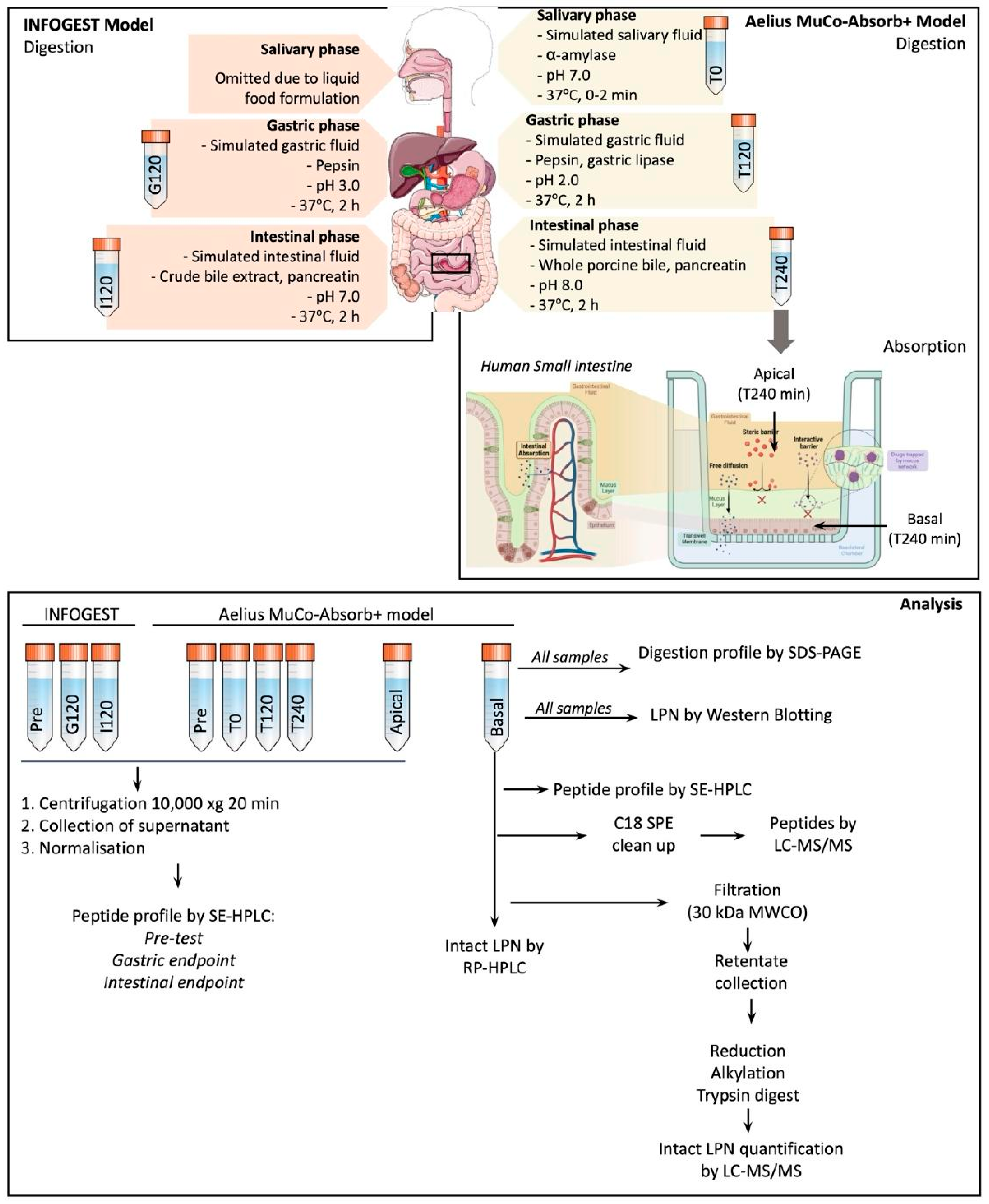
Figure 2.
Western blots of (A) matrix only, (B) bmLPN, (C) rhLPNLab, and (D) rhLPNPilot formulated in matrix during INFOGEST digestion, using monoclonal primary antibody MAB222P (BBI Solutions). Digestions were carried out using D2: 0.7344 mg/mL lactopontin (LPN). Samples were analysed under reducing conditions, and 1.2 μg protein equivalent was resolved per lane. Images are representative of triplicate digestion reactions. T0, pre-test sample prior to digestion; G30, G60, and G120, gastric digestion timepoints; I30, I60, and I120, intestinal digestion timepoints; Ladder, molecular weight marker; X, blot negative control. Panel A contained matrix-only digestion samples without added LPN and was included to assess non-specific antibody binding arising from matrix components or INFOGEST enzymes. Individual digestion components, including pepsin, pancreatin, and bile, were additionally loaded as controls. The final lane in Panel A contained purified bmLPN (1.2 μg) as a positive control. Panels B–D contained digestion reactions with bmLPN, rhLPNLab, or rhLPNPilot, respectively. The final lane in each panel contained the corresponding unformulated LPN standard (1.2 μg). The corresponding SDS-PAGE gels are shown in Supplementary Figure S1.
Figure 2.
Western blots of (A) matrix only, (B) bmLPN, (C) rhLPNLab, and (D) rhLPNPilot formulated in matrix during INFOGEST digestion, using monoclonal primary antibody MAB222P (BBI Solutions). Digestions were carried out using D2: 0.7344 mg/mL lactopontin (LPN). Samples were analysed under reducing conditions, and 1.2 μg protein equivalent was resolved per lane. Images are representative of triplicate digestion reactions. T0, pre-test sample prior to digestion; G30, G60, and G120, gastric digestion timepoints; I30, I60, and I120, intestinal digestion timepoints; Ladder, molecular weight marker; X, blot negative control. Panel A contained matrix-only digestion samples without added LPN and was included to assess non-specific antibody binding arising from matrix components or INFOGEST enzymes. Individual digestion components, including pepsin, pancreatin, and bile, were additionally loaded as controls. The final lane in Panel A contained purified bmLPN (1.2 μg) as a positive control. Panels B–D contained digestion reactions with bmLPN, rhLPNLab, or rhLPNPilot, respectively. The final lane in each panel contained the corresponding unformulated LPN standard (1.2 μg). The corresponding SDS-PAGE gels are shown in Supplementary Figure S1.
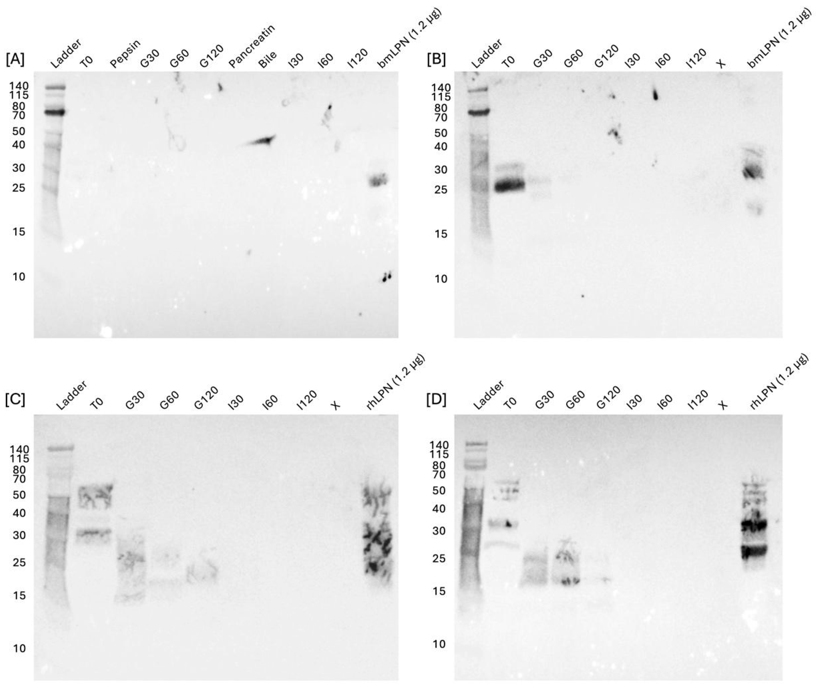
Figure 3.
Western blots of (A) bmLPN and (B) rhLPNPilot in matrix at D2: 0.7344 mg/mL during MuCo-Absorb+ modelling, using monoclonal primary antibody MAB222P (BBI Solutions). Samples were analysed under reducing conditions, and 0.52 μg protein equivalent was resolved per lane. Images are representative of triplicate digestion reactions. Pre-test samples included LPN formulated in a matrix prior to digestion. T0, salivary digestion; T120, endpoint of gastric digestion; T240, endpoint of intestinal digestion; apical r1 and r2, duplicate apical compartment samples collected after 4 h absorption modelling; Ladder, molecular weight marker; X, blot negative control (deionised water in loading buffer). The final lane contains the blot positive control consisting of purified LPN only in the loading buffer (without matrix).
Figure 3.
Western blots of (A) bmLPN and (B) rhLPNPilot in matrix at D2: 0.7344 mg/mL during MuCo-Absorb+ modelling, using monoclonal primary antibody MAB222P (BBI Solutions). Samples were analysed under reducing conditions, and 0.52 μg protein equivalent was resolved per lane. Images are representative of triplicate digestion reactions. Pre-test samples included LPN formulated in a matrix prior to digestion. T0, salivary digestion; T120, endpoint of gastric digestion; T240, endpoint of intestinal digestion; apical r1 and r2, duplicate apical compartment samples collected after 4 h absorption modelling; Ladder, molecular weight marker; X, blot negative control (deionised water in loading buffer). The final lane contains the blot positive control consisting of purified LPN only in the loading buffer (without matrix).
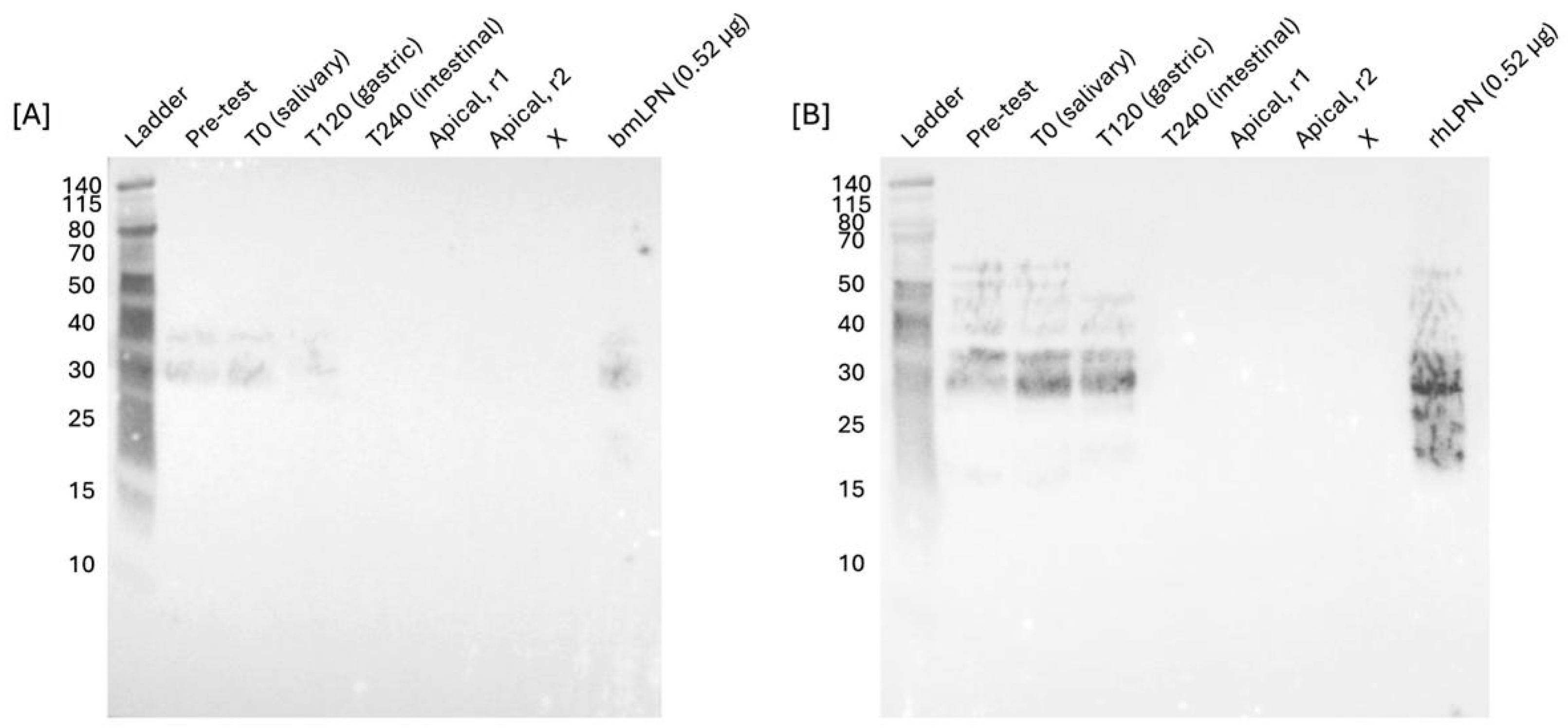
Figure 4.
(A) Peptide profiles following digestion with the MuCo-Absorb+ model, obtained using size-exclusion high-performance liquid chromatography (SE-HPLC) for matrix (blue), bmLPN (purple) and rhLPNPilot (teal) digesta, where LPN was included in formulations at D2: 0.7344 mg/mL. (B) Molecular weight distribution of peptides determined by SE-HPLC. Results are expressed as the percentage of the total chromatographic area under the curve (% total AUC) represented by peptide fractions of >5 kDa, 1–5 kDa, and <1 kDa and are shown as the mean of triplicate digestions with duplicate technical (injection) replicates (± SD). Molecular weights were estimated using a linear regression of log-transformed molecular weights of peptide standards against elution volume.
Figure 4.
(A) Peptide profiles following digestion with the MuCo-Absorb+ model, obtained using size-exclusion high-performance liquid chromatography (SE-HPLC) for matrix (blue), bmLPN (purple) and rhLPNPilot (teal) digesta, where LPN was included in formulations at D2: 0.7344 mg/mL. (B) Molecular weight distribution of peptides determined by SE-HPLC. Results are expressed as the percentage of the total chromatographic area under the curve (% total AUC) represented by peptide fractions of >5 kDa, 1–5 kDa, and <1 kDa and are shown as the mean of triplicate digestions with duplicate technical (injection) replicates (± SD). Molecular weights were estimated using a linear regression of log-transformed molecular weights of peptide standards against elution volume.
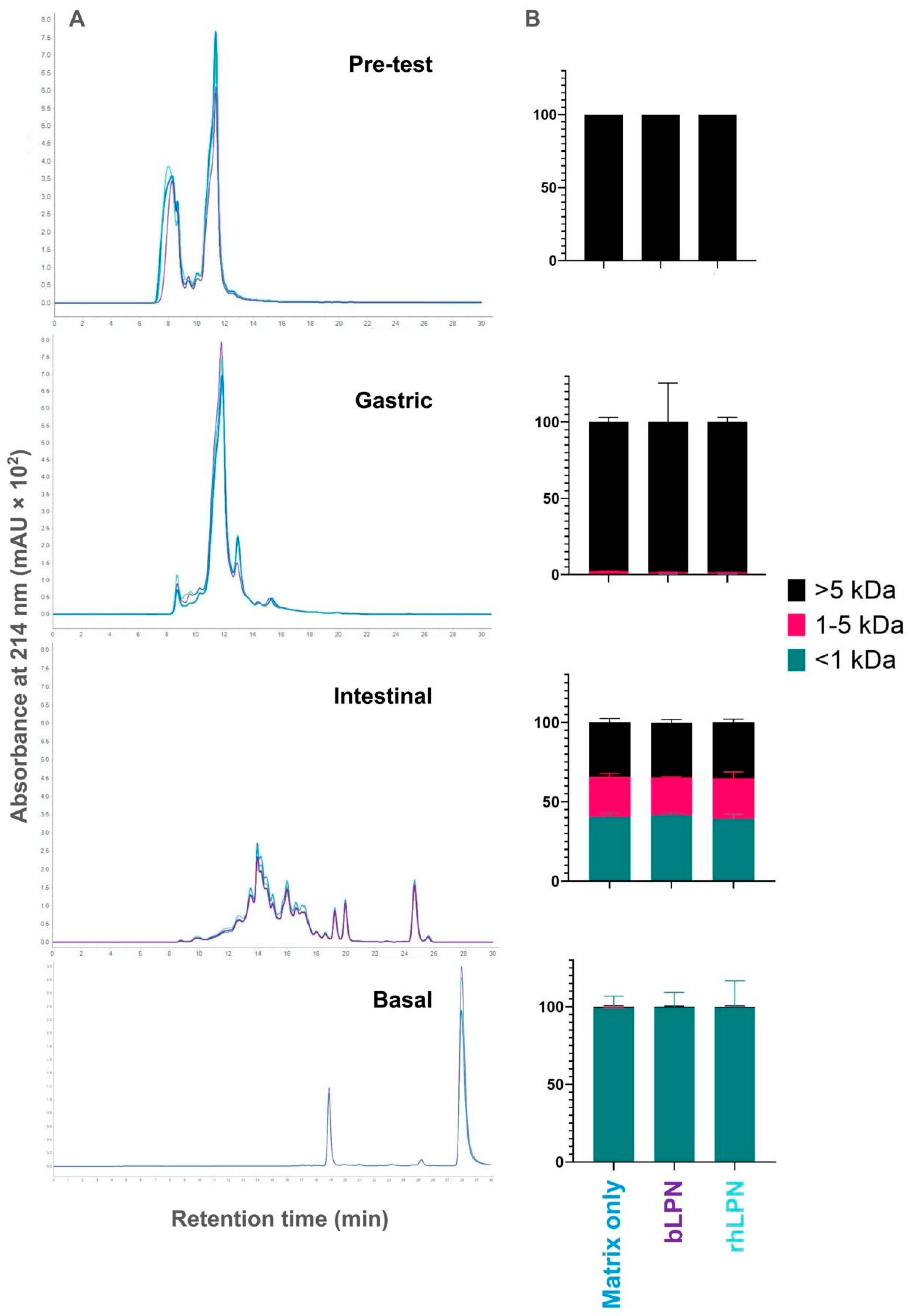
Figure 5.
(A) Representative peptide profiles obtained by SE-HPLC following INFOGEST digestion of the matrix (light orange), bmLPN (pink), and rhLPNPilot (dark orange), and following MuCo-Absorb+ model digestion of the matrix (blue), bmLPN (purple), and rhLPNPilot (teal). (B) Molecular weight distribution of peptides at the end of digestion determined by SE-HPLC. Results are expressed as the percentage of the total chromatographic area under the curve (% total AUC) represented by peptide fractions of >5 kDa, 1–5 kDa, and <1 kDa and are shown as the mean of triplicate digestions with duplicate technical (injection) replicates (± SD). Molecular weights were estimated using a linear regression of log-transformed molecular weights of peptide standards against elution volume.
Figure 5.
(A) Representative peptide profiles obtained by SE-HPLC following INFOGEST digestion of the matrix (light orange), bmLPN (pink), and rhLPNPilot (dark orange), and following MuCo-Absorb+ model digestion of the matrix (blue), bmLPN (purple), and rhLPNPilot (teal). (B) Molecular weight distribution of peptides at the end of digestion determined by SE-HPLC. Results are expressed as the percentage of the total chromatographic area under the curve (% total AUC) represented by peptide fractions of >5 kDa, 1–5 kDa, and <1 kDa and are shown as the mean of triplicate digestions with duplicate technical (injection) replicates (± SD). Molecular weights were estimated using a linear regression of log-transformed molecular weights of peptide standards against elution volume.
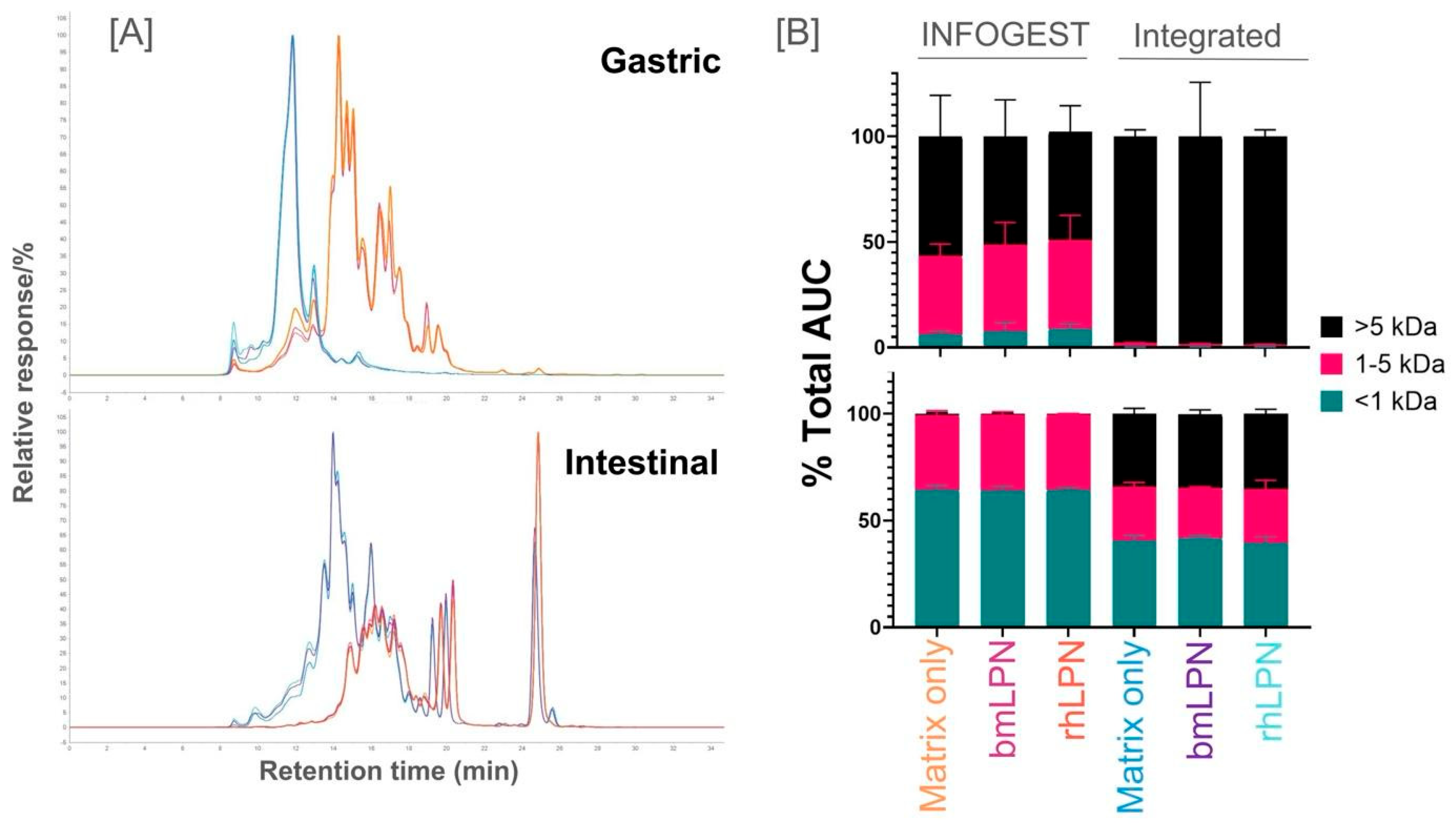
Figure 6.
The abundance of β-Lactoglobulin (BLG)-derived peptides detected in basolateral samples by LC-MS/MS following MuCo-Absorb+ modelling, as the mean of triplicate experiments ± SD. Letters of the compact letter display represent grouping based on one-way Brown-Forsythe and Welch ANOVA with Dunnett’s T3 test. Groups sharing the same letter are not significantly different, whereas groups with different letters are significantly different at α = 0.05.
Figure 6.
The abundance of β-Lactoglobulin (BLG)-derived peptides detected in basolateral samples by LC-MS/MS following MuCo-Absorb+ modelling, as the mean of triplicate experiments ± SD. Letters of the compact letter display represent grouping based on one-way Brown-Forsythe and Welch ANOVA with Dunnett’s T3 test. Groups sharing the same letter are not significantly different, whereas groups with different letters are significantly different at α = 0.05.
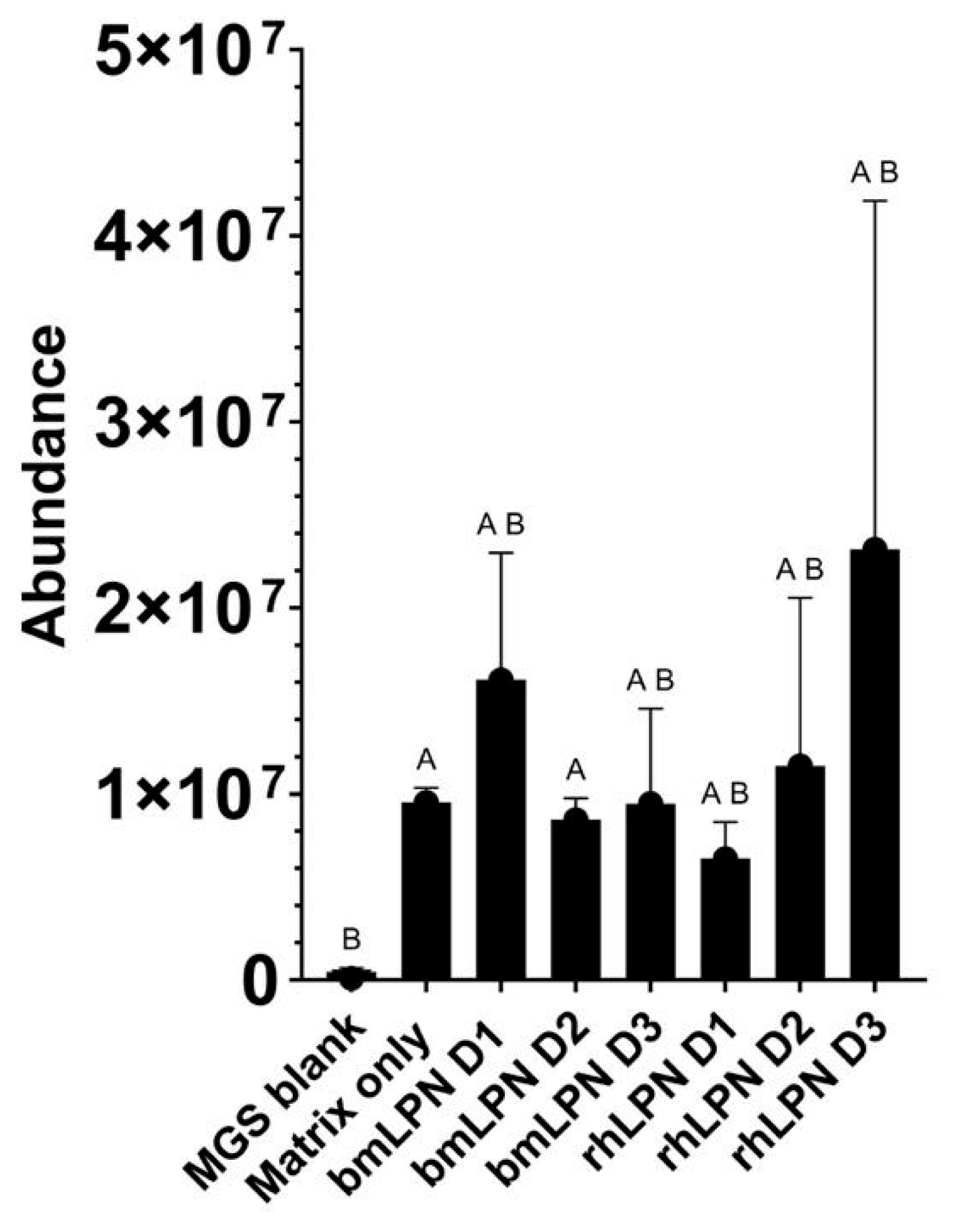
Figure 7.
(A) Effect of rhLPNPilot and bmLPN digestion products on Caco-2 cell viability following 4 h exposure to samples generated in the MuCo-Absorb+ model. Cell viability was assessed following exposure to digestion products at D1 (0.0864 mg/mL), D2 (0.7344 mg/mL), and D3 (3.672 mg/mL). No reduction in viability was observed for either protein source at any dose. Data are presented as the mean of triplicate digests applied to duplicate wells ± SD. Statistical analysis was performed using a linear mixed-effects model with treatment as a fixed effect and plate as a random effect. (B) Transepithelial electrical resistance (TEER) before and after 4 h exposure to digestion products of rhLPNPilot and bmLPN generated in the MuCo-Absorb+ model. No significant effects of protein source, dose, or their interaction were observed. Data are presented as the mean of triplicate digests applied to duplicate wells ± SD.
Figure 7.
(A) Effect of rhLPNPilot and bmLPN digestion products on Caco-2 cell viability following 4 h exposure to samples generated in the MuCo-Absorb+ model. Cell viability was assessed following exposure to digestion products at D1 (0.0864 mg/mL), D2 (0.7344 mg/mL), and D3 (3.672 mg/mL). No reduction in viability was observed for either protein source at any dose. Data are presented as the mean of triplicate digests applied to duplicate wells ± SD. Statistical analysis was performed using a linear mixed-effects model with treatment as a fixed effect and plate as a random effect. (B) Transepithelial electrical resistance (TEER) before and after 4 h exposure to digestion products of rhLPNPilot and bmLPN generated in the MuCo-Absorb+ model. No significant effects of protein source, dose, or their interaction were observed. Data are presented as the mean of triplicate digests applied to duplicate wells ± SD.
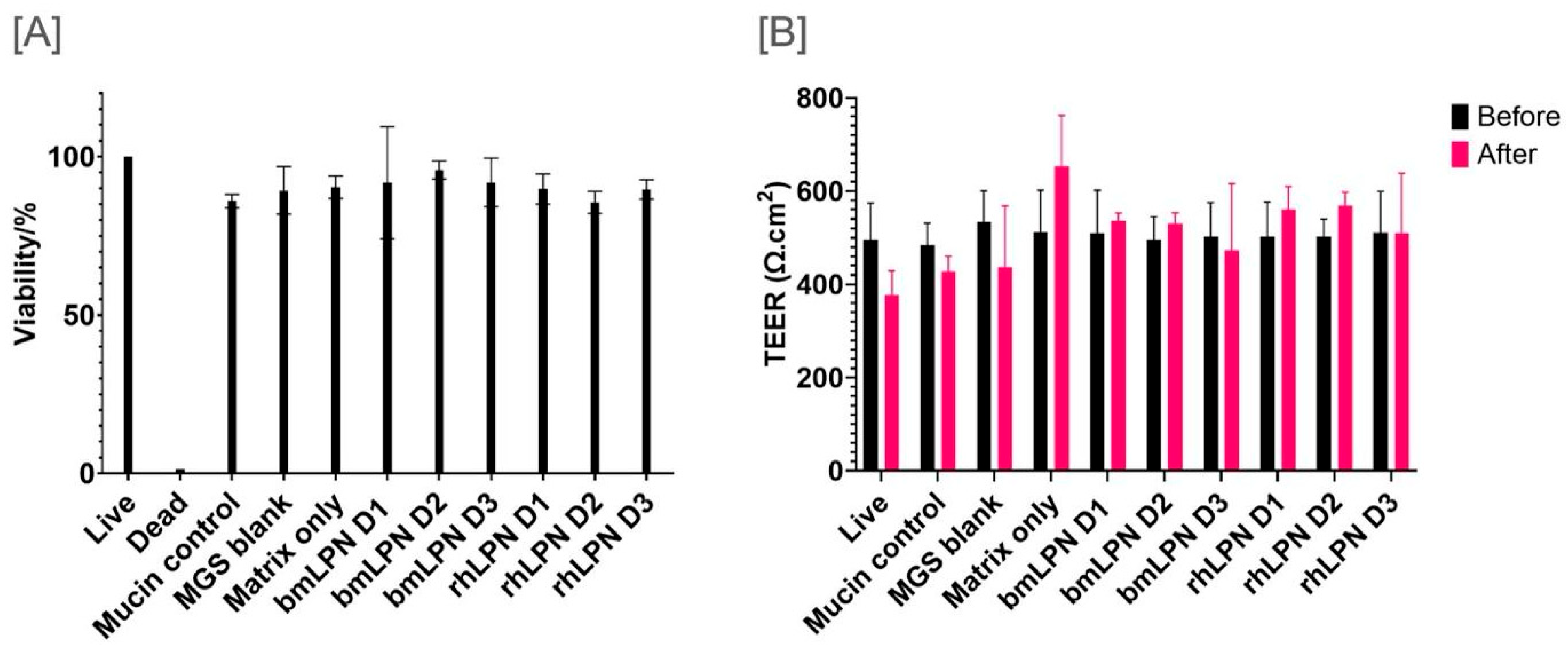

Table 1.
Proteins identified in basolateral samples from the Aelius MuCo-Absorb+ model by SPE-LC-MS/MS. Protein identifications were assigned by matching detected peptides to a custom subset of the SwissProt-reviewed database using Spectrum Mill MS Proteomics Software Rev BI.07.11.216 (Agilent). Confident protein identification was defined as detection of at least two unique peptides at <1.2% false discovery rate (FDR). The number of unique peptides is presented as the mean of triplicate experiments.
Table 1.
Proteins identified in basolateral samples from the Aelius MuCo-Absorb+ model by SPE-LC-MS/MS. Protein identifications were assigned by matching detected peptides to a custom subset of the SwissProt-reviewed database using Spectrum Mill MS Proteomics Software Rev BI.07.11.216 (Agilent). Confident protein identification was defined as detection of at least two unique peptides at <1.2% false discovery rate (FDR). The number of unique peptides is presented as the mean of triplicate experiments.
| Protein | Species | UniProt PAN | Number of unique peptides (mean of three replicates) | ||||||||
| MGS blank | Matrix | bmLPN | rhLPN | ||||||||
| D1 | D2 | D3 | D1 | D2 | D3 | ||||||
| Proteins attributed to the formulation: | |||||||||||
| Beta lactoglobulin | Bos taurus | P02754 | 11 | 26 | 29 | 27 | 32 | 27 | 28 | 27 | |
| Kappa casein | Bos taurus | P02668 | 4 | 12 | 9 | 10 | 10 | 11 | 12 | 8 | |
| Beta casein | Bos taurus | P02666 | 6 | 6 | 10 | 4 | 11 | 12 | 8 | 9 | |
| Alpha lactalbumin | Bos taurus | P00711 | 0 | 2 | 3 | 2 | 5 | 3 | 2 | 3 | |
| Glycosylation-dependent cell adhesion molecule 1 | Bos taurus | P80195 | 0 | 0 | 1 | 0 | 0 | 0 | 2 | 0 | |
| Proteins attributed to the digestion phase of the Aelius MuCo-Absorb+ model: | |||||||||||
| Pepsin A | Sus scrofa | P00791 | 7 | 5 | 9 | 3 | 1 | 5 | 6 | 3 | |
| Proteins attributed to the absorption phase of the Aelius MuCo-Absorb+ model: | |||||||||||
| Serum albumin | Bos taurus | P02769 | 7 | 9 | 1 | 0 | 0 | 3 | 1 | 1 | |
| Serrotransferrin | Bos taurus | Q29443 | 2 | 3 | 0 | 0 | 0 | 0 | 0 | 1 | |
| Alpha-2-HS-glycoprotein | Bos taurus | P12763 | 2 | 2 | 0 | 0 | 0 | 1 | 0 | 0 | |
| Alpha-1-antiproteinase | Bos taurus | P34955 | 1 | 2 | 0 | 0 | 0 | 0 | 0 | 1 | |
Disclaimer/Publisher’s Note: The statements, opinions and data contained in all publications are solely those of the individual author(s) and contributor(s) and not of MDPI and/or the editor(s). MDPI and/or the editor(s) disclaim responsibility for any injury to people or property resulting from any ideas, methods, instructions or products referred to in the content. |
© 2026 by the authors. Licensee MDPI, Basel, Switzerland. This article is an open access article distributed under the terms and conditions of the Creative Commons Attribution (CC BY) license (http://creativecommons.org/licenses/by/4.0/).
Copyright: This open access article is published under a Creative Commons CC BY 4.0 license, which permit the free download, distribution, and reuse, provided that the author and preprint are cited in any reuse.


