

Submitted:
06 June 2026
Posted:
08 June 2026
You are already at the latest version
Abstract
Environmental pollutants pose a serious threat to global health and are key contributors to mitochondrial dysfunction. Mitochondria play a crucial role in cellular bioenergetics, redox regulation, Ca²⁺ homeostasis, and cell death signaling. A growing body of evidence indicates that heavy metals and pesticides may directly and indirectly affect mitochondrial transport systems, ion channels, and membrane protein complexes. Impairment of the adenine nucleotide translocator, phosphate carrier, mitochondrial Ca²⁺ uniporter complex, voltage-dependent anion channel, and ATP synthase alters mitochondrial metabolite fluxes and redox balance, compromises mitochondrial Ca²⁺ homeostasis, and promotes oxidative stress, leading to mitochondrial dysfunction. These alterations converge on the opening of the mitochondrial permeability transition pore, leading to dissipation of the mitochondrial membrane potential, impaired oxidative phosphorylation, ATP depletion, mitochondrial swelling, release of pro-apoptotic factors, and cell death. This review summarizes the current understanding of the molecular mechanisms by which heavy metals and pesticides impair mitochondrial transport systems, leading to altered mitochondrial function and permeability transition, promoting the development of various human pathologies, including neurodegenerative and cardiovascular diseases, metabolic syndrome, inflammation, and cancer. Furthermore, emerging strategies targeting mitochondria and the regulation of the mitochondrial permeability transition pore are discussed as potential approaches to mitigate pollutant-induced mitochondrial dysfunction and permeability transition.
Keywords:
mitochondrial permeability transition pore
; mitochondrial dysfunction
; mitochondrial transporters
; mitochondrial toxicology
; environmental pollutants
; oxidative stress
; calcium signaling
; ATP synthase
; ANT
; MCU complex
1. Introduction
Mitochondria are central hubs of cellular metabolism and signaling, integrating bioenergetic processes with adaptive stress responses. Although historically recognized for their role in ATP production through oxidative phosphorylation (OXPHOS), mitochondria are now understood to act as multifunctional organelles that coordinate metabolic fluxes, redox balance, Ca2+ homeostasis, and signaling, in addition to being involved in innate immunity, cellular adaptation to environmental stress, and the determination of cellular fate [1,2]. A distinctive feature of mitochondrial physiology is the strict compartmentalization of metabolic reactions across mitochondrial membranes. The inner mitochondrial membrane is highly impermeable to most metabolites and ions, requiring specialized transport systems to regulate the exchange of substrates between the cytosol and the mitochondrial matrix. Transport processes are mainly mediated by the Mitochondrial Carrier Family, also known as the Solute Carrier Family 25 (SLC25), the largest group of mitochondrial membrane transporters in eukaryotic cells [3]. This family includes more than fifty carriers responsible for transporting nucleotides, tricarboxylic acid (TCA) cycle intermediates, amino acids, cofactors, and inorganic ions across the inner mitochondrial membrane. These carriers act as metabolic mediators that link mitochondrial bioenergetics with cytosolic pathways and cellular signaling networks; alterations in their transport activity can rapidly influence mitochondrial bioenergetics, redox balance, and cellular metabolism, leading to congenital human diseases [3,4,5,6].
Mitochondrial dysfunction can be induced by exposure to xenobiotics and is increasingly recognized as a key pathogenic mechanism in numerous chronic diseases, ranging from neurodegenerative and cardiovascular disorders to metabolic syndrome and cancer [7,8]. Human populations are continuously exposed to a wide range of environmental pollutants. Increasing evidence indicates that many toxicants can target mitochondria, impairing ATP production, Ca2+ signaling, and redox homeostasis [9,10]. As the primary intracellular generators of reactive oxygen species (ROS), mitochondria play a central role in toxicity-induced dysfunction, which exacerbates oxidative damage and triggers complex cellular stress signaling pathways [11].
Mitochondrial transport systems act as metabolic gatekeepers that translate environmental stress into bioenergetic responses. Through these fundamental molecular interfaces, toxicants can perturb metabolite flows, ionic homeostasis, and the signaling pathways that regulate mitochondrial permeability transition. Such alterations increase ROS production and alter the mitochondrial membrane potential (ΔΨm), which are central drivers of mitochondrial dysfunction in various human diseases [12,13]. In this context, a key event is the opening of a high-conductance channel known as the mitochondrial permeability transition pore (mPTP), which plays a crucial role in determining the cell’s fate [14,15]. The mPTP acts as a bioenergetic checkpoint that links mitochondrial stress to cell death pathways [16,17,18]. Although the precise molecular identity of the mPTP is still debated, numerous findings indicate that certain mitochondrial transport systems may be directly or indirectly involved in regulating the mitochondrial permeability transition [16]. Despite growing evidence linking environmental toxicants to mitochondrial dysfunction, the specific role of mitochondrial transport systems in mediating this toxicity remains incompletely understood. In particular, the molecular mechanisms through which environmental pollutants alter metabolite transport and promote permeability transition have only recently begun to be explored; elucidating them is pivotal, as mitochondrial transport systems function as early molecular sensors of metabolic stress, providing a critical link between environmental exposure, mitochondrial dysfunction, and disease pathogenesis.
This review focuses on the emerging role of certain mitochondrial transport systems as mediators of environmental toxicity. An overview is provided of their functional properties and how heavy metals and pesticides affect their activity, leading to bioenergetic dysregulation and mitochondrial dysfunction. Emphasis is placed on the biochemical mechanisms linking their impairment to mPTP activation and the mitochondrial permeability transition. The pathological implications of these processes are also examined, highlighting how compromised mitochondrial transport systems contribute to the onset of various human diseases. Finally, promising therapeutic strategies and future research directions to mitigate pollutant-induced mitochondrial dysfunction are discussed.
2. Transport Systems Likely Involved in mPTP Formation or Regulation
The mPTP is a multiprotein complex located in the inner mitochondrial membrane. It exhibits pleiotropic properties that depend on its conductance state, ranging from a low-conductance ion-selective channel to a high-conductance megapore permeable to solutes up to 1.5 kDa when activated. Its opening facilitates massive Ca2+ influx, causing mitochondrial osmotic swelling, loss of the electrical gradient across the inner mitochondrial membrane, and ΔΨm dissipation. These events impair cellular energy production, leading to bioenergetic collapse and the release of pro-apoptotic factors such as cytochrome c and apoptosis-inducing factor. Therefore, the mPTP is a critical regulator of mitochondrial-mediated cell death and is involved in numerous pathological conditions, including ischemia-reperfusion injury, neurodegenerative diseases, cancer, and toxicant-induced cellular damage [14,19,20]. A growing body of experimental evidence indicates that specific mitochondrial transport proteins—including the adenine nucleotide translocator (ANT), the phosphate carrier (PiC), the Ca2+ uniporter (MCU) complex, the voltage-dependent anion channel (VDAC), and ATP synthase dimers—may play key structural or regulatory roles in pore formation and/or activation [15,21,22,23]. Heavy metals and pesticides can alter the structure and function of these transport systems, thereby making mitochondria more susceptible to mPTP opening [10].
2.1. Adenine Nucleotide Translocator
ANT, also known as the ADP/ATP carrier, is one of the most abundant proteins in the inner mitochondrial membrane. It catalyzes the electrogenic exchange of cytosolic ADP for mitochondrial ATP, coupling OXPHOS to cellular energy requirements [24]. ANT operates via an alternating-access mechanism in which it undergoes structural transitions between two alternative conformations: the cytoplasmic-state (c-state, open toward the intermembrane space) and the matrix-state (m-state, open toward the mitochondrial matrix). According to this mechanism, with a single central substrate binding site, the binding of ADP (or ATP) triggers conformational changes between the two states [19]. Such changes allow for the sequential binding and release of ADP and ATP while preventing simultaneous exposure of the nucleotide binding site to both the mitochondrial matrix and the intermembrane space [25]. Beyond its canonical role in nucleotide exchange, this translocator has long been implicated in mPTP regulation. Pharmacological studies have demonstrated that ANT ligands, such as ADP or bongkrekic acid (BKA), stabilize the carrier in the m-state—a conformation that inhibits mPTP activation. Conversely, atractyloside and carboxyatractyloside (AT and CAT, respectively) lock the carrier in the c-state, which renders ANT highly susceptible to mPTP formation. The latter process is also promoted by Ca2+ overload, oxidative stress, and the action of certain pro-oxidants on specific ANT cysteines; these modifications induce intramolecular cross-linking on the matrix surface of the translocator, driving mPTP opening. Such findings indicate that ANT conformational changes may influence the structural transition from a nucleotide transporter to a non-selective, high-conductance pore [19]. Genetic studies have supported ANT involvement in the mitochondrial permeability transition. For instance, the deletion of multiple ANT isoforms in mouse models can significantly alter mitochondrial Ca2+ retention capacity and modulate susceptibility to the mitochondrial permeability transition [26]. These studies suggest that ANT may serve both as a structural component of the pore and as a key regulatory element that controls mPTP opening. ANT interacts with the mitochondrial matrix protein cyclophilin D (CypD), a peptidyl-prolyl isomerase that targets a conserved proline residue within a matrix loop of this translocator. CypD binds to ANT primarily when the latter is in the c state; this interaction lowers the pore opening threshold, promoting conformational changes that facilitate the formation of high-conductance channels under conditions of Ca2+ overload and oxidative stress [27].
ANT is tightly bound to cardiolipin, a phospholipid found almost exclusively in the inner mitochondrial membrane and rich in unsaturated fatty acids. Cardiolipin binding is necessary to stabilize the active dimeric structure of mitochondrial carriers, often capping the N-terminal ends of transmembrane a-helices [28]. This phospholipid maintains the active conformation of these transporters only when it is present in its intact form, containing four unsaturated acyl chains. Under conditions of ischemic stress or ROS accumulation, cardiolipin undergoes peroxidation that alters its molecular structure, resulting in a loss of affinity for mitochondrial carriers. Specifically, oxidation modifies its acyl chains, changing their shape from conical to a geometry that no longer fits the specific protein binding sites [29]. In general, the binding of cardiolipin to a carrier is based on interactions between the negative charges of the cardiolipin phosphate groups and the positive amino acid residues of the carrier. Cardiolipin oxidation can reduce charge density or alter the position of phosphate groups, thus weakening the bond, destabilizing protein conformation, and reducing transport activity. Furthermore, oxidized cardiolipin (CLox) translocates to the outer mitochondrial membrane, a process that destabilizes both mitochondrial carriers and respiratory supercomplexes; this translocation provides a signaling platform that lowers the threshold for Ca2+-induced mPTP opening. Specifically, CLox interacts with proteins such as CypD, VDAC, and Bcl-2 family members (Bax/Bak), facilitating the recruitment of factors that trigger pore opening, thereby promoting cytochrome c release and apoptotic cell death [30].
Recent findings have highlighted that ANT and CypD are two key players in the regulation of mPTP opening under pathological conditions like ischemia-reperfusion. A “two-pore” model for mPTP opening has been hypothesized, suggesting that while ANT and CypD are both integral to the process, they may operate through distinct but overlapping pathways. The simultaneous ablation of both components has been shown to completely abolish pore opening, confirming their synergistic role [31]. In vivo studies conducted on dystrophic mouse models have demonstrated that the deletion of the ANT1 gene (mainly expressed in oxidative tissues such as the heart and skeletal muscles), combined with that of the gene encoding CypD, provided nearly complete protection against necrosis, suggesting that the ANT/CypD complex acts synergistically in regulating mPTP opening [32]. Furthermore, CypD seems to play a dual role in managing mitochondrial damage via mPTP dynamics; specifically, it modulates intermittent mPTP opening to control Ca2+ overload, preserve mitochondrial integrity, and alleviate damage during hepatic ischemic stress [33].
Other studies have highlighted that ANT inhibition plays a dual role in regulating cell death, with its outcome depending heavily on the cellular context—particularly on the presence of severe lipotoxic stress [27]. Under basal conditions, generalized ANT inhibition (e.g., by CAT) is toxic because it blocks the essential exchange of adenine nucleotides, leading to bioenergetic failure and apoptosis. Conversely, under conditions of severe lipotoxicity, such as palmitate overload, specific conformational inhibition of ANT via BKA can prevent cell death. This protective mechanism is directly linked to the regulation of mPTP dynamics. Specifically, during lipotoxic stress, BKA locks the carrier in the m-state, a conformation that prevents pore opening, thereby reducing mitochondrial ROS production, preserving ΔΨm, and preventing cell death [27]. Consequently, while the indiscriminate blockade of nucleotide transport triggers toxicity, strategically targeting ANT conformations to prevent mPTP opening represents a promising therapeutic avenue to alleviate lipotoxic damage.
2.2. Mitochondrial Phosphate Carrier
The mitochondrial PiC mediates a proton-coupled transport of inorganic phosphate (Pi) from the cytosol to the mitochondrial matrix. Its role is essential for ATP synthesis, as Pi is required for ADP phosphorylation by the F1F0-ATP synthase complex [3]. PiC transport activity is also relevant for ensuring effective mitochondrial Ca2+ handling and is strongly influenced by the presence of cardiolipin [34]. PiC has long been hypothesized to be a component or regulator of the mPTP. In this regard, experimental studies have demonstrated that genetic deletion of SLC25A3 (the human gene encoding PiC) does not prevent mPTP opening, indicating that PiC is not a direct component of the pore complex. However, this deletion desensitizes the mPTP, as indicated by a significant decrease in mitochondrial susceptibility to Ca2+-induced permeability transition and an increased Ca2+-retention capacity, suggesting that PiC could regulate mPTP formation [35]. The influx of Pi can alter the ionic strength and osmolarity of the mitochondrial matrix. Since the mPTP is sensitive to volume and membrane potential, these alterations can promote matrix swelling and subsequent mitochondrial permeabilization. It is known that high Pi levels in the matrix lower the threshold required for mPTP opening; moreover, Pi synergizes with Ca2+ to promote mPTP opening via the formation of Ca2+-phosphate precipitates in the matrix. Under stressful conditions, such as during ischemia-reperfusion injury, this synergistic effect leads to persistent, irreversible mPTP activation [36]. Therefore, Pi could act as an indirect key inducer of the mPTP, likely interacting with regulatory components of the pore complex (including CypD) to stabilize conformational states that promote pore opening. Consistently, recent evidence indicates that the inducing effects of Pi are linked to the presence of CypD [19]. Inorganic polyphosphate (polyP) is a potent activator of the mPTP complex [37]. This effect seems to depend largely on the length of the polyP chain; specifically, a reduction in chain length decreases Ca2+-induced pore opening, as observed in murine cardiomyocytes [38]. PolyP has been proposed to act as an integral structural component of the mPTP channel; according to this model, Ca2+-induced mPTP activation is associated with the de novo assembly of a macromolecular complex composed of the c-subunit ring of ATP synthase, polyP, and polyhydroxybutyrate, with polyP modulating the selectivity and voltage dependence of the channel [39]. Beyond its mitochondrial role, polyP can modulate other ion channels involved in cellular Ca2+ homeostasis, including Transient Receptor Potential Melastatin 2 and Transient Receptor Potential Ankyrin 1 [40]. Both are transient receptor potential channels that serve as cellular sensors for various environmental and endogenous stimuli, including hazardous chemicals and altered redox status. Under oxidative stress, these channels mediate massive Ca2+ influx from the extracellular space into the cytoplasm, thereby dysregulating downstream signaling pathways and contributing to inflammation, cancer, neurodegeneration, and ischemia/reperfusion injury [41,42].
2.3. Mitochondrial Ca2+ Uniporter Complex
The MCU complex is a high-capacity channel located on the inner mitochondrial membrane that allows large amounts of cytosolic Ca2+—released from the ER or extracellular sources—to enter the mitochondrial matrix. The MCU core pore-forming subunit is regulated by several accessory proteins, including MICU1, MICU2, EMRE, and MCUb, which collectively control the sensitivity and gating properties of the channel [43,44]. This complex is essential for regulating mitochondrial Ca2+ uptake, metabolic coupling, and cell death signaling. Mitochondrial Ca2+ plays a fundamental role in metabolic regulation; matrix Ca2+ activates several enzymes of the TCA cycle, including pyruvate dehydrogenase, isocitrate dehydrogenase, and α-ketoglutarate dehydrogenase, thereby stimulating NADH production and enhancing OXPHOS [45]. However, excessive mitochondrial Ca2+ accumulation is one of the most powerful triggers of the mitochondrial permeability transition. MCU-dependent matrix Ca2+ overload stimulates mitochondrial respiration, resulting in increased ROS production; this phenomenon acts in a feed-forward loop, lowering the threshold for Ca2+-induced mPTP opening. In addition, elevated mitochondrial matrix Ca2+ promotes the interaction between CypD and mPTP components, leading to conformational changes that induce pore opening [46]. While CypD catalyzes mPTP opening, this process is significantly less efficient without Ca2+ influx through the MCU complex [23].
2.4. Voltage-Dependent Anion Channel
VDAC is the major pore-forming protein of the outer mitochondrial membrane, serving as the primary conduit for metabolite exchange. VDAC allows the diffusion of ATP, ADP, Pi, and other small metabolites, thus coordinating mitochondrial metabolism with cellular energy demands [47,48]. Structurally, it forms a β-barrel channel composed of 19 β-strands that create a pore capable of accommodating negatively charged metabolites. VDAC gating properties modulate mitochondrial permeability by switching between an open, high-conductance state and a closed, low-conductance state, depending on the transmembrane potential. In general, the channel is permeable to ATP/ADP and anions at low potential values. Conversely, at high potentials, VDAC is in a closed state characterized by low conductance (approximately under 50%) and is selective for cations, becoming virtually impermeable to polyanionic metabolites such as ATP, thereby limiting cellular energy production. Ca2+ flow is greater in the closed than in the open state; this phenomenon is essential for apoptotic signaling and mitochondrial Ca2+ homeostasis [49].
VDAC gating involves the N-terminal α-helix moving away from the pore wall to reorient toward the center, accompanied by a significant deformation of the β-barrel [50]. Gating is significantly influenced by lipid composition, with phosphatidylethanolamine and cardiolipin modulating gating asymmetry and affecting structural stability. A very recent study has demonstrated that while phosphatidylethanolamine enhances stability in human VDAC3, cardiolipin can promote an open-like state [51]. Metabolic state can modulate VDAC transition, since high NADH concentrations or specific ATP/ADP ratios can induce conformational changes, thus shifting the channel toward a closed state with reduced permeability to anionic respiratory substrates. Such metabolic modulation contributes to suppressing mitochondrial metabolism in cancer cells; under high glycolytic flux, free tubulin binds to and blocks VDAC pores, while hexokinase II recruitment to VDAC stabilizes the channel to facilitate direct ATP coupling for glycolysis [52].
VDAC has not been proposed as an obligatory structural component of the mPTP [16], even if it plays an important regulatory role in the mitochondrial permeability transition and apoptotic signaling. Consistently, it has been hypothesized that VDAC oligomerization may lead to the formation of a mega-pore that mediates the release of cytochrome c and other pro-apoptotic factors [53]. VDAC1 plays a key role in regulating mitochondrial Ca2+ homeostasis, being a critical component of the signaling machinery that links the ER to the mitochondria, i.e., the mitochondria-associated membranes (MAMs). At these junctions, VDAC interacts with the inositol-1,4,5-triphosphate receptor (IP3R) located on the ER via the glucose-regulated chaperone protein 75 (Grp75), allowing for a massive Ca2+ influx from the ER into the intermembrane space; subsequently, the cation can enter the mitochondrial matrix primarily through the MCU complex [54].
2.5. ATP Synthase
The F1F0-ATP synthase, or simply ATP synthase, is a multiprotein complex located primarily at the edges of the mitochondrial cristae and is capable of forming dimers or higher-order complexes, which contribute to the typical curvature of the cristae. One of the most debated hypotheses regarding the molecular identity of the mPTP concerns the direct involvement of ATP synthase and its dimers [15,55]. Specifically, ATP synthase is considered a leading candidate for directly forming or driving pore assembly, with a particular focus on its c-subunit ring or dimeric interface as the potential pore-forming components. Genetic and functional studies have suggested that the c-subunit ring acts as a channel that opens upon the dissociation of the F1 domain under stressful conditions [56]. CypD is capable of binding to the oligomycin-sensitivity conferring protein (OSCP) subunit of ATP synthase, inducing conformational changes that are transmitted to the F0 domain; this seems to support mPTP opening, especially under conditions of mitochondrial Ca2+ overload. At the catalytic site, when the natural cofactor Mg2+ is replaced by Ca2+, the protein complex could dissociate, forming the mPTP in the F0 domain [57]. According to other studies, CypD binding could lower the Ca2+ threshold required for mPTP opening, acting as a key modulator of the transition [19]. On this basis, under stressful conditions such as high Ca2+ levels and oxidative damage, conformational changes in ATP synthase could transform it from an energy-producing enzyme into a non-specific channel. However, other authors point out that the role of ATP synthase in this context remains a matter of debate, as available data vary depending on the species and the experimental systems used; moreover, alternative models suggest that ATP synthase may regulate the pore rather than form it directly [20].
Early reconstitution studies have shown that purified ATP synthase dimers embedded in lipid bilayers exhibit channel activity with properties typical of the mPTP, demonstrating that channel opening is Ca2+-dependent and pharmacologically regulated [58]. Specifically, conformational changes occurring in these dimeric complexes under conditions of high matrix Ca2+ levels and oxidative stress could generate a high-conductance pathway. According to this model, either the c-subunit ring of the F0 domain or the interface between ATP synthase monomers could constitute the structural basis of the pore [58,59]. Notably, experiments led on mitochondria lacking the c-subunit ring have revealed that while the classic mPTP cannot form, low-conductance channels sensitive to cyclosporine A (CsA) persist [60]. Furthermore, yeast mutant strains lacking the specific F1F0-ATP synthase dimerization subunits exhibit resistance to mPTP opening, supporting that dimerization is necessary for pore assembly [61]. Conversely, alternative models have suggested that the enzyme may instead act as a negative regulator of the mPTP by stabilizing the inner mitochondrial membrane. In this scenario, the loss of ATP synthase activity leads to increased susceptibility to pore formation and exacerbated necrotic damage, suggesting that the complex inhibits mPTP formation rather than promoting it [62]. Ultimately, despite this abundance of genetic and biochemical evidence, the molecular debate remains open.
3. Environmental Pollutants as Modulators of Mitochondrial Dysfunction
A growing body of research indicates that mitochondrial dysfunction constitutes a key mechanistic link between exposure to environmental pollutants and the pathogenesis of numerous chronic diseases, including neurodegenerative disorders, cardiovascular diseases, metabolic syndrome, and cancer [9,63]. Environmental pollutants can disrupt mitochondrial homeostasis through multiple converging mechanisms. Several toxicological studies have primarily focused on the inhibition of respiratory chain complexes, leading to increased oxidative stress, altered mitochondrial dynamics, damage to mitochondrial DNA (mtDNA), and mutations [10,63]. Recent investigations have highlighted the critical role of mitochondrial transport systems in mediating toxicant-induced mitochondrial dysfunction [64]. Perturbation of mitochondrial transport processes can impair ATP synthesis, alter mitochondrial redox balance, affect Ca2+ signaling, and increase mitochondrial susceptibility to permeability transition. Indeed, many toxicant-induced mitochondrial alterations converge on mPTP activation [14,19].
3.1. Heavy Metals Affect Mitochondrial Function and Ca2+ Homeostasis
Heavy metals represent one of the most extensively studied classes of environmental toxicants. Aluminum (Al), cadmium (Cd), mercury (Hg), arsenic (As), lead (Pb), chromium (Cr), and others are capable of accumulating within biological tissues and intracellular compartments, including mitochondria. Their bioaccumulation leads to oxidative stress and interferes with several signaling pathways, influencing cell metabolism, growth, proliferation, survival, and apoptosis, thereby promoting the onset of many human diseases, including pulmonary, gastrointestinal, renal, cardiovascular, reproductive, and neurodegenerative disorders, as well as several types of cancer [65].
Heavy metal-induced mitochondrial toxicity occurs through several mechanisms. Iron can induce ferroptosis, a form of non-apoptotic cell death characterized by extensive lipid peroxidation [66]. Cd and Pb can alter the balance between mitochondrial fusion and fission, resulting in mitochondrial dysfunction and energetic failure [67]. Certain heavy metals interfere with components of the mitochondrial electron transport chain, leading to electron leakage and increased ROS production, resulting in oxidative stress, mPTP opening, mitochondrial swelling, and apoptotic cell death [68]. In addition, heavy metals inhibit antioxidant defense systems, including intracellular glutathione, glutathione reductase, glutathione peroxidase, superoxide dismutase (SOD), catalase, and the thioredoxin pathways, thus exacerbating ROS-induced cellular damage [65]. Mitochondrial ROS overload causes oxidative damage to mtDNA, cardiolipin oxidation, as well as structural and functional alterations in mitochondrial proteins. Intact cardiolipin preserves the structure and activity of mitochondrial transporters located in the inner mitochondrial membrane; cardiolipin oxidation impairs the function of SLC25 members, promoting mitochondrial dysfunction and several diseases [69]. Furthermore, mitochondrial transporters contain cysteine residues that are highly susceptible to oxidative modifications, including S-glutathionylation and the formation of disulfide bridges; these alterations can disrupt transport activity and metabolic fluxes across the inner mitochondrial membrane [5].
Heavy metal exposure promotes mitochondrial Ca2+ overload through the overexpression of Nipsnap homolog 2, a positive regulator of L-type Ca2+ channels, resulting in uncontrolled mitochondrial Ca2+ influx [64]. Furthermore, heavy metals interfere with ER homeostasis, triggering mitochondrial Ca2+ accumulation. In particular, heavy metals activate phospholipase C at the plasma membrane, leading to the hydrolysis of phosphatidylinositol 4,5-bisphosphate and the production of diacylglycerol and IP3. The latter binds to IP3Rs on the ER, causing Ca2+ release and subsequent mitochondrial Ca2+ transfer through MAMs, with entry into the mitochondrial matrix occurring primarily via the MCU complex. In addition, at MAMs, ryanodine receptors located on the ER membrane can interact with VDAC to promote mitochondrial Ca2+ uptake [64].
Intracellular ROS overproduction and Ca2+ overload reinforce each other. Heavy metals such as As, Cr, Cd, Hg, and Pb promote lipid peroxidation and increase plasma membrane permeability, facilitating Ca2+ influx [70]. Because of their high affinity for thiol groups, these metals inhibit Na+/K+-ATPase activity, leading to intracellular Na+ accumulation, altered Na+/Ca2+ exchange activity, and a rise in cytosolic Ca2+ levels that in turn stimulate mitochondrial Ca2+ uptake [64,71]. Intracellular Ca2+ overload activates phospholipase A2, resulting in enhanced arachidonic acid generation. This compound promotes Ca2+ channel opening and further Ca2+ influx, while also activating protein kinase C and NADPH oxidase, thereby amplifying ROS production [64]. Arachidonic acid impairs mitochondrial respiratory chain function, disrupts the cytochrome oxidase system, and compromises electron transport, leading to the generation of superoxide anions and hydrogen peroxide, triggering Ca2+-dependent apoptosis through the mitochondrial pathway [64,72]. Furthermore, arachidonic acid contributes to oxidative stress via cyclooxygenase and lipoxygenase pathways, exacerbating mitochondrial oxidative damage [64].
Mitochondrial Ca2+ homeostasis is a critical determinant of metabolic activity and permeability transition sensitivity. Elevated cytosolic Ca2+ concentrations stimulate Ca2+ uptake into the mitochondrial matrix primarily through the MCU complex. Within the mitochondrial matrix, the presence of Ca2+ activates several dehydrogenases of the TCA cycle, thereby stimulating mitochondrial metabolism [73]. However, matrix Ca2+ overload leads to mitochondrial dysfunction, promotes ROS overproduction, and induces mPTP opening, resulting in the release of cytochrome c and mtDNA; these events activate pro-inflammatory pathways and trigger apoptosis or necrosis [74]. In this context, recent studies have shown that the deletion of Transmembrane Protein 65, a mitochondrial Na+/Ca2+ exchanger that mediates Ca2+ efflux from the mitochondrial matrix to the cytoplasm, results in mitochondrial Ca2+ overload, which in turn promotes the mitochondrial permeability transition [75].
3.1.1. Effects of Heavy Metals on ANT
ANT is highly susceptible to modulation by heavy metals due to its strategic localization and its abundance of reactive cysteine residues. ANT inhibition can occur via thiol binding or through metal-induced oxidative modifications, which can impair ATP export, leading to bioenergetic collapse and increased susceptibility to permeability transition. In this regard, ANT modifications promote a non-specific, high-conductance pore-like conformation linked to the mPTP, which results in mitochondrial swelling, ΔΨm dissipation, release of cytochrome c, and subsequent apoptosis [68,76,77,78].
Based on recent literature data, As, Hg, Cd, Pb, and others can act as direct or indirect ANT inhibitors, interfering with nucleotide exchange processes [68]. Certain thiol reagents and various heavy metals (Cd, Hg, As, and Pb) have a strong affinity for the thiol groups of this transporter, leading to thiol oxidation, structural alterations, and impaired transport activity [79]. Hg, methylmercury, and Cu are known to interact with the thiol groups of specific ANT cysteine residues, acting as potent inhibitors of the carrier, activating mPTP opening and inducing cell death [80,81]. Arsenic compounds can interfere with ANT function indirectly by binding to lipoic acid-containing enzymes and depleting cellular thiols such as glutathione, thereby enhancing oxidative stress and promoting ANT oxidation and dysfunction [82]. Cadmium has been suggested to induce mPTP opening, causing a loss of ΔΨm and mitochondrial dysfunction. In particular, pre-incubation with protective compounds containing thiol groups can partially reverse cadmium-induced effects, suggesting that the damage specifically targets protein thiol groups [83]. Notably, some authors have shown that BKA, a specific ANT inhibitor, can completely inhibit cadmium-induced mPTP opening and loss of ΔΨm, indicating that this transporter acts as a key mediator in the process [84]. Heavy metal-induced ROS overproduction can also oxidize surrounding phospholipids (e.g., cardiolipin), destabilizing the local membrane environment and compromising the structural integrity and functional cycle of this transporter [29,85]. Heavy metals can also enhance mPTP opening via Ca2+ overload [86], which facilitates the interaction between ANT and CypD. It is known that CsA forms a CsA-CypD complex that prevents CypD from inducing pore opening in response to elevated Ca2+ levels; some studies have demonstrated that CsA fails to prevent permeability transition and cadmium-triggered apoptosis, highlighting that cadmium-induced mPTP opening depends on a direct interaction with ANT [84].
From a medical perspective, ANT inhibition by heavy metals causes severe damage to energy-intensive tissues such as the liver, heart, and kidneys, contributing to the development of hepatic and cardiac dysfunction, as well as chronic renal disease [87]. Neurons also require a large amount of energy; therefore, metal-induced ATP depletion leads to neuronal death, contributing to the pathogenesis of various neurological disorders, including Alzheimer’s, Parkinson’s, and Huntington’s disease, as well as amyotrophic lateral sclerosis [77].
3.1.2. Effects of Heavy Metals on PiC
Heavy metals can target mammalian PiCs through different mechanisms, including molecular mimicry, binding to thiol groups, disruption of the proton gradient across the inner mitochondrial membrane, and oxidative damage, thus causing mitochondrial dysfunction and cell death [88]. Some heavy metals (particularly metalloids) act as structural analogs of Pi. Specifically, vanadate and arsenate competitively inhibit mitochondrial Pi transport by competing for the Pi-binding sites on PiC, thereby preventing efficient Pi uptake into the mitochondrial matrix [89]. In mitochondria, arsenate can replace Pi during ATP synthesis, forming ADP-arsenate, which is unstable and hydrolyzes, leading to decreased ATP levels.
Eukaryotic PiCs have essential cysteine residues with reactive thiol groups that can be covalently bound by As, Hg, Pb, and Cd. These bonds impair the ability of the carrier to switch between the different conformations required for the transport cycle; indeed, Hg, several thiol-reactive compounds, and, to a lesser extent, As have been shown to strongly inhibit PiC transport activity [34,90].
Pi transport in mammals is often coupled with the movement of other ions. Since heavy metals disrupt ion transport, interfere with membrane integrity, and dissipate electrochemical gradients [88,91], carriers that rely on co-transport mechanisms, like PiC, cannot function properly. Recent research has also established that mammalian PiC does not exclusively transport phosphate, but acts as a crucial importer of copper into the mitochondrial matrix, required for the assembly of cytochrome c oxidase, a terminal enzyme in the electron transport chain [92]. Heavy metals can compete for or disrupt copper-binding ability; indeed, arsenate can inhibit copper uptake, interfering with the transport mechanism, thus leading to mitochondrial copper deficiency, impaired cytochrome c oxidase activity, and subsequent respiratory failure [93].
Heavy metal-induced ROS cause oxidative modifications such as S-glutathionylation, sulfenylation, and carbonylation of PiC, further compromising its structural integrity and transport efficiency [85]. Arsenic compounds can exacerbate PiC dysfunction indirectly through the depletion of intracellular glutathione and by binding to vicinal thiol groups, thereby amplifying oxidative stress and enhancing PiC susceptibility to redox-dependent inactivation [82]. Furthermore, heavy metal-induced lipid peroxidation (especially of cardiolipin) alters the physicochemical environment required for PiC activity [29]. A critical consequence of PiC deficiency is the disruption of mitochondrial Pi availability, which limits ATP synthase activity and leads to bioenergetic failure, thereby promoting ΔΨm dissipation and increasing susceptibility to mPTP opening under conditions of elevated Ca2+ and oxidative stress, ultimately triggering cell death pathways [85,94].
3.1.3. Effects of Heavy Metals on the MCU Complex
The MCU complex is a critical molecular target of heavy metals, due to its finely tuned dependence on electrochemical gradients and redox-sensitive regulatory mechanisms [95]. Heavy metals can directly interact with thiol-containing residues, redox-sensitive domains, and regulatory subunits of this complex, leading to conformational changes that impair MICU1/MICU2 gatekeeping function and increase the probability of pathological channel opening under low Ca2+ conditions [44]. Hg and Cd, due to their strong thiophilicity, can bind to cysteine residues within MCU-associated proteins or alter the redox state of the mitochondrial matrix, thereby disrupting the tight coupling between mitochondrial ΔΨm and Ca2+ uptake, leading to uncontrolled Ca2+ influx and mitochondrial depolarization [64]. Cd, Hg, and Pb disrupt MCU-mediated Ca2+ homeostasis primarily by elevating cytosolic Ca2+ levels through membrane damage and interference with Ca2+ transport systems, thereby indirectly enhancing mitochondrial Ca2+ uptake via MCU and promoting mitochondrial Ca2+ overload [64]. In this regard, Cd has been shown to activate the IP3R-MCU signaling axis, resulting in mitochondrial Ca2+ accumulation and apoptosis [96]. Heavy metal-induced ROS can modify components of the MCU complex, alter channel kinetics, and enhance mitochondrial Ca2+ uptake, thus establishing a vicious cycle between Ca2+ overload and ROS generation [97]. Indeed, excessive MCU-mediated Ca2+ uptake leads to electron leakage from the mitochondrial respiratory chain and mitochondrial ROS overproduction, resulting in damage to mitochondrial proteins, lipids, and DNA, with subsequent mitochondrial dysfunction [98]. Heavy metal-induced mitochondrial Ca2+ overload via MCU is a key trigger of downstream pathological events, including mPTP activation, ΔΨm dissipation, release of pro-apoptotic factors, and initiation of apoptotic or ferroptotic cell death pathways, all of which are closely linked to increased ROS levels and bioenergetic collapse [99].
3.1.4. Effects of Heavy Metals on VDAC
VDAC represents a critical interface through which environmental toxicants can perturb mitochondrial homeostasis and cellular fate [100]. Heavy metals can exhibit severe toxicity partly due to their high affinity for thiol groups, as well as through their ability to bind to specific VDAC histidine residues; this event promotes metal-catalyzed oxidation leading to protein carbonylation, structural destabilization, and dysregulation of channel gating, thereby compromising ion selectivity and mitochondrial permeability [101]. The ability of toxic metals to mimic physiologically relevant ions enables their aberrant transport through mitochondrial pathways, potentially involving VDAC, thus facilitating their bioaccumulation [102].
Heavy metal toxicity can also impact VDAC by inducing oxidative stress, either through direct ROS generation via redox cycling (e.g., iron, copper, chromium) or by disabling antioxidant defenses. Oxidative modifications of VDAC and associated proteins can shift the carrier from its physiologically open, anion-selective state, to partially closed conformations that promote cation flux, particularly Ca2+, further amplifying mitochondrial Ca2+ overload, a key trigger of mPTP opening and apoptotic signaling cascades [64]. Arsenic trioxide provides a paradigmatic example of metal-mediated VDAC targeting, as it induces VDAC-dependent mPTP opening, inducing cytochrome c release and caspase activation, with subsequent apoptotic cell death [103]. Similarly, Cr6+ has been shown to upregulate VDAC1 expression and disrupt intracellular Ca2+ homeostasis, indicating that some heavy metals can modulate not only VDAC function but also its transcription and translation, enhancing mitochondrial vulnerability to toxic insults [104].
Heavy metals also interfere with protein-protein interactions involving VDAC, such as its association with hexokinase and Bcl-2 family proteins, thereby disrupting the fine balance between pro-survival metabolic coupling and pro-apoptotic signaling on the mitochondrial surface [105]. Given the key role of VDAC in MAMs, several authors have demonstrated that the formation and stability of the IP3R-Grp75-VDAC complex are promoted by environmental toxicants, leading to mitochondrial Ca2+ overload and ROS overproduction [106,107]. Heavy metals and pesticides cause oxidative damage and ER stress. Cellular stress often leads to overexpression of Grp75 or increased proximity between the ER and mitochondria, physically stabilizing the IP3R-Grp75-VDAC bridge to respond to energy demands or cell death signaling [108,109]. In this regard, Cd has been shown to induce neurotoxicity by increasing the interaction between IP3R and VDAC1 via Grp75 in neuronal cells, resulting in a massive influx of mitochondrial Ca2+ and subsequent apoptosis [110].
3.1.5. Effects of Heavy Metals on ATP Synthase
Heavy metals act through converging mechanisms that include direct coordination with amino acid residues of ATP synthase, perturbation of membrane lipid-protein interactions, and induction of oxidative stress, ultimately resulting in defective ATP production and mitochondrial dysfunction [64]. The susceptibility of ATP synthase to heavy metals like Cd and Hg is largely driven by their high affinity for thiol groups. Notably, thiol groups are critical for redox regulation of ATP synthase activity and structural stabilization; moreover, the thiol group of cysteine at position 141 in the OSCP subunit appears to be involved in mPTP regulation [111]. Other studies have shown that cross-linking of vicinal dithiols in ATP synthase using reagents that link cysteine residues can have opposite effects on mPTP modulation [112].
Cd has been found to reduce ATP synthesis by disrupting the electron transport chain, causing oxidative stress and a loss of ΔΨm [113]. Other authors have found that Cd can inhibit ATP synthase activity, reduce ATP production, and enhance oxidative stress, with subsequent mitochondrial damage and apoptotic cell death [114].
Similarly, Hg covalently binds to thiol groups in mitochondrial respiratory chain complexes and ATP synthase, impairing mitochondrial function [115]. Furthermore, Hg can reduce the expression level of subunits 6 and 8 of human ATP synthase [116].
Arsenate mimics the γ phosphate of ATP, competing with Pi at the active site of the enzyme, thus inhibiting ATP synthesis and halting cellular energy production [117].
Toxic hydrophobic organotin compounds can enter the ion channel of ATP synthase, blocking the proton flow and preventing ATP synthesis [118].
Notably, other metals targeting ATP synthase can be used to treat certain human diseases. This is the case with Cr3+, which accumulates in the mitochondria and binds to the beta subunit of ATP synthase, inhibiting its activity; thereby increasing the AMP/ATP ratio, activating AMP-activated protein kinase, and improving glucose metabolism [119].
Another toxic mechanism involves disruption of the lipid microenvironment, particularly cardiolipin, which is tightly associated with ATP synthase and supports the structural integrity of its components [120]. Cardiolipin oxidation or displacement is frequently observed under metal-induced oxidative stress; such modifications can alter proton channel architecture and compromise proton flux through ATP synthase.
Heavy metals also interfere with the supramolecular organization of ATP synthase. Structural studies have demonstrated that ATP synthase dimers induce membrane curvature and contribute to cristae morphology and the proper function of the complex; moreover, cardiolipin promotes ATP synthase dimerization [57]. Heavy metal-induced oxidative damage and lipid peroxidation are likely to destabilize dimer interfaces, impairing cristae organization. Loss of dimer integrity disrupts the spatial organization of proton flux and may promote conformational plasticity of the complex. Increasing evidence suggests that, under pathological conditions, such as Ca2+ overload or oxidative stress, ATP synthase can undergo structural rearrangements that contribute to the formation of a high-conductance channel resembling the mPTP [15,16,57].
3.2. Pesticides Inhibit Mitochondrial Respiratory Chain Activity and Interfere with Mitochondrial Fluxes
Pesticides represent another major class of environmental toxicants. They induce oxidative stress by the generation of ROS and reactive nitrogen species (RNS), which have numerous harmful effects on human health, causing neurodegenerative disorders, cardiovascular diseases, endocrine imbalances, renal damage, skeletal problems, respiratory disorders, and reproductive system dysfunction [121]. Oxidative stress is a critical driver of mitochondrial dysfunction and can trigger different signaling pathways, including RNS signaling, Keap1/Nrf2/ARE, TNFR1/TNF-α, MAPKs, NF-κB, and mitochondrial apoptosis pathways [122].
Pesticides cause the deterioration of mitochondrial transport systems primarily through indirect mechanisms, rather than through specific direct interactions. The processes involved include the inhibition of electron flow through the mitochondrial electron transport chain, oxidative damage, and membrane-dependent mechanisms. Inhibition of mitochondrial respiration alters the proton gradient across the inner mitochondrial membrane and impairs ΔΨm. Since the activity of ANT, PiC, the MCU complex, and ATP synthase is driven by the electrochemical gradient, such alterations can disrupt metabolite fluxes and reduce ATP synthesis. Furthermore, the loss of ΔΨm can alter the gating properties of VDAC [123].
Several pesticides exert their toxic effects at the level of the electron transport chain. Rotenone, a broad-spectrum botanical insecticide, pesticide, and piscicide, is one of the best-characterized mitochondrial toxicants. It acts as a potent inhibitor of complex I, blocking electron transfer from NADH to ubiquinone, thus promoting electron leakage and superoxide production within mitochondria; this results in oxidative damage to mitochondrial proteins and lipids [124]. Notably, at the brain level, rotenone seems to decrease ischemia-induced injury by inhibiting the mitochondrial permeability transition [125]. Conversely, more recent studies have demonstrated that rotenone activates the NLR family pyrin domain-containing 3 inflammasome through mPTP opening and mtDNA-mediated cGAS–STING signaling [126].
Maneb is a manganese-containing dithiocarbamate fungicide extensively used in agriculture to control fungal diseases. It inhibits complex III, disrupting the mitochondrial proton gradient, which leads to reduced ATP production, ROS overproduction, and mitochondrial apoptosis [127].
The herbicide paraquat undergoes continuous cycles of reduction and reoxidation in complexes I and III, generating large amounts of superoxide radicals [128]. Paraquat-generated ROS can oxidize critical functional groups within mitochondrial transport systems, increasing mPTP sensitivity to Ca2+ modulation and promoting its opening with subsequent cell death [129]. Co-exposure to paraquat and maneb has been shown to synergistically exacerbate oxidative stress and mitochondrial dysfunction in neuroblastoma cells [130]. Paraquat can also induce mitochondrial toxicity by binding directly to VDAC1; this interaction triggers massive superoxide anion production, resulting in lipid peroxidation, compromised membrane fluidity, mitochondrial membrane rupture, and cell death [131].
Roundup, a glyphosate-based herbicide, has been found to depress mitochondrial respiration by partially inhibiting complexes II and III, while OXPHOS can be affected by both a direct and an indirect effect on the ATPase activity, ultimately disrupting mitochondrial membrane integrity [132]. Roundup has recently been found to increase hydrogen peroxide production, leading to inhibited mitochondrial respiration, depolarization of mitochondrial membrane potential, and mitochondrial swelling [133].
Organophosphates and carbamates induce oxidative damage, impairing mitochondrial function; in fact, ROS generated upon pesticide exposure can promote carbonylation and oxidative degradation of mitochondrial transport proteins [134].
ROS overproduction contributes to mtDNA damage and lipid peroxidation; the latter can further destabilize the functional conformation of mitochondrial carriers, impairing transport processes. In this regard, atrazine exposure has been demonstrated to induce ROS overproduction with subsequent cardiolipin oxidation and mitochondrial dysfunction [135]. In addition, chlorpyrifos, a widely used broad-spectrum organophosphate pesticide, has been reported to impair mitochondrial function via increasing ROS generation, decreasing ΔΨm, and inducing lipid peroxidation with subsequent mitochondrial dysfunction [136].
Similar to heavy metals, pesticides can also induce ER stress and modulate ER-mitochondria contact sites and cytosolic Ca2+ levels, altering mitochondrial Ca2+ signaling. In this regard, cypermethrin, a synthetic pyrethroid broad-spectrum pesticide, has been found to induce mitochondrial Ca2+overload and apoptosis in Sertoli cells through enhanced ER-mitochondria coupling via the IP3R1-GRP75-VDAC1 complex [109]. Paraquat has been found to induce ER stress and mitochondrial Ca2+ overload through redox-driven oxidative stress [137]. ER stress increases Ca2+ release and upregulates MCU expression [138], thereby leading to mitochondrial dysfunction and mPTP opening. Rotenone indirectly enhances MCU activity by disrupting the interaction between the complex I and MCU, which halts the physiological degradation of the MCU protein. This post-translational stabilization leads to an accumulation of functional MCU channels on the inner mitochondrial membrane, increasing mitochondrial Ca2+ uptake [139].
ATP synthase is a critical target of pesticide-induced mitochondrial toxicity. Organotin compounds, historically used as biocides and pesticides, are well-known inhibitors of the F0 domain, where they disrupt proton translocation by interacting with hydrophobic regions of the membrane-embedded domain [140]. This kind of interaction collapses the proton motive force and inhibits ATP synthesis [117]. Paraquat can impair ATP synthase activity indirectly by generating ROS that oxidatively modify key subunits, including the redox-sensitive OSCP subunit. Conformational and oxidative alterations of OSCP may promote the transition of ATP synthase to a pore-forming structure associated with the mPTP [141]. In particular, the oxidation of cysteine at position 141 in OSCP seems to facilitate mPTP opening [111]. Emerging evidence also suggests that under severe oxidative or metabolic stress conditions, including those induced by pesticides, ATP synthase may undergo conformational rearrangements contributing to the formation of high-conductance channels resembling the mPTP [55,142]. In addition, pesticide-induced lipid peroxidation disrupts cardiolipin interactions required for ATP synthase dimer stability, leading to cristae disorganization and reduced bioenergetic efficiency [120,143].
4. Therapeutic Strategies Targeting Mitochondria and Permeability Transition
Given the central role of mitochondrial dysfunction in cellular damage, considerable research has focused on therapeutic strategies to preserve mitochondrial integrity and prevent the mitochondrial permeability transition. Heavy metals and pesticides generally converge on a limited number of mitochondrial stress pathways, including oxidative stress, Ca2+ overload, disruption of mitochondrial bioenergetics, as well as destabilization of mitochondrial membranes and transport systems, all of which collectively promote mPTP opening. Consequently, interventions targeting mitochondrial transport systems and regulators of mitochondrial permeability transition have emerged as promising therapeutic approaches [19]. Recent advances in mitochondrial pharmacology have highlighted several strategies to improve mitochondrial function, including CypD inhibitors, modulators of mitochondrial Ca2+ transport, mitochondria-targeted antioxidants, redox-based approaches, VDAC targeting, and emerging nanoparticle-based delivery systems. These interventions aim to mitigate oxidative damage, stabilize ΔΨm, restore metabolic fluxes, and prevent the bioenergetic collapse that often precedes cell death [144].
4.1. CypD Inhibition and Control of mPTP Opening
CypD plays a critical role in regulating mPTP opening. CypD interacts with several inner mitochondrial membrane proteins, thereby influencing the conformational state of the pore-forming complex [144]. Under conditions of mitochondrial stress, such as Ca2+ overload, oxidative damage, or ATP depletion, CypD undergoes post-translational modifications, including acetylation, phosphorylation, and oxidation, which enhance its binding to the inner mitochondrial membrane and promote mPTP opening. This interaction facilitates conformational rearrangements of ATP synthase dimers and other mitochondrial membrane proteins, leading to the formation of a high-conductance pore that dissipates ΔΨm and causes osmotic swelling of the mitochondrial matrix [14].
CypD inhibition represents one of the most widely studied strategies for preventing permeability transition, since this phenomenon is associated with the development of severe human diseases, including ischemia-reperfusion injury and neurodegeneration [145]. The immunosuppressant drug CsA was one of the first compounds shown to inhibit mPTP opening by binding to the active site of CypD and preventing permeability transition under stress conditions [16]. Experimental models have demonstrated that genetic deletion or pharmacological inhibition of CypD confers resistance to permeability transition induced by oxidative stress and Ca2+ overload [146]. However, the clinical use of CsA for mitochondrial protection is limited by its immunosuppressive properties [147]. Consequently, recent research has focused on developing non-immunosuppressive cyclophilin inhibitors, including sanglifehrin derivatives and other small molecules targeting the CypD catalytic pocket [145]. These compounds can stabilize ΔΨm, reduce mitochondrial swelling, and prevent the release of pro-apoptotic factors.
4.2. Targeting Mitochondrial Ca2+ Transport
Mitochondrial Ca2+ overload promotes ROS generation, increases matrix osmotic pressure, and triggers mPTP opening, playing a critical role in several pathological processes [148]. Therefore, modulation of mitochondrial Ca2+ transport represents a key strategy to prevent mitochondrial dysfunction. Interventions aimed at modulating mitochondrial Ca2+ homeostasis, such as Ca2+ buffering agents and regulators of ER-mitochondria contact sites, can prevent mitochondrial Ca2+ overload and mPTP opening [149]. Pharmacological inhibition of MCU-mediated Ca2+ uptake, using compounds such as Ru360, has been shown to reduce mitochondrial Ca2+ overload and prevent permeability transition in multiple experimental models [150,151]. Another important regulator of mitochondrial Ca2+ homeostasis is the mitochondrial Na+/Ca2+ exchanger, which mediates Ca2+ efflux from the mitochondrial matrix; its impairment leads to pathological Ca2+ accumulation in mitochondria [152]. Dual targeting of mitochondrial Ca2+ pathways, by inhibiting MCU influx and activating Na+/Ca2+ efflux, could effectively stabilize Ca2+ homeostasis during environmental stress [153,154].
4.3. Mitochondria-Targeted Antioxidants and Redox-Based Approaches
Oxidative stress is a major contributor to mitochondrial damage. Possible therapeutic strategies converge on the attenuation of mitochondrial redox imbalance, stabilization of protein conformation, and preservation of the lipid microenvironment. Conventional antioxidants often fail to adequately protect mitochondria because they do not accumulate efficiently within the mitochondrial matrix. In order to overcome this limitation, several mitochondria-targeted antioxidants (MTAs) have been developed [149,155]. One of the best-studied compounds is mitoquinone mesylate (MitoQ), consisting of a ubiquinone moiety linked to a lipophilic triphenylphosphonium cation. The latter allows for selective, ΔΨm-driven accumulation of the compound within mitochondria. In this context, MitoQ acts as a redox-active antioxidant that cycles between ubiquinone and ubiquinol to efficiently scavenge ROS, thus stabilizing ΔΨm, improving mitochondrial respiration, and preventing oxidative damage to mitochondrial membranes and proteins [156].
Another promising MTA is SS-31 (elamipretide), a small mitochondria-penetrating peptide that selectively binds cardiolipin in the inner mitochondrial membrane. Interaction with cardiolipin stabilizes mitochondrial cristae architecture and promotes the structural organization of the respiratory chain supercomplexes [157]. By stabilizing cardiolipin-protein interactions, SS-31 improves electron transport efficiency, reduces ROS generation, and prevents mitochondrial membrane depolarization, thus mitigating mitochondrial dysfunction, as shown in multiple disease models [158,159]. Recent studies have also reported the development of novel SS-31 derivatives capable of enhancing mitochondrial ATP synthesis while reducing inflammatory responses [160].
Other molecules, such as 10-(6′-plastoquinonyl)decyltriphenylphosphonium (SkQ1, a plastoquinone derivative), and 2-(2,2,6,6-Tetramethylpiperidin-1-oxyl-4-ylamino)-2-oxoethyl)triphenylphosphonium chloride (MitoTEMPO, a superoxide scavenger) can accumulate within mitochondria and efficiently neutralize ROS [161,162]. MTAs not only reduce ROS burden but also stabilize membrane potential and prevent lipid peroxidation [161].
Thiol-protective agents such as N-acetylcysteine preserve thiol redox balance and prevent oxidative modifications of cysteine residues under stress conditions [163], thereby preserving the activity of transport systems and mitochondrial bioenergetics, as well as reducing susceptibility to mPTP opening.
Furthermore, supplementation with metabolic intermediates and cofactors, such as coenzyme Q10 and lipoic acid, has been shown to support mitochondrial bioenergetics [164].
A review by Meng and Wu has highlighted the use of small molecules that enhance mitochondrial bioenergetics and oxidative phosphorylation efficiency [165]. According to these authors, the most promising OXPHOS-supporting molecules are coenzyme Q10, idebenone (a synthetic coenzyme Q10 analog), methylene blue (a redox cycler enhancing electron transfer efficiency), and SS-31, while notable antioxidants are N-acetylcysteine, MitoQ, SkQ1, TempoL (a SOD mimetic), and EPI-743 (vatiquinone). Energy-reprogramming molecules can improve ATP production by altering metabolic fluxes; the most promising in this category are dichloroacetate (a compound that activates pyruvate dehydrogenase), bezafibrate (a PPAR agonist that increases mitochondrial biogenesis), trimetazidine (a molecule that shifts substrate use toward glucose oxidation), and metformin (a mild complex I modulator that activates AMP-activated protein kinase) [165].
4.4. VDAC Targeting
Pharmacological modulation of VDAC represents a complex but promising therapeutic avenue. Small molecules such as VBIT-4 and VBIT-12 have been developed to selectively inhibit VDAC oligomerization, preventing aberrant interactions with pro-apoptotic proteins, thereby reducing apoptosis and mitochondrial dysfunction under stress conditions [166,167]. Furthermore, targeting VDAC interactions with pro-apoptotic proteins of the Bcl-2 family can modulate mitochondrial outer membrane permeability; in this regard, peptides and small molecules that disrupt Bax/VDAC interactions exert protective effects against toxin-induced cell death [168].
4.5. Nanoparticle-Based Delivery Systems
Nanoparticle-based delivery systems can be engineered to selectively accumulate in mitochondria and deliver therapeutic molecules directly to these organelles [169]. For example, nanoparticle-mediated delivery of inhibitors targeting mitochondrial permeability transition has recently been proposed as a strategy to suppress mitochondrial dysfunction in fibrotic and inflammatory diseases [170]. These emerging approaches could significantly improve the pharmacological targeting of mitochondria and facilitate the development of novel therapeutic strategies for diseases associated with mitochondria-targeting environmental toxicants.
5. Conclusions and Future Perspectives
Mitochondria are increasingly recognized as critical intracellular targets of heavy metals and pesticides. Over the past decade, a growing body of evidence has demonstrated that mitochondrial dysfunction represents a key mechanistic link between environmental pollutants and the development of numerous pathological conditions, including neurodegenerative disorders, cardiovascular diseases, metabolic syndrome, and accelerated aging [63]. One of the most important advances in mitochondrial toxicology has been the recognition that heavy metals and pesticides do not merely inhibit respiratory chain complexes but also affect mitochondrial transport processes, ion homeostasis, and signaling pathways. The impairment of these processes can initiate a cascade of events, including reduced ATP synthesis, dysregulated Ca2+ signaling, and mitochondrial ROS overproduction, which converge on the activation of the mPTP, a critical regulatory node that determines mitochondrial fate under conditions of cellular stress [14,19]. Recent studies have significantly advanced our understanding of the molecular architecture and regulation of the mPTP (Figure 1); increasing evidence supports the involvement of ATP synthase dimers, the ANT, and regulatory proteins such as CypD in the formation and regulation of the pore [16,145,171]. Despite substantial progress in this field, several important knowledge gaps remain. First, the structural dynamics of the protein complexes implicated in pore formation under pathological conditions remain incompletely understood. Future studies combining cryo-electron microscopy, molecular dynamics simulations, and biochemical approaches will likely provide new insights. Second, the specific mechanisms through which environmental toxicants interact with mitochondrial transporters remain poorly characterized. Several heavy metals and pesticides can induce oxidative alterations in mitochondrial proteins, but the functional consequences of these changes for transport activity and metabolic fluxes have yet to be thoroughly studied at the molecular level. Detailed biochemical studies investigating their effects on individual mitochondrial carriers will therefore be essential to better understand the mechanisms of mitochondrial toxicology. Another important area for future research concerns the role of mitochondrial Ca2+ signaling in environmental toxicant-induced mitochondrial dysfunction. The MCU complex plays a key role in regulating mitochondrial Ca2+ homeostasis, yet its involvement in environmental toxicology remains relatively underexplored. Understanding how environmental pollutants modulate mitochondrial Ca2+ transport could provide new opportunities for therapeutic intervention.
Advances in mitochondria-targeted therapeutics also represent a promising direction for future research. The study and development of compounds capable of selectively targeting mitochondrial proteins, including CypD inhibitors, MCU modulators, and mitochondria-targeted antioxidants, may provide novel opportunities for preventing or mitigating mitochondrial dysfunction associated with environmental exposure [145]. In addition, emerging nanomedicine approaches could enable the targeted delivery of therapeutic agents directly to mitochondria, improving treatment efficacy and reducing systemic toxicity.
Finally, integrating mitochondrial toxicology with environmental epidemiology and systems biology approaches could facilitate the translation of mechanistic insights into clinical and public health applications. High-throughput omics technologies, including metabolomics, proteomics, and mitochondrial genomics, could provide powerful tools for identifying biomarkers of mitochondrial dysfunction associated with exposure to environmental toxicants [172,173,174].
Author Contributions
Conceptualization, R.C., G.G., and G.L.; writing—original draft preparation, R.C., G.G., and G.L.; writing—review and editing, R.C. and G.L.; visualization, R.C. and G.L.; supervision, R.C. and G.G. All authors have read and agreed to the published version of the manuscript.
Funding
This research received no external funding.
Institutional Review Board Statement
Not applicable.
Informed Consent Statement
Not applicable.
Data Availability Statement
No new data were created or analyzed in this study. Data sharing not applicable.
Conflicts of Interest
The authors declare no conflicts of interest.
Abbreviations
The following abbreviations are used in this manuscript:
| ADP | Adenosine Diphosphate |
| ANT | Adenine Nucleotide Translocator |
| ARE | Antioxidant Response Element |
| AT | Atractyloside |
| ATP | Adenosine Triphosphate |
| Bak | Bcl-2 Homologous Antagonist/Killer |
| BAX | Bcl-2-Associated X Protein |
| Bcl-2 | B-cell lymphoma 2 |
| BKA | Bongkrekic Acid |
| CAT | Carboxyatractyloside |
| CLox | Oxidized Cardiolipin |
| CsA | Cyclosporine A |
| CypD | Cyclophilin D |
| ΔΨm | Mitochondrial Membrane Potential |
| ER | Endoplasmic Reticulum |
| Grp75 | Glucose-Regulated Chaperone Protein 75 |
| IP3R | Inositol-1,4,5-triphosphate Receptor |
| Keap1 | Kelch-like ECH-Associated Protein 1 |
| MAMs | Mitochondria-associated membranes |
| MAPKs | Mitogen-Activated Protein Kinases |
| MCU | Mitochondrial Calcium Uniporter |
| MitoQ | Mitoquinone Mesylate |
| mPTP | Mitochondrial Permeability Transition Pore |
| MTA | Mitochondria-Targeted Antioxidant |
| mtDNA | Mitochondrial DNA |
| NADH | Nicotinamide Adenine Dinucleotide |
| NF-κB | Nuclear factor-κB |
| Nrf2 | Nuclear Factor Erythroid 2-Related Factor 2 |
| OSCP | Oligomycin-Sensitivity Conferring Protein |
| OSCP | Oligomycin Sensitivity Conferral Protein |
| OXPHOS | Oxidative Phosphorylation |
| PiC | Phosphate Carrier |
| polyP | Inorganic Polyphosphate |
| PPAR | Peroxisome Proliferator–Activated Receptor |
| RNS | Reactive Nitrogen Species |
| ROS | Reactive Oxygen Species |
| SkQ1 | 10-(6′-plastoquinonyl)decyltriphenylphosphonium |
| SLC25 | Solute Carrier Family 25 |
| SOD | Superoxide Dismutase |
| TCA | Tricarboxylic Acid |
| Tempo | 2,2,6,6-Tetramethylpiperidine 1-oxyl |
| TempoL | 4-Hydroxy-2,2,6,6-tetramethylpiperidin-1-oxyl |
| TNFR1 | TNF Receptor 1 |
| TNFα | Tumor Necrosis Factor Alpha |
| VBIT-12 | N-[[1-(1-naphthalenylmethyl)-4-(phenylamino)-4-piperidinyl]carbonyl]-glycine |
| VBIT-4 | N-(4-chlorophenyl)-4-hydroxy-3-[4-[4-(trifluoromethoxy)phenyl]piperazin-1-yl]butanamide |
| VDAC | Voltage-Dependent Anion Channel |
References
- Spinelli, J.B.; Haigis, M.C. The Multifaceted Contributions of Mitochondria to Cellular Metabolism. Nat. Cell Biol. 2018, 20, 745–754. [Google Scholar] [CrossRef] [PubMed]
- Murphy, M.P.; Hartley, R.C. Mitochondria as a Therapeutic Target for Common Pathologies. Nat. Rev. Drug Discov. 2018, 17, 865–886. [Google Scholar] [CrossRef] [PubMed]
- Palmieri, F. The Mitochondrial Transporter Family SLC25: Identification, Properties and Physiopathology. Mol. Asp. Med. 2013, 34, 465–484. [Google Scholar] [CrossRef]
- Palmieri, F. Mitochondrial Transporters of the SLC25 Family and Associated Diseases: A Review. J. Inher Metab. Disea 2014, 37, 565–575. [Google Scholar] [CrossRef]
- Palmieri, F.; Monné, M. Discoveries, Metabolic Roles and Diseases of Mitochondrial Carriers: A Review. Biochim. Et. Biophys. Acta (BBA) —Mol. Cell Res. 2016, 1863, 2362–2378. [Google Scholar] [CrossRef] [PubMed]
- Palmieri, F.; Scarcia, P.; Monné, M. Diseases Caused by Mutations in Mitochondrial Carrier Genes SLC25: A Review. Biomolecules 2020, 10, 655. [Google Scholar] [CrossRef]
- Nunnari, J.; Suomalainen, A. Mitochondria: In Sickness and in Health. Cell 2012, 148, 1145–1159. [Google Scholar] [CrossRef]
- Ventura, N.; Haendeler, J. Role of Mitochondria in Environmentally and Dietary Modulated Age-Associated Diseases. Cells 2023, 12, 404. [Google Scholar] [CrossRef]
- Reddam, A.; McLarnan, S.; Kupsco, A. Environmental Chemical Exposures and Mitochondrial Dysfunction: A Review of Recent Literature. Curr. Envir Health Rpt 2022, 9, 631–649. [Google Scholar] [CrossRef]
- Li, K.; Geng, Y.; Lin, B.; Xi, Z. Molecular Mechanisms Underlying Mitochondrial Damage, Endoplasmic Reticulum Stress, and Oxidative Stress Induced by Environmental Pollutants. Toxicol. Res. 2023, 12, 1014–1023. [Google Scholar] [CrossRef]
- Shadel, G.S.; Horvath, T.L. Mitochondrial ROS Signaling in Organismal Homeostasis. Cell 2015, 163, 560–569. [Google Scholar] [CrossRef]
- Kowalczyk, P.; Sulejczak, D.; Kleczkowska, P.; Bukowska-Ośko, I.; Kucia, M.; Popiel, M.; Wietrak, E.; Kramkowski, K.; Wrzosek, K.; Kaczyńska, K. Mitochondrial Oxidative Stress-A Causative Factor and Therapeutic Target in Many Diseases. Int. J. Mol. Sci. 2021, 22, 13384. [Google Scholar] [CrossRef] [PubMed]
- Bartman, S.; Coppotelli, G.; Ross, J.M. Mitochondrial Dysfunction: A Key Player in Brain Aging and Diseases. CIMB 2024, 46, 1987–2026. [Google Scholar] [CrossRef]
- Bernardi, P.; Rasola, A.; Forte, M.; Lippe, G. The Mitochondrial Permeability Transition Pore: Channel Formation by F-ATP Synthase, Integration in Signal Transduction, and Role in Pathophysiology. Physiol. Rev. 2015, 95, 1111–1155. [Google Scholar] [CrossRef] [PubMed]
- Carraro, M.; Gerle, C.; Bernardi, P. The Mitochondrial Permeability Transition Pore: Past, Present, and Future. Annu. Rev. Biophys. 2026, 55, 93–112. [Google Scholar] [CrossRef] [PubMed]
- Bernardi, P.; Gerle, C.; Halestrap, A.P.; Jonas, E.A.; Karch, J.; Mnatsakanyan, N.; Pavlov, E.; Sheu, S.-S.; Soukas, A.A. Identity, Structure, and Function of the Mitochondrial Permeability Transition Pore: Controversies, Consensus, Recent Advances, and Future Directions. Cell Death Differ. 2023, 30, 1869–1885. [Google Scholar] [CrossRef]
- Kritskaya, K.A.; Stelmashchuk, O.A.; Abramov, A.Y. Point of No Return—What Is the Threshold of Mitochondria With Permeability Transition in Cells to Trigger Cell Death. J. Cell. Physiol. 2025, 240, e31521. [Google Scholar] [CrossRef]
- Nesci, S. The Mitochondrial Permeability Transition Pore in Cell Death: A Promising Drug Binding Bioarchitecture. Med. Res. Rev. 2020, 40, 811–817. [Google Scholar] [CrossRef]
- Carrer, A.; Laquatra, C.; Tommasin, L.; Carraro, M. Modulation and Pharmacology of the Mitochondrial Permeability Transition: A Journey from F-ATP Synthase to ANT. Molecules 2021, 26, 6463. [Google Scholar] [CrossRef]
- Frigo, E.; Tommasin, L.; Lippe, G.; Carraro, M.; Bernardi, P. The Haves and Have-Nots: The Mitochondrial Permeability Transition Pore across Species. Cells 2023, 12, 1409. [Google Scholar] [CrossRef]
- Bonora, M.; Giorgi, C.; Pinton, P. Molecular Mechanisms and Consequences of Mitochondrial Permeability Transition. Nat. Rev. Mol. Cell Biol. 2022, 23, 266–285. [Google Scholar] [CrossRef]
- Bround, M.J.; Bers, D.M.; Molkentin, J.D. A 20/20 View of ANT Function in Mitochondrial Biology and Necrotic Cell Death. J. Mol. Cell. Cardiol. 2020, 144, A3–A13. [Google Scholar] [CrossRef]
- Endlicher, R.; Drahota, Z.; Štefková, K.; Červinková, Z.; Kučera, O. The Mitochondrial Permeability Transition Pore—Current Knowledge of Its Structure, Function, and Regulation, and Optimized Methods for Evaluating Its Functional State. Cells 2023, 12, 1273. [Google Scholar] [CrossRef]
- Cimadamore-Werthein, C.; Jaiquel Baron, S.; King, M.S.; Springett, R.; Kunji, E.R. Human Mitochondrial ADP/ATP Carrier SLC25A4 Operates with a Ping-pong Kinetic Mechanism. EMBO Rep. 2023, 24, EMBR202357127. [Google Scholar] [CrossRef]
- Ruprecht, J.J.; King, M.S.; Zögg, T.; Aleksandrova, A.A.; Pardon, E.; Crichton, P.G.; Steyaert, J.; Kunji, E.R.S. The Molecular Mechanism of Transport by the Mitochondrial ADP/ATP Carrier. Cell 2019, 176, 435–447.e15. [Google Scholar] [CrossRef]
- Karch, J.; Bround, M.J.; Khalil, H.; Sargent, M.A.; Latchman, N.; Terada, N.; Peixoto, P.M.; Molkentin, J.D. Inhibition of Mitochondrial Permeability Transition by Deletion of the ANT Family and CypD. Sci. Adv. 2019, 5, eaaw4597. [Google Scholar] [CrossRef]
- Belosludtseva, N.V.; Ilzorkina, A.I.; Serov, D.A.; Dubinin, M.V.; Talanov, E.Yu.; Karagyaur, M.N.; Primak, A.L.; Liu, J.; Belosludtsev, K.N. ANT-Mediated Inhibition of the Permeability Transition Pore Alleviates Palmitate-Induced Mitochondrial Dysfunction and Lipotoxicity. Biomolecules 2024, 14, 1159. [Google Scholar] [CrossRef]
- Klingenberg, M. Cardiolipin and Mitochondrial Carriers. Biochim. Et. Biophys. Acta (BBA) —Biomembranes 2009, 1788, 2048–2058. [Google Scholar] [CrossRef]
- Paradies, G.; Paradies, V.; Ruggiero, F.M.; Petrosillo, G. Role of Cardiolipin in Mitochondrial Function and Dynamics in Health and Disease: Molecular and Pharmacological Aspects. Cells 2019, 8, 728. [Google Scholar] [CrossRef]
- Emaus, K.J.; Fogo, G.M.; Wider, J.M.; Sanderson, T.H. The Role of Cardiolipin in Mitochondrial Dynamics and Quality Control in Neuronal Ischemia/Reperfusion Injury. Cell Death Dis. 2025, 16, 494. [Google Scholar] [CrossRef]
- Patel, P.; Mendoza, A.; Ramirez, D.; Robichaux, D.; Molkentin, J.D.; Karch, J. The Adenine Nucleotide Translocase Family Underlies Cardiac Ischemia-Reperfusion Injury through the Mitochondrial Permeability Pore Independently of Cyclophilin D. Sci. Adv. 2024, 10, eadp7444. [Google Scholar] [CrossRef]
- Bround, M.J.; Havens, J.R.; York, A.J.; Sargent, M.A.; Karch, J.; Molkentin, J.D. ANT-Dependent MPTP Underlies Necrotic Myofiber Death in Muscular Dystrophy. Sci. Adv. 2023, 9, eadi2767. [Google Scholar] [CrossRef]
- Liu, J.; Wu, C.; Lin, Z.; Ma, M.; Ma, W.; Yu, X.; Wang, K.; Zeng, B. Cyclophilin D (PPIF) and MPTP in Hepatic Ischemia-Reperfusion Injury: Insights into Mechanisms. Front. Immunol. 2025, 16, 1575242. [Google Scholar] [CrossRef]
- Seifert, E.L.; Ligeti, E.; Mayr, J.A.; Sondheimer, N.; Hajnóczky, G. The Mitochondrial Phosphate Carrier: Role in Oxidative Metabolism, Calcium Handling and Mitochondrial Disease. Biochem. Biophys. Res. Commun. 2015, 464, 369–375. [Google Scholar] [CrossRef]
- Kwong, J.Q.; Davis, J.; Baines, C.P.; Sargent, M.A.; Karch, J.; Wang, X.; Huang, T.; Molkentin, J.D. Genetic Deletion of the Mitochondrial Phosphate Carrier Desensitizes the Mitochondrial Permeability Transition Pore and Causes Cardiomyopathy. Cell Death Differ. 2014, 21, 1209–1217. [Google Scholar] [CrossRef]
- Rozich, E.; Ozkurede, U.; Pakkiriswami, S.; Gemilere, R.; Azarin, S.M.; Liu, J.C. Mitochondrial Oxidative Stress, Calcium and Dynamics in Cardiac Ischaemia-reperfusion Injury. J. Physiol. 2025, JP287770. [Google Scholar] [CrossRef]
- Montilla, M.; Pavas-Escobar, N.; Guatibonza-Arévalo, I.M.; Múnera, A.; Rivera-Melo, R.E.; Ruiz, F.A. Inorganic Polyphosphate: An Emerging Regulator of Neuronal Bioenergetics and Its Implications in Neuroprotection. Biomolecules 2025, 15, 1060. [Google Scholar] [CrossRef]
- Seidlmayer, L.K.; Gomez-Garcia, M.R.; Shiba, T.; Porter, G.A.; Pavlov, E.V.; Bers, D.M.; Dedkova, E.N. Dual Role of Inorganic Polyphosphate in Cardiac Myocytes: The Importance of polyP Chain Length for Energy Metabolism and mPTP Activation. Arch. Biochem. Biophys. 2019, 662, 177–189. [Google Scholar] [CrossRef]
- Akosah, Y.; Hambardikar, V.; Neginskaya, M.; Morris, S.; Solesio, M.E.; Pavlov, E.V. Differential Effects of Endo- and Exopolyphosphatase Expression on the Induction of the Mitochondrial Permeability Transition Pore. Biochim. Et. Biophys. Acta (BBA) —Biomembranes 2025, 1867, 184408. [Google Scholar] [CrossRef]
- Seidlmayer, L.K.; Juettner, V.V.; Kettlewell, S.; Pavlov, E.V.; Blatter, L.A.; Dedkova, E.N. Distinct mPTP Activation Mechanisms in Ischaemia–Reperfusion: Contributions of Ca2+, ROS, pH, and Inorganic Polyphosphate. Cardiovasc. Res. 2015, 106, 237–248. [Google Scholar] [CrossRef]
- Sultana, S.; Xie, Y.; Kamal, M.S.; Li, W. Pathophysiological Significance of the TRPM2 Ion Channel as a Potential Target in Cancer, Neurological Disorders, and Ischemia/Reperfusion Injury. Pharmacol. Res. 2026, 225, 108132. [Google Scholar] [CrossRef]
- Yan, Q.; Gao, C.; Li, M.; Lan, R.; Wei, S.; Fan, R.; Cheng, W. TRP Ion Channels in Immune Cells and Their Implications for Inflammation. IJMS 2024, 25, 2719. [Google Scholar] [CrossRef]
- Pathak, T.; Trebak, M. Mitochondrial Ca2+ Signaling. Pharmacol. Ther. 2018, 192, 112–123. [Google Scholar] [CrossRef]
- Kamer, K.J.; Mootha, V.K. The Molecular Era of the Mitochondrial Calcium Uniporter. Nat. Rev. Mol. Cell Biol. 2015, 16, 545–553. [Google Scholar] [CrossRef]
- Denton, R.M. Regulation of Mitochondrial Dehydrogenases by Calcium Ions. Biochim. Et. Biophys. Acta (BBA) —Bioenergetics 2009, 1787, 1309–1316. [Google Scholar] [CrossRef]
- Bernardi, P.; Di Lisa, F. The Mitochondrial Permeability Transition Pore: Molecular Nature and Role as a Target in Cardioprotection. J. Mol. Cell. Cardiol. 2015, 78, 100–106. [Google Scholar] [CrossRef]
- Shoshan-Barmatz, V.; Shteinfer-Kuzmine, A.; Verma, A. VDAC1 at the Intersection of Cell Metabolism, Apoptosis, and Diseases. Biomolecules 2020, 10, 1485. [Google Scholar] [CrossRef]
- Amsalem, Z.; Arif, T.; Shteinfer-Kuzmine, A.; Chalifa-Caspi, V.; Shoshan-Barmatz, V. The Mitochondrial Protein VDAC1 at the Crossroads of Cancer Cell Metabolism: The Epigenetic Link. Cancers 2020, 12, 1031. [Google Scholar] [CrossRef]
- Tan, W.; Colombini, M. VDAC Closure Increases Calcium Ion Flux. Biochim. Et. Biophys. Acta (BBA) —Biomembranes 2007, 1768, 2510–2515. [Google Scholar] [CrossRef]
- Zachariae, U.; Schneider, R.; Briones, R.; Gattin, Z.; Demers, J.-P.; Giller, K.; Maier, E.; Zweckstetter, M.; Griesinger, C.; Becker, S.; et al. β-Barrel Mobility Underlies Closure of the Voltage-Dependent Anion Channel. Structure 2012, 20, 1540–1549. [Google Scholar] [CrossRef]
- Rawat, A.; Mahalakshmi, R. Protein–Lipid Interplay Governs Ion Channel Gating and Bioenergetics in Human Mitochondrial VDAC3. Protein Sci. 2026, 35, e70486. [Google Scholar] [CrossRef]
- Conti Nibali, S.; De Siervi, S.; Luchinat, E.; Magrì, A.; Messina, A.; Brocca, L.; Mantovani, S.; Oliviero, B.; Ahmed, M.H.; Mondelli, M.U.; et al. VDAC1-Interacting Molecules Promote Cell Death in Cancer Organoids through Mitochondrial-Dependent Metabolic Interference. iScience 2024, 27, 109853. [Google Scholar] [CrossRef]
- Caterino, M.; Ruoppolo, M.; Mandola, A.; Costanzo, M.; Orrù, S.; Imperlini, E. Protein–Protein Interaction Networks as a New Perspective to Evaluate Distinct Functional Roles of Voltage-Dependent Anion Channel Isoforms. Mol. Biosyst. 2017, 13, 2466–2476. [Google Scholar] [CrossRef]
- Koren, D.T.; Malik, C.; Siddiqui, S.I.; Shrivastava, R.; Ghosh, S. Modulation of Mitochondrial Voltage Dependent Anion Channel: Studies on Bilayer Electrophysiology. Front. Physiol. 2025, 16, 1666994. [Google Scholar] [CrossRef]
- Tommasin, L.; Carrer, A.; Nata, F.B.; Frigo, E.; Fogolari, F.; Lippe, G.; Carraro, M.; Bernardi, P. Adenine Nucleotide Translocator and ATP Synthase Cooperate in Mediating the Mitochondrial Permeability Transition. J. Physiol. 2025, JP287147. [Google Scholar] [CrossRef]
- Mnatsakanyan, N.; Jonas, E.A. ATP Synthase C-Subunit Ring as the Channel of Mitochondrial Permeability Transition: Regulator of Metabolism in Development and Degeneration. J. Mol. Cell. Cardiol. 2020, 144, 109–118. [Google Scholar] [CrossRef]
- Garone, C.; Pietra, A.; Nesci, S. From the Structural and (Dys)Function of ATP Synthase to Deficiency in Age-Related Diseases. Life 2022, 12, 401. [Google Scholar] [CrossRef]
- Giorgio, V.; Von Stockum, S.; Antoniel, M.; Fabbro, A.; Fogolari, F.; Forte, M.; Glick, G.D.; Petronilli, V.; Zoratti, M.; Szabó, I.; et al. Dimers of Mitochondrial ATP Synthase Form the Permeability Transition Pore. Proc. Natl. Acad. Sci. U.S.A. 2013, 110, 5887–5892. [Google Scholar] [CrossRef]
- Alavian, K.N.; Beutner, G.; Lazrove, E.; Sacchetti, S.; Park, H.-A.; Licznerski, P.; Li, H.; Nabili, P.; Hockensmith, K.; Graham, M.; et al. An Uncoupling Channel within the C-Subunit Ring of the F1 FO ATP Synthase Is the Mitochondrial Permeability Transition Pore. Proc. Natl. Acad. Sci. U.S.A. 2014, 111, 10580–10585. [Google Scholar] [CrossRef]
- Neginskaya, M.A.; Solesio, M.E.; Berezhnaya, E.V.; Amodeo, G.F.; Mnatsakanyan, N.; Jonas, E.A.; Pavlov, E.V. ATP Synthase C-Subunit-Deficient Mitochondria Have a Small Cyclosporine A-Sensitive Channel, but Lack the Permeability Transition Pore. Cell Rep. 2019, 26, 11–17.e2. [Google Scholar] [CrossRef]
- Carraro, M.; Giorgio, V.; Šileikytė, J.; Sartori, G.; Forte, M.; Lippe, G.; Zoratti, M.; Szabò, I.; Bernardi, P. Channel Formation by Yeast F-ATP Synthase and the Role of Dimerization in the Mitochondrial Permeability Transition. J. Biol. Chem. 2014, 289, 15980–15985. [Google Scholar] [CrossRef]
- Pekson, R.; Liang, F.G.; Axelrod, J.L.; Lee, J.; Qin, D.; Wittig, A.J.H.; Paulino, V.M.; Zheng, M.; Peixoto, P.M.; Kitsis, R.N. The Mitochondrial ATP Synthase Is a Negative Regulator of the Mitochondrial Permeability Transition Pore. Proc. Natl. Acad. Sci. U.S.A. 2023, 120, e2303713120. [Google Scholar] [CrossRef]
- Zuo, M.; Ye, M.; Lin, H.; Liao, S.; Xing, X.; Liu, J.; Wu, D.; Huang, Z.; Ren, X. Mitochondrial Dysfunction in Environmental Toxicology: Mechanisms, Impacts, and Health Implications. Chem. Res. Toxicol. 2024, 37, 1794–1806. [Google Scholar] [CrossRef]
- Sun, Q.; Li, Y.; Shi, L.; Hussain, R.; Mehmood, K.; Tang, Z.; Zhang, H. Heavy Metals Induced Mitochondrial Dysfunction in Animals: Molecular Mechanism of Toxicity. Toxicology 2022, 469, 153136. [Google Scholar] [CrossRef]
- Jomova, K.; Alomar, S.Y.; Nepovimova, E.; Kuca, K.; Valko, M. Heavy Metals: Toxicity and Human Health Effects. Arch. Toxicol. 2025, 99, 153–209. [Google Scholar] [CrossRef]
- Sinha, B.K. Ferroptosis in Toxicology: Present and Future. IJMS 2025, 26, 6658. [Google Scholar] [CrossRef]
- Elmorsy, E.M.; Al Doghaither, H.A.; Al-Ghafari, A.B.; Aldarmahi, A.A.; Abdeen, A.; Ibrahim, S.F.; Albarakati, R.G.; Amer, S. Cadmium and Lead Induce Mitochondrial Dysfunction in Ovarian Theca Cells: Mechanisms of Oxidative Stress and Bioenergetic Collapse. Food Chem. Toxicol. 2025, 202, 115531. [Google Scholar] [CrossRef]
- Korotkov, S.M. Mitochondrial Oxidative Stress Is the General Reason for Apoptosis Induced by Different-Valence Heavy Metals in Cells and Mitochondria. IJMS 2023, 24, 14459. [Google Scholar] [CrossRef]
- Chicco, A.J.; Sparagna, G.C. Role of Cardiolipin Alterations in Mitochondrial Dysfunction and Disease. Am. J. Physiol.-Cell Physiol. 2007, 292, C33–C44. [Google Scholar] [CrossRef]
- Balali-Mood, M.; Naseri, K.; Tahergorabi, Z.; Khazdair, M.R.; Sadeghi, M. Toxic Mechanisms of Five Heavy Metals: Mercury, Lead, Chromium, Cadmium, and Arsenic. Front. Pharmacol. 2021, 12, 643972. [Google Scholar] [CrossRef]
- Hegyi, B.; Pölönen, R.-P.; Hellgren, K.T.; Ko, C.Y.; Ginsburg, K.S.; Bossuyt, J.; Mercola, M.; Bers, D.M. Cardiomyocyte Na+ and Ca2+ Mishandling Drives Vicious Cycle Involving CaMKII, ROS, and Ryanodine Receptors. Basic Res. Cardiol. 2021, 116, 58. [Google Scholar] [CrossRef] [PubMed]
- Penzo, D.; Petronilli, V.; Angelin, A.; Cusan, C.; Colonna, R.; Scorrano, L.; Pagano, F.; Prato, M.; Di Lisa, F.; Bernardi, P. Arachidonic Acid Released by Phospholipase A2 Activation Triggers Ca2+-Dependent Apoptosis through the Mitochondrial Pathway. J. Biol. Chem. 2004, 279, 25219–25225. [Google Scholar] [CrossRef] [PubMed]
- De Stefani, D.; Rizzuto, R.; Pozzan, T. Enjoy the Trip: Calcium in Mitochondria Back and Forth. Annu. Rev. Biochem. 2016, 85, 161–192. [Google Scholar] [CrossRef] [PubMed]
- Xu, X.; Pang, Y.; Fan, X. Mitochondria in Oxidative Stress, Inflammation and Aging: From Mechanisms to Therapeutic Advances. Sig Transduct. Target Ther. 2025, 10, 190. [Google Scholar] [CrossRef]
- Zhang, J.L.; Chang, Y.-C.; Lai, P.-H.; Yeh, H.-I.; Tsai, C.-W.; Huang, Y.-L.; Liu, T.-Y.; Lee, I.-C.; Foulon, N.; Xu, Y.; et al. TMEM65 Functions as the Mitochondrial Na+/Ca2+ Exchanger. Nat. Cell Biol. 2025, 27, 1301–1310. [Google Scholar] [CrossRef]
- Korotkov, S.M.; Novozhilov, A.V. A Comparative Study on the Effects of the Lysine Reagent Pyridoxal 5-Phosphate and Some Thiol Reagents in Opening the Tl+-Induced Mitochondrial Permeability Transition Pore. IJMS 2023, 24, 2460. [Google Scholar] [CrossRef]
- Cheng, H.; Yang, B.; Ke, T.; Li, S.; Yang, X.; Aschner, M.; Chen, P. Mechanisms of Metal-Induced Mitochondrial Dysfunction in Neurological Disorders. Toxics 2021, 9, 142. [Google Scholar] [CrossRef]
- Costantini, P.; Belzacq, A.-S.; Vieira, H.L.; Larochette, N.; De Pablo, M.A.; Zamzami, N.; Susin, S.A.; Brenner, C.; Kroemer, G. Oxidation of a Critical Thiol Residue of the Adenine Nucleotide Translocator Enforces Bcl-2-Independent Permeability Transition Pore Opening and Apoptosis. Oncogene 2000, 19, 307–314. [Google Scholar] [CrossRef]
- Vyssokikh, M.Y.; Brdiczka, D. The Function of Complexes between the Outer Mitochondrial Membrane Pore (VDAC) and the Adenine Nucleotide Translocase in Regulation of Energy Metabolism and Apoptosis. Acta Biochim Pol. 2003, 50, 389–404. [Google Scholar] [CrossRef]
- Chen, Y.; Wu, L.; Liu, J.; Ma, L.; Zhang, W. Adenine Nucleotide Translocase: Current Knowledge in Post-translational Modifications, Regulations and Pathological Implications for Human Diseases. FASEB J. 2023, 37, e22953. [Google Scholar] [CrossRef]
- Sailer, J.; Nagel, J.; Akdogan, B.; Jauch, A.T.; Engler, J.; Knolle, P.A.; Zischka, H. Deadly Excess Copper. Redox Biol. 2024, 75, 103256. [Google Scholar] [CrossRef]
- Flora, S.J.S. Arsenic-Induced Oxidative Stress and Its Reversibility. Free Radic. Biol. Med. 2011, 51, 257–281. [Google Scholar] [CrossRef]
- Zazueta, C.; Sánchez, C.; Garcı́a, N.; Correa, F. Possible Involvement of the Adenine Nucleotide Translocase in the Activation of the Permeability Transition Pore Induced by Cadmium. Int. J. Biochem. Cell Biol. 2000, 32, 1093–1101. [Google Scholar] [CrossRef]
- Li, M.; Xia, T.; Jiang, C.-S.; Li, L.-J.; Fu, J.-L.; Zhou, Z.-C. Cadmium Directly Induced the Opening of Membrane Permeability Pore of Mitochondria Which Possibly Involved in Cadmium-Triggered Apoptosis. Toxicology 2003, 194, 19–33. [Google Scholar] [CrossRef]
- Zorov, D.B.; Juhaszova, M.; Sollott, S.J. Mitochondrial Reactive Oxygen Species (ROS) and ROS-Induced ROS Release. Physiol. Rev. 2014, 94, 909–950. [Google Scholar] [CrossRef] [PubMed]
- Koyama, H.; Kamogashira, T.; Yamasoba, T. Heavy Metal Exposure: Molecular Pathways, Clinical Implications, and Protective Strategies. Antioxidants 2024, 13, 76. [Google Scholar] [CrossRef]
- Turkington, R.E.; Hukriede, N.A.; Ho, J.; Jayasundara, N.; Sanders, A.P. Metal Mechanisms of Mitochondrial Toxicity: Recent Review of Arsenic, Cadmium, and Lead-Induced Nephrotoxicity. Env. Sci. Pollut. Res. 2025, 32, 14439–14451. [Google Scholar] [CrossRef] [PubMed]
- Cheng, Y.; Zhao, Y.; Chen, C.; Zhang, F. Heavy Metals Toxicity: Mechanism, Health Effects, and Therapeutic Interventions. MedComm 2025, 6, e70241. [Google Scholar] [CrossRef] [PubMed]
- Ballatori, N. Transport of Toxic Metals by Molecular Mimicry. Env. Health Perspect. 2002, 110, 689–694. [Google Scholar] [CrossRef]
- Curcio, R.; Frattaruolo, L.; Marra, F.; Pesole, G.; Vozza, A.; Cappello, A.R.; Fiorillo, M.; Lauria, G.; Ahmed, A.; Fiermonte, G.; et al. Two Functionally Different Mitochondrial Phosphate Carriers Support Drosophila Melanogaster OXPHOS throughout Distinct Developmental Stages. Biochim. Et. Biophys. Acta (BBA) —Mol. Cell Res. 2024, 1871, 119615. [Google Scholar] [CrossRef]
- Belyaeva, E.A. Modulators of Mitochondrial ATP-Sensitive Potassium Channel Affect Cytotoxicity of Heavy Metals: Action on Isolated Rat Liver Mitochondria and AS-30D Ascites Hepatoma Cells. Ecotoxicol. Environ. Saf. 2023, 256, 114829. [Google Scholar] [CrossRef] [PubMed]
- Boulet, A.; Vest, K.E.; Maynard, M.K.; Gammon, M.G.; Russell, A.C.; Mathews, A.T.; Cole, S.E.; Zhu, X.; Phillips, C.B.; Kwong, J.Q.; et al. The Mammalian Phosphate Carrier SLC25A3 Is a Mitochondrial Copper Transporter Required for Cytochrome c Oxidase Biogenesis. J. Biol. Chem. 2018, 293, 1887–1896. [Google Scholar] [CrossRef]
- Zhu, X.; Boulet, A.; Buckley, K.M.; Phillips, C.B.; Gammon, M.G.; Oldfather, L.E.; Moore, S.A.; Leary, S.C.; Cobine, P.A. Mitochondrial Copper and Phosphate Transporter Specificity Was Defined Early in the Evolution of Eukaryotes. eLife 2021, 10, e64690. [Google Scholar] [CrossRef]
- Varanyuwatana, P.; Halestrap, A.P. The Roles of Phosphate and the Phosphate Carrier in the Mitochondrial Permeability Transition Pore. Mitochondrion 2012, 12, 120–125. [Google Scholar] [CrossRef] [PubMed]
- De Stefani, D.; Raffaello, A.; Teardo, E.; Szabò, I.; Rizzuto, R. A Forty-Kilodalton Protein of the Inner Membrane Is the Mitochondrial Calcium Uniporter. Nature 2011, 476, 336–340. [Google Scholar] [CrossRef]
- Liu, H.; Wang, R.; OuYang, H.; Wang, Y.; Wu, J.; Li, M.; Hu, Y.; Yao, Y.; Liu, Y.; Ji, Y. Cadmium Induced Mouse Spermatogonia Apoptosis via Mitochondrial Calcium Overload Mediated by IP3R-MCU Signal Pathway. Toxicology 2023, 486, 153448. [Google Scholar] [CrossRef]
- Wang, Y.; Li, X.; Zhao, F. MCU-Dependent mROS Generation Regulates Cell Metabolism and Cell Death Modulated by the AMPK/PGC-1α/SIRT3 Signaling Pathway. Front. Med. 2021, 8, 674986. [Google Scholar] [CrossRef]
- Yang, X.; Chen, Y.; Zheng, G.; Nie, Q.; Zhang, P. Mitochondrial Calcium Uniporter (MCU)-Mediated Calcium Overload in Psychoactive Drug Neurotoxicity: From Pathogenesis to Therapeutic Targets. IJMS 2025, 26, 4732. [Google Scholar] [CrossRef]
- Marmolejo-Garza, A.; Krabbendam, I.E.; Luu, M.D.A.; Brouwer, F.; Trombetta-Lima, M.; Unal, O.; O’Connor, S.J.; Majerníková, N.; Elzinga, C.R.S.; Mammucari, C.; et al. Negative Modulation of Mitochondrial Calcium Uniporter Complex Protects Neurons against Ferroptosis. Cell Death Dis. 2023, 14, 772. [Google Scholar] [CrossRef]
- Daniilidis, M.; Günsel, U.; Broutzakis, G.; Leitl, K.D.; Janowski, R.; Fredriksson, K.; Niessing, D.; Gatsogiannis, C.; Hagn, F. Structural Basis of Apoptosis Induction by the Mitochondrial Voltage-Dependent Anion Channel. Nat. Commun. 2025, 16, 9481. [Google Scholar] [CrossRef] [PubMed]
- Li, Y.; Liu, C.; Qi, M.; Ye, T.; Kang, Y.; Wang, Y.; Wang, X.; Xue, H. Effect of the Metal Ion-Induced Carbonylation Modification of Mitochondrial Membrane Channel Protein VDAC on Cell Vitality, Seedling Growth and Seed Aging. Front. Plant Sci. 2023, 14, 1138781. [Google Scholar] [CrossRef] [PubMed]
- Marchetti, C. Role of Calcium Channels in Heavy Metal Toxicity. ISRN Toxicol. 2013, 2013, 1–9. [Google Scholar] [CrossRef]
- Zheng, Y.; Shi, Y.; Tian, C.; Jiang, C.; Jin, H.; Chen, J.; Almasan, A.; Tang, H.; Chen, Q. Essential Role of the Voltage-Dependent Anion Channel (VDAC) in Mitochondrial Permeability Transition Pore Opening and Cytochrome c Release Induced by Arsenic Trioxide. Oncogene 2004, 23, 1239–1247. [Google Scholar] [CrossRef]
- Yi, X.; Xiao, F.; Zhong, X.; Duan, Y.; Liu, K.; Zhong, C. A Ca2+ Chelator Ameliorates Chromium (VI)-Induced Hepatocyte L-02 Injury via down-Regulation of Voltage-Dependent Anion Channel 1 (VDAC1) Expression. Environ. Toxicol. Pharmacol. 2017, 49, 27–33. [Google Scholar] [CrossRef]
- Dubey, A.K.; Godbole, A.; Mathew, M.K. Regulation of VDAC Trafficking Modulates Cell Death. Cell Death Discov. 2016, 2, 16085. [Google Scholar] [CrossRef]
- Li, Y.; Zhang, Q.; Liu, X.; Wang, Y.; Yang, C.; Wu, Y.; Xiao, B.; Feng, Y.; Wu, A.; Yi, J.; et al. Citrinin-Induced Intestinal Onset of Pyroptosis via the IP3R1–GRP75–VDAC1 Complex-Mediated Mitochondrial Oxidative Stress. J. Agric. Food Chem. 2025, 73, 5803–5815. [Google Scholar] [CrossRef]
- Du, X.; Gao, Y.; Song, S.; Hui, Q.; Wang, Z.; Ji, M.; Li, M.; Duan, S.; Liu, S.; Wang, Z.; et al. Mechanisms and Targeted Prevention of Abnormal Ductular Reaction Caused by a Low Concentration of Benzo(a)Pyrene. Cell Death Dis. 2025, 16, 714. [Google Scholar] [CrossRef]
- Chen, H.; Dai, X.; Xiong, Z.; Cao, H.; Xing, C.; Li, H.; Gao, X.; Hu, M.; Yang, F. Dual-Pathway Mechanism of Vanadium-Induced Hepatotoxicity in Ducks: Synergistic Crosstalk between Glucose Homeostasis Disruption and NADH/FSP1/COQ10 Axis-Driven Ferroptosis. Int. J. Biol. Sci. 2026, 22, 43–59. [Google Scholar] [CrossRef]
- Zhang, R.; Wang, X.-X.; Xie, J.; Yao, T.; Guo, Q.; Wang, Q.; Ding, Z.; Zhang, J.-P.; Zhang, M.-R.; Xu, L.-C. Cypermethrin Induces Sertoli Cell Apoptosis through Endoplasmic Reticulum-Mitochondrial Coupling Involving IP3R1-GRP75-VDAC1. Reprod. Toxicol. 2024, 124, 108552. [Google Scholar] [CrossRef] [PubMed]
- Wang, T.; Zhu, Q.; Cao, B.; Cai, Y.; Wen, S.; Bian, J.; Zou, H.; Song, R.; Gu, J.; Liu, X.; et al. Ca2+ Transfer via the ER-Mitochondria Tethering Complex in Neuronal Cells Contribute to Cadmium-Induced Autophagy. Cell Biol. Toxicol. 2022, 38, 469–485. [Google Scholar] [CrossRef] [PubMed]
- Carraro, M.; Jones, K.; Sartori, G.; Schiavone, M.; Antonucci, S.; Kucharczyk, R.; Di Rago, J.-P.; Franchin, C.; Arrigoni, G.; Forte, M.; et al. The Unique Cysteine of F-ATP Synthase OSCP Subunit Participates in Modulation of the Permeability Transition Pore. Cell Rep. 2020, 32, 108095. [Google Scholar] [CrossRef]
- Algieri, C.; Trombetti, F.; Pagliarani, A.; Ventrella, V.; Nesci, S. The Mitochondrial F1FO-ATPase Exploits the Dithiol Redox State to Modulate the Permeability Transition Pore. Arch. Biochem. Biophys. 2021, 712, 109027. [Google Scholar] [CrossRef]
- Branca, J.J.V.; Pacini, A.; Gulisano, M.; Taddei, N.; Fiorillo, C.; Becatti, M. Cadmium-Induced Cytotoxicity: Effects on Mitochondrial Electron Transport Chain. Front. Cell Dev. Biol. 2020, 8, 604377. [Google Scholar] [CrossRef] [PubMed]
- Wang, Y.; Chi, H.; Xu, F.; He, Z.; Li, Z.; Wu, F.; Li, Y.; Zhang, G.; Peng, X.; Yu, S.; et al. Cadmium Chloride-Induced Apoptosis of HK-2 Cells via Interfering with Mitochondrial Respiratory Chain. Ecotoxicol. Environ. Saf. 2022, 236, 113494. [Google Scholar] [CrossRef] [PubMed]
- Nesci, S.; Trombetti, F.; Pirini, M.; Ventrella, V.; Pagliarani, A. Mercury and Protein Thiols: Stimulation of Mitochondrial F1FO-ATPase and Inhibition of Respiration. Chem.-Biol. Interact. 2016, 260, 42–49. [Google Scholar] [CrossRef] [PubMed]
- Elmorsy, E.M.; Al Doghaither, H.A.; Al-Ghafari, A.B.; Amer, S.; Fawzy, M.S.; Toraih, E.A. Fucoxanthin Mitigates Mercury-Induced Mitochondrial Toxicity in the Human Ovarian Granulosa Cell Line. Reprod. Toxicol. 2025, 132, 108855. [Google Scholar] [CrossRef]
- Althaher, A.R.; Alwahsh, M. An Overview of ATP Synthase, Inhibitors, and Their Toxicity. Heliyon 2023, 9, e22459. [Google Scholar] [CrossRef]
- Von Ballmoos, C.; Brunner, J.; Dimroth, P. The Ion Channel of F-ATP Synthase Is the Target of Toxic Organotin Compounds. Proc. Natl. Acad. Sci. U.S.A. 2004, 101, 11239–11244. [Google Scholar] [CrossRef] [PubMed]
- Wang, H.; Hu, L.; Li, H.; Lai, Y.-T.; Wei, X.; Xu, X.; Cao, Z.; Cao, H.; Wan, Q.; Chang, Y.-Y.; et al. Mitochondrial ATP Synthase as a Direct Molecular Target of Chromium(III) to Ameliorate Hyperglycaemia Stress. Nat. Commun. 2023, 14, 1738. [Google Scholar] [CrossRef]
- Duncan, A.L.; Robinson, A.J.; Walker, J.E. Cardiolipin Binds Selectively but Transiently to Conserved Lysine Residues in the Rotor of Metazoan ATP Synthases. Proc. Natl. Acad. Sci. U.S.A. 2016, 113, 8687–8692. [Google Scholar] [CrossRef]
- Kaur, R.; Choudhary, D.; Bali, S.; Bandral, S.S.; Singh, V.; Ahmad, M.A.; Rani, N.; Singh, T.G.; Chandrasekaran, B. Pesticides: An Alarming Detrimental to Health and Environment. Sci. Total Environ. 2024, 915, 170113. [Google Scholar] [CrossRef]
- Sule, R.O.; Condon, L.; Gomes, A.V. A Common Feature of Pesticides: Oxidative Stress—The Role of Oxidative Stress in Pesticide-Induced Toxicity. Oxidative Med. Cell. Longev. 2022, 2022, 5563759. [Google Scholar] [CrossRef]
- Rostovtseva, T.K.; Queralt-Martín, M.; Rosencrans, W.M.; Bezrukov, S.M. Targeting the Multiple Physiologic Roles of VDAC With Steroids and Hydrophobic Drugs. Front. Physiol. 2020, 11, 446. [Google Scholar] [CrossRef]
- Tat, J.; Heskett, K.; Boss, G.R. Acute Rotenone Poisoning: A Scoping Review. Heliyon 2024, 10, e28334. [Google Scholar] [CrossRef]
- Morkuniene, R.; Rekuviene, E.; Kopustinskiene, D.M. Rotenone Decreases Ischemia-Induced Injury by Inhibiting Mitochondrial Permeability Transition: A Study in Brain. In Mitochondria; Methods in Molecular Biology; Tomar, N., Ed.; Springer US: New York, NY, 2022; Vol. 2497, pp. 63–72. ISBN 978-1-0716-2308-4. [Google Scholar]
- Mun, Y.; Kim, J.; Choi, Y.-J.; Lee, B.-H. cGAS–STING–NF-κB Axis Mediates Rotenone-Induced NLRP3 Inflammasome Activation Through Mitochondrial DNA Release. Antioxidants 2025, 14, 1276. [Google Scholar] [CrossRef]
- Liu, C.; Liu, Z.; Fang, Y.; Liao, Z.; Zhang, Z.; Yuan, X.; Yu, T.; Yang, Y.; Xiong, M.; Zhang, X.; et al. Exposure to Dithiocarbamate Fungicide Maneb in Vitro and in Vivo: Neuronal Apoptosis and Underlying Mechanisms. Environ. Int. 2023, 171, 107696. [Google Scholar] [CrossRef]
- Silva, R.; Sobral, A.F.; Dinis-Oliveira, R.J.; Barbosa, D.J. The Link Between Paraquat and Demyelination: A Review of Current Evidence. Antioxidants 2024, 13, 1354. [Google Scholar] [CrossRef]
- Huang, C.-L.; Chao, C.-C.; Lee, Y.-C.; Lu, M.-K.; Cheng, J.-J.; Yang, Y.-C.; Wang, V.-C.; Chang, W.-C.; Huang, N.-K. Paraquat Induces Cell Death Through Impairing Mitochondrial Membrane Permeability. Mol. Neurobiol. 2016, 53, 2169–2188. [Google Scholar] [CrossRef]
- Da Silva, S.; Da Costa, C.D.L.; Naime, A.A.; Santos, D.B.; Farina, M.; Colle, D. Mechanisms Mediating the Combined Toxicity of Paraquat and Maneb in SH-SY5Y Neuroblastoma Cells. Chem. Res. Toxicol. 2024, 37, 1269–1282. [Google Scholar] [CrossRef]
- Shimada, H.; Hirai, K.-I.; Simamura, E.; Hatta, T.; Iwakiri, H.; Mizuki, K.; Hatta, T.; Sawasaki, T.; Matsunaga, S.; Endo, Y.; et al. Paraquat Toxicity Induced by Voltage-Dependent Anion Channel 1 Acts as an NADH-Dependent Oxidoreductase. J. Biol. Chem. 2009, 284, 28642–28649. [Google Scholar] [CrossRef]
- Peixoto, F. Comparative Effects of the Roundup and Glyphosate on Mitochondrial Oxidative Phosphorylation. Chemosphere 2005, 61, 1115–1122. [Google Scholar] [CrossRef]
- Rihani, R.; Charles, A.-L.; Oulehri, W.; Geny, B. The Effects of Glyphosate and Roundup® Herbicides on the Kidneys’ Cortex and the Medulla and on Renal Tubular Cells’ Mitochondrial Respiration and Oxidative Stress. Molecules 2025, 30, 2335. [Google Scholar] [CrossRef]
- Leung, M.C.K.; Meyer, J.N. Mitochondria as a Target of Organophosphate and Carbamate Pesticides: Revisiting Common Mechanisms of Action with New Approach Methodologies. Reprod. Toxicol. 2019, 89, 83–92. [Google Scholar] [CrossRef]
- Karadayian, A.G.; Paez, B.; Bustamante, J.; Lores-Arnaiz, S.; Czerniczyniec, A. Mitochondrial Dysfunction Due to in Vitro Exposure to Atrazine and Its Metabolite in Striatum. J. Biochem Amp Mol. Tox 2023, 37, e23232. [Google Scholar] [CrossRef]
- Montanarí, C.; Franco-Campos, F.; Taroncher, M.; Rodríguez-Carrasco, Y.; Zingales, V.; Ruiz, M.J. Chlorpyrifos Induces Cytotoxicity via Oxidative Stress and Mitochondrial Dysfunction in HepG2 Cells. Food Chem. Toxicol. 2024, 192, 114933. [Google Scholar] [CrossRef]
- Xiao, P.; Wu, S.; Wang, Z.; Shen, G.; Shi, X. Biotoxicity of Paraquat to Lung Cells Mediated by Endoplasmic Reticulum-Mitochondria Interaction. J. Mol. Histol. 2024, 55, 1063–1077. [Google Scholar] [CrossRef]
- Duan, W.; Liu, C.; Zhou, J.; Yu, Q.; Duan, Y.; Zhang, T.; Li, Y.; Fu, G.; Sun, Y.; Tian, J.; et al. Upregulation of Mitochondrial Calcium Uniporter Contributes to Paraquat-Induced Neuropathology Linked to Parkinson’s Disease via Imbalanced OPA1 Processing. J. Hazard. Mater. 2023, 453, 131369. [Google Scholar] [CrossRef]
- Balderas, E.; Eberhardt, D.R.; Lee, S.; Pleinis, J.M.; Sommakia, S.; Balynas, A.M.; Yin, X.; Parker, M.C.; Maguire, C.T.; Cho, S.; et al. Author Correction: Mitochondrial Calcium Uniporter Stabilization Preserves Energetic Homeostasis during Complex I Impairment. Nat. Commun. 2022, 13, 3532. [Google Scholar] [CrossRef]
- Nesci, S.; Ventrella, V.; Trombetti, F.; Pirini, M.; Pagliarani, A. The Mitochondrial F1FO-ATPase Desensitization to Oligomycin by Tributyltin Is Due to Thiol Oxidation. Biochimie 2014, 97, 128–137. [Google Scholar] [CrossRef]
- Giorgio, V.; Fogolari, F.; Lippe, G.; Bernardi, P. OSCP Subunit of Mitochondrial ATP Synthase: Role in Regulation of Enzyme Function and of Its Transition to a Pore. Br. J. Pharmacol. 2019, 176, 4247–4257. [Google Scholar] [CrossRef]
- Bernardi, P.; Carraro, M.; Lippe, G. The Mitochondrial Permeability Transition: Recent Progress and Open Questions. FEBS J. 2022, 289, 7051–7074. [Google Scholar] [CrossRef] [PubMed]
- Musatov, A.; Sedlák, E. Role of Cardiolipin in Stability of Integral Membrane Proteins. Biochimie 2017, 142, 102–111. [Google Scholar] [CrossRef]
- Amanakis, G.; Murphy, E. Cyclophilin D: An Integrator of Mitochondrial Function. Front. Physiol. 2020, 11, 595. [Google Scholar] [CrossRef]
- Haleckova, A.; Benek, O.; Zemanová, L.; Dolezal, R.; Musilek, K. Small-molecule Inhibitors of Cyclophilin D as Potential Therapeutics in Mitochondria-related Diseases. Med. Res. Rev. 2022, 42, 1822–1855. [Google Scholar] [CrossRef]
- Veres, B.; Eros, K.; Antus, C.; Kalman, N.; Fonai, F.; Jakus, P.B.; Boros, E.; Hegedus, Z.; Nagy, I.; Tretter, L.; et al. Cyclophilin D-dependent Mitochondrial Permeability Transition Amplifies Inflammatory Reprogramming in Endotoxemia. FEBS Open Bio 2021, 11, 684–704. [Google Scholar] [CrossRef]
- Zhang, C.; Cheng, Y.; Liu, D.; Liu, M.; Cui, H.; Zhang, B.; Mei, Q.; Zhou, S. Mitochondria-Targeted Cyclosporin A Delivery System to Treat Myocardial Ischemia Reperfusion Injury of Rats. J. Nanobiotechnol 2019, 17, 18. [Google Scholar] [CrossRef] [PubMed]
- D’Angelo, D.; Rizzuto, R. The Mitochondrial Calcium Uniporter (MCU): Molecular Identity and Role in Human Diseases. Biomolecules 2023, 13, 1304. [Google Scholar] [CrossRef]
- Zong, Y.; Li, H.; Liao, P.; Chen, L.; Pan, Y.; Zheng, Y.; Zhang, C.; Liu, D.; Zheng, M.; Gao, J. Mitochondrial Dysfunction: Mechanisms and Advances in Therapy. Sig Transduct. Target Ther. 2024, 9, 124. [Google Scholar] [CrossRef]
- Xu, X.; Zhou, B.; Liu, J.; Ma, Q.; Zhang, T.; Wu, X. Ru360 Alleviates Postoperative Cognitive Dysfunction in Aged Mice by Inhibiting MCU-Mediated Mitochondrial Dysfunction. NDT 2023, Volume 19, 1531–1542. [Google Scholar] [CrossRef] [PubMed]
- Salazar-Ramírez, F.D.J.; Luévano-Martínez, L.A.; Méndez-Fernández, A.; Bernal-Ramírez, J.; Morales-Ochoa, C.A.; Silva-Platas, C.; Cabrera-Orefice, A.; Murrieta, A.C.; Velasco-Bolom, J.L.; Mendiola-Garza, G.; et al. Mitochondrial Ca2+ Overload Is a Pivotal Risk Factor for Lethal Ventricular Arrhythmias Due to the Oxidation of Mitochondrial Respirasome and Energetic Failure. Br. J. Pharmacol. 2026, 183, 1404–1426. [Google Scholar] [CrossRef]
- Fan, M.; Tsai, C.-W.; Zhang, J.; Zhang, J.; Krishnan, A.R.; Liu, T.-Y.; Huang, Y.-L.; Aydin, D.; Du, S.; Sobecks, B.L.; et al. Structure and Mechanism of the Mitochondrial Calcium Transporter NCLX. Nature 2025, 646, 1272–1280. [Google Scholar] [CrossRef]
- Deng, S.; Tayefi, F.; Jin, Y. Metabolic-Stress-Induced Mitochondrial Calcium Dysregulation: A Central Hub in Diabetic Cardiomyopathy Pathogenesis and Treatment. Front. Endocrinol. 2025, 16, 1696344. [Google Scholar] [CrossRef]
- Liu, Y.; Jin, W.; Zhou, H.; Wang, X.; Ren, H.; Yang, X.; Luo, K.; Dou, X. Role of Mitochondrial Ca2+ in Stroke: From Molecular Mechanism to Treatment Strategy (Review). Mol. Med. Rep. 2025, 32, 1–9. [Google Scholar] [CrossRef]
- Franczyk, B.; Bojdo, K.; Chłądzyński, J.; Hossa, K.; Krawiranda, K.; Krupińska, N.; Kustosik, N.; Leszto, K.; Lisińska, W.; Wieczorek, A.; et al. Rational Design of Mitochondria-Targeted Antioxidants: From Molecular Determinants to Clinical Perspectives. Drugs Drug Candidates 2026, 5, 9. [Google Scholar] [CrossRef]
- Farshad, A.; Wehrend, A. MitoQ as a Mitochondria-Targeted Antioxidant in Sperm Cryopreservation: An Updated Review on Its Mechanisms, Efficacy, and Future Perspectives. Antioxidants 2025, 14, 1350. [Google Scholar] [CrossRef] [PubMed]
- Russo, S.; De Rasmo, D.; Rossi, R.; Signorile, A.; Lobasso, S. SS-31 Treatment Ameliorates Cardiac Mitochondrial Morphology and Defective Mitophagy in a Murine Model of Barth Syndrome. Sci. Rep. 2024, 14, 13655. [Google Scholar] [CrossRef]
- Zhu, Y.; Luo, M.; Bai, X.; Li, J.; Nie, P.; Li, B.; Luo, P. SS-31, a Mitochondria-Targeting Peptide, Ameliorates Kidney Disease. Oxidative Med. Cell. Longev. 2022, 2022, 1295509. [Google Scholar] [CrossRef] [PubMed]
- Szeto, H.H. First-in-class Cardiolipin-protective Compound as a Therapeutic Agent to Restore Mitochondrial Bioenergetics. Br. J. Pharmacol. 2014, 171, 2029–2050. [Google Scholar] [CrossRef]
- Li, M.; Kong, D.; Meng, L.; Wang, Z.; Bai, Z.; Wu, G. Discovery of Novel SS-31 ( D -Arg-dimethylTyr-Lys-Phe-NH2 ) Derivatives as Potent Agents to Ameliorate Inflammation and Increase Mitochondrial ATP Synthesis. RSC Adv. 2024, 14, 29789–29799. [Google Scholar] [CrossRef]
- Fields, M.; Marcuzzi, A.; Gonelli, A.; Celeghini, C.; Maximova, N.; Rimondi, E. Mitochondria-Targeted Antioxidants, an Innovative Class of Antioxidant Compounds for Neurodegenerative Diseases: Perspectives and Limitations. IJMS 2023, 24, 3739. [Google Scholar] [CrossRef]
- Zinovkin, R.A.; Lyamzaev, K.G.; Chernyak, B.V. Current Perspectives of Mitochondria-Targeted Antioxidants in Cancer Prevention and Treatment. Front. Cell Dev. Biol. 2023, 11, 1048177. [Google Scholar] [CrossRef]
- Raghu, G.; Berk, M.; Campochiaro, P.A.; Jaeschke, H.; Marenzi, G.; Richeldi, L.; Wen, F.-Q.; Nicoletti, F.; Calverley, P.M.A. The Multifaceted Therapeutic Role of N-Acetylcysteine (NAC) in Disorders Characterized by Oxidative Stress. CN 2021, 19, 1202–1224. [Google Scholar] [CrossRef]
- Hargreaves, I.P. Coenzyme Q10 as a Therapy for Mitochondrial Disease. Int. J. Biochem. Cell Biol. 2014, 49, 105–111. [Google Scholar] [CrossRef]
- Meng, L.; Wu, G. Recent Advances in Small Molecules for Improving Mitochondrial Disorders. RSC Adv. 2023, 13, 20476–20485. [Google Scholar] [CrossRef]
- Ben-Hail, D.; Begas-Shvartz, R.; Shalev, M.; Shteinfer-Kuzmine, A.; Gruzman, A.; Reina, S.; De Pinto, V.; Shoshan-Barmatz, V. Novel Compounds Targeting the Mitochondrial Protein VDAC1 Inhibit Apoptosis and Protect against Mitochondrial Dysfunction. J. Biol. Chem. 2016, 291, 24986–25003. [Google Scholar] [CrossRef] [PubMed]
- Xu, Y.; Tummala, S.R.; Chen, X.; Vardi, N. VDAC in Retinal Health and Disease. Biomolecules 2024, 14, 654. [Google Scholar] [CrossRef] [PubMed]
- Arbel, N.; Shoshan-Barmatz, V. Voltage-Dependent Anion Channel 1-Based Peptides Interact with Bcl-2 to Prevent Antiapoptotic Activity. J. Biol. Chem. 2010, 285, 6053–6062. [Google Scholar] [CrossRef]
- Burska, A.N.; Raish, K.E.; Bayandy, D.; Paunov, V.N. Emerging Nano-Delivery Systems for Targeting Mitochondria. Nanoscale 2026, 18, 1835–1864. [Google Scholar] [CrossRef] [PubMed]
- Jia, F.; Xia, X.; Song, Y.; Chen, Y.; Pang, J.; Zhang, X.; Xu, Q. Nanomedicine Guiding Mitochondrial Function in the Treatment of Cardiovascular Diseases: Mechanisms, Latest Developments, and Clinical Translation. Ageing Res. Rev. 2026, 118, 103135. [Google Scholar] [CrossRef]
- Murphy, E. Cyclophilin D Regulation of the Mitochondrial Permeability Transition Pore. Curr. Opin. Physiol. 2022, 25, 100486. [Google Scholar] [CrossRef]
- Ferreira, C.R.; Lima Gomes, P.C.F.D.; Robison‡, K.M.; Cooper‡, B.R.; Shannahan, J.H. Implementation of Multiomic Mass Spectrometry Approaches for the Evaluation of Human Health Following Environmental Exposure. Mol. Omics 2024, 20, 296–321. [Google Scholar] [CrossRef] [PubMed]
- Schäfer, J.A.; Sutandy, F.X.R.; Münch, C. Omics-Based Approaches for the Systematic Profiling of Mitochondrial Biology. Mol. Cell 2023, 83, 911–926. [Google Scholar] [CrossRef]
- Rattray, N.J.W.; Deziel, N.C.; Wallach, J.D.; Khan, S.A.; Vasiliou, V.; Ioannidis, J.P.A.; Johnson, C.H. Beyond Genomics: Understanding Exposotypes through Metabolomics. Hum. Genom. 2018, 12, 4. [Google Scholar] [CrossRef] [PubMed]
Figure 1.
Heavy metals and pesticides cause detrimental effects leading to mitochondrial dysfunction, altered permeability transition, and cell death. Heavy metal- and pesticide-induced ROS overproduction leads to oxidative stress, altered mitochondrial dynamics, mtDNA damage, impaired antioxidant systems, membrane lipid peroxidation, and impaired activity of mitochondrial proteins. Alterations in the transport activity of ANT, PiC, MCU, ATP synthase and VDAC cause mitochondrial dysfunction and dysregulation of calcium homeostasis. Mitochondrial calcium overload, mediated by alterations in MAMs and MCU, induces mPTP opening via cyclophilin D, a process to which alterations in ATP synthase and ANT contribute. This event leads to loss of ΔΨm, ATP depletion, release of proapoptotic factors, and activation of cell death pathways. For further details and abbreviations, refer to the text.
Figure 1.
Heavy metals and pesticides cause detrimental effects leading to mitochondrial dysfunction, altered permeability transition, and cell death. Heavy metal- and pesticide-induced ROS overproduction leads to oxidative stress, altered mitochondrial dynamics, mtDNA damage, impaired antioxidant systems, membrane lipid peroxidation, and impaired activity of mitochondrial proteins. Alterations in the transport activity of ANT, PiC, MCU, ATP synthase and VDAC cause mitochondrial dysfunction and dysregulation of calcium homeostasis. Mitochondrial calcium overload, mediated by alterations in MAMs and MCU, induces mPTP opening via cyclophilin D, a process to which alterations in ATP synthase and ANT contribute. This event leads to loss of ΔΨm, ATP depletion, release of proapoptotic factors, and activation of cell death pathways. For further details and abbreviations, refer to the text.
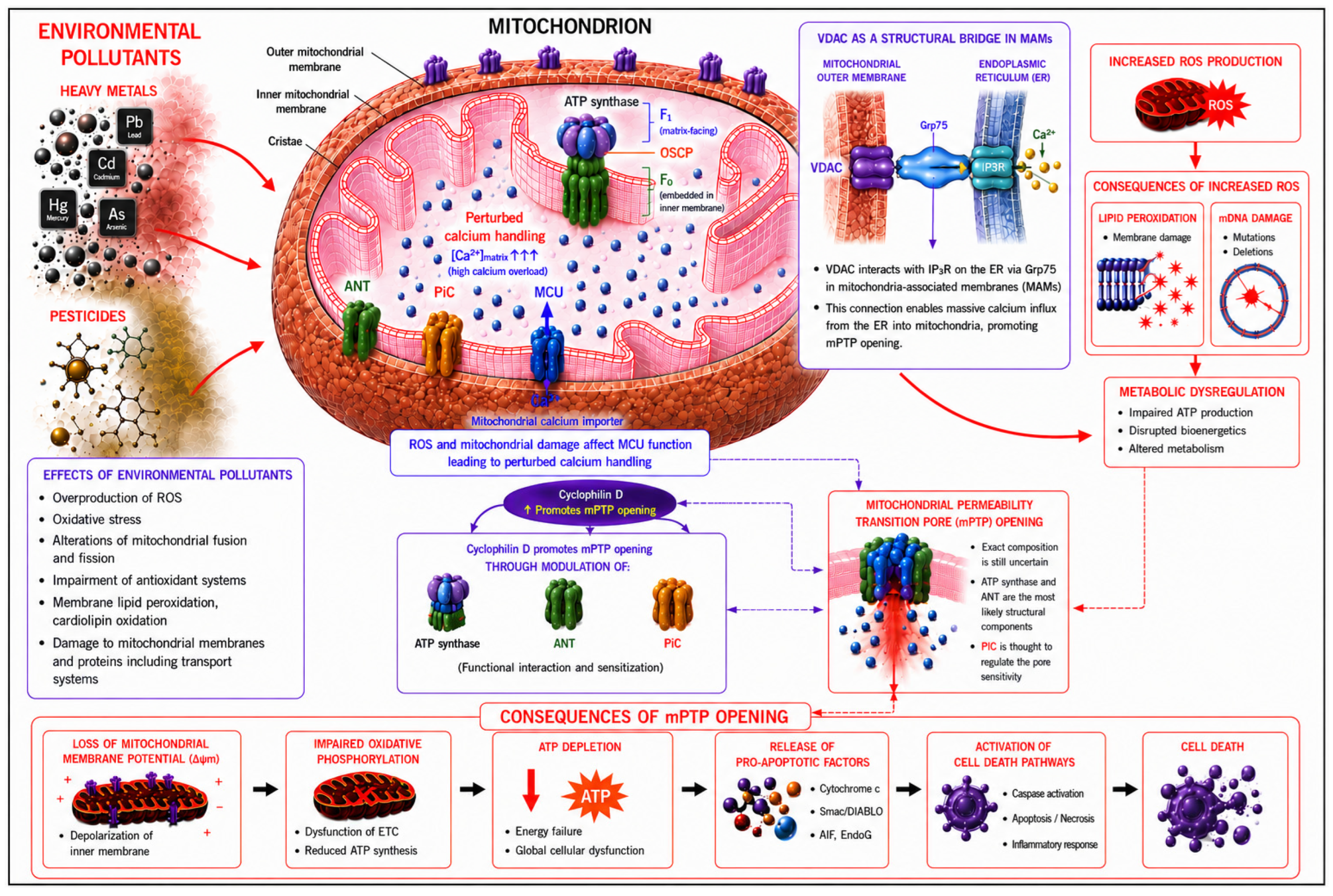
Disclaimer/Publisher’s Note: The statements, opinions and data contained in all publications are solely those of the individual author(s) and contributor(s) and not of MDPI and/or the editor(s). MDPI and/or the editor(s) disclaim responsibility for any injury to people or property resulting from any ideas, methods, instructions or products referred to in the content. |
© 2026 by the authors. Licensee MDPI, Basel, Switzerland. This article is an open access article distributed under the terms and conditions of the Creative Commons Attribution (CC BY) license (http://creativecommons.org/licenses/by/4.0/).
Copyright: This open access article is published under a Creative Commons CC BY 4.0 license, which permit the free download, distribution, and reuse, provided that the author and preprint are cited in any reuse.


