
Preprint
Review

This version is not peer-reviewed.

Functional Renal Reserve-Guided Personalized Renoprotection in Chronic Kidney Disease: Extending the KDIGO CGA Framework

A peer-reviewed version of this preprint was published in:
Biomedicines 2026, 14(7), 1478. https://doi.org/10.3390/biomedicines14071478

Submitted:

04 June 2026

Posted:

05 June 2026

You are already at the latest version

Abstract
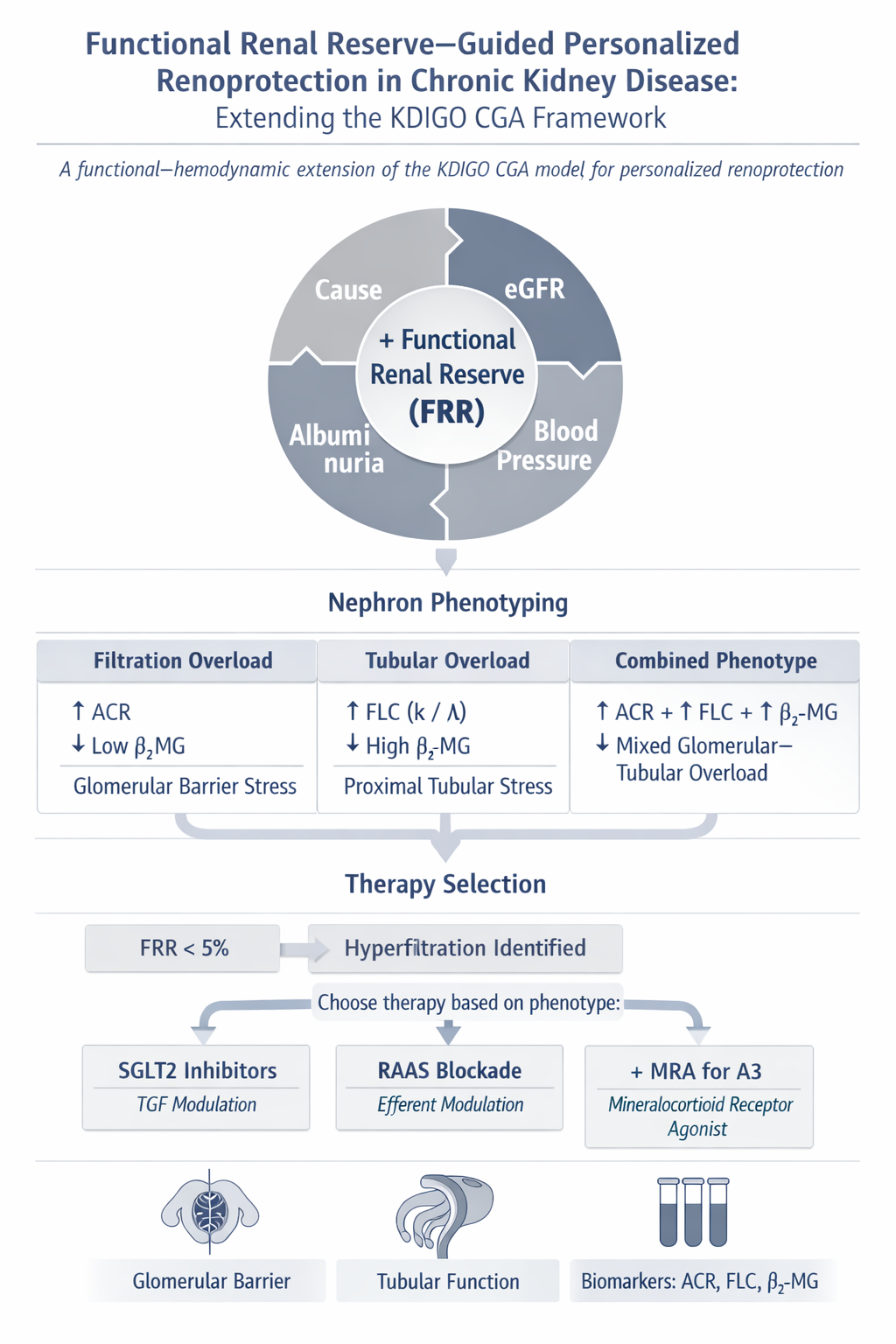
The Kidney Disease: Improving Global Outcomes (KDIGO) classification of chronic kidney disease (CKD) is based on the cause of disease, the category of estimated glomerular filtration rate (eGFR), and the category of albuminuria. This framework is indispensable for risk stratification, yet it does not always identify the functional and hemodynamic mechanism that maintains the current filtration level and drives future progression. In particular, a normal or only moderately reduced eGFR does not exclude relative hyperfiltration of the remaining nephrons, and albuminuria reflects not only glomerular barrier injury but also the limited capacity of the proximal tubule to endocytose and metabolically process filtered proteins. In this conceptual review, we propose a functional-hemodynamic extension of the KDIGO CGA model: Cause + GFR + Albuminuria + Functional Renal Reserve + Blood Pressure. Within this framework, functional renal reserve (FRR) is considered a dynamic stress test of nephron reactivity, whereas blood pressure acts as an essential hemodynamic and therapeutic modifier that influences the safety, sequencing, and intensity of renoprotection. Detailed antihypertensive treatment is beyond the scope of this article; the KDIGO 2021 recommendation of a target systolic blood pressure below 120 mmHg in adults with CKD and elevated blood pressure, when tolerated and measured in a standardized manner, is used as a clinical reference point. A central element of the proposed algorithm is FRR. A zero or negative FRR under standardized testing may indicate an "actionable hyperfiltration phenotype": a clinically meaningful state in which total eGFR does not reflect the true workload imposed on individual nephrons. Depending on the combination of urinary albumin-to-creatinine ratio, eGFR, FRR, blood pressure, metabolic phenotype, and tubular overload markers, the proposed approach may support RAAS-blockade-first, SGLT2-inhibitor-first, early dual therapy, or staged triple renoprotection. A mechanistic distinction is also emphasized. Sodium-glucose cotransporter 2 inhibitors (SGLT2i) directly inhibit proximal tubular sodium and glucose reabsorption, but their key anti-hyperfiltration effect is mediated by increased sodium delivery to the macula densa, restoration of tubuloglomerular feedback, and increased afferent arteriolar tone. By contrast, renin-angiotensin-aldosterone system inhibitors (RAASi) predominantly modulate the efferent/postglomerular compartment and may improve downstream peritubular perfusion, a mechanism potentially relevant to albumin and protein handling by the proximal tubule. The proposed model does not replace KDIGO; rather, it adds physiological phenotyping to the existing risk map. More precise evaluation of proteinuria, including the albumin-to-total-protein ratio, first-morning urine sampling, low-molecular-weight proteins, and tubular markers, may prevent misclassification of physiological, orthostatic, postglomerular, or tubular proteinuria as progressive glomerular CKD. The model requires prospective validation but may help develop practical algorithms for personalized renoprotection, particularly in patients with low or moderate albuminuria, normal or moderately reduced eGFR, diabetes, obesity, hypertension, solitary kidney, reduced nephron mass, or an uncertain progression trajectory.
Keywords: 
;  ;  ;  ;  ;  ;  ;  ;  ;  ;  

1. Conceptual Framework: From KDIGO CGA to Functional Renoprotection

The classical KDIGO model describes CKD along three axes: cause, GFR, and albuminuria. In daily practice, this model identifies risk category, surveillance intensity, the need for additional evaluation, and broad therapeutic direction [1,67]. However, when clinicians select renoprotective treatment, the practical question is often more granular: which drug should be introduced first, how rapidly should therapies be combined, when should treatment be intensified, and in what sequence should RAAS inhibitors, SGLT2 inhibitors, and non-steroidal mineralocorticoid receptor antagonists (ns-MRAs) be used?
We propose that KDIGO CGA should be regarded not as a final drug-selection algorithm, but as the necessary first layer. A second layer should include functional renal reserve, blood pressure, volume status, eGFR slope, the phenotype of proteinuria or albuminuria, and additional tubular markers. This extension builds on the previously proposed idea of adding blood pressure to CKD prognosis based on GFR and albuminuria categories [2] and translates it into a more functional model by placing FRR as a dynamic stress test of nephron reserve [3,4,5]. The model can therefore be expressed as:
Cause + GFR + Albuminuria + Functional Renal Reserve + Blood Pressure.
This sequence reflects the logic of the present article. Structural and functional risk is first established using KDIGO; the nephron reserve is then assessed functionally; and blood pressure is subsequently used as a critical hemodynamic modifier that determines drug choice, dose, titration speed, and the safety of combination therapy. We therefore do not propose a new antihypertensive algorithm. Rather, we suggest using blood pressure as an additional clinical coordinate that modifies the sequencing of kidney-protective therapy.
This additional functional-hemodynamic layer may identify the dominant pathophysiological node: efferent-glomerular, afferent/TGF-mediated, proximal tubular, postglomerular-peritubular, inflammatory-fibrotic, or mixed. Therapy can then be individualized: RAASi-first for a pressure-driven or albuminuric phenotype; SGLT2i-first for a TGF-mediated hyperfiltration phenotype with normal or low-normal blood pressure; early dual therapy for mixed phenotypes; and staged triple therapy for high residual risk when functional reserve is preserved [6,7,8,9].
The purpose of this concept is not to revise KDIGO, but to build a physiological extension above it. Such an extension may be particularly useful when eGFR and urinary albumin-to-creatinine ratio (UACR) provide an incomplete or misleading picture: normal eGFR despite reduced nephron mass, low albuminuria despite marked tubular overload, elevated blood pressure with minimal albuminuria, a pronounced eGFR dip after therapy initiation, or uncertainty about which disease-modifying drug should be introduced first.

2. Blood Pressure in the Proposed Model: A Risk Criterion, Not a Separate Treatment Algorithm

Blood pressure is not formally a CKD stage within the KDIGO CGA system. Clinically, however, it is a third major vector of CKD progression alongside eGFR and albuminuria. Systemic pressure, intraglomerular pressure, pulse pressure, sodium status, volume load, nocturnal non-dipping, and vascular stiffness may profoundly modify both progression risk and the tolerability of renoprotective therapy [2,10].
In this review, blood pressure is used not as a stand-alone treatment target but as a hemodynamic modifier of the proposed algorithm. Comprehensive blood pressure management requires a separate discussion, including antihypertensive combinations, standardized office measurement, home monitoring, ambulatory blood pressure monitoring, nocturnal profiles, and arterial stiffness. For this reason, we do not place blood pressure above FRR in the conceptual hierarchy, but rather consider it after FRR as the parameter that determines therapeutic sequencing and safety.
The practical reference point remains the KDIGO 2021 recommendation that adults with CKD and elevated blood pressure should be treated to a target systolic blood pressure below 120 mmHg, when tolerated and using standardized office measurement [10]. This does not imply that every patient with CKD should undergo aggressive pressure reduction at any cost. In patients with low FRR, advanced age, diuretic therapy, heart failure, or hypovolemia risk, excessive blood pressure reduction may amplify an eGFR dip and limit the feasibility of combined renoprotection [75].
In the proposed algorithm, FRR answers the question: is the kidney operating with reserve, or already at its functional limit? Blood pressure answers a different question: which route for lowering intrarenal stress is safer, and what should be introduced first-efferent/postglomerular modulation with RAAS blockade, afferent/TGF modulation with SGLT2 inhibition, or cautious staged combination therapy?

3. Contemporary Therapeutic Context: Three Core Classes of Renoprotection

Current evidence has moved nephrology toward a model of multiple foundational renoprotective therapies. The first component is RAAS blockade, which remains central in proteinuric and albuminuric CKD, particularly in diabetes and hypertension. The second component is SGLT2 inhibition, which reduces the risk of CKD progression across a wide range of eGFR and albuminuria, including in patients without diabetes. The third component is ns-MRA therapy, especially finerenone, which reduces inflammatory-fibrotic and cardiovascular residual risk in patients with type 2 diabetes, CKD, and persistent albuminuria.
The CREDENCE, DAPA-CKD, and EMPA-KIDNEY trials established that SGLT2 inhibitors reduce CKD progression and adverse cardiorenal outcomes [11,12,13]. Meta-analyses indicate that SGLT2i reduce the risk of CKD progression by approximately 30-40% in patients with eGFR below 60 mL/min/1.73 m2 and/or severe albuminuria, with benefit observed across different baseline eGFR and UACR strata [14,15].
Most major SGLT2i trials were conducted on a background of established or recommended RAAS blockade when tolerated [16], and real-world audits and network meta-analyses continue to evaluate how RAASi, SGLT2i, and aldosterone/mineralocorticoid-pathway therapies are combined in practice [61,62]. Therefore, in conventional evidence-based pathways, SGLT2i are frequently added to stable RAASi therapy. Yet real-world practice is broader than randomized trial design. A patient may have low blood pressure, A1 albuminuria, absent functional reserve, high hypovolemia risk, solitary kidney, obesity, diabetes without albuminuria, or early CKD with preserved eGFR. In these scenarios, the question of where to start remains clinically relevant.
RAASi, SGLT2i, and ns-MRAs should not be viewed as competing therapies. They address different components of CKD pathophysiology. RAASi primarily reduce efferent arteriolar tone, intraglomerular pressure, and may improve postglomerular peritubular perfusion [9,17]. SGLT2i directly reduce proximal tubular sodium and glucose reabsorption, increase sodium delivery to the macula densa, restore tubuloglomerular feedback, increase afferent arteriolar tone, and reduce intraglomerular pressure [8,18,19]. ns-MRAs reduce mineralocorticoid-mediated inflammation, fibrosis, and vascular-cardiorenal residual risk [20,21,22]. Personalized treatment should therefore depend on which mechanism is dominant in the individual patient.

4. Albuminuria as a Two-Compartment Phenomenon

Understanding the antiproteinuric effects of renoprotective therapies requires revisiting the physiology of glomerular filtration and tubular protein handling. Historically, it was assumed that approximately 3-4 g of albumin per day pass through the glomerular filter and are almost entirely reabsorbed by the proximal tubule. This estimate appears in the literature: with an albumin sieving coefficient of approximately 0.0006 and normal GFR, calculated albumin filtration may reach several grams per day [23,24].
However, contemporary data have substantially refined this view. Studies in patients with congenital megalin deficiency, a condition that nearly abolishes proximal endocytic reabsorption, show urinary albumin excretion mostly within the microalbuminuric range rather than in grams per day [25]; this observation also helps challenge the notion that microalbuminuria is always the earliest purely glomerular event in diabetic kidney disease [60]. This suggests that, under physiological conditions in humans, only a limited amount of albumin crosses the glomerular barrier; massive gram-level filtration followed by complete reabsorption is unlikely.
In the normal kidney, the glomerular filtration barrier permits passage mainly of low-molecular-weight proteins and a limited quantity of albumin. Nearly all of this filtered protein load is retrieved by the proximal tubule via the megalin-cubilin endolysosomal system, which explains the near absence of protein in final urine [26,27,28,29,56,57,58,59].
Albuminuria is therefore not merely a sign of a "leaky filter." It is a two-compartment phenomenon. The first compartment is the glomerular component: how much albumin crosses the filtration barrier, determined by intraglomerular pressure, podocyte integrity, the endothelial glycocalyx, the glomerular basement membrane, and filtration selectivity. The second compartment is the tubular component: how much filtered albumin can be recovered and metabolically processed by the proximal tubule, determined by peritubular perfusion, oxygen supply, the megalin-cubilin system, endolysosomal capacity, and the degree of tubular workload.
This two-compartment concept is essential for individualized renoprotection. If the fall in albuminuria induced by RAASi is driven mainly by reduced intraglomerular pressure, part of the effect may overlap mechanistically with SGLT2i. If a substantial part of the effect is related to improved downstream peritubular perfusion and restored tubular protein handling, then RAASi plus SGLT2i becomes not simply additive, but pathophysiologically complementary.

5. KDIGO Albuminuria and Terminological Clarification

Using the KDIGO classification, urinary albumin-to-creatinine ratio should be interpreted as follows: UACR below 3 mg/mmol corresponds to A1, normal or mildly increased albuminuria; UACR 3-30 mg/mmol corresponds to A2, moderately increased albuminuria; and UACR above 30 mg/mmol corresponds to A3, severely increased albuminuria, where clinically significant glomerular or podocyte injury and/or marked intraglomerular hypertension are more likely.
Albuminuria and non-selective proteinuria should not be conflated, and the albumin-to-total-protein ratio may help distinguish glomerular from non-glomerular sources in selected clinical settings [32,79]. Even when UACR exceeds 30 mg/mmol, the measurement still quantifies albumin rather than the full spectrum of urinary proteins. Assessment of non-selectivity requires additional tests: total urine protein, protein-to-creatinine ratio, the albumin-to-total-protein ratio, IgG, transferrin, alpha1-microglobulin, beta2-microglobulin, retinol-binding protein, and, when appropriate, urine protein electrophoresis.
Nevertheless, A3 albuminuria is a strong clinical signal. It may reflect a glomerular barrier lesion, podocyte injury, endothelial or glycocalyx dysfunction, or pronounced intraglomerular hypertension. In such cases, RAASi generally remains the most physiologically grounded first-line therapy, with early SGLT2i addition and, when indicated, ns-MRA therapy.

5.1. Practical Phenotyping of Proteinuria Before Renoprotective Drug Selection

Before selecting a renoprotective sequence, it is important to establish whether persistent proteinuria is glomerular, tubular, mixed, postglomerular, or physiological/orthostatic. This step is frequently overlooked when only UACR or a dipstick is available. Bökenkamp emphasized several practical principles: dipsticks detect mainly albumin; protein concentration varies with urine flow; a first-morning urine sample is essential when orthostatic proteinuria is suspected; and 24 h collections should be interpreted cautiously because incomplete or prolonged collection is common [30].
Orthostatic proteinuria is characterized by absent proteinuria in the recumbent position, typically in the first-morning void, with proteinuria appearing later during the day. It is common in adolescents and young adults, has an excellent prognosis, and should not be misclassified as progressive glomerular CKD [30,31]. Conversely, persistent first-morning proteinuria, abnormal sediment, hypoalbuminemia, edema, reduced eGFR, or hypertension should prompt further evaluation.
The albumin-to-total-protein ratio provides an additional practical clue. A predominantly albuminuric pattern suggests a glomerular component, whereas a low albumin fraction with marked low-molecular-weight proteinuria suggests tubular proteinuria. Intermediate values may reflect mixed or postglomerular sources [32]. This distinction is directly relevant to the proposed algorithm because the dominant site of protein handling determines whether the therapeutic target should be glomerular pressure, proximal tubular workload, peritubular perfusion, or residual inflammatory-fibrotic risk [32,79].
Table 1. Practical phenotyping of proteinuria before renoprotective therapy selection.
Phenotype Urinary clues Clinical clues Relationship to FRR and therapy
Glomerular Albumin/total protein usually >60-70%; UACR A2-A3; low-molecular-weight proteins absent or only moderately increased Hematuria, casts, and low serum albumin may be present in nephrotic syndrome Low FRR with A2-A3 suggests a filtration/pressure phenotype: RAASi-first plus early SGLT2i
Tubular Albumin/total protein <30-40%; prominent low-molecular-weight proteins such as alpha1-microglobulin, beta2-microglobulin, RBP, cystatin C, or FLC Serum albumin is often normal; edema is absent; Fanconi syndrome, Dent disease, or drug-related tubular injury may be present Low FRR plus low-molecular-weight proteinuria suggests tubular overload or a tubulointerstitial node; cautious staged therapy is required
Mixed High UACR plus increased low-molecular-weight proteins; intermediate albumin/total protein ratio Glomerular disease with secondary tubular overload or tubulointerstitial injury High progression risk: early dual therapy; staged triple therapy when indicated and when FRR is preserved or borderline
Postglomerular
bleeding
Albumin/total protein approximately 50-60%; low-molecular-weight proteins do not dominate Macroscopic hematuria, clots, urological symptoms; no dysmorphic erythrocytes or casts Should not be interpreted as a hyperfiltration phenotype; urological/postglomerular evaluation is required
Orthostatic/
physiological
First-morning urine negative; daytime samples positive; total protein may be high during the day Normal serum albumin, blood pressure, urinary sediment, and eGFR; common in adolescents and young adults Persistent proteinuria must be confirmed before renoprotection; transient and postural causes should be excluded

6. Tubular Proteinuria, Megalin-Cubilin, and Low-Molecular-Weight Markers

The proximal tubule is equipped with a high-capacity endocytic system. Megalin, cubilin, ClC-5, the early and late endosomal compartments, and the lysosomal pathway work together to reclaim low-molecular-weight proteins and a limited amount of filtered albumin [27,28,29,33,34,35,36,37]. When this system is genetically or functionally impaired, urinary protein composition changes even when the glomerular barrier is not the primary lesion.
Dent disease provides a useful model. ClC-5 or OCRL-related proximal tubular dysfunction causes low-molecular-weight proteinuria, hypercalciuria, nephrocalcinosis, nephrolithiasis, and progressive CKD in a subset of patients [34,36,39]. In such disorders, the magnitude of total proteinuria may be misleading if interpreted as a purely glomerular process. The urine is enriched with low-molecular-weight proteins, whereas albuminuria is often modest relative to total protein.
This is relevant to personalized renoprotection because tubular overload may coexist with glomerular stress; the composition of urinary proteins can therefore be more informative than total protein alone [56,57,58,59]. A patient may have A1 or A2 albuminuria but still have marked proximal tubular stress, reflected by urinary beta2-microglobulin, alpha1-microglobulin, retinol-binding protein, cystatin C, or free light chains. In such a phenotype, eGFR and UACR alone may underestimate the degree of nephron stress.

7. Free Light Chains and Beta2-Microglobulin as Indicators of Nephron Overload

Free light chains (FLCs) may be viewed as sensitive indicators of which nephron compartment is overloaded, particularly when interpreted together with eGFR, tubular markers, and functional testing [45,53]. In early CKD, especially when eGFR is still normal or only mildly reduced, increased urinary FLCs may reflect proximal tubular overload, hypoxia, and impaired megalin-cubilin-mediated reabsorption. In more advanced CKD, reduced filtration and systemic accumulation may contribute more substantially. The pattern of FLC increase can therefore provide indirect information about whether the limiting node is primarily tubular, glomerular, or mixed.
Beta2-microglobulin (beta2-MG; approximately 11.8 kDa) is freely filtered and almost completely reabsorbed by the proximal tubule. Increased urinary beta2-MG is a sensitive marker of proximal tubular dysfunction and may reflect structural or functional tubular injury independent of GFR [38]. In the context of FRR testing, elevated beta2-MG may suggest that absent reserve is not merely a glomerular pressure problem but also a sign of proximal tubular overload or tubulointerstitial stress.
Combining UACR, FLCs, beta2-MG, and FRR may help determine whether the nephron is limited by the glomerular filtration barrier, the proximal tubular reabsorptive system, or both. This can guide whether RAASi, SGLT2i, or combination therapy is most physiologically appropriate.
Table 2. Conceptual interpretation of nephron overload markers.
Marker Glomerular component Tubular component Combined phenotype
ACR Markedly increased Mildly increased or unchanged Markedly increased
FLC Markedly increased in advanced filtration impairment Increased with proximal tubular overload Markedly increased
Beta2-MG Markedly increased in advanced filtration impairment Increased with tubular injury or impaired reabsorption Markedly increased
FRR 0% or negative 0-5% 0% or negative

8. Limitations of eGFR: Why Normal eGFR Does Not Exclude CKD or Hyperfiltration

KDIGO uses eGFR and albuminuria as core axes of CKD prognosis, and both lower eGFR and higher albuminuria predict adverse kidney outcomes across populations [71]. Yet eGFR represents the sum of filtration by all functioning nephrons, not the workload imposed on each individual nephron. A patient with reduced nephron number may maintain a normal total eGFR through increased single-nephron GFR. In such circumstances, normal eGFR may conceal relative hyperfiltration and a diminished capacity to adapt to additional stress [7,40,41,42,43].
Absolute hyperfiltration is usually defined as GFR above the expected range for age, sex, and body size. Relative hyperfiltration is more difficult to detect. It may occur when total eGFR is normal or mildly reduced, while the remaining nephrons already operate at high single-nephron filtration. This situation is relevant in diabetes, obesity, insulin resistance, solitary kidney, nephron loss after nephrectomy or congenital reduction in nephron mass, ADPKD, transplant recipients, and some glomerular diseases [43,66,69,70,74].
Creatinine-based equations, including CKD-EPI and FAS, are valuable but cannot identify single-nephron hyperfiltration. The FAS equation adjusts creatinine to age- and sex-specific Q values and can provide age-adapted assessment, while CKD-EPI remains a widely used creatinine-based estimator; neither directly measures nephron workload or reserve [40,41,68]. Cystatin C or combined creatinine-cystatin C equations may improve the accuracy of GFR estimation [42], yet they still do not replace functional testing of renal reserve.

9. Illustrative Calculation: Apparent Stability of eGFR and the Need for Functional Assessment

Consider a teaching example of a 61-year-old man with serum creatinine 1.36 mg/dL and CKD-EPI eGFR approximately 56 mL/min/1.73 m2. A simplified FAS calculation without age correction may misleadingly suggest a higher value. However, the correct FAS equation in adults older than 40 years incorporates the age multiplier 0.988^(age - 40). With Scr/Q approximately 1.36/0.90 = 1.51, the first part of the equation yields approximately 71 mL/min/1.73 m2; applying the age multiplier 0.988^21 gives approximately 55 mL/min/1.73 m2. Thus, CKD-EPI and FAS converge, supporting CKD G3a if persistence is documented.
This example does not demonstrate absolute hyperfiltration. Rather, it illustrates a more important point: neither CKD-EPI nor FAS answers whether the remaining nephron mass has functional reserve. If protein or amino acid loading increases GFR, reserve is preserved; if GFR fails to increase or falls, the kidney may already be functioning near its adaptive limit. FRR therefore provides information that eGFR equations cannot supply.

10. Functional Renal Reserve as a Stress Test of Nephron Reserve

Functional renal reserve is the capacity of the kidney to increase GFR in response to a physiological or pharmacological stimulus, most commonly protein or amino acid loading. It represents the difference between basal GFR and the maximally achievable filtration response under standardized conditions [3,4,44,73].
A preserved FRR suggests that functioning nephrons retain adaptive capacity. A low, zero, or negative FRR suggests that the kidney is already operating close to maximal filtration, or that it cannot appropriately recruit additional filtration because of vascular stiffness, nephron loss, tubulointerstitial injury, or hemodynamic maladaptation. In the present model, a zero or negative FRR is interpreted as a marker of an actionable hyperfiltration phenotype when the clinical context supports this interpretation.
This does not mean that FRR alone diagnoses the mechanism of injury. Instead, FRR identifies whether reserve exists. The mechanism must then be inferred from UACR, protein phenotype, tubular markers, blood pressure, eGFR slope, diabetes, obesity, heart failure, solitary kidney, ADPKD, transplant status, and response to initial therapy. FRR is thus a functional gatekeeper rather than a stand-alone biomarker.

11. What Happens to the Nephron During Hyperfiltration?

Hyperfiltration is not a benign increase in filtration. It reflects maladaptive recruitment of glomerular hemodynamics, tubular transport, oxygen consumption, and vascular tone. In reduced nephron mass, the remaining nephrons increase single-nephron GFR to preserve total filtration. Initially compensatory, this state may increase glomerular capillary pressure, podocyte stress, protein leak, proximal tubular workload, peritubular hypoxia, and inflammatory-fibrotic signaling [6,7,50,51,54,55,76].
If FRR is preserved, a further increase in GFR can still be elicited after a standardized load. If FRR is absent or negative, the nephron may already be operating at or beyond its adaptive ceiling. Such a patient may be particularly sensitive to any drug that alters afferent or efferent tone, volume status, or tubular sodium handling. This explains why the same therapeutic class may produce a desired eGFR dip in one patient and an excessive decline or albuminuria worsening in another.

12. Two Mechanisms of the Antialbuminuric Effect of RAAS Blockade

The classical explanation for the antialbuminuric effect of RAAS blockade is efferent arteriolar vasodilation, reduced intraglomerular pressure, and reduced albumin filtration. This mechanism is well established and underlies KDIGO recommendations for ACE inhibitors or angiotensin receptor blockers in patients with A2 or A3 albuminuria, particularly in diabetes and hypertension [1,17].
However, this may not be the only mechanism. Peritubular capillaries are downstream of the glomerulus and arise from the efferent arteriole. Excessive efferent vasoconstriction may impair postglomerular perfusion. RAAS activation may therefore sustain both intraglomerular hypertension and peritubular hypoperfusion, thereby promoting tubulointerstitial hypoxia and impairing tubular reabsorption of filtered proteins [9,50,51,64,76].
From this perspective, RAAS blockade may reduce albuminuria through two pathways: by reducing albumin passage across the glomerular barrier through lower intraglomerular pressure, and by improving downstream peritubular perfusion, reducing proximal tubular ischemia, and restoring megalin-cubilin-mediated reabsorption and degradation of filtered proteins.
This distinction matters for personalized therapy. If albuminuria is primarily driven by glomerular pressure and barrier leak, RAASi and SGLT2i partly converge on the common final pathway of lowering intraglomerular pressure. If peritubular ischemia and impaired tubular protein handling contribute substantially, RAASi and SGLT2i become particularly complementary: RAASi may improve the efferent-peritubular component, while SGLT2i reduce proximal sodium-glucose transport and restore TGF.

13. SGLT2i: Direct Tubular Target and Afferent Hemodynamic Output

The expression "SGLT2i-first in a tubular-hyperfiltration phenotype" may be imprecise. A more accurate term is "proximal tubular/TGF-mediated hyperfiltration phenotype" or simply "TGF-mediated phenotype." SGLT2 inhibitors act directly in the proximal tubule by inhibiting SGLT2-dependent sodium and glucose reabsorption. Their main anti-hyperfiltration hemodynamic output, however, is mediated by increased sodium delivery to the macula densa, restoration of tubuloglomerular feedback, increased afferent arteriolar tone, and reduced intraglomerular pressure [8,18,19,52,63,77].
Thus, SGLT2i should not be described as drugs that directly restore megalin-cubilin-mediated protein endocytosis. Their action is to reduce proximal sodium-glucose transport work, lower proximal tubular oxygen demand, restore TGF, and correct maladaptive hyperfiltration. RAASi, in contrast, more directly influence the efferent/postglomerular compartment and may affect peritubular perfusion.
This distinction is clinically important. If SGLT2i initiation produces an eGFR dip accompanied by lower UACR, the response may represent desirable hemodynamic unloading of hyperfiltration. If both creatinine and albuminuria rise, afferent restriction may have reduced glomerular pressure without improving downstream peritubular perfusion or tubular protein processing. In such a phenotype, adding RAAS blockade or prioritizing RAASi may be more logical, provided blood pressure and potassium allow it.

14. Mechanistic Separation: RAASi, SGLT2i, and ns-MRAs

In the proposed model, RAASi, SGLT2i, and ns-MRAs are not ranked as stronger or weaker agents. Instead, they are positioned according to the pathophysiological node they predominantly address.
RAASi can be conceptualized as efferent/postglomerular therapy. Their key actions include reducing efferent arteriolar tone, intraglomerular pressure, and albuminuria, and possibly improving postglomerular peritubular perfusion. The most logical starting scenarios are A2/A3 albuminuria, elevated blood pressure, pressure-driven phenotype, diabetic kidney disease, glomerular hypertension, and signs of a filtration node.
SGLT2i can be conceptualized as proximal tubular/TGF-afferent therapy. By inhibiting SGLT2, they reduce proximal sodium and glucose reabsorption, increase sodium delivery to the macula densa, restore TGF, increase afferent tone, reduce maladaptive glomerular hyperfiltration, and lower tubular transport work, hypoxia, and volume-metabolic overload. The most logical starting scenarios are diabetes, obesity, heart failure, TGF-mediated hyperfiltration, zero or negative FRR with A1/A2 albuminuria, normal or low-normal blood pressure, and evidence of proximal tubular overload.
Ns-MRA therapy can be conceptualized as anti-inflammatory and anti-fibrotic residual-risk therapy. Finerenone reduces mineralocorticoid-mediated inflammatory and fibrotic processes and has demonstrated clinical benefit in patients with CKD and type 2 diabetes, particularly with persistent albuminuria on background RAAS blockade [20,21,22]. In this algorithm, ns-MRA therapy is most often the third component of staged triple therapy rather than a universal first-line agent.

15. Three Variants of Low FRR: Filtration, TGF-Proximal, and Mixed Nodes

A central point of the proposed concept is that low FRR indicates exhaustion of functional reserve, but not the mechanism of that exhaustion. The mechanism must be inferred from the accompanying phenotype. Three main patterns can be distinguished.

15.1. Filtration/Glomerular Node

This pattern is dominated by injury or overload of the glomerular filtration barrier. Podocytes are injured, the filtration barrier becomes stiff or sclerotic, intraglomerular pressure is high, and afferent-efferent regulation has already reached maximal compensation. Clues include A2/A3 albuminuria, FRR close to zero or negative, normal or reduced eGFR, absent GFR increase after load, and possible hematuria or systemic features of glomerular disease. The therapeutic implication is RAASi priority followed by early SGLT2i addition; in diabetes with persistent albuminuria, ns-MRA therapy may be added when potassium and eGFR allow.

15.2. TGF-Proximal/Afferent Node

This is an under-recognized but clinically important pattern. The proximal tubule reabsorbs sodium, glucose, amino acids, and filtered proteins and participates in TGF regulation. When proximal sodium-glucose reabsorption is chronically increased, less sodium reaches the macula densa, TGF signaling is displaced, and the afferent arteriole may remain excessively dilated. The nephron may operate at its limit while FRR is low. Clues include normal or mildly increased UACR, normal or reduced eGFR, FRR close to zero, diabetes, obesity, high sodium intake, volume overload, and urinary tubular markers. This is a TGF-mediated hyperfiltration phenotype, for which SGLT2i-first may be physiologically coherent, followed by RAASi if UACR rises, blood pressure increases, or a pressure phenotype emerges.

15.3. Mixed Node

The most severe phenotype combines filtration and TGF-proximal overload. Clues include A2/A3 albuminuria, reduced eGFR, zero or negative FRR, elevated blood pressure, tubular markers, diabetes, obesity, heart failure, tubulointerstitial hypoxia, and high residual risk. The nephron has exhausted reserve in both glomerular and tubular-TGF compartments. The therapeutic implication is not monotherapy but early combined anti-hyperfiltration treatment with RAASi plus SGLT2i, followed by ns-MRA when indicated and safe, with tight monitoring of blood pressure, sodium, weight, and volume status.

16. Preserved FRR and the Possibility of Staged Triple Renoprotection

Preserved FRR is not a guarantee of safety, but it can be interpreted as a functional marker of adaptive capacity. In patients with preserved FRR, stable hemodynamics, and indications for combined therapy, staged intensification with RAASi, SGLT2i, and ns-MRA may be undertaken more confidently. This is particularly relevant in diabetes, A2/A3 albuminuria, high cardiorenal risk, and preserved ability to respond to physiological stress.
Every step of intensification should include monitoring of serum creatinine, eGFR, potassium, blood pressure, volume status, UACR, symptoms of hypotension, the eGFR dip at 2-4 weeks, and reassessment at 8-12 weeks. KDIGO recommends monitoring blood pressure, creatinine, and potassium 2-4 weeks after initiating or increasing RAASi; ACE inhibitors or ARBs are generally continued if creatinine rises by no more than 30% within 4 weeks. Ns-MRA therapy requires particularly careful potassium monitoring [1,10,16].
The practical meaning of preserved FRR is not that all agents can be prescribed aggressively at the same time, but that the kidney demonstrates adaptive capacity. Staged intensification is therefore more acceptable when FRR is preserved, whereas in zero or negative FRR slow titration with one new agent at a time may be safer.

17. Algorithm for Functional Renal Reserve-Guided Personalized Renoprotection in CKD

Table 3. Proposed algorithm for selecting initial and subsequent renoprotective therapy according to UACR, FRR, blood pressure, and clinical phenotype.
UACR, mg/mmol FRR BP / clinical phenotype Interpretation Initial strategy Next step
<3 (A1) Preserved Normal BP, stable eGFR, no diabetes, HF, obesity, solitary kidney, or reduced nephron mass Low current risk; functional reserve preserved Observation; salt, weight, protein and BP control; repeat UACR/eGFR SGLT2i only for specific indications: T2D, HF, or adverse eGFR slope
<3 (A1) Zero or negative Normal or low-normal BP; diabetes, obesity, solitary kidney, familial risk, or reduced nephron mass Hidden/relative hyperfiltration: eGFR maintained by maximal nephron workload SGLT2i-first to correct proximal tubular/TGF-mediated hyperfiltration and increase afferent tone Add RAASi if UACR rises, BP increases, or pressure phenotype appears
<3 (A1) Zero or negative Elevated BP Hidden hyperfiltration plus systemic/intraglomerular pressure RAASi-first if BP and potassium allow Early SGLT2i addition after 2-6 weeks; monitor creatinine, potassium, and BP
<3 (A1) Preserved Elevated BP Pressure-driven phenotype with preserved reserve RAASi-first SGLT2i for T2D, HF, reduced eGFR, or adverse slope; FRR supports safer titration
3-30 (A2) Preserved Normal or moderately elevated BP Early albuminuric CKD; reserve still present RAASi if BP/diabetes; SGLT2i for eGFR/T2D/HF indications Assess UACR response at 8-12 weeks; combine if reduction is insufficient
3-30 (A2) Zero or negative Any BP, especially diabetes, obesity, reduced eGFR, or adverse slope Albuminuria plus exhausted reserve = high progression risk Early dual therapy: RAASi + SGLT2i; order depends on BP, volume, and potassium In T2D with persistent UACR >3 mg/mmol, consider ns-MRA
>30 (A3) Any Often elevated BP; hematuria or systemic features may be present Severe albuminuric/glomerular phenotype; barrier injury plus pressure likely RAASi as foundational therapy + early SGLT2i addition Exclude GN/podocytopathy; consider biopsy; in T2D consider ns-MRA
>30 (A3) Zero or negative Any BP A3 plus exhausted reserve = maximal risk; nephrons operate at their limit Not monotherapy: combined anti-hyperfiltration strategy RAASi + SGLT2i, then ns-MRA when indicated and potassium is normal
Any UACR Preserved High risk but stable hemodynamics Functional capacity for intensification exists Titrate renoprotection more confidently Staged triple therapy when indicated: RAASi + SGLT2i + ns-MRA
Any UACR Zero or negative Low BP, advanced age, diuretics, HF, or hypovolemia risk Exhausted reserve plus high risk of excessive eGFR dip Start with one agent, low dose, slow titration Monitor at 1-2 and 4 weeks; avoid aggressive simultaneous loading

18. Practical Interpretation of the Algorithm

The algorithm is intended to support clinical reasoning rather than replace guideline-based indications. In an A1 patient with preserved FRR and stable eGFR, observation and correction of lifestyle, sodium intake, body weight, and blood pressure may be sufficient unless another indication for SGLT2i exists. In an A1 patient with zero or negative FRR, normal blood pressure, and a metabolic or reduced-nephron-mass phenotype, SGLT2i-first may be justified as a TGF-directed anti-hyperfiltration strategy.
In an A2 patient with low FRR, the combination of albuminuria and exhausted reserve suggests higher risk and supports early dual therapy. In A3 albuminuria, the filtration/pressure component is strong enough that RAAS blockade remains foundational, unless contraindicated. SGLT2i should be added early when tolerated, and ns-MRA therapy should be considered in type 2 diabetes with persistent albuminuria, normal potassium, and appropriate eGFR.
The algorithm also provides a framework for interpreting early changes after therapy initiation, including albuminuria changes that may function as surrogate markers of long-term kidney risk [72]. A modest eGFR dip with UACR reduction is often desirable and may indicate hemodynamic unloading. A creatinine rise with stable or reduced urea and reduced UACR may not represent true structural deterioration. A creatinine rise accompanied by increased albuminuria, symptomatic hypotension, volume depletion, or elevated urea requires reassessment of the phenotype, dose, volume status, and drug sequence [46,47,48,49,78].

19. Illustrative Therapeutic Trajectories: How the Algorithm May Work in Practice

The following scenarios are presented as phenotype-based clinical vignettes, not as identifiable case reports or a clinical case series. They are intended to illustrate how FRR, UACR, tubular markers, blood pressure, and early treatment response may guide interpretation and therapeutic sequencing.

19.1. IgA Nephropathy: Creatinine and Albuminuria Increase after SGLT2i

In a patient with IgA nephropathy, empagliflozin initiation may be followed by increased creatinine and increased urinary albumin. If FRR is low or negative, this response may indicate a mixed phenotype in which afferent/TGF modulation reduces glomerular inflow but does not correct peritubular hypoperfusion or tubular protein handling. If blood pressure and potassium allow, adding RAAS blockade or switching priority toward RAASi may be more coherent than simply escalating SGLT2i. If the rise is excessive or accompanied by clinical hypovolemia, temporary discontinuation and re-phenotyping may be necessary.

19.2. ADPKD: Creatinine Increase after SGLT2i

In autosomal dominant polycystic kidney disease, architectural distortion and reduced effective nephron mass may produce a low or borderline FRR. SGLT2i initiation may cause a creatinine increase, especially when renal perfusion reserve is limited. This does not necessarily indicate irreversible injury, but it requires careful assessment of volume status, blood pressure, urea, urinary markers, and UACR response. Low-dose initiation may be reasonable in selected patients, but the evidence base for ADPKD remains less robust than for classical diabetic or albuminuric CKD.

19.3. Type 1 Diabetes with High FRR: Creatinine Dip Without True Functional Deterioration

In type 1 diabetes with high FRR, for example 48%, SGLT2i may produce a creatinine increase while urea decreases and albuminuria falls. This pattern may represent a beneficial hemodynamic dip rather than true loss of kidney function, particularly if the patient is clinically euvolemic and UACR improves. A high FRR suggests adaptive capacity, making staged combination therapy potentially safer, although SGLT2i use in type 1 diabetes requires careful ketoacidosis risk assessment and adherence to local regulatory recommendations.

19.4. Chronic Glomerulonephritis: Different Responses to Dapagliflozin and Empagliflozin

A patient with chronic glomerulonephritis may experience eGFR decline on dapagliflozin but eGFR improvement or stabilization after switching to empagliflozin. This scenario does not prove class heterogeneity in an individual patient, but it illustrates that drug-specific pharmacodynamics, volume effects, adherence, dose, and baseline phenotype may influence response. Repeating FRR before and after switching may help determine whether the second response reflects improved hemodynamic compatibility rather than random variability. Recent observational data comparing dapagliflozin and empagliflozin in advanced CKD support the need for cautious within-class evaluation [65].

19.5. Hereditary Nephrotic syndrome: FRR 12% and Different Responses to SGLT2i

In hereditary nephrotic syndrome with FRR around 12%, dapagliflozin may reduce eGFR from 50 to 47 mL/min/1.73 m2, discontinuation may be followed by eGFR recovery to 55, and empagliflozin may maintain eGFR around 54 while reducing albuminuria from 323 to 211 mg/mmol. This pattern suggests a borderline but present reserve and highlights that the same mechanistic class may have different tolerability in a given patient. The therapeutic decision should be based on the integrated response: eGFR slope, UACR reduction, symptoms, blood pressure, and tubular markers.

19.6. Low-Dose SGLT2i in ADPKD

In ADPKD with low or borderline FRR, low-dose empagliflozin, for example 5 mg, may cause only a small creatinine increase. This may be interpreted as a cautious afferent/TGF intervention in a kidney with limited reserve. The aim is not aggressive eGFR reduction but gentle modulation of maladaptive hemodynamics, provided volume status, blood pressure, and symptoms remain stable.

19.7. Nephrotic Syndrome: UACR Falls on SGLT2i But Rises after Finerenone

In nephrotic syndrome, SGLT2i may reduce UACR, while addition of finerenone may paradoxically increase UACR in some circumstances. If FRR is preserved, this may reflect transient hemodynamic adjustment or background variability. If FRR is low, however, additional mineralocorticoid receptor modulation may destabilize renal hemodynamics or volume balance in a vulnerable nephron phenotype. This scenario supports staged therapy rather than simultaneous multi-drug initiation.

19.8. Transplanted Solitary Kidney and Native Solitary Kidney: High Sensitivity to Hemodynamic Intervention

In a transplanted solitary kidney, low-dose empagliflozin may increase creatinine when FRR is low, reflecting the limited adaptive capacity of a single functioning renal unit. Similarly, in a native solitary kidney, finerenone may reduce eGFR and increase urea when reserve is extremely limited. These examples illustrate why solitary kidney status should be considered a high-sensitivity phenotype. Therapy may still be beneficial, but dosing, sequencing, and monitoring must be conservative.

20. A Practical Stepwise Algorithm Based on FRR

A practical workflow may be summarized as follows. First, classify CKD according to KDIGO CGA and confirm persistence of albuminuria or proteinuria. Second, phenotype proteinuria using first-morning urine, UACR, total protein-to-creatinine ratio, albumin-to-total-protein ratio, sediment, and low-molecular-weight proteins. Third, assess eGFR slope, blood pressure, volume status, metabolic phenotype, and nephron-mass context. Fourth, measure FRR under a standardized protocol when the clinical question is whether the kidney is working with reserve or at its limit.
If FRR is preserved, staged intensification may be considered when there are guideline-based indications and hemodynamics are stable. If FRR is zero or negative, therapy should be individualized by phenotype: RAASi-first for pressure/albuminuric patterns, SGLT2i-first for TGF-mediated metabolic or volume phenotypes with low-normal blood pressure, early dual therapy for mixed patterns, and cautious single-agent initiation when hypovolemia or hypotension risk is high.
The early response should be read physiologically. A desired eGFR dip is modest, early, and accompanied by lower albuminuria or improved clinical status. An adverse dip is excessive, progressive, accompanied by rising urea, worsening albuminuria, hypotension, symptoms, or signs of volume depletion. In the latter case, therapy sequence, dose, volume status, and the presumed phenotype should be reconsidered.

21. Emerging Therapeutic Targets and Future Directions

The proposed framework is compatible with emerging CKD therapies. Endothelin receptor antagonists, aldosterone synthase inhibitors, GLP-1 receptor agonists, anti-inflammatory strategies, anti-fibrotic approaches, and microbiome-directed interventions may all be interpreted through the same logic: what pathophysiological node is dominant, and does the kidney have functional reserve to tolerate additional intervention?
Future studies should evaluate whether FRR predicts the magnitude and safety of eGFR dip, the antiproteinuric response to RAASi or SGLT2i, the tolerability of ns-MRA, and long-term kidney outcomes. Prospective trials could stratify patients by UACR, blood pressure, tubular markers, nephron-mass status, and FRR to test whether treatment sequencing can be optimized beyond current guideline categories.

22. Limitations of the Concept

This review proposes a conceptual framework rather than a validated clinical prediction model. FRR methodology is not yet standardized across centers, and thresholds such as FRR below 5%, zero FRR, or negative FRR require further validation. Protein or amino acid loading protocols, timing, dietary state, hydration, background therapy, and GFR measurement method may all influence results [3,4,44,73].
The proposed proteinuria phenotyping approach also requires broader implementation of urinary markers that are not routinely measured in many clinics. UACR, total protein, albumin-to-total-protein ratio, beta2-MG, FLCs, cystatin C, and other low-molecular-weight proteins may not be universally available. Moreover, the clinical scenarios presented here are illustrative and should not be interpreted as evidence of efficacy or harm for specific drug sequences.
Finally, the model does not replace guideline indications. KDIGO-based treatment recommendations remain the foundation. The proposed extension is intended to support personalized sequencing, interpretation of early hemodynamic responses, and hypothesis generation for future clinical studies.

23. Conclusions

KDIGO CGA remains the essential starting point for CKD classification and risk assessment. However, eGFR and albuminuria alone do not always reveal whether the remaining nephrons are functioning with adaptive reserve or at their physiological limit. Albuminuria itself should be interpreted as a two-compartment phenomenon, reflecting both glomerular passage and proximal tubular handling of filtered proteins.
Functional renal reserve may help identify an actionable hyperfiltration phenotype and guide the sequencing of RAASi, SGLT2i, and ns-MRA therapy. Blood pressure should be interpreted as a hemodynamic modifier that determines the safety and order of therapy rather than as a separate algorithm within this review. Proteinuria phenotyping, including the albumin-to-total-protein ratio and low-molecular-weight markers, can prevent misclassification and refine treatment choice.
In summary, KDIGO provides the risk map, FRR shows the functional reserve of the nephron, and blood pressure determines the hemodynamic safety and sequencing of renoprotection. This functional-hemodynamic extension of KDIGO CGA requires prospective validation but may offer a practical path toward precision renoprotection in CKD.

Author Contributions

Conceptualization, D.I. and A.G.; methodology, D.I., A.G., V.B. and M.I.; writing-original draft preparation, D.I.; writing-review and editing, D.I., A.G.; visualization, D.I., V.B., and M.I.; supervision, D.I. All authors have read and agreed to the published version of the manuscript.

Funding

This research received no external funding

Institutional Review Board Statement

Not applicable. This article is a conceptual review and does not report identifiable patient data or original human-subject research.

Data Availability Statement

No new data were created or analyzed in this study. Data sharing is not applicable to this article.

Acknowledgments

The authors gratefully acknowledge the clinical staff of Prof. D. Ivanov’s Nephrology Clinic for their continuous support in routine nephrology care and for practical clinical discussions that helped refine the real-world relevance of the proposed functional–hemodynamic approach to personalized renoprotection.

Conflicts of Interest

The authors declare no conflicts of interest.

Abbreviations

ABPM — ambulatory blood pressure monitoring
ACEi — angiotensin-converting enzyme inhibitor
ACR — albumin-to-creatinine ratio
ADPKD — autosomal dominant polycystic kidney disease
AKI — acute kidney injury
ARB — angiotensin receptor blocker
BP — blood pressure
CGA — cause, glomerular filtration rate, and albuminuria
CKD — chronic kidney disease
CKD-EPI — Chronic Kidney Disease Epidemiology Collaboration
CV — cardiovascular
DKD — diabetic kidney disease
eGFR — estimated glomerular filtration rate
ESKD — end-stage kidney disease
FAS — full age spectrum
FLC — free light chains
FRR — functional renal reserve
GBM — glomerular basement membrane
GFR — glomerular filtration rate
HF — heart failure
IgA — immunoglobulin A
KDIGO — Kidney Disease: Improving Global Outcomes
LMW — low molecular weight
mGFR — measured glomerular filtration rate
MRA — mineralocorticoid receptor antagonist
ns-MRA — non-steroidal mineralocorticoid receptor antagonist
RAAS — renin–angiotensin–aldosterone system
RAASi — renin–angiotensin–aldosterone system inhibitor
RCT — randomized controlled trial
RFR — renal functional reserve
SBP — systolic blood pressure
SGLT2 — sodium–glucose cotransporter 2
SGLT2i — sodium–glucose cotransporter 2 inhibitor
T1D — type 1 diabetes
T2D — type 2 diabetes
TGF — tubuloglomerular feedback
UACR — urinary albumin-to-creatinine ratio
UPCR — urinary protein-to-creatinine ratio
β2-MG — beta-2 microglobulin

References

  1. Kidney Disease: Improving Global Outcomes (KDIGO) CKD Work Group. KDIGO 2024 Clinical Practice Guideline for the Evaluation and Management of Chronic Kidney Disease. Kidney Int. 2024, 105(4S), S117–S314. [CrossRef]
  2. Ivanov, D.; Mintser, O.; Ivanova, M. SP325 Adding elevated BP for CKD prognosis by GFR and albuminuria category. Nephrol. Dial. Transplant. 2015, 30 (Suppl 3), iii486. [Google Scholar] [CrossRef]
  3. Palsson, R.; Waikar, S.S. Renal functional reserve revisited. Adv. Chronic Kidney Dis. 2018, 25(3), e1–e8. [Google Scholar] [CrossRef]
  4. Armenta, A.; Madero, M.; Rodriguez-Iturbe, B. Functional reserve of the kidney. Clin. J. Am. Soc. Nephrol. 2022, 17(3), 458–466. [Google Scholar] [CrossRef]
  5. Ivanov, D.; Gozhenko, A.; Ivanova, M. Functional renal reserve: clinical implementation: Monograph; ScientificWorld-NetAkhatAV: Karlsruhe, 2025; p. 158 p. ISBN 978-3-98924-063-6. [Google Scholar] [CrossRef]
  6. Brenner, B.M.; Meyer, T.W.; Hostetter, T.H. Dietary protein intake and the progressive nature of kidney disease: the role of hemodynamically mediated glomerular injury. N. Engl. J. Med. 1982, 307(11), 652–659. [Google Scholar] [CrossRef] [PubMed]
  7. Helal, I.; Fick-Brosnahan, G.M.; Reed-Gitomer, B.; Schrier, R.W. Glomerular hyperfiltration: definitions, mechanisms and clinical implications. Nat. Rev. Nephrol. 2012, 8, 293–300. [Google Scholar] [CrossRef] [PubMed]
  8. Vallon, V.; Thomson, S.C. The tubular hypothesis of nephron filtration and diabetic kidney disease. Nat. Rev. Nephrol. 2020, 16, 317–336. [Google Scholar] [CrossRef] [PubMed]
  9. Satoh, M. Glomerular pressure and tubular oxygen supply: a critical dual target in kidney protection. Hypertens. Res. 2025, 48, 1–8. [Google Scholar] [CrossRef]
  10. Kidney Disease: Improving Global Outcomes (KDIGO) Blood Pressure Work Group. KDIGO 2021 Clinical Practice Guideline for the Management of Blood Pressure in Chronic Kidney Disease. Kidney Int. 2021, 99(3S), S1–S87. [CrossRef]
  11. Perkovic, V.; Jardine, M.J.; Neal, B.; Bompoint, S.; Heerspink, H.J.L.; et al. Canagliflozin and renal outcomes in type 2 diabetes and nephropathy. N. Engl. J. Med. 2019, 380, 2295–2306. [Google Scholar] [CrossRef]
  12. Heerspink, H.J.L.; Stefansson, B.V.; Correa-Rotter, R.; Chertow, G.M.; Greene, T.; Hou, F.F.; et al. Dapagliflozin in patients with chronic kidney disease. N. Engl. J. Med. 2020, 383, 1436–1446. [Google Scholar] [CrossRef]
  13. The EMPA-KIDNEY Collaborative Group. Empagliflozin in patients with chronic kidney disease. N. Engl. J. Med. 2023, 388, 117–127. [Google Scholar] [CrossRef]
  14. Li, N.; Lv, D.; Zhu, X.; Wei, P.; Gui, Y.; Liu, S.; Zhou, E.; Zheng, M.; Zhou, D.; Zhang, L. Effects of SGLT2 inhibitors on renal outcomes in patients with chronic kidney disease: a meta-analysis. Front. Med. 2021, 8, 728089. [Google Scholar] [CrossRef]
  15. Neuen, B.L.; Fletcher, R.A.; Anker, S.D.; Bhatt, D.L.; Butler, J.; Cherney, D.Z.I.; Docherty, K.F.; Inzucchi, S.E.; Jardine, M.J.; Mahaffey, K.W.; McCausland, F.R.; McGuire, D.K.; McMurray, J.J.V.; Neal, B.; Packer, M.; Patel, S.M.; Perkovic, V.; Sabatine, M.S.; Sardell, R.J.; Solomon, S.D.; Vaduganathan, M.; Wanner, C.; Wheeler, D.C.; Zannad, F.; Haynes, R.; Staplin, N.; Herrington, W.G.; Heerspink, H.J.L. SGLT2 inhibitors and kidney outcomes by glomerular filtration rate and albuminuria: a meta-analysis. JAMA 2025. [Google Scholar] [CrossRef]
  16. Kidney Disease: Improving Global Outcomes (KDIGO) Diabetes Work Group. KDIGO 2022 Clinical Practice Guideline for Diabetes Management in Chronic Kidney Disease. Kidney Int. 2022, 102(5S), S1–S127. [CrossRef]
  17. Remuzzi, G.; Perico, N.; Macia, M.; Ruggenenti, P. The role of renin-angiotensin-aldosterone system in the progression of chronic kidney disease. Kidney Int. 2005, 68 (Suppl 99), S57–S65. [Google Scholar] [CrossRef]
  18. Cherney, D.Z.I.; Perkins, B.A.; Soleymanlou, N.; Maione, M.; Lai, V.; Lee, A.; Fagan, N.M.; Woerle, H.J.; Johansen, O.E.; Broedl, U.C.; von Eynatten, M. Renal hemodynamic effect of sodium-glucose cotransporter 2 inhibition in patients with type 1 diabetes mellitus. Circulation 2014, 129(5), 587–597. [Google Scholar] [CrossRef]
  19. van Bommel, E.J.M.; Muskiet, M.H.A.; Tonneijck, L.; Kramer, M.H.H.; Nieuwdorp, M.; van Raalte, D.H. SGLT2 inhibition in the diabetic kidney—from mechanisms to clinical outcome. Clin. J. Am. Soc. Nephrol. 2017, 12(4), 700–710. [Google Scholar] [CrossRef] [PubMed]
  20. Bakris, G.L.; Agarwal, R.; Anker, S.D.; Pitt, B.; Ruilope, L.M.; Rossing, P.; et al. Effect of finerenone on chronic kidney disease outcomes in type 2 diabetes. N. Engl. J. Med. 2020, 383, 2219–2229. [Google Scholar] [CrossRef] [PubMed]
  21. Pitt, B.; Filippatos, G.; Agarwal, R.; Anker, S.D.; Bakris, G.L.; Rossing, P.; et al. Cardiovascular events with finerenone in kidney disease and type 2 diabetes. N. Engl. J. Med. 2021, 385, 2252–2263. [Google Scholar] [CrossRef] [PubMed]
  22. Agarwal, R.; Filippatos, G.; Pitt, B.; Anker, S.D.; Rossing, P.; Joseph, A.; et al. Cardiovascular and kidney outcomes with finerenone in patients with type 2 diabetes and chronic kidney disease: the FIDELITY pooled analysis. Eur. Heart J. 2022, 43(6), 474–484. [Google Scholar] [CrossRef] [PubMed]
  23. Tojo, A.; Kinugasa, S. Mechanisms of glomerular albumin filtration and tubular reabsorption. Int. J. Nephrol. 2012, 2012, 481520. [Google Scholar] [CrossRef] [PubMed]
  24. Comper, W.; Vuchkova, J.; McCarthy, K. New insights into proteinuria/albuminuria. Front. Physiol. 2022, 13991756. [Google Scholar] [CrossRef]
  25. Storm, T.; Tranebjærg, L.; Frykholm, C.; Birn, H.; Verroust, P.J.; Nevéus, T.; Sundelin, B.; Hertz, J.; Holmström, G.; Ericson, K.; Christensen, E.I.; Nielsen, R. Renal phenotypic investigations of megalin-deficient patients: novel insights into tubular proteinuria and albumin filtration. Nephrol. Dial. Transplant. 2013, 28(3), 585–591. [Google Scholar] [CrossRef]
  26. Maack, T.; Johnson, V.; Kau, S.T.; Figueiredo, J.; Sigulem, D. Renal filtration, transport, and metabolism of low-molecular-weight proteins: a review. Kidney Int. 1979, 16(3), 251–270. [Google Scholar] [CrossRef]
  27. Nielsen, R.; Christensen, E.I. Proteinuria and events beyond the slit. Pediatr. Nephrol. 2010, 25, 813–822. [Google Scholar] [CrossRef] [PubMed]
  28. Weisz, O.A. Endocytic adaptation to functional demand by the kidney proximal tubule. J. Physiol. 2021, 599. [Google Scholar] [CrossRef]
  29. Edwards, A.; Long, K.; Baty, C.; Shipman, K.; Weisz, O. Modelling normal and nephrotic axial uptake of albumin and other filtered proteins along the proximal tubule. J. Physiol. 2022, 600. [Google Scholar] [CrossRef]
  30. Bökenkamp, A. Proteinuria—take a closer look! Pediatr. Nephrol. 2020, 35, 533–541. [Google Scholar] [CrossRef]
  31. Rytand, D.A.; Spreiter, S. Prognosis in postural (orthostatic) proteinuria: forty to fifty-year follow-up of six patients after diagnosis by Thomas Addis. N. Engl. J. Med. 1981, 305(11), 618–621. [Google Scholar] [CrossRef]
  32. Smith, E.R.; Cai, M.M.X.; McMahon, L.P.; Wright, D.A.; Holt, S.G. The value of simultaneous measurements of urinary albumin and total protein in proteinuric patients. Nephrol. Dial. Transplant. 2012, 27(4), 1534–1541. [Google Scholar] [CrossRef] [PubMed]
  33. Leheste, J.; Rolinski, B.; Vorum, H.; Hilpert, J.; Nykjaer, A.; Jacobsen, C.; Aucouturier, P.; Moskaug, J.; Otto, A.; Christensen, E.I.; Willnow, T.E. Megalin knockout mice as an animal model of low molecular weight proteinuria. Am. J. Pathol. 1999, 155(4), 1361–1370. [Google Scholar] [CrossRef]
  34. Sakakibara, N.; Nozu, K. Tubular proteinuria due to hereditary endocytic receptor disorder of the proximal tubule: Dent disease and chronic benign proteinuria. Pediatr. Nephrol. 2025, 40, 3367–3377. [Google Scholar] [CrossRef]
  35. Dachy, A.; Paquot, F.; Debray, G.; Bovy, C.; Christensen, E.I.; Collard, L.; Jouret, F. In-depth phenotyping of a Donnai–Barrow patient helps clarify proximal tubule dysfunction. Pediatr. Nephrol. 2015, 30, 1027–1031. [Google Scholar] [CrossRef]
  36. Norden, A.G.W.; Lapsley, M.; Igarashi, T.; Kelleher, C.L.; Lee, P.J.; Matsuyama, T.; Scheinman, S.J.; Shiraga, H.; Sundin, D.P.; Thakker, R.V.; Unwin, R.J.; Verroust, P.J.; Moestrup, S.K. Urinary megalin deficiency implicates abnormal tubular endocytic function in Fanconi syndrome. J. Am. Soc. Nephrol. 2002, 13(1), 125–133. [Google Scholar] [CrossRef]
  37. Nielsen, R.; Christensen, E.I.; Birn, H. Megalin and cubilin in proximal tubule protein reabsorption: from experimental models to human disease. Kidney Int. 2016, 89(1), 58–67. [Google Scholar] [CrossRef] [PubMed]
  38. Herget-Rosenthal, S.; van Wijk, J.A.; Bröcker-Preuss, M.; Bökenkamp, A. Increased urinary cystatin C reflects structural and functional renal tubular impairment independent of glomerular filtration rate. Clin. Biochem. 2007, 40(13-14), 946–951. [Google Scholar] [CrossRef]
  39. Bökenkamp, A.; Ariceta, G.; Böckenhauer, D.; Devuyst, O.; Emma, F.; van Bennekom, D.; Levtchenko, E.; Sayer, J.; Servais, A.; Vargas, R.; Zaniew, M.; Prikhodina, L. Dent disease: clinical practice recommendations. Nephrol. Dial. Transplant. 2025, 40(5), 852–864. [Google Scholar] [CrossRef] [PubMed]
  40. Pottel, H.; Hoste, L.; Dubourg, L.; Ebert, N.; Schaeffner, E.; Eriksen, B.O.; Melsom, T.; Lamb, E.J.; Rule, A.D.; Turner, S.T.; Glassock, R.J.; De Souza, V.; Selistre, L.; Mariat, C.; Martens, F.; Delanaye, P. An estimated glomerular filtration rate equation for the full age spectrum. Nephrol. Dial. Transplant. 2016, 31(5), 798–806. [Google Scholar] [CrossRef]
  41. Pottel, H.; Delanaye, P.; Schaeffner, E.; Dubourg, L.; Eriksen, B.O.; Melsom, T.; Lamb, E.J.; Rule, A.D.; Turner, S.T.; Glassock, R.J.; et al. Estimating glomerular filtration rate for the full age spectrum from serum creatinine and cystatin C. Nephrol. Dial. Transplant. 2017, 32(3), 497–507. [Google Scholar] [CrossRef]
  42. Inker, L.A.; Eneanya, N.D.; Coresh, J.; Tighiouart, H.; Wang, D.; Sang, Y.; Crews, D.C.; Doria, A.; Estrella, M.M.; Froissart, M.; Grams, M.E.; Greene, T.; Grubb, A.; Gudnason, V.; Gutierrez, O.M.; Kalil, R.; Karger, A.B.; Mauer, M.; Navis, G.; Nelson, R.G.; Poggio, E.D.; Rodby, R.; Rossing, P.; Rule, A.D.; Selvin, E.; Seegmiller, J.C.; Shlipak, M.G.; Torres, V.E.; Yang, W.; Ballew, S.H.; Couture, S.J.; Powe, N.R.; Levey, A.S. Chronic Kidney Disease Epidemiology Collaboration. New creatinine- and cystatin C-based equations to estimate GFR without race. N. Engl. J. Med. 2021, 385, 1737–1749. [Google Scholar] [CrossRef]
  43. Denic, A.; Glassock, R.J.; Rule, A.D. Single-nephron glomerular filtration rate in healthy adults. N. Engl. J. Med. 2017, 376, 2349–2357. [Google Scholar] [CrossRef]
  44. Jufar, A.H.; Lankadeva, Y.R.; May, C.N.; Cochrane, A.D.; Bellomo, R.; Evans, R.G. Renal functional reserve: from physiological phenomenon to clinical biomarker and beyond. Am. J. Physiol.-Regul. Integr. Comp. Physiol. 2020, 319(6), R690–R702. [Google Scholar] [CrossRef] [PubMed]
  45. Ivanov, D.D.; Gozhenko, A.I.; Savitskaya, L.M. Renoprotection and its association with eGFR and renal functional reserve. Počki 2018, 7(4), 238–244. [Google Scholar] [CrossRef]
  46. Kraus, B.J.; Weir, M.R.; Bakris, G.L.; Mattheus, M.; Cherney, D.Z.I.; Sattar, N.; Heerspink, H.J.L.; Ritter, I.; von Eynatten, M.; Zinman, B.; Wanner, C. Characterization and implications of the initial estimated glomerular filtration rate “dip” upon sodium-glucose cotransporter-2 inhibition with empagliflozin in the EMPA-REG OUTCOME trial. Kidney Int. 2021, 99, 750–762. [Google Scholar] [CrossRef]
  47. Xie, Y.; Bowe, B.; Gibson, A.K.; McGill, J.B.; Maddukuri, G.; Al-Aly, Z. Clinical implications of estimated glomerular filtration rate dip following sodium-glucose cotransporter-2 inhibitor initiation on cardiovascular and kidney outcomes. J. Am. Heart Assoc. 2021, 10(11), e020237. [Google Scholar] [CrossRef]
  48. Wheeler, D.C.; Stefansson, B.V.; Jongs, N.; Chertow, G.M.; Greene, T.; Hou, F.F.; McMurray, J.J.V.; Correa-Rotter, R.; Rossing, P.; Toto, R.D.; Langkilde, A.M.; Heerspink, H.J.L. Effects of dapagliflozin on the rate of decline in kidney function in patients with chronic kidney disease with and without type 2 diabetes: a prespecified analysis from the DAPA-CKD trial. Lancet Diabetes Endocrinol. 2021, 9, 743–754. [Google Scholar] [CrossRef] [PubMed]
  49. Chuang, M.H.; Tang, Y.S.; Chen, J.Y.; Pan, H.C.; Liao, H.W.; Chu, W.K.; Cheng, C.Y.; Wu, V.C.; Heung, M. Abrupt decline in estimated glomerular filtration rate after initiating sodium-glucose cotransporter 2 inhibitors predicts clinical outcomes: a systematic review and meta-analysis. Diabetes Metab. J. 2024, 48, 240–254. [Google Scholar] [CrossRef]
  50. Nangaku, M. Chronic hypoxia and tubulointerstitial injury: a final common pathway to end-stage renal failure. J. Am. Soc. Nephrol. 2006, 17(1), 17–25. [Google Scholar] [CrossRef] [PubMed]
  51. Mimura, I.; Nangaku, M. The suffocating kidney: tubulointerstitial hypoxia in end-stage renal disease. Nat. Rev. Nephrol. 2010, 6, 667–678. [Google Scholar] [CrossRef]
  52. Muskiet, M.H.A.; Tonneijck, L.; Smits, M.M.; Kramer, M.H.H.; Diamant, M.; Joles, J.A.; van Raalte, D.H. Renal haemodynamic effects of sodium-glucose cotransporter 2 inhibitors in hyperfiltering people with type 1 diabetes and people with type 2 diabetes. Kidney Int. 2020, 97(4), 667–670. [Google Scholar] [CrossRef]
  53. Gozhenko, A.; Ivanov, D. What can a tandem of a pathophysiologist and a clinician bring to clinical practice? Kidneys 2024, 13(2), 96–100. [Google Scholar] [CrossRef]
  54. Taal, M.W.; Brenner, B.M. The hyperfiltration theory: a paradigm shift in nephrology. Kidney Int. 1996, 50(2), 731–732. [Google Scholar] [CrossRef]
  55. Hostetter, T.H. Hyperfiltration and glomerulosclerosis. Semin. Nephrol. 2003, 23(2), 194–199. [Google Scholar] [CrossRef] [PubMed]
  56. Edwards, A.; Christensen, E.; Unwin, R.; Norden, A. Predicting the protein composition of human urine in normal and pathological states: quantitative description based on Dent1 disease (CLCN5 mutation). J. Physiol. 2020, 599. [Google Scholar] [CrossRef] [PubMed]
  57. Lillehoj, E.; Poulik, M. Normal and abnormal aspects of proteinuria. Part I: Mechanisms, characteristics and analyses of urinary protein. Part II: Clinical considerations. Exp. Pathol. 1986, 29(1), 1–28. [Google Scholar] [CrossRef]
  58. Christensen, E.I.; Kristoffersen, I.B.; Grann, B.; Thomsen, J.S.; Andreasen, A.; Nielsen, R. A well-developed endolysosomal system reflects protein reabsorption in segment 1 and 2 of rat proximal tubules. In Kidney International; 2020. [Google Scholar] [CrossRef]
  59. Gburek, J.; Konopska, B.; Gołąb, K. Renal handling of albumin — from early findings to current concepts. Int. J. Mol. Sci. 2021, 22, 5809. [Google Scholar] [CrossRef] [PubMed]
  60. Thethi, T.; Batuman, V. Challenging the conventional wisdom on diabetic nephropathy: Is microalbuminuria the earliest event? J. Diabetes Its Complicat. 2019, 33(3), 191–192. [Google Scholar] [CrossRef]
  61. Teh, J.; Harris, L.; Corcoran, N.; Lappin, D. #1452 Halting the progression of chronic kidney disease; an audit of the use of renin-angiotensin-aldosterone blockers and SGLT-2 inhibitors. Nephrol. Dial. Transplant. 2024. [Google Scholar] [CrossRef]
  62. Yang, S.; Zhao, L.; Mi, Y.; He, W. Effects of sodium-glucose cotransporter 2 inhibitors and aldosterone antagonists, in addition to renin-angiotensin system antagonists, on major adverse kidney outcomes in patients with type 2 diabetes and chronic kidney disease: a systematic review and network meta-analysis. Diabetes Obes. Metab. 2022, 24, 2159–2168. [Google Scholar] [CrossRef]
  63. Heerspink, H.J.L.; Perkins, B.A.; Fitchett, D.H.; Husain, M.; Cherney, D.Z.I. Sodium glucose cotransporter 2 inhibitors in the treatment of diabetes mellitus: cardiovascular and kidney effects, potential mechanisms, and clinical applications. Circulation 2016, 134(10), 752–772. [Google Scholar] [CrossRef]
  64. Gaupp, C.; Schmid, B.; Tripal, P.; Edwards, A.; Daniel, C.; Zimmermann, S.; Goppelt-Struebe, M.; Willam, C.; Rosen, S.; Schley, G. Reconfiguration and loss of peritubular capillaries in chronic kidney disease. Sci. Rep. 2023, 13, 18907. [Google Scholar] [CrossRef]
  65. Lin, S.H.; Chen, J.J.; Hsiao, C.C.; Fang, Y.W.; Chen, M.T.; Tsai, M.H.; Yang, H.Y. Cardiorenal outcomes of dapagliflozin vs. empagliflozin in advanced chronic kidney disease and diabetes. Clin. Pharmacol. Ther. 2026. [Google Scholar] [CrossRef]
  66. Tonneijck, L.; Muskiet, M.H.A.; Smits, M.M.; van Bommel, E.J.M.; Heerspink, H.J.L.; van Raalte, D.H.; Joles, J.A. Glomerular hyperfiltration in diabetes: mechanisms, clinical significance, and treatment. J. Am. Soc. Nephrol. 2017, 28(4), 1023–1039. [Google Scholar] [CrossRef]
  67. Levin, A.; Stevens, P.E.; Bilous, R.W.; Coresh, J.; De Francisco, A.L.M.; De Jong, P.E.; Griffith, K.E.; Hemmelgarn, B.R.; Iseki, K.; Lamb, E.J.; Levey, A.S.; Riella, M.C.; Shlipak, M.G.; Wang, H.; White, C.T.; Winearls, C.G. Kidney disease: improving global outcomes (KDIGO) CKD Work Group. KDIGO 2012 clinical practice guideline for the evaluation and management of chronic kidney disease. Kidney Int. Suppl. 2013, 3, 1–150. [Google Scholar]
  68. Levey, A.S.; Stevens, L.A.; Schmid, C.H.; Zhang, Y.L.; Castro, A.F.; Feldman, H.I.; Kusek, J.W.; Eggers, P.; Van Lente, F.; Greene, T.; Coresh, J. CKD-EPI. A new equation to estimate glomerular filtration rate. Ann. Intern. Med. 2009, 150, 604–612. [Google Scholar] [CrossRef]
  69. Dwyer, J.P.; Parving, H.H.; Hunsicker, L.G.; Ravid, M.; Remuzzi, G.; Lewis, J.B. Renal dysfunction in the presence of normoalbuminuria in type 2 diabetes: results from the DEMAND study. Cardiorenal Med. 2012, 2, 1–10. [Google Scholar] [CrossRef]
  70. Spoto, B.; Pisano, A.; Zoccali, C. Insulin resistance in chronic kidney disease: a systematic review. Am. J. Physiol.-Ren. Physiol. 2016, 311, F1087–F1108. [Google Scholar] [CrossRef]
  71. Gansevoort, R.T.; Matsushita, K.; van der Velde, M.; Astor, B.C.; Woodward, M.; Levey, A.S.; de Jong, P.E.; Coresh, J. Lower estimated GFR and higher albuminuria are associated with adverse kidney outcomes. Kidney Int. 2011, 80, 93–104. [Google Scholar] [CrossRef]
  72. Heerspink, H.J.L.; Greene, T.; Tighiouart, H.; Gansevoort, R.T.; Coresh, J.; Simon, A.L.; Chan, T.M.; Hou, F.F.; Lewis, J.B.; Locatelli, F.; Praga, M.; Schena, F.P.; Levey, A.S.; Inker, L.A. Change in albuminuria as a surrogate endpoint for progression of kidney disease: a meta-analysis of treatment effects in randomised clinical trials. Lancet Diabetes Endocrinol. 2019, 7, 128–139. [Google Scholar] [CrossRef]
  73. Bosch, J.P.; Saccaggi, A.; Lauer, A.; Ronco, C.; Belledonne, M.; Glabman, S. Renal functional reserve in humans: effect of protein intake on glomerular filtration rate. Am. J. Med. 1983, 75(6), 943–950. [Google Scholar] [CrossRef]
  74. Denic, A.; Glassock, R.J.; Rule, A.D. Structural and functional changes with the aging kidney. Adv. Chronic Kidney Dis. 2016, 23(1), 19–28. [Google Scholar] [CrossRef]
  75. Cheung, A.K.; Rahman, M.; Reboussin, D.M.; Craven, T.E.; Greene, T.; Kimmel, P.L.; Cushman, W.C.; Hawfield, A.T.; Johnson, K.C.; Lewis, C.E.; Oparil, S.; Rocco, M.V.; Sink, K.M.; Whelton, P.K.; Wright, J.T., Jr.; Ambrosius, W.T. Effects of intensive BP control in CKD. J. Am. Soc. Nephrol. 2017, 28(9), 2812–2823. [Google Scholar] [CrossRef]
  76. Fine, L.G.; Norman, J.T. Chronic hypoxia as a mechanism of progression of chronic kidney diseases: from hypothesis to novel therapeutics. Kidney Int. 2008, 74(7), 867–872. [Google Scholar] [CrossRef]
  77. Kidokoro, K.; Cherney, D.Z.I.; Bozovic, A.; Nagasu, H.; Satoh, M.; Kanda, E.; Sasaki, T.; Kashihara, N. Evaluation of glomerular hemodynamic function by empagliflozin in diabetic mice using in vivo imaging. Circulation 2019, 140(4), 303–315. [Google Scholar] [CrossRef]
  78. Jongs, N.; Chertow, G.M.; Greene, T.; McMurray, J.J.V.; Langkilde, A.M.; Correa-Rotter, R.; Kashihara, N.; Rossing, P.; Sjöström, C.D.; Stefansson, B.V.; Toto, R.D.; Wheeler, D.C.; Heerspink, H.J.L. Correlates and consequences of an acute change in eGFR in response to the SGLT2 inhibitor dapagliflozin in patients with CKD. J. Am. Soc. Nephrol. 2022, 33(11), 2094–2107. [Google Scholar] [CrossRef]
  79. Ohisa, N.; Yoshida, K.; Matsuki, R.; Suzuki, H.; Miura, H.; Ohisa, Y.; Murayama, N.; Kaku, M.; Sato, H. A comparison of urinary albumin-total protein ratio to phase-contrast microscopic examination of urine sediment for differentiating glomerular and nonglomerular bleeding. Am. J. Kidney Dis. 2008, 52(2), 235–241. [Google Scholar] [CrossRef]
Disclaimer/Publisher’s Note: The statements, opinions and data contained in all publications are solely those of the individual author(s) and contributor(s) and not of MDPI and/or the editor(s). MDPI and/or the editor(s) disclaim responsibility for any injury to people or property resulting from any ideas, methods, instructions or products referred to in the content.
Copyright: This open access article is published under a Creative Commons CC BY 4.0 license, which permit the free download, distribution, and reuse, provided that the author and preprint are cited in any reuse.
Prerpints.org logo

Preprints.org is a free preprint server supported by MDPI in Basel, Switzerland.

Subscribe

© 2026 MDPI (Basel, Switzerland) unless otherwise stated

Accessibility

Disclaimer

Terms of Use

Privacy Policy

Privacy Settings