

Submitted:
03 June 2026
Posted:
04 June 2026
You are already at the latest version
Abstract
Radiotherapy for glioblastoma remains anchored to postoperative structural MRI and anatomy-based target definitions, despite marked spatial heterogeneity and dynamic biological change during chemoradiotherapy. Advanced MRI, amino-acid PET, radiomics, and habitat imaging can characterize tumor biology beyond conventional anatomy, yet their relevance to radiotherapy planning and their evidentiary maturity differ substantially. This narrative review examines the current evidence for biologically informed radiotherapy in glioblastoma and proposes an MRI-centered actionability framework that organizes imaging-derived findings into five levels of evidentiary maturity, from descriptive or associative signals to intervention-ready biomarkers, to guide literature interpretation, multidisciplinary discussion, and prospective protocol design. Standard MRI-based planning remains the clinical backbone, supported by contemporary guidelines and the absence of randomized evidence demonstrating benefit from routine biologically guided target modification. Advanced MRI is discussed as the most practical serial platform for treatment-course reassessment, amino-acid PET as a selective complementary tool for metabolic clarification and recurrence assessment, and radiomics and habitat imaging as promising but not yet intervention-ready research layers. Across modalities, prospective evidence supports feasibility more consistently than clinical benefit. The proposed framework is intended to separate biologically informative findings from those sufficiently validated to justify a defined radiotherapy consequence, and to structure the translational path toward intervention-grade evidence in glioblastoma radiotherapy.
Keywords:
glioblastoma
; biologically informed radiotherapy
; adaptive radiotherapy
; advanced MRI
; amino-acid PET
; FET PET
; radiomics
; habitat imaging
; target delineation
; actionability framework
1. Introduction
Glioblastoma remains a disease in which radiotherapy is indispensable as part of standard combined-modality treatment [1,2], yet treatment planning still relies predominantly on a postoperative anatomic snapshot built around contrast enhancement, the resection cavity, and selected T2/FLAIR abnormalities [3,4]. Although target delineation has evolved toward smaller and more standardized margins, standard planning remains fundamentally anatomy-based rather than biology-based [3,4].
The limitation of this approach is not simply that conventional MRI is incomplete, but that glioblastoma is intrinsically spatially heterogeneous and temporally dynamic. Different tumor regions can vary in cellularity, vascularity, permeability, metabolic activity, and treatment sensitivity, and these properties can evolve during chemoradiotherapy. Advanced MRI, amino-acid PET, and downstream quantitative approaches such as radiomics and habitat imaging have expanded the ability to characterize biologically relevant tissue beyond standard anatomy. However, the central translational problem is no longer the absence of candidate imaging biomarkers—it is the lack of clear evidence showing when imaging-defined abnormalities are sufficiently interpretable, spatially reliable, technically reproducible, and clinically validated to justify a defined radiotherapy consequence [5,6,7].
This distinction is especially important because clinically useful imaging is not necessarily treatment-guiding imaging. The evidentiary threshold for radiotherapy modification is higher than that required for diagnostic interpretation: to justify contour modification, focal boosting, or adaptive replanning, an imaging-defined abnormality must be spatially reliable, technically reproducible, temporally interpretable, and linked to a predefined treatment consequence with prospective evidence of safety and clinical value [3,8,9]. Recent prospective and randomized data further illustrate this gap, showing that biologically informed or imaging-adapted strategies can be technically feasible and biologically plausible without yet demonstrating definitive clinical benefit [10,11,12,13].
This narrative review addresses a deliberately structured question: what can advanced imaging already support in clinical glioblastoma radiotherapy, what remains investigational, and what level of evidence should be required before treatment modification is justified? To organize this evidence, we propose an MRI-centered actionability framework that separates imaging-derived findings into five levels of evidentiary maturity, from descriptive signals to intervention-ready biomarkers, intended to guide literature interpretation, multidisciplinary discussion, and prospective protocol design [10,11,12,13].
Literature search. A targeted search was performed in PubMed/MEDLINE and Scopus (last updated 27 April 2026), combining terms related to glioblastoma, radiotherapy, advanced imaging, and adaptive treatment. Priority was given to clinical guidelines, consensus recommendations, randomized and prospective studies, recurrence-pattern analyses, and clinically relevant reviews. Retrospective studies were included when providing radiotherapy-relevant spatial data. This article is a narrative review; no PRISMA flow diagram or formal risk-of-bias assessment was performed.
2. Current Clinical Foundation: Standard MRI-Based Planning
Contemporary guideline-based practice reflects both the centrality of structural MRI and the persistent uncertainty surrounding optimal target definition in glioblastoma. The most authoritative European guidance, the ESTRO-EANO target delineation guideline, supports a single-phase strategy centered on the postoperative resection cavity and residual contrast-enhancing tumor on T1-weighted MRI, with a CTV margin of approximately 15 mm adapted to anatomical barriers and postoperative anatomy, and a PTV margin typically not exceeding 3 mm when modern image guidance is available [3]. The guideline does not support indiscriminate inclusion of all T2/FLAIR abnormalities, recognizing that hyperintensity can reflect a mixture of edema, postoperative change, and infiltrative non-enhancing tumor that are not equivalent from a planning perspective [3]. The ASTRO clinical practice guideline for WHO grade 4 diffuse glioma is concordant with this approach [4].
Standard MRI-based planning remains clinically defensible because it performs reasonably well in the context for which it was designed. Patterns-of-failure studies consistently show that most recurrences occur within or near the high-dose region, supporting progressive margin reduction without obvious loss of disease control [14,15]. At the same time, structural MRI is poorly suited to distinguishing viable non-enhancing tumor from edema or postoperative change, and cannot directly characterize metabolic activity or treatment-resistant biological phenotypes. These limitations are precisely why advanced imaging has become attractive for radiotherapy: not because standard MRI has failed, but because it leaves unresolved questions about biological extent, recurrence-prone subregions, and target evolution during treatment [3,7,10,11]. The appropriate next step is therefore incremental rather than substitutive, with advanced imaging used as a selective, protocol-driven extension of standard planning rather than a routine replacement.
3. Where Advanced Imaging Is Already Clinically Useful
3.1. Advanced MRI for Biological Contextualization
Before considering any imaging modality as a guide for treatment modification, it is essential to distinguish between clinical usefulness and intervention readiness. Diffusion- and perfusion-based MRI can provide biologically meaningful information on hypercellularity, vascular aggressiveness, and evolving treatment response [7,16,17], while serial MRI can capture relevant target dynamics during chemoradiotherapy that are invisible on the initial planning scan [18]. From a practical radiotherapy perspective, the most established contribution of advanced MRI is improved biological contextualization of regions that appear uncertain on standard postoperative imaging: better appreciation of non-enhancing but suspicious tissue, improved interpretation of interval changes, and more informed multidisciplinary discussion in cases where standard MRI alone is insufficiently specific [3,7,17]. The ESTRO-EANO guideline explicitly states that functional and metabolic MR imaging roles in target delineation remain ill-defined and that these modalities should currently be used within prospective trials rather than for routine delineation [3].
3.2. Amino-Acid PET as a Selective Complementary Tool
Amino-acid tracers such as FET, MET, and FDOPA provide favorable lesion-to-background contrast and are supported for several clinically relevant applications in glioma care [8,9]. The strongest current evidence supports three broad areas: differentiation of neoplastic from non-neoplastic lesions when conventional imaging remains equivocal; delineation of glioma extent, particularly in non-enhancing disease where amino-acid uptake can identify metabolically active tumor beyond contrast enhancement; and differentiation of glioma recurrence from treatment-related changes, including pseudoprogression and radionecrosis, where conventional MRI frequently remains ambiguous [8,9]. The 2025 RANO/EANO PET update characterizes amino-acid PET as strongly recommended for delineation of glioma extent for local therapy planning and for differentiating recurrence from treatment-related changes when MRI is equivocal [9]. These are clinically established adjunctive roles. However, the ability of amino-acid PET to improve outcomes when used to modify radiotherapy volumes or adaptive strategies remains unproven, and both PET RANO 1.0 and the updated RANO/EANO recommendations emphasize the need for further prospective validation before broader treatment-modifying use [8,9].
The fact that an imaging modality improves interpretation does not mean it is ready to change treatment. In radiotherapy, the treatment consequence is spatial and potentially irreversible, and the evidentiary threshold for routine modification has not yet been met by any advanced imaging domain in glioblastoma [3,8,9].
4. Target Delineation and Recurrence-Site Logic
4.1. Advanced MRI Risk Mapping Beyond Structural Extent
Hypercellular regions on high b-value diffusion imaging and hyperperfused regions on perfusion MRI can extend beyond contrast-enhancing tumor and may represent aggressive, treatment-resistant disease [10,11,17]. In the University of Michigan phase II program, these regions were used prospectively to define biologically based boost volumes, demonstrating technical feasibility and hypothesis-generating survival signals, but not definitive superiority over standard treatment [10]. The subsequent interim analysis of the response-adaptive phase II study further showed that persistent and newly developing hypercellular/hyperperfused subvolumes can be tracked during treatment without interrupting chemoradiotherapy, again supporting feasibility rather than mature efficacy [11]. These data suggest that advanced MRI can generate spatially meaningful biological maps relevant to radiotherapy planning. However, the most defensible current consequence remains biological risk overlay on standard target volumes, selective scrutiny of suspicious regions, and prospective use in protocol-driven studies [3,10,11,17].
4.2. Amino-Acid PET and Spatial Discordance
The ESTRO-EANO guideline recognizes amino-acid PET, particularly FET PET, as a valuable additional tool for target delineation in non-enhancing regions, while noting that it remains under investigation as a basis for routine PET-driven margin modification [3]. A systematic review by Horsley et al. synthesizing 20 relevant studies found a consistent pattern: MRI-derived and amino-acid-PET-derived volumes are complementary rather than interchangeable [19]. The PET-defined biological tumor volume was often larger than T1-enhancing disease but smaller than T2/FLAIR abnormality, and a substantial component of PET-avid disease frequently lay outside both the enhancing core and standard MRI-defined regions [19]. This spatial discordance is clinically relevant because it suggests that standard postoperative MRI may incompletely represent viable tumor extent in a subset of patients. However, PET-based volume definition remains influenced by tracer selection, acquisition timing, segmentation method, and multimodality registration [3,8,9,19], and there is no uniform evidence-based rule indicating that all PET-avid regions should be boosted or used to modify standard target geometry outside prospective protocols.
4.3. Recurrence-Site Association and the High-Risk Subvolume Concept
Several studies have shown that biologically defined subregions identified before radiotherapy are spatially associated with later failure. Early MET-PET studies demonstrated that metabolically active tumor volumes could extend beyond contrast-enhancing abnormalities and that pretreatment uptake patterns were associated with the site of subsequent recurrence [20,21]. A prospective dual-time-point FET-PET study similarly found that pre-irradiation volumes defined by MRI and FET-PET could predict glioblastoma recurrence patterns [22], and FET-PET/MRI-guided pattern-of-failure analyses have further shown that PET-defined and MRI-defined volumes provide clinically relevant but non-interchangeable spatial information [23]. Horsley et al. concluded that amino-acid PET is a strong predictor of subsequent relapse sites across the literature [19], a finding that provides region-level biological credibility beyond patient-level prognostication.
This recurrence-site enrichment is the conceptual foundation of the high-risk subvolume idea, and it is where advanced imaging is most directly relevant to radiotherapy reasoning. However, spatial association with recurrence is not proof that acting on a region will improve outcomes. The transition from recurrence-site enrichment to therapeutic intervention requires a substantially higher evidentiary standard. Advanced MRI and amino-acid PET currently justify closer scrutiny of suspicious non-enhancing tissue, prospective annotation of candidate high-risk subregions, and biologically informed reasoning for salvage or re-irradiation in selected cases [3,9,19,21,22,23], but not routine contour redesign or automatic boost selection outside prospective protocols.
5. Trial-Level Reality: Feasibility Without Proven Benefit
5.1. The GLIAA Trial
The most important randomized evidence in this field comes from the multicenter GLIAA trial, which compared FET-PET-guided versus CE-T1 MRI-guided target delineation for re-irradiation in recurrent glioblastoma [12]. GLIAA showed that FET-PET-based planning was feasible and safe but did not result in significant clinical benefit: median progression-free survival was 4.0 months in the FET-PET group versus 4.9 months in the CE-T1 MRI group, with no significant difference in the primary endpoint [12]. The investigators’ interpretation is directly relevant here: despite the known diagnostic utility of FET-PET in recurrent glioblastoma, the difference between PET-defined and MRI-defined target volumes was not sufficient to improve clinical outcomes after re-irradiation [12]. GLIAA does not disprove the biological or diagnostic value of amino-acid PET, but it weakens any assumption that biologically richer target delineation automatically translates into progression-free survival benefit.
5.2. Early-Phase Studies in Newly Diagnosed Glioblastoma
The University of Michigan phase II study used biologically based target volume definition with dose intensification to hypercellular and hyperperfused regions identified by high b-value diffusion and DCE perfusion MRI, demonstrating feasibility, acceptable toxicity, and promising survival signals relative to historical controls [10]. The subsequent interim analysis of the response-adaptive phase II trial showed that persistent and newly developing hypercellular/hyperperfused regions could be incorporated into adaptive boost replanning without interrupting chemoradiotherapy, meeting predefined interim safety criteria [11]. These studies establish that advanced MRI-guided biologically based radiotherapy is technically implementable and sufficiently credible for prospective testing. What they do not establish is that such approaches have already improved survival in a way robust enough to justify routine clinical adoption.
5.3. Cautionary Evidence from Adaptive Metabolic Imaging
The randomized phase II NRG-RTOG1106/ECOG-ACRIN 6697 trial in stage III NSCLC tested whether mid-treatment FDG PET/CT could guide individualized adaptive dose escalation [13]. Although conducted outside glioblastoma, it tests the same translational assumption underlying many imaging-adaptive paradigms: that residual or evolving metabolic activity during treatment can identify the appropriate target for intensification. PET-adapted dose escalation was feasible and safe, but did not improve local-regional control, progression-free survival, or overall survival [13]. This result illustrates a general principle directly relevant to glioblastoma: imaging-guided adaptation can be technically successful and biologically plausible without producing measurable clinical benefit. In glioblastoma specifically, on-treatment imaging changes may reflect tumor biology, edema, inflammation, blood-brain barrier disruption, or treatment-related change [8,9,16], making interval signal evolution difficult to interpret as a reliable adaptive trigger.
The current trial landscape therefore supports a consistent conclusion: standard MRI-based planning remains the clinical backbone because no alternative imaging-guided strategy has demonstrated superior outcomes in a randomized setting [3,4,12], while advanced MRI and amino-acid PET can refine biological understanding and be incorporated into prospective workflows [10,11,12,19,20,21,22,23]. Biologically guided approaches should be presented as feasible and investigational, not as clinically mature. The currently available intervention-oriented evidence is summarized in Table 1 and shows a consistent pattern: biologically informed imaging has crossed the threshold of feasibility, but not yet that of intervention-grade clinical benefit.
6. What Adaptation Is Currently Credible
6.1. Geometry-Driven Adaptation
A distinction should be made between geometry-driven adaptation, which responds to measurable anatomic change, and biologically triggered adaptation, which depends on the interpretation of imaging biomarkers as treatment-resistant or recurrence-prone disease. In current glioblastoma practice, the former is more mature and immediately defensible than the latter.
Serial imaging studies have shown that the target does not remain static throughout chemoradiotherapy. Changes in cavity configuration, migration of enhancing tissue, evolving edema, and shifts in surrounding anatomy can occur during treatment and may affect the adequacy of the original plan [6,18]. Offline replanning based on interval MRI currently represents the most credible form of adaptation in glioblastoma because it addresses a problem that is anatomically concrete and clinically measurable, rather than depending on layers of biological inference. In the study by Şenkesen et al., interim MRI-guided adaptation reduced doses to organs at risk and normal brain while maintaining acceptable recurrence patterns [24]. Paczona et al. similarly reported that PTV reduction based on interim MRI enabled sparing of critical normal tissue without compromising survival [25]. Both studies remain non-randomized, but they support the practical value of interval MRI for geometry-sensitive treatment adjustment.
6.2. MR-Guided Workflows and Candidate Biologic Adaptation
The emergence of MR-guided radiotherapy and MR-linac workflows has expanded the technical possibilities of adaptation considerably. Daily onboard MRI can improve soft tissue visualization, permit serial evaluation of postoperative cavity dynamics, and potentially support smaller margins with greater confidence than cone-beam-CT-based workflows [6]. Early clinical experience with 1.5T MR-Linac adaptive radiotherapy supports feasibility and manageable acute toxicity [26], but does not yet demonstrate superiority in survival, neurocognition, or quality of life over conventional image-guided treatment. MR-guided workflows should therefore be described as high-fidelity technical enablers, not as validated superiority strategies.
Candidate biologic adaptation, incorporating hypercellular and hyperperfused regions identified by advanced MRI into dose-intensified or response-adaptive replanning, has been shown to be technically implementable without interrupting treatment [10,11]. However, as discussed in Section 5, this remains an investigational extension that has cleared the hurdle of technical feasibility but not yet that of intervention-grade clinical validation. The most accurate current description is therefore a research program, not a clinically mature paradigm. Technical sophistication and dosimetric elegance are not sufficient justifications for adaptive change; any redistribution of dose must ultimately be validated against recurrence patterns and clinical endpoints.
7. Technical and Translational Bottlenecks
7.1. Acquisition, Segmentation, and Registration
The principal obstacle to biologically informed radiotherapy in glioblastoma is not the absence of promising imaging signals, but the fragility of the translational chain linking image abnormality to biological interpretation and then to treatment consequence. An imaging-defined region can only influence contouring, boosting, or replanning if it functions as a spatially reliable treatment instruction rather than merely as an informative biomarker.
Advanced MRI remains vulnerable to substantial acquisition and post-processing heterogeneity, and the ESTRO-EANO guideline explicitly states that functional and metabolic MR techniques should currently be confined to prospective trials rather than routine delineation [3]. Amino-acid PET faces analogous challenges: PET RANO 1.0 emphasizes standardized acquisition and reconstruction, harmonization across institutions in multicenter studies, and central review when PET contributes to study endpoints [8]. Joint EANM/EANO/RANO/SNMMI procedure standards similarly emphasize harmonized acquisition, reconstruction, interpretation, and reporting [27]. Image registration adds a further layer of uncertainty (thresholding, segmentation, manual correction, and MRI/CT/PET co-registration all influence the apparent location and extent of candidate subvolumes [3,8,27,28]) making technical validation an integral part of clinical credibility rather than a preliminary methods exercise.
7.2. External Validation and Multicenter Portability
Single-center performance is insufficient for any biomarker intended to alter treatment. A region that appears reproducibly suspicious in one institution may not be defined identically, segmented similarly, or interpreted the same way elsewhere. PET RANO 1.0 explicitly calls for multicenter standardization of regulatory frameworks, multimodality workflows, PET camera harmonization, tracer availability, and hardware and software components before PET can be cleanly implemented in trials at scale [8]. The 2025 RANO/EANO PET update highlights the scarcity of class 1 evidence showing that PET incorporation into workflows improves patient outcomes, making clear that broader clinical translation still requires more than local technical success [9]. The Michigan adaptive phase II program incorporated quality assurance and end-to-end testing to ensure that image acquisition and processing were accurate and reproducible enough for prospective trial use [11], a level of technical discipline that remains uncommon and illustrates a broader point: a threshold or imaging signature that works well within a locked institutional pipeline should still be regarded as locally promising, not broadly actionable, until portability has been tested prospectively.
7.3. Biological Validation
Technical reproducibility alone is insufficient. A region becomes clinically credible only when linked to meaningful biology and spatially relevant outcomes. Survival association at the patient level is not enough for radiotherapy adaptation because the decision itself is spatial: a biomarker that predicts poor prognosis but does not reliably identify where treatment-relevant disease resides is of limited value for contour refinement or focal intervention. The validation hierarchy required for treatment action demands: biological plausibility; spatial association with recurrence or pathology; reproducibility across institutions; and linkage to an intervention-relevant endpoint in a prospective treatment study. The sequential requirements of this translational pathway are illustrated in Figure 1. At present, many imaging-derived candidates in glioblastoma remain strongest only at the first or second level [10,11,17,19,20,21,22,23].
7.4. Workflow and Resource Constraints
Even a technically sound biomarker can fail clinically if it does not fit real-world workflow. Serial advanced MRI requires scanner access, protocol consistency, rapid image review, and staff able to interpret interval change during active treatment. Amino-acid PET adds tracer availability, scheduling logistics, reimbursement variability, and expert interpretation. MR-guided workflows create still greater demands in staffing, on-table time, and quality assurance [6,26]. In practice, translation will likely need to be tiered: guideline-based planning for all patients, selective advanced MRI or amino-acid PET for anatomically or biologically ambiguous cases, offline MRI-based replanning for meaningful geometric change, and biologically guided boosting only within prospective protocols. Such a tiered approach allows the field to develop rigorously while remaining compatible with real-world radiotherapy practice.
8. Radiomics, Delta-Radiomics, and Habitat Imaging: Research Layers, Not Treatment-Guidance Tools
Radiomics offers a way to transform conventional and advanced imaging into quantitative descriptors that may capture biologically relevant patterns not appreciable by visual inspection alone. In principle this is attractive for radiotherapy: if robust imaging features could identify treatment-resistant phenotypes or recurrence-prone subregions, they might eventually refine risk-adapted planning or selective intensification. In practice, however, the current radiomics literature remains much stronger in retrospective risk modeling than in prospective treatment guidance. Most studies address classification, molecular prediction, or survival stratification at the patient level rather than the region level, and a model that predicts poor prognosis does not indicate where dose should be modified or which subvolume should be boosted. Standardization remains a major challenge: the Image Biomarker Standardization Initiative provided an essential step toward harmonized feature definitions [29], but feature standardization alone does not resolve segmentation variability, scanner effects, temporal drift, or the need for prospective validation [29,30]. Many studies remain limited by small retrospective cohorts, heterogeneous MRI protocols, high-dimensional feature spaces with multiple-testing risk, and scarce prospective locked-model testing [29,30]. Delta-radiomics compounds these challenges by adding timepoint consistency, interval dependency, treatment-related confounding, and feature fragility across repeated acquisitions, problems that are especially acute in glioblastoma, where serial imaging changes during chemoradiotherapy can reflect tumor biology, edema, inflammation, treatment effect, and shifting cavity geometry simultaneously [29,30,31]. The most defensible current role of both radiomics and delta-radiomics is therefore structured hypothesis generation: identifying candidate patterns of risk, enriching exploratory analyses, and informing future trial design rather than directly guiding treatment modification.
Habitat imaging is perhaps the most radiotherapy-facing of these downstream quantitative domains because it attempts to convert multiparametric heterogeneity into spatially explicit compartments: regions such as hypercellular-hyperperfused, infiltrative, or metabolically active tissue that could, in theory, be more relevant to radiotherapy than any single modality alone [32,33,34,35]. This conceptual proximity to contouring and focal boosting is precisely why habitat imaging must be discussed carefully. Habitat definitions vary across studies, clustering strategies are not standardized, and the biological labels assigned to imaging-derived habitats are often inferred from combinations of imaging features rather than directly validated against histopathology or recurrence maps [30,32,33,34,35]. A cluster labeled as hypercellular or treatment-resistant may represent a plausible imaging phenotype without being a histologically validated biological compartment, a distinction that is crucial for radiotherapy, because visually compelling habitat maps can create the impression of spatial actionability before the underlying biological meaning has been proven. Habitat imaging should therefore be framed as a promising spatial research approach that requires reproducibility testing, pathology or recurrence-site linkage, multicenter portability, and prospective intervention testing before it can justify target modification or focal dose escalation.
9. The MRI-Centered Actionability Framework
9.1. Rationale and Structure
The MRI-centered actionability framework proposed in this review is an author-derived conceptual tool intended to support structured interpretation of imaging-derived findings in glioblastoma radiotherapy, to guide prospective imaging-guided study design, and to clarify the evidentiary threshold required before an imaging abnormality can be linked to a treatment consequence. It should not be used as a stand-alone clinical decision rule.
The framework is qualitative rather than score-based. Assignment to a given level depends on the overall maturity of evidence supporting a specific imaging finding in a specific clinical context, including biological plausibility, spatial reliability, technical reproducibility, recurrence-site or pathology correlation, and prospective linkage to a predefined radiotherapy action. The same imaging modality may therefore occupy different levels depending on setting: amino-acid PET used to clarify equivocal recurrence has a different evidentiary status from amino-acid PET used to justify routine dose escalation in newly diagnosed glioblastoma. Standard structural MRI remains the guideline-supported clinical backbone for target delineation and should not be confused with Level 5 validation of biologically guided treatment modification: Level 5 refers specifically to an imaging-defined biomarker prospectively validated to support a predefined treatment change with clinically meaningful benefit.
9.2. Five Levels of Actionability
Level 1 — Descriptive or associative signal. An imaging feature is associated with prognosis, molecular class, or global treatment response, but has no reliable spatial consequence for radiotherapy. Many radiomics models and patient-level imaging biomarkers remain at this stage. They may support stratification or research annotation but do not indicate where treatment should be changed [29,30,31].
Level 2 — Biologically plausible spatial abnormality. The imaging finding is spatially localized and biologically credible but not yet validated strongly enough to support a treatment consequence. Examples include suspicious diffusion or perfusion abnormalities extending beyond contrast enhancement, or a quantitative habitat map that appears plausible but lacks reproducibility and prospective validation [10,11,17,29,32,33,34,35].
Level 3 — Spatially suspicious region with recurrence-oriented relevance. The finding is biologically plausible and supported by recurrence-site association, pathology correlation, or consistent radiotherapy-facing literature. Advanced MRI-defined hypercellular/hyperperfused subregions and amino-acid-PET-defined biological tumor volumes often fall into this category. These findings justify closer scrutiny, prospective annotation, and protocol-based study enrichment, but not yet routine treatment modification [10,11,19,20,21,22,23].
Level 4 — Multimodally supported candidate target for protocolized intervention. A Level 4 signal is spatially credible, technically well characterized, and supported by convergent evidence from more than one imaging domain or repeated timepoints. Such findings may reasonably define candidate subvolumes for prospective biologically guided boosting or adaptation within specialized protocols but still lack definitive evidence that acting on them improves clinical outcome [10,11,12,18,23].
Level 5 — Intervention-relevant biomarker. At this level, an imaging-defined finding supports a predefined treatment modification and has been prospectively validated to justify routine clinical use, including reproducibility, biological credibility, and evidence that treatment modification improves a clinically meaningful endpoint without compromising safety. At present, no imaging domain in glioblastoma routinely satisfies this level [3,4,8,9,12,13].
This hierarchy is intentionally conservative. It is designed not to deny progress, but to ensure that different kinds of evidence are not conflated. Figure 2 summarizes the framework as a stepwise hierarchy linking evidentiary maturity to the strongest radiotherapy consequence currently justifiable for a given imaging finding.
9.3. Operational Implications
Level 5 should currently be understood as a theoretical benchmark rather than an achieved category for biologically informed radiotherapy in glioblastoma. Randomized evidence has not shown improved clinical outcomes from PET-guided target refinement in recurrent glioblastoma [12], MRI-guided biologic intensification strategies in newly diagnosed disease remain early-phase [10,11], and comparator evidence from adaptive metabolic imaging outside glioblastoma reinforces that feasibility and biological rationale do not guarantee efficacy [13].
In practical terms, the framework supports a deliberately narrow but useful operational rule: the stronger the proposed treatment consequence, the stronger the evidentiary requirement must be. Diagnostic clarification requires less evidence than contour modification; contour modification requires less evidence than focal dose escalation; and adaptive treatment redistribution requires the highest level of validation. In baseline imaging, standard structural MRI remains the planning backbone, while advanced MRI and amino-acid PET can be used selectively to clarify uncertain extent or enrich biological reasoning in selected cases [3,4,9,17,19]. During treatment, serial MRI may justify replanning when there is meaningful geometric evolution [6,18,24,25], while on-treatment biological changes on advanced MRI or PET should still be interpreted with caution given the multiple competing processes that can drive interval signal evolution [8,9,11,16]. This rule is not restrictive—it is what allows biologically informed radiotherapy to develop rigorously rather than rhetorically. The operational meaning of each actionability level, together with the radiotherapy consequences it can and cannot justify, is summarized in Table 2.
10. Future Directions
The next phase of biologically informed radiotherapy in glioblastoma should move from imaging enrichment to intervention-grade testing. The field no longer lacks candidate imaging signals; it lacks sufficiently standardized, biologically validated, and prospectively tested rules linking those signals to predefined radiotherapy actions.
First, multicenter harmonization of MRI and PET acquisition is a prerequisite. Diffusion, perfusion, and amino-acid PET metrics are highly sensitive to acquisition parameters, reconstruction methods, scanner characteristics, and post-processing choices. Prospective trials should use harmonized imaging protocols, scanner qualification, predefined quality-assurance procedures, and central imaging review whenever imaging-derived subvolumes are intended to influence contouring, boosting, or adaptive replanning [5,8,27,29].
Second, imaging-processing pipelines should be locked before clinical implementation. Segmentation methods, registration procedures, threshold definitions, radiomic feature extraction, and habitat-clustering approaches should be predefined rather than optimized retrospectively. Without locked pipelines, it remains impossible to determine whether a proposed biomarker is truly reproducible or merely dependent on local technical choices [29,30,31,32,33,34,35].
Third, future trials should define explicit imaging-to-treatment rules. A biologically suspicious abnormality should not simply be described, its intended radiotherapy consequence should be specified in advance, whether annotation, multidisciplinary review, target refinement, focal boosting, adaptive replanning, or stratification. Each action carries a different evidentiary threshold and a different risk of toxicity or inappropriate intensification.
Fourth, prospective spatial validation should become a central endpoint. Patient-level prognostic association is insufficient for radiotherapy adaptation. Imaging-derived abnormalities must be tested against recurrence maps, spatial patterns of failure, and pathology when available. This is particularly important for candidate high-risk subvolumes identified by advanced MRI, amino-acid PET, or habitat-based approaches [19,20,21,22,23].
Fifth, future studies should move beyond feasibility endpoints and incorporate clinically meaningful outcomes: progression-free survival, overall survival, local control, marginal failure, neurologic toxicity, neurocognitive outcomes, and quality of life. Neurocognition and patient-reported outcomes are especially important in glioblastoma, where any gain in spatial precision must be weighed against the risk of functional harm in patients with limited prognosis.
Sixth, imaging-guided strategies should be integrated with molecular risk stratification. MGMT promoter methylation, IDH status, TERT promoter alterations, EGFR amplification, and methylation-based tumor profiling all influence prognosis, recurrence behavior, and treatment sensitivity [2]. The most clinically useful models will combine imaging-defined spatial risk with molecular features, extent of resection, and established clinical variables. Practically, translation should follow a tiered structure—standard MRI-based planning for all patients, selective advanced imaging for ambiguous cases, offline replanning for meaningful geometric change, and biologically guided boosting only within prospective protocols—allowing rigorous development while remaining compatible with real-world radiotherapy practice.
11. Conclusions
Biologically informed radiotherapy in glioblastoma has crossed the threshold of biological plausibility and technical feasibility, but not yet that of intervention-grade clinical validation. Standard MRI-based planning remains the guideline-supported backbone of current practice, and no advanced imaging domain currently satisfies routine Level 5 criteria for treatment modification.
Advanced MRI is the most practical serial platform for treatment-course reassessment and protocol-based biological enrichment. Amino-acid PET provides a valuable complementary layer for metabolic clarification, recurrence-versus-treatment-effect assessment, and selected salvage or re-irradiation reasoning. Radiomics, delta-radiomics, and habitat imaging expand the conceptual vocabulary of imaging heterogeneity but remain research tools until technical reproducibility, biological validation, and prospective intervention testing are demonstrated.
The MRI-centered actionability framework proposed here is intended to translate this evidence landscape into a practical tool, separating findings that are descriptive, biologically plausible, recurrence-oriented, or protocol-level candidates from those sufficiently validated to justify routine treatment modification. The decisive next step is the design of prospective, preferably multicenter trials in which imaging-defined abnormalities are linked to predefined, technically reproducible, and clinically measurable treatment actions. Only through this transition can biologically informed radiotherapy fulfill its translational promise and improve outcomes for patients with glioblastoma.
Author Contributions
Conceptualization, F.D. and P.T.; investigation, F.D., G.R., G.Bat., P.P., T.C. and M.V.; resources, A.C., G.Bag., A.P., M.A.M. and S.C.; writing — original draft preparation, F.D.; writing — review and editing, F.D., G.R., G.Bat., P.P., T.C., M.V., A.C., G.Bag., A.P., M.A.M., S.C. and P.T.; visualization, F.D.; supervision, P.T.; project administration, F.D. and P.T. All authors have read and agreed to the published version of the manuscript.
Funding
This research received no external funding.
Institutional Review Board Statement
Not applicable. This narrative review did not involve human participants, human data, or animal subjects.
Informed Consent Statement
Not applicable.
Data Availability Statement
No new data were created or analyzed in this study. Data sharing is not applicable to this article.
Conflicts of Interest
The authors declare no conflicts of interest.
References
- Stupp, R.; Mason, W.P.; van den Bent, M.J.; et al. Radiotherapy plus concomitant and adjuvant temozolomide for glioblastoma. N Engl. J. Med. 2005, 352(10), 987–996. [Google Scholar] [CrossRef]
- Weller, M.; van den Bent, M.; Preusser, M.; et al. EANO guidelines on the diagnosis and treatment of diffuse gliomas of adulthood. Nat. Rev. Clin. Oncol. 2021, 18(3), 170–186. [Google Scholar] [CrossRef] [PubMed]
- Niyazi, M.; Andratschke, N.; Bendszus, M.; et al. ESTRO-EANO guideline on target delineation and radiotherapy details for glioblastoma. Radiother. Oncol. 2023, 184, 109663. [Google Scholar] [CrossRef]
- Yeboa, D.N.; Braunstein, S.E.; Cabrera, A.; et al. Radiation therapy for WHO grade 4 adult-type diffuse glioma: an ASTRO clinical practice guideline. Pract. Radiat. Oncol. 2025, 15(5), 451–471. [Google Scholar] [CrossRef] [PubMed]
- O’Connor, J.P.B.; Aboagye, E.O.; Adams, J.E.; et al. Imaging biomarker roadmap for cancer studies. Nat. Rev. Clin. Oncol. 2017, 14(3), 169–186. [Google Scholar] [CrossRef]
- Tseng, C.L.; Zeng, K.L.; Mellon, E.A.; et al. Evolving concepts in margin strategies and adaptive radiotherapy for glioblastoma: a new future is on the horizon. Neuro Oncol. 2024, 26 (Suppl 1), S3–S16. [Google Scholar] [CrossRef]
- Breen, W.G.; Aryal, M.P.; Cao, Y.; Kim, M.M. Integrating multi-modal imaging in radiation treatments for glioblastoma. Neuro Oncol. 2024, 26 (Suppl 1), S17–S25. [Google Scholar] [CrossRef]
- Albert, N.L.; Galldiks, N.; Ellingson, B.M.; et al. PET-based response assessment criteria for diffuse gliomas (PET RANO 1.0): a report of the RANO group. Lancet Oncol. 2024, 25(1), e29–e41. [Google Scholar] [CrossRef]
- Galldiks, N.; Lohmann, P.; Aboian, M.; et al. Update to the RANO working group and EANO recommendations for the clinical use of PET imaging in gliomas. Lancet Oncol. 2025, 26(8), e436–e447. [Google Scholar] [CrossRef]
- Kim, M.M.; Sun, Y.; Aryal, M.P.; et al. A phase 2 study of dose-intensified chemoradiation using biologically based target volume definition in patients with newly diagnosed glioblastoma. Int. J. Radiat. Oncol. Biol. Phys. 2021, 110(3), 792–803. [Google Scholar] [CrossRef] [PubMed]
- Kim, M.M.; Aryal, M.P.; Suresh, K.; et al. A phase 2 study of multiparametric magnetic resonance imaging-guided high-dose response-adaptive radiation therapy with concurrent temozolomide in patients with newly diagnosed glioblastoma: results from an interim analysis. Int. J. Radiat. Oncol. Biol. Phys. 2025, 122(3), 605–610. [Google Scholar] [CrossRef] [PubMed]
- Grosu, A.L.; Weber, W.A.; Graf, E.; et al. O-(2-[18F]fluoroethyl)-L-tyrosine-PET-guided versus contrast-enhanced T1-weighted MRI-guided re-irradiation in patients with recurrent glioblastoma (GLIAA/NOA-10 ARO2013-01): a multicentre, open-label, randomised trial. Lancet Oncol. 2026, 27(2), 169–180. [Google Scholar] [CrossRef]
- Kong, F.M.; Hu, C.; Pryma, D.A.; et al. Primary results of NRG-RTOG1106/ECOG-ACRIN 6697: a randomized phase II trial of individualized adaptive (chemo)radiotherapy using midtreatment 18F-fluorodeoxyglucose positron emission tomography/computed tomography in stage III non-small cell lung cancer. J. Clin. Oncol. 2024, 42(33), 3935–3946. [Google Scholar] [CrossRef] [PubMed]
- McDonald, M.W.; Shu, H.K.G.; Curran, W.J., Jr.; Crocker, I.R. Pattern of failure after limited margin radiotherapy and temozolomide for glioblastoma. Int. J. Radiat. Oncol. Biol. Phys. 2011, 79(1), 130–136. [Google Scholar] [CrossRef]
- Paulsson, A.K.; McMullen, K.P.; Peiffer, A.M.; et al. Limited margins using modern radiotherapy techniques does not increase marginal failure rate of glioblastoma. Am. J. Clin. Oncol. 2014, 37(2), 177–181. [Google Scholar] [CrossRef]
- Wen, P.Y.; van den Bent, M.; Youssef, G.; et al. RANO 2.0: update to the response assessment in neuro-oncology criteria for high- and low-grade gliomas in adults. J. Clin. Oncol. 2023, 41(33), 5187–5199. [Google Scholar] [CrossRef] [PubMed]
- Henriksen, O.M.; del Mar Álvarez-Torres, M.; Figueiredo, P.; et al. High-grade glioma treatment response monitoring biomarkers: a position statement on the evidence supporting the use of advanced MRI techniques in the clinic, and the latest bench-to-bedside developments. Part 1: perfusion and diffusion techniques. Front Oncol. 2022, 12, 810263. [Google Scholar] [CrossRef]
- Stewart, J.; Sahgal, A.; Lee, Y.; et al. Quantitating interfraction target dynamics during concurrent chemoradiation for glioblastoma: a prospective serial imaging study. Int. J. Radiat. Oncol. Biol. Phys. 2021, 109(3), 736–746. [Google Scholar] [CrossRef]
- Horsley, P.J.; Bailey, D.L.; Schembri, G.; Hsiao, E.; Drummond, J.; Back, M.F. The role of amino acid PET in radiotherapy target volume delineation for adult-type diffuse gliomas: a review of the literature. Crit. Rev. Oncol. Hematol. 2025, 205, 104552. [Google Scholar] [CrossRef]
- Grosu, A.L.; Weber, W.A.; Riedel, E.; et al. L-(methyl-11C) methionine positron emission tomography for target delineation in resected high-grade gliomas before radiotherapy. Int. J. Radiat. Oncol. Biol. Phys. 2005, 63(1), 64–74. [Google Scholar] [CrossRef]
- Lee, I.H.; Piert, M.; Gomez-Hassan, D.; et al. Association of 11C-methionine PET uptake with site of failure after concurrent temozolomide and radiation for primary glioblastoma multiforme. Int. J. Radiat. Oncol. Biol. Phys. 2009, 73(2), 479–485. [Google Scholar] [CrossRef]
- Harat, M.; Małkowski, B.; Makarewicz, R. Pre-irradiation tumour volumes defined by MRI and dual time-point FET-PET for the prediction of glioblastoma multiforme recurrence: a prospective study. Radiother. Oncol. 2016, 120(2), 241–247. [Google Scholar] [CrossRef] [PubMed]
- Lundemann, M.; Costa, J.C.; Law, I.; et al. Patterns of failure for patients with glioblastoma following O-(2-[18F]fluoroethyl)-L-tyrosine PET- and MRI-guided radiotherapy. Radiother. Oncol. 2017, 122(3), 380–386. [Google Scholar] [CrossRef]
- Şenkesen, Ö.; Tezcanlı, E.; Abacıoğlu, M.U.; et al. Limited field adaptive radiotherapy for glioblastoma: changes in target volume and organ at risk doses. Radiat. Oncol. J. 2022, 40(1), 9–19. [Google Scholar] [CrossRef]
- Paczona, V.R.; Végváry, Z.; Kelemen, G.; et al. Magnetic resonance imaging in glioblastoma radiotherapy — beyond treatment adaptation. Phys. Imaging Radiat. Oncol. 2025, 34, 100754. [Google Scholar] [CrossRef]
- Jamora, K.; Wada, M.; Khor, R.; et al. Clinical outcomes and quality of life in glioblastoma patients treated with MR-guided adaptive radiotherapy using a 1.5 T MR-Linac in an Australian setting. Clin. Transl. Radiat. Oncol. 2026, 58, 101132. [Google Scholar] [CrossRef]
- Law, I.; Albert, N.L.; Arbizu, J.; et al. Joint EANM/EANO/RANO practice guidelines/SNMMI procedure standards for imaging of gliomas using PET with radiolabelled amino acids and [18F]FDG: version 1.0. Eur. J. Nucl. Med. Mol. Imaging 2019, 46(3), 540–557. [Google Scholar] [CrossRef] [PubMed]
- Brighi, C.; Puttick, S.; Li, S.; et al. A novel semiautomated method for background activity and biological tumour volume definition to improve standardisation of 18F-FET PET imaging in glioblastoma. EJNMMI Phys. 2022, 9(1), 9. [Google Scholar] [CrossRef]
- Zwanenburg, A.; Vallières, M.; Abdalah, M.A.; et al. The image biomarker standardization initiative: standardized quantitative radiomics for high-throughput image-based phenotyping. Radiology 2020, 295(2), 328–338. [Google Scholar] [CrossRef] [PubMed]
- Martin, P.; Holloway, L.; Metcalfe, P.; Koh, E.S.; Brighi, C. Challenges in glioblastoma radiomics and the path to clinical implementation. Cancers 2022, 14(16), 3897. [Google Scholar] [CrossRef]
- Nardone, V.; Reginelli, A.; Rubini, D.; et al. Delta radiomics: an updated systematic review. Radiol. Med. 2024, 129(8), 1197–1214. [Google Scholar] [CrossRef]
- Li, S.; Dai, Y.; Chen, J.; Yan, F.; Yang, Y. MRI-based habitat imaging in cancer treatment: current technology, applications, and challenges. Cancer Imaging 2024, 24(1), 107. [Google Scholar] [CrossRef]
- Dextraze, K.; Saha, A.; Beig, N.; et al. Spatial habitats from multiparametric MR imaging are associated with signaling pathway activities and survival in glioblastoma. Oncotarget 2017, 8(68), 112992–113001. [Google Scholar] [CrossRef] [PubMed]
- Beig, N.; Bera, K.; Prasanna, P.; et al. Radiogenomic-based survival risk stratification of tumor habitat on Gd-T1w MRI is associated with biological processes in glioblastoma. Clin. Cancer Res. 2020, 26(8), 1866–1876. [Google Scholar] [CrossRef] [PubMed]
- Kim, M.; Park, J.E.; Kim, H.S.; et al. Spatiotemporal habitats from multiparametric physiologic MRI distinguish tumor progression from treatment-related change in post-treatment glioblastoma. Eur. Radiol. 2021, 31(8), 6374–6383. [Google Scholar] [CrossRef] [PubMed]
Figure 1.
From imaging signal to radiotherapy consequence: the translational validation pathway. Four sequential criteria must be met before an imaging-derived finding can justify a defined radiotherapy action. Most current glioblastoma imaging candidates demonstrate biological plausibility and spatial recurrence association (steps 1–2), but multicenter technical reproducibility (step 3) and prospective intervention linkage with clinical outcome evidence (step 4) remain unmet. Level 5, routine clinical use, is indicated with a dashed border to reflect its status as a target not yet reached in glioblastoma radiotherapy.
Figure 1.
From imaging signal to radiotherapy consequence: the translational validation pathway. Four sequential criteria must be met before an imaging-derived finding can justify a defined radiotherapy action. Most current glioblastoma imaging candidates demonstrate biological plausibility and spatial recurrence association (steps 1–2), but multicenter technical reproducibility (step 3) and prospective intervention linkage with clinical outcome evidence (step 4) remain unmet. Level 5, routine clinical use, is indicated with a dashed border to reflect its status as a target not yet reached in glioblastoma radiotherapy.
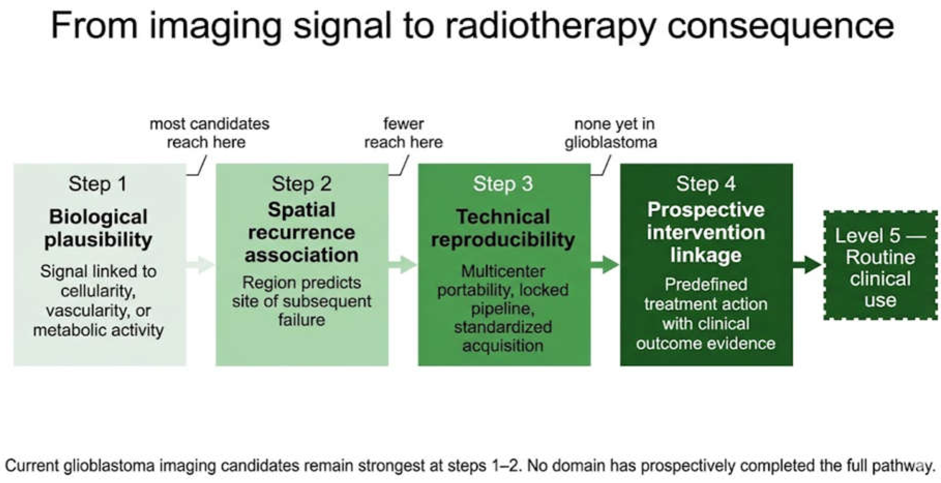
Figure 2.
MRI-centered actionability framework for imaging-derived findings in glioblastoma radiotherapy. The framework organizes imaging-derived findings into five levels of evidentiary maturity, from Level 1 (descriptive signal) at the base to Level 5 (intervention-ready biomarker) at the apex. For each level, the strongest radiotherapy consequence currently justified is indicated on the right. The left axis reflects increasing evidentiary maturity. The framework is a qualitative author-proposed conceptual tool, not an externally validated classification system. In current glioblastoma practice, no advanced imaging domain routinely reaches Level 5 status for treatment modification.
Figure 2.
MRI-centered actionability framework for imaging-derived findings in glioblastoma radiotherapy. The framework organizes imaging-derived findings into five levels of evidentiary maturity, from Level 1 (descriptive signal) at the base to Level 5 (intervention-ready biomarker) at the apex. For each level, the strongest radiotherapy consequence currently justified is indicated on the right. The left axis reflects increasing evidentiary maturity. The framework is a qualitative author-proposed conceptual tool, not an externally validated classification system. In current glioblastoma practice, no advanced imaging domain routinely reaches Level 5 status for treatment modification.
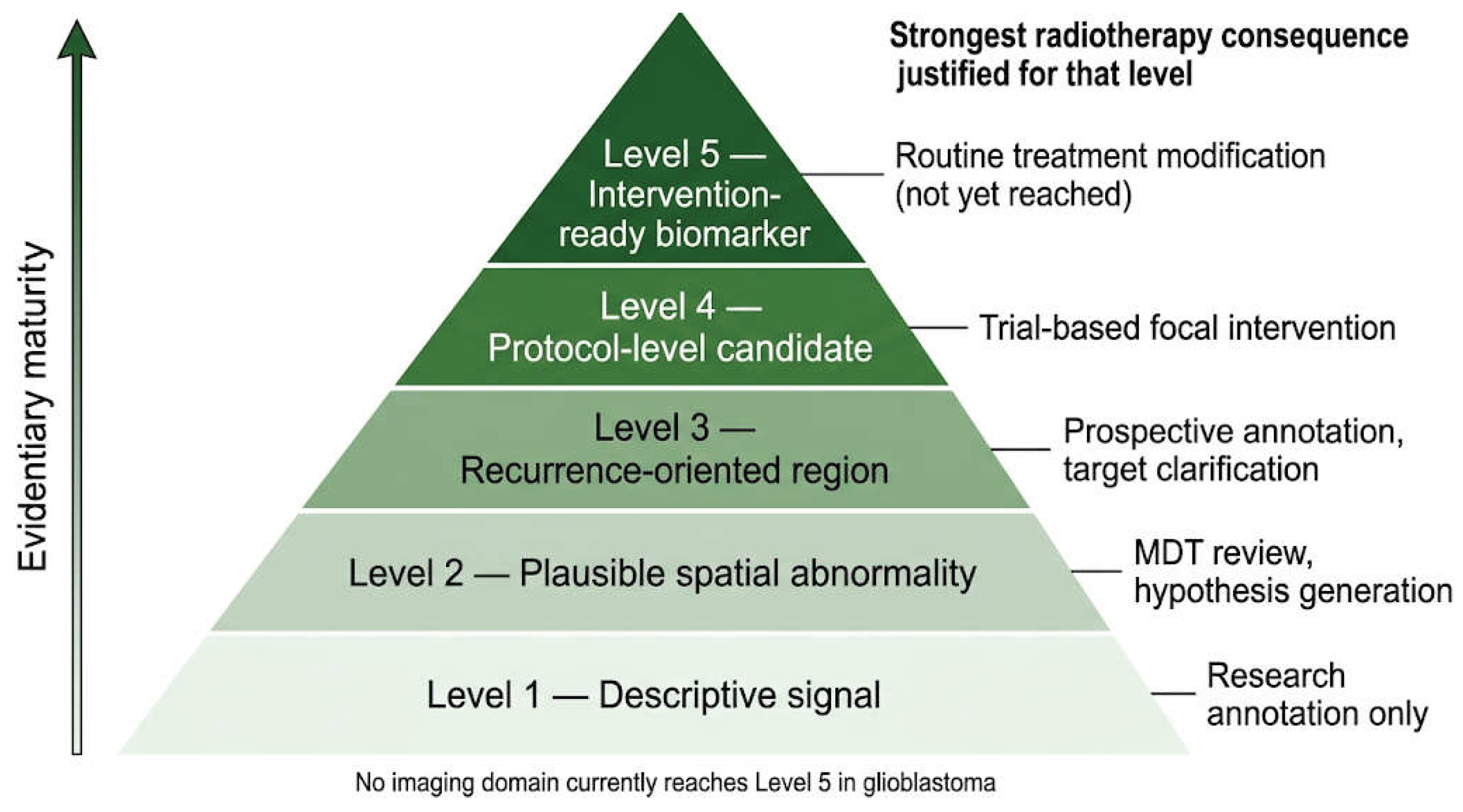

Table 1.
Prospective and randomized studies relevant to imaging-guided radiotherapy in glioblastoma and key cautionary comparator evidence. The available evidence supports feasibility more consistently than intervention-grade clinical benefit. PFS, progression-free survival; OS, overall survival; MRI, magnetic resonance imaging; PET, positron emission tomography; FET, O-(2-[¹⁸F]fluoroethyl)-L-tyrosine; CE-T1, contrast-enhanced T1-weighted MRI; DWI, diffusion-weighted imaging; DCE, dynamic contrast-enhanced; FDG, fluorodeoxyglucose; NSCLC, non-small cell lung cancer.
Table 1.
Prospective and randomized studies relevant to imaging-guided radiotherapy in glioblastoma and key cautionary comparator evidence. The available evidence supports feasibility more consistently than intervention-grade clinical benefit. PFS, progression-free survival; OS, overall survival; MRI, magnetic resonance imaging; PET, positron emission tomography; FET, O-(2-[¹⁸F]fluoroethyl)-L-tyrosine; CE-T1, contrast-enhanced T1-weighted MRI; DWI, diffusion-weighted imaging; DCE, dynamic contrast-enhanced; FDG, fluorodeoxyglucose; NSCLC, non-small cell lung cancer.
| Study | Setting | Imaging/Intervention | Design / N | Main Result | Interpretation for Actionability |
| Grosu et al., 2026 — GLIAA/NOA-10 ARO2013-01 [12] | Recurrent glioblastoma | FET PET-guided versus CE-T1 MRI-guided re-irradiation target delineation | Multicenter open-label randomized trial; 200 randomized, 195 treated per protocol | FET PET-guided planning was feasible and safe but did not improve PFS or clinical outcome compared with CE-T1 MRI-guided planning | Randomized negative/neutral evidence; weakens any assumption that biologically refined PET-based target definition automatically improves outcome |
| Kim et al., 2021 [10] | Newly diagnosed glioblastoma | Dose-intensified chemoradiation to biologically defined hypercellular/hyperperfused subvolumes using high b-value DWI and DCE MRI | Single-arm phase II; N = 26 | Feasible with acceptable toxicity and promising survival signal relative to historical expectations | Hypothesis-generating; supports prospective testing but not routine implementation |
| Kim et al., 2025 [11] | Newly diagnosed glioblastoma | Multiparametric MRI-guided high-dose response-adaptive radiotherapy with mid-treatment boost replanning | Interim phase II analysis; N = 16 of planned 30 | Response-adaptive replanning was feasible without interrupting chemoradiotherapy and met interim safety criteria | Early feasibility evidence; not yet efficacy-proving |
| Kong et al., 2024 – NRG-RTOG1106/ECOG-ACRIN 6697 [13] | Stage III NSCLC comparator trial | Mid-treatment FDG PET/CT-guided adaptive dose escalation | Randomized phase II; N = 127 | PET-adapted escalation was feasible and safe but did not improve local-regional control, PFS, or OS | Cautionary comparator; demonstrates that metabolically guided adaptation can be rational and feasible without improving efficacy |
Table 2.
MRI-centered actionability framework for imaging-derived findings in glioblastoma radiotherapy. Each level reflects a qualitative category of evidentiary maturity. The same imaging modality may occupy different levels depending on clinical context. No advanced imaging domain currently satisfies Level 5 criteria for routine treatment modification in glioblastoma. ADC, apparent diffusion coefficient; MRI, magnetic resonance imaging; PET, positron emission tomography; rCBV, relative cerebral blood volume; RT, radiotherapy.
Table 2.
MRI-centered actionability framework for imaging-derived findings in glioblastoma radiotherapy. Each level reflects a qualitative category of evidentiary maturity. The same imaging modality may occupy different levels depending on clinical context. No advanced imaging domain currently satisfies Level 5 criteria for routine treatment modification in glioblastoma. ADC, apparent diffusion coefficient; MRI, magnetic resonance imaging; PET, positron emission tomography; rCBV, relative cerebral blood volume; RT, radiotherapy.
| Level | Evidence Category | Typical Example | What It Can Justify Today | What It Cannot Justify |
| 1 | Descriptive or associative signal without reliable spatial treatment consequence | Patient-level radiomic risk score or molecular prediction model | Research stratification and exploratory annotation | Contouring, boosting, or adaptive replanning |
| 2 | Biologically plausible spatial abnormality with insufficient validation for treatment consequence | Isolated low-ADC or high-rCBV region beyond contrast enhancement; exploratory habitat compartment | Closer scrutiny, multidisciplinary interpretation, and hypothesis generation | Routine target modification or dose escalation |
| 3 | Spatially suspicious region supported by recurrence-site association, pathology correlation, or consistent radiotherapy-facing evidence | Amino-acid PET biological tumor volume; hypercellular/hyperperfused subregion on advanced MRI | Prospective annotation, selective target clarification, and protocol-based enrichment | Routine contour redesign or standard-of-care boosting |
| 4 | Multimodally supported candidate target suitable for protocolized intervention in specialized settings | Concordant advanced MRI plus amino-acid PET abnormality; persistent or emerging high-risk region in a prospective adaptive workflow | Trial-based or protocol-based focal intervention | Routine clinical adaptation outside prospective frameworks |
| 5 | Intervention-relevant biomarker prospectively validated to support a predefined treatment modification with clinical benefit | No current routine glioblastoma example | Routine treatment modification, if prospectively validated | Any claim of current routine readiness in glioblastoma |
Disclaimer/Publisher’s Note: The statements, opinions and data contained in all publications are solely those of the individual author(s) and contributor(s) and not of MDPI and/or the editor(s). MDPI and/or the editor(s) disclaim responsibility for any injury to people or property resulting from any ideas, methods, instructions or products referred to in the content. |
© 2026 by the authors. Licensee MDPI, Basel, Switzerland. This article is an open access article distributed under the terms and conditions of the Creative Commons Attribution (CC BY) license (http://creativecommons.org/licenses/by/4.0/).
Copyright: This open access article is published under a Creative Commons CC BY 4.0 license, which permit the free download, distribution, and reuse, provided that the author and preprint are cited in any reuse.


