Submitted:
01 June 2026
Posted:
02 June 2026
You are already at the latest version
Abstract
The development of 3D printing high impact denture bases is challenging, as materials exhibiting both high flexural strength/modulus and fracture toughness are required. Nowadays, most of the commercially available 3D printing denture bases contain signifi-cant amounts of crosslinking monomers and therefore behave as brittle materials. In this contribution, urethane dimethacrylate DMA1/(octahydro-4,7-methano-1H-indenyl)methyl acrylate (OMIMA) 1/1 (wt/wt) formulations containing a poly(ɛ-caprolactone)-polydimethylsiloxane-poly(ɛ-caprolactone) (PCL-PDMS-PCL) triblock copolymer (BCP1) and fumed silica SiO2-NPs were evaluated for DLP 3D printing of frac-ture tough denture bases. The post-curing step was performed at various temperatures (RT, 60°C, 80°C, 100°C and 120°C). This parameter was shown to strongly influence the Tg and mechanical properties of 3D printed materials. A post-curing temperature of 100°C was found to be ideal. Under these conditions, 3D printed materials exhibiting excellent mechanical properties were successfully obtained. Furthermore, the amounts of BCP1 and SiO2-NPs were varied. The formulation containing 8.0 wt% of BCP1 and 10.0 wt% of SiO2-NPs was able to fulfill the ISO 20795-1:2013 requirements in terms of flexural strength/modulus and fracture toughness for denture bases with improved impact re-sistance. This material showed better performance than the commercially available for-mulations Printodent® GR-14.2 denture HI and Lucitone Digital PrintTM 3D denture base.
Keywords:
denture base
; block copolymers
; high impact
; fracture toughness
; 3D printing
1. Introduction
Complete dentures have been traditionally manufactured using compression and injection molding techniques [1]. Heat cure denture bases are typically obtained via thermal polymerization of a methyl methacrylate (MMA) based resin (small amounts of dimethacrylates are frequently added) [1,2]. The resulting materials mainly consist of PMMA and exhibit a low crosslinking density. They can therefore be efficiently reinforced via the addition of toughening agents such as core-shell particles. Although excellent and clinically successful materials were developed using this procedure, traditional manufacturing of complete dentures remains complex and time-consuming. The development of CAD (computer-aided design) – CAM (computer-aided manufacturing) technology has revolutionized the dental industry. Using this digital approach, denture bases can be either prepared by subtractive (SM) or by additive manufacturing (AM) [3,4,5,6,7,8,9]. Following the SM workflow, pre-polymerized PMMA based denture bases are milled out of discs using specific milling machines. Although materials exhibiting excellent mechanical properties (flexural strength/modulus, fracture toughness, etc.) are obtained, SM involves significant material waste. On the other hand, AM, also known as 3D printing, produces less waste. It is a fast and cost-effective technology that enables the manufacturing of denture bases with high precision [10,11,12,13,14]. Stereolithography (SLA) and Digital Light Processing (DLP) are mainly used in dental laboratories and practices. Both technologies produce 3D printed materials via the photopolymerization of a resin layer by layer. SLA requires a laser light source to cure the resin, whereas DLP uses a light projector. Various 3D printing denture bases are available on the market. As MMA cannot be efficiently used for 3D printing due to its volatility and low reactivity, the chemical composition of 3D printing denture bases had to be completely modified. They mainly consist of a mixture of dimethacrylates, resulting in printed dentures that are highly crosslinked and brittle.
Denture base materials are subjected to various stresses, such as repeated masticatory forces. High impact forces could also be generated as a result of an accidental dropping of the denture. Such stresses could lead to a fracture of the denture base. For these reasons, fracture tough denture bases are preferred. Two families of traditionally manufactured denture base materials can be found on the market: conventional and high impact denture bases. The ISO 20795-1:2013 standard for light-activated denture bases does not specify any lower limit regarding fracture toughness. On the other hand, high impact denture bases must exhibit high maximum stress intensity factor (Kmax) and total work of fracture (Wf) values (Kmax ≥ 1.9 MPa m1/2 and Wf ≥ 900 J m-2). Geiger et al. as well as Hetzler et al. recently evaluated several commercially available 3D printing denture bases and clearly showed that these materials did not fulfill the requirements for materials with improved impact resistance (Kmax and Wf were significantly lower than the minimum required values) [15,16]. To the best of our knowledge, Lucitone Digital PrintTM 3D denture base (Dentsply Sirona) is the only 3D printing denture base material able to exceed the minimum required fracture toughness for high impact materials. However, recent studies reported quite low flexural strength and modulus values using this material [17,18]. There is therefore a strong need for the development of fracture tough 3D printing denture bases presenting mechanical properties in the same range as the state-of-the-art PMMA based materials.
In the last few years, our research group reported a highly efficient technology to significantly improve the fracture toughness of 3D printable (meth)acrylate based resins. This technology lies in the incorporation of block copolymers (BCPs) in a low crosslinking density network [19,20,21,22]. This network is typically obtained via the photopolymerization of a resin containing a polymerizable urethane macromonomer combined with a monofunctional monomer as diluent. Both diblock and triblock copolymers were evaluated. Amongst the tested toughening agents, poly(ɛ-caprolactone)-polydimethylsiloxane-poly(ɛ-caprolactone) (PCL-PDMS-PCL) triblock copolymers were shown to be particularly efficient. Indeed, the addition of BCP1 (PCL-PDMS-PCL: 1000 g mol-1- 2000 g mol-1- 1000 g mol-1) to a DMA1/OMIMA monomer mixture led to fracture tough materials exhibiting excellent mechanical properties (Figure 1) [22]. Using an optimized amount of BCP1, the ISO 20795-1:2013 standard requirements for high impact denture bases were successfully reached. It is however worth mentioning that the materials were photocured in molds.
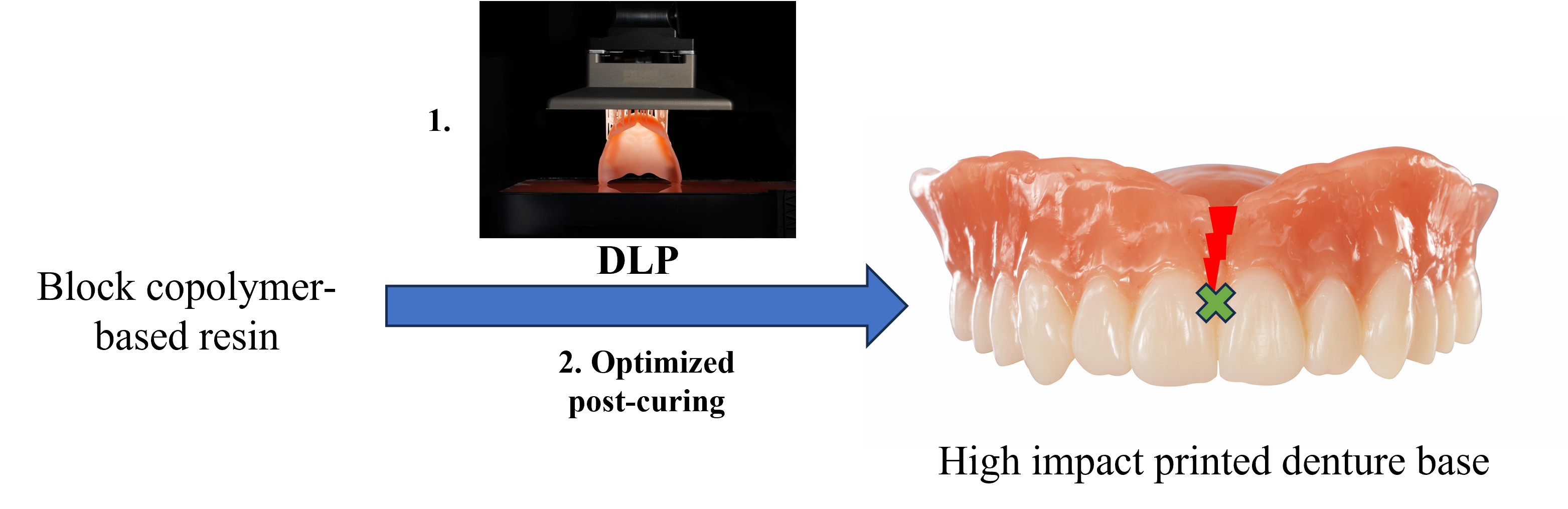
The objective of this contribution is to evaluate this BCP toughening technology for DLP 3D printing of fracture tough denture base materials. The DMA1/OMIMA/BCP1 combination was selected for this investigation, as it provided the highest fracture toughness up to now. Several research groups recently reported the strong influence of the post-curing step on the mechanical properties of printed materials [23,24,25,26]. Particularly, it was shown that heating during the post-processing step was advantageous and led to higher flexural strength/modulus values and improved conversion. For this reason, the selected 3D printed BCP1 based resins were post-cured using different conditions (variation of the temperature and of the irradiation time). In this article, the influence of the post-curing conditions on the flexural strength, flexural modulus, and fracture toughness of 3D printed BCP1 based materials is discussed. The difference of mechanical properties between bulk-cured (in a mold) and 3D printed materials is also addressed.
2. Materials and Methods
2.1. Materials
The monomer (octahydro-4,7-methano-1H-indenyl)methyl acrylate (OMIMA) was provided by Arkema and the initiator Genocure TPO (2,4,6-trimethylbenzoyldiphenylphosphine oxide) by Rahn AG. DMA1 and BCP1 were synthesized according to previously reported procedures [22]. Fumed silica nanoparticles (SiO2-NPs) silanized with 3-methacryloxypropyltrimethoxysilane, and exhibiting a specific surface area (BET) of 25 – 45 m2 g-1, were used as received from Evonik.
2.2. Formulation of Photopolymerizable BCP1 Based Monomer Mixtures
DMA1 and the monofunctional monomer OMIMA were mixed under magnetic stirring for several hours at 50 °C until full dissolution of the very highly viscous DMA1. 1.0 wt% TPO as well as BCP1 were subsequently added and stirring was continued until full solubilization of BCP1. Various BCP1 amounts were evaluated (4.0 wt%, 5.0 wt%, 6.0 wt%, 7.0 wt% and 8.0 wt%). These values represent the BCP1 content in the monomer mixture (without taking the filler into account). After addition of SiO2-NPs, the material was mixed and homogenized for 2×2 min at 2000 U min-1 using a SpeedMixer DAC 600.1.
2.3. Measurement of the Mechanical Properties, Glass-Transition Temperature, and Double Bond Conversion
2.3.1. Bulk Cured BCP1 Containing Materials
2.3.1.1. Flexural Strength and Flexural Modulus
Flexural strength (FS) and flexural modulus (FM) were assessed according to a slightly modified ISO 20795-1:2013 method. Specimens (3.3 mm x 10 mm x 64 mm) were prepared using stainless-steel molds (n = 6). The molds were filled with the photopolymerizable monomer mixture and covered with polyester film (50 µm) to avoid oxygen inhibition. Materials were light-cured in a LED cube 100 (20 mW cm−2 @ 405 nm and 100 mW cm−2 @ 450 nm, Hönle) controlled by a LED Powerdrive (Hönle) for 10 min for each side. After complete cure, the specimens were allowed to cool down to RT, removed from the molds and sanded from all sides with 600grit silicon carbide sandpaper. Specimens were then stored in water at 37 °C for 50 h. Measurement of FS/FM was carried out in a three-point bending test (span: 50 mm) with a cross-head speed of 5 mm min-1 using a Z2.5/TS universal testing machine (Zwick-Roell). The measurement was carried out in water at 37 °C (the specimens were immersed in a tempered water bath during the measurement).
2.3.1.2. Fracture Toughness
Fracture toughness was measured according to slightly modified ISO 20795-1:2013 method. Both the maximum stress intensity factor (Kmax) and the total work of fracture (Wf) were determined using single-edge notched beam (SENB) specimens (n = 6). A stainless-steel mold (4 mm × 8 mm × 40 mm) was filled with a photopolymerizable monomer mixture. The mold was covered with a polyester film (50 µm) to avoid oxygen inhibition. Materials were light-cured in a LED cube 100 (20 mW cm−2 @ 405 nm and 100 mW cm−2 @ 450 nm, Hönle) controlled by a LED Powerdrive (Hönle) for 10 min from both sides. After polymerization of the bulk specimens, a 1.0 mm wide and 3.0 mm deep notch of rectangular shape was prepared using a circular saw with diamond blade. A pre-crack was then prepared at the bottom of the notch by striking a razorblade with gentle pressure to a depth of 0.3 mm. Specimens were stored in water for seven days at 37 °C, dabbed with paper and subsequently loaded to break at RT with a span of 32 mm and at a crosshead speed of 1.0 mm min-1 using a universal testing machine (Zwick Z2.5, Zwick/Roell). Kmax (in MPa m1/2) was calculated as follows:
where f(x) is a geometrical function dependent on x:
with x = (a/ht) and ht as the height of the specimen (8 mm), bt its width (4 mm), lt the span length (32 mm), a the crack length (3 mm + pre-crack depth with razor blade), and Pmax the maximum load exerted on the specimens (in N).
The total fracture work Wf (in J m-2) was calculated as follows:
where U is the recorded area under the load/deflection curve.
2.3.2. 3D Printed Materials
2.3.2.1. Commercially Available 3D Printing Denture Base Materials
Two formulations were evaluated: Printodent® GR-14.2 denture HI (Pro3Dure) and Lucitone Digital PrintTM 3D denture base (Dentsply Sirona). They were printed using the ASIGA UV MAX 385 (Asiga) with a layer thickness of 100 μm and the printing parameters recommended by the manufacturers. Post-processing was carried out according to the manufacturers’ instructions, which included ultrasonic cleaning in isopropyl alcohol and light exposure in a post-curing device. Printodent® GR-14.2 denture HI was post-processed in a Otoflash G171 unit (NK-Optik GmbH), whereas Lucitone Digital PrintTM 3D denture base was post-cured in a Digital Cure large capacity unit (Dentsply Sirona).
2.3.2.2. BCP1 Containing Materials: 3D Printing Workflow
All samples were printed to their final dimensions on an DLP printer (PrograPrint PR5, Ivoclar Vivadent AG) using the parameter set “ProArt Print Splint”. Specimens were printed without supports and on their thinner side in horizontal orientation (in y-direction) with deactivated burn-in-layers. Excess resin was removed with wipes and specimens were post cured for 10 min for each side in a LED cube 100 (20 mW cm−2 @ 405 nm and 100 mW cm−2 @ 450 nm, Hönle) controlled by a LED Powerdrive (Hönle). The specimens were placed on a custom made pre-heated heating plate with adjustable temperature (set between RT and 120 °C) during the post-curing. After complete cure, the samples were allowed to cool down to RT and sanded from all sides with 600grit silicon carbide sandpaper, except for the NIR samples which were measured without sanding.
2.3.2.3. Flexural Strength and Flexural Modulus
Printed specimens (3.3 mm × 10 mm × 64 mm, n = 6) were stored in water at 37 °C for 50 h. The measurement was performed similarly to 2.3.1.1..
2.3.2.4. Fracture Toughness
SENB specimens (4.0 mm × 8.0 mm × 40 mm, n = 6) that already contained a 1.0 mm wide and 3.0 mm deep notch of rectangular shape (designed in the STL file) were 3D printed. After printing, the notch was cleaned carefully with paper towels to remove any excess resin, and the specimens were post-cured as described before. A pre-crack was then prepared at the bottom of the notch by striking a razorblade with gentle pressure to a depth of 0.3 mm. Specimens were stored in water for seven days at 37 °C and dabbed. The fracture toughness measurement was performed similarly to 2.3.1.2..
2.3.2.5. Glass-Transition Temperature (DMTA)
DMTA measurements were performed with printed specimens of the dimensions 5 mm × 2 mm × 40 mm on an Anton Paar MCR 301 device with a CTD 600 oven and a SRF 5 fixture. The measurements were carried out in torsion mode with a frequency of 1 Hz, a normal force (FN) of –1 N, and a strain of 0.05%. A temperature spectrum was monitored from 25 °C to 250 °C with a heating rate of 2 K min−1. The glass transition temperature (Tg) was defined as the temperature corresponding to the maximum of the loss factor (tan δ) curve.
2.3.2.6. Double Bond Conversion (NIR Spectrometry)
Spectra of the uncured material (film thickness 1.0 mm) and of printed, circular specimens (d = 15 mm, h = 1.0 mm, n = 3) were measured by NIR spectroscopy using an Invenio R spectrometer (Bruker, 16 Scans, 8 cm-1 Resolution, 3000 – 10000 cm-1). The (meth)acrylate overtone peak at 6165 cm-1 was integrated for both spectra. DBC was calculated by the following equation:
Acured and Auncured correspond to the integrated areas in the NIR spectrum (Acured: area of the (meth)acrylate peak for the cured material, Auncured: area of the (meth)acrylate peak for the uncured material).
2.3.3. IvoBase High Impact
IvoBase high impact was cured using the injection-molding system IvoBase according to the manufacurer’s instructions. FS/FM and fracture toughness were measured according to ISO 20795-1:2013.
3. Results
3.1. Difference Between 3D Printed and Bulk-Cured Materials
As a starting point for this work, a DMA1/OMIMA (1/1: wt/wt) formulation containing 5.0 wt% of BCP1 and 3.0 wt% of SiO2-NPs was either photocured in bulk (in a mold, photocuring conditions: 20 mW cm−2 @ 405 nm and 100 mW cm−2 @ 450 nm, 10 min for each side) or 3D printed. The following 3D printing workflow was selected: The specimens were firstly 3D printed using a PrograPrint PR5, cleaned with wipes, and finally post-cured at RT in a LED cube 100 curing unit using the same irradiation as for the bulk cured specimens (20 mW cm−2 @ 405 nm and 100 mW cm−2 @ 450 nm, 10 min for each side). Both the FS/FM and the fracture toughness were measured (Table 1). The results clearly showed that, under these conditions, the 3D printed material exhibited significantly lower mechanical properties. FS, FM, Kmax and Wf were far below the targeted ISO 20795-1:2013 values for materials with improved impact resistance (FS > 65 MPa, FM > 2000 MPa, Kmax ≥ 1.9 MPa m1/2 and Wf ≥ 900 J m-2).
3.2. 3D Printing of BCP1 Containing Formulations: Influence of the Post-Curing Conditions on the Mechanical Properties
In order to improve the mechanical properties of 3D printed materials, the post-curing conditions were subsequently modified. Various temperatures were additionally selected for the post-processing step: 60 °C, 80 °C, 100 °C and 120 °C. FM, FS, Kmax and Wf were assessed for each post-curing temperature (Figure 2, Figure 3, Figure 4 and Figure 5).
The results demonstrated that this parameter has a strong influence on the mechanical properties. Indeed, FS and FM obtained at RT and 60 °C were significantly lower than the values measured at 80 °C, 100 °C, and 120 °C. However, no difference was noticed between the specimens post-cured at 80 °C, 100 °C, or 120 °C. Using elevated temperatures during the post-processing step enabled the 3D printed BCP1 based material to fulfill the ISO 20795-1:2013 requirements for type 4 (light-activated materials) denture base polymers in terms of FS (FS > 65 MPa) and FM (FM > 2000 MPa) and to reach similar values than for the bulk-cured material (Table 1). Moreover, the post-curing temperature also significantly impacted the fracture toughness of the 3D printed specimens. Indeed, a lower Kmax value was obtained for the specimens post-cured at RT compared to those that were post-cured at elevated temperatures. Interestingly, regardless of the selected temperature (60 °C, 80 °C, 100 °C, or 120 °C), no significant differences were observed among the Kmax values, all of which exceeded 1.9 MPa m1/2. Under these conditions, the ISO 20795-1:2013 requirements concerning materials with improved impact resistance were therefore met. A similar trend was evident for Wf: Post-curing at RT produced a lower value, whereas elevated temperatures yielded comparable and consistently higher Wf values. Unfortunately, for each tested post-curing temperature, the Wf was lower than the minimally required 900 J m-2 for high impact denture bases (ISO 20795-1:2013) and did not reach the value obtained with the bulk-cured material.
The double-bond conversion (DBC) and Tg of printed materials were subsequently measured (Table 2). High DBC values (> 95%) were reached for each post-curing condition. The RT post-processed material exhibited somehow a lower DBC. The DMTA results clearly showed that the higher the post-curing temperature is, the higher the Tg. The influence of the post-curing irradiation time on the mechanical properties of the 3D printed materials was also investigated. 3D printed specimens were post-cured at 100 °C during 2×5 min, 2×10 min and 2×20 min. Similar FS, FM, Kmax and Wf values were obtained, independently of the post-curing time (Table 3).
3.3. 3D Printing of BCP1 Containing Formulations: Influence of the BCP1 and SiO2-NP Contents on the Mechanical Properties
The influence of the BCP1 content on both the FS/FM and the fracture toughness of DLP printed materials was studied (Figure 6, Figure 7, Figure 8 and Figure 9). DMA1/OMIMA (1/1: wt/wt) formulations containing 4.0 wt%, 6.0 wt%, and 7.0 wt% of BCP1 were first selected (each formulation additionally contained 3.0 wt% SiO2-NPs).
The following trends can clearly be identified: The higher the BCP1 content, the lower the FS and FM values. On the other hand, Wf increases together with the BCP1 amounts. A BCP1 content of at least 6.0 wt% was needed to reach the minimum requirements for high impact in terms of Kmax and Wf (ISO 20795-1: 2013). However, FS and FW were slightly below the targeted values. A formulation containing 8.0 wt% BCP1 and having an increased SiO2-NPs content of 10.0 wt% was finally prepared. This resin was able to fulfill all requirements for materials with improved impact resistance.
3.4. Comparison with the State of the Art
Two commercially available 3D printing denture base materials were also evaluated. Moreover, IvoBase high impact was selected as PMMA based reference material. FS, FM as well as Kmax and Wf were measured (Table 4).
Printodent® GR-14.2 denture HI exhibited satisfactory FS and FM values. However, the printed material was brittle and presented extremely low Kmax and Wf values. On the other hand, a high fracture toughness was obtained using Lucitone Digital PrintTM 3D denture base. However, the measured FS was significantly lower than the required 65 MPa (ISO20795-1: 2013). Interestingly, the mechanical properties measured with the optimized DMA1/OMIMA formulation (8.0 wt% BCP1; 10.0 wt% SiO2-NPs content) and with the PMMA based material (IvoBase high impact) were closing similar range.
4. Discussion
The objective of this work was to transfer the BCP toughening technology to DLP 3D printing. A resin formulation based on a DMA1/OMIMA/BCP1 combination was selected (with TPO as photoinitiator) for this study. Our research group recently showed that this formulation was suitable for the development of light-cured high impact denture bases, due to the self-assembly of BCP1 in the monomer mixture, acting as toughening agent [22]. However, these materials were photocured in a mold, raising the question of whether this technology can be effectively transferred to DLP 3D printing.
To answer this question, the BCP1 containing formulation (DMA1/OMIMA (1/1: wt/wt) + 5.0 wt% BCP1 + 3.0 wt% SiO2-NPs) was printed using a standard workflow. A PrograPrint PR5 was selected as DLP printer. Following printing, specimens were post-cured in a LED cube 100 curing unit at RT (20 mW cm−2 @ 405 nm and 100 mW cm−2 @ 450 nm, 10 min from both sides). The mechanical properties (FS/FM and fracture toughness) were measured and compared with the values of the bulk-cured material (Figure 2, Figure 3, Figure 4 and Figure 5 and Table 1). A significant drop in the mechanical properties was observed when the material was 3D printed, to values far below the minimum targeted requirements for high impact denture bases. This strong decrease might be due to the fact that a lower DBC was measured with the printed material (Table 3).
In order to improve the mechanical properties of 3D printed BCP1 based materials, we subsequently decided to focus on the variation of the post-curing conditions. The post-curing temperature has recently been shown to play a key role on the physical properties of printed materials [23,24,25,26]. As an example, K. Qi et al. printed a dimethacrylate based nanocomposite using SLA technology and evaluated the post-curing at either 40 °C or 80 °C [24]. The specimens that were post-cured at 80 °C exhibited significantly higher FS and FM. Therefore, different temperatures (60 °C, 80 °C, 100 °C, and 120 °C) were selected for the post-curing step in the present work. The results clearly demonstrated that the post-curing temperature has a strong influence on FS and FM (Figure 2 and Figure 3). Materials post-cured above 80 °C exhibited FS and FM values exceeding the minimum requirements for type 4 (light-activated materials) denture base polymers (ISO 20795-1:2013). These results are therefore in agreement with the conclusions of K. Qi et al. [24]. It is worth mentioning that both the FS and FM of these printed materials were similar to the bulk cured specimens. The same trend was observed for the fracture toughness (Figure 4 and Figure 5). Both Kmax and Wf values were strongly improved upon heating during the post-curing step. Unfortunately, and independently of the selected post-curing temperature, the measured Wf remained significantly below the 900 J m-2 threshold required for high impact denture bases. This outcome might suggest that the network structure of printed versus bulk cured materials is different (different crosslinking density). It might also be due to the presence of layers in the printed material or to the different photocuring process (in two steps for printed materials: Irradiation during printing, followed by a second irradiation in a post-curing unit). Nonetheless, it must be highlighted that the obtained fracture toughness is significantly improved in comparison to conventional denture bases. As an example, ProBase Hot (heat cure PMMA-based conventional denture base material from Ivoclar) exhibits a maximum stress intensity factor (Kmax) of 1.44 ± 0.18 MPa m1/2 and a total work of fracture (Wf) of 270 ± 30 J m-2 [27].
The post-curing temperature was also shown to have a strong influence on DBC and Tg of printed materials (Table 2). Heating during the post-curing step led to an increase in these values, which might explain the trend observed with regard to the values obtained for FS and FM. The Tg of the fully cured materials is slightly below 80 °C. With post-curing the material above the Tg, improvement in the polymer chain mobility is created, resulting in higher DBC, FS and FM. 100 °C seems to be the most suitable post-curing temperature as no further advantages were observed at 120 °C. Using these conditions, the irradiation time was varied. The results showed that this parameter did not have any influence on the mechanical properties of printed materials (Table 3).
Moreover, it is well known that the amount of BCP strongly influences the fracture toughness as well as FS/FM of bulk light-cured materials [42]. In this contribution, DMA1/OMIMA (1/1: wt/wt) formulations containing 4.0 wt%, 6.0 wt%, and 7.0 wt% of BCP1 were also 3D printed and evaluated. As expected, higher BCP1 content negatively influenced both FS and FM (Figure 6 and Figure 7). On the other hand, the higher the amounts of BCP1, the higher the Wf. An addition of 6.0 wt% of BCP1 was sufficient to exceed the minimum required Kmax and Wf values for high impact denture bases (Figure 8 and Figure 9). Unfortunately, FS and FM were slightly below the aimed values. In order to further optimize the formulation, both the BCP1 (8.0 wt%) and the SiO2-NP (10.0 wt%) contents were increased. The addition of higher inorganic filler amounts was expected to improve FS and FM values. This strategy proved effective, as the new resin was able to fulfill all criteria (FS, FM, Kmax and Wf) for materials with improved impact resistance (ISO 20795-1:2013). Two commercially available 3D printing denture base materials (Printodent® GR-14.2 denture HI and Lucitone Digital PrintTM 3D denture base) were additionally selected and evaluated (Table 4). Printodent® GR-14.2 denture HI was found to be extremely brittle and therefore rather unsuitable for denture bases. According to the state of the art, Lucitone Digital PrintTM 3D denture base is currently the only 3D printing material that exhibits high fracture toughness. Our results confirm this claim, as both Kmax and Wf were above the minimum required values for high impact materials (ISO 20795-1:2013). Moreover, our measurements confirm the observations of Lawson et al. and Coldea et al.: FS remains significantly lower than the aimed 65 MPa [17,18]. In this context, the optimized BCP1 containing resin is the only one able to fully meet the ISO20795-1:2013 requirements and can therefore be considered as a 3D printing high impact denture base. Even if the PMMA based gold standard material (IvoBase high impact) still exhibits higher FS and Wf values, the differences are modest (Table 4). In the near future, 3D printing denture bases should be able to exhibit similar mechanical properties than PMMA based reference materials.
5. Conclusion
In this contribution, DMA1/OMIMA containing various amounts of BCP1 were successfully printed using DLP technology. Interestingly, it was shown that the transfer of bulk photocuring (in molds) to 3D printing led to a decrease in fracture toughness. Although the addition of BCP1 clearly led to a strong increase of Kmax and Wf of 3D printed materials, the improvement was not as strong as for bulk-cured resins. This study also clearly highlighted the impact of the post-curing temperature on the 3D printed materials mechanical properties. It seems to be essential to heat the printed material above Tg during the post-processing step in order to obtain the best properties. Using an optimized formulation and an improved 3D printing workflow, it was possible to fulfill all the requirements for denture bases with improved impact resistance (ISO 20795-1:2013). This work therefore clearly demonstrates the high potential of the BCP toughening technology for the development of 3D printing high impact denture bases that would exhibit similar mechanical properties than the gold standard PMMA based materials.
Author Contributions
Conceptualization, K.R. and Y.C.; methodology, K.R., I. L., S.O., B.G. and Y.C.; software, K.R.; validation, K.R. and Y.C.; investigation, K.R., I. L., S.O., B.G. and Y.C.; resources, K.R. and Y.C.; data curation, K.R.; writing—original draft preparation, K.R., L.G. and Y.C.; visualization, Y.C.; supervision, K.R. and Y.C.; project administration, K.R. and Y.C. All authors have read and agreed to the published version of the manuscript.
Funding
This research received no external funding.
Data Availability Statement
The data presented in this study are available on request from the corresponding author.
Conflicts of Interest
The authors declare no conflicts of interest.
Abbreviations
The following abbreviations are used in this manuscript:
| MDPI | Multidisciplinary Digital Publishing Institute |
| DOAJ | Directory of open access journals |
| TLA | Three letter acronym |
| LD | Linear dichroism |
References
- Anusavice, K. J.; Shen, C.; Rawls, H. R. Phillips’science of dental materials, 12th edition; Elsevier Saunders: St Louis, Missouri, 2013; pp. 474–485. [Google Scholar]
- Alla, R. K.; Raghavendra Swamy, K. N.; Vyas, R.; Konakanchi, A. Conventional and contemporary polymers for the fabrication of denture prosthesis: part I – Overview, composition and properties. Int. J. Appl. Dent. Sci. 2015, 1, 82–89. [Google Scholar]
- Biglin, M. S.; Baytaroğlu, E. N.; Erdem, A.; Dilber, E. A review of computer-aided design/computer-aided manufacture techniques for removable denture fabrication. Eur. J. Dent. 2016, 10, 286–291. [Google Scholar]
- Maeda, Y.; Minoura, M.; Tsutsumi, S.; Okada, M.; Nokubi, T. A CAD/CAM system for removable denture. Part I: Fabrication of complete dentures. Int. J. Prosthodont 1994, 7, 17–21. [Google Scholar] [PubMed]
- Baba, N. Z.; AlRumaih, H. S.; Goodacre, B. J.; Goodacre, C. J. Current techniques in CAD/CAM denture fabrication. Gen. Dent. 2016, 64, 23–28. [Google Scholar] [PubMed]
- Pacquet, W.; Benoit, A.; Hatège-Kimana, C.; Wulfman, C. Mechanical Properties of CAD/CAM Denture Base Resins. Int. J. Prosthodont 2019, 32, 104. [Google Scholar] [CrossRef] [PubMed]
- Srinivasan, M.; Kalberer, N.; Kamnoedboon, P.; Mekki, M.; Durual, S.; Özcan, M.; Müller, F. CAD-CAM complete denture resins: an evaluation of biocompatibility, mechanical properties, and surface characteristics. J. Dent. 2021, 114, 103785. [Google Scholar] [CrossRef]
- Perea-Lowery, L.; Gibreel, M.; Vallittu, P. K.; Lassila, L. V. 3D-Printed vs. Heat-polymerizing and autopolymerizing denture base acrylic resins. Materials 2021, 14, 5781. [Google Scholar] [CrossRef]
- Batisse, C.; Nicolas, E. Comparison of CAD/CAM and conventional denture base resins: A systematic review. Appl. Sci. 2021, 11, 5990. [Google Scholar] [CrossRef]
- Andjela, L.; Abdurahmanovich, V. M.; Vladimirovna, S. N.; Mikhailovna, D. I.; Yurievich, D. D.; Alekseevna, M. Y. A review on Vat photopolymerization 3D-printing processes for dental application. Dent. Mater. 2022, 38, E284–E296. [Google Scholar] [CrossRef]
- Della Bona, A.; Cantelli, V.; Britto, V. T.; Collares, K. F.; Stansbury, J. W. 3D printing restorative materials using a stereolithographic technique: a systematic review. Dent. Mater. 2021, 37, 336–350. [Google Scholar] [CrossRef]
- Tigmeanu, C. V.; Ardelean, L. C.; Rusu, L.-C.-; Negrutiu, M.-L. Additive manufactured polymers in dentistry, current state-of-the-art and future perspectives-a review. Polymers 2022, 14, 3658. [Google Scholar] [CrossRef]
- Balhaddad, A. A.; Garcia, I. M.; Mokeem, L.; Alsahafi, R.; Majeed-Saidan, A.; Albagami, H. H.; Hhan, A. S.; Ahmad, S.; Mezzomo Collares, F.; Della Bona, A.; Melo, M. A. S. Three-dimensional (3D) printing in dental practice: Applications, areas of interest, and level of evidence. Clin. Oral Investig. 2023, 27, 2465–2491. [Google Scholar] [CrossRef]
- Goodacre, B. J. 3D printing of complete dentures: A narrative review. Int. J. Prosthodont 2024, 37, s159–s164. [Google Scholar] [CrossRef]
- Geiger, V.; Mayinger, F.; Hoffmann, M.; Reymus, M.; Stawarkzyk, B. Fracture toughness, work of fracture, flexural strength and elastic modulus of 3D-printed denture base resins in two measurement environments after artificial aging. J. Mech. Behav. BioMed Mater. 2024, 150, 106234. [Google Scholar] [CrossRef] [PubMed]
- Hetzler, S.; Rehm, S.; Räther, S.; Rues, S.; Zenthöfer, A.; Rammelsberg, P.; Schwindling, F. S. Fracture toughness, work of fracture and hardness of 3D-printed denture base resins. Materials 2025, 18, 4338. [Google Scholar] [CrossRef] [PubMed]
- Lawson, N. C.; Safadi, Y.; Alford, A.; Aggarwal, H.; Bora, P. V.; Lawson, T. J.; Givan, D. A. Flexural strength, fracture toughness, translucency, stain resistance, and water sorption of 3D-printed, milled, and conventional denture base materials. J. Prosthodont 2026, 35, 553–560. [Google Scholar] [CrossRef] [PubMed]
- Coldea, A.; Mayinger, F.; Meinen, J.; Hoffmann, F.; Stawarczyk, B. Mechanical properties of 3D printed denture base polymers. J. Prosthet. Dent. 2025, 133, 1361 e1–e8. [Google Scholar] [CrossRef]
- Demleitner, M.; Schönl, S.; Angermann, J.; Fässler, P.; Lamparth, I.; Rist, K.; Schnur, T.; Catel, Y.; Rosenfeldt, S.; Retsch, M.; Ruckdäschel, H.; Altstädt, V. Influence of block copolymer concentration and resin crosslink density on the properties of UV-curable methacrylate resin systems. Macromol. Mater. Eng. 2022, 307, 2200320. [Google Scholar] [CrossRef]
- Schönl, F.; Demleitner, M.; Angermann, J.; Fässler, P.; Lamparth, I.; Rist, K.; Schnur, T.; Catel, Y.; Rosenfeldt, S.; Ruckdäschel, H. Synthesis and evaluation of novel urethane macromonomers for the formulation of fracture tough 3D printable dental materials. J. Mecha Behav. BioMed Mater. 2024, 160, 106737. [Google Scholar] [CrossRef]
- Ott, E.; Fässler, P.; Grob, B.; Rist, K.; Vidal, L.; Lalevée, J.; Catel, Y. Evaluation of novel urethane dimethacrylates as crosslinkers for the development of fracture tough dental materials containing a poly(ε-caprolactone)-polydimethylsiloxane-poly(ε-caprolactone) triblock copolymer. J. Appl. Polym. Sci. 2024, 141, e55724. [Google Scholar] [CrossRef]
- Fässler, P.; Lamparth, I.; Omeragic, S.; Grob, B.; Rist, K.; Cousin, F.; Vidal, L.; Lalevée, J.; Catel, Y. Block copolymers: Efficient toughening agents for the preparation of 3D-printable high impact denture base materials. Dent. Mater. 2025, 41, 839–849. [Google Scholar] [CrossRef]
- Khosravani, M. R.; Ayatollahi, M. R.; Reinicke, T. Effects of post-processing techniques on the mechanical characterization of additively manufactured parts. J. Manuf. Process. 2023, 107, 98–114. [Google Scholar] [CrossRef]
- Qi, K.; Hada, T.; Ren, X.; Iwaki, M.; Minakuchi, S.; Kanazawa, M. Effects of post-polymerization conditions on the mechanical properties of 3D-printed dental resin nanocomposite. J. Prosthodont Res. 2025, 69, 553–561. [Google Scholar] [CrossRef] [PubMed]
- Staffova, M.; Ondreas, F.; Svatik, J.; Zboncak, M.; Jancar, J.; Lepcio, P. 3D printing and post-curing optimization of photopolymerized structures: Basic concepts and effective tools for improved thermomechanical properties. Polym. Test. 2022, 108, 107499. [Google Scholar] [CrossRef]
- Cheadle, A. M. G.; Maier, E.; Palin, W. M.; Tomson, P. L.; Poologasundarampillai, G.; Hadis, M. A. The impact of modifying 3D printing parameters on mechanical strength and physical properties in vat photopolymerisation. Sci. Rep. 2025, 15, 12592. [Google Scholar] [CrossRef]
- Zappini, G.; Kammann, A.; Wachter, W. Comparison of fracture tests of denture base materials. J. Prosthet. Dent. 2003, 90, 578–585. [Google Scholar] [CrossRef]
Figure 1.
Structures of DMA1, BCP1 and OMIMA.

Figure 2.
FS of printed materials that were post-cured at different temperatures. Material: DMA1/OMIMA (1/1: wt/wt) + 5.0 wt% BCP1 + 3.0 wt% SiO2-NPs. The red line represents the minimum required value according to ISO 20795-1:2013 for type 4 (light-activated materials) denture base polymers.
Figure 2.
FS of printed materials that were post-cured at different temperatures. Material: DMA1/OMIMA (1/1: wt/wt) + 5.0 wt% BCP1 + 3.0 wt% SiO2-NPs. The red line represents the minimum required value according to ISO 20795-1:2013 for type 4 (light-activated materials) denture base polymers.

Figure 3.
FM of printed materials that were post-cured at different temperatures. Material: DMA1/OMIMA (1/1: wt/wt) + 5.0 wt% + 3.0 wt% SiO2-NPs. The red line represents the minimum required value according to ISO 20795-1:2013 for type 4 (light-activated materials) denture base polymers.
Figure 3.
FM of printed materials that were post-cured at different temperatures. Material: DMA1/OMIMA (1/1: wt/wt) + 5.0 wt% + 3.0 wt% SiO2-NPs. The red line represents the minimum required value according to ISO 20795-1:2013 for type 4 (light-activated materials) denture base polymers.

Figure 4.
Kmax of printed materials that were post-cured at different temperatures. Material: DMA1/OMIMA (1/1: wt/wt) + 5.0 wt% BCP1 + 3.0 wt% SiO2-NPs. The red line represents the minimum required value according to ISO 20795-1:2013 for denture bases exhibiting improved impact resistance (high impact).

Figure 5.
Wf of printed materials that were post-cured at different temperatures. Material: DMA1/OMIMA (1/1: wt/wt) + 5.0 wt% + 3.0 wt% SiO2-NPs. The red line represents the minimum required value according to ISO 20795-1:2013 for denture bases exhibiting improved impact resistance (High impact).
Figure 5.
Wf of printed materials that were post-cured at different temperatures. Material: DMA1/OMIMA (1/1: wt/wt) + 5.0 wt% + 3.0 wt% SiO2-NPs. The red line represents the minimum required value according to ISO 20795-1:2013 for denture bases exhibiting improved impact resistance (High impact).

Figure 6.
FS of printed DMA1/OMIMA (1/1: wt/wt) materials containing various amounts of BCP1 and SiO2-NPs (*: 3.0 wt%; **: 10.0 wt%). The red line represents the minimum required value according to ISO 20795-1:2013 for type 4 (light-activated materials) denture base polymers.
Figure 6.
FS of printed DMA1/OMIMA (1/1: wt/wt) materials containing various amounts of BCP1 and SiO2-NPs (*: 3.0 wt%; **: 10.0 wt%). The red line represents the minimum required value according to ISO 20795-1:2013 for type 4 (light-activated materials) denture base polymers.

Figure 7.
FM of printed DMA1/OMIMA (1/1: wt/wt) materials containing various amounts of BCP1 and SiO2-NPs (*: 3.0 wt%; **: 10.0 wt%). The red line represents the minimum required value according to ISO 20795-1:2013 for type 4 (light-activated materials) denture base polymers.
Figure 7.
FM of printed DMA1/OMIMA (1/1: wt/wt) materials containing various amounts of BCP1 and SiO2-NPs (*: 3.0 wt%; **: 10.0 wt%). The red line represents the minimum required value according to ISO 20795-1:2013 for type 4 (light-activated materials) denture base polymers.

Figure 8.
Kmax of printed DMA1/OMIMA (1/1: wt/wt) materials containing various amounts of BCP1 and SiO2-NPs (*: 3.0 wt%; **: 10.0 wt%). The red line represents the minimum required value according to ISO 20795-1:2013 for denture bases exhibiting improved impact resistance (High impact).
Figure 8.
Kmax of printed DMA1/OMIMA (1/1: wt/wt) materials containing various amounts of BCP1 and SiO2-NPs (*: 3.0 wt%; **: 10.0 wt%). The red line represents the minimum required value according to ISO 20795-1:2013 for denture bases exhibiting improved impact resistance (High impact).

Figure 9.
Wf of printed DMA1/OMIMA (1/1: wt/wt) materials containing various amounts of BCP1 and SiO2-NPs (*: 3.0 wt%; **: 10.0 wt%). The red line represents the minimum required value according to ISO 20795-1:2013 for denture bases exhibiting improved impact resistance (High impact).
Figure 9.
Wf of printed DMA1/OMIMA (1/1: wt/wt) materials containing various amounts of BCP1 and SiO2-NPs (*: 3.0 wt%; **: 10.0 wt%). The red line represents the minimum required value according to ISO 20795-1:2013 for denture bases exhibiting improved impact resistance (High impact).


Table 1.
FS/FM, Kmax and Wf of bulk cured and 3D printed (post-curing at RT) DMA1/OMIMA (1/1: wt/wt) materials containing 5.0 wt% of BCP1 and 3.0 wt% of SiO2-NPs.
Table 1.
FS/FM, Kmax and Wf of bulk cured and 3D printed (post-curing at RT) DMA1/OMIMA (1/1: wt/wt) materials containing 5.0 wt% of BCP1 and 3.0 wt% of SiO2-NPs.
| Post-curing irradiation time (min) | FS ISO 50 h 37 °C (MPa) | FM ISO 50 h 37 °C (MPa) | Kmax (MPa m1/2) | Wf (J m-2) |
| Bulk curing | 62.7 ± 1.7 | 2028 ± 119 | 2.25 ± 0.05 | 1439 ± 31 |
| 3D printing | 52.2 ± 1.2 | 1516 ± 42 | 1.62 ± 0.10 | 460 ± 45 |
Table 2.
Tg and DBC of printed materials that were post-cured at different temperatures. Material: DMA1/OMIMA (1/1: wt/wt) + 5.0 wt% BCP1 + 3.0 wt% SiO2-NPs.
Table 2.
Tg and DBC of printed materials that were post-cured at different temperatures. Material: DMA1/OMIMA (1/1: wt/wt) + 5.0 wt% BCP1 + 3.0 wt% SiO2-NPs.
| Post-curing temperature (°C) | DBC (%) | Tg (°C) |
| RT | 95 ± 1 | 65.5 |
| 60 | 98 ± 1 | 70.0 |
| 80 | 99 ± 1 | 73.0 |
| 100 | 100 | 74.0 |
| 120 | 100 | 76.2 |
Table 3.
FS, FM, Kmax, and Wf measured after various post-curing irradiation times at 100 °C. Material: DMA1/OMIMA (1/1: wt/wt) + 5.0 wt% BCP1 + 3.0 wt% SiO2-NPs.
Table 3.
FS, FM, Kmax, and Wf measured after various post-curing irradiation times at 100 °C. Material: DMA1/OMIMA (1/1: wt/wt) + 5.0 wt% BCP1 + 3.0 wt% SiO2-NPs.
| Post-curing irradiation time (min) | FS ISO 50 h 37 °C (MPa) | FM ISO 50 h 37 °C (MPa) | Kmax (MPa m1/2) | Wf (J m-2) |
| 2 x 5 | 67.0 ± 1.4 | 2135 ± 47 | 2.07 ± 0.17 | 677 ± 80 |
| 2 x 10 | 68.6 ± 1.6 | 2198 ± 58 | 2.06 ± 0.10 | 686 ± 62 |
| 2 x 20 | 69.2 ± 0.6 | 2145 ± 38 | 2.02 ± 0.11 | 615 ± 64 |
Table 4.
FS, FM, Kmax, and Wf of four commercially available materials.
| Material | FS ISO 50 h 37 °C (MPa) | FM ISO 50 h 37 °C (MPa) | Kmax (MPa m1/2) | Wf (J m-2) |
| Printodent® GR-14.2 denture HI | 69.2 ± 1.8 | 2153 ± 76 | 0.82 ± 0.04 | 79 ± 10 |
| Lucitone Digital PrintTM 3D denture base | 56.7 ± 1.9 | 2144 ± 121 | 1.92 ± 0.09 | 1272 ± 177 |
| IvoBase high impact | 73.8 ± 2.0 | 2361 ± 53 | 2.12 ± 0.07 | 1366 ± 72 |
Disclaimer/Publisher’s Note: The statements, opinions and data contained in all publications are solely those of the individual author(s) and contributor(s) and not of MDPI and/or the editor(s). MDPI and/or the editor(s) disclaim responsibility for any injury to people or property resulting from any ideas, methods, instructions or products referred to in the content. |
© 2026 by the authors. Licensee MDPI, Basel, Switzerland. This article is an open access article distributed under the terms and conditions of the Creative Commons Attribution (CC BY) license (http://creativecommons.org/licenses/by/4.0/).
Copyright: This open access article is published under a Creative Commons CC BY 4.0 license, which permit the free download, distribution, and reuse, provided that the author and preprint are cited in any reuse.


