

Submitted:
28 May 2026
Posted:
29 May 2026
You are already at the latest version
Abstract
Hantavirus cardiopulmonary syndrome (HCPS) is characterized by endothelial dysfunction and sudden vascular collapse, with a stereotyped biphasic course: a prodromal febrile phase with thrombocytopenia, followed in severe cases by explosive non-cardiogenic pulmonary oedema, shock and death (case-fatality 30–45% for HCPS causing species). We propose that HCPS is fundamentally a disease of kinin dysregulation, in which an endothelial IL-18/IFN-γ autoinflammatory loop predisposes to uncontrolled contact-system activation and bradykinin-driven capillary leak.Pathogenic hantaviruses can inhibit endothelial cell death pathways, maintaining long-lived endothelial “viral factories” that continue to trigger type I interferon and non-canonical caspase-4 activation. This pathway could drive selective IL-18 release (without IL-1β surge), which in concert with IL-12 licenses an NK/CD8 IFN-γ response. IFN-γ signals back on the endothelium in a self-sustaining loop that has no intrinsic off-switch because the virus maintains endothelial persistence without triggering cell death pathways. This loop's primary effect is not acute lethality but dysregulation of the kinin system resulting in chronic IFN-γ signalling that remodels the kinin-receptor repertoire in the inflammatory environment, with upregulation of the inducible B1 receptor (BKB1R) that can act as a synergistic receptor for B2 receptor (BKB2R). The clinical fingerprint is rising ferritin with disproportionately low CRP.Around day 7–10, antibody-mediated changes in viral-endothelial-platelet interactions may unmask a procoagulant endothelial surface, triggering FXII autoactivation and a bradykinin burst. On an endothelium already dysregulated by chronic IFN-γ, the newly induced BKB1R amplifies the BKB2R-mediated permeability response, precipitating explosive capillary leak. These inflammatory events inevitably produce TNF/IL-1/IL-6, that will happen in any antimicrobial response that normally aid immune recruitment and tissue repair. However, when standing IFN-γ remains elevated and compensatory mechanisms (IL-1Ra, IL-10) fail to contain these innate signals, TNF/IL-1/IL-6 potentiate endothelial dysfunction and risk amplification toward systemic shock. Critically, icatibant (a selective B2R antagonist) is sufficient to arrest progression, a clinical observation that unifies hantavirus pathogenesis with the bradykinin-dysregulation framework previously established for severe COVID-19.This model identifies kinin-system antagonism (icatibant) as the rational primary intervention, with short-acting JAK inhibition as supportive but balanced modulators of the inflammatory dysregulation driver. Here, the balance is critical, while JAK/IFN-γ blockade reduces the dysregulation substrate, excessive suppression of type I/II IFN impairs antiviral immunity and risks enabling viral persistence. The predictions are testable with existing assays and drugs, the time to test them is now.
Keywords:
hantavirus
; ANDV
; Andes
; Bradykinin
; platelets
; endothial cell
; cytokines
; ferritin
; IL‐18
; BKB2R
; icatibant
; JAKinh
; IFNg
; caspase‐4
; HCPS
Introduction
Hantaviruses are rodent-borne RNA viruses causing two syndromes: haemorrhagic fever with renal syndrome (HFRS) in Eurasia and hantavirus cardiopulmonary syndrome (HCPS) in the Americas [1]. Both share a central feature, non-cytopathic infection of microvascular endothelial cells coupled with increased vascular permeability, but not through cell lysis [2,3]. Instead, permeability arises from functional disruption of the endothelial barrier rather than cell death, placing hantaviruses within the non-cytopathic, endothelial-tropic hemorrhagic fevers, alongside dengue and arenaviruses such as Lassa, in contrast to the directly cytopathic filoviruses (Ebola, Marburg) [4,5,6].
The clinical course of HCPS is strikingly biphasic. After 1–6 weeks incubation, symptomatic disease begins with a 2–7-day febrile prodrome of fever, myalgias, abdominal pain and headache, but notably sparse in respiratory symptoms [1]. A subset of patients then deteriorates within hours into non-cardiogenic pulmonary edema, respiratory failure and refractory shock, with most deaths occurring in the first 24 hours of admission [1,7]. Survivors can recover quickly once supported through the cardiopulmonary phase, suggesting that the underlying endothelial dysfunction is reversible [8,9].
The laboratory signature is distinctive and mechanistically informative: early and progressive thrombocytopenia, a platelet count <40,000/µL predicts mortality, relative leukocytosis with lymphopenia, immunoblastosis, hemoconcentration, and markedly elevated D-dimers without overt thromboembolism [1,10]. Critically, IL-6 and CRP remain modest while IL-18, IFN-γ and IFN-γ-induced chemokines (CXCL9, CXCL10) are conspicuously elevated, a signature of IL-18/IFN-γ-driven pathology rather than classical cytokine storm or HLH/MAS [7].
No specific antiviral or immunomodulatory therapy is licensed for HCPS. Randomized trials of ribavirin and, high-dose corticosteroids were negative, and supportive care usually avoids aggressive fluid resuscitation because of the vascular leakage [11,12,13]. Yet two individual cases of severe Puumala HFRS were given icatibant, a selective B2-receptor bradykinin antagonist, with stabilization of disease progression and recovery, a hint that has been overlooked [14,15]. Although the kallikrein-kinin system (KKS) has been sporadically investigated in select viral infections, including one early study in dengue hemorrhagic fever in 1975 [16] and a few reports on viral hepatitis, Influenza, and viral hemorrhagic fevers (VHF) through the 1990s, mostly reported in Russian language literature [16,17,18,19], systematic exploration of KKS dysregulation as a driver of viral pathogenesis has been limited. While Joseph and Kaplan established the theoretical foundation for understanding bradykinin-forming pathways in innate inflammation [20,21,22], their work focused primarily on hereditary angioedema (HAE) and chronic inflammatory conditions, leaving acute viral pathogenesis largely unexplored through a KKS lens. A significant but notable exception was a 2013 study demonstrating that hantavirus directly activates the plasma kallikrein-kinin system to induce endothelial hyperpermeability [23], yet this mechanistic insight did not catalyze broader investigation into KKS dysfunction across the spectrum of hemorrhagic viral infections.
Among early investigations of dysregulated bradykinin signaling in severe COVID-19, our mechanistic hypothesis [24] contributed to broader attention to the KKS as a pathophysiological axis in viral disease. The hypothesis proposed that ACE2 downregulation impairs bradykinin inactivation, thereby driving unopposed B1/B2 receptor activation and vascular leak [24]. This hypothesis has been independently substantiated by different mechanistic studies documenting elevated plasma and tissue kallikrein activity, accumulation of kinin peptides, and mast cell-mediated endothelial dysfunction [25,26,27,28,29,30,31,32]. Moreover, clinical proof-of-concept has emerged from one randomized trials and one exploratory studies demonstrating beneficial clinical outcomes of bradykinin B2 receptor antagonism (icatibant) in COVID-19, while one randomized trial did not show efficacy [33,34,35,36]. Plasma kallikrein inhibition (with lanadelumab) demonstrated no effect in mild COVID-19 [37]. Lanadelumab was also enrolled in the COMMUNITY platform trial (NCT04590586), an ambitious multi-sponsor, adaptive design study launched in late 2020 through the COVID R&D Alliance involving Amgen, UCB, and other major pharmaceutical companies across global sites. Despite this enormous coordinated effort, peer-reviewed efficacy outcomes for the lanadelumab arm have not yet been published. After promising data from a pilot study, a subsequent larger trial with C1 esterase inhibitor replacement demontrated no effect in severe COVID-19 pneumonia [38,39]. Several mechanistic reviews further support KKS involvement in viral infection [40,41,42].
Recognizing that both hantavirus cardiopulmonary syndrome and severe COVID-19 manifest as non-cytopathic, endothelial-tropic diseases with dysregulated vascular permeability as a final common pathway [30], we propose that the detailed mechanistic KKS framework that is now established in COVID-19 provides a robust template for systematically reinvestigating kallikrein-kinin system dysfunction in hantavirus disease. The 2013 hantavirus KKS observation, viewed through the lens of modern mechanistic understanding and clinical therapeutic validation from COVID-19, warrants comprehensive translational investigation in this parallel viral setting.
Hantavirus Disease as Kinin Dysregulation, a Testable Mechanistic Framework
Non-Lytic Infection Sustains a Productively Infected Endothelium
Hantaviruses replicate to high titres in endothelial cells without inducing cell death [2,43] (Figure 1). Hantaviruses, including Andes virus, actively blocks endothelial cell death pathways through caspase-3 inhibition and BCL2 upregulation [44,45]. The consequence is a long-lived, productively infected endothelial cell that continuously exposes viral RNA and other pathogen-associated molecular patterns (PAMPs) to its cytosol while remaining morphologically intact. This enforced survival is the pivot-point of the disease, it removes the natural off-switch that would terminate inflammation in normal infections, enabling instead a self-sustaining deregulatory loop.
Type I Interferon, GBPs and Caspase-4 Drive Selective IL-18 Release
Cytosolic viral RNA engages RIG-I and cGAS–STING sensors, triggering type I interferon (IFN-α/β) production [46,47]. Type I IFN upregulates guanylate-binding proteins (GBPs), which activate caspase-4 (the non-canonical inflammasome caspase) [48]. Caspase-4 can cleave gasdermin D (GSDMD) and IL-18, generating sublytic pores that permit selective release of mature IL-18 [49,50,51], and the apoptotic and necroptotic executioners that have been suppressed by viral anti-death proteins keep this process ongoing. The result is high IL-18 release without the IL-1β/IL-6 surge of classical inflammasome activation, precisely the laboratory pattern observed in hantavirus infection (Figure 1).
Platelet Recruitment and a Platelet-Adherent Endothelial Layer
Infected endothelial cells recruit circulating platelets to their luminal surface [52]. Hantavirus has also been shown to attach to b3-integrins on platelets and therefore might lead and drive an interaction between virus, platelets and endothelium that will lead to an aggregate layering the pulmonary endothelium [52] (Figure 1). Adherent platelets become activated, and degranulate, releasing inorganic polyphosphate (polyP), zinc and phosphatidylserine-bearing microparticles [53] (Figure 1). This adhesive interaction could contribute to the progressive prodromal thrombocytopenia that is one of the earliest and most reliable laboratory findings associated with HCPS, often preceding overt vascular leak by days [8,52,54].
A central hypothesis is that, in this prodromal configuration, a "platelet carpet", may partially shield the endothelial membrane from contact-system activation (Figure 1). While polyP is inherently procoagulant, the endothelial surface might remain covered and relatively inaccessible to FXII to trigger fulminant activation in the prodromal stage. This is the key reason why FXII activation and bradykinin generation are restrained during the prodrome, even as the circulating platelet count falls. The virus attached to the platelets in the aggregate and the infected endothelium are thus protected by a dynamic equilibrium. Platelets are continuously recruited and consumed, resulting in the falling platelet count, but the supply is sufficient to maintain endothelial surface coverage. The platelets serve a dual purpose, they deposit the raw material (polyP, zinc, PS) that will later fuel contact-system activation, but for now they shield it from access. This is a remarkable example of viral exploitation of host hemostasis, as the virus maintains a "Trojan carpet" of platelets that simultaneously sustains infection by preventing the immune counterattack that exposition of the infected endothelial cells would trigger (Figure 1).
The IL-18/IFN-γ Loop Is Self-Amplifying and Dysregulates the Kinin System
Mature IL-18, synergizing with IL-12, drives NK cells and Th1 effector cells to produce IFN-γ [55]. IFN-γ then signals back on the surviving endothelium through JAK1/JAK2 and STAT1, upregulating interferon-stimulated genes including GBPs, caspase-4, and chemokines (CXCL9, CXCL10, CXCL11) [49,56,57,58,59]. This recruits more CD8-T cells and NK cells, perpetuating the loop, IFN-γ → caspase-4 → IL-18 → more IFN-γ [49,60,61]. Because the infected endothelial cell does not enter a cell death program [44], this loop escalates as a self-sustaining autoinflammatory state, even despite IL-18BP (which is the natural inhibitor of IL-18 [62]). Critically, prolonged high-level IFN-γ signaling does more than sustain inflammation, it dysregulates the kinin system. IFN-γ upregulates the inducible BKB1R on endothelial cells [63]. The endothelium is thus remodeled toward kinin sensitivity, over days 1–7, IFN-γ signaling creates a surface primed for increased bradykinin signaling due to upregulation of BKB1R that works synergistically with BKB2R, should the contact system ever be activated [64,65].
Day 7-10, Contact-System Activation and the Bradykinin Burst
Around day 7–10, neutralizing IgG antibodies appear and the virus is significantly neutralized halting virus-induced pathological host dysregulation [1,66]. In vitro data has shwn that platelet adherence to infected endothelial cells is β3-integrins dependent and can be neutralized by ANDV-specific neutralizing antibodies [52]. We hypothesize that these neutralizing antibodies reduces the protective coating on platelets that shields the endothelial surface exposing an anionic endothelial surface. These dynamics result in a biphasic platelet behavior characterized by inhibition of adhesion with progressive thrombocytopenia and subsequent restoration of platelet-endothelial interactions with circulating platelet counts stabalizing and eventually rising again. This exposed surface may be decorated with residual polyphosphate (polyP), zinc and phosphatidylserine from activated and possible now dying endothelial cells. This is an ideal scaffold for FXII autoactivation, polyP aligns and stabilizes FXII, and zinc bridges histidine residues to the negatively charged surface and FXII then autoactivates (with zinc as cofactor, the dominant source are platelets) into FXIIa, cleaving prekallikrein to generate plasma kallikrein, which reciprocally amplifies FXII activation and cleaves high-molecular-weight kininogen to release bradykinin [22,67,68]. Bradykinin encounters endothelial B2 receptors that are constitutive, and can initiate an acute permeability response, and the newly induced B1 receptors, which are inducible, pro-inflammatory, and amplify the permeability response [22,69]. The consequence is explosive non-cardiogenic pulmonary oedema, the signature of HCPS.
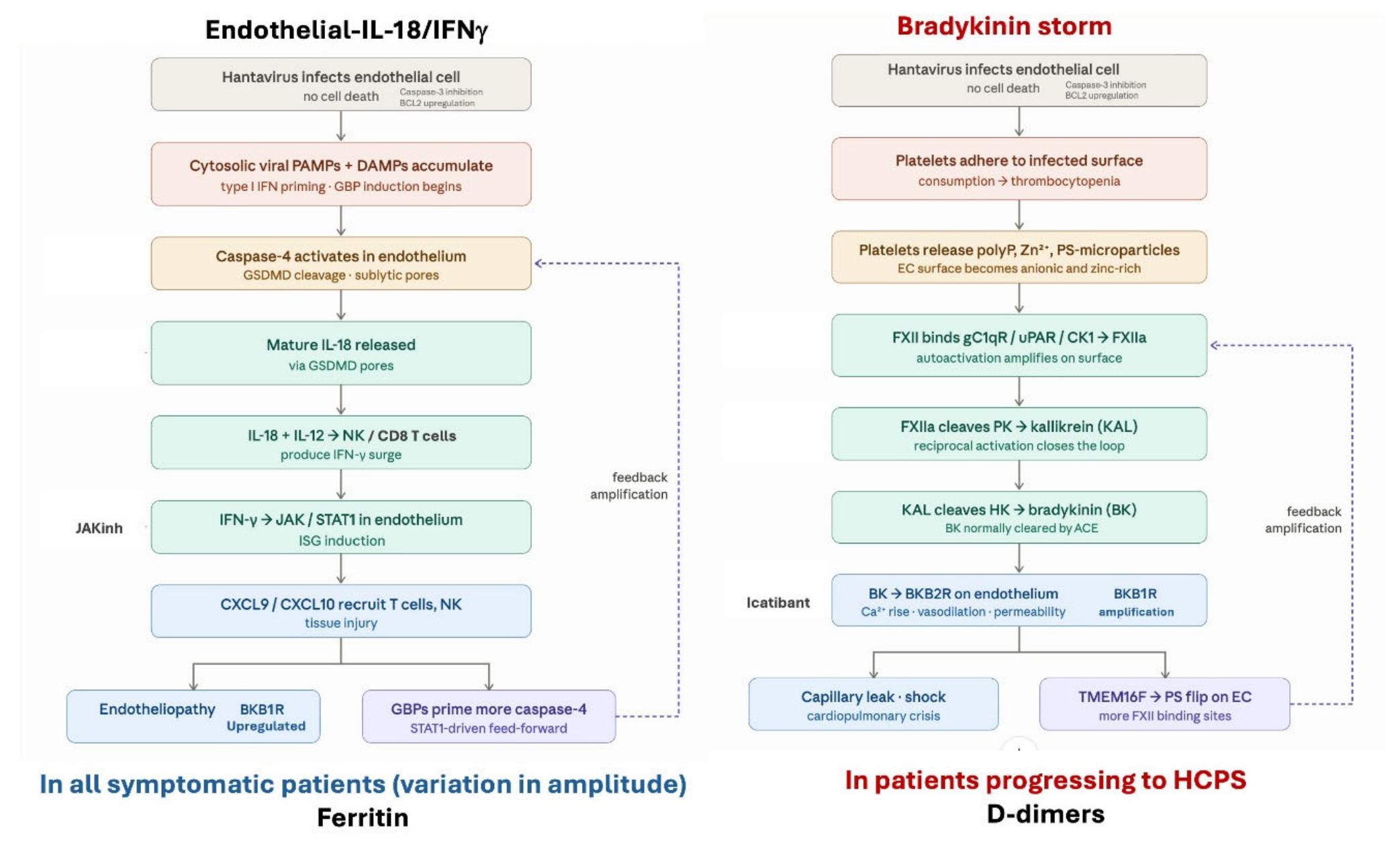
The same day 7-10 events that unmasks the contact surface might also deliver a more significant TNF/IL-1/IL-6 signal, from virus-specific T cells, from recruited monocytes/macrophages, or from lytic death of infected endothelium. This combination of TNF/IL-1/IL-6, when not kept in control by natural counterregulatory mechanisms, might synergizes with the standing high IFN-γ a combination of cytokines known to induce cytokine-driven shock [70]. Once initiated, the bradykinin burst is self-amplifying through TMEM16F-dependent phosphatidylserine externalization on the endothelial surface [71,72], creating more FXII-binding sites [73]. Without intervention, the cascade does not self-terminate until the supply of prekallikrein and HMWK is exhausted or the patient dies. With intervention, such as icatibant to block B2R, the loop is broken, and because most endothelial cells are not lytically destroyed, recovery can be remarkably rapid (Figure 3).
Biomarkers of Dysregulation and Collapse
The biphasic model predicts a distinct biomarker hierarchy.
Days 1–7 (prodromal phase and dysregulation phase). Ferritin rises and tracks the IL-18/IFN-γ loop, regardless of clinical disease severity. IL-18 and CXCL9/CXCL10 are elevated. CRP and IL-6 remain disproportionately low (in Supplement Table 4 of [7]. Thrombocytopenia progresses as platelets adhere to infected endothelium. This phase is present in all symptomatic patients; mild or severe [7] (Figure 1).
Days 7–10 (transition and collapse). Platelet count trajectory accalerating downward is halted and might even rise already as the protective aggregate (the 'Trojan carpet') is consumed and cleared, with progressively increasing exposure of the affected endothelial layer to the contact system. D-dimers rise markedly, reflecting fibrin generation from contact-system activation in the subendothelial space and its subsequent turnover by kallikrein-driven fibrinolysis. Functional C1-inhibitor activity begins to decline as the contact cascade engages. Free bradykinin rises and acts on B2R, more potently due to additional B1R expression that has been build up in the inflammatory phase. IL-1/TNF/IL-6 will emerge as a secondary response, and has to be kept in control by host anti-inflammatory responses such as IL-1Ra and IL-10. This is the window of vulnerability to catastrophic progression (Figure 2). These predictions are testable with existing assays. Clinical trajectory, cytokine dynamics, D-dimer kinetics, combined with platelet trajectory, should prospectively identify patients at imminent risk of collapse, enabling early intervention with icatibant.
Mechanism-Matched Therapeutic Targets
B2R Antagonism (Icatibant)
Icatibant, a selective B2-receptor antagonist (with a very short half life 1-2h), demonstrated efficacy in hereditary angioedema (HAE) and received FDA approval for this indication [75], Based on this rationale, icatibant was evaluated in two exploratory COVID-19 trials, where it was administered as a 30 mg subcutaneous injection, with doses repeated at 6-or 8-hour intervals [33,35,36]. Its efficacy in two cases of severe Puumala HFRS demonstrates proof-of-concept, blocking B2R may be sufficient to interrupt the bradykinin-driven cascade in severe hantavirus infection and arrest progression [14,15]. The short half-life allows rapid titration and discontinuation once the patient stabilizes, minimizing prolonged immunosuppression. Alternative oral B2R antagonists in development (deucrictibant) might reach therapeutic exposure faster and have a longer half-life, potentially enabling deployment in remote endemic regions with limited ICU infrastructure [76].
Contact-System Upstream Inhibition
If icatibant is insufficient, or as an adjunct, contact-system inhibitors may be appropriate. C1-esterase inhibitor (C1-INH, plasma-derived or recombinant) simultaneously inhibits FXIIa and kallikrein and has a long clinical track record in hereditary angio-oedema [77]. Specific FXIIa inhibitors are in development [78]. Ecallantide (kallikrein inhibitor) is another licensed option [79]. These agents work upstream of bradykinin generation and may be more comprehensive in blocking the cascade, though they have longer half-lives.
JAK Inhibition and IFN-γ Modulation
Short-acting JAK inhibitors (JAKinh; baricitinib, ruxolitinib) can be layered in as supportive therapy, targeting the dysregulation driver. However, the balance is critical. First, the endothelium is not lytically destroyed. Normal barrier function returns within days once the bradykinin burst is interrupted and viral driven host pathology wanes. “Over suppression” of IFN-γ, and with JAKinh also type I IFN, is unnecessary and risks impairing the adaptive immune response needed to counteract viral replication and clear the infection. Second, hantaviruses persist in vivo longer than once appreciated, with viral RNA detectable in blood for weeks after clinical resolution. Prolonged or intense IFN-γ/type I IFN blockade could enable viral persistence and late relapse, a risk particularly acute in a patient already immunocompromised by ECMO and ICU stay.
Therefore, IFN-γ/JAK modulation is best deployed selectively. JAK inhibition or anti-IFN-γ titrated against ferritin trajectory in high-amplitude loops, discontinued once the patient stabilizes or the ferritin begins to fall. The therapeutic portfolio should be serial and biomarker-guided, not prolonged or prophylactic, and needs further investigation.
Brief TNF/IL-1/IL-6 Blockade for the Second Hit Leading to Shock
Short-acting recombinant IL-1 receptor antagonist (anakinra), TNF-neutralizing agents, or anti-IL-6 (tocilizumab) may blunt the potential TNF/IL-1/IL-6 second hit if given at the right time. A very short half-live would make them ideal for an acute, biomarker-triggered strategy (rising CRP). The primary mechanism of systemic collapse and shock is the combination of high IFN-γ that synergizes with IL-1/TNF/IL-6 and the bradykinin-driven vascular leakage, not TNF/IL-1/IL-6 per se, these blocking agents are supportive, not primary.
Why short-acting drugs are essential
The pharmacology must match the biology. Hantavirus infection in recovering patients is reversible, viral replication is self-limited, and then suppressed by adaptive immunity, and prolonged immunosuppression risks opportunistic infection in a patient emerging from ICU. A panel of biomarker-titrated, short-acting interventions, (eg icatibant, anakinra, short-acting JAK inhibition), allows for rapid escalation on deterioration and rapid de-escalation once stabilization begins. Long-half-life immunosuppression therefore must be considered carefully. However, this has to be explored in a well designed clinical trial.
Conclusions: Kinin Dysregulation as a Unifying Framework
Hantavirus cardiopulmonary syndrome is not a cytokine storm in the conventional sense. We proposse that it is a disease of kinin dysregulation, in which a non-lytic, endothelial-tropic virus sustains a self-amplifying IL-18/IFN-γ loop that dysregulates the bradykinin system, priming the endothelium for explosive bradykinin-mediated capillary leak. The clinical observation that icatibant (a selective BKB2R antagonist) might stop a progressing cascade in the severe early stage of HCPS (two case reports published), coupled with the parallel clinical experiences of icatibant in COVID-19 and the described distinctive clincial picture in Hantavirus, cytokine profile and laboratory phenotype (high CXCL9, CXCL10, reltaively low IL-6, and elevated D-dimer), supports the IFN-axis and kinin system as the central pathogenic pathway.
Figure 3 synthesizes these observations into a dual-pathway mechanistic model, illustrating how the same endothelial infection simultaneously drives a self-amplifying IL-18/IFN-γ loop (left) and a contact-system-bradykinin cascade (Bradykinin storm, right), with each arm feeding back on the other to produce the distinctive HCPS phenotype.
This model rationalizes the failure of broad anti-inflammatory, antiviral, and volume-resuscitation strategies. It also identifies mechanism-matched, short-acting, biomarker-guided interventions, and icatibant as the primary bradykinin-blocking layer, short-acting JAK/IFN-γ modulation to reduce dysregulation amplitude, and minimal, reversible immunosuppression tailored to the patient's trajectory. The model unifies hantavirus pathogenesis with the bradykinin hypothesis of severe COVID-19 and opens a framework applicable across endothelial-tropic viral hemorrhagic fevers. The predictions are testable, the biomarkers are measurable with existing assays, the drugs are licensed or in trials, and the time to test them, in a disease that has no current specific therapy and carries a 30–45% case-fatality rate, is now.
Abbreviations
| ACE | Angiotensin-Converting Enzyme |
| BCL2 | B-cell lymphoma 2 (anti-apoptotic protein) |
| BK | Bradykinin |
| BKB1R | Bradykinin B1 receptor |
| BKB2R | Bradykinin B2 receptor |
| CK1 | Cytokeratin 1 |
| CXCL9/10 | C-X-C Motif Chemokine Ligand 9 and 10 |
| DAMPs | Damage-Associated Molecular Patterns |
| EC | Endothelial Cell |
| FXII / FXIIa | Coagulation Factor XII / activated Factor XII |
| gC1qR | Globular C1q Receptor |
| GBP | Guanylate-Binding Proteins |
| GSDMD | Gasdermin D |
| HCPS | Hantavirus Cardiopulmonary Syndrome |
| HK | High Molecular Weight Kininogen |
| IFN / IFN-γ | Interferon / Interferon-gamma |
| IL-12 / IL-18 | Interleukin-12 / Interleukin-18 |
| ISG | Interferon-Stimulated Genes |
| JAK | Janus Kinase |
| JAKinh | JAK inhibitor (therapeutic agent) |
| KAL | Kallikrein |
| NK | Natural Killer cell |
| PAMPs | Pathogen-Associated Molecular Patterns |
| PK | Prekallikrein |
| polyP | Polyphosphate |
| PS | Phosphatidylserine |
| STAT1 | Signal Transducer and Activator of Transcription 1 |
| TMEM16F | Transmembrane protein 16F (phospholipid scramblase) |
| uPAR | Urokinase Plasminogen Activator Receptor |
| Zn²⁺ | Zinc ions |
| Icatibant | Synthetic BKB2R antagonist (therapeutic agent) |
References
- Vial, P.A.; Ferres, M.; Vial, C.; Klingstrom, J.; Ahlm, C.; Lopez, R.; et al. Hantavirus in humans: a review of clinical aspects and management. Lancet Infect. Dis. 2023, 23(9), e371–e82. [Google Scholar] [CrossRef]
- Hepojoki, J.; Vaheri, A.; Strandin, T. The fundamental role of endothelial cells in hantavirus pathogenesis. Front Microbiol. 2014, 5, 727. [Google Scholar] [CrossRef] [PubMed]
- Gavrilovskaya, I.N.; Gorbunova, E.E.; Mackow, N.A.; Mackow, E.R. Hantaviruses direct endothelial cell permeability by sensitizing cells to the vascular permeability factor VEGF, while angiopoietin 1 and sphingosine 1-phosphate inhibit hantavirus-directed permeability. J. Virol. 2008, 82(12), 5797–806. [Google Scholar] [CrossRef]
- Horton, L.E.; Cross, R.W.; Hartnett, J.N.; Engel, E.J.; Sakabe, S.; Goba, A.; et al. Endotheliopathy and Platelet Dysfunction as Hallmarks of Fatal Lassa Fever. Emerg. Infect. Dis. 2020, 26(11), 2625–37. [Google Scholar] [CrossRef] [PubMed]
- Kunz, S. The role of the vascular endothelium in arenavirus haemorrhagic fevers. Thromb. Haemost. 2009, 102(6), 1024–9. [Google Scholar] [CrossRef] [PubMed]
- Spiropoulou, C.F.; Srikiatkhachorn, A. The role of endothelial activation in dengue hemorrhagic fever and hantavirus pulmonary syndrome. Virulence 2013, 4(6), 525–36. [Google Scholar] [CrossRef]
- Martinez, V.P.; Di Paola, N.; Alonso, D.O.; Perez-Sautu, U.; Bellomo, C.M.; Iglesias, A.A.; et al. Super-Spreaders" and Person-to-Person Transmission of Andes Virus in Argentina. N. Engl. J. Med. 2020, 383(23), 2230–41. [Google Scholar] [CrossRef]
- Duchin, J.S.; Koster, F.T.; Peters, C.J.; Simpson, G.L.; Tempest, B.; Zaki, S.R.; et al. Hantavirus pulmonary syndrome: a clinical description of 17 patients with a newly recognized disease. The Hantavirus Study Group. N. Engl. J. Med. 1994, 330(14), 949–55. [Google Scholar] [CrossRef]
- Hallin, G.W.; Simpson, S.Q.; Crowell, R.E.; James, D.S.; Koster, F.T.; Mertz, G.J.; et al. Cardiopulmonary manifestations of hantavirus pulmonary syndrome. Crit. Care Med. 1996, 24(2), 252–8. [Google Scholar] [CrossRef]
- Lopez, R.; Vial, C.; Graf, J.; Calvo, M.; Ferres, M.; Mertz, G.; et al. Platelet Count in Patients with Mild Disease at Admission is Associated with Progression to Severe Hantavirus Cardiopulmonary Syndrome. Viruses 2019, 11(8). [Google Scholar] [CrossRef]
- Mertz, G.J.; Miedzinski, L.; Goade, D.; Pavia, A.T.; Hjelle, B.; Hansbarger, C.O.; et al. Placebo-controlled, double-blind trial of intravenous ribavirin for the treatment of hantavirus cardiopulmonary syndrome in North America. Clinical infectious diseases: an official publication of the Infectious Diseases Society of America 2004, 39(9), 1307–13. [Google Scholar] [CrossRef]
- Vial, P.A.; Valdivieso, F.; Ferres, M.; Riquelme, R.; Rioseco, M.L.; Calvo, M.; et al. High-dose intravenous methylprednisolone for hantavirus cardiopulmonary syndrome in Chile: a double-blind, randomized controlled clinical trial. Clinical infectious diseases: an official publication of the Infectious Diseases Society of America 2013, 57(7), 943–51. [Google Scholar] [CrossRef]
- Chang, B.; Crowley, M.; Campen, M.; Koster, F. Hantavirus cardiopulmonary syndrome. Semin. Respir. Crit. Care Med. 2007, 28(2), 193–200. [Google Scholar] [CrossRef]
- Antonen, J.; Leppanen, I.; Tenhunen, J.; Arvola, P.; Makela, S.; Vaheri, A.; et al. A severe case of Puumala hantavirus infection successfully treated with bradykinin receptor antagonist icatibant. Scand. J. Infect. Dis. 2013, 45(6), 494–6. [Google Scholar] [CrossRef]
- Laine, O.; Leppanen, I.; Koskela, S.; Antonen, J.; Makela, S.; Sinisalo, M.; et al. Severe Puumala virus infection in a patient with a lymphoproliferative disease treated with icatibant. Infect. Dis. (Lond) 2015, 47(2), 107–11. [Google Scholar] [CrossRef]
- Edelman, R.; Nimmannitya, S.; Colman, R.W.; Talamo, R.C.; Top, F.H., Jr. Evaluation of the plasma kinin system in dengue hemorrhagic fever. J. Lab Clin. Med. 1975, 86(3), 410–21. [Google Scholar] [PubMed]
- Kramariev, S.O. Prekallikrein and kallikrein inhibitors in the blood plasma in viral hepatitis in children. Pediatr. Akus. Ginekol. 1979, 6, 9–11. [Google Scholar]
- Starshov, P.D.; Egorova, M.I.; Beziaeva, L.A. [State of the kallikrein-kinin, blood coagulation and fibrinolytic systems of blood in various forms of influenza]. Ter. Arkh 1980, 52(3), 125–8. [Google Scholar] [PubMed]
- Fedorchenko Iu, L.; Sirotin, B.Z.; Rozhkovskaia, E.N. Vascular permeability, microcirculation and biologically active substances in patients with hemorrhagic fever with renal syndrome. Ter. Arkh 1990, 62(6), 71–4. [Google Scholar] [PubMed]
- Joseph, K.; Kaplan, A.P. Formation of bradykinin: a major contributor to the innate inflammatory response. Adv. Immunol. 2005, 86, 159–208. [Google Scholar]
- Kaplan, A.P.; Joseph, K. Pathogenic mechanisms of bradykinin mediated diseases: dysregulation of an innate inflammatory pathway. Adv. Immunol. 2014, 121, 41–89. [Google Scholar] [PubMed]
- Kaplan, A.P.; Joseph, K.; Silverberg, M. Pathways for bradykinin formation and inflammatory disease. J. Allergy Clin. Immunol. 2002, 109(2), 195–209. [Google Scholar] [CrossRef]
- Taylor, S.L.; Wahl-Jensen, V.; Copeland, A.M.; Jahrling, P.B.; Schmaljohn, C.S. Endothelial cell permeability during hantavirus infection involves factor XII-dependent increased activation of the kallikrein-kinin system. PLoS Pathog. 2013, 9(7), e1003470. [Google Scholar] [CrossRef]
- van de Veerdonk, F.L.; Netea, M.G.; van Deuren, M.; van der Meer, J.W.; de Mast, Q.; Bruggemann, R.J.; et al. Kallikrein-kinin blockade in patients with COVID-19 to prevent acute respiratory distress syndrome. Elife 2020, 9. [Google Scholar] [CrossRef]
- de Maat, S.; de Mast, Q.; Danser, A.H.J.; van de Veerdonk, F.L.; Maas, C. Impaired Breakdown of Bradykinin and Its Metabolites as a Possible Cause for Pulmonary Edema in COVID-19 Infection. Semin Thromb. Hemost. 2020, 46(7), 835–7. [Google Scholar] [CrossRef]
- Garvin, M.R.; Alvarez, C.; Miller, J.I.; Prates, E.T.; Walker, A.M.; Amos, B.K.; et al. A mechanistic model and therapeutic interventions for COVID-19 involving a RAS-mediated bradykinin storm. Elife 2020, 9. [Google Scholar] [CrossRef]
- Tabassum, A.; Iqbal, M.S.; Sultan, S.; Alhuthali, R.A.; Alshubaili, D.I.; Sayyam, R.S.; et al. Dysregulated Bradykinin: Mystery in the Pathogenesis of COVID-19. Mediat. Inflamm. 2022, 2022, 7423537. [Google Scholar] [CrossRef]
- Karamyan, V.T. Between two storms, vasoactive peptides or bradykinin underlie severity of COVID-19? Physiol. Rep. 2021, 9(5), e14796. [Google Scholar] [CrossRef] [PubMed]
- Alfaro, E.; Diaz-Garcia, E.; Garcia-Tovar, S.; Zamarron, E.; Mangas, A.; Galera, R.; et al. Impaired Kallikrein-Kinin System in COVID-19 Patients' Severity. Front. Immunol. 2022, 13, 909342. [Google Scholar] [CrossRef]
- Asselta, R.; Paraboschi, E.M.; Mantovani, A.; Duga, S. ACE2 and TMPRSS2 variants and expression as candidates to sex and country differences in COVID-19 severity in Italy. Aging 2020, 12(11), 10087–98. [Google Scholar] [CrossRef] [PubMed]
- Martens, C.P.; Van Mol, P.; Wauters, J.; Wauters, E.; Gangnus, T.; Noppen, B.; et al. Dysregulation of the kallikrein-kinin system in bronchoalveolar lavage fluid of patients with severe COVID-19. EBioMedicine 2022, 83, 104195. [Google Scholar] [CrossRef]
- Nagashima, S.; Dutra, A.A.; Arantes, M.P.; Zeni, R.C.; Klein, C.K.; de Oliveira, F.C.; et al. COVID-19 and Lung Mast Cells: The Kallikrein-Kinin Activation Pathway. Int. J. Mol. Sci. 2022, 23(3). [Google Scholar] [CrossRef] [PubMed]
- Malchair, P.; Giol, J.; Garcia, V.; Rodriguez, O.; Ruibal, J.C.; Zarauza, A.; et al. Three-Day Icatibant on Top of Standard Care in Patients With Coronavirus Disease 2019 Pneumonia: A Randomized, Open-Label, Phase 2, Proof-of-Concept Trial. Clinical infectious diseases: an official publication of the Infectious Diseases Society of America 2023, 76(10), 1784–92. [Google Scholar] [CrossRef]
- Malchair, P.; Giol, J.; Jacob, J.; Villoria, J.; Carnaval, T.; Videla, S. Pathophysiology of COVID-19: A Post Hoc Analysis of the ICAT-COVID Clinical Trial of the Bradykinin Antagonist Icatibant. Pathogens 2025, 14(6). [Google Scholar] [CrossRef]
- van de Veerdonk, F.L.; Kouijzer, I.J.E.; de Nooijer, A.H.; van der Hoeven, H.G.; Maas, C.; Netea, M.G.; et al. Outcomes Associated With Use of a Kinin B2 Receptor Antagonist Among Patients With COVID-19. JAMA Netw. Open 2020, 3(8), e2017708. [Google Scholar] [CrossRef]
- Consortium, I.S.C. Report of the first seven agents in the I-SPY COVID trial: a phase 2, open label, adaptive platform randomised controlled trial. EClinicalMedicine 2023, 58, 101889. [Google Scholar]
- Engel, J.J.; van Linge, C.; Wiersinga, W.J.; Kouijzer, I.J.E.; de Mast, Q.; Hassing, R.J.; et al. Intravenous lanadelumab for the treatment of moderately ill COVID-19 patients. Br. J. Clin. Pharmacol. 2026. [Google Scholar] [CrossRef] [PubMed]
- Urwyler, P.; Leimbacher, M.; Charitos, P.; Moser, S.; Heijnen, I.; Trendelenburg, M.; et al. Recombinant C1 inhibitor in the prevention of severe COVID-19: a randomized, open-label, multi-center phase IIa trial. Front. Immunol. 2023, 14, 1255292. [Google Scholar] [CrossRef]
- Urwyler, P.; Moser, S.; Charitos, P.; Heijnen, I.; Rudin, M.; Sommer, G.; et al. Treatment of COVID-19 With Conestat Alfa, a Regulator of the Complement, Contact Activation and Kallikrein-Kinin System. Front. Immunol. 2020, 11, 2072. [Google Scholar] [CrossRef]
- Coelho, S.V.A.; Augusto, F.M.; Arruda, L.B. Potential Pathways and Pathophysiological Implications of Viral Infection-Driven Activation of Kallikrein-Kinin System (KKS). Viruses 2024, 16(2). [Google Scholar] [CrossRef]
- Conway, E.M.; Mackman, N.; Warren, R.Q.; Wolberg, A.S.; Mosnier, L.O.; Campbell, R.A.; et al. Understanding COVID-19-associated coagulopathy. Nat. Rev. Immunol. 2022, 22(10), 639–49. [Google Scholar] [CrossRef]
- Kaplan, A.P.; Ghebrehiwet, B. Pathways for bradykinin formation and interrelationship with complement as a cause of edematous lung in COVID-19 patients. J. Allergy Clin. Immunol. 2021, 147(2), 507–9. [Google Scholar] [CrossRef]
- Yanagihara, R.; Silverman, D.J. Experimental infection of human vascular endothelial cells by pathogenic and nonpathogenic hantaviruses. Arch. Virol. 1990, 111(3-4), 281–6. [Google Scholar] [CrossRef]
- Gupta, S.; Braun, M.; Tischler, N.D.; Stoltz, M.; Sundstrom, K.B.; Bjorkstrom, N.K.; et al. Hantavirus-infection confers resistance to cytotoxic lymphocyte-mediated apoptosis. PLoS Pathog. 2013, 9(3), e1003272. [Google Scholar] [CrossRef]
- Sola-Riera, C.; Garcia, M.; Ljunggren, H.G.; Klingstrom, J. Hantavirus inhibits apoptosis by preventing mitochondrial membrane potential loss through up-regulation of the pro-survival factor BCL-2. PLoS Pathog. 2020, 16(2), e1008297. [Google Scholar] [CrossRef]
- Sun, L.; Wu, J.; Du, F.; Chen, X.; Chen, Z.J. Cyclic GMP-AMP synthase is a cytosolic DNA sensor that activates the type I interferon pathway. Science 2013, 339(6121), 786–91. [Google Scholar] [CrossRef]
- Yoneyama, M.; Kikuchi, M.; Natsukawa, T.; Shinobu, N.; Imaizumi, T.; Miyagishi, M.; et al. The RNA helicase RIG-I has an essential function in double-stranded RNA-induced innate antiviral responses. Nat. Immunol. 2004, 5(7), 730–7. [Google Scholar] [CrossRef] [PubMed]
- Wandel, M.P.; Kim, B.H.; Park, E.S.; Boyle, K.B.; Nayak, K.; Lagrange, B.; et al. Guanylate-binding proteins convert cytosolic bacteria into caspase-4 signaling platforms. Nat. Immunol. 2020, 21(8), 880–91. [Google Scholar] [CrossRef] [PubMed]
- Elkayam, E.; Gervais, F.G.; Wu, H.; Crackower, M.A.; Lieberman, J. New insights into the noncanonical inflammasome point to caspase-4 as a druggable target. Nat. Rev. Immunol. 2025, 25(8), 558–68. [Google Scholar] [CrossRef] [PubMed]
- Devant, P.; Dong, Y.; Mintseris, J.; Ma, W.; Gygi, S.P.; Wu, H.; et al. Structural insights into cytokine cleavage by inflammatory caspase-4. Nature 2023, 624(7991), 451–9. [Google Scholar] [CrossRef]
- Shi, X.; Sun, Q.; Hou, Y.; Zeng, H.; Cao, Y.; Dong, M.; et al. Recognition and maturation of IL-18 by caspase-4 noncanonical inflammasome. Nature 2023, 624(7991), 442–50. [Google Scholar] [CrossRef] [PubMed]
- Gavrilovskaya, I.N.; Gorbunova, E.E.; Mackow, E.R. Pathogenic hantaviruses direct the adherence of quiescent platelets to infected endothelial cells. J. Virol. 2010, 84(9), 4832–9. [Google Scholar] [CrossRef]
- Verhoef, J.J.; Barendrecht, A.D.; Nickel, K.F.; Dijkxhoorn, K.; Kenne, E.; Labberton, L.; et al. Polyphosphate nanoparticles on the platelet surface trigger contact system activation. Blood 2017, 129(12), 1707–17. [Google Scholar] [CrossRef] [PubMed]
- Graziano, K.L.; Tempest, B. Hantavirus pulmonary syndrome: a zebra worth knowing. Am. Fam. Physician 2002, 66(6), 1015–20. [Google Scholar] [PubMed]
- Yoshimoto, T.; Takeda, K.; Tanaka, T.; Ohkusu, K.; Kashiwamura, S.; Okamura, H.; et al. IL-12 up-regulates IL-18 receptor expression on T cells, Th1 cells, and B cells: synergism with IL-18 for IFN-gamma production. J. Immunol. 1998, 161(7), 3400–7. [Google Scholar] [CrossRef]
- Valenzuela, N.M. JAKinibs prevent persistent, IFNgamma-autonomous endothelial cell inflammation and immunogenicity. Am. J. Physiol. Cell Physiol. 2023, 325(1), C186–C207. [Google Scholar] [CrossRef]
- Santos, J.C.; Boucher, D.; Schneider, L.K.; Demarco, B.; Dilucca, M.; Shkarina, K.; et al. Human GBP1 binds LPS to initiate assembly of a caspase-4 activating platform on cytosolic bacteria. Nat. Commun. 2020, 11(1), 3276. [Google Scholar] [CrossRef]
- Soler, D.; Chapman, T.R.; Poisson, L.R.; Wang, L.; Cote-Sierra, J.; Ryan, M.; et al. CCR8 expression identifies CD4 memory T cells enriched for FOXP3+ regulatory and Th2 effector lymphocytes. J. Immunol. 2006, 177(10), 6940–51. [Google Scholar] [CrossRef]
- Ng, C.T.; Fong, L.Y.; Abdullah, M.N.H. Interferon-gamma (IFN-gamma): Reviewing its mechanisms and signaling pathways on the regulation of endothelial barrier function. Cytokine 2023, 166, 156208. [Google Scholar] [CrossRef]
- Iwai, Y.; Hemmi, H.; Mizenina, O.; Kuroda, S.; Suda, K.; Steinman, R.M. An IFN-gamma-IL-18 signaling loop accelerates memory CD8+ T cell proliferation. PLoS ONE 2008, 3(6), e2404. [Google Scholar] [CrossRef]
- Nielsen, C.M.; Wolf, A.S.; Goodier, M.R.; Riley, E.M. Synergy between Common gamma Chain Family Cytokines and IL-18 Potentiates Innate and Adaptive Pathways of NK Cell Activation. Front. Immunol. 2016, 7, 101. [Google Scholar] [CrossRef] [PubMed]
- Harel, M.; Fauteux-Daniel, S.; Rodriguez, E.; Palmer, G.; Gabay, C. IL-18 Binding Protein-Producing Cells Attenuate Anemia in Murine Macrophage Activation Syndrome. J. Immunol. 2023, 210(11), 1790–803. [Google Scholar] [CrossRef] [PubMed]
- Koumbadinga, G.A.; Desormeaux, A.; Adam, A.; Marceau, F. Effect of interferon-gamma on inflammatory cytokine-induced bradykinin B1 receptor expression in human vascular cells. Eur. J. Pharmacol. 2010, 647(1-3), 117–25. [Google Scholar] [CrossRef]
- Kuhr, F.; Lowry, J.; Zhang, Y.; Brovkovych, V.; Skidgel, R.A. Differential regulation of inducible and endothelial nitric oxide synthase by kinin B1 and B2 receptors. Neuropeptides 2010, 44(2), 145–54. [Google Scholar] [CrossRef]
- Marceau, F.; Bachelard, H.; Bouthillier, J.; Fortin, J.P.; Morissette, G.; Bawolak, M.T.; et al. Bradykinin receptors: Agonists, antagonists, expression, signaling, and adaptation to sustained stimulation. Int. Immunopharmacol. 2020, 82, 106305. [Google Scholar] [CrossRef]
- Bharadwaj, M.; Nofchissey, R.; Goade, D.; Koster, F.; Hjelle, B. Humoral immune responses in the hantavirus cardiopulmonary syndrome. J. Infect. Dis. 2000, 182(1), 43–8. [Google Scholar] [CrossRef]
- Wang, Y.; Ivanov, I.; Smith, S.A.; Gailani, D.; Morrissey, J.H. Polyphosphate, Zn(2+) and high molecular weight kininogen modulate individual reactions of the contact pathway of blood clotting. J. Thromb. Haemost. 2019, 17(12), 2131–40. [Google Scholar] [CrossRef]
- Muller, F.; Mutch, N.J.; Schenk, W.A.; Smith, S.A.; Esterl, L.; Spronk, H.M.; et al. Platelet polyphosphates are proinflammatory and procoagulant mediators in vivo. Cell. 2009, 139(6), 1143–56. [Google Scholar] [CrossRef]
- Ruiz, S.; Vardon-Bounes, F.; Buleon, M.; Guilbeau-Frugier, C.; Seguelas, M.H.; Conil, J.M.; et al. Kinin B1 receptor: a potential therapeutic target in sepsis-induced vascular hyperpermeability. J. Transl. Med. 2020, 18(1), 174. [Google Scholar] [CrossRef]
- Karki, R.; Sharma, B.R.; Tuladhar, S.; Williams, E.P.; Zalduondo, L.; Samir, P.; et al. Synergism of TNF-alpha and IFN-gamma Triggers Inflammatory Cell Death, Tissue Damage, and Mortality in SARS-CoV-2 Infection and Cytokine Shock Syndromes. Cell. 2021, 184(1), 149–68 e17. [Google Scholar] [CrossRef] [PubMed]
- Schmaier, A.A.; Anderson, P.F.; Chen, S.M.; El-Darzi, E.; Aivasovsky, I.; Kaushik, M.P.; et al. TMEM16E regulates endothelial cell procoagulant activity and thrombosis. J. Clin. Invest. 2023, 133(11). [Google Scholar] [CrossRef]
- Suzuki, J.; Umeda, M.; Sims, P.J.; Nagata, S. Calcium-dependent phospholipid scrambling by TMEM16F. Nature 2010, 468(7325), 834–8. [Google Scholar] [CrossRef] [PubMed]
- Didiasova, M.; Wujak, L.; Schaefer, L.; Wygrecka, M. Factor XII in coagulation, inflammation and beyond. Cell Signal. 2018, 51, 257–65. [Google Scholar] [CrossRef] [PubMed]
- Hirano, T.; Murakami, M. COVID-19: A New Virus, but a Familiar Receptor and Cytokine Release Syndrome. Immunity 2020, 52(5), 731–3. [Google Scholar] [CrossRef]
- Cicardi, M.; Banerji, A.; Bracho, F.; Malbran, A.; Rosenkranz, B.; Riedl, M.; et al. Icatibant, a new bradykinin-receptor antagonist, in hereditary angioedema. N. Engl. J. Med. 2010, 363(6), 532–41. [Google Scholar] [CrossRef]
- Valerieva, A.; Caballero, T.; Magerl, M.; Frade, J.P.; Audhya, P.K.; Craig, T. Advent of oral medications for the treatment of hereditary angioedema. Clin. Transl. Allergy 2024, 14(9), e12391. [Google Scholar] [CrossRef]
- Busse, P.J.; Fok, J.S.; Kamdar, S.S.; Longhurst, H.J.; Riedl, M.A. Updates on the Current and Evolving Treatment for Hereditary Angioedema. J. Allergy Clin. Immunol. Pract. 2026, 14(2), 375–83. [Google Scholar] [CrossRef]
- Craig, T.J.; Reshef, A.; Li, H.H.; Jacobs, J.S.; Bernstein, J.A.; Farkas, H.; et al. Efficacy and safety of garadacimab, a factor XIIa inhibitor for hereditary angioedema prevention (VANGUARD): a global, multicentre, randomised, double-blind, placebo-controlled, phase 3 trial. Lancet 2023, 401(10382), 1079–90. [Google Scholar] [CrossRef]
- Zanichelli, A.; De Angeli, G.; Baroni, I.; Mansi, M.; Caravella, G.; Caruso, R. Hereditary angioedema treatment beyond biologics: current state of preventive and on-demand approaches and new perspectives. Expert Opin. Pharmacother. 2025, 26(10), 1221–8. [Google Scholar] [CrossRef] [PubMed]
Figure 1.
Early-phase HCPS pathophysiology: Non-lytic hantavirus infection, innate immune activation, and formation of the protective platelet aggregate "Trojan carpet". Andes virus (ANDV) infects pulmonary microvascular endothelial cells non-lytically, evading cell death through upregulation of anti-apoptotic BCL-2 and caspase-3 inhibition mediated by the viral nucleocapsid protein. Infected endothelial cells activate both Type I interferon (IFN-α/β) signaling, which (reflected by elevated IP10) and recruits innate immune cells, and a distinctive endothelial-driven caspase-4 dependent IL-18 response (reflected by elevated serum ferritin). IL-18, in concert with Type II interferon (IFN-γ) produced by activated NK and CD8+ T cells, amplifies a feed-forward immune response (reflected by CXCL9) and recruitment of additional lymphocytes. Despite vigorous cytotoxic immune activation, infected endothelial cells resist killing due to hantavirus-mediated cell detah signalling suppression. Platelets adhere to the infected endothelial surface and form a protective aggregate scaffold, the "Trojan carpet", composed of platelets, fibrin, and polyphosphate (PolyP) enriched with zinc ions (Zn²⁺) and upregulated bradykinin B1 receptor (BKB1R). This aggregate physically shields the underlying endothelial surface from Factor XII (FXII) and the contact system, preventing autoactivation of FXII. Endothelial caspase-4 activation is maintained, reflecting innate immune stress. This phase represents a precarious equilibrium where protective mechanisms temporarily contain vascular leakage despite massive immune activation.
Figure 1.
Early-phase HCPS pathophysiology: Non-lytic hantavirus infection, innate immune activation, and formation of the protective platelet aggregate "Trojan carpet". Andes virus (ANDV) infects pulmonary microvascular endothelial cells non-lytically, evading cell death through upregulation of anti-apoptotic BCL-2 and caspase-3 inhibition mediated by the viral nucleocapsid protein. Infected endothelial cells activate both Type I interferon (IFN-α/β) signaling, which (reflected by elevated IP10) and recruits innate immune cells, and a distinctive endothelial-driven caspase-4 dependent IL-18 response (reflected by elevated serum ferritin). IL-18, in concert with Type II interferon (IFN-γ) produced by activated NK and CD8+ T cells, amplifies a feed-forward immune response (reflected by CXCL9) and recruitment of additional lymphocytes. Despite vigorous cytotoxic immune activation, infected endothelial cells resist killing due to hantavirus-mediated cell detah signalling suppression. Platelets adhere to the infected endothelial surface and form a protective aggregate scaffold, the "Trojan carpet", composed of platelets, fibrin, and polyphosphate (PolyP) enriched with zinc ions (Zn²⁺) and upregulated bradykinin B1 receptor (BKB1R). This aggregate physically shields the underlying endothelial surface from Factor XII (FXII) and the contact system, preventing autoactivation of FXII. Endothelial caspase-4 activation is maintained, reflecting innate immune stress. This phase represents a precarious equilibrium where protective mechanisms temporarily contain vascular leakage despite massive immune activation.
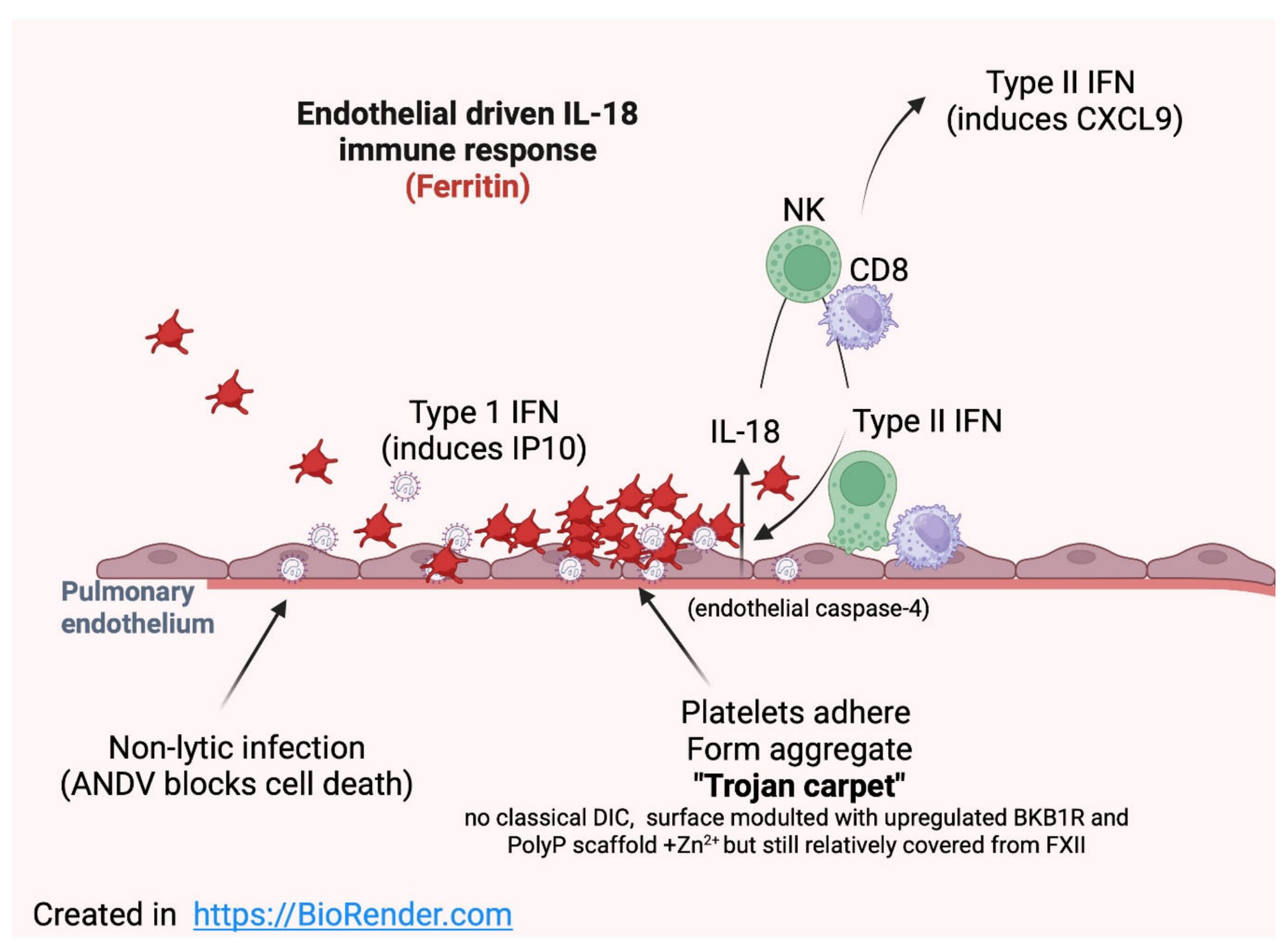
Figure 2.
Pathophysiology of HCPS during the transition-collapse phase (Days 7–10): Clearance of the protective platelet aggregate and contact system activation. During the prodromal phase, a protective aggregate of platelets, fibrin, and immune cells (the "Trojan carpet") accumulates on the surface of hantavirus-infected endothelial cells, preventing exposure to the contact system (Figure 1). By Days 7–10, as this aggregate is progressively cleared by immune surveillance, the underlying endothelial surface becomes exposed to Factor XII (FXII) and the contact system machinery. FXII activation initiates a cascade leading to bradykinin generation. Bradykinin acts through BKB2 receptors (BKB2R) on endothelial cells, combined with direct endothelial effects of hantavirus infection (RhoA activation, VE-cadherin internalization, and resistance to induction of cell death pathways), resulting in massive vascular leakage and non-cardiogenic pulmonary edema. Elevated D-dimer reflects ongoing coagulation and fibrinolysis. This contact system activation, occurring precisely when cellular immunity (NK cells, CD8+ T cells) and humoral immunity (Neutralizing Ab) is most vigorous, creating a self-perpetuating loop of inflammation and vascular dysfunction with no intrinsic resolution mechanism, as infected endothelial cells persist without undergoing apoptosis. PolyP, polyphosphate; Zn²⁺, zinc ions. Probably teh combination of neutralizing the virus-induced pathological conditions and the depletion of components from these zymogen systems leads to a halt of teh bradykinin storm and can therefore be self-limiting.
Figure 2.
Pathophysiology of HCPS during the transition-collapse phase (Days 7–10): Clearance of the protective platelet aggregate and contact system activation. During the prodromal phase, a protective aggregate of platelets, fibrin, and immune cells (the "Trojan carpet") accumulates on the surface of hantavirus-infected endothelial cells, preventing exposure to the contact system (Figure 1). By Days 7–10, as this aggregate is progressively cleared by immune surveillance, the underlying endothelial surface becomes exposed to Factor XII (FXII) and the contact system machinery. FXII activation initiates a cascade leading to bradykinin generation. Bradykinin acts through BKB2 receptors (BKB2R) on endothelial cells, combined with direct endothelial effects of hantavirus infection (RhoA activation, VE-cadherin internalization, and resistance to induction of cell death pathways), resulting in massive vascular leakage and non-cardiogenic pulmonary edema. Elevated D-dimer reflects ongoing coagulation and fibrinolysis. This contact system activation, occurring precisely when cellular immunity (NK cells, CD8+ T cells) and humoral immunity (Neutralizing Ab) is most vigorous, creating a self-perpetuating loop of inflammation and vascular dysfunction with no intrinsic resolution mechanism, as infected endothelial cells persist without undergoing apoptosis. PolyP, polyphosphate; Zn²⁺, zinc ions. Probably teh combination of neutralizing the virus-induced pathological conditions and the depletion of components from these zymogen systems leads to a halt of teh bradykinin storm and can therefore be self-limiting.
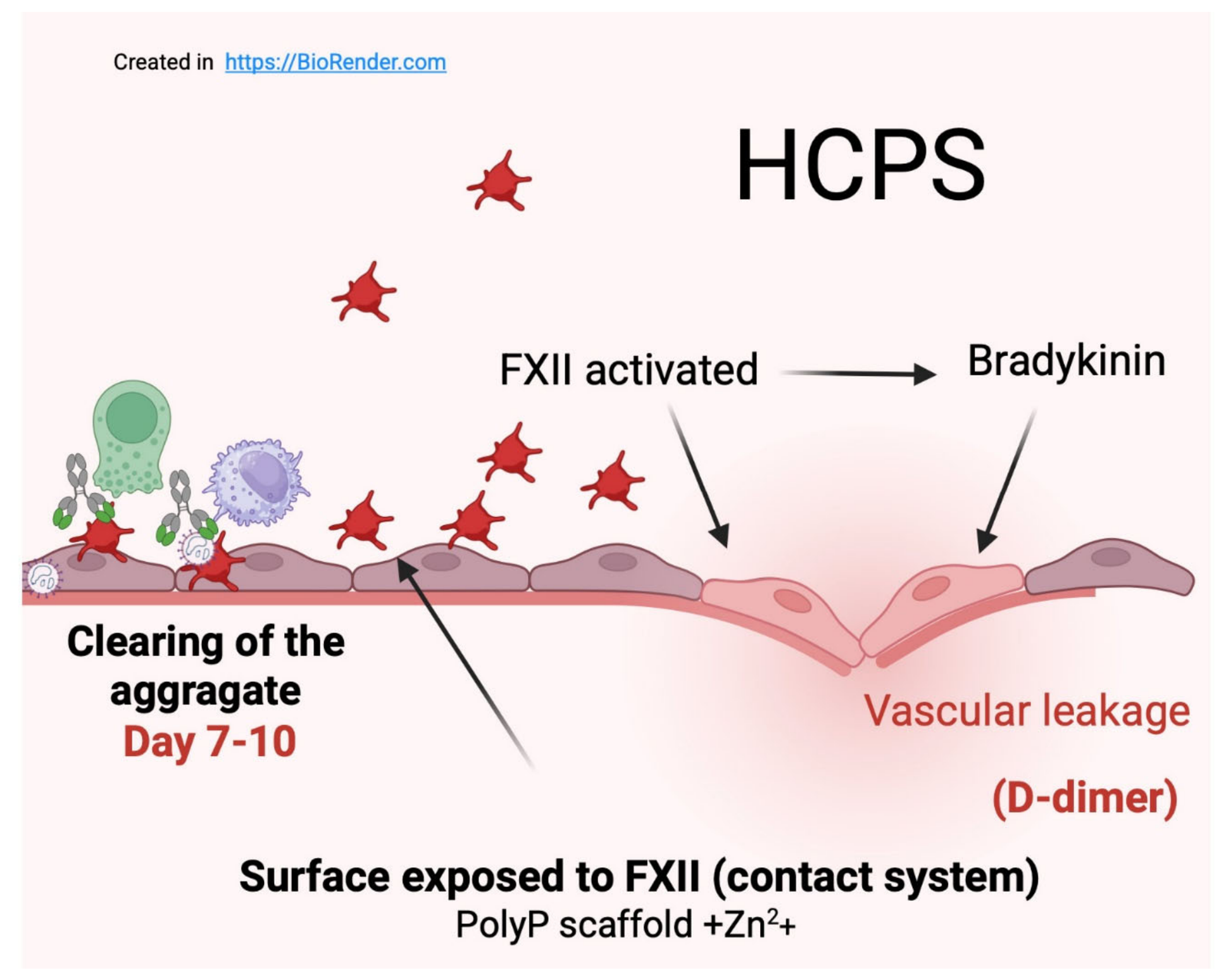
Figure 3.
Dual immunopathological pathways in Hantavirus infection: Endothelial IL-18/IFN-γ signalling and the bradykinin storm. The figure illustrates two parallel, interconnected pathogenic cascades initiated upon Hantavirus infection of endothelial cells. Both pathways are characterised by the absence of direct cytopathic cell death, facilitated by viral caspase-3 inhibition and BCL2 upregulation. Left panel; Endothelial IL-18/IFNγ pathway (present in all symptomatic patients; amplitude reflected by serum ferritin). Accumulation of cytosolic viral PAMPs and DAMPs triggers type I IFN priming and GBP induction. Subsequent caspase-4 activation in the endothelium leads to IL-18 and GSDMD cleavage and sublytic pore formation, enabling non-lytic release of mature IL-18. IL-18, in concert with IL-12, drives IFN-γ production by NK cells and CD8⁺ T cells. IFN-γ signals via JAK/STAT1, inducing ISGs and chemokines including CXCL9/CXCL10, which results in an endotheliopathy. A STAT1-driven feed-forward loop is established whereby GBPs prime further caspase-4 activation, and BKB1R becomes upregulated on the endothelial surface. Therapeutic target: JAK inhibition (JAKinh). Right panel; Bradykinin storm (predominant in patients progressing to HCPS; reflected by elevated D-dimers). Platelet adhesion to infected endothelium leads to their consumption (thrombocytopenia) and release of polyP, Zn²⁺, and PS-exposing microparticles, rendering the EC surface anionic and zinc-rich. This environment promotes FXII binding to surface receptors gC1qR, uPAR, and CK1, triggering autoactivation to FXIIa. FXIIa cleaves PK to KAL, which in turn cleaves HK to release BK, normally degraded by ACE. Excess BK signals through BKB2R on the endothelium, causing Ca²⁺ influx, vasodilation, and increased permeability, and through BKB1R (upregulated via the left pathway), further amplifying the response. Additionally, BK activates TMEM16F, inducing PS flip on EC membranes, generating new FXII binding sites and closing a self-amplifying feedback loop. The net result is capillary leak and eventually shock. Therapeutic target: icatibant (BKB2R antagonist). Dashed arrows indicate cross-pathway feedback amplification loops.
Figure 3.
Dual immunopathological pathways in Hantavirus infection: Endothelial IL-18/IFN-γ signalling and the bradykinin storm. The figure illustrates two parallel, interconnected pathogenic cascades initiated upon Hantavirus infection of endothelial cells. Both pathways are characterised by the absence of direct cytopathic cell death, facilitated by viral caspase-3 inhibition and BCL2 upregulation. Left panel; Endothelial IL-18/IFNγ pathway (present in all symptomatic patients; amplitude reflected by serum ferritin). Accumulation of cytosolic viral PAMPs and DAMPs triggers type I IFN priming and GBP induction. Subsequent caspase-4 activation in the endothelium leads to IL-18 and GSDMD cleavage and sublytic pore formation, enabling non-lytic release of mature IL-18. IL-18, in concert with IL-12, drives IFN-γ production by NK cells and CD8⁺ T cells. IFN-γ signals via JAK/STAT1, inducing ISGs and chemokines including CXCL9/CXCL10, which results in an endotheliopathy. A STAT1-driven feed-forward loop is established whereby GBPs prime further caspase-4 activation, and BKB1R becomes upregulated on the endothelial surface. Therapeutic target: JAK inhibition (JAKinh). Right panel; Bradykinin storm (predominant in patients progressing to HCPS; reflected by elevated D-dimers). Platelet adhesion to infected endothelium leads to their consumption (thrombocytopenia) and release of polyP, Zn²⁺, and PS-exposing microparticles, rendering the EC surface anionic and zinc-rich. This environment promotes FXII binding to surface receptors gC1qR, uPAR, and CK1, triggering autoactivation to FXIIa. FXIIa cleaves PK to KAL, which in turn cleaves HK to release BK, normally degraded by ACE. Excess BK signals through BKB2R on the endothelium, causing Ca²⁺ influx, vasodilation, and increased permeability, and through BKB1R (upregulated via the left pathway), further amplifying the response. Additionally, BK activates TMEM16F, inducing PS flip on EC membranes, generating new FXII binding sites and closing a self-amplifying feedback loop. The net result is capillary leak and eventually shock. Therapeutic target: icatibant (BKB2R antagonist). Dashed arrows indicate cross-pathway feedback amplification loops.
Disclaimer/Publisher’s Note: The statements, opinions and data contained in all publications are solely those of the individual author(s) and contributor(s) and not of MDPI and/or the editor(s). MDPI and/or the editor(s) disclaim responsibility for any injury to people or property resulting from any ideas, methods, instructions or products referred to in the content. |
© 2026 by the authors. Licensee MDPI, Basel, Switzerland. This article is an open access article distributed under the terms and conditions of the Creative Commons Attribution (CC BY) license (http://creativecommons.org/licenses/by/4.0/).
Copyright: This open access article is published under a Creative Commons CC BY 4.0 license, which permit the free download, distribution, and reuse, provided that the author and preprint are cited in any reuse.


