

Submitted:
28 May 2026
Posted:
29 May 2026
You are already at the latest version
Abstract
Background/Objectives: Chemotherapy-induced peripheral neuropathy (CIPN) affects up to 85% of patients receiving neurotoxic regimens, often leading to dose reduction and impaired quality of life, yet effective preventive or therapeutic options remain scarce. Emerging evidence implicates chemotherapy-induced gut microbiota dysbiosis in CIPN pathogenesis via a gut–nerve axis. Concurrently, rectal ozone insufflation (ROI) can modulate the gut microbiota and reduce inflammation. This narrative re-view aims to synthesise evidence on the role of gut dysbiosis in CIPN, evaluate the microbiota-modulating capacity of rectal ozone therapy (OT), and examine the biological plausibility of ozone as a microbiota-targeting intervention for CIPN. Methods: A comprehensive literature search was conducted across PubMed, Scopus, Web of Science, and grey literature for studies published through March 2026 using terms related to CIPN, gut microbiota, dysbiosis, the gut–nerve axis, and OT. Preclinical studies, clinical reports, and systematic reviews were included and narratively synthesised according to the three objectives. Results: Neurotoxic agents induce dysbiosis marked by reduced microbial diversity, loss of short-chain fatty acid-producing bacteria, and expansion of pro-inflammatory taxa. Preclinical models demonstrate a causal role for specific microbial communities in CIPN, with microbiota depletion or faecal transplantation modulating neuropathic phenotypes. In human cohorts, dysbiosis severity correlates with CIPN symptoms. Preclinical studies show that ROI restores microbial balance, enhances short-chain fatty acid levels, and strengthens intestinal barrier function via Nrf2/HO-1 and sirtuin 1 (SIRT1) pathways. Preliminary retrospective data report sustained symptom improvement in CIPN patients receiving OT, though no study has directly linked ozone-induced microbiota changes to clinical outcomes. Conclusions: Convergent evidence supports a strong biological rationale for investi-gating ROI as a microbiota-targeting intervention in CIPN. However, high-quality randomised controlled trials with longitudinal microbiome profiling are needed to establish mechanistic causality and guide future supportive care strategies in oncology.
Keywords:
gut microbiota
; dysbiosis
; chemotherapy-induced peripheral neuropathy
; gut–nerve axis
; ozone therapy
; rectal ozone insufflation
; microbiota modulation
; cancer treatment toxicity
; neuroinflammation
; supportive oncology
1. Introduction
Chemotherapy-induced peripheral neuropathy (CIPN) is a frequent, often persistent toxicity of many frontline cytotoxic regimens, with major implications for dose intensity, survival, and quality of life [1]. Growing evidence links CIPN to chemotherapy-driven gut microbiota dysbiosis and gut–nerve crosstalk [2], highlighting the microbiota–brain axis as a new therapeutic target, to which redox- and immunity-modulating strategies such as ozone therapy (OT) may be conceptually relevant [3,4].
CIPN is a predominantly sensory peripheral neuropathy with numbness, tingling, burning pain, and “glove and stocking” distribution; motor and autonomic involvement occur in some patients [5]. Symptoms may emerge during treatment and persist in ~30% of patients for ≥1 year after chemotherapy, substantially impairing function and quality of life [5,6]. Mechanistically, CIPN reflects axonal degeneration, microtubule disruption, mitochondrial dysfunction, altered ion channel expression, and neuroinflammation in peripheral and central pathways [1].
Across neurotoxic agents (platinums, taxanes, vinca alkaloids, bortezomib, thalidomide), an estimated 19–85% of patients develop CIPN during treatment [1]. CIPN is a major dose-limiting toxicity, often necessitating dose reduction, delay, or premature discontinuation, with potential compromise of treatment efficacy and survival [7]. Long-term CIPN contributes substantially to survivorship burden and healthcare costs [8].
Guidelines consistently conclude there is no effective pharmacologic prevention of CIPN; several proposed agents are discouraged [5,7]. For established painful CIPN, duloxetine is the only drug with moderate evidence and guideline endorsement; however, its analgesic benefit is modest, appears to be maintained only during treatment, may be associated with adverse effects, and many patients remain symptomatic [9,10]. Other pharmacologic and non-pharmacologic approaches (gabapentinoids, tricyclics, topical agents, neuromodulatory and rehabilitative strategies) show inconsistent or limited efficacy and lack robust evidence [9]. This therapeutic gap supports exploration of novel disease-modifying strategies.
Multiple reviews and preclinical studies now implicate the microbiota–gut–brain axis in CIPN pathogenesis. Chemotherapy disrupts gut microbial composition and barrier integrity, promoting dysbiosis, systemic leakage of microbial products, and immune activation [11,12]. Dysbiotic microbiota can drive glial and immune cell activation, neuroinflammation, and spinal microgliosis, contributing to neuropathic pain [11,12,13,14]. Antibiotic-induced microbiota depletion or faecal microbiota transplantation (FMT) modulate chemotherapy-induced mechanical and thermal hypersensitivity and associated cytokine profiles in rodent models, underscoring a causal role for gut microbes in CIPN [13,15].
Chemotherapeutic agents such as oxaliplatin, paclitaxel, and cisplatin induce characteristic shifts in gut microbiota, including altered Bacteroidetes/Firmicutes ratios and depletion of barrier-supporting taxa such as Akkermansia muciniphila [15]. These changes are associated with increased intestinal permeability, enhanced translocation of lipopolysaccharide (LPS), and activation of Toll-like receptor 4 (TLR4)–dependent inflammatory cascades in dorsal root ganglia (DRG) and spinal cord [13,15]. Blocking or attenuating dysbiosis—via antibiotics, probiotics, FMT, or other microbiota-targeted manipulations—reduces macrophage and microglial activation, pro-inflammatory cytokines (e.g., IL-6, TNF-α), and behavioural signs of neuropathic pain in animal models [15]. Collectively, these data position chemotherapy-induced dysbiosis as a central upstream driver of neuroimmune mechanisms in CIPN.
Given the established role of gut microbiota dysbiosis in the pathogenesis of CIPN—driving neuroinflammation, immune–glial activation, and barrier dysfunction—therapeutic strategies aimed at restoring microbial homeostasis represent a promising but underexplored avenue [15]. OT, particularly through rectal insufflation (ROI), offers a compelling biological rationale in this context. [16] Beyond its well-recognized pleiotropic effects on redox signaling, tissue oxygenation, and immune modulation, OT has been shown to modify the intestinal mucosal oxidative balance, microcirculation, and local immune milieu.[17]. Recent preclinical evidence suggests that ROI can positively influence gut microbial ecosystems [4,18]. Consequently, OT emerges as a potential microbiota-modulating intervention that may simultaneously address two interconnected drivers of CIPN: dysbiosis and neuroinflammation. This mechanistic convergence—coupled with the current lack of effective preventive strategies for CIPN—strongly justifies exploring OT as an adjunctive, biology-targeted tool to mitigate chemotherapy-induced neurotoxicity through optimization of the gut–nerve axis [19].
This narrative review aims to provide a comprehensive and updated overview of the pathophysiological mechanisms underlying CIPN, current therapeutic limitations, and emerging strategies targeting key biological pathways. Particular emphasis is placed on the role of gut microbiota dysbiosis as a mechanistic contributor to neurotoxicity and as a potential therapeutic target, as well as on the rationale for microbiota-modulating interventions.
In this context, the present work builds upon our previous review on OT in CIPN [20], extending its scope to specifically address microbiota-related mechanisms. It also complements our ongoing clinical research protocol [19], which investigates the modulation of gut microbiota through ROI in patients with radiotherapy/chemotherapy-induced pelvic toxicity. By integrating these lines of research, this review proposes a translational framework linking microbiota alterations, systemic and neuroinflammatory pathways, and the potential role of OT as a microbiota-modulating intervention in CIPN.
2. Materials and Methods
2.1. Search Strategy and Information Sources
A comprehensive literature search was conducted to identify relevant studies examining the relationship between gut microbiota dysbiosis, CIPN, and OT. The following electronic databases were searched from inception through March 2026: PubMed/MEDLINE, Scopus, Web of Science, and Google Scholar for grey literature. The search strategy combined controlled vocabulary (MeSH terms where applicable) and free-text keywords, including: “chemotherapy-induced peripheral neuropathy” OR “CIPN” OR “neuropathic pain” AND “gut microbiota” OR “dysbiosis” OR “microbiome” AND “ozone therapy” OR “rectal ozone insufflation” OR “medical ozone” AND “gut–nerve axis” OR “microbiota–gut–brain axis”. Additional studies were identified by manual screening of reference lists of included articles and relevant systematic reviews.
2.2. Eligibility Criteria
Preclinical studies (in vivo animal models), clinical reports (including retrospective studies, case series, and prospective observational studies), systematic reviews, and narrative reviews published in peer-reviewed journals were considered eligible if they addressed at least one of the following: (i) the impact of neurotoxic chemotherapeutic agents (taxanes, platinum compounds, vinca alkaloids) on gut microbiota composition; (ii) the mechanistic role of gut microbiota dysbiosis in CIPN pathogenesis via the gut–nerve axis; (iii) the effects of ROI on gut microbiota modulation, intestinal barrier function, or inflammatory pathways; or (iv) clinical outcomes of OT in patients with CIPN or related neuropathic conditions. No restrictions on language or publication date were applied. Conference abstracts and unpublished data were excluded.
2.3. Data Synthesis and Presentation
Given the heterogeneity of study designs, outcome measures, and experimental models, a narrative synthesis approach was adopted. Evidence was organised thematically according to the three predefined objectives: (1) the pathogenic role of gut microbiota dysbiosis in CIPN; (2) the microbiota-modulating capacity of ROI; and (3) the biological plausibility of OT as a microbiota-targeting intervention for CIPN. Key findings were extracted and summarised descriptively, with particular attention to causal evidence from faecal microbiota transplantation studies, preclinical mechanistic data, and preliminary clinical outcomes.
2.4. Data Availability Statement
All data presented in this review are derived from previously published studies cited in the reference list. No new primary data, custom computer code, or proprietary materials were generated or analysed during this study. The search strategy and inclusion criteria are fully disclosed to enable replication. The corresponding author may be contacted for additional documentation or clarification regarding the synthesis process.
2.5. Ethical Approval
As this manuscript is a narrative review of published literature and does not involve direct human or animal subjects, primary ethical approval was not required. All included studies cited in this review had obtained appropriate institutional ethics approval as reported in their respective original publications.
2.6. Generative Artificial Intelligence Disclosure
During the preparation of this work, the authors used generative artificial intelligence (GenAI) solely for superficial text editing purposes, including grammar correction, spelling checks, punctuation standardisation, and formatting consistency. No GenAI tools were used to generate substantive content, data, tables, figures, or interpretative analysis, nor were they employed in study design, literature search, data extraction, or conclusion formulation. The authors assume full responsibility for the accuracy, originality, and integrity of all content presented in this manuscript.
3. Pathophysiology of CIPN
CIPN results from the convergence of direct neurotoxicity, neuroinflammation, and oxidative stress/mitochondrial damage. These mechanisms, common to taxanes, platinum compounds, and vinca alkaloids (Table 1), produce a distal “glove-and-stocking” sensory neuropathy characterized by axonal degeneration and neuropathic pain [1,21,22].
Neurotoxic Mechanisms of Chemotherapy involves:
Distal Axonal Damage and Axonal Transport: - Disruption of microtubules interferes with anterograde and retrograde axonal transport, leading to distal degeneration of axonal segments and membrane remodelling [21,23].
Mitochondrial Dysfunction and Oxidative Stress: a) Taxanes, platinum compounds, vinca alkaloids, and bortezomib induce axonal mitotoxicity, mitochondrial swelling and vacuolization, opening of the permeability transition pore, cytochrome c release, and neuronal apoptosis [1,22]. b) Mitochondrial damage increases Reactive Oxygen Species (ROS) production, resulting in bioenergetic failure and neuropathy progression [24].
3.1. Neuroinflammation in CIPN
CIPN is associated with immune processes and neuroinflammation in the peripheral nerve, DRG, and spinal cord [22,23]. Chemotherapy activates microglia and astrocytes in the dorsal spinal cord, as well as macrophages/lymphocytes in the nerve and DRG, with subsequent cytokine production [23]. Levels of TNF-α, IL-1β, IL-6, and other chemokines increase, while anti-inflammatory cytokines (IL-10, IL-4) decrease, creating a pronociceptive environment [25]. These mediators enhance the excitability of DRG and dorsal horn neurons (peripheral and central sensitization), contributing to allodynia and hyperalgesia [7].
3.2. Oxidative Stress and Redox Imbalance
Oxidative stress is a central mechanism: damaged mitochondria generate excessive ROS, causing damage to neuronal lipids, proteins, and DNA [22]. The peripheral nervous system is particularly vulnerable due to its high phospholipid content and weak antioxidant defenses [24]. Dysfunction of antioxidant systems (including dysregulation of nuclear factor erythroid 2-related factor 2 (Nrf2) and enzymes such as superoxide dismutase (SOD) and glutathione-S-transferases) is linked to neurotoxicity from cisplatin and other agents [26].
CIPN arises from the interplay of distal axonal damage mediated by microtubule disruption, mitochondrial dysfunction with oxidative stress, and neuroinflammation driven by cytokines and glial/immune cells. These processes shared, albeit with nuances, among taxanes, platinum compounds, and vinca alkaloids, converge to produce a long-lasting, painful, and often persistent sensory neuropathy.
4. Gut Microbiota and Chemotherapy-Induced Dysbiosis
4.1. Baseline Role of Gut Microbiota in Host Homeostasis
In the eubiotic state, the gut microbiota maintains a diverse and symbiotic community that supports multiple physiological functions [27,28]:
- Short-chain fatty acids (SCFAs). Microbial fermentation of dietary fibre produces acetate, propionate, and butyrate. These metabolites regulate intestinal barrier integrity by upregulating tight junction proteins including occluding, claudin-1, and zonula ocludens (ZO-1). Butyrate serves as the primary energy source for colonocytes and exerts anti-inflammatory effects through inhibition of histone deacetylases (HDACs) and suppression of Nuclear Factor kappa B (NF-κB) and NLRP3 inflammasome signalling [27,29].
- Intestinal barrier integrity. SCFAs, along with indole derivatives and secondary bile acids, maintain epithelial barrier function. Acetate produced by Bifidobacterium spp. enhances barrier protection and epithelial defense. Disruption of this barrier is associated with increased systemic inflammation and susceptibility to infection [27,29].
- Immune modulation. Gut microorganisms interact with mucosal immune cells at the gut-associated lymphoid tissue (GALT) and mesenteric lymph nodes, providing signals (pathogen-associated molecular patterns (PAMPs), damage-associated molecular patterns (DAMPs)) to antigen-presenting cells that modulate both local and systemic immunity. SCFAs signal through G protein-coupled receptors (GPCRs) such as GPR43, GPR41, and GPR109A to control immune homeostasis, promote regulatory T cell differentiation, and reduce inflammatory cytokine expression [27,30].
4.2. Chemotherapy-Induced Dysbiosis
Chemotherapy produces profound compositional and functional alterations in the gut microbiota [31]:
- Decreased alpha diversity. In patients receiving oral fluoropyrimidines, alpha diversity decreased significantly, particularly in those who developed diarrhoea. Studies in myeloablative chemotherapy (without concomitant antibiotics) confirmed severe reductions in diversity [32].
- Loss of SCFA-producing bacteria. Chemotherapy consistently reduces Firmicutes (including Faecalibacterium and Roseburia) and Actinobacteria (including Bifidobacterium). In a study of 28 patients with non-Hodgkin’s lymphoma, Firmicutes and Actinobacteria decreased significantly (p = 0.0002) following chemotherapy. In patients with chemotherapy-induced diarrhoea, Bifidobacterium abundance decreased significantly (p = 0.019), while it increased in those without diarrhoea [32,33,34].
- Expansion of pathobionts. Proteobacteria increased significantly (p = 0.0002) after chemotherapy, particularly Enterobacteriaceae. Chemotherapy-induced epithelial cell death releases purine-containing metabolites that drive transcriptional reprogramming of Enterobacteriaceae, promoting their respiration and purine utilization-dependent expansion [33,34].
- Metabolic alterations. Dysbiosis is associated with reduced capacity for nucleotide metabolism (p = 0.0001), energy metabolism (p = 0.001), and metabolism of cofactors and vitamins (p = 0.006), with increased glycan metabolism and xenobiotic biodegradation. Metabolites such as TMAO (trimethylamine N-oxide), produced by certain bacteria, have been associated with tumour promotion in various cancer types [35,36].
4.3. Intestinal Barrier Dysfunction
- “Leaky gut” (increased intestinal permeability). Chemotherapeutic agents directly and rapidly damage proliferating intestinal epithelial cells, compromising tight junctions and increasing paracellular permeability. In murine models, among common agents, oxaliplatin causes the most severe intestinal damage in murine models, followed by 5-FU and irinotecan. Loss of SCFA production exacerbates barrier dysfunction: oral butyrate improved epithelial permeability and prevented irinotecan-induced increases in β-glucuronidase activity [37,38].
- Bacterial translocation. Barrier disruption permits passage of bacteria and their products from the intestinal lumen into mesenteric lymph nodes and secondary lymphoid organs. Cyclophosphamide treatment was associated with translocation of Gram-positive bacteria such as Enterococcus hirae and Lactobacillus johnsonii to the spleen and lymph nodes, stimulating Th17 and Th1 immune responses. This phenomenon has a dual role: it may contribute to antitumor efficacy but also to systemic toxicity [38].
- Systemic immune activation. Bacterial translocation and release of LPS and other pathogen-associated molecular patterns (PAMPs) activate inflammatory pathways including TLR4/MyD88/NF-κB and NOD/RIP2/NF-κB, with production of proinflammatory cytokines (TNF-α, IL-1β, IL-6, IFN-γ). Chemotherapy-associated dysbiosis correlates with gastrointestinal toxicity, neutropenia, and even cardiotoxicity. In the context of CAR-T cell therapy, prior dysbiosis (from broad-spectrum antibiotics) was associated with higher incidence of cytokine release syndrome [39].
Collectively, chemotherapy generates a vicious cycle: direct epithelial damage promotes dysbiosis, dysbiosis reduces production of protective SCFAs, which aggravates barrier dysfunction and facilitates bacterial translocation with systemic immune activation, perpetuating inflammation and gastrointestinal toxicity [40].
5. The Gut–Nerve Axis in CIPN
5.1. Mechanistic Pathways: Microbiota - Nervous System Signaling
The gut microbiota communicates with the peripheral and central nervous systems through three principal routes that are relevant to CIPN:
Immune pathways: Chemotherapy-induced dysbiosis increases intestinal permeability, allowing bacterial products, particularly LPS, to enter systemic circulation. LPS activates TLR4 on DRG neurons, satellite glial cells, and spinal cord glia, triggering downstream MyD88/NF-κB and MAPK (ERK1/2, p38) signaling cascades that produce proinflammatory cytokines (TNF-α, IL-1β, IL-6) and chemokines (MCP-1/CCL2) [12,41]. This immune-mediated signaling recruits macrophages into the DRG, which further amplifies neuroinflammation and contributes to intraepidermal nerve fiber loss [42]. Additionally, the DAMP molecule high mobility group box-1 (HMGB1) is released during chemotherapy-induced tissue injury and activates both TLR4 and RAGE receptors on spinal microglia, upregulating proinflammatory cytokines during the early stages of neuropathic pain [43]. In long-term germ cell tumor survivors, elevated plasma HMGB1(a marker of gut microbial translocation) was associated with worse CIPN symptom burden [44].
Metabolic pathways: Gut microbiota-derived metabolites directly modulate nociceptive signaling. Chemotherapy-induced expansion of Clostridium species increases production of deoxycholic acid (DCA), a secondary bile acid. DCA elevates serum CCL5 and induces CCR5 overexpression in DRG neurons through the bile acid receptor TGR5, contributing to neuronal hyperexcitability. Notably, the CCR5 antagonist maraviroc suppressed paclitaxel-induced neuropathic pain in this model [45]. Conversely, loss of SCFA-producing bacteria reduces circulating butyrate and propionate, removing their anti-inflammatory and neuromodulatory protective effects [46].
Vagal pathway (emerging evidence, with caveats). The vagus nerve represents a direct neural route for gut-to-brain communication. Roseburia intestinalis-derived butyrate has been shown to activate vagal neurons through GPR41 receptors, enhancing vagal neurotransmission to the nucleus tractus solitarius (NTS) and suppressing the central amygdala (CeA), a brain region involved in pain perception. Vagal knockout of Gpr41 abolished these analgesic effects [47]. However, this evidence derives from postherpetic neuralgia models rather than CIPN specifically, and the relevance of vagal signaling to CIPN, which primarily involves peripheral sensory neurons and DRG, remains to be established. The gut microbiota’s intersection between the microbiome-gut-brain axis and the neuroimmune-endocrine axis forms a complex network that can directly or indirectly affect key components involved in CIPN manifestations [48].
5.2. Role of SCFAs
Butyrate as an anti-inflammatory and epigenetic modulator. Sodium butyrate (NaB) has demonstrated significant neuroprotective effects in CIPN models through multiple mechanisms:
- Anti-inflammatory effects. In an oxaliplatin-induced peripheral neuropathy (OIPN) rat model, NaB significantly decreased mechanical and cold allodynia scores and reduced sciatic nerve TNF-α levels while increasing IL-10 and nerve growth factor (NGF) expression. NaB also increased immunohistochemical expression of FOXP-3 (regulatory T cell marker) and PPAR-γ, indicating immunomodulatory and anti-inflammatory properties. Prophylactic NaB (before OIPN induction) showed greater neuroprotection than concurrent administration [49].
- Epigenetic regulation via HDAC inhibition. Butyrate acts as a potent HDAC inhibitor. In bone cancer pain models, butyrate supplementation decreased HDAC2 expression in the spinal dorsal horn and increased μ-opioid receptor (MOR) expression, potentiating morphine analgesia. In chronic constriction injury models, sodium butyrate (200–400 mg/kg oral, 14 days) attenuated cold allodynia, mechanical allodynia, and thermal hyperalgesia, with concurrent reduction in sciatic nerve TNF-α. Butyrate also increased expression of PPAR-α and PPAR-γ receptors while reducing COX-2, iNOS, TNF-α, and neuronal activation marker c-Fos, with analgesic effects mediated partly through ATP-dependent K⁺ channels [50].
Impact on neuropathic pain in CIPN. Propionic acid has also been shown to suppress nociceptive neuronal excitability via GPR41 signaling-mediated inhibition of voltage-gated Ca²⁺ channels in the central terminals of spinal nociceptive neurons [51].
5.3. Microbiota-Driven Neuroinflammation
LPS/ TLR4/ neuroinflammation cascade. TLR4 has emerged as a central mediator linking gut-derived endotoxemia to CIPN. Key findings include:
- TLR4 expression increases in DRG neurons as early as day 1 after oxaliplatin treatment and persists through day 14. Co-treatment with TLR4 antagonists (LPS-RS) or minocycline attenuated hyperalgesia and blocked downstream MyD88 and TRIF signalling [52].
- TLR4 antisense oligodeoxynucleotides prevented CIPN development induced by all three major drug classes (oxaliplatin, paclitaxel, bortezomib) and reversed established CIPN from oxaliplatin and paclitaxel [41].
Glial activation. Both peripheral and central glial cells are critically involved: a) Spinal microglia proliferate in paclitaxel-treated mice harboring pain-sensitive microbiota (C57BL/6) but not in those with pain-resistant microbiota (129SvEv), directly linking gut microbial composition to central glial activation [8]. b) Astrocytes in the spinal dorsal horn show increased GFAP expression in CIPN. FMT from healthy donors reduced astrocytic GFAP expression and TLR4/p38MAPK pathway activation in both the colon and spinal cord of paclitaxel-treated rats [53]. c) Paclitaxel activates TLR4 on spinal astrocytes (but not microglia or neurons in the spinal cord), sensitizing TRPV1 receptors and impairing capsaicin-induced tachyphylaxis, leading to sustained nociceptive hyperactivity. Sustained minocycline preincubation mitigated this sensitization [54]. d) In the DRG, TLR4 activation leads to MCP-1 upregulation, macrophage infiltration, TNF-α production, and ultimately loss of intraepidermal nerve fibers, a hallmark of clinical CIPN [42].
5.4. Preclinical Evidence of Causality
Microbiota depletion - reduced CIPN. Multiple independent studies demonstrate that antibiotic-mediated gut microbiota depletion prevents or attenuates CIPN. In oxaliplatin-treated mice, antibiotic cocktail feeding prevented thermal hyperalgesia and mechanical allodynia and inhibited proinflammatory cytokine production (TNF-α, IL-1β, IL-6) in DRG. Antibiotic treatment starting simultaneously with chemotherapy produced immediate analgesia [55]. Importantly, antibiotic treatment starting after establishment of oxaliplatin-induced neuropathic pain did not significantly reverse it, suggesting that gut microbiota may play a more critical role in CIPN initiation than maintenance for this drug class [55].
FMT: transferring pain phenotypes. The most compelling causal evidence comes from reciprocal FMT experiments. Pain-sensitive donors - pain phenotype: Reciprocal microbiota transfers between C57BL/6 (pain-sensitive) and 129SvEv (pain-resistant) mice demonstrated that the gut microbiota determines paclitaxel-induced pain sensitivity. Mice receiving the pain-sensitive B6 microbiota developed CIPN with spinal microglia proliferation, while those receiving the pain-resistant 129 microbiota were protected, with notable absence of infiltrating immune cells [8]. Healthy donors – protection: FMT from healthy rats to paclitaxel-treated rats significantly alleviated mechanical allodynia and thermal hyperalgesia, reduced TLR4/p38MAPK pathway activation and astrocytic GFAP expression in the spinal dorsal horn [53]. Restoration of microbiota - pain rescue: In antibiotic-depleted mice protected from CIPN, transplantation of faecal bacteria from specific-pathogen-free mice partially restored the gut microbiota and fully rescued the behaviourally expressed neuropathic pain. Akkermansia, Bacteroides, and Desulfovibrionaceae were identified as taxa potentially playing key roles [55]. In oxaliplatin-treated rats, antibiotic-mediated gut microbiota depletion significantly attenuated neuropathic pain, reduced plasma LPS levels and serum proinflammatory cytokines (TNF-α, IL-6, IL-1β), and suppressed TLR4/MyD88/NF-κB signaling in DRG neurons. Conversely, in the OIPN model, FMT reversed these protective effects, leading to the reappearance of neuropathic pain, increased plasma LPS, decreased intestinal barrier markers, and reactivation of the TLR4/MyD88/NF-κB pathway in DRG [56].
5.5. Human Evidence
Microbiota–symptom correlations. Clinical data linking gut microbiota to CIPN in humans remain limited but are growing. In a cohort of 70 early-stage breast cancer patients receiving taxane-based chemotherapy, decreases in microbiota alpha diversity during treatment were associated with worse neuropathy symptoms and general pain after controlling for baseline symptoms. Larger shifts in beta diversity also coincided with more severe neuropathy. SCFA-producing bacteria were depleted in patients with neuropathy at the final infusion, and decreases in fecal SCFA levels correlated with more severe neuropathy symptoms. Circulating neurofilament light chain (NFL), a putative CIPN biomarker, increased during treatment [46]. In breast cancer patients with different paclitaxel-induced neuropathy grades, microbiome and metabolome analysis revealed that severe neuropathy was associated with expansion of Clostridium species and elevated DCA levels, linking a specific microbiota-metabolite axis to CIPN severity [45]. In 194 long-term germ cell tumour survivors (median 10-year follow-up), higher plasma HMGB-1 levels (a biomarker of gut microbial translocation) were associated with worse overall CIPN20 scores (p = 0.048) and worse motor function (p = 0.036), suggesting that gut microbial translocation may contribute to persistent CIPN even years after treatment [44]. A case report of a breast cancer survivor with long-standing CIPN showed that cannabidiol plus multimodal exercise enriched gut genera producing SCFAs and was associated with clinically meaningful improvements in CIPN symptoms and quality of life [57].
6. Ozone Therapy: Biological and Molecular Effects
6.1. Fundamentals of Ozone Therapy
Ozone as a pro-drug. Ozone (O₃) is not a drug or medicine in the conventional sense, as it lacks specific cellular receptors and is extremely reactive with biological matrices. Instead, ozone functions as a pro-drug or bioregulator that generates effector molecules upon contact with biological fluids [58]. When gaseous ozone impacts blood or tissues, it is instantly consumed through reactions with polyunsaturated fatty acids (PUFAs) bound to albumin and other biomolecules, producing two classes of messenger molecules that mediate its therapeutic effects [59].
The interaction of ozone with biological substrates generates: a) Hydrogen peroxide. This represents the early-phase messenger. When ozone reacts with unsaturated fatty acids in aqueous environments (such as lung lining fluids or plasma), the net reaction produces one mole of H₂O₂ per mole of ozone consumed, along with two moles of aldehydes. H₂O₂ triggers immediate biochemical pathways in blood cells and tissues [59]. b) Lipid oxidation products (LOPs). These constitute the late-phase messengers and include alkenals (such as 4-hydroxynonenal [4-HNE]), aldehydes, and other lipoperoxides collectively termed “ozonides.” LOPs act as signal transducers that enter cells throughout the body, where they interact with the Nrf2-Keap1 protein complex to initiate adaptive antioxidant responses. The therapeutic dosage range is typically 1-40 μg/mL (depending on protocol), within which ozone triggers a hormetic and adaptive cellular response rather than oxidative damage [60].
6.2. Redox Signaling and Hormesis
OT operates through the paradoxical mechanism of hormesis, particularly mitohormesis, whereby low-dose oxidative stress triggers protective cellular adaptations [61].
Activation of Nrf2/HO-1 pathway. The Nrf2 (nuclear factor erythroid 2-related factor 2) pathway is the central mechanism underlying ozone’s therapeutic effects: LOPs, particularly 4-HNE, react with the Nrf2-Keap1 protein complex in the cytoplasm. Under basal conditions, Keap1 sequesters Nrf2 and promotes its degradation. Ozone-derived LOPs modify cysteine residues on Keap1, releasing Nrf2 and allowing its nuclear translocation to chromatin sites of active transcription [62]. Nuclear Nrf2 binds to electrophile-responsive elements (EpRE) in DNA, upregulating expression of antioxidant enzymes (superoxide dismutase [SOD], catalase, glutathione peroxidase (GPx)), phase II detoxification enzymes (NAD(P)H quinone oxidoreductase 1 [NQO1]), and heme oxygenase-1 (HO-1) [63]. In human studies, three sessions of major autohemotherapy (MAH) with ozone (35 μg/mL) significantly increased Nrf2 levels in peripheral blood mononuclear cells immediately after ozone exposure (p < 0.01) and 30 minutes post-reinfusion (p < 0.05). After the treatment series, SOD and catalase activities increased (p < 0.05), demonstrating sustained antioxidant adaptation [63]. Ozone-induced EpRE activation can be completely reversed by ectopic expression of Keap1, confirming the Nrf2-dependent mechanism [64].
Activation of SIRT1 and related pathways. Ozone activates the AMPK/FOXO/mTOR/SIRT1 pathway, which regulates cellular energy metabolism, autophagy, and longevity[58]. SIRT1 is a NAD⁺-dependent deacetylase involved in mitochondrial biogenesis and stress resistance. The activation of these pathways contributes to ozone’s pleiotropic effects on cellular survival and metabolic adaptation [60]. Antioxidant adaptation. The repetition of graduated oxidative stresses through serial ozone treatments induces a multiform adaptive response (oxidative preconditioning). With progressive OT, the upregulated protective enzymes reverse chronic oxidative stress induced by inflammation, blocking disease progression and improving quality of life. This represents a shift from acute pro-oxidant stimulus to sustained antioxidant capacity—the hallmark of hormesis [61].
6.3. Anti-Inflammatory and Immunomodulatory Effects
Reduction of proinflammatory cytokines. OT consistently demonstrates potent anti-inflammatory effects across multiple experimental models. In rheumatoid arthritis models, ozone treatment significantly decreased synovial levels of TNF-α and IL-12p70 while increasing IL-10 (anti-inflammatory cytokine). The most pronounced effects occurred with repeated treatments (at 2-, 24-, and 48-hours post-induction), indicating that sustained OT is more effective than single acute application[65]. In another arthritis model, ozone reduced TNF-α protein concentrations and decreased TNF-α and IL-1β mRNA levels, while reestablishing cellular redox balance and reducing nitric oxide and fructolysine (a marker of glycation) [66]. In psoriasis, topical ozone treatment significantly decreased IL-17A, IL-22, and IL-23 expression in peripheral blood CD4⁺ T cells and inhibited activation of Th17 cells in spleen tissue [67]. In COPD models, ROI decreased secretion of IL-4, IL-17A, TNF-α, IL-1β, and IL-6 in lung tissue [68].
Modulation of the NF-κB pathway. Ozone exerts anti-inflammatory effects through Nrf2/NF-κB crosstalk. Ozone upregulates Nrf2, which in turn inhibits the TLR4/NF-κB pathway. In COPD models, ROI upregulated Nrf2 expression, decreased ROS levels, and downregulated expression of TLR4, NF-κB, and phosphorylated NF-κB (p-NF-κB). When Nrf2 was pharmacologically inhibited (ML385), the anti-inflammatory effects of ozone were abolished, confirming Nrf2-dependent NF-κB suppression [68]. In psoriasis, high-throughput sequencing confirmed that ozone treatment significantly suppressed imiquimod (IMQ)-induced activation of the Toll-like receptor 2 (TLR2)/NF-κB signalling pathway in skin lesions[67]. In neuropathic pain models, intrathecal ozone injection suppressed the GluR6-NF-κB/p65 signalling pathway, reducing spinal expression of IL-1β, IL-6, TNF-α, and NF-κB/p65 [69].
In addition, ozone promotes a cytokine shift from proinflammatory to anti-inflammatory profiles, with initial transient ROS generation followed by sustained anti-inflammatory signalling [70].
6.4. Effects on Microcirculation and Oxygen Metabolism
Relevance to peripheral nerve. OT exerts multiple effects on microcirculation and tissue oxygenation that are particularly relevant to peripheral nerve health. Improved microcirculation and perfusion: OT promotes blood circulation and reduces ischemia in both peripheral and central tissues. In peripheral arterial disease (PAD), oxygen-OT improves tissue perfusion, reduces hypoxia, and enhances healing processes. The therapy is effective in low-perfusion syndromes and appears to prevent complications such as amputation in PAD patients [71,72].
Effects on peripheral nerve injury. In a rat sciatic nerve crush injury model, OT (0.7 mg/kg) administered for 4 weeks produced significant histological improvements: Reduced nerve diameter and decreased thickness of perineurium and epineurium compared to untreated injury (p < 0.001), indicating reduced oedema and fibrosis. Decreased vascular congestion and vacuolization (p < 0.05), suggesting improved microvascular function. Lower S100 immunoreactivity (a marker of nerve injury and glial activation) compared to untreated injury (p < 0.05). Ozone improved sciatic nerve recovery without increasing fibrotic tissue, a critical advantage for functional nerve regeneration [73].
Oxygen metabolism and mitochondrial function. OT targets mitochondria and their turnover/biogenesis, acting as a tuner of fundamental cellular survival mechanisms. Through mitohormesis, ozone enhances mitochondrial structure and function, which is particularly important for energy-demanding peripheral nerves [60]. Ozone indirectly triggers the HIF-1α (hypoxia-inducible factor 1α) pathway, which regulates cellular adaptation to oxygen availability, and activates HO-1 signalling and the NO/iNOS biochemical machinery, all of which contribute to improved tissue oxygenation and vascular function [58].
6.5. Clinical Applications in Neuropathic Pain and CIPN
In experimental neuropathic pain models, ozone activates AMPK (AMP-activated protein kinase), which suppresses microglial activation and normalizes pain-related signalling molecules (PKCγ, NMDA receptor, ERK). The anti-nociceptive effect of ozone was abolished by AMPK antagonists, confirming AMPK-dependent mechanisms [74,75].
Overall, OT operates through a sophisticated hormetic mechanism: controlled low-dose oxidative stress generates H₂O₂ and LOPs that activate Nrf2/HO-1 and SIRT1 pathways, inducing antioxidant adaptation; suppresses NF-κB-mediated inflammation and proinflammatory cytokines; and improves microcirculation and oxygen metabolism in peripheral nerves. These pleiotropic effects position ozone as a promising adjuvant therapy for CIPN, though robust clinical trial data are still needed [20].
7. Clinical Evidence of Ozone Therapy in CIPN
7.1. Available Clinical Studies
The clinical evidence for OT in CIPN consists entirely of uncontrolled, preliminary reports from our research group, supplemented by broader cancer survivor studies and a recent narrative review [20].
Case series and retrospective studies directly addressing CIPN. CIPN pain series (2022): A preliminary report of 7 patients with chronic pain secondary to grade II-III CIPN treated with ROI. This represents the first published clinical report specifically evaluating OT for CIPN-related pain[76]. CIPN numbness/tingling series (2025): A retrospective study of 15 patients (8 female/7 male, median age 66 years) with persistent numbness and tingling secondary to grade 2-3 CIPN. Ozone was administered by ROI over 40 planned sessions across 4 months, with initial concentration of 10 μg/mL progressively increased to 30 μg/mL and gas volume from 180 mL to 300 mL per session [77].
Broader cancer survivor studies including CIPN patients. Cancer survivor and health-related quality of life (HRQOL) study (2023): A prospective evaluation of 26 cancer survivors with chronic side effects of radiotherapy and chemotherapy (including but not limited to CIPN) assessed health-related quality of life (EQ-5D-5L) and toxicity grade (CTCAE v5.0) before and after OT [78]. Cancer symptom scoping review (2025): A scoping review identified 16 articles evaluating medical ozone treatment for pain, fatigue, anxiety, and depression in cancer patients, providing a broader context for ozone’s role in cancer symptom management [79]. Recent evidence supports OT potential in managing CIPN. In a preliminary clinical report, 7 patients with chronic grade II-III CIPN received ozone treatment via ROI. Median pain (Visual Analogue Scale (VAS)) decreased from 7 (baseline) to 4 at treatment end (p = 0.004), with sustained improvement at 3 months (VAS 5.5, p = 0.008) and 6 months (VAS 6, p = 0.008). Half of the patients showed improvement in CTCAE toxicity grade [74]. A 2025 narrative review—the most comprehensive to date—summarized 18 experimental studies and 27 clinical reports (1995–2025) offering preliminary evidence supporting ozone’s role in CIPN management. The hypothesized mechanisms include modulation of oxidative stress, inflammation, microcirculation, and nerve regeneration. However, clinical evidence remains limited, and multiple randomized controlled trials are currently ongoing [20].
7.2. Clinical Outcomes
Pain (VAS). In the 7-patient CIPN pain series, ROI produced statistically significant and clinically meaningful pain reduction. Median VAS decreased from 7 (range 5-8) at baseline to 4 (range 2-6) at end of treatment (p = 0.004). All patients except one showed clinically relevant pain improvement (defined as ≥2-point VAS reduction)[76]. In the broader cancer survivor cohort (n = 26), all dimensions of the EQ-5D-5L questionnaire, including pain/discomfort, were significantly improved (p < 0.05), with significant improvement in the overall EQ-5D-5L index (p < 0.001)[78]. In a separate case series of 6 cancer patients with refractory pelvic pain secondary to cancer treatment, VAS decreased from 7.8 ± 2.1 to 2.8 ± 3.8 after 3 months of OT (p = 0.020) [20].
Paraesthesia (numbness and tingling). The 15-patient retrospective study specifically addressed sensory CIPN symptoms: After ozone treatment, 67% of patients reported a decrease in numbness and tingling of ≥50% (p = 0.002) [77]. This is particularly notable given that the ASCO CIPN guideline acknowledges there is no effective clinical management option for numbness and tingling, with duloxetine providing only modest benefit for pain specifically [20,80].
Toxicity grade (CTCAE). OT demonstrated improvement in CIPN toxicity grading: In the pain series, 50% of patients showed improvement in CTCAE v5.0 toxicity grade[76]. In the numbness/tingling series, 47% of patients experienced a decrease in the grade of CIPN toxicity (p = 0.016) [77]. In the broader cancer survivor study, the overall grade of toxicity was significantly decreased (p < 0.001)[78].
7.3. Durability of Response
Six-month follow-up data. Both CIPN-specific studies demonstrated sustained benefit beyond the treatment period: Pain outcomes. Median VAS was 5.5 (range 1.8-6.3) at 3 months post-treatment (p = 0.008) and 6 (range 2.6-6.6) at 6 months post-treatment (p = 0.008), indicating statistically significant improvement persisting at 6 months despite some attenuation from end-of-treatment values [76]. Numbness/tingling outcomes. The improvements in both CIPN toxicity grade and self-reported numbness/tingling were maintained at 3- and 6-month follow-up after the end of ozone treatment [77]. Broader cancer survivor outcomes. In the radiation-induced rectal bleeding series (n = 12), OT effects were maintained over a median follow-up of 104 months (range 52-119), suggesting potential for very long-term benefit in some cancer treatment-related toxicities [81].
The durability of response is noteworthy given that ozone treatment was administered over a finite period (~4 months) and benefits persisted after cessation, suggesting that ozone may induce lasting biological changes (e.g., sustained Nrf2-mediated antioxidant adaptation, microbiota remodelling) rather than merely providing symptomatic relief during active treatment [76,77].
7.4. Safety Profile
Adverse events. OT by rectal insufflation demonstrates a favourable safety profile across the available literature. In the CIPN studies, no serious adverse events were reported. The most common side effect was soft and temporary flatulence lasting several hours after each session [76,77,81]. In the fibromyalgia rectal insufflation study (24 sessions, 8 mg ozone per session), transient meteorism (abdominal bloating/gas) was the most frequently reported side effect[82]. In the evidence gap map encompassing 26 systematic reviews of OT across multiple indications, no serious adverse effects were reported [83]. When the correct dose is administered within the therapeutic range, no side effects have been reported in neurological applications [71]. In the broader oncology literature, ozone autohemotherapy in 50 cancer patients with fatigue showed no side effects, with 70% achieving significant symptom improvement [84].
Important safety caveats. The therapeutic window is narrow: beneficial hormetic effects (by rectal way or MAH protocol) occur at 10-40 μg/mL, while potential toxicity may occur at doses >80 μg/mL. This necessitates precise dosing and trained administration [60].
Regulatory considerations. The regulatory status of OT varies significantly across jurisdictions. OT is not officially allowed in many countries, though private medical services use it worldwide. The global legal status of OT is highly polarized. It is a standard complementary practice in parts of Europe and Latin America, but an illegal or unapproved procedure in the US, Canada, and the UK. The primary drivers of legality are not scientific consensus on efficacy, but rather regulatory frameworks regarding natural substances and economic incentives for clinical trials [83].
7.5. Critical Appraisal
Absence of microbiome analysis. No clinical study of OT in CIPN has included gut microbiota assessment: Despite the strong mechanistic rationale linking ozone and microbiota modulation / SCFA production / neuroprotection, no human study has simultaneously measured microbiota changes and CIPN outcomes during OT [19,20]. The first studies designed to prospectively investigate ROI’s impact on gut microbiota in cancer patients are currently ongoing (NCT07259681 in 38 gynaecological cancer patients with pelvic toxicity and NCT06799351 in 42 patients with CIPN). These studies will use 16S rRNA sequencing of stool samples pre- and post-intervention [19]. Without microbiome data, the proposed mechanism linking OT to CIPN improvement via gut microbiota modulation remains entirely hypothetical in the clinical setting, supported only by preclinical evidence and biological plausibility [19].
8. Integrative Mechanistic Model: Ozone Therapy, Gut Microbiota, and CIPN
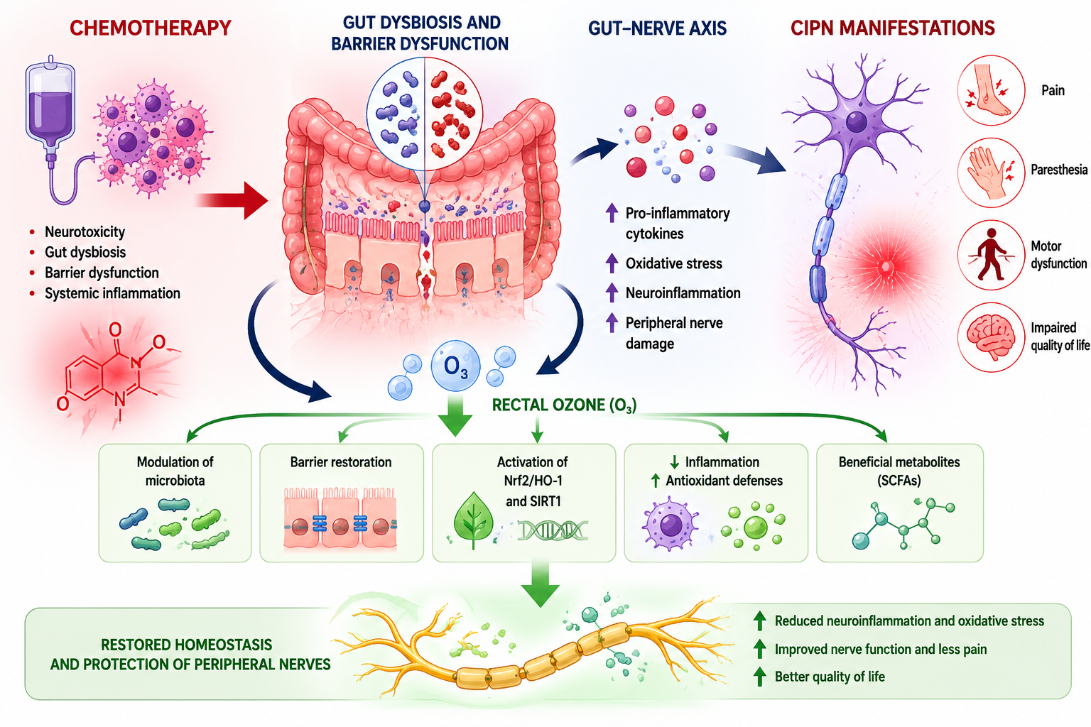
The following integrative model synthesizes the evidence presented in sections 3–7 into a unified mechanistic framework linking chemotherapy-induced dysbiosis to peripheral neuropathy and proposing OT as a multi-target intervention acting through the gut–nerve axis (Figure 1).
8.1. The Pathogenic Cascade: Chemotherapy / Dysbiosis / CIPN
The pathogenic arm of the model follows a sequential, self-amplifying cascade: Step 1: Chemotherapy-induced dysbiosis. Cytotoxic agents (platinum compounds, taxanes, vinca alkaloids) directly damage the intestinal epithelium and profoundly alter gut microbiota composition. This results in decreased alpha diversity, loss of SCFA-producing commensals (Faecalibacterium prausnitzii, Roseburia intestinalis, Lactobacillus, Bifidobacterium), and expansion of pathobionts (Enterobacteriaceae, Fusobacterium). Simultaneously, chemotherapy disrupts tight junction proteins and increases intestinal permeability (“leaky gut”), enabling bacterial translocation [19].
Step 2: Metabolic and immune derangement. The loss of SCFA-producing bacteria leads to decreased butyrate and propionate production, removing their anti-inflammatory, epigenetic (HDAC inhibition), and barrier-protective effects. Concurrently, pathobiont expansion increases production of proinflammatory metabolites including LPS and trimethylamine N-oxide (TMAO). LPS translocate across the compromised intestinal barrier into systemic circulation [47].
Step 3: Neuroinflammation and peripheral nerve damage. Circulating LPS activates the TLR4/NF-κB signalling pathway in DRG neurons, satellite glial cells, and spinal cord microglia, triggering release of proinflammatory cytokines (TNF-α, IL-1β, IL-6). This neuroinflammatory cascade sensitizes peripheral nociceptors, activates spinal glial cells, and damages peripheral nerve fibres, manifesting clinically as pain, numbness, tingling, and functional impairment characteristic of CIPN [20]. The loss of butyrate-mediated neuroprotection (via GPR41/GPR43 signalling and HDAC inhibition) further exacerbates neuronal vulnerability. Preclinical evidence demonstrates causality: FMT from pain-sensitive donors transfers the neuropathic phenotype, while FMT from resistant donors confers protection [20,47].
8.2. The Therapeutic Intervention: Ozone / Microbiota Restoration / CIPN Improvement
The therapeutic arm proposes OT as a multi-level intervention that reverses the pathogenic cascade at multiple nodes: Node 1: Redox control and hormetic adaptation. ROI generates H₂O₂ and LOPs (particularly 4-HNE) that activate the Nrf2/Keap1/ARE pathway, upregulating endogenous antioxidant enzymes (SOD, catalase, GSH-Px, HO-1) and the SIRT1/AMPK/FOXO axis. This shifts the cellular redox environment from oxidative distress (caused by chemotherapy) toward oxidative eustress, creating conditions favourable for commensal bacterial recovery [60,63,64]. Node 2: Gut microbiota modulation. The restored redox environment, combined with ozone’s direct antimicrobial effects against pathobionts and its enhancement of intestinal oxygenation, promotes recolonization by beneficial bacteria (Lactobacillus, Bifidobacterium, SCFA producers). Preclinical evidence demonstrates that ROI increases faecal butyrate and propionate while decreasing TMA/TMAO. These microbiota changes are functionally significant—antibiotic depletion experiments confirm that ozone’s protective effects are microbiota-dependent [3,4,85]. Node 3: Intestinal barrier restoration. OT enhances tight junction protein expression and reduces intestinal permeability through both direct effects (Nrf2-mediated cytoprotection of epithelial cells) and indirect effects (increased butyrate production by restored commensals, which serves as the primary energy source for colonocytes and reinforces barrier integrity). This prevents LPS translocation and breaks the systemic inflammatory cascade [4,86].
Node 4: Anti-inflammatory signalling. Ozone activates Nrf2/NF-κB crosstalk, whereby upregulated Nrf2 suppresses the TLR4/NF-κB pathway. This reduces systemic and local proinflammatory cytokines (TNF-α, IL-1β, IL-6) while increasing anti-inflammatory mediators (IL-10). In the spinal cord, ozone suppresses the GluR6-NF-κB/p65 pathway and activates AMPK, which inhibits microglial activation and normalizes pain-related signalling molecules (PKCγ, NMDA receptor, ERK) [67,68,74,75].
Node 5: Gut–nerve axis modulation and neuroprotection. The restored SCFA production—particularly butyrate—exerts direct neuroprotective effects through multiple mechanisms: GPR41/GPR43 receptor activation on DRG neurons (modulating neuronal excitability), HDAC inhibition (epigenetic regulation of inflammatory gene expression in glial cells), and enhancement of peripheral nerve microcirculation (improved oxygen delivery to vasa nervorum). Ozone additionally improves nerve recovery by reducing perineurial oedema, vascular congestion, and S100 immunoreactivity (glial activation marker) without increasing fibrosis[73]. The convergence of restored SCFAs, reduced neuroinflammation, and improved microcirculation may provide a biological rationale to clinical improvement in pain, paraesthesia, and CIPN toxicity grade, with effects sustained at 6 months post-treatment [76,77].
8.3. Evidence Strength and Knowledge Gaps
The strength of evidence varies substantially across the proposed mechanistic nodes:
The strongest evidence supports individual components of the model: ozone activates Nrf2/HO-1 and suppresses NF-κB (shown preclinically and in human peripheral blood mononuclear cells, PBMCs); ROI increases Lactobacillus, Bifidobacterium, and faecal SCFAs while decreasing TMAO (demonstrated in ApoE⁻/⁻ mice) [63,64,68] ; ozone restores intestinal barrier function in a microbiota-dependent manner (confirmed by antibiotic depletion experiments); and butyrate from Roseburia intestinalis [3,85] directly alleviates neuropathic pain via GPR41/GPR43 signalling [47].
Critically, the complete integrated pathway—from OT through microbiota modulation to CIPN improvement—has never been tested as a unified hypothesis, preclinical or clinical. The clinical CIPN studies did not include microbiome analysis, and the preclinical microbiome studies did not use CIPN models [77]. Two ongoing clinical studies are the first attempt to bridge this gap by simultaneously measuring microbiota changes and clinical outcomes during OT in cancer patients with CIPN (NCT06706544) and with radiotherapy/chemotherapy-induced pelvic toxicity (NCT07259681) [19].
The model also highlights a self-amplifying pathogenic loop (dysbiosis / barrier dysfunction / inflammation / oxidative stress / worsened dysbiosis) that OT may interrupt at multiple points simultaneously, potentially explaining why a single intervention produces effects across diverse endpoints (pain, paraesthesia, toxicity grade) with sustained benefit after treatment cessation [77].
9. Conclusions
This narrative review has examined the convergence of three rapidly evolving fields—CIPN, gut microbiota science, and OT—to construct an integrative mechanistic hypothesis proposing that OT may ameliorate CIPN through modulation of the gut–nerve axis.
The central thesis of this review rests on a logical chain of evidence assembled from independent research streams that, while individually substantiated, have never been tested as a unified pathway in a single study. The integrative model proposes that chemotherapy-induced dysbiosis—characterized by loss of SCFA-producing commensals and expansion of pathobionts—drives intestinal barrier dysfunction, systemic LPS translocation, and neuroinflammation via the TLR4/NF-κB axis, ultimately contributing to peripheral nerve damage. OT, acting as a hormetic pro-drug, is hypothesized to interrupt this cascade at multiple nodes: restoring redox homeostasis through Nrf2/HO-1 activation, remodelling gut microbiota composition toward SCFA-producing taxa, reinforcing intestinal barrier integrity, and suppressing neuroinflammatory signalling through Nrf2-mediated NF-κB inhibition [19].
The strength of this hypothesis lies not in any single line of evidence but in the mechanistic coherence across domains. The demonstration that Roseburia intestinalis-derived butyrate directly alleviates neuropathic pain via GPR41/GPR43 signalling provides a molecular bridge between microbiota restoration and neuroprotection [87]. The finding that ROI increases Lactobacillus, Bifidobacterium, and faecal SCFAs in a microbiota-dependent manner (abolished by antibiotics) establishes biological plausibility for ozone as a microbiota-modulating intervention [18,56]. And the first human cohort study demonstrating that gut microbiota disruption during chemotherapy correlates with CIPN sensory symptoms and pain validates the clinical relevance of the gut–nerve axis in this context [46].
However, mechanistic coherence does not constitute proof of efficacy. The model remains a hypothesis constructed from parallel evidence streams rather than a validated therapeutic pathway. Each mechanistic node has been demonstrated independently, but the complete chain—from ozone administration through microbiota modulation to measurable CIPN improvement mediated by specific molecular intermediaries—remains untested as an integrated sequence.
The significance of this hypothesis extends beyond OT itself. It represents a paradigm shift in conceptualizing CIPN—from a purely neurotoxic phenomenon to a systems-level disorder involving the gut–nerve axis—and opens new avenues for mechanism-based prevention and treatment. If validated, this framework could transform CIPN management from reactive symptom control to proactive, microbiota-guided intervention, potentially applicable to the estimated 30-70% of chemotherapy-treated patients who develop this debilitating complication [12].
Three priorities will determine whether this hypothesis advances from theoretical framework to clinical reality: 1. Completion of the first RCT of OT for CIPN with integrated microbiome and metabolomic endpoints, using sham-controlled design and validated CIPN assessment instruments. 2. Validation of a multi-modal biomarker panel (microbiota composition, faecal SCFAs, serum NFL, inflammatory markers) capable of tracking the gut–nerve axis in real time during chemotherapy and intervention. 3. Mechanistic confirmation through preclinical studies directly testing whether ozone’s neuroprotective effects in CIPN models are abolished by antibiotic-mediated microbiota depletion and recapitulated by SCFA supplementation alone.
Until these milestones are achieved, the integrative model presented here should be regarded as a research framework and hypothesis-generating synthesis rather than a basis for clinical practice. The convergence of microbiome science, redox biology, and neuroimmunology has created an unprecedented opportunity to address one of supportive oncology’s most persistent unmet needs—but realizing this opportunity demands the rigorous, well-designed clinical investigation that the hypothesis deserves and the patients require [88].
Author Contributions
Conceptualization, all authors; writing—original draft preparation about ozone concepts, B.C., G.M.-S., and F.R.-E.; writing—original draft preparation about microbiota concepts, B.C., E.C.-L., G.M.-S., J.E.-P, J.L.-M., R.M-P. and F.R.-E.; writing—original draft preparation about molecular concepts, B.C., G.M.-S., C.P.-S., R.M.-A., M.F.-T., and F.R.-E.; writing—original draft preparation about radiotherapy-induced toxicity, chemotherapy-induced toxicity, and clinical cancer management, B.C., G.M.-S., M.F., S.G., A.R.-V., C.A., G.B., R.M-P; writing—original draft preparation about ozone treatment, B.C., A.C.-M., L.C.-H., D.P.-R., and J.A.D.-G.; writing—original draft preparation, B.C., G.M.-S and J.M.G.-M.; writing—review and editing, B.C., G.M.-S., J.L.-M., and F.R.-E.; funding grants: CIGC/23’-24, CGC/2025/12551 and PI 23/01324 by B.C. and PIFIISC25/52 and PIFIISC24/37 by B.C. and J.L.-M. All authors have read and agreed to the published version of the manuscript.
Funding
This work was supported by the following grants: i) PI23/01324 from the Instituto de Salud Carlos III (ISCIII, Spanish Ministry of Science and Innovation, Madrid, Spain) and the European Regional Development Fund (ERDF); ii) CIGC’23-24 and CGC/2025/12551 from the Cabildo de Gran Canaria (Las Palmas, Spain); and iii) PIFIISC25/52 and PIFIISC24/37 from the Fundación Canaria Instituto de Investigación Sanitaria de Canarias (FIISC, Las Palmas, Spain). Some authors affiliated with the Research Unit and IISC were supported by grants from the Cabildo de Gran Canaria.
Institutional Review Board Statement
Not applicable.
Informed Consent Statement
Not applicable.
Data Availability Statement
Data supporting reported results in this review can be found in the respective references.
Acknowledgments
During the preparation of this manuscript, the authors used OpenAI and Open Evidence AI, for the purpose of supporting the linguistic editing of the manuscript. The authors have reviewed and edited the output and take full responsibility for the content of this publication.
Conflicts of Interest
In 2023, BC received financial support for a European grant application from Hänsler Medical GmbH (Iffezheim, Germany). The authors declare no other conflicts of interest. The funders had no role in the design of the study; in the collection, analyses, or interpretation of data; in the writing of the manuscript, or in the decision to publish the results. All authors confirm that they had full access to all the data in the study and accept responsibility to submit for publication.
Abbreviations
The following abbreviations are used in this manuscript:
| 4-HNE | 4-hydroxynonenal |
| AMPK | AMP-activated protein kinase |
| CIPN | Chemotherapy-induced peripheral neuropathy |
| CTCAE | Common Terminology Criteria for Adverse Events |
| DCA | Deoxycholic acid |
| DRG | Dorsal root ganglion |
| FMT | Faecal microbiota transplantation |
| GALT | Gut-associated lymphoid tissue |
| GenAI | Generative artificial intelligence |
| HDAC | Histone deacetylase |
| HIF-1α | Hypoxia-inducible factor 1-alpha |
| HO-1 | Heme oxygenase-1 |
| H₂O₂ | Hydrogen peroxide |
| IL | Interleukin |
| LPS | Lipopolysaccharide |
| LOPs | Lipid oxidation products |
| MAH | Major autohemotherapy |
| MAPK | Mitogen-activated protein kinase |
| MOR | μ-opioid receptor |
| NaB | Sodium butyrate |
| NFL | Neurofilament light chain |
| NF-κB | Nuclear factor kappa-light-chain-enhancer of activated B cells |
| Nrf2 | Nuclear factor erythroid 2-related factor 2 |
| NTS | Nucleus tractus solitarius |
| OT | Ozone therapy |
| PAD | Peripheral arterial disease |
| PAMP | Pathogen-associated molecular pattern |
| PBMC | Peripheral blood mononuclear cell |
| PUFA | Polyunsaturated fatty acid |
| RCT | Randomized controlled trial |
| ROI | Rectal ozone insufflation |
| ROS | Reactive oxygen species |
| SCFAs | Short-chain fatty acids |
| SIRT1 | Sirtuin 1 |
| SOD | Superoxide dismutase |
| TLR4 | Toll-like receptor 4 |
| TMAO | Trimethylamine N-oxide |
| TNF-α | Tumour necrosis factor alpha |
| VAS ZO-1 |
Visual Analogue Scale Zonula Ocludens |
References
- Chen, X.; Gan, Y.; Au, N.P.B.; Ma, C.H.E. Current understanding of the molecular mechanisms of chemotherapy-induced peripheral neuropathy. Front Mol. Neurosci. 2024, 17, 1345811. [Google Scholar] [CrossRef]
- Luan, W.W.; Gu, H.W.; Qiu, D.; Ding, X.; Liu, P.M.; Hashimoto, K.; Yang, J.J.; Wang, X.M. Repeated administration of esketamine ameliorates mechanical allodynia in mice with chemotherapy-induced peripheral neuropathy: A role of gut microbiota and metabolites. Neurochem Int. 2025, 185, 105961. [Google Scholar] [CrossRef] [PubMed]
- Wang, Q.; Liu, C.Z.; Li, B.T.; Yu, X.Q.; Zhang, J.Y.; Wang, Z.T.; Liao, L.J.; Liu, X.D. Ozone controls the metabolism of tryptophan protecting against sepsis-induced intestinal damage by activating aryl hydrocarbon receptor. World J. Gastroenterol. 2025, 31, 105411. [Google Scholar] [CrossRef]
- Su, Z.; Lin, J.; Zeng, X.; Li, X.; Hou, Q.; Wang, Q.; Liu, C.; Qin, J.; Li, Y.; Zhang, J.; et al. Ozone water enema activates SIRT1-Nrf2/HO-1 pathway to ameliorate gut dysbiosis in mice receiving COVID-19 patient-derived faecal microbiota. J. Med. Microbiol. 2025, 74. [Google Scholar] [CrossRef] [PubMed]
- Mezzanotte, J.N.; Grimm, M.; Shinde, N.V.; Nolan, T.; Worthen-Chaudhari, L.; Williams, N.O.; Lustberg, M.B. Updates in the Treatment of Chemotherapy-Induced Peripheral Neuropathy. Curr. Treat. Options Oncol. 2022, 23, 29–42. [Google Scholar] [CrossRef]
- Avallone, A.; Bimonte, S.; Cardone, C.; Cascella, M.; Cuomo, A. Pathophysiology and Therapeutic Perspectives for Chemotherapy-induced Peripheral Neuropathy. Anticancer Res. 2022, 42, 4667–4678. [Google Scholar] [CrossRef]
- Mattar, M.; Umutoni, F.; Hassan, M.A.; Wamburu, M.W.; Turner, R.; Patton, J.S.; Chen, X.; Lei, W. Chemotherapy-Induced Peripheral Neuropathy: A Recent Update on Pathophysiology and Treatment. Life 2024, 14. [Google Scholar] [CrossRef] [PubMed]
- Ramakrishna, C.; Corleto, J.; Ruegger, P.M.; Logan, G.D.; Peacock, B.B.; Mendonca, S.; Yamaki, S.; Adamson, T.; Ermel, R.; McKemy, D.; et al. Dominant Role of the Gut Microbiota in Chemotherapy Induced Neuropathic Pain. Sci. Rep. 2019, 9, 20324. [Google Scholar] [CrossRef]
- Jesus Palma, A.C.; Antunes Junior, C.R.; Barreto, E.S.R.; Alencar, V.B.; Souza, A.; Mathias, C.M.C.; Lins-Kusterer, L.E.F.; Azi, L.; Kraychete, D.C. Pharmacological Treatment of Chemotherapy-Induced Neuropathy: A Systematic Review of Randomized Clinical Trials. Pain Manag Nurs. 2025, 26, 249–263. [Google Scholar] [CrossRef]
- Smith, E.M.; Pang, H.; Cirrincione, C.; Fleishman, S.; Paskett, E.D.; Ahles, T.; Bressler, L.R.; Fadul, C.E.; Knox, C.; Le-Lindqwister, N.; et al. Effect of duloxetine on pain, function, and quality of life among patients with chemotherapy-induced painful peripheral neuropathy: a randomized clinical trial. JAMA 2013, 309, 1359–1367. [Google Scholar] [CrossRef]
- Rani, M.; Akhilesh; Chouhan, D.; Uniyal, A.; Tiwari, V. Fecal Microbiota Transplantation-Mediated Rebalancing of the Gut-Brain Axis Alleviates Cisplatin-Induced Neuropathic Pain. ACS Chem. Neurosci. 2024, 10.1021/acschemneuro.4c00267. [Google Scholar] [CrossRef]
- Shatunova, S.; Aktar, R.; Peiris, M.; Lee, J.Y.P.; Vetter, I.; Starobova, H. The role of the gut microbiome in neuroinflammation and chemotherapy-induced peripheral neuropathy. Eur. J. Pharmacol. 2024, 979, 176818. [Google Scholar] [CrossRef] [PubMed]
- Corriero, A.; Giglio, M.; Inchingolo, F.; Moschetta, A.; Varrassi, G.; Puntillo, F. Gut Microbiota Modulation and Its Implications on Neuropathic Pain: A Comprehensive Literature Review. Pain Ther. 2024, 13, 33–51. [Google Scholar] [CrossRef] [PubMed]
- Shen, S.; Lim, G.; You, Z.; Ding, W.; Huang, P.; Ran, C.; Doheny, J.; Caravan, P.; Tate, S.; Hu, K.; et al. Gut microbiota is critical for the induction of chemotherapy-induced pain. Nat. Neurosci. 2017, 20, 1213–1216. [Google Scholar] [CrossRef]
- Chen, C.; Wang, G.Q.; Li, D.D.; Zhang, F. Microbiota-gut-brain axis in neurodegenerative diseases: molecular mechanisms and therapeutic targets. Mol. BioMed 2025, 6, 64. [Google Scholar] [CrossRef] [PubMed]
- Clavo, B.; Santana-Rodríguez, N.; Llontop, P.; Gutiérrez, D.; Suárez, G.; López, L.; Rovira, G.; Martínez-Sánchez, G.; González, E.; Jorge, I.J.; et al. Ozone Therapy as Adjuvant for Cancer Treatment: Is Further Research Warranted? Evid.-Based Complement. Altern. Med. eCAM 2018, 2018, 7931849. [Google Scholar] [CrossRef]
- Viebahn-Haensler, R.; Leon Fernandez, O.S. Mitochondrial Dysfunction, Its Oxidative Stress-Induced Pathologies and Redox Bioregulation through Low-Dose Medical Ozone: A Systematic Review. Molecules 2024, 29. [Google Scholar] [CrossRef]
- Li, R.; Wang, Y.; Ji, X.; Han, Q.; He, K.; Zhao, H.; Li, H. Ozone rectal insufflation inhibits the development of atherosclerosis by modulating gut microbiota and short-chain fatty acids in ApoE−/− mice. Front. Microbiol. 2025, 16, 1–13. [Google Scholar] [CrossRef]
- Clavo, B.; Cordoba-Lanus, E.; Martinez-Sanchez, G.; Federico, M.; Canovas-Molina, A.; Pinero, J.E.; Vargas-Prado, A.M.; Ramchandani, A.; Zajac, M.; Ribeiro, I.; et al. Modulating the Gut Microbiota via Rectal Ozone Insufflation in Gynecological Cancer Patients with Radiotherapy/Chemotherapy-Induced Pelvic Toxicity: A Proposed Clinical Study Protocol. J. Clin. Med. 2025, 14. [Google Scholar] [CrossRef]
- Clavo, B.; Canovas-Molina, A.; Federico, M.; Martinez-Sanchez, G.; Benitez, G.; Galvan, S.; Ramallo-Farina, Y.; Fabelo, H.; Cazorla-Rivero, S.; Lago-Moreno, E. Ozone Treatment in the Management of Chemotherapy-Induced Peripheral Neuropathy: A Review of Rationale and Research Directions. Cancers 2025, 17. [Google Scholar] [CrossRef]
- Zajaczkowska, R.; Kocot-Kepska, M.; Leppert, W.; Wrzosek, A.; Mika, J.; Wordliczek, J. Mechanisms of Chemotherapy-Induced Peripheral Neuropathy. Int. J. Mol. Sci. 2019, 20. [Google Scholar] [CrossRef] [PubMed]
- Kacem, H.; Cimini, A.; d’Angelo, M.; Castelli, V. Molecular and Cellular Involvement in CIPN. Biomedicines 2024, 12. [Google Scholar] [CrossRef]
- Fumagalli, G.; Monza, L.; Cavaletti, G.; Rigolio, R.; Meregalli, C. Neuroinflammatory Process Involved in Different Preclinical Models of Chemotherapy-Induced Peripheral Neuropathy. Front Immunol. 2020, 11, 626687. [Google Scholar] [CrossRef] [PubMed]
- Klein, I.; Lehmann, H.C. Pathomechanisms of Paclitaxel-Induced Peripheral Neuropathy. Toxics 2021, 9. [Google Scholar] [CrossRef]
- Kerckhove, N.; Collin, A.; Conde, S.; Chaleteix, C.; Pezet, D.; Balayssac, D. Long-Term Effects, Pathophysiological Mechanisms, and Risk Factors of Chemotherapy-Induced Peripheral Neuropathies: A Comprehensive Literature Review. Front Pharmacol. 2017, 8, 86. [Google Scholar] [CrossRef]
- Gupta, P.; Makkar, T.K.; Goel, L.; Pahuja, M. Role of inflammation and oxidative stress in chemotherapy-induced neurotoxicity. Immunol. Res. 2022, 70, 725–741. [Google Scholar] [CrossRef]
- Mann, E.R.; Lam, Y.K.; Uhlig, H.H. Short-chain fatty acids: linking diet, the microbiome and immunity. Nat. Rev. Immunol. 2024, 24, 577–595. [Google Scholar] [CrossRef]
- Tan, J.K.; Macia, L.; Mackay, C.R. Dietary fiber and SCFAs in the regulation of mucosal immunity. J. Allergy Clin. Immunol. 2023, 151, 361–370. [Google Scholar] [CrossRef]
- Fu, Y.; Lyu, J.; Wang, S. The role of intestinal microbes on intestinal barrier function and host immunity from a metabolite perspective. Front Immunol. 2023, 14, 1277102. [Google Scholar] [CrossRef]
- Xie, L.; Alam, M.J.; Marques, F.Z.; Mackay, C.R. A major mechanism for immunomodulation: Dietary fibres and acid metabolites. Semin Immunol. 2023, 66, 101737. [Google Scholar] [CrossRef] [PubMed]
- Zhao, L.Y.; Mei, J.X.; Yu, G.; Lei, L.; Zhang, W.H.; Liu, K.; Chen, X.L.; Kolat, D.; Yang, K.; Hu, J.K. Role of the gut microbiota in anticancer therapy: from molecular mechanisms to clinical applications. Signal Transduct. Target Ther. 2023, 8, 201. [Google Scholar] [CrossRef]
- Kawasaki, Y.; Kakimoto, K.; Tanaka, Y.; Shimizu, H.; Nishida, K.; Numa, K.; Kinoshita, N.; Tatsumi, Y.; Nakazawa, K.; Koshiba, R.; et al. Relationship between Chemotherapy-Induced Diarrhea and Intestinal Microbiome Composition. Digestion 2023, 104, 357–369. [Google Scholar] [CrossRef]
- Montassier, E.; Gastinne, T.; Vangay, P.; Al-Ghalith, G.A.; Bruley des Varannes, S.; Massart, S.; Moreau, P.; Potel, G.; de La Cochetiere, M.F.; Batard, E.; et al. Chemotherapy-driven dysbiosis in the intestinal microbiome. Aliment Pharmacol. Ther. 2015, 42, 515–528. [Google Scholar] [CrossRef]
- Zwielehner, J.; Lassl, C.; Hippe, B.; Pointner, A.; Switzeny, O.J.; Remely, M.; Kitzweger, E.; Ruckser, R.; Haslberger, A.G. Changes in human fecal microbiota due to chemotherapy analyzed by TaqMan-PCR, 454 sequencing and PCR-DGGE fingerprinting. PLoS ONE 2011, 6, e28654. [Google Scholar] [CrossRef]
- Zhou, L.; Li, B.; Ren, J.; Wang, S.; Wang, J. Microbiome-mediated regulation of chemoradiotherapy response. Front Oncol. 2025, 15, 1659467. [Google Scholar] [CrossRef] [PubMed]
- Loganathan, T.; Doss, C.G. Gut microbiota and its influence on the Gut-Brain axis in comparison with chemotherapy patients and cancer-free control data in Breast cancer-A computational perspective. PLoS ONE 2025, 20, e0324742. [Google Scholar] [CrossRef]
- Cheatham, S.M.; Rehman, Z.; Arastonejad, M.; Kane, R.; Ahmad, N.; Luffman, N.; Harada, H.; Zhang, Y.; Tyc, K.M.; Gewirtz, D.A.; et al. Butyrate prevents chemotherapy-induced gastrointestinal toxicity and microbial dysbiosis. Sci. Rep. 2025, 16, 785. [Google Scholar] [CrossRef]
- Dahlgren, D.; Lennernas, H. Review on the effect of chemotherapy on the intestinal barrier: Epithelial permeability, mucus and bacterial translocation. BioMed Pharmacother. 2023, 162, 114644. [Google Scholar] [CrossRef] [PubMed]
- Chen, R.; Liu, L.; Lin, Y.; Yang, B.; Wang, J. Chemotherapy-induced gastrointestinal dysfunction: Mechanisms and integrative Western-Chinese medicine strategies. Pharmacol. Res. 2026, 227, 108191. [Google Scholar] [CrossRef]
- Peng, W.; Fan, X.; Shi, H.; Jiang, Y.; Fan, L.; Xing, Y.; Peng, Y.; He, Y.; Zou, W.; Jiang, M. Gut Microbiota and Chemotherapy-Induced Gastrointestinal Toxicity: Mechanisms and Intervention Strategies. Dig. Dis. 2026, 44, 99–110. [Google Scholar] [CrossRef] [PubMed]
- Araldi, D.; Khomula, E.V.; Bonet, I.J.M.; Bogen, O.; Green, P.G.; Levine, J.D. Role of pattern recognition receptors in chemotherapy-induced neuropathic pain. Brain 2024, 147, 1025–1042. [Google Scholar] [CrossRef]
- Zhang, H.; Li, Y.; de Carvalho-Barbosa, M.; Kavelaars, A.; Heijnen, C.J.; Albrecht, P.J.; Dougherty, P.M. Dorsal Root Ganglion Infiltration by Macrophages Contributes to Paclitaxel Chemotherapy-Induced Peripheral Neuropathy. J. Pain 2016, 17, 775–786. [Google Scholar] [CrossRef]
- Moraes, T.R.; Veras, F.P.; Barchuk, A.R.; Nogueira, E.S.C.; Kanashiro, A.; Galdino, G. Spinal HMGB1 participates in the early stages of paclitaxel-induced neuropathic pain via microglial TLR4 and RAGE activation. Front Immunol. 2024, 15, 1303937. [Google Scholar] [CrossRef]
- Chovanec, M. Chemotherapy induced peripheral neuropathy (CIPN) and biomarkers of gut microbial translocation in long-term survivors of testicular germ cell tumors. J. Clin. Oncol. 2023, 41, abstr e17032. [Google Scholar] [CrossRef]
- Zhong, S.; Liu, F.; Giniatullin, R.; Jolkkonen, J.; Li, Y.; Zhou, Z.; Lin, X.; Liu, C.; Zhang, X.; Liu, Z.; et al. Blockade of CCR5 suppresses paclitaxel-induced peripheral neuropathic pain caused by increased deoxycholic acid. Cell Rep. 2023, 42, 113386. [Google Scholar] [CrossRef]
- Beales, J.T.; Mohanty, V.; Seng, M.M.; Tapp, Z.M.; Sardesai, S.D.; Williams, N.O.; Gatti-Mays, M.E.; Stover, D.G.; Sudheendra, P.K.; Wesolowski, R.; et al. Gut microbiota and metabolite disruption during breast cancer chemotherapy is associated with peripheral neuropathy sensory symptoms and pain. Brain Behav. Immun. 2026, 135, 106489. [Google Scholar] [CrossRef]
- Jiang, Y.; Huang, Z.; Sun, W.; Huang, J.; Xu, Y.; Liao, Y.; Jin, T.; Li, Q.; Ho, I.H.T.; Zou, Y.; et al. Roseburia intestinalis-derived butyrate alleviates neuropathic pain. Cell Host Microbe 2025, 33, 104–118 e107. [Google Scholar] [CrossRef]
- Guo, R.; Chen, L.H.; Xing, C.; Liu, T. Pain regulation by gut microbiota: molecular mechanisms and therapeutic potential. Br. J. Anaesth. 2019, 123, 637–654. [Google Scholar] [CrossRef]
- AlKady, A.R.; Abd Elmaaboud, M.A.; Eltantawy, A.F.; Abdin, A.A. The potential amelioration of oxaliplatin-induced peripheral neuropathy in Sprague Dawley rats by sodium butyrate via targeting neuro-immuno-inflammatory axis. Toxicol. Appl. Pharmacol. 2026, 506, 117651. [Google Scholar] [CrossRef]
- Yuan, W.; Xiao, J.; Liao, H.; Xie, Z.; Zhao, Y.; Li, C.; Zhou, K.; Song, X.J. Lactobacillus rhamnosus GG and butyrate supplementation in rats with bone cancer reduces mechanical allodynia and increases expression of mu-opioid receptor in the spinal cord. Front Mol. Neurosci. 2023, 16, 1207911. [Google Scholar] [CrossRef]
- Sashide, Y.; Takeda, M. Gut microbiota-derived short-chain fatty acid suppresses the excitability of rat nociceptive secondary neurons via G-protein-coupled receptor 41 signaling. Mol. Pain 2025, 21, 17448069251320233. [Google Scholar] [CrossRef] [PubMed]
- Illias, A.M.; Yu, K.J.; Hwang, S.H.; Solis, J.; Zhang, H.; Velasquez, J.F.; Cata, J.P.; Dougherty, P.M. Dorsal root ganglion toll-like receptor 4 signaling contributes to oxaliplatin-induced peripheral neuropathy. Pain 2022, 163, 923–935. [Google Scholar] [CrossRef] [PubMed]
- Shi, H.; Chen, M.; Zheng, C.; Yinglin, B.; Zhu, B. Fecal Microbiota Transplantation Alleviated Paclitaxel-Induced Peripheral Neuropathy by Interfering with Astrocytes and TLR4/p38MAPK Pathway in Rats. J. Pain Res. 2023, 16, 2419–2432. [Google Scholar] [CrossRef]
- Slepicka, J.; Palecek, J. Glial Activation Enhances Spinal TRPV1 Receptor Sensitivity in a Paclitaxel Model of Neuropathic Pain. Physiol. Res. 2025, 74, 677–691. [Google Scholar] [CrossRef]
- Ma, P.; Mo, R.; Liao, H.; Qiu, C.; Wu, G.; Yang, C.; Zhang, Y.; Zhao, Y.; Song, X.J. Gut microbiota depletion by antibiotics ameliorates somatic neuropathic pain induced by nerve injury, chemotherapy, and diabetes in mice. J. Neuroinflammation 2022, 19, 169. [Google Scholar] [CrossRef]
- Liu, Z.; Zhang, T.; Wang, S.; Yin, H.; Shao, X.; Gao, L.; Lu, X. Gut Microbiota: A Critical Regulator of Oxaliplatin-Induced Peripheral Neurotoxicity Development 10.1016/j.neuro.2026.103460. Neurotoxicology 2026, 103460. [Google Scholar] [CrossRef]
- Vigano, M.; Kubal, S.; Lu, Y.; Habib, S.; Samarani, S.; Cama, G.; Viau, C.; Farzin, H.; Koudieh, N.; Xia, J.; et al. Impact of Cannabidiol and Exercise on Clinical Outcomes and Gut Microbiota for Chemotherapy-Induced Peripheral Neuropathy in Cancer Survivors: A Case Report. Pharmaceuticals 2024, 17. [Google Scholar] [CrossRef]
- Cenci, A.; Macchia, I.; La Sorsa, V.; Sbarigia, C.; Di Donna, V.; Pietraforte, D. Mechanisms of Action of Ozone Therapy in Emerging Viral Diseases: Immunomodulatory Effects and Therapeutic Advantages With Reference to SARS-CoV-2. Front Microbiol. 2022, 13, 871645. [Google Scholar] [CrossRef]
- Tricarico, G.; Travagli, V. The Relationship between Ozone and Human Blood in the Course of a Well-Controlled, Mild, and Transitory Oxidative Eustress. Antioxidants 2021, 10. [Google Scholar] [CrossRef]
- Chirumbolo, S.; Tirelli, U.; Franzini, M.; Pandolfi, S.; Ricevuti, G.; Vaiano, F.; Valdenassi, L. Ozone in the adjunct medical treatment. The round personality of a molecule with hormetic properties. Hum. Exp. Toxicol. 2023, 42, 9603271231218926. [Google Scholar] [CrossRef] [PubMed]
- Ye, Y.; Yu, Z.H.; Nie, L.; Wang, F.X.; Mu, G. Beyond Paradox: Ozone Therapy as a Hormetic Modulator in Ischemia-Reperfusion Injury - An Update. Cell Biochem Biophys. 2025. [Google Scholar] [CrossRef]
- Malatesta, M.; Tabaracci, G.; Pellicciari, C. Low-Dose Ozone as a Eustress Inducer: Experimental Evidence of the Molecular Mechanisms Accounting for Its Therapeutic Action. Int. J. Mol. Sci. 2024, 25. [Google Scholar] [CrossRef]
- Re, L.; Martinez-Sanchez, G.; Bordicchia, M.; Malcangi, G.; Pocognoli, A.; Morales-Segura, M.A.; Rothchild, J.; Rojas, A. Is ozone pre-conditioning effect linked to Nrf2/EpRE activation pathway in vivo? A preliminary result. Eur. J. Pharmacol. 2014, 742, 158–162. [Google Scholar] [CrossRef]
- Galie, M.; Costanzo, M.; Nodari, A.; Boschi, F.; Calderan, L.; Mannucci, S.; Covi, V.; Tabaracci, G.; Malatesta, M. Mild ozonisation activates antioxidant cell response by the Keap1/Nrf2 dependent pathway. Free Radic. Biol. Med. 2018, 124, 114–121. [Google Scholar] [CrossRef]
- Tartari, A.P.S.; Moreira, F.F.; Pereira, M.; Carraro, E.; Cidral-Filho, F.J.; Salgado, A.I.; Kerppers, I.I. Anti-inflammatory Effect of Ozone Therapy in an Experimental Model of Rheumatoid Arthritis. Inflammation 2020, 43, 985–993. [Google Scholar] [CrossRef]
- Vaillant, J.D.; Fraga, A.; Díaz, M.T.; Mallok, A.; Viebahn-Hänsler, R.; Fahmy, Z.; Barberá, A.; Delgado, L.; Menéndez, S.; Fernández, O.S.L. Ozone oxidative postconditioning ameliorates joint damage and decreases pro-inflammatory cytokine levels and oxidative stress in PG/PS-induced arthritis in rats. Eur. J. Pharmacol. 2013, 714, 318–324. [Google Scholar] [CrossRef]
- Zeng, J.; Lei, L.; Zeng, Q.; Yao, Y.; Wu, Y.; Li, Q.; Gao, L.; Du, H.; Xie, Y.; Huang, J.; et al. Ozone Therapy Attenuates NF-kappaB-Mediated Local Inflammatory Response and Activation of Th17 Cells in Treatment for Psoriasis. Int. J. Biol. Sci. 2020, 16, 1833–1845. [Google Scholar] [CrossRef]
- Zhu, X.; Zhao, Q.; Wang, P.; Cao, S.; Williams, J.P.; An, J. Ozone rectal insufflation attenuates lung inflammation by inhibiting the TLR4/NF-kappaB pathway through upregulation of Nrf2 in mice with COPD-like pathology. Int. Immunopharmacol. 2025, 167, 115676. [Google Scholar] [CrossRef]
- Zhang, W.; Wang, F.; Zhang, L.; Sun, T.; Fu, Z. Intrathecal injection of ozone alleviates CCI-induced neuropathic pain via the GluR6-NF-kappaB/p65 signalling pathway in rats. Mol. Med. Rep. 2021, 23. [Google Scholar] [CrossRef]
- Martínez-Sánchez, G.; Schwartz, A.; Di-Donna, V. Potential Cytoprotective Activity of Ozone Therapy in SARS-CoV-2/COVID-19. Antioxidants 2020, 9. [Google Scholar] [CrossRef]
- Masan, J.; Sramka, M.; Rabarova, D. The possibilities of using the effects of ozone therapy in neurology. Neuro Endocrinol. Lett. 2021, 42, 13–21. [Google Scholar]
- Juchniewicz, H.; Lubkowska, A. Oxygen-Ozone (O(2)-O(3)) Therapy in Peripheral Arterial Disease (PAD): A Review Study. Ther. Clin. Risk Manag 2020, 16, 579–594. [Google Scholar] [CrossRef] [PubMed]
- Somay, H.; Emon, S.T.; Uslu, S.; Orakdogen, M.; Meric, Z.C.; Ince, U.; Hakan, T. The histological effects of ozone therapy on sciatic nerve crush injury in rats. World Neurosurg. 2017. [Google Scholar] [CrossRef] [PubMed]
- Ruan, S.; Jia, R.; Hu, L.; Liu, Y.; Tian, Q.; Jiang, K.; Xia, X.; Tao, X.; Liu, W.T.; Pan, Y.; et al. Ozone promotes macrophage efferocytosis and alleviates neuropathic pain by activating the AMPK/Gas6-MerTK/SOCS3 signaling pathway. Front Immunol. 2024, 15, 1455771. [Google Scholar] [CrossRef]
- Lu, L.; Pan, C.; Chen, L.; Hu, L.; Wang, C.; Han, Y.; Yang, Y.; Cheng, Z.; Liu, W.-T. AMPK activation by peri-sciatic nerve administration of ozone attenuates CCI-induced neuropathic pain in rats. J. Mol. Cell Biol. 2017, 9, 132–143. [Google Scholar] [CrossRef]
- Clavo, B.; Rodriguez-Abreu, D.; Galvan, S.; Federico, M.; Martinez-Sanchez, G.; Ramallo-Farina, Y.; Antonelli, C.; Benitez, G.; Rey-Baltar, D.; Jorge, I.J.; et al. Long-term improvement by ozone treatment in chronic pain secondary to chemotherapy-induced peripheral neuropathy: A preliminary report. Front Physiol. 2022, 13, 935269. [Google Scholar] [CrossRef]
- Clavo, B.; Rodriguez-Abreu, D.; Galvan-Ruiz, S.; Federico, M.; Canovas-Molina, A.; Ramallo-Farina, Y.; Antonilli, C.; Benitez, G.; Fabelo, H.; Garcia-Lourve, C.; et al. Long-Term Effects of Ozone Treatment in Patients with Persistent Numbness and Tingling Secondary to Chemotherapy-Induced Peripheral Neuropathy. A Retrospective Study. Integr. Cancer Ther. 2025, 24, 15347354241307038. [Google Scholar] [CrossRef]
- Clavo, B.; Canovas-Molina, A.; Ramallo-Farina, Y.; Federico, M.; Rodriguez-Abreu, D.; Galvan, S.; Ribeiro, I.; Marques da Silva, S.C.; Navarro, M.; Gonzalez-Beltran, D.; et al. Effects of Ozone Treatment on Health-Related Quality of Life and Toxicity Induced by Radiotherapy and Chemotherapy in Symptomatic Cancer Survivors. Int. J. Env. Res. Public Health 2023, 20. [Google Scholar] [CrossRef]
- Lei, L.; Xue, X.; Feng, G.; Wang, X.; Jiang, Y. Medical ozone treatment for pain, fatigue, anxiety, and depression in cancer patients: a scoping review. Front Psychol. 2025, 16, 1687754. [Google Scholar] [CrossRef]
- Loprinzi, C.L.; Lacchetti, C.; Bleeker, J.; Cavaletti, G.; Chauhan, C.; Hertz, D.L.; Kelley, M.R.; Lavino, A.; Lustberg, M.B.; Paice, J.A.; et al. Prevention and Management of Chemotherapy-Induced Peripheral Neuropathy in Survivors of Adult Cancers: ASCO Guideline Update. J. Clin. Oncol. 2020, 38, 3325–3348. [Google Scholar] [CrossRef] [PubMed]
- Clavo, B.; Santana-Rodriguez, N.; Llontop, P.; Gutierrez, D.; Ceballos, D.; Mendez, C.; Rovira, G.; Suarez, G.; Rey-Baltar, D.; Garcia-Cabrera, L.; et al. Ozone Therapy in the Management of Persistent Radiation-Induced Rectal Bleeding in Prostate Cancer Patients. Evid. Based Complement Altern. Med. 2015, 2015, 480369. [Google Scholar] [CrossRef] [PubMed]
- Hidalgo-Tallon, J.; Menendez-Cepero, S.; Vilchez, J.S.; Rodriguez-Lopez, C.M.; Calandre, E.P. Ozone therapy as add-on treatment in fibromyalgia management by rectal insufflation: an open-label pilot study. J. Altern. Complement Med. 2013, 19, 238–242. [Google Scholar] [CrossRef]
- Serra, M.E.G.; Baeza-Noci, J.; Mendes Abdala, C.V.; Luvisotto, M.M.; Bertol, C.D.; Anzolin, A.P. The role of ozone treatment as integrative medicine. An evidence and gap map. Front Public Health 2022, 10, 1112296. [Google Scholar] [CrossRef] [PubMed]
- Tirelli, U.; Cirrito, C.; Pavanello, M.; Del Pup, L.; Lleshi, A.; Berretta, M. Oxygen-ozone therapy as support and palliative therapy in 50 cancer patients with fatigue - A short report. Eur. Rev. Med. Pharmacol. Sci. 2018, 22, 8030–8033. [Google Scholar] [CrossRef]
- Li, R.; Wang, Y.; Ji, X.; Han, Q.; He, K.; Zhao, H.; Li, H. Ozone rectal insufflation inhibits the development of atherosclerosis in ApoE-/-mice, which is mediated by the regulation of gut microbiota and metabolites. Front Microbiol. 2025, 16, 1597695. [Google Scholar] [CrossRef] [PubMed]
- Yu, Q.; Yang, X.; Zhang, C.; Zhang, X.; Wang, C.; Chen, L.; Liu, X.; Gu, Y.; He, X.; Hu, L.; et al. AMPK activation by ozone therapy inhibits tissue factor-triggered intestinal ischemia and ameliorates chemotherapeutic enteritis. FASEB J. 2020, 34, 13005–13021. [Google Scholar] [CrossRef]
- Allani, M.; Nath, G.; Juyal, G.; Chandra Joshi, M.; Tiwari, V. Fecal microbiota transplantation attenuates neuropathic pain in rats via gut microbiota-mediated immunomodulation of ion channels and nociceptors. Microb. Pathog. 2026, 212, 108275. [Google Scholar] [CrossRef]
- Hajjar, R.; Mars, R.A.T.; Kashyap, P.C. Harnessing the microbiome for cancer therapy. Nat. Rev. Microbiol. 2026. [Google Scholar] [CrossRef]
Figure 1.
Proposed Integrative Mechanistic Model: Ozone Therapy, Gut Microbiota, and CIPN. Abbreviations: CIPN: chemotherapy-induced peripheral neuropathy; SCFAs: short-chain fatty acids; LPS: lipopolysaccharide; TLR4: Toll-like receptor 4; PRRs: pattern recognition receptors; TMAO: trimethylamine N-oxide; O3: ozone; H2O2: hydrogen peroxide; LOPs: lipid oxidation products; Nrf2: nuclear factor erythroid 2–related factor 2; HO-1: heme oxygenase-1; SIRT1: sirtuin 1; NF-κB: nuclear factor kappa B; Treg: regulatory T cells.
Figure 1.
Proposed Integrative Mechanistic Model: Ozone Therapy, Gut Microbiota, and CIPN. Abbreviations: CIPN: chemotherapy-induced peripheral neuropathy; SCFAs: short-chain fatty acids; LPS: lipopolysaccharide; TLR4: Toll-like receptor 4; PRRs: pattern recognition receptors; TMAO: trimethylamine N-oxide; O3: ozone; H2O2: hydrogen peroxide; LOPs: lipid oxidation products; Nrf2: nuclear factor erythroid 2–related factor 2; HO-1: heme oxygenase-1; SIRT1: sirtuin 1; NF-κB: nuclear factor kappa B; Treg: regulatory T cells.
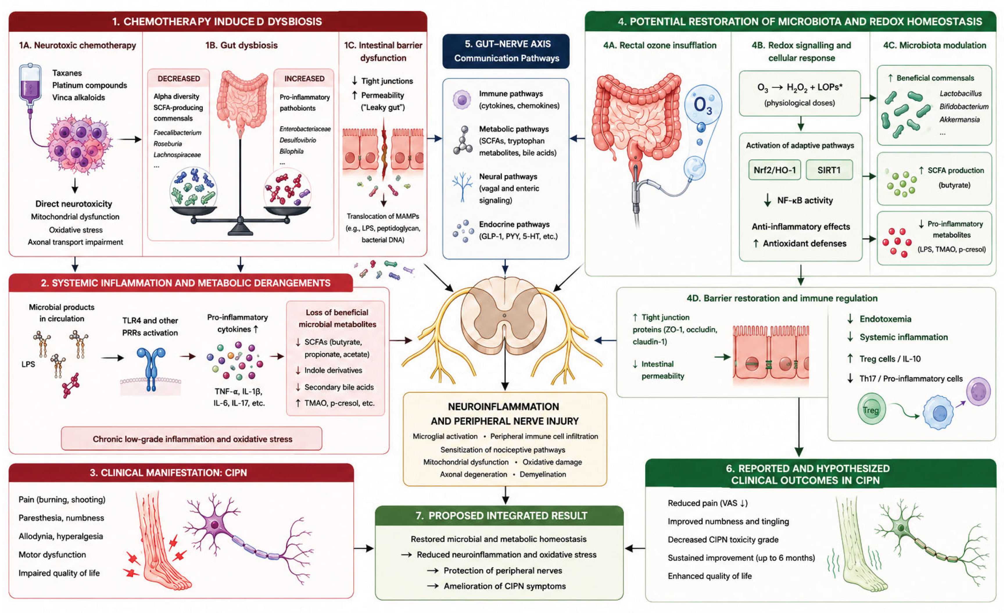

| Drug Class | Key Mechanism | Axonal Effect |
|---|---|---|
| Taxanes | Microtubule stabilization | Transport blockade, dying -back |
| Platinum compounds | DNA adducts (including mitochondrial) | DRG apoptosis, fiber degeneration |
| Vinca alkaloids | Tubulin polymerization blockade | Microtubule and transport disruption |
Legend: DRG, dorsal root ganglion.
Disclaimer/Publisher’s Note: The statements, opinions and data contained in all publications are solely those of the individual author(s) and contributor(s) and not of MDPI and/or the editor(s). MDPI and/or the editor(s) disclaim responsibility for any injury to people or property resulting from any ideas, methods, instructions or products referred to in the content. |
© 2026 by the authors. Licensee MDPI, Basel, Switzerland. This article is an open access article distributed under the terms and conditions of the Creative Commons Attribution (CC BY) license (http://creativecommons.org/licenses/by/4.0/).
Copyright: This open access article is published under a Creative Commons CC BY 4.0 license, which permit the free download, distribution, and reuse, provided that the author and preprint are cited in any reuse.


