

Submitted:
27 May 2026
Posted:
29 May 2026
You are already at the latest version
Abstract
The glomerular filtration barrier (GFB), localized at the interface of blood and urine, is crucial for renal filtration. Slit diaphragm (SD), a specialized intercellular junction between adjacent podocytes, is a significant component of GFB. Several transmembrane proteins (nephrin, NEPH1, and TRPC6) and adapter proteins (podocin and CD2AP) are tethered together and form a highly complex SD. Non-synonymous single-nucleotide polymorphisms (nsSNPs) in SD proteins cause congenital nephrotic syndrome, characterized by massive proteinuria. Nevertheless, the physicochemical consequences of these mutations remain elusive. We performed an integrated computational analysis of 25 clinically associated nsSNPs across five core SD proteins. We considered AlphaFold-derived structures and assessed the effects of SNPs on thermodynamic stability, secondary structure, aggregation propensity, and surface hydrophobicity. ΔΔG and RMSD are physicochemical descriptors, whereas aggregation and solubility scores reflect surface remodeling. The results suggest that some SNPs are destabilizing and confer high aggregation risk (podocin-D160G, TRPC6-R895C); some elicit surface remodeling (podocin-R168H, NEPH1-S573L); and variants exert dual effects on solubility and aggregation (TRPC6-R895C, podocin-V180M, CD2AP-T374A). Overall, most amino acid substitutions in SD proteins destabilize protein folding and promote aggregation, highlighting the role of physicochemical events in podocyte injury. Although the study provides quantitative insights into how amino acid substitutions alter the behavior of SD proteins, it has limitations, including reliance on AlphaFold-derived structures and the absence of experimental validation. Collectively, this work provides insights into mutation-driven physicochemical remodeling of SD proteins and establishes a rational framework for prioritizing clinically relevant variants for experimental validation and therapeutic interventions.
Keywords:
kidney
; podocyte
; slit-diaphragm
; nephrotic syndrome
; proteinuria
; podocin
; nephrin
; EPH1
; CD2AP
; TRPC6
Introduction
The slit diaphragm SD is a specialized intercellular junction that bridges the adjacent foot processes of podocytes, serving as a critical determinant of the permselectivity of the glomerular filtration barrier (GFB) [1,2,3]. The glomerular filtration barrier (GFB), comprising the fenestrated endothelium, glomerular basement membrane, and podocyte layer, enables efficient plasma ultrafiltration while limiting macromolecular loss into primary urine [4,5,6]. The negatively charged SD impedes the efflux of serum albumin, helping to maintain oncotic pressure and fluid balance [7,8]. The loss of negative charge on the podocyte surface diminishes electrostatic repulsion, resulting in the loss of albumin and other large molecules, such as immunoglobulins, in the urine [2,9]. Besides contributing to glomerular permselectivity, the SD serves as a dynamic signaling platform that regulates podocyte cytoskeleton and podocyte adhesion to the basement membrane [10]. The SD architecture is maintained by a multimeric network of transmembrane and scaffolding proteins, including nephrin, podocin, CD2-associated protein (CD2AP), NEPH1, and transient receptor potential cation channel 6 (TRPC6) [11,12,13,14,15,16,17]. These proteins couple structural integrity with intracellular signaling, which is essential for maintaining the absolute function of podocytes. Mutations in genes encoding proteins that constitute SD perturb the SD complex, compromising filtration selectivity and causing congenital nephrotic syndrome (CNS), a condition characterized by massive proteinuria, hypoalbuminemia, and edema [2,5,6,18,19,20,21]. Urinary loss of 3 grams or more of protein per 24 hours is considered nephrotic-range proteinuria [22].
CNS is one of the most frequent glomerular diseases among children, whereas most of the children with CNS respond to steroid treatment, but 10-20% of the children are steroid resistant [23]. Therefore, CNS may be steroid-sensitive (SSNS) or steroid-resistant (SRNS), with the latter frequently progressing to focal segmental glomerulosclerosis (FSGS) and end-stage renal disease (ESRD) [24]. Numerous SRNS cases are linked to missense mutations or nonsynonymous single-nucleotide polymorphisms (nsSNPs) in SD-associated genes [25,26]. Similarly, nephrin mutations impair folding and trafficking, preventing proper SD assembly, while NEPH1 variants destabilize intracellular signaling interfaces, leading to SD disruption and podocyte effacement [27,28,29,30]. For instance, overexpression of mutant TRPC6 in mice recapitulates human FSGS phenotypes, highlighting the pathogenic role of TRPC6-mediated calcium dysregulation in podocytes [19,20]. CD2AP instability, as observed in FSGS, restricts the flexibility of intrinsically disordered regions (IDRs) and weakens interactions with SD partners, promoting podocyte injury [17,31,32]. Likewise, pathogenic podocin mutations alter oligomerization and interaction dynamics, thereby reducing the stability of the SD complex [24]. Despite a greater understanding with few mutations in SD proteins, the quantitative physicochemical consequences of a large number of disease-associated mutations remain poorly defined. Although previous studies have identified general mechanisms such as protein misfolding, mistargeting, and disrupted interactions in SD dysfunction, it remains unclear how individual amino acid substitutions specifically alter the physical and structural properties of these proteins. In particular, we still lack a quantitative understanding of how such mutations affect protein stability, solubility, folding, and surface behavior, which are essential for proper oligomerization and slit diaphragm assembly. This study addresses this gap by systematically analyzing how disease-associated substitutions reshape the physicochemical characteristics of key SD proteins, thereby linking molecular alterations to podocyte barrier failure.
This study systematically analyzes 25 pathogenic nsSNPs across five core SD proteins, podocin, nephrin, CD2AP, NEPH1, and TRPC6, to elucidate their impact on physicochemical and structural properties critical for SD integrity. Variants were selected based on clinical association, recurrence in genetic databases, and localization within conserved or functionally relevant regions. Using AlphaFold-based structural modeling and computational prediction, we characterize mutation-induced physicochemical perturbations to refine genotype-phenotype relationships. Together, this knowledge deepens our understanding of the structure–function relationship of slit diaphragm proteins and offers greater insight into the pathogenic consequences of specific variants. These findings provide a quantitative framework for deciphering SD dysfunction and highlight potential molecular targets for therapeutic restoration of GFB function in the CNS.
Methods
Retrieval of Protein Structures and Single-Nucleotide Polymorphisms (SNPs): Predicted three-dimensional models of SD proteins; NPHS1 (nephrin), NPHS2 (podocin), KIRREL1 (NEPH1), TRPC6, and CD2AP were retrieved from the AlphaFold Protein Structure Database. Clinically validated nonsynonymous single-nucleotide polymorphisms (nsSNPs) associated with CNS were extracted from UniProtKB/Swiss-Prot, with cross-referencing to dbSNP, ClinVar, and Ensembl. Only variants with established clinical relevance or strong experimental support were selected for downstream computational analyses [33].
Functional impact prediction: The functional effects of nsSNPs were predicted using SIFT, PolyPhen-2, PANTHER, and MutPred2. Variants with SIFT ≤ 0.05 were classified as intolerant or damaging. PolyPhen-2 scores ≥ 0.85 indicated “probably damaging,” while 0.15–0.85 denoted “possibly damaging.” PANTHER analysis incorporated evolutionary conservation through PSEP (Position-Specific Evolutionary Preservation). MutPred2 assigned pathogenicity probabilities; scores > 0.75 indicated high-confidence pathogenic variants. These analyses were used collectively to predict the likely molecular effects of each substitution on protein function [34,35,36].
Variant Selection and Clinical Relevance: Nonsynonymous SNPs were curated from UniProt and cross-validated using ClinVar and published literature. The inclusion criteria comprised classification as pathogenic/likely pathogenic, recurrence across databases, localization within conserved functional domains, and a documented association with nephrotic syndrome phenotypes (CNS, SRNS, or FSGS). Variants with conflicting annotations or low-confidence evidence were excluded. Where available, allele frequency and disease severity were considered to prioritize clinically relevant mutations. The genetic, physicochemical, and clinical characteristics of all selected variants are summarized in SupplementaryTable S2.
Protein stability analysis: To assess thermodynamic stability changes induced by nsSNPs, the CUPSAT web server was used. The change in Gibbs free energy (ΔΔG, kcal/mol) between the wild-type (WT) and mutant forms was calculated. Variants with ΔΔG < –0.5 kcal/mol were considered destabilizing, whereas ΔΔG > +0.5 kcal/mol indicated stabilization. These data were cross-validated with structural deviation (RMSD) and aggregation analyses to ensure consistency [37].
Secondary structure and aggregation propensity analysis: The Pasta 2.0 server was used to estimate alterations in α-helix, β-sheet, and coil composition for each mutant relative to the WT. A comparative analysis identified variations in the aggregation-prone residues between WT and mutants. An increased proportion and redistribution of beta-sheet content were interpreted as potential indicators of enhanced aggregation propensity, consistent with its association with amyloidogenic behavior.
Surface hydrophobicity and structural deviation analysis: Hydrophobic and electrostatic surface properties were computed using Protein Surface Analyzer (PSA) and AggScore tools from the Schrödinger BioLuminate suite. Surfaces were color-coded according to electrostatic potential: positive (blue), negative (red), hydrophobic (green), and neutral/polar (gray). Root-mean-square deviation (RMSD) values were calculated by superimposing WT and mutant structures in Schrödinger Maestro using Cα backbone atoms. Variants were categorized as minimally deviating (RMSD < 2 Å), moderately deviating (2–4 Å), or substantially altered (> 4 Å) [38].
Identification of Aggregation-Prone Residues and Solubility Assessment: Structure-based aggregation analysis was performed using Aggrescan3D, which quantifies aggregation propensity and solubility from 3D structural models. Comparative studies of WT and mutant forms were conducted to identify shifts in aggregation-prone residues and changes in solubility. Positive aggregation scores indicate an increased aggregation tendency or reduced solubility, while negative scores reflect enhanced solubility, and near-zero values represent residues with no influence on aggregation. [39].
All structural analyses in this study were performed using AlphaFold-predicted models, which represent static, monomeric conformations generated in the absence of cellular context. While AlphaFold provides high-accuracy structural predictions for many folded regions, it does not explicitly account for membrane embedding, oligomerization, lipid interactions, mechanical forces, post-translational modifications, or the dynamic cellular environment. In addition, intrinsically disordered regions (IDRs) and multimeric assemblies, which are characteristic features of several slit diaphragm proteins, are not fully captured by current structure prediction algorithms. Further, Alternative servers were not used due to lower accuracy and lack of full-length models compared to AlphaFold.” Accordingly, all analyses were conducted as relative, comparative assessments between wild-type and mutant proteins, and the resulting physicochemical parameters should be interpreted as predictive trends rather than definitive representations of in vivo conformations or functional states. The overall computational workflow is summarized in Figure 1.
Results
Overview of Mutation-Induced Physicochemical Alterations: A total of 25 nsSNPs across five slit diaphragm (SD) proteins, podocin, nephrin, CD2AP, NEPH1, and TRPC6, were evaluated to determine their structural and physicochemical consequences. Functional prediction tools (SIFT, PolyPhen-2, PANTHER, MutPred2) consistently classified the majority of variants as deleterious or pathogenic. Detailed functional prediction and stability scores are summarized in (Supplementary Table S1). Approximately 80% of the variants were destabilizing according to CUPSAT (ΔΔG < –0.5 kcal/mol), indicating widespread thermodynamic compromise. RMSD deviations ranging from 1.2 Å to >7 Å further suggested substantial conformational remodeling. Collectively, these findings indicate that nsSNPs can significantly alter the SD protein architecture, potentially weakening the integrity of the GFB. A comprehensive summary of the physicochemical parameters is presented in Table 1.
ΔΔG and RMSD values reported in this study should not be interpreted as direct indicators of pathogenicity. Instead, these parameters are presented as supportive physicochemical descriptors that reflect predicted thermodynamic shifts and modeled structural deviations, respectively. Given that protein stability and conformation in vivo are influenced by membrane environments, interaction partners, post-translational modifications, and cellular quality-control mechanisms, changes in ΔΔG and RMSD alone cannot be equated with functional impairment or disease causation. Accordingly, these metrics are used here to identify relative trends in mutation-induced effects and to prioritize variants for further experimental Validation, rather than to establish definitive mechanistic conclusions.
Structural and Aggregation Effects of Podocin Variants: Seven podocin variants (R138Q, D160G, R168H, V180M, R229Q, V290M, A297V) exhibited marked physicochemical perturbations. PolyPhen-2 predicted R138Q, D160G, R168H, and V290M as probably damaging (score ≥ 0.85), while MutPred2 classified all except R229Q as pathogenic. Most substitutions were destabilizing, although R168H showed mild stabilization (ΔΔG = +0.54 kcal/mol) accompanied by a large RMSD (>7.39 Å), suggesting local structural reorganization. Secondary-structure analysis revealed a reduced α-helix and a significant increase in β-sheet content, particularly in D160G, indicative of partial unfolding and conformational shifts. Aggrescan 3D analysis revealed that D160G showed an increased propensity to aggregate. Interestingly, V290M was associated with a substantial increase in solubility, while A297V demonstrated a significant reduction. The shifts in the aggregation scores (decrease) were accompanied by changes in the solubility (increase) for V180M, and vice versa in the case of R138Q, indicating a subtle dual impact on both aggregation behavior and solubility ( Supplementary Figure 1A). Hydrophobic surface analysis revealed increased solvent-exposed hydrophobicity in R138Q, which may promote non-specific interactions and clustering (Figure 2A, B, and C). Similarly, D160G displayed a pronounced decrease, most likely reflecting local conformational rearrangements or burial of previously exposed hydrophobic residues. Aggregation-prone surface patch analysis (AggScore) further supported the above findings. D160G and R168H are associated with decreased negative and positive surface potentials, aligning with its moderately stabilizing yet structurally flexible behavior. However, R229Q disrupted charge balance, increasing the negative surface potential (red), while A297V expanded a hydrophobic surface patch (green), and both variants suggested an increased risk of aggregation. Together, these results indicate that podocin nsSNPs disrupt folding equilibrium and generate aggregation-prone intermediates that may weaken SD stability. Further, the Comprehensive physicochemical profiling of podocin variants is provided in Supplementary Table S3.
Destabilizing and Aggregation-Prone Nephrin Mutants: Seven nephrin variants (W64S, A107E, I171N, R256W, R367C, C528F, C623F) were all predicted to be damaging by most damage prediction tools. CUPSAT identified every substitution as destabilizing. Among these, R367C demonstrated the strongest pathogenic signature, with an RMSD greater than 4 Å, reduced solubility, and increased hydrophobic exposure, suggesting impaired adhesion or altered interaction dynamics in the mutant (Figure 3A,B). Additionally, electrostatic surface analysis revealed a loss of positive potential, indicating perturbations in charge distribution and patch formation, which further suggests a predisposition to aggregation (Table 2; Figure 3C). Aggregation analysis confirmed increased aggregation scores for R367C and R256W, consistent with their decreased solubility, while A107E and I171N showed a substantial increase in solubility (Supplementary Figure 1B). These findings align with nephrin’s known susceptibility to misfolding and trafficking defects, reinforcing its central role in maintaining the structural integrity of the SD. The detailed physicochemical parameters for nephrin variants are summarized in (Supplementary Table 3).
Aggregation propensity scores reported in this study should not be interpreted as evidence of in vivo aggregation within podocytes or at the slit diaphragm. Instead, these predictions reflect relative changes in surface physicochemical properties, including hydrophobic exposure and residue clustering, that may influence folding behavior under simplified conditions. In cellular environments, protein homeostasis is tightly regulated by molecular chaperones, endoplasmic reticulum quality-control systems, post-translational modifications, and membrane insertion pathways. Therefore, increased aggregation scores should be viewed as comparative indicators of altered surface chemistry rather than direct markers of pathogenic protein aggregation.
CD2AP Variants Alter Surface Chemistry and Protein Flexibility: Two CD2AP variants (K301M, T374A) were evaluated. T374A was the only variant in the dataset classified as deleterious by SIFT and exhibited the most significant structural deviation (RMSD > 5 Å). Both variants showed destabilizing ΔΔG values. Hydrophobic surface mapping revealed reduced solvent-exposed hydrophobicity, suggesting a more compact conformation. Despite this, the aggregation analysis showed reduced solubility, especially for K301M (Supplementary Figure 1C). T374A exhibited a minor increase in α-helix content, indicating localized refolding. These alterations suggest that CD2AP mutations reduce structural flexibility and may compromise the cytoskeletal coupling of SD. The detailed physicochemical characterization of CD2AP variants is highlighted in Supplementary Table 4. Moreover, the detailed physicochemical parameters are summarized in Table 3.
NEPH1 Mutations Exhibit Distinct Stability and Aggregation Profiles: Two NEPH1 variants (S573L, R440C) exhibited contrasting structural effects. S573L was stabilizing (ΔΔG = 1.32 kcal/mol). Still, it displayed increased β-sheet content and an elevated hydrophobic surface area, along with an increased tendency to aggregate, suggesting that even stabilizing mutants may increase the aggregation risk due to changes in surface exposure. (Figure 4A; Supplementary Figure 1D). R440C was destabilizing (ΔΔG = -0.64 kcal/mol), showed reduced solubility, and increased hydrophobic patch formation (Figure 4C,D). Despite differing stability profiles, both mutations altered surface polarity and aggregation tendencies, highlighting the dual contribution of structural stability and hydrophobicity to NEPH1 pathogenicity. The Physicochemical properties of NEPH1 variants are summarized in Table 4. Moreover, Comprehensive physicochemical profiling of NEPH1 variants is summarized in Supplementary Table S4.
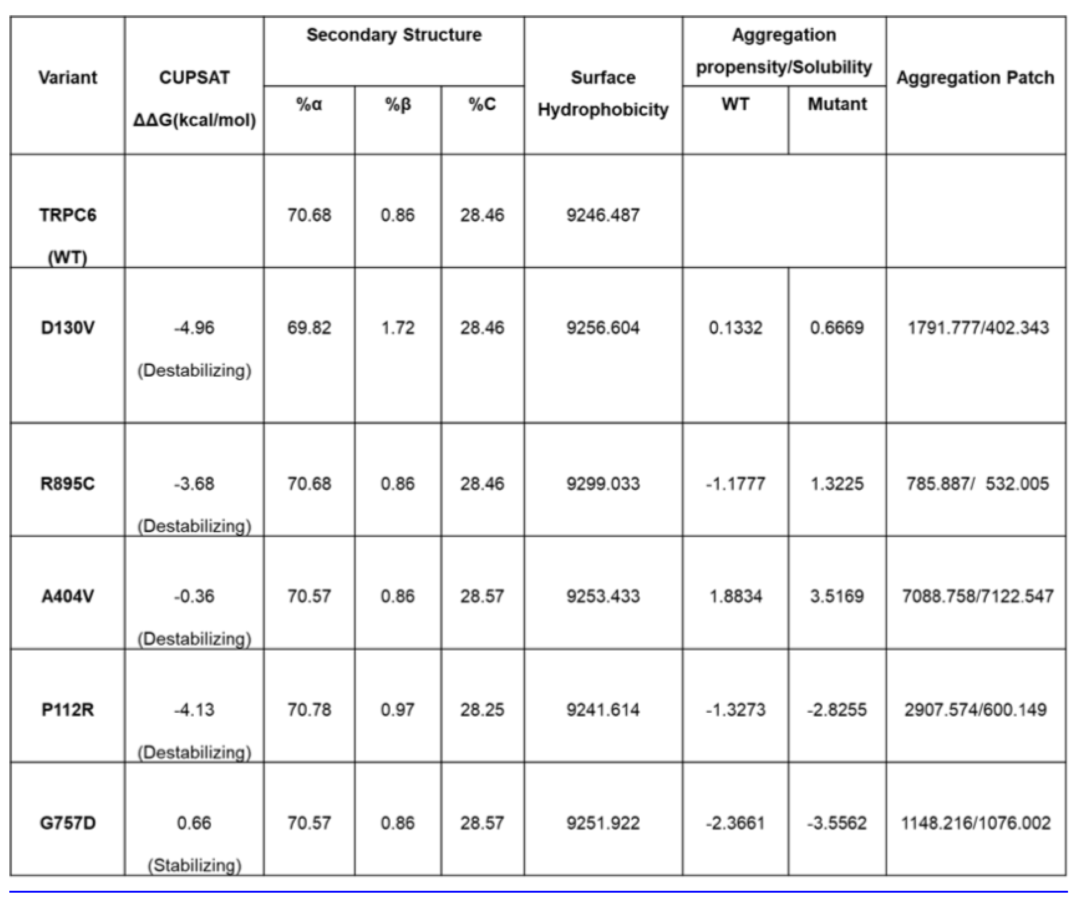
Heterogeneous Physicochemical Consequences of TRPC6 Variants: Seven TRPC6 variants (P112R, D130V, G162R, A404V, G757D, L780P, R895C) showed diverse physicochemical outcomes. Six were predicted as damaging. G757D emerged as a potentially stabilizing variant, with a low RMSD of <2.0 Å and a positive ΔΔG of> 0.5 kcal/mol, suggesting structural preservation. R895C was the most pathogenic, exhibiting an RMSD > 4 Å, drastically reduced solubility, and extensive formation of hydrophobic patches (Figure 5A). D130V exhibited increased β-sheet content (Figure S2) and, alongside high aggregation propensities, was also observed in A404V (Supplementary Figure 1E). Surface patch mapping showed that the A404V transitioned from a positive to a hydrophobic, aggregation-prone surface (Figure 5B,D). Conversely, G757D showed stabilizing ΔΔG, minimal structural deviation, and low aggregation, indicating preserved structural integrity. However, G757D and P112R showed increased solubility (Supplementary Figure 1E). A heightened aggregation potential and a hydrophobic surface patch were observed in R895C (Table 5), which exhibited the most pronounced reduction in solubility (Supplementary Figure 1E), indicating it is likely aggregation-prone and functionally compromised. Aggscore analysis further supported this, revealing more extensive hydrophobic and neutral patches. These findings demonstrate the mutation-specific sensitivity of TRPC6 to misfolding, relevant to its role in familial FSGS. The Comprehensive TRPC6 variant analysis is shown in Table 5. Moreover, the Detailed physicochemical characterization of TRPC6 variants is provided in Supplementary Table 4.
Cross-Protein Trends in Solubility and Aggregation Behavior: Comparison across all SD proteins revealed a strong inverse relationship between solubility and aggregation propensity, with some nsSNPs exhibiting dual effects on both properties. Variants such as R895C (TRPC6), R138Q (podocin), and V180M (podocin) exhibited elevated aggregation propensity and reduced solubility, rendering them structurally compromised. Additionally, T374A (CD2AP) showed a mild increase in aggregation. (Supplementary Figure. 1A–E). These findings suggest that nsSNP-induced pathogenicity is driven not only by thermodynamic instability but also by surface hydrophobicity, charge redistribution, and altered folding dynamics, which collectively disrupt the integrity of the SD complex and glomerular filtration function.
It is important to emphasize that the physicochemical alterations reported here are derived from static structural models and therefore reflect relative mutation-induced trends rather than direct biological outcomes. The observed changes in predicted stability, surface hydrophobicity, aggregation propensity, and RMSD values should be interpreted within the limitations of AlphaFold-based modeling, which does not incorporate membrane environments, oligomeric assembly, lipid interactions, or transcellular adhesion complexes that are fundamental to slit diaphragm function.
Discussion
The SD is the critical component that maintains the selective permeability of the GFB, and its precise molecular architecture dictates both the body’s fluid balance and blood oncotic pressure by preventing the leakage of macromolecules into the urine [5,6,40,41,42,43,44]. Even a minor disruption of this complex protein network, comprising podocin, nephrin, CD2AP, NEPH1, and TRPC6, results in podocyte dysfunction and proteinuria [2,10,15]. In this study, we provide a comprehensive physicochemical perspective on how single amino acid substitutions across several key SD proteins remodel their intrinsic stability, surface chemistry, and aggregation behavior. By integrating multi-parametric computational analyses, our findings reveal that both destabilizing and paradoxically stabilizing mutations can compromise protein behavior by altering hydrophobicity, folding dynamics, and intermolecular interactions. This work bridges the fields of structural bioinformatics and renal physiology, offering a molecular narrative that connects genotype to phenotype in nephrotic syndrome and establishes a quantitative framework for understanding podocyte barrier failure.
Protein misfolding and aggregation are widely recognized as key molecular events that drive conformational diseases [45,46,47]. Reduced protein stability often promotes misfolding and degradation, while paradoxically, specific stabilizing mutations can also exert deleterious effects by locking proteins into non-functional or aggregation-prone conformations [48,49,50]. This dual behavior is evident in our analysis, in which both destabilizing and stabilizing variants showed potential pathogenicity, depending on their surface chemistry and conformational plasticity. Among podocin variants, R138Q and D160G were markedly destabilizing and showed increased aggregation, reinforcing podocin’s critical role in maintaining SD complex integrity [42]. Interestingly, R168H displayed stabilizing behavior but with elevated aggregation propensity due to altered surface hydrophobicity, illustrating that structural rigidity does not necessarily equate to functional neutrality. This finding aligns with general principles of protein aggregation disorders, where altered surface exposure of hydrophobic residues can drive pathogenic misassembly [51]. For nephrin, all analyzed variants were predicted to be damaging, with R367C showing pronounced hydrophobic surface exposure and reduced solubility features that promote aggregation and misfolding. These physicochemical disruptions complement previous reports linking nephrin missense mutations to defective folding and intracellular trafficking, leading to impaired SD assembly and filtration barrier failure [52]. The RMSD deviations exceeding 4 Å further indicate substantial conformational remodeling that may compromise intermolecular recognition within the SD complex.
The CD2AP variant T374A exhibited significant structural deviation and was uniquely classified as deleterious by SIFT, suggesting its potential to perturb CD2AP-mediated scaffolding functions. The reduced solvent-exposed hydrophobic surfaces in both T374A and K301M mutants suggest compaction or conformational tightening, which could diminish the flexibility of intrinsically disordered regions (IDRs). Such conformational constraints are consistent with previous findings that CD2AP instability contributes to restricted IDR dynamics, impaired SD interactions, and podocyte effacement in NS and focal segmental glomerulosclerosis [53].For NEPH1, the S573L variant, though thermodynamically stabilizing, exhibited increased β-sheet content and hydrophobic surface exposure, indicating that enhanced stability can paradoxically promote aggregation by altering secondary structure distribution. In contrast, R440C was destabilizing yet shared similar aggregation-promoting characteristics, underscoring the importance of both internal stability and surface chemistry in defining pathogenic potential. These results suggest that subtle shifts in NEPH1 folding dynamics can significantly impact the organization of the SD complex and signal transduction. TRPC6 variants demonstrated the most heterogeneous physicochemical effects. The R895C substitution displayed combined destabilization, hydrophobic surface expansion, and markedly reduced solubility, identifying it as the most aggregation-prone and pathogenic mutation. These findings are consistent with earlier evidence implicating TRPC6 gain-of-function mutations in the pathogenesis of FSGS and podocyte injury [54]. The enrichment of hydrophobic and neutral surface patches observed in the R895C and A404V mutants further supports the likelihood of aberrant intermolecular interactions and misfolding that compromise ion channel function and stability of the SD.
Collectively, this integrative computational study advances current understanding by quantitatively linking amino acid substitutions to alterations in protein stability, solubility, and aggregation behavior, which collectively regulate the assembly and interaction dynamics of SD proteins. These insights provide a molecular framework explaining how specific mutations disrupt podocyte homeostasis and drive nephrotic pathology. Nevertheless, several limitations must be acknowledged. The study primarily relies on computational predictions derived from AlphaFold-generated structural models, as experimentally determined structures of SD proteins remain limited. Although AlphaFold offers high confidence in structured regions, predictions within IDRs and protein-protein interfaces may lack atomic precision, potentially influencing stability or aggregation estimates. Additionally, while computational tools provide valuable high-throughput insights, experimental Validation through biophysical, structural, and cellular assays remains essential to confirm the predicted pathogenic mechanisms.
Limitations: All mechanistic interpretations presented in this study should be considered hypothesis-generating rather than definitive. While the observed physicochemical alterations are biologically plausible and consistent with known slit diaphragm dysfunction mechanisms, the conclusions are derived from computational predictions and therefore do not establish direct causality. Accordingly, proposed links between mutation-induced physicochemical changes and impaired barrier integrity, cytoskeletal coupling, or adhesion defects are framed as testable hypotheses that require experimental Validation. Future studies employing cellular assays, protein-protein interaction analyses, membrane reconstitution systems, and molecular dynamics simulations will be essential to substantiate these predictions and define their mechanistic relevance in vivo. Despite these limitations, our findings establish a robust framework for interpreting the physicochemical consequences of disease-associated SD variants. By integrating structure-based modeling with predictive analytics, this study lays the foundation for precision nephrology approaches that aim to identify destabilizing mutations, prioritize clinically relevant variants, and inform therapeutic design targeting podocyte dysfunction. The computational pipeline developed here may be broadly applicable to other podocyte-associated or protein-misfolding diseases, supporting molecular diagnosis and targeted drug discovery in kidney pathophysiology.
Conclusion
This study presents a comprehensive physicochemical framework for understanding how pathogenic nonsynonymous single-nucleotide polymorphisms (nsSNPs) remodel slit diaphragm (SD) proteins and compromise podocyte barrier integrity. Through an integrated multi-parametric computational analysis of podocin, nephrin, CD2AP, NEPH1, and TRPC6, we demonstrate that both destabilizing and paradoxically stabilizing mutations can disrupt protein behavior by altering surface hydrophobicity, solubility, and secondary-structure dynamics. Importantly, our findings reveal that pathogenicity is not solely driven by global structural destabilization but is governed by a delicate interplay between folding kinetics, surface polarity, and intermolecular recognition. Several high-impact variants, including podocin-D160G and TRPC6-R895C, exhibited strong aggregation signatures and reduced solubility, highlighting their potential to impair SD assembly and weaken glomerular filtration. Conversely, stabilizing variants such as podocin-R168H and NEPH1-S573L also enhanced aggregation propensity through surface remodeling, underscoring that increased stability does not necessarily equate to functional preservation. Collectively, these results advance the molecular understanding of podocyte dysfunction by revealing convergent physicochemical mechanisms underlying SD disruption. The computational framework established here serves as a platform for variant prioritization for experimental Validation. It provides a rational basis for developing targeted therapeutic strategies to restore SD integrity and glomerular barrier function in nephrotic syndrome.
Supplementary Materials
The following supporting information can be downloaded at the website of this paper posted on Preprints.org.
Author Contributions
Swetha Gajula: Conceptualization, Methodology, Formal analysis, Data curation, Writing original draft, and Validation. Abrar H Qadri: Validation, Writing, Editing, and Visualization. Hari Krishnan Padmanabhan: Methodology, Formal analysis, Data curation. Anil K Pasupulati: Conceptualization, Supervision, Editing, Project administration, and Funding acquisition.
Data availability statement
The data presented in this study are available from the corresponding author upon reasonable request.
Artificial intelligence use disclosure
During the preparation of this manuscript, ChatGPT (OpenAI) was used exclusively for English language editing and grammatical correction. The authors reviewed and edited the output and take full responsibility for the manuscript’s content.
Funding Information and Acknowledgments
AKP acknowledges funding from the Indian Council of Medical Research and Anusandhan National Research Foundation.
Declaration of competing interest
The authors declare no conflict of interest.
References
- Anil Kumar, P.; Welsh, G.I.; Saleem, M.A.; Menon, R.K. Molecular and cellular events mediating glomerular podocyte dysfunction and depletion in diabetes mellitus. Front Endocrinol. 2014, 5, 151. [Google Scholar] [CrossRef]
- Bazzi, C.; Bakoush, O.; Gesualdo, L. Proteinuria: from molecular to clinical applications in glomerulonephritis. Int. J. Nephrol. 2012, 2012, 424968. [Google Scholar] [CrossRef]
- Ebefors, K.; Lassén, E.; Anandakrishnan, N.; Azeloglu, E.U.; Daehn, I.S. Modeling the Glomerular Filtration Barrier and Intercellular Crosstalk. In Frontiers in Physiology Volume; 2021; pp. 12–2021. [Google Scholar]
- Daehn, I.S.; Duffield, J.S. The glomerular filtration barrier: a structural target for novel kidney therapies. Nat. Rev. Drug Discov. 2021, 20, 770–788. [Google Scholar] [CrossRef]
- Grahammer, F.; Schell, C.; Huber, T.B. The podocyte slit diaphragm--from a thin grey line to a complex signalling hub. Nat. Rev. Nephrol. 2013, 9, 587–98. [Google Scholar] [CrossRef] [PubMed]
- Grahammer, F.; et al. A flexible, multilayered protein scaffold maintains the slit in between glomerular podocytes. JCI Insight 1 2016. [Google Scholar] [CrossRef] [PubMed]
- Fissell, W.H.; Miner, J.H. What Is the Glomerular Ultrafiltration Barrier? J. Am. Soc. Nephrol. 2018, 29, 2262–2264. [Google Scholar] [CrossRef]
- Williamson, C.R.; Pantic, U.V.; Wang, A.Y.; Jones, N. Revisiting nephrin signaling and its specialized effects on the uniquely adaptable podocyte. Biochem J. 2025, 482, 763–88. [Google Scholar] [CrossRef]
- Ballermann, B.J.; Nyström, J.; Haraldsson, B. The Glomerular Endothelium Restricts Albumin Filtration. In Frontiers in Medicine; 2021; pp. 8–2021. [Google Scholar]
- Cunanan, J.; Zhang, D.; Peired, A.J.; Barua, M. Podocytes in health and glomerular disease. Front Cell Dev. Biol. 2025, 13, 1564847. [Google Scholar] [CrossRef]
- Birtasu, A.N.; et al. The molecular architecture of the kidney slit diaphragm. bioRxiv 2023. [Google Scholar] [CrossRef]
- Kocylowski, M.K.; et al. A slit-diaphragm-associated protein network for dynamic control of renal filtration. Nat. Commun. 2022, 13. [Google Scholar] [CrossRef]
- Qadri, A.H.; Prajapati, J.; Praghna, D.; Sinha, A.; Pasupulati, A.K. Structural and functional insights of the podocyte slit diaphragm complex. Tissue Barriers 2575198. [CrossRef]
- Mulukala Narasimha, S.K.; Kar, P.P.; Vadrevu, R.; Pasupulati, A.K. Intrinsically disordered regions mediate macromolecular assembly of the Slit diaphragm proteins associated with Nephrotic syndrome. Mol. Simul. 2019, 45, 603–613. [Google Scholar] [CrossRef]
- Mulukala, S.K.N.; Irukuvajjula, S.S.; Kumar, K.; Garai, K.; Venkatesu, P.; Vadrevu, R.; Pasupulati, A.K. Structural features and oligomeric nature of human podocin domain. Biochem. Biophys. Rep. 2020, 23, 100774. [Google Scholar] [CrossRef] [PubMed]
- Mulukala, S.K.N.; Kambhampati, V.; Qadri, A.H.; Pasupulati, A.K. Evolutionary conservation of intrinsically unstructured regions in slit-diaphragm proteins. PLoS ONE 2021, 16, e0254917. [Google Scholar] [CrossRef] [PubMed]
- Qadri, A.H.; Tripathi, R.M.; Faheem, I.; Meshram, S.; Reddem, E.R.; Ramachandran, R.; Pasupulati, A.K. CD2AP’s Structure and Oligomerization are compromised by the K301M mutation: implications for Nephrotic syndrome. bioRxiv 2026, 2026.01.08.698362. [Google Scholar]
- Ebefors, K.; Lassén, E.; Anandakrishnan, N.; Azeloglu, E.U.; Daehn, I.S. Modeling the Glomerular Filtration Barrier and Intercellular Crosstalk. Front Physiol. 2021, 12, 689083. [Google Scholar] [CrossRef]
- Lee, J.X.; Tan, Y.J.; Ismail, N.A.S. NPHS Mutations in Pediatric Patients with Congenital and Steroid-Resistant Nephrotic Syndrome. Int. J. Mol. Sci. 2024, 25. [Google Scholar] [CrossRef]
- Liu, J.; Wang, W. Genetic basis of adult-onset nephrotic syndrome and focal segmental glomerulosclerosis. Front Med. 2017, 11, 333–339. [Google Scholar] [CrossRef]
- Benoit, G.; Machuca, E.; Antignac, C. Hereditary nephrotic syndrome: a systematic approach for genetic testing and a review of associated podocyte gene mutations. Pediatr. Nephrol. 2010, 25, 1621–1632. [Google Scholar] [CrossRef]
- Hull, R.P.; Goldsmith, D.J. Nephrotic syndrome in adults. BMJ 2008, 336, 1185–9. [Google Scholar] [CrossRef]
- Pokrajac, D.; Kamber, A.H.; Karasalihovic, Z. Children with Steroid-Resistant Nephrotic Syndrome: A Single -Center Experience. Mater. Sociomed. 2018, 30, 84–88. [Google Scholar] [CrossRef]
- Vivarelli, M.; Gibson, K.; Sinha, A.; Boyer, O. Childhood nephrotic syndrome. The Lancet 2023, 402, 809–824. [Google Scholar] [CrossRef] [PubMed]
- Zhao, J.; Zhang, S.; Jiang, Y.; Liu, Y.; Zhu, Q. Mutation analysis of pathogenic non-synonymous single nucleotide polymorphisms (nsSNPs) in WFS1 gene through computational approaches. Sci. Rep. 2023, 13, 6774. [Google Scholar] [CrossRef]
- Sarker, D.K.; Ray, P.; Salam, F.B.A.; Uddin, S.J. Exploring the impact of deleterious missense nonsynonymous single nucleotide polymorphisms in the DRD4 gene using computational approaches. Sci. Rep. 2025, 15, 3150. [Google Scholar] [CrossRef]
- Santín, S.; et al. Nephrin mutations cause childhood- and adult-onset focal segmental glomerulosclerosis. Kidney Int. 2009, 76, 1268–76. [Google Scholar] [CrossRef] [PubMed]
- Sellin, L.; Huber, T.B.; Gerke, P.; Quack, I.; Pavenstadt, H.; Walz, G. NEPH1 defines a novel family of podocin interacting proteins. FASEB J. 2003, 17, 115–7. [Google Scholar] [CrossRef]
- Völker, L.A.; et al. Comparative analysis of Neph gene expression in mouse and chicken development. Histochem Cell Biol. 2012, 137, 355–66. [Google Scholar] [CrossRef]
- Weber, S.; Gribouval, O.; Esquivel, E.L.; Morinière, V.; Tête, M.-J.; Legendre, C.; Niaudet, P.; Antignac, C. NPHS2 mutation analysis shows genetic heterogeneityof steroid-resistant nephrotic syndrome and lowpost-transplant recurrence. Kidney Int. 2004, 66, 571–579. [Google Scholar] [CrossRef] [PubMed]
- Vandal, M.; et al. CD2AP at the junction of nephropathy and Alzheimer’s disease. Mol. Neurodegener. 2025, 20, 63. [Google Scholar] [CrossRef]
- Gao, S.; et al. An updated analysis of the association between CD2-associated protein gene rs9349407 polymorphism and Alzheimer’s disease in Chinese population. In Frontiers in Neuroinformatics Volume; 2022; pp. 16–2022. [Google Scholar]
- Sim, N.L.; Kumar, P.; Hu, J.; Henikoff, S.; Schneider, G.; Ng, P.C. SIFT web server: predicting effects of amino acid substitutions on proteins. Nucleic Acids Res. 2012, 40, W452–7. [Google Scholar] [CrossRef]
- Adzhubei, I.A.; Schmidt, S.; Peshkin, L.; Ramensky, V.E.; Gerasimova, A.; Bork, P.; Kondrashov, A.S.; Sunyaev, S.R. A method and server for predicting damaging missense mutations. Nat. Methods 2010, 7, 248–9. [Google Scholar] [CrossRef]
- Mi, H.; Muruganujan, A.; Casagrande, J.T.; Thomas, P.D. Large-scale gene function analysis with the PANTHER classification system. Nat. Protoc. 2013, 8, 1551–66. [Google Scholar] [CrossRef]
- Pejaver, V.; et al. Inferring the molecular and phenotypic impact of amino acid variants with MutPred2. Nat. Commun. 2020, 11, 5918. [Google Scholar] [CrossRef]
- Parthiban, V.; Gromiha, M.M.; Schomburg, D. CUPSAT: prediction of protein stability upon point mutations. Nucleic Acids Res. 2006, 34, W239–42. [Google Scholar] [CrossRef]
- Walsh, I.; Seno, F.; Tosatto, S.C.; Trovato, A. PASTA 2.0: an improved server for protein aggregation prediction. Nucleic Acids Res. 2014, 42, W301–7. [Google Scholar] [CrossRef]
- Kuriata, A.; Iglesias, V.; Pujols, J.; Kurcinski, M.; Kmiecik, S.; Ventura, S. Aggrescan3D (A3D) 2.0: prediction and engineering of protein solubility. Nucleic Acids Res. 2019, 47, W300–w307. [Google Scholar] [CrossRef] [PubMed]
- Adair, B.D.; Altintas, M.M.; Möller, C.C.; Arnaout, M.A.; Reiser, J. Structure of the kidney slit diaphragm adapter protein CD2-associated protein as determined with electron microscopy. J. Am. Soc. Nephrol. 2014, 25, 1465–73. [Google Scholar] [CrossRef]
- Allison, S.J. Insights into protein scaffolds of the slit diaphragm. Nat. Rev. Nephrol. 2016, 12, 441–441. [Google Scholar] [CrossRef] [PubMed]
- Padmanaban, H.K.; Bheemireddy, S.; Mulukala, S.K.N.; Dunna, N.R.; Pasupulati, A.K. Structural conformations of intrinsically disordered proteins of podocyte slit-diaphragm. Comput. Struct. Biotechnol. Rep. 2025, 2, 100060. [Google Scholar] [CrossRef]
- Qadri, A.H.; Prajapati, J.; Faheem, I.; Bhattacharjee, U.; Padmanaban, H.K.; Mulukala, S.K.; Pasupulati, A.K. Biophysical characterization and insights into the oligomeric nature of CD2-associated protein. Int. J. Biochem Mol. Biol. 2024, 15, 20–33. [Google Scholar] [CrossRef] [PubMed]
- van de Leemput, J.; Wen, P.; Han, Z. Using Drosophila Nephrocytes to Understand the Formation and Maintenance of the Podocyte Slit Diaphragm. Front Cell Dev. Biol. 2022, 10, 837828. [Google Scholar] [CrossRef] [PubMed]
- Singh, S.M.; Kongari, N.; Cabello-Villegas, J.; Mallela, K.M. Missense mutations in dystrophin that trigger muscular dystrophy decrease protein stability and lead to cross-beta aggregates. Proc. Natl. Acad. Sci. U S A 2010, 107, 15069–74. [Google Scholar] [CrossRef]
- Soto, C.; Pritzkow, S. Protein misfolding, aggregation, and conformational strains in neurodegenerative diseases. Nat. Neurosci. 2018, 21, 1332–1340. [Google Scholar] [CrossRef]
- Chaturvedi, S.K.; Siddiqi, M.K.; Alam, P.; Khan, R.H. Protein misfolding and aggregation: Mechanism, factors and detection. Process Biochem. 2016, 51, 1183–1192. [Google Scholar] [CrossRef]
- Witham, S.; Takano, K.; Schwartz, C.; Alexov, E. A missense mutation in CLIC2 associated with intellectual disability is predicted by in silico modeling to affect protein stability and dynamics. Proteins 2011, 79, 2444–54. [Google Scholar] [CrossRef]
- Louros, N.; Schymkowitz, J.; Rousseau, F. Mechanisms and pathology of protein misfolding and aggregation. Nat. Rev. Mol. Cell Biol. 2023, 24, 912–933. [Google Scholar] [CrossRef]
- Akbarian, M.; Chen, S.H. Instability Challenges and Stabilization Strategies of Pharmaceutical Proteins. Pharmaceutics 2022, 14. [Google Scholar] [CrossRef] [PubMed]
- Zhuo, L.; Huang, L.; Yang, Z.; Li, G.; Wang, L. A comprehensive analysis of NPHS1 gene mutations in patients with sporadic focal segmental glomerulosclerosis. BMC Med. Genet 2019, 20, 111. [Google Scholar] [CrossRef]
- Haraldsson, B.; Nyström, J.; Deen, W.M. Properties of the glomerular barrier and mechanisms of proteinuria. Physiol. Rev. 2008, 88, 451–87. [Google Scholar] [CrossRef]
- Isojima, T.; et al. LMX1B mutation with residual transcriptional activity as a cause of isolated glomerulopathy. Nephrol. Dial. Transpl. 2014, 29, 81–8. [Google Scholar] [CrossRef]
- Bierzynska, A.; Saleem, M. Recent advances in understanding and treating nephrotic syndrome. F1000Res 2017, 6, 121. [Google Scholar] [CrossRef]
Figure 1.
Computational workflow for slit-diaphragm protein analysis: Schematic representation of the integrated in silico pipeline used in this study. Clinically relevant nsSNPs were retrieved from UniProt and ClinVar, followed by pathogenicity filtering using SIFT and PolyPhen-2. Wild-type protein structures were obtained from AlphaFold. Functional impact was assessed using PANTHER and MutPred2. Protein stability changes were evaluated using CUPSAT, DynaMut2, and DUET. Secondary structure alterations were predicted using PASTA 2.0. Aggregation propensity and solubility were analyzed using Aggrescan3D, while surface hydrophobicity mapping was performed using Schrödinger Protein Surface Analyzer (PSA). Structural deviations between wild-type and mutant proteins were quantified by RMSD calculations in Maestro. All outputs were integrated for comprehensive physicochemical interpretation.
Figure 1.
Computational workflow for slit-diaphragm protein analysis: Schematic representation of the integrated in silico pipeline used in this study. Clinically relevant nsSNPs were retrieved from UniProt and ClinVar, followed by pathogenicity filtering using SIFT and PolyPhen-2. Wild-type protein structures were obtained from AlphaFold. Functional impact was assessed using PANTHER and MutPred2. Protein stability changes were evaluated using CUPSAT, DynaMut2, and DUET. Secondary structure alterations were predicted using PASTA 2.0. Aggregation propensity and solubility were analyzed using Aggrescan3D, while surface hydrophobicity mapping was performed using Schrödinger Protein Surface Analyzer (PSA). Structural deviations between wild-type and mutant proteins were quantified by RMSD calculations in Maestro. All outputs were integrated for comprehensive physicochemical interpretation.
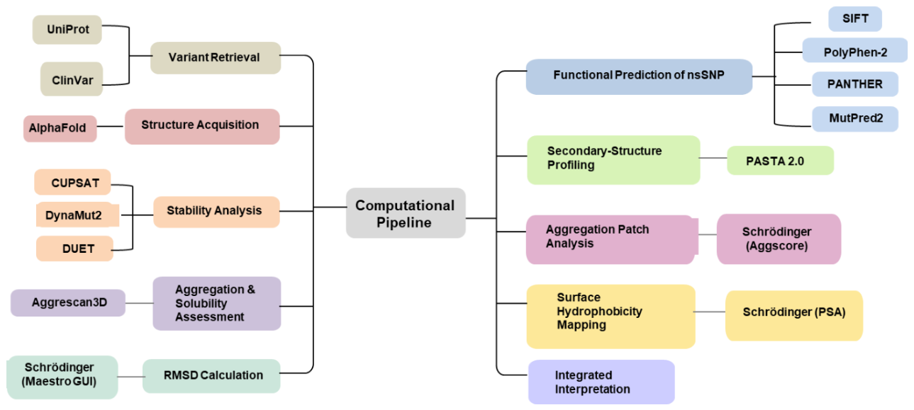
Figure 2.
Hydrophobicity and surface-patch profiles of podocin variants: (A) Comparative surface hydrophobicity mapping of wild-type (WT) podocin and the R138Q mutant showing localized alterations in solvent-exposed hydrophobic regions. (B) Electrostatic surface patch comparison between WT and R229Q podocin highlighting redistribution of positive and negative surface potentials. (C) Quantitative comparison of solvent-accessible surface area (SASA, Ų) among WT and podocin variants. (D) Comparative analysis of aggregation-prone surface patch areas (Ų) for WT and mutant podocin proteins, indicating mutation-dependent alterations in aggregation propensity.
Figure 2.
Hydrophobicity and surface-patch profiles of podocin variants: (A) Comparative surface hydrophobicity mapping of wild-type (WT) podocin and the R138Q mutant showing localized alterations in solvent-exposed hydrophobic regions. (B) Electrostatic surface patch comparison between WT and R229Q podocin highlighting redistribution of positive and negative surface potentials. (C) Quantitative comparison of solvent-accessible surface area (SASA, Ų) among WT and podocin variants. (D) Comparative analysis of aggregation-prone surface patch areas (Ų) for WT and mutant podocin proteins, indicating mutation-dependent alterations in aggregation propensity.
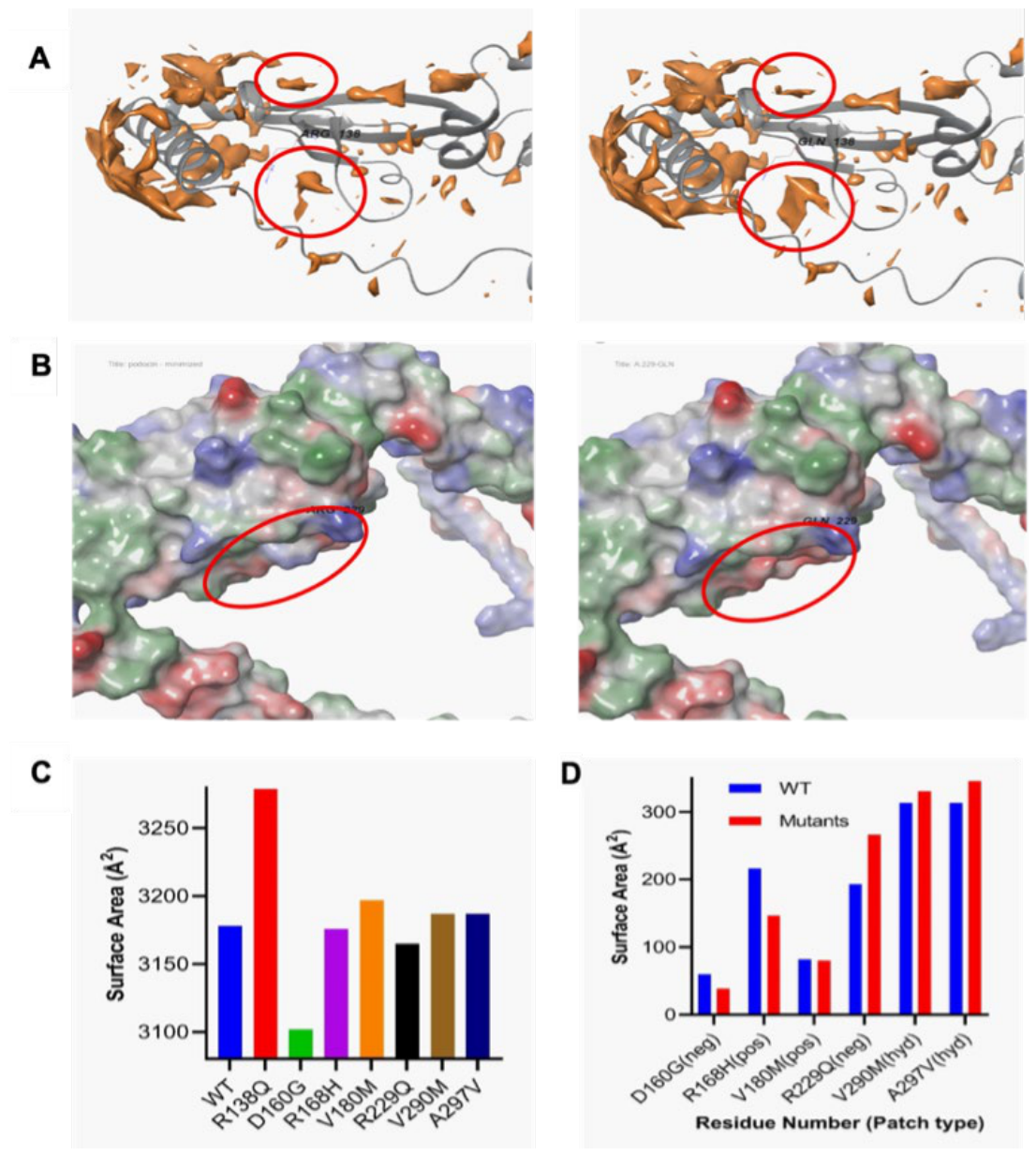
Figure 3.
Comparative surface hydrophobicity profiles of nephrin WT and mutants. (A) The R367C substitution introduces a localized increase in surface hydrophobicity, promoting aggregation propensity. (B) Quantitative comparison of solvent-accessible surface area (Ų) between wild-type and mutant nephrin variants. The R367C mutant exhibits a marked increase in overall surface exposure relative to WT.(C) Comparative analysis of aggregation-prone surface patches (Ų) in WT and mutant nephrin. The R367C variant shows a significant reduction in patch area, suggesting local structural rearrangements that may modulate aggregation behavior.
Figure 3.
Comparative surface hydrophobicity profiles of nephrin WT and mutants. (A) The R367C substitution introduces a localized increase in surface hydrophobicity, promoting aggregation propensity. (B) Quantitative comparison of solvent-accessible surface area (Ų) between wild-type and mutant nephrin variants. The R367C mutant exhibits a marked increase in overall surface exposure relative to WT.(C) Comparative analysis of aggregation-prone surface patches (Ų) in WT and mutant nephrin. The R367C variant shows a significant reduction in patch area, suggesting local structural rearrangements that may modulate aggregation behavior.
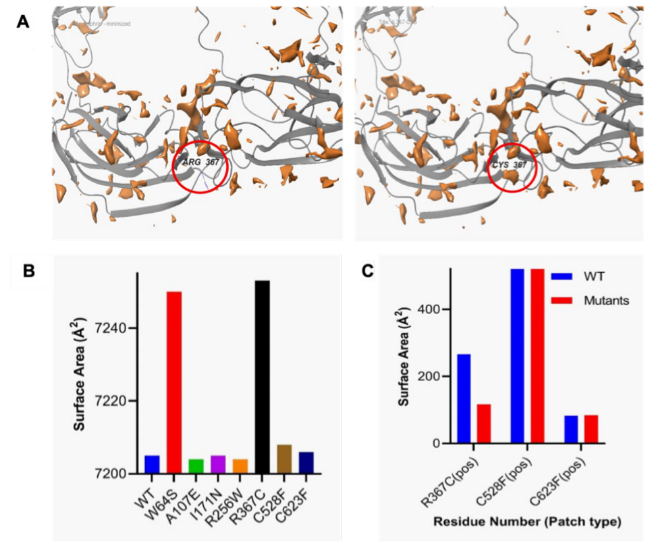
Figure 4.
Surface patch and hydrophobicity analysis of NEPH1 WT and mutants. (A) The R440C mutation leads to the loss of the positive electrostatic patch (blue) and alters adjacent hydrophobic (green) and negative (red) surface regions, indicating local charge redistribution. (B) Comparative surface hydrophobicity profiles of NEPH1 variants represented as solvent-accessible surface area (Ų). The S573L mutant exhibits a pronounced increase in surface exposure relative to WT. (C) Quantitative comparison of aggregation-prone patch areas (Ų) between WT and mutant NEPH1 proteins. The R440C variant displays a significant expansion in patch surface area, suggesting enhanced aggregation propensity and altered surface topology.
Figure 4.
Surface patch and hydrophobicity analysis of NEPH1 WT and mutants. (A) The R440C mutation leads to the loss of the positive electrostatic patch (blue) and alters adjacent hydrophobic (green) and negative (red) surface regions, indicating local charge redistribution. (B) Comparative surface hydrophobicity profiles of NEPH1 variants represented as solvent-accessible surface area (Ų). The S573L mutant exhibits a pronounced increase in surface exposure relative to WT. (C) Quantitative comparison of aggregation-prone patch areas (Ų) between WT and mutant NEPH1 proteins. The R440C variant displays a significant expansion in patch surface area, suggesting enhanced aggregation propensity and altered surface topology.
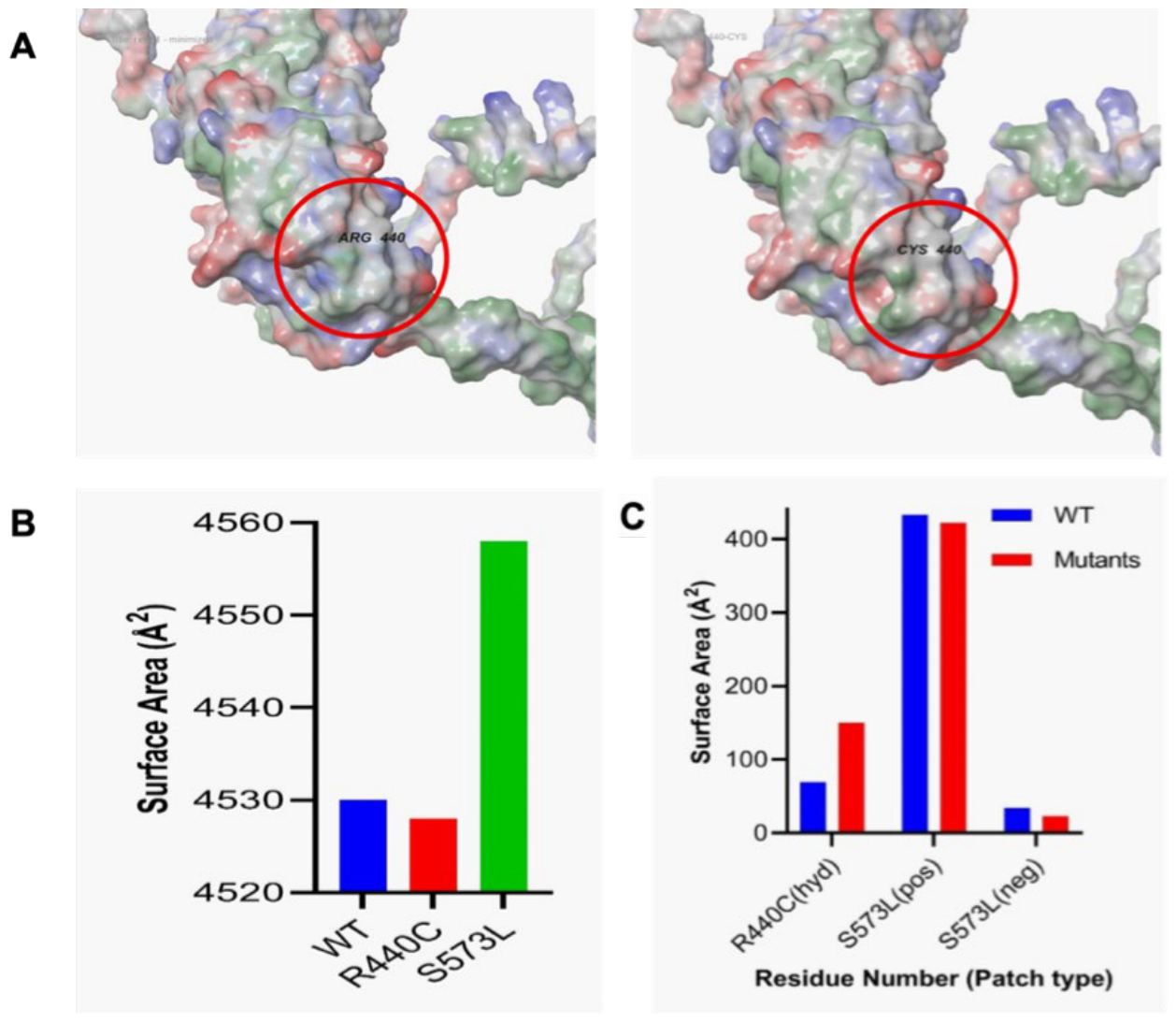
Figure 5.
Comparative surface area and patch analysis of TRPC6 WT and mutants. (A) Structural mapping of local surface area alterations showing WT Arg895 (left) and mutant Cys895 (right). The R895C substitution increases hydrophobic surface exposure, indicating enhanced aggregation propensity. (B) Surface patch analysis of the A404V variant reveals disruption of the positive electrostatic potential (blue) and emergence of a hydrophobic patch (green). The accompanying bar graph compares solvent-accessible surface areas (Ų), with R895C exhibiting a marked increase relative to WT.(C) Quantitative assessment of aggregation-prone surface patches (Ų) among TRPC6 variants. The G757D mutant displays a reduction in patch area, suggesting local structural rearrangements that may influence protein stability and interaction dynamics. (D) Comparative aggregation-prone surface patch area analysis of WT and mutant TRPC6 variants.
Figure 5.
Comparative surface area and patch analysis of TRPC6 WT and mutants. (A) Structural mapping of local surface area alterations showing WT Arg895 (left) and mutant Cys895 (right). The R895C substitution increases hydrophobic surface exposure, indicating enhanced aggregation propensity. (B) Surface patch analysis of the A404V variant reveals disruption of the positive electrostatic potential (blue) and emergence of a hydrophobic patch (green). The accompanying bar graph compares solvent-accessible surface areas (Ų), with R895C exhibiting a marked increase relative to WT.(C) Quantitative assessment of aggregation-prone surface patches (Ų) among TRPC6 variants. The G757D mutant displays a reduction in patch area, suggesting local structural rearrangements that may influence protein stability and interaction dynamics. (D) Comparative aggregation-prone surface patch area analysis of WT and mutant TRPC6 variants.
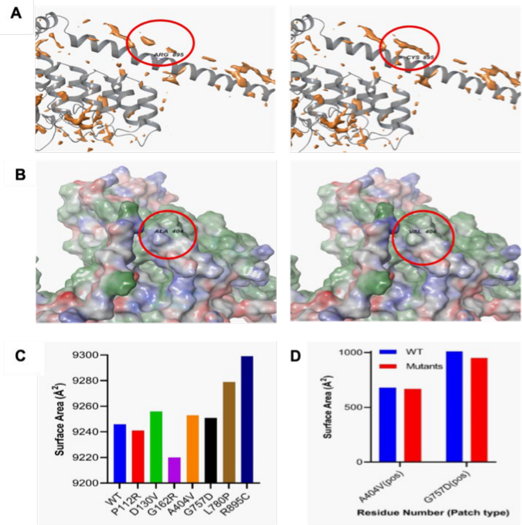

Table 1.
Physicochemical characterization of slit diaphragm (SD) protein variants: Comparative summary of predicted physicochemical parameters for 25 nsSNPs across five SD proteins podocin, nephrin, CD2AP, NEPH1, and TRPC6. The table lists functional predictions (PolyPhen-2, MutPred), thermodynamic stability changes (ΔΔG, kcal/mol; CUPSAT), secondary structure composition (α-helix, β-sheet, coil), surface hydrophobicity, and aggregation/solubility scores relative to wild-type (WT). Destabilizing variants exhibit increased hydrophobicity。
Table 1.
Physicochemical characterization of slit diaphragm (SD) protein variants: Comparative summary of predicted physicochemical parameters for 25 nsSNPs across five SD proteins podocin, nephrin, CD2AP, NEPH1, and TRPC6. The table lists functional predictions (PolyPhen-2, MutPred), thermodynamic stability changes (ΔΔG, kcal/mol; CUPSAT), secondary structure composition (α-helix, β-sheet, coil), surface hydrophobicity, and aggregation/solubility scores relative to wild-type (WT). Destabilizing variants exhibit increased hydrophobicity。
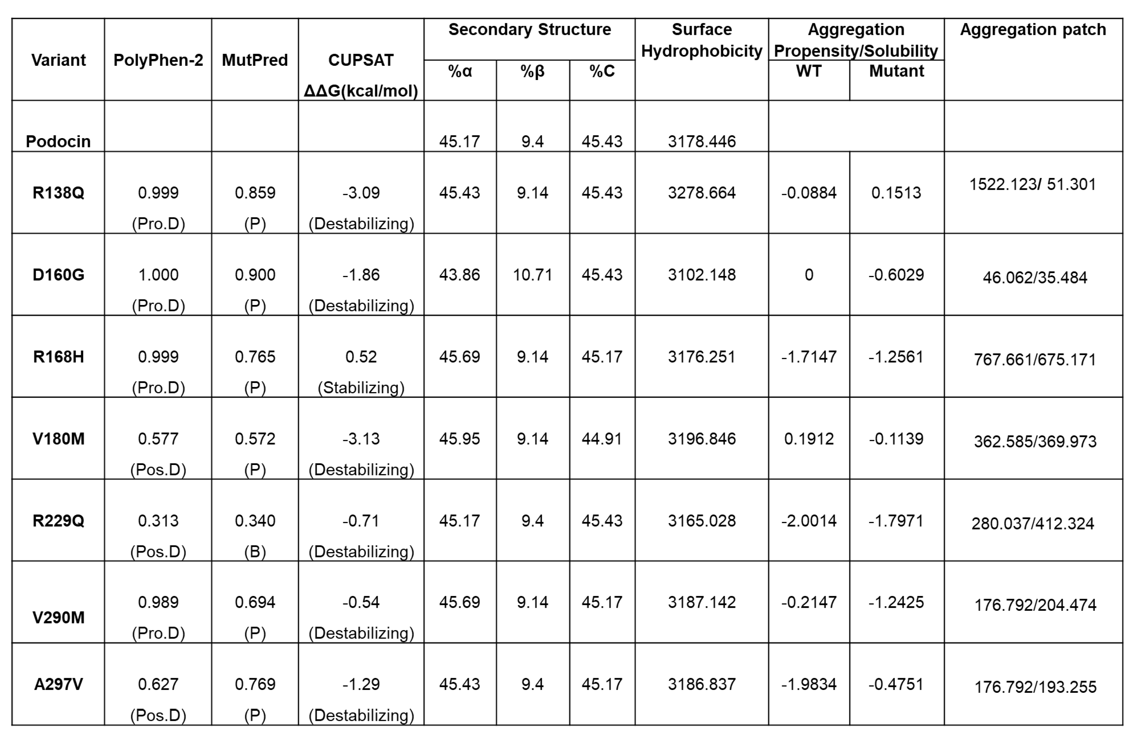 |
Table 2.
Integrated physicochemical characterization of slit diaphragm (SD) protein variants: Evaluvation of 25 pathogenic nsSNPs across five SD proteins podocin, nephrin, CD2AP, NEPH1, and TRPC6—showing predicted effects on protein stability, solubility, secondary structure, hydrophobicity, and aggregation propensity. Functional impact (SIFT, PolyPhen-2, PANTHER, MutPred) and stability changes (ΔΔG; CUPSAT) are compared against wild-type (WT) proteins. Several destabilizing variants (e.g., podocin R138Q, TRPC6
R895C) showed increased hydrophobic exposure and strong aggregation signatures, while some stabilizing substitutions (podocin R168H, NEPH1 S573L) also enhanced aggregation through urface remodeling. Overall,thes atterns reveal convergent physicochemical mechanisms contributing to SD disruption and podocyte
dysfunction in nephrotic syndrome.
Table 2.
Integrated physicochemical characterization of slit diaphragm (SD) protein variants: Evaluvation of 25 pathogenic nsSNPs across five SD proteins podocin, nephrin, CD2AP, NEPH1, and TRPC6—showing predicted effects on protein stability, solubility, secondary structure, hydrophobicity, and aggregation propensity. Functional impact (SIFT, PolyPhen-2, PANTHER, MutPred) and stability changes (ΔΔG; CUPSAT) are compared against wild-type (WT) proteins. Several destabilizing variants (e.g., podocin R138Q, TRPC6
R895C) showed increased hydrophobic exposure and strong aggregation signatures, while some stabilizing substitutions (podocin R168H, NEPH1 S573L) also enhanced aggregation through urface remodeling. Overall,thes atterns reveal convergent physicochemical mechanisms contributing to SD disruption and podocyte
dysfunction in nephrotic syndrome.
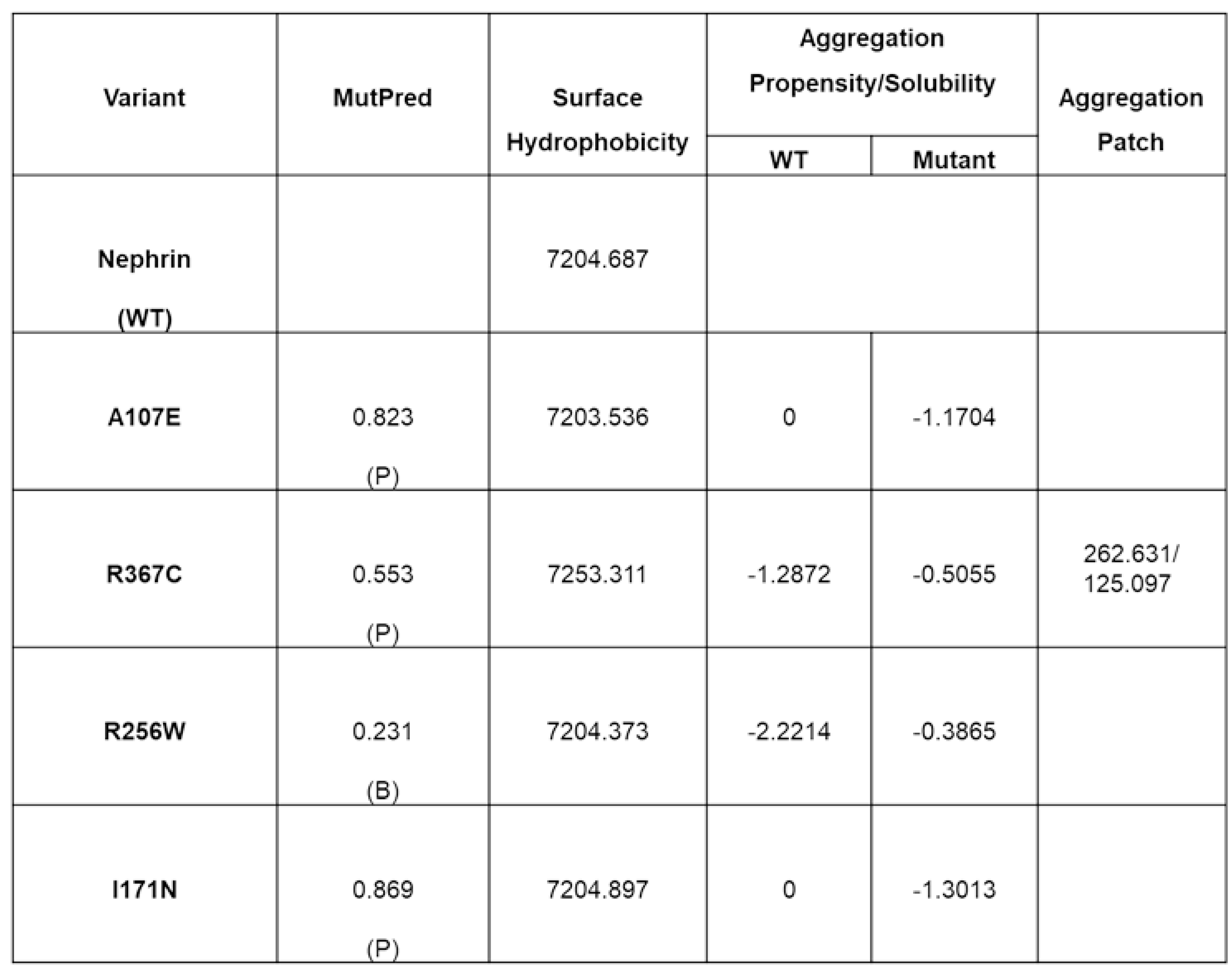 |
Table 3.
Physicochemical characterization of CD2AP variants: Profiling of wild-type (WT) CD2AP with two missense variants (K301M, T374A) showing predicted effects on structural stability, secondary-structure composition (α-helix, β-sheet, coil), surface hydrophobicity, and aggregation/solubility profiles. Both substitutions introduced measurable shifts in secondary-structure content and surface properties, with K301M exhibiting reduced solubility and T374A displaying increased α-helical content, indicative of localized structural reorganization and altered folding behavior.
Table 3.
Physicochemical characterization of CD2AP variants: Profiling of wild-type (WT) CD2AP with two missense variants (K301M, T374A) showing predicted effects on structural stability, secondary-structure composition (α-helix, β-sheet, coil), surface hydrophobicity, and aggregation/solubility profiles. Both substitutions introduced measurable shifts in secondary-structure content and surface properties, with K301M exhibiting reduced solubility and T374A displaying increased α-helical content, indicative of localized structural reorganization and altered folding behavior.
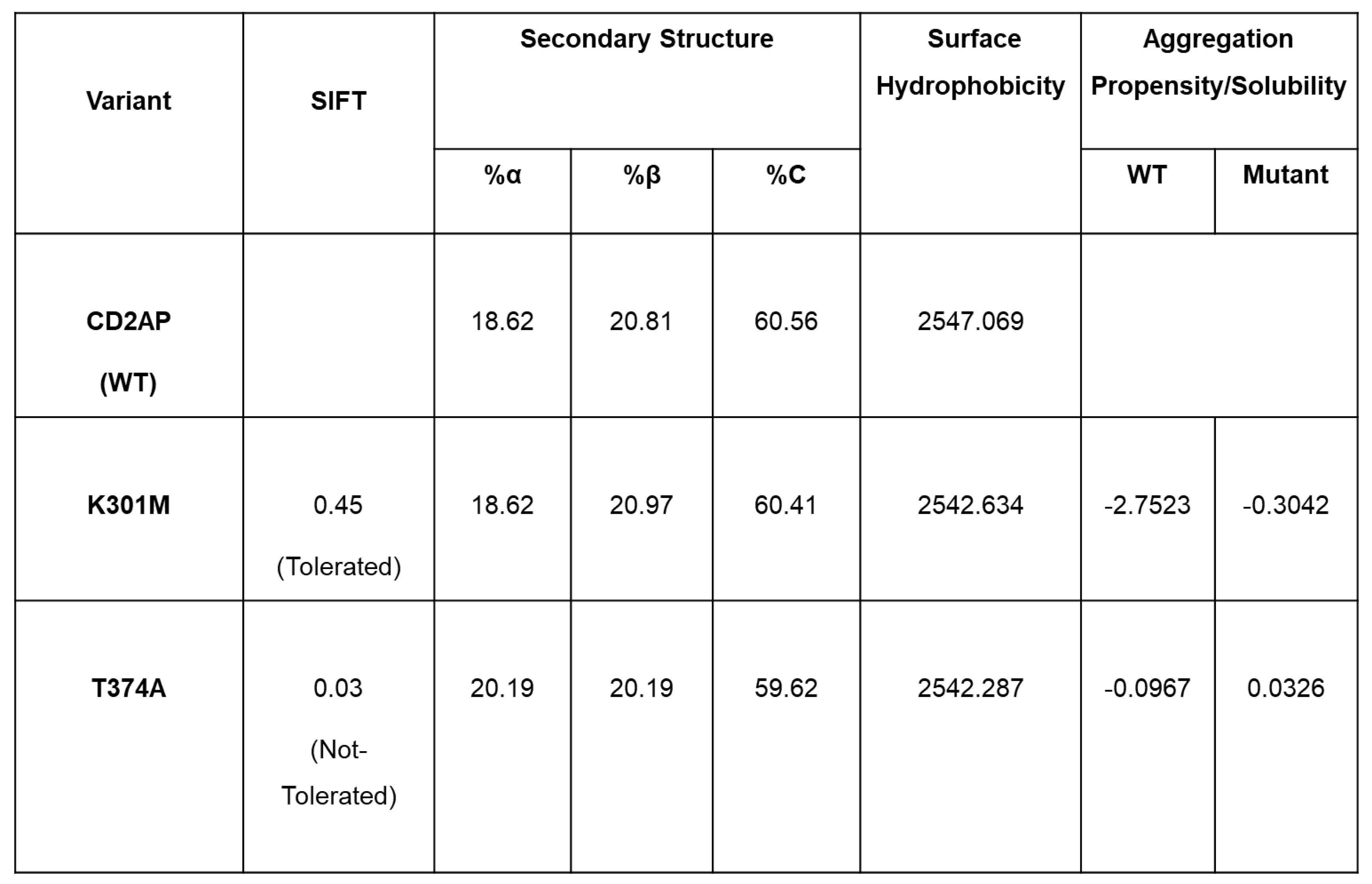 |
Table 4.
Physicochemical characterization of NEPH1 variants: Analysis of wild-type (WT) NEPH1 and two missense variants (R440C, S573L) highlighting predicted effects on protein stability, secondary-structure composition, surface hydrophobicity, and solubility. CUPSAT ΔΔG values classify R440C as destabilizing and S573L as stabilizing. S573L shows increased β-sheet content and elevated hydrophobic surface area, suggesting altered folding with enhanced aggregation potential. In contrast, R440C demonstrates reduced solubility and subtle hydrophobic changes, consistent with destabilization and aggregation propensity.
Table 4.
Physicochemical characterization of NEPH1 variants: Analysis of wild-type (WT) NEPH1 and two missense variants (R440C, S573L) highlighting predicted effects on protein stability, secondary-structure composition, surface hydrophobicity, and solubility. CUPSAT ΔΔG values classify R440C as destabilizing and S573L as stabilizing. S573L shows increased β-sheet content and elevated hydrophobic surface area, suggesting altered folding with enhanced aggregation potential. In contrast, R440C demonstrates reduced solubility and subtle hydrophobic changes, consistent with destabilization and aggregation propensity.
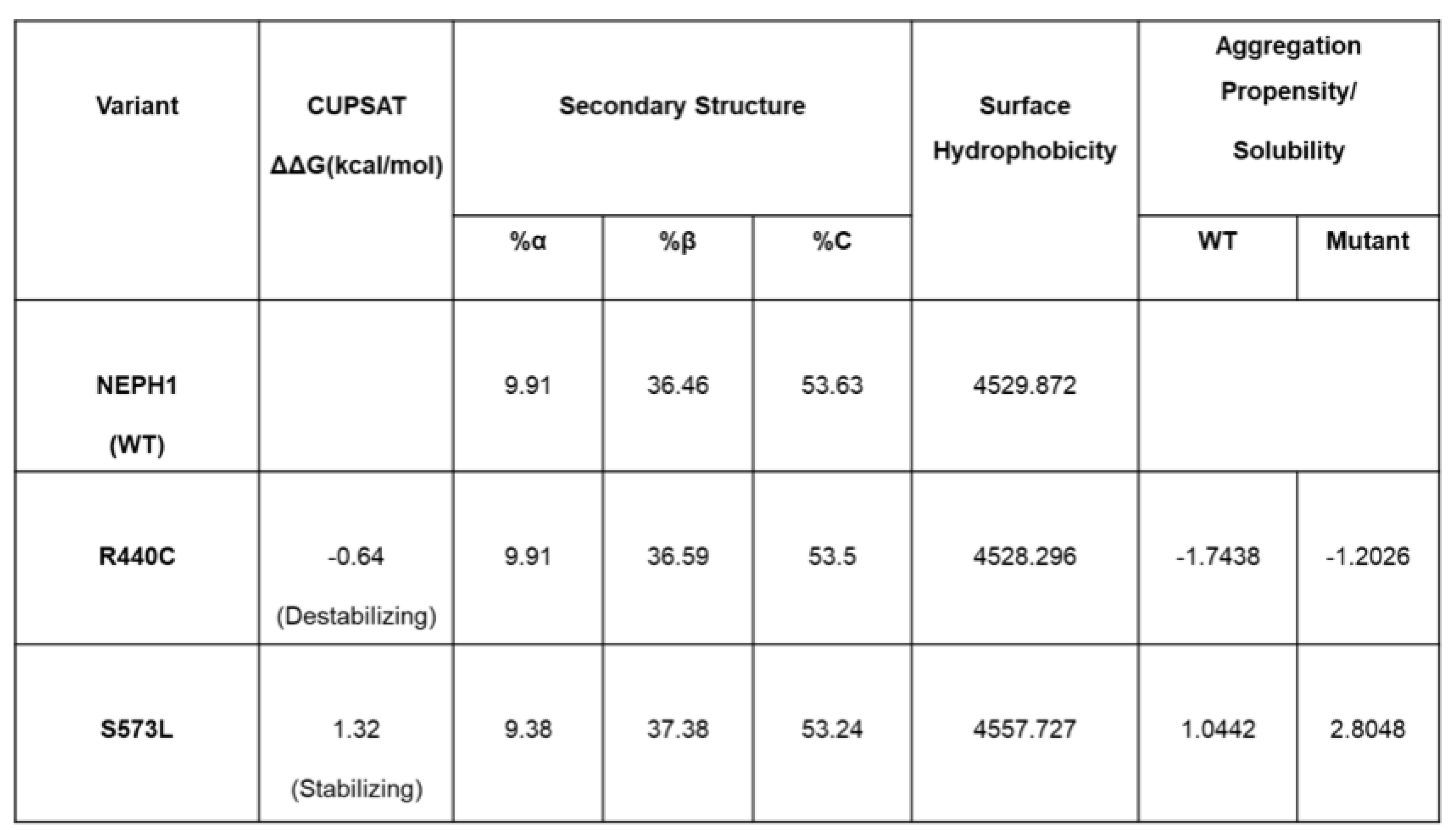 |
Table 5.
Physicochemical characterization of TRPC6 variants: Comparative summary of TRPC6 wild-type (WT) and mutant variants (D130V, R895C, A404V, P112R, and G757D) showing predicted effects on protein stability, secondary structure, hydrophobicity, and aggregation propensity. CUPSAT ΔΔG values identify D130V, R895C, A404V, and P112R as destabilizing, while G757D is stabilizing. Most destabilizing variants display increased surface hydrophobicity and aggregation scores, particularly R895C and A404V, suggesting elevated aggregation risk. G757D shows minimal secondary structural alteration but moderate stabilization with decreased aggregation potential.
Table 5.
Physicochemical characterization of TRPC6 variants: Comparative summary of TRPC6 wild-type (WT) and mutant variants (D130V, R895C, A404V, P112R, and G757D) showing predicted effects on protein stability, secondary structure, hydrophobicity, and aggregation propensity. CUPSAT ΔΔG values identify D130V, R895C, A404V, and P112R as destabilizing, while G757D is stabilizing. Most destabilizing variants display increased surface hydrophobicity and aggregation scores, particularly R895C and A404V, suggesting elevated aggregation risk. G757D shows minimal secondary structural alteration but moderate stabilization with decreased aggregation potential.
 |
Disclaimer/Publisher’s Note: The statements, opinions and data contained in all publications are solely those of the individual author(s) and contributor(s) and not of MDPI and/or the editor(s). MDPI and/or the editor(s) disclaim responsibility for any injury to people or property resulting from any ideas, methods, instructions or products referred to in the content. |
© 2026 by the authors. Licensee MDPI, Basel, Switzerland. This article is an open access article distributed under the terms and conditions of the Creative Commons Attribution (CC BY) license (http://creativecommons.org/licenses/by/4.0/).
Copyright: This open access article is published under a Creative Commons CC BY 4.0 license, which permit the free download, distribution, and reuse, provided that the author and preprint are cited in any reuse.


