
Submitted:
28 May 2026
Posted:
28 May 2026
You are already at the latest version
Abstract
Background: Pancreatic ductal adenocarcinoma (PDAC) is an aggressive malignancy with a poor prognosis, even in resectable disease. Molecular alterations in the four major driver genes (KRAS, SMAD4, CDKN2A, and TP53) have been associated with disease progression and patient outcomes. However, reliable prognostic biomarkers remain limited, particularly in resected PDAC, where recurrence rates remain high. Case presentation: We report the case of a 71-year-old woman incidentally diagnosed with PDAC carrying KRAS G12D/SMAD4 co-mutations with low baseline neutrophil-to-lymphocyte ratio (NLR) and circulating cell-free DNA (ccfDNA) levels. The patient underwent distal pancreatectomy with splenectomy and portal vein resection with multiple postoperative complications requiring re-intervention. She experienced a disease stability under adjuvant chemotherapy, and subsequent second lines of treatment, achieving an overall survival (OS) of 53-months. Conclusions: This case highlights the potential value of integrating molecular alterations with inflammatory and liquid biopsy biomarkers, including NLR and ccfDNA levels for improving risk stratification in PDAC. A focused review of the literature further supports the need for multimodal prognostic assessment in resected disease.
Keywords:
pancreatic ductal adenocarcinoma
; neutrophil-to-lymphocyte ratio
; ccfDNA
; KRAS G12D
; SMAD4
; prognosis
1. Introduction and Clinical Significance
Pancreatic ductal adenocarcinoma (PDAC) is one of the most aggressive cancers, with an annual death rate approaching incidence. Despite advances in diagnosis and treatment, most cases are detected at advanced stages, when curative surgery is no longer feasible.
In contrast, incidentally detected pancreatic lesions may allow earlier diagnosis and surgical intervention. Resected early-stage PDAC and high-risk pancreatic lesions in asymptomatic individuals are associated with improved survival, compared with patients with symptomatic disease, largely due to smaller tumor burden and higher resectability at diagnosis [1].
Genetically, more than 90% of PDAC cases harbor a KRAS mutation [2], predominantly with codon 12 alterations, KRAS G12D being the most common [3,4]. This variant has previously been associated with more aggressive tumors and worse clinical outcomes in resectable disease prognosis [4,5,6,7,8]. Loss of SMAD4 function is linked to tumor progression and metastatic dissemination and represents an established unfavorable prognostic factor for OS [9,10]. Several studies demonstrated that the combination of KRAS and SMAD4 mutations was an independent prognostic factor for progression-free survival (PFS) and OS, and patients with both KRAS and SMAD4 mutations have significantly shorter PFS and OS than those without this mutational profile [7,8,10,11,12,13].
Importantly, the co-occurrence of KRAS G12D and SMAD4 mutations is defined as an independent predictor of shorter PFS and OS, suggesting the existence of a high-risk molecular subtype of PDAC with limited therapeutic responsiveness to standard chemotherapy.
This article reports a rare case of incidentally detected PDAC harboring a KRAS G12D/SMAD4 co-mutation that achieved a prolonged period of stable disease and OS and presents a narrative review of the literature, emphasizing available treatments and their clinical significance. Beyond genomic alterations, emerging biomarkers such as ccfDNA) and systemic inflammatory markers, including NLR, have shown potential prognostic value in pancreatic cancer, although their clinical utility remains under investigation
2. Case Presentation
A 71-year-old woman with a medical history of hypertension, prior cholecystectomy for gallstone disease, and a family history of rectal adenocarcinoma had a screening computed tomography (CT) colonography in June 2019 that showed a 25 mm mass in the pancreatic body. The tumor was invading and stenosing the portal vein confluence without arterial invasion or adenopathy (Figure 1A).
The patient was admitted to the Gastroenterology Clinic in July 2019. No abnormality was found upon physical examination, and laboratory tests showed normal values of carbohydrate antigen 19-9 (CA19-9), carcinoembryonic antigen (CEA), alpha-fetoprotein (AFP) levels, and total bilirubin. The patient was enrolled in a prospective observational study (PANCNGS: October 2018 – December 20221) after providing informed consent. The study was approved by the institutional review board (IRB: 23878/14.06.2018) and included endoscopic ultrasound (EUS)-guided tissue acquisition and fine needle aspiration (FNA) of the pancreatic mass (Figure 1B) and peripheral blood sampling. EUS-FNA of the pancreatic lesion (Figure 1B) confirmed well-differentiated PDAC (Figure 1C-D).
Genomic DNA (gDNA) and ccfDNA were isolated using the same protocols that we detailed in our prior research [14,15]. The ccfDNA concentration was quantified using a Qubit 3.0 Fluorometer (Life Technologies) and measured in ng/μL, eluted in 25 μL of ultra-pure water, and measured 6.3 ng/mL. The NLR was calculated as the ratio between the peripheral blood neutrophil and lymphocyte counts and was 4.49. Subsequently, targeted amplicon-based next-generation sequencing (NGS) was carried out using the Illumina NextSeq500 platform. NGS analysis revealed a pathogenic mutation in KRAS (G12D), along with alterations in SMAD4, ARID1A, TSC2, TP53, EGFR, and ERBB2 (HER2), identified in FNA or in both FNA and ccfDNA samples (Table 1).
Following multidisciplinary tumor board evaluation, the disease was classified as resectable PDAC, and surgical resection was recommended.
Variants detected at low variant allele frequency (VAF) likely reflect tumor heterogeneity and ultra-deep sequencing sensitivity. Clinical significance of these alterations should be interpreted in the context of longitudinal molecular monitoring.
On July 23, 2019, the patient underwent a distal pancreatectomy with splenectomy and lateral resection of the portal vein. Pathological examination confirmed PDAC (pT2N1Mx, lymphovascular invasion positive, perineural invasion positive, stage IIB, AJCC 8th edition) [16]. Six days later, the patient developed diffuse abdominal pain and bilious output from the surgical drain. Abdominal CT scan revealed a perforated gastric ulcer with generalized peritonitis (Figure 2A). An emergency surgical re-intervention was performed on July 31, 2019, including segmental gastrectomy, double-layer gastrorrhaphy, peritoneal lavage, and drainage. Four days after the second surgical procedure, the patient presented with severe abdominal pain, and elevated drain fluid lipase (9 993 U/L) and amylase (3 996 U/L) levels significantly increased, consistent with acute pancreatitis of the pancreatic remnant. The supramesocolic accumulation (Figure 2B) was evacuated via laparotomy.
Following postoperative recovery, adjuvant chemotherapy with gemcitabine monotherapy was started in November 2019 due to the patient’s low performance status (ECOG 2-3), and it was continued until April 2020, when the restage CT scan showed progressing disease (Figure 2C, 2D).
Second-line treatment with gemcitabine plus capecitabine was administered from May 2020 to October 2021, achieving stable disease during the initial six cycles. Third-line therapy with nano-liposomal irinotecan (Onivyde®) plus 5-fluorouracil (5-FU) was initiated in December 2021 and continued until August 2022, when disease progression involved the superior mesenteric vein, superior mesenteric artery, and celiac trunk, accompanied by rising CA19-9 (114 U/L) (Figure 2E). In September 2022, fourth-line therapy with mFOLFOX6 was started following a multidisciplinary tumor board discussion. Disease stabilization was maintained until April 2023, when clinical deterioration occurred, accompanied by a marked increase in CA19-9 (555 U/L). Imaging revealed progression of liver metastasis and new peritoneal nodules (Figure 2F). The patient’s condition progressively declined, and she died in November 2023, after a 53-month OS. The patient’s treatment timeline is illustrated in Figure 3.
3. Discussion
Prognostic Value of ccfDNA Concentration and NLR in PDAC
In a previously published prospective cohort study including 82 patients with PDAC, we demonstrated that higher values of NLR (≥ 3.31) were linked with worse OS (4 vs. 10 months; log rank p = 0.011), and an elevated ccfDNA concentration (≥ 25.79 ng/mL) was strongly associated with shorter OS (4 vs. 8 months; log rank p = 0.009). According to the results of the multivariable Cox regression analysis, the baseline concentration of ccfDNA was an independent prognostic factor for OS (HR 0.45, 95% CI 0.21-0.97, p = 0.041). Furthermore, when ccfDNA levels and NLR were combined, patients with both biomarkers negative (ccfDNA concentration < 25.79 ng/ml and NLR < 3.31) showed significantly longer OS than patients with one or both biomarkers positive [14]. In various studies, multivariate analysis demonstrated that patients with higher NLR (cut-off values ranged from 2 to 5) had a worse prognosis than those with lower NLR [17,18,19,20]. This patient had a ccfDNA concentration of less than 25.79 ng/ml and an NLR of less than 5, which may indicate a favorable biological profile.
KRAS G12D/SMAD4 Co-Mutation’s Impact on Chemotherapy Responsiveness and Survival
About 90% of invasive PDACs have KRAS mutations, which appear early in the disease. Although KRAS mutations occur too frequently to be considered appropriate prognostic factors, previous reports mention that the KRAS mutant subtype G12V or G12D is associated with poor prognosis [21]. Several studies reported that the presence of the KRAS G12D in the primary tumor was a negative prognostic factor, including within the subgroup that received chemotherapy [22,23,24,25].
The impact of SMAD4 loss on chemotherapy responsiveness may vary depending on certain treatment regimens and disease stages, even though it is linked to a worse prognosis and shorter survival [9,26,27].
SMAD4 mutations were identified as a predictive factor for relapse-free survival (RFS) out of the four driver genes. Additionally, it was discovered that the combination of SMAD4 and KRAS mutations was an independent predictor of OS and RFS [28].
Nevertheless, SMAD4 deletion significantly accelerated the growth of pancreatic tumors when paired with the carcinogenic KRAS G12D mutation, resulting in a marked decrease in survival [13,29,30]. SMAD4 mutations predict poor response to chemotherapy and are associated with early metastatic spread. Currently not targetable, but may serve as a biomarker for aggressive disease needing intensified monitoring or alternative therapeutic strategies [9,31]. At the moment of the NGS panel, the frequency of SMAD4 mutation was extremely low, but taking into account the aggressive phenotype these mutations generate, it could be highly significant during patient follow-up.
The co-occurrence of KRAS G12D and SMAD4 mutations defines a high-risk molecular subtype, commonly associated with poor prognosis, enhanced metastatic potential, and limited responsiveness to standard chemotherapy [7,8,10,11,12,13]. In a retrospective cohort study, Shen et al. reported that patients with KRAS G12D mutation had inferior survival among those with resectable disease [4]. Similarly, Yokose et al. demonstrated that the combination of KRAS and SMAD4 mutations is an independent poor prognostic factor for recurrence and survival in resected PDAC [11].
Impact of KRAS Mutation Subtypes and Co-Occurring Mutations on Treatment Response and Prognostic
It has been demonstrated that TP53 mutations do not significantly impact prognosis in patients with KRAS G12D mutations [32]. TP53 mutation, frequently observed in PDAC, adds to the aggressive biology, reflecting a high degree of genomic instability and resistance to apoptosis [10,33,34]. Alterations in ARID1A and TSC2 further suggest epigenetic dysregulation and mTOR pathway activation, respectively, both of which are linked to tumor progression and treatment resistance [35,36,37,38].
Although KRAS mutations have long been considered “undruggable”, the recent development of G12D-specific inhibitors (e.g., MRTX1133) opens the door for targeted therapy in clinical trials [31,39,40].
ARID1A mutations are associated with increased tumor immunogenicity, especially when accompanied by high tumor mutational burden or microsatellite instability [36]. The high frequency of occurrence of these mutations in our case may suggest sensitivity to immune checkpoint inhibitors (e.g., anti-PD-1/PD-L1), particularly in the context of a broader immunogenic signature.
ERBB2 (HER2) mutation cases are suitable for HER2-targeted therapy. If confirmed by IHC or FISH as HER2 overexpression or amplification, this may justify the use of HER2-targeted agents (e.g., trastuzumab, pertuzumab, or trastuzumab deruxtecan)[41,42,43,44].
EGFR mutations are of limited significance for PDAC. They require a specific mutational context (e.g., L858R or exon 19 deletion) to be actionable. Historically, EGFR inhibitors (e.g., erlotinib) showed modest benefits in PDAC [45,46]; re-evaluation may be warranted if a sensitizing EGFR mutation is present.
Most variants detected in this case (except KRAS G12D) were identified at low variant allele frequencies (VAFs), likely reflecting the ultra-deep sequencing strategy used. Despite their low abundance, such mutations may hold significant prognostic and therapeutic relevance, particularly when monitored over time. Serial molecular profiling using the same targeted panel may enable early detection of emerging subclones with pathogenic potential or therapy resistance. Thus, these low-frequency variants should not be dismissed, but rather integrated into the longitudinal molecular surveillance strategy for this patient or in similar cases.
Postoperative complications following pancreatic surgery are often associated with treatment delays and the omission of adjuvant chemotherapy [47]. In this case, the patient’s PFS during the first line of chemotherapy (gemcitabine-based regimens) was 18 months, which is comparable to the ESPAC4 trial [48,49]. Under the second line of chemotherapy (Onivyde® +5-FU), the PFS was 9 months (in contrast to the NAPOLI-1 study, where the median PFS was 6.1 months [50]). Additionally, the patient’s tumor marker values were noticeably decreased throughout the second regimen. Notably, treatment was well tolerated despite the patient’s postoperative course.
The incidental diagnosis of an early-stage (resectable) PDAC with KRAS G12D and SMAD4 mutations, which experienced multiple postoperative complications and a prolonged period of stable disease under adjuvant chemotherapy, both on the first and second lines of chemotherapy, exemplifies this case’s uniqueness. Many clinical trials are currently evaluating the safety and effectiveness of KRAS G12D therapy in solid tumors, including PDAC. Enrollment in a trial investigating KRAS G12D inhibitors should be considered if available. However, early diagnosis and surgery remain the cornerstone of increased OS and PFS, even in the presence of negative molecular markers.
4. Conclusions
This case highlights prolonged survival in resected PDAC harboring a KRAS G12D/SMAD4 co-mutation, a molecular profile usually associated with a poor prognosis. Low baseline NLR and ccfDNA levels may have contributed to a more favorable clinical course. Integrating molecular and circulating biomarkers may improve prognostic stratification in resected PDAC.
Author Contributions
Conceptualization, B.V. and A.E.C. R.A.I; investigation, B.V., R.A.I., A.E.C., A.S., S.O.D., I.G.L., G.B., C.G.; writing—original draft preparation, B.V., A.E.C., R.A.I.; writing—review and editing, A.E.C., C.G. All authors have read and agreed to the published version of the manuscript.
Funding
The article processing charges were funded by Carol Davila University of Medicine and Pharmacy, Bucharest, Romania.
Institutional Review Board Statement
The study was conducted in accordance with the Declaration of Helsinki and was approved by the Fundeni Clinical Institute Ethics Committee (IRB: 23878/14.06.2018).
Informed Consent Statement
Written informed consent has been obtained from the patient to publish this paper.
Data Availability Statement
The clinical data and investigations supporting the findings of this case report are included within the article.
Conflicts of Interest
The authors declare no conflicts of interest.
Abbreviations
The following abbreviations are used in this manuscript:
| PDAC | Pancreatic Ductal Adenocarcinoma |
| NLR | Neutrophil-to-lymphocyte ratio |
| ccfDNA | Circulating cell-free DNA |
| OS | Overall Survival |
| PFS | Progression-Free Survival |
| CT | Computed Tomography |
| CA19-9 | Carbohydrate Antigen 19-9 |
| CEA | Carcinoembryonic Antigen |
| AFP | Alpha-Fetoprotein |
| EUS-FNA | Endoscopic Ultrasound-Guided Tissue Acquisition and Fine Needle Aspiration |
| gDNA | Genomic DNA |
| NGS | Next Generation Sequencing |
| 5-FU | 5-fluorouracil |
| RFS | relapse-free survival |
| VAFs | variant allele frequencies |
References
- Takikawa, Tetsuya; Kikuta, Kazuhiro; Hamada, Shin; Kume, Kiyoshi; Miura, Shin. Clinical features and prognostic impact of asymptomatic pancreatic cancer. Sci. Rep. 2022, no. 0123456789, 1–11. [Google Scholar] [CrossRef]
- Singhi, Aatur D.; et al. Real-Time Targeted Genome Profile Analysis of Pancreatic Ductal Adenocarcinomas Identifies Genetic Alterations That. Gastroenterology 2019, vol. 156(no. 8), 2242–2253.e4. [Google Scholar] [CrossRef]
- Buscail, Louis. Role of oncogenic KRAS in the diagnosis, prognosis and treatment of pancreatic cancer. Nat. Rev. Gastroenterol. Hepatol. [CrossRef]
- Shen, Henry; et al. KRAS G12D Mutation Subtype in Pancreatic Ductal Adenocarcinoma : Does It Influence Prognosis or Stage of Disease at Presentation ? Cell 2022. [Google Scholar] [CrossRef]
- Qiu, Ganbin; Gu, Yujun; Ji, Yayun; Jiang, Hui. Clinical Effect of Driver Mutations of KRAS, CDKN2A/P16, TP53, and SMAD4 in Pancreatic Cancer: A Meta-Analysis. Genet. Test. Mol. Biomark. 2020, vol. 24(no. 12), 777–788. [Google Scholar] [CrossRef]
- Laubender, P.; Neumann, Jens. KRAS mutation status is not predictive for objective response to anti-EGFR treatment with erlotinib in patients with advanced pancreatic cancer. J. Gastroenterol. 2013, 544–548. [Google Scholar] [CrossRef]
- Qian, Zhi Rong. , Association of Alterations in Main Driver Genes With Outcomes of Patients With Resected Pancreatic Ductal Adenocarcinoma. JAMA Oncol. 2017, vol. 02215, 1–6. [Google Scholar] [CrossRef] [PubMed]
- Biankin, Andrew V. , DPC4/Smad4 Expression and Outcome in Pancreatic Ductal Adenocarcinoma. J. Clin. Oncol. 2023, vol. 20(no. 23), 4531–4542. [Google Scholar] [CrossRef] [PubMed]
- Fei, Naomi; et al. SMAD4 loss is associated with response to neoadjuvant chemotherapy plus hydroxychloroquine in patients with pancreatic adenocarcinoma. Clin. Transl. Sci. 2021, no. January, 1822–1829. [Google Scholar] [CrossRef] [PubMed]
- Stefanoudakis, Dimitrios; Frountzas, Maximos; Schizas, Dimitrios; Michalopoulos, Nikolaos V; Drakaki, Alexandra; Toutouzas, Konstantinos G. Significance of TP53, CDKN2A, SMAD4 and KRAS in Pancreatic Cancer. Curr. Issues Mol. Biol. 2024, 2827–2844. [Google Scholar] [CrossRef]
- Yokose, Takahiro; et al. Combination of KRAS and SMAD4 mutations in formalin-fixed paraffin-embedded tissues as a biomarker for pancreatic cancer. 2019, 2174–2182. [Google Scholar] [CrossRef]
- Izeradjene, Kamel; et al. KrasG12D and Smad4/Dpc4 Haploinsufficiency Cooperate to Induce Mucinous Cystic Neoplasms and Invasive Adenocarcinoma of the Pancreas. Cancer Cell 2007, no. March, 229–243. [Google Scholar] [CrossRef]
- Bardeesy, Nabeel. , Smad4 is dispensable for normal pancreas development yet critical in progression and tumor biology of pancreas cancer. Genes Dev. 2006, 3130–3146. [Google Scholar] [CrossRef]
- Varzaru, Bianca; et al. Prognostic Value of Circulating Cell-Free DNA Concentration and Neutrophil-to-Lymphocyte Ratio in Patients with Pancreatic Ductal Adenocarcinoma : A Prospective Cohort Study. Int. J. Mol. Sci. 2024. [Google Scholar] [CrossRef]
- Bunduc, Stefania; et al. Endoscopic ultrasound-guided fine-needle aspiration pancreatic adenocarcinoma samples yield adequate DNA for next-generation sequencing: A cohort analysis Stefania. World J. Gastroenterol. 2023, vol. 29(no. 18), 2864–2874. [Google Scholar] [CrossRef]
- Amin, M.B.; Edge, S.; Greene, F.; Byrd, D.R.; Brookland, R.K.; Washington, M.K.; Gershenwald, J.E.; Compton, C.C.; Hess, K.R.; Sullivan, A.J. CC Cancer Staging Manual, 8th ed.; Springer: New York, 2017. [Google Scholar]
- Stotz, M.; et al. Increased neutrophil-lymphocyte ratio is a poor prognostic factor in patients with primary operable and inoperable pancreatic cancer. Br. J. Cancer 2013, vol. 109(no. 2), 416–421. [Google Scholar] [CrossRef]
- Mowbray, Nicholas G.; Griffith, David; Hammoda, Mohammed; Shingler, Guy; Kambal, Amir; Al-Sarireh, Bilal. A meta-analysis of the utility of the neutrophil-to-lymphocyte ratio in predicting survival after pancreatic cancer resection. In HPB; Elsevier B.V., 01 May 2018; vol. 20, no. 5, pp. 379–384. [Google Scholar] [CrossRef]
- Luo, Guopei. , Blood Neutrophil–Lymphocyte Ratio Predicts Survival in Patients with Advanced Pancreatic Cancer Treated with Chemotherapy. Ann. Surg. Oncol. 2015, vol. 22(no. 2), 670–676. [Google Scholar] [CrossRef] [PubMed]
- Varzaru, Bianca. , Real-Life Results of Palliative Chemotherapy in Metastatic Pancreatic Ductal Adenocarcinoma. Cancers . 2023, vol. 15(no. 13), 1–11. [Google Scholar] [CrossRef]
- Bournet, Barbara; Muscari, Fabrice; Buscail, Camille; Assenat, Eric; Barthet, Marc. KRAS G12D Mutation Subtype Is A Prognostic Factor for Advanced Pancreatic Adenocarcinoma. Clin. Transl. Gastroenterol. 2016, vol. 7(no. 3), e157–8. [Google Scholar] [CrossRef] [PubMed]
- Sinn, Bruno V; et al. KRAS Mutations in Codon 12 or 13 Are Associated With Worse Prognosis in Pancreatic Ductal Adenocarcinoma. Pancreas 2014, vol. 43(no. 4), 578–583. [Google Scholar] [CrossRef] [PubMed]
- Shin, Sang Hyun; et al. Genetic Alterations of K-ras, p53, c-erbB-2, and DPC4 in Pancreatic Ductal Adenocarcinoma and Their Correlation With Patient Survival. Pancreas 2013, vol. 42(no. 2), 216–222. [Google Scholar] [CrossRef]
- Franko, Jan. , Loss of Heterozygosity Predicts Poor Survival After Resection of Pancreatic Adenocarcinoma. J. Gastrointest. Surg. 2008, 1664–1673. [Google Scholar] [CrossRef]
- Vans, James D. E.; Ampbell, Fiona C.; Emoine, Nicholas L.; Eoptolemos, John P. N. K-RAS ONCOGENE SUBTYPE MUTATIONS ARE ASSOCIATED WITH SURVIVAL BUT NOT EXPRESSION OF P53, P16 INK4A, P21 WAF-1, CYCLIN D1, ERBB-2 AND ERBB-3 IN RESECTED PANCREATIC DUCTAL ADENOCARCINOMA. Int. J. Cancer 2000, vol. 474, no. January, 469–474. [Google Scholar]
- Wang, Xiaofeng; et al. Hypoxic tumor-derived exosomal miR-301a mediates M2 macrophage polarization via PTEN/PI3Kg to promote pancreatic cancer metastasis. Cancer Res. 2018, vol. 78(no. 16), 4586–4598. [Google Scholar] [CrossRef] [PubMed]
- Wang, Zusen; et al. SMAD4 Y353C promotes the progression of PDAC. BMC Cancer 2019, 1–12. [Google Scholar] [CrossRef] [PubMed]
- Yokose, Takahiro; et al. Combination of KRAS and SMAD4 mutations in formalin-fixed paraffin-embedded tissues as a biomarker for pancreatic cancer. Cancer Sci. 2019, 2174–2182. [Google Scholar] [CrossRef] [PubMed]
- Sun, Yingjia. Impact of KRAS Mutation Subtypes and Co-Occurring Mutations on Response and Outcome in Advanced NSCLC Patients following First-Line Treatment. J. Clin. Med. 2022. [Google Scholar] [CrossRef]
- Yousef, Abdelrahman. , Impact of KRAS mutations and co-mutations on clinical outcomes in pancreatic ductal adenocarcinoma. Precis. Oncol. 2024. [Google Scholar] [CrossRef]
- Cook, Shelly; Li, Bingyu; Zhang, Qiong; Castaneda, Claire. Targeted Therapies in Pancreatic Cancer : A New Era of Precision Medicine. Biomedicines 2024, 1–13. [Google Scholar]
- Jiang, Yubo; et al. Molecular characterization and prognostic implications of KRAS mutations in pancreatic cancer patients : insights from multi-cohort analysis. Precis. Oncol. 2025, 1–11. [Google Scholar] [CrossRef]
- Mello, Stephano S.; Valente, Liz J.; Raj, Nitin; Hruban, Ralph H.; Curtis, Christina; Attardi, Laura D. A p53 Super-tumor Suppressor Reveals a Tumor Suppressive p53-Ptpn14-Yap Axis in Pancreatic Article A p53 Super-tumor Suppressor Reveals a Tumor Suppressive p53-Ptpn14-Yap Axis in Pancreatic Cancer. Cancer Cell 2017, vol. 32(no. 4), 460–473.e6. [Google Scholar] [CrossRef]
- Mello, Stephano S.; et al. Multifaceted role for p53 in pancreatic cancer suppression. PNAS 2023, 1–9. [Google Scholar] [CrossRef] [PubMed]
- LIU, M.I.N.G.Y.A.N.G. Arid1a: A Gatekeeper in the Development of Pancreatic Cancer From a Rare Precursor Lesion. Gastroenterology 2022, 371–373. [Google Scholar] [CrossRef]
- Li, Ruichao; Xiong, Guangbing; Zhao, Jun; Yang, Lin. Targeting the alterations of ARID1A in pancreatic cancer : tumorigenesis, prediction of treatment, and prognostic value. Am. J. Transl. Res. 2022, vol. 14(no. 9), 5952–5964. [Google Scholar]
- Kearns, Bridger; et al. ARID1A and Its Impact Across the Hallmarks of Cancer. Int. J. Mol. Sci. 2025, 1–14. [Google Scholar] [CrossRef]
- Tomihara, Hideo; Loss; et al. Loss of ARID1A promotes epithelial-mesenchymal transition and sensitizes pancreatic tumors to proteotoxic stress. Cancer Res. 2022, vol. 81(no. 2), 332–343. [Google Scholar] [CrossRef]
- Corbet, Cyril; Bannoura, Sahar F.; Khan, Husain Yar; Azmi, Asfar S.; Hobbs, Aaron; Wang, Boshi. KRAS G12D targeted therapies for pancreatic cancer : Has the fortress been conquered ? Front. Oncol. 2022, no. November, 1–9. [Google Scholar] [CrossRef]
- Sobhani, Navid; Pittacolo, Matteo; Angelo, Alberto D. Recent Anti-KRAS G12D Therapies : A ‘ Possible Impossibility ’ for Pancreatic Ductal Adenocarcinoma. Cancers . 2025, 1–23. [Google Scholar]
- Falk, Markus; et al. Response of an HER2-Mutated NSCLC Patient to Trastuzumab Deruxtecan and Monitoring of Plasma ctDNA Levels by Liquid Biopsy. Curr. Oncol. 2023, 1692–1698. [Google Scholar] [CrossRef]
- Ryu, Ki Hyun; Kwon, Ah-Young; Han, Song-Hee. The Prognostic Impact of HER2 Genetic and Protein Expression in Pancreatic Carcinoma — HER2 Protein and Gene in Pancreatic Cancer. In Diagnostics; 2021. [Google Scholar]
- Zhu, Kunrui; Yang, Xinyi; Tai, Hebei; Zhong, Xiaorong; Luo, Ting; Zheng, Hong. HER2 - targeted therapies in cancer : a systematic review. Biomark. Res. 2024, 1–17. [Google Scholar] [CrossRef] [PubMed]
- Xu, Luying; Xie, Yuxin; Gou, Qiheng; Cai, Rui; Bao, Rong. HER2-targeted therapies for HER2-positive early-stage breast cancer : present and future. Front. Pharmacol. 2024, no. September, 1–16. [Google Scholar] [CrossRef]
- Mucciolo, Giulia B. Gianluca; Henrı’quez, Joaquı’n Araos; Jihad, Muntadher; Teles, Sara Pinto; Manansala, Judhell S.; Li, Wenlong; Ashworth, Sally; Lloyd, Eloise G.; Cheng, Priscilla S.W.; Luo, Weike; Anand, Akanksha; Sawle, Ashley; Piskorz, Anna. EGFR-activated myofibroblasts promote metastasis of pancreatic cancer ll EGFR-activated myofibroblasts promote metastasis of pancreatic cancer. Cancer Cell 2024, 101–118. [Google Scholar] [CrossRef]
- Sheahan, Anjali V; Biankin, Andrew V; Parish, Christopher R.; Khachigian, Levon M. “Targeted therapies in the management of locally advanced and metastatic pancreatic cancer: a systematic review,” 2018. Available online: www.oncotarget.com.
- Ryan, P.; Bilimoria, Karl Y.; Tomlinson, James S.; Bentrem, David J. Postoperative Complications Reduce Adjuvant Chemotherapy Use in Resectable Pancreatic Cancer. Ann. Surg. 2014, vol. 260(no. 2), 372–377. [Google Scholar] [CrossRef] [PubMed]
- Palmer, Daniel H. , “Pancreatic Adenocarcinoma : Long-Term Outcomes of Adjuvant Therapy in the ESPAC4 Phase III Trial,” Gastrointest. Cancer 2023, vol. 43(no. 10). [Google Scholar] [CrossRef] [PubMed]
- Bertolo, Aaron; Hodul, Pamela; Kim, Dae Won. ESPAC-4 long-term follow-up : shall we choose gemcitabine and capecitabine as adjuvant therapy for elderly and low-risk pancreatic adenocarcinoma following surgical resection ? 2026, vol. 9(no. 7). [Google Scholar] [CrossRef]
- Wang-Gillam, Andrea; et al. NAPOLI-1 phase 3 study of liposomal irinotecan in metastatic pancreatic cancer : Final overall survival analysis and characteristics of long-term survivors. Eur. J. Cancer 2015, vol. 108, 78–87. [Google Scholar] [CrossRef] [PubMed]
Figure 1.
Diagnostic CT imaging, EUS, and histopathological findings: (A) a 25 mm pancreatic body tumor stenosing and infiltrating the portal vein confluence without arterial invasion or adenopathy; (B) EUS and qualitative EUS elastography: a well-defined pancreatic body tumor of 37/17 mm diameter and surrounding large blood vessels; qualitative EUS elastography shows a predominant blue pattern indicating increased stiffness; (C) and (D) hematoxylin-eosin staining: atypical cancerous cells that could indicate a low-grade pancreatic adenocarcinoma diagnosis; (C) Cytology: smear with atypical cohesive epithelial cells, tachychromatic nuclei, and reduced pleomorphism. (D) Paraffin block: atypical epithelial flaps with glandular outlines and focal desmoplastic stroma.
Figure 1.
Diagnostic CT imaging, EUS, and histopathological findings: (A) a 25 mm pancreatic body tumor stenosing and infiltrating the portal vein confluence without arterial invasion or adenopathy; (B) EUS and qualitative EUS elastography: a well-defined pancreatic body tumor of 37/17 mm diameter and surrounding large blood vessels; qualitative EUS elastography shows a predominant blue pattern indicating increased stiffness; (C) and (D) hematoxylin-eosin staining: atypical cancerous cells that could indicate a low-grade pancreatic adenocarcinoma diagnosis; (C) Cytology: smear with atypical cohesive epithelial cells, tachychromatic nuclei, and reduced pleomorphism. (D) Paraffin block: atypical epithelial flaps with glandular outlines and focal desmoplastic stroma.
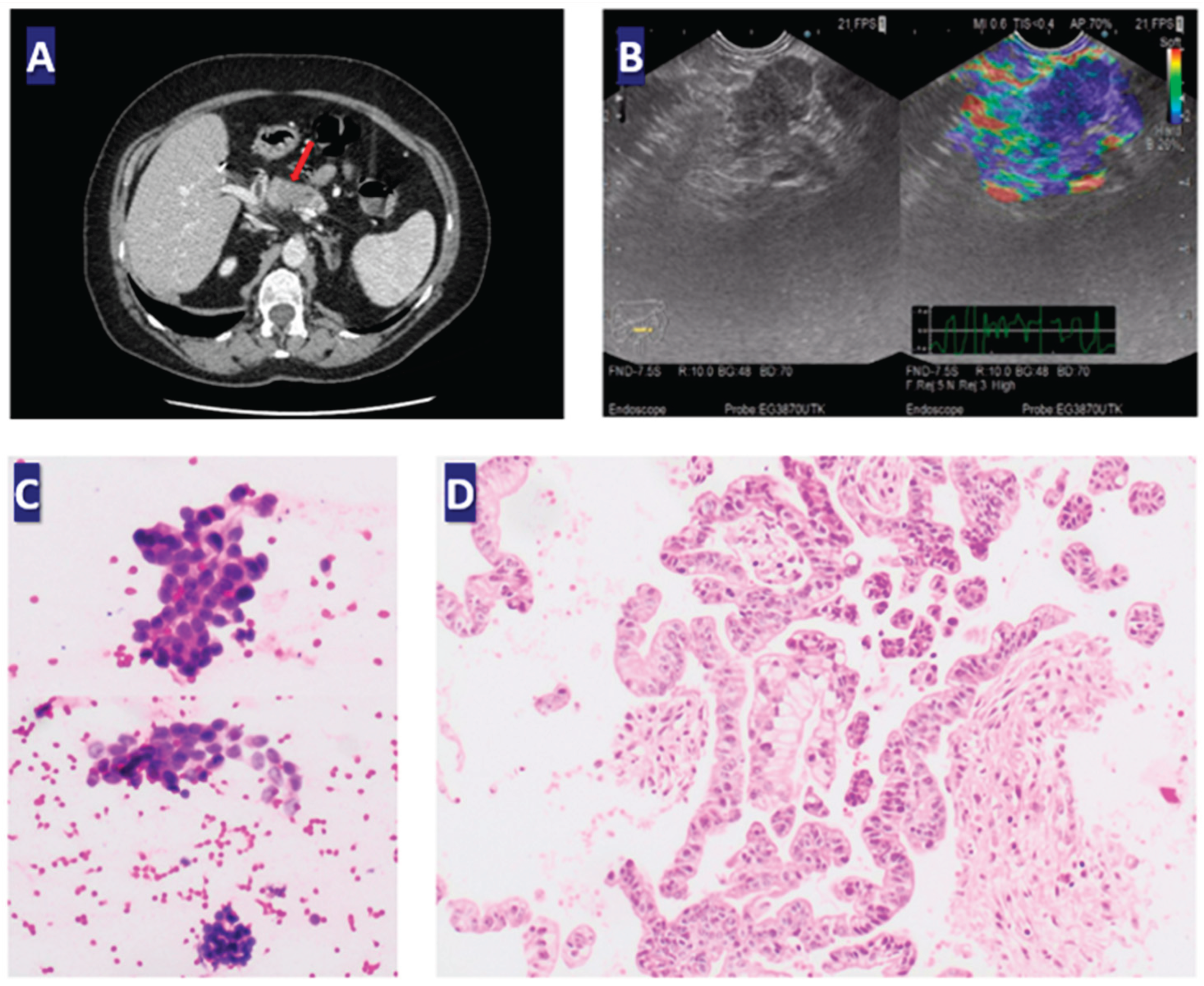
Figure 2.
Follow-up CT scans: (A) a fistula located on the posterior gastric wall with intraperitoneal extravasation of the oral contrast, intraperitoneal air bubbles, and parafluid collection adjacent to the pancreatectomy site, in the mesogastric and left subphrenic regions; (B) no evidence of the previously mentioned small air bubbles, except for one at the level of the laparotomy incision; (C) and (D) progressive disease: mesenteric and peritoneal infiltration, with adenopathy and peritoneal nodules; (E) the tumor involved the superior mesenteric vein, superior mesenteric artery, and celiac trunk; (F) dimensional progression of the nodular hepatic lesion and new peritoneal nodules. .
Figure 2.
Follow-up CT scans: (A) a fistula located on the posterior gastric wall with intraperitoneal extravasation of the oral contrast, intraperitoneal air bubbles, and parafluid collection adjacent to the pancreatectomy site, in the mesogastric and left subphrenic regions; (B) no evidence of the previously mentioned small air bubbles, except for one at the level of the laparotomy incision; (C) and (D) progressive disease: mesenteric and peritoneal infiltration, with adenopathy and peritoneal nodules; (E) the tumor involved the superior mesenteric vein, superior mesenteric artery, and celiac trunk; (F) dimensional progression of the nodular hepatic lesion and new peritoneal nodules. .
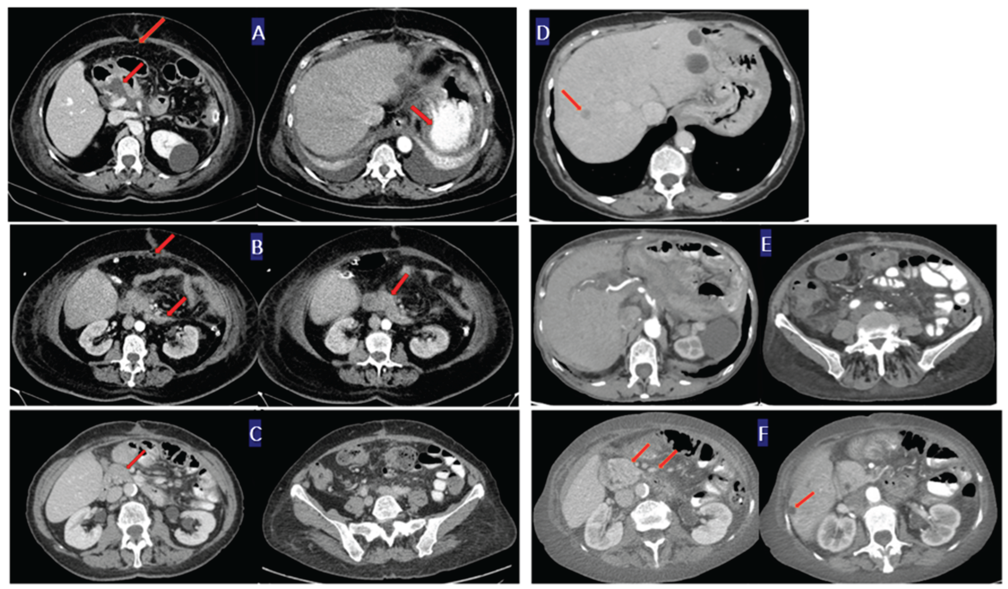
Figure 3.
Treatment timeline. GB: gemcitabine-based regimens. Onivyde® + (5-FU): nano-liposomal irinotecan + 5 – fluorouracil; mFOLFOX6: modified FOLFOLX-6.
Figure 3.
Treatment timeline. GB: gemcitabine-based regimens. Onivyde® + (5-FU): nano-liposomal irinotecan + 5 – fluorouracil; mFOLFOX6: modified FOLFOLX-6.
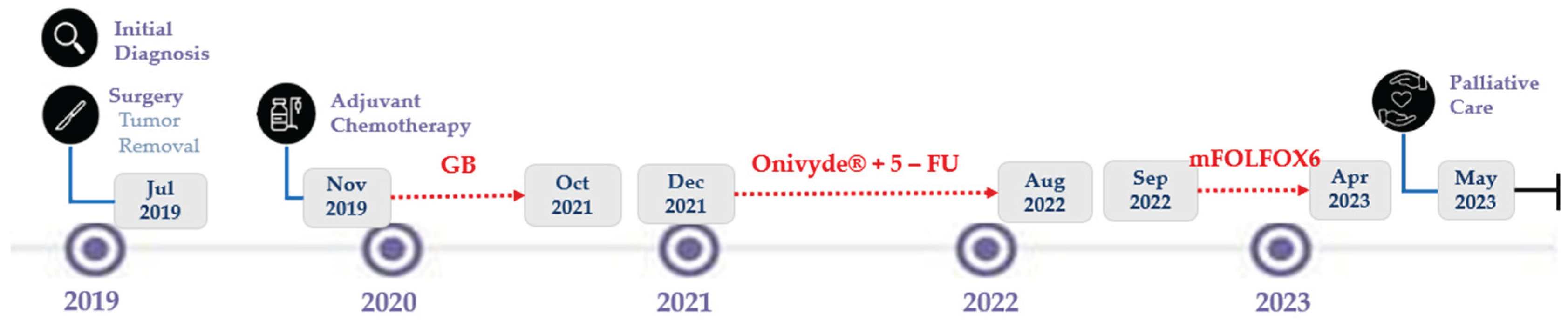

Table 1.
Molecular profile of the tumor and circulating cell-free DNA in the reported PDAC case.
| Gene | Alteration | Sample source | Detection context | Clinical relevance | Title 1 | Title 2 | Title 3 | ||
|---|---|---|---|---|---|---|---|---|---|
| KRAS | G12D mutation | FNA + ccfDNA | High-confidence pathogenic driver | Key oncogenic driver associated with aggressive PDAC biology and poor prognosis | entry 1 | data | data | ||
| SMAD4 | Inactivating alteration | FNA + ccfDNA | Co-occurring driver alteration | Tumor suppressor loss is linked to metastatic potential and reduced survival | entry 2 | data | data 1 | ||
| TP53 | Mutation (unspecified variant) | FNA | Detected in tumor tissue | Genomic instability contributes to tumor progression | |||||
| ARID1A | Loss-of-function mutation | FNA + ccfDNA (low VAF) | Subclonal/low-frequency variant | Epigenetic dysregulation: potential role in tumor aggressiveness | |||||
| TSC2 | Pathogenic alteration | FNA + ccfDNA (low VAF) | Subclonal variant | Possible activation of the mTOR pathway | |||||
| EGFR | Mutation (unspecified) | FNA + ccfDNA (low VAF) | Low-frequency detection | Limited actionable relevance in PDAC unless the sensitizing mutation is confirmed | |||||
| ERBB2 (HER2) | Mutation (not amplified) | FNA + ccfDNA (low VAF) | Low-frequency variant | Potential therapeutic relevance if confirmed amplification/overexpression |
Disclaimer/Publisher’s Note: The statements, opinions and data contained in all publications are solely those of the individual author(s) and contributor(s) and not of MDPI and/or the editor(s). MDPI and/or the editor(s) disclaim responsibility for any injury to people or property resulting from any ideas, methods, instructions or products referred to in the content. |
© 2026 by the authors. Licensee MDPI, Basel, Switzerland. This article is an open access article distributed under the terms and conditions of the Creative Commons Attribution (CC BY) license (http://creativecommons.org/licenses/by/4.0/).
Copyright: This open access article is published under a Creative Commons CC BY 4.0 license, which permit the free download, distribution, and reuse, provided that the author and preprint are cited in any reuse.


