

Submitted:
26 May 2026
Posted:
27 May 2026
You are already at the latest version
Abstract
Background: Invasive neuromodulation therapies such as deep brain stimulation (DBS) have become increasingly important for treating refractory neurological and psychiatric disorders, including movement disorders, epilepsy, obsessive-compulsive disorder, and more. Despite increasing clinical use, optimizing brain stimulation remains a major challenge. Key questions remain regarding how to select optimal stimulation targets, determine electrode placement, program the large parameter space of stimulation settings, understand mechanisms of action, and adapt therapy as symptoms fluctuate or progress over time. Because direct clinical symptom measurement can be variable, delayed, or difficult to quantify intraoperatively, physiologic biomarkers hold potential to navigate these challenges. One promising approach is to measure the neural responses elicited by brief pulses of electrical stimulation, based on the assumption that stimulation-evoked potentials reflect the complex connectivity of activated brain networks. Objective: Following PRISMA guidelines, we systematically review studies investigating how such stimulation-evoked potentials, particularly cerebro-cerebral evoked potentials (CCEPs), can improve invasive brain stimulation in humans. Results: Across movement disorders, epilepsy, and psychiatry, stimulation-evoked potentials have been used to determine network engagement, refine target localization, guide postoperative device programming and, in emerging cases, inform adaptive or state-dependent stimulation strategies. Conclusion: We highlight common methodological approaches, key findings, and limitations across these applications. Finally, we discuss future directions needed to transition from retrospective circuit characterization toward prospective, patient-specific guidance of brain stimulation therapies.
Keywords:
cortico-cortical evoked potential
; effective connectivity
; neuromodulation
; stimulation parameters
; DBS targeting
1. Introduction
Invasive brain stimulation can be a transformative intervention for patients with treatment-refractory neurological and psychiatric disorders[1]. In movement disorders such as Parkinson’s disease (PD), essential tremor (ET), and dystonia, deep brain stimulation (DBS) is a standard-of-care that alleviates motor symptoms and improves quality of life[2]. Invasive stimulation therapies (DBS and responsive neurostimulation, RNS) have also shown promise for epilepsy, greatly reducing seizure frequency in most patients but less commonly leading to sustained seizure freedom[3]. DBS has more recently been applied to psychiatric conditions, including obsessive-compulsive disorder (OCD; FDA-approved in the U.S. under a humanitarian device exemption) and depression (undergoing clinical trials), offering a promising option for treatment-refractory individuals, though it has higher variability in treatment response rates than in epilepsy and movement disorders[4].
Despite growing interest and rapidly improving mechanistic understanding, clinical applications of invasive brain stimulation face several challenges that restrict their use and effectiveness. First, for many indications, the optimal brain region to stimulate, whether in general or in individual patients, remains unknown. Second, the process of personalizing stimulation settings typically relies on evaluating changes in symptoms, which can require timescales of weeks to months (e.g., in epilepsy and psychiatric disorders)[5,6]. Delayed ability to measure symptom reduction, in combination with continuing improvements to DBS technology that dramatically increase the complexity of parameters available, makes empirical personalization of DBS settings infeasible[7,8]. Lastly, DBS is typically delivered using constant high-frequency stimulation, but different temporal patterns of stimulation and lower frequencies may produce different, perhaps more beneficial, effects on circuits.
Acute neurophysiological biomarkers may be able to explain treatment outcomes and prospectively optimize therapy. One such biomarker is the cerebro-cerebral evoked potential (CCEP), a characteristic waveform observed in electrophysiological recordings following single pulses of stimulation. CCEPs reflect the connectivity of activated brain networks[9], and features of the CCEP waveform, such as the timing of positive and negative peaks, can be associated with activation of specific anatomical pathways, providing mechanistic insight[10,11]. Trial-averaged CCEPs can be measured rapidly, typically within a few seconds, and offer a high signal-to-noise ratio allowing for accurate estimation with relatively few repetitions[12] - a significant benefit for practical clinical applications, e.g. during surgery or navigating large DBS parameter spaces. Single-pulse stimulation is largely well tolerated by patients, with little discomfort or adverse effects[13]. Finally, CCEPs provide a within-subject functional measurement that does not rely on predefined anatomical or behavioral assumptions, complementing image-based modeling approaches. Thus, CCEPs hold potential to improve various challenges facing invasive brain stimulation across disorders.
Here, we systematically review studies that examine the potential for CCEPs and other forms of stimulation-evoked potentials to improve invasive brain stimulation in humans. We highlight common methodological approaches, differing clinical goals, key findings, and limitations across applications. Finally, we discuss future directions needed to transition from retrospective circuit characterization toward prospective, patient-specific guidance of brain stimulation therapies.
2. Methods
This systematic review was conducted following Preferred Reporting Items for Systematic Reviews (PRISMA) guidelines.
Terminology
Many terms exist to describe neural responses evoked by single-pulse electrical stimulation (SPES). Though these evoked potentials were initially referred to as cortico-cortical evoked potentials, many other terms have been used: cerebro-cerebral evoked potential (CCEP), pulse-evoked potential (PEP), single-pulse evoked potential (SPEP), cortical evoked potential (cEP), brain stimulation evoked potential (BSEP), direct cortical response (DCR), DBS evoked potential (DBSEP), DBS local evoked potential (DLEP), evoked resonant neural activity (ERNA), and evoked compound action potential (ECAP). These terms primarily differ based on stimulation and recording location, the stimulation device, the circuit which generated the response, and the time scale of the evoked response. For clarity and consistency, in this review we broadly referred to these phenomena as CCEPs by adopting the more general terminology of cerebro-cerebral evoked potentials[14].
Seminal work on CCEPs identified an early and late component of the evoked potential waveform shape, termed N1 and N2[15]. The N1 is a sharp, negative peak that typically appears between 10 and 50 ms after stimulation, and it is considered to reflect direct connectivity. The N2 is a broader, negative peak that appears later and it is believed to reflect multisynaptic interactions[16]. However, the CCEP waveform shape is often complex, and these components might display inverted polarity, different latencies, and additional components or waveform features depending on the circuit, location of stimulation/recording electrodes with respect to the cortical folds, and behavioral state[17,18,19].
Literature Search Strategy
We searched the PubMed database for relevant studies. This search was conducted in November 2025 and included studies from 2014 to April 2026.

Table 1.
Systematic review search terms for cerebro-cerebral evoked potential studies. We used three categories of search terms, all combined with AND. These search terms were agreed upon by the first authors (K.T and E.C). Left: the terminology used to describe CCEPs or other electrophysiological biomarkers. Middle: the stimulation modality used. Right: clinical indication of brain stimulation.
Table 1.
Systematic review search terms for cerebro-cerebral evoked potential studies. We used three categories of search terms, all combined with AND. These search terms were agreed upon by the first authors (K.T and E.C). Left: the terminology used to describe CCEPs or other electrophysiological biomarkers. Middle: the stimulation modality used. Right: clinical indication of brain stimulation.
|
Search term 1: Electrophysiological Biomarkers |
Search term 2: Stimulation Modality |
Search term 3: Clinical Indication |
|
|
|
This search resulted in 1,341 articles. We included studies that directly compared CCEPs to a clinical outcome of a brain stimulation device or a secondary measure related to clinical outcomes of brain stimulation. Many CCEP studies explored applications in epilepsy outside the scope of therapeutic brain stimulation – for example, delineating the epileptogenic network or eloquent cortex and identifying optimal surgical resection areas – all of which we excluded. Furthermore, we excluded studies which used both non-invasive stimulation and non-invasive recording modalities (such as transcranial magnetic stimulation with scalp EEG) but included studies with at least one invasive modality. We excluded all nonhuman studies and reviews. We used Rayyan,[20] an assistive tool for systematic review screening, to screen studies by title and abstract (K.T.) and organize included and excluded studies. Two reviewers (K.T. and E.C.) independently evaluated each article and reached an agreement if there was a discrepancy.
Critical Appraisal
The quality of included studies was assessed by four reviewers (K.T., E.C., G.L.N., L.M.). Most included studies were observational or case studies and analyzed feasibility or mechanisms. Therefore, we performed a critical appraisal for each study without a risk-of-bias assessment (Table 2).
3. Results
Study Information
From the 1,341 articles on PubMed, we included 30 articles. In addition, 16 articles were identified through expert recommendations or references. In total, we included 46 papers: 31 in movement disorders, 9 in epilepsy, and 6 in neuropsychiatry (Figure 1). All identified studies are summarized in Table 2.
Study methodologies and clinical applications
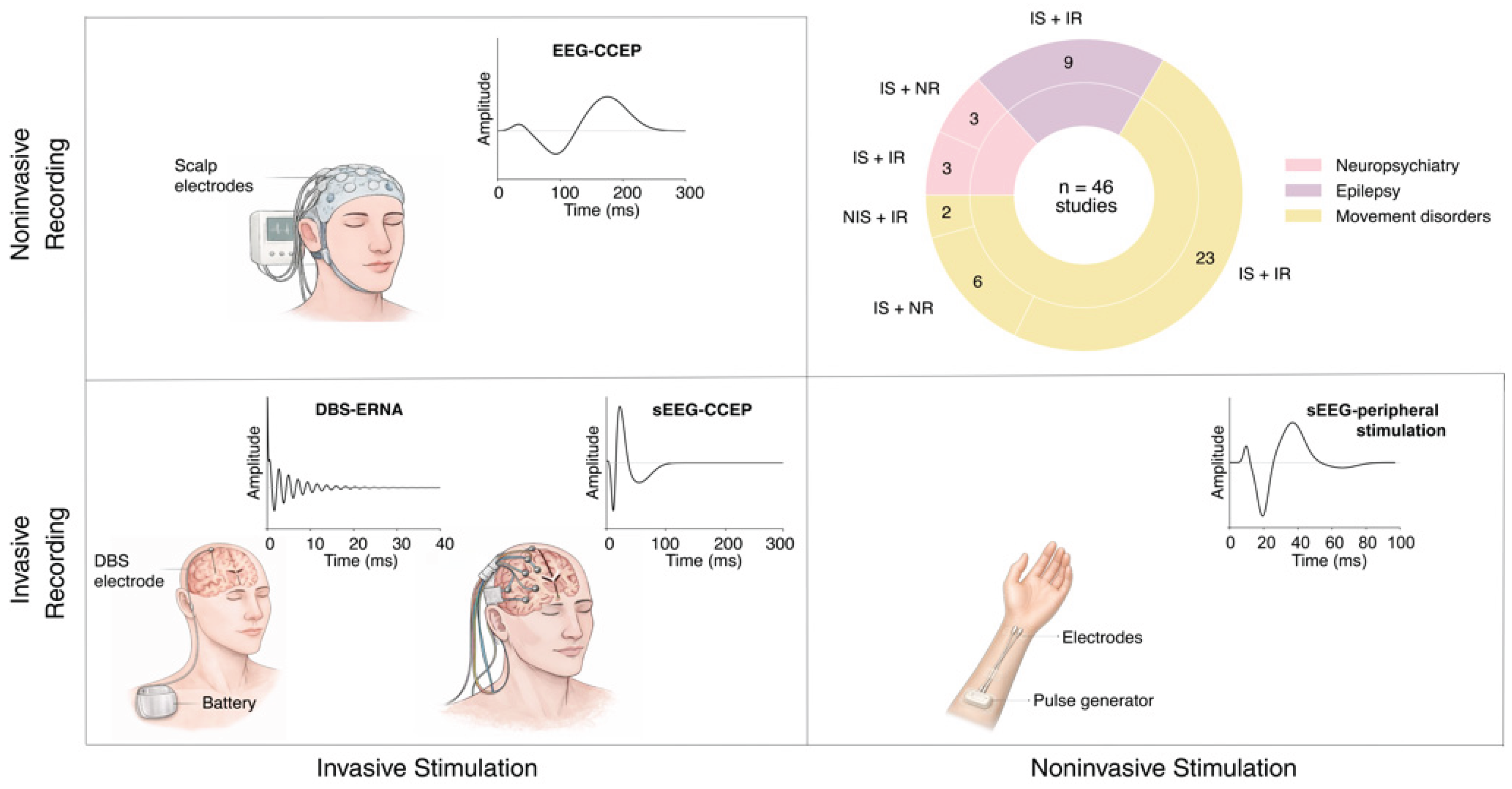
Stimulation and recording modalities: Most studies used both invasive stimulation and recording. Forty-four studies used invasive stimulation methods using implanted DBS leads (34) or sEEG electrodes, grids, or strips (10). Two studies performed nerve stimulation of the wrist to elicit evoked responses. Thirty-seven of the studies used invasive recordings: 16 used sEEG depth electrodes or electrocorticography (ECoG), 20 used DBS leads, and 1 used both ECoG and DBS. The other 9 used only non-invasive recording through either scalp electroencephalogram (EEG), electromyography (EMG), magnetoencephalopathy (MEG), or a combination (Figure 2).
Outcome measures: We organized each of the 46 studies by the type of outcome data to which CCEPs were compared. Twenty-seven studies compared CCEPs directly to clinical outcomes, including reduction in seizure frequency, motor symptoms, or psychiatric symptoms, or else used CCEPs to differentiate the most clinically beneficial contacts from others. Seven studies compared CCEPs to another physiologic biomarker, such as oscillations or gamma power, or compared CCEPs before and after high-frequency DBS-like stimulation. Seven studies compared imaging or tractography to CCEPs. The remaining 5 studies compared CCEPs to multiple outcome measures.
Clinical applications: Lastly, we identified three clinical applications for which CCEPs were investigated to guide brain stimulation. Fourteen studies (all in movement disorders) examined whether CCEPs could predict the ideal contact configuration for therapeutic benefit. Ten studies (3 movement disorders, 3 epilepsy, 4 psychiatry) investigated CCEPs as a measure to guide target selection or lead placement. Twelve studies (8 movement disorders, 3 epilepsy, 1 neuropsychiatry) examined whether CCEPs can quantify functional or state-dependent mechanisms of stimulation. The remaining 10 studies investigated multiple clinical applications.
Table 2.
Summary of CCEP studies, extracted data, and quality assessments. For each study, the first authors (K.T. and E.C.) extracted the following information: stimulation and recording modalities, secondary measures being compared to CCEPs, and clinical goals of the study. a = NHLBI Quality Assessment Tool for Observational Cohort and Cross-Sectional Studies, b = Joanna Briggs Institute (JBI) Critical Appraisal Checklist for Case Reports, c = Quality In Prognosis Studies (QUIPS), d = NHLBI Tool for Case Series Studies.
Table 2.
Summary of CCEP studies, extracted data, and quality assessments. For each study, the first authors (K.T. and E.C.) extracted the following information: stimulation and recording modalities, secondary measures being compared to CCEPs, and clinical goals of the study. a = NHLBI Quality Assessment Tool for Observational Cohort and Cross-Sectional Studies, b = Joanna Briggs Institute (JBI) Critical Appraisal Checklist for Case Reports, c = Quality In Prognosis Studies (QUIPS), d = NHLBI Tool for Case Series Studies.
| Reference | Indication | Cohort Size | Stimulation Method | Recording Method | Secondary Measure | Clinical Application | Quality Score |
| (Kobayashi et al., 2024) | Epilepsy | 12 | sEEG | sEEG | Clinical Outcome | Lead Placement | 9/14a |
| (Damiani et al., 2025) | Epilepsy | 37 | sEEG | sEEG | Clinical Outcome | Lead Placement | 8/14a |
| (Cheng et al., 2025) | Epilepsy | 11 | sEEG | sEEG | Imaging, Tractography | Lead Placement | 10/14a |
| (Munot et al., 2023) | Epilepsy | 14 | sEEG | sEEG | Before vs After Stimulation | Mechanism | 5/14a |
| (Huang et al., 2025) | Epilepsy | 2 | sEEG/grid/strip | sEEG | Biomarker | Mechanism | 8/9d |
| (Gregg et al., 2026) | Epilepsy | 10 | sEEG | sEEG | Clinical Outcome, Before vs After Stimulation | Mechanism | 8/14a |
| (Keller et al., 2018) | Epilepsy | 8 | sEEG | sEEG/ECoG | Before vs After Stimulation | Mechanism AND Lead Placement | 9/14a |
| (Wang et al., 2020) | Epilepsy | 5 | DBS (ANT) | DBS | Clinical Outcome | Mechanism AND Lead Placement | 8/14a |
| (Cole et al., 2026) | Epilepsy | 81 | sEEG | sEEG/ECoG | imaging | Mechanism AND Lead Placement | 9/14a |
| (Sanger et al., 2018) | Movement Disorder (Dystonia) |
15 | peripheral nerve stimulation | sEEG | Clinical Outcome | Lead Placement | 9/14a |
| (Hernandez-Martin et al., 2020) | Movement Disorder (Dystonia) | 14 | nerve (median, wrist level) | sEEG | Imaging, Tractography | Lead Placement | 8/14a |
| (Soroushmojdeh et al.i, 2025) | Movement Disorder (Dystonia) | 13 | DBS | DBS | Imaging, Tractography, Biomarker | Mechanism | 10/14a |
| (Ni et al., 2018) | Movement Disorder (Dystonia) | 8 | DBS (GPi), TMS | EEG, EMG | Before vs After Stimulation | Mechanism | 8/14a |
| (Wiest et al., 2023) | Movement Disorder (Dystonia) | 7 | DBS (STN) | DBS | Imaging, Tractography | Mechanism AND Lead Placement | 8/14a |
| (Bhanpuri et al., 2014) | Movement Disorder (Dystonia) | 11 | DBS (GPi), TMS | EEG, EMG | Clinical Outcome | Contact Selection | 9/14a |
| (Johnson et al., 2025) | Movement Disorder (Dystonia) | 8 | DBS (GP) | DBS | Clinical Outcome | Contact Selection | 9/14a |
| (Boddu et al., 2024) | Movement Disorder (Dystonia) | 1 | DBS (GPi), TMS | DBS | Clinical Outcome | Contact Selection | 8/8b |
| (Kent et al., 2014) | Movement Disorder (ET) | 17 | DBS | DBS | Clinical Outcome | Mechanism | 9/14a |
| (Bogaert et al., 2025) | Movement Disorder (PD) | 14 | DBS (STN) | DBS | Clinical Outcome | Lead Placement | 8/14a |
| (Sinclair et al., 2018) | Movement Disorder (PD) | 19 | DBS (STN) | DBS | Clinical Outcome | Lead Placement AND Contact Selection | 9/14a |
| (Ozturk et al., 2021) | Movement Disorder (PD) | 13 | DBS (STN) | DBS | Biomarker | Mechanism | 8/14a |
| (Sanabria et al., 2022) | Movement Disorder (PD) | 1 | DBS (GPi) | DBS | Biomarker | Mechanism | 6/8b |
| (Wiest et al., 2020) | Movement Disorder (PD) | 14 | DBS (STN) | DBS | Biomarker, Clinical Outcome | Mechanism | 8/14a |
| (Wiest et al., 2025) | Movement Disorder (PD) | 4 | DBS (STN) | DBS | Biomarker | Mechanism | 9/14a |
| (Sinclair et al., 2019) | Movement Disorder (PD) | 21 | DBS (STN) | DBS | Clinical Outcome | Mechanism | 10/14a |
| (Ren et al., 2026) | Movement Disorder (PD) | 863 | DBS (STN) | ECoG | Imaging, Tractography | Mechanism | 10/14a |
| (Connolly et al., 2021) | Movement Disorder (PD) | 1 | DBS (STN) | ECoG, EMG | Clinical Outcome | Mechanism AND Contact Selection | 7/8b |
| (Borgheai et al., 2021) | Movement Disorder (PD) | 15 | DBS (STN) | ECoG, DBS, EMG | Clinical Outcome | Mechanism AND Contact Selection | 8/14a |
| (Steiner et al., 2024) | Movement Disorder (PD) | 30 | DBS (STN) | DBS | Clinical Outcome | Mechanism AND Lead Placement | 9/14a |
| (Testini et al., 2025) | Movement Disorder (PD) | 12 | DBS (STN, GPi) | EMG | Clinical Outcome | Contact Selection | 8/14a |
| (Sinclair et al., 2022) | Movement Disorder (PD) | 21 | DBS (STN) | DBS | Clinical Outcome | Contact Selection | 10/14a |
| (Johnson et al., 2023) | Movement Disorder (PD) | 22 | DBS (GPi) | DBS | Clinical Outcome | Contact Selection | 9/14a |
| (Xu et al., 2022) | Movement Disorder (PD) | 14 | DBS (STN) | DBS | Clinical Outcome | Contact Selection | 9/14a |
| (Xu et al., 2022) | Movement Disorder (PD) | 47 | DBS (STN) | DBS | Clinical Outcome | Contact Selection | 10/14a |
| (Bahners et al., 2024) | Movement Disorder (PD) | 32 | DBS (STN) | dry EEG | Clinical Outcome | Contact Selection | 8/14a |
| (Miocinovic et al., 2018) | Movement Disorder (PD) | 10 | DBS (STN, GPi) | ECoG | Clinical Outcome | Contact Selection | 7/14a |
| (Spooner et al., 2023) | Movement Disorder (PD) | 24 | DBS (STN) | MEG | Clinical Outcome | Contact Selection | 9/14a |
| (Cole et al., 2025) | Movement Disorder (PD) | 54 | DBS (STN) | EMG | Clinical Outcome | Contact Selection | 10/14a |
| (Opri et al., 2025) | Movement Disorder (PD) | 31 | DBS (STN) | DBS | Clinical Outcome | Contact Selection AND Lead Placement | 9/14a |
| (Nagao et al., 2026) | Movement Disorder (PD) | 12 | DBS | DBS | Clinical Outcome | Contact Selection and Mechanism | Moderate Riskc |
| (Lee et al., 2025) | OCD | 1 | sEEG | sEEG | Clinical Outcome | Lead Placement | 7/8b |
| (Bogaert et al., 2025) | OCD | 10 | DBS | EEG | Clinical Outcome, Imaging, Tractography | Lead Placement | 9/14a |
| (Smith et al., 2022) | Depression | 8 | DBS (SCC) | EEG | Imaging, Tractography | Lead Placement | 8/14a |
| (Adkinson et al., 2022) | Depression | 2 | DBS (SCC/VCVS) | sEEG | Imaging, Tractography | Lead Placement | 5/8b |
| (Desai et al., 2025) | Depression | 10 | DBS (SCC) | EEG | Imaging, Tractography, Clinical Outcome | Mechanism | 9/14a |
| (Scangos et al., 2021) | Depression | 1 | sEEG | sEEG | Clinical Outcome | Mechanism AND Lead Placement | 7/8b |
Clinical applications of CCEPs
Movement Disorders
DBS is widely used to treat motor symptoms of movement disorders. The targets – the subthalamic nucleus (STN), globus pallidus internus (GPi), and ventralis intermedius nucleus of the thalamus (VIM) – are commonly used clinically and therapeutically effective[2]. Primary challenges of DBS for movement disorders remain practical: improving efficiency and reliability of implantation surgery, quickly finding each patient’s most effective parameters, and improving scalability[7,21]. Furthermore, recent advancements in adaptive DBS (aDBS) may offer additional benefit to patients with high degrees of symptom or medication fluctuations[22]. We identified 31 studies of CCEPs in movement disorders.
Parkinson’s Disease: No studies leveraged CCEPs to select personalized targets for PD. The most investigated CCEP was ERNA, a quickly decaying oscillatory waveform recorded from non-stimulated DBS contacts during either STN or GPi DBS. Four studies retrospectively compared ERNA between different stimulating DBS contacts and found that a larger ERNA amplitude can accurately predict the contact chosen for chronic stimulation[23,24,25,26]. A similar pattern was observed using both cortical (ECoG) and non-invasively recorded (EEG, MEG) CCEPs[27,28,29]. One study demonstrated that contacts prospectively selected to maximize ERNA achieve comparable symptom relief to conventional programming[30]. Additionally, the anatomical location that elicits greatest ERNA coincided with the most clinically effective subregions of the STN and GPi, indicating that ERNA may help optimize lead placement[31,32,33]. Cortical CCEPs were strongest in the somato-cognitive action network, where reduced connectivity was associated with better clinical outcomes[34].
Beyond contact selection, several studies showed that CCEPs reflect physiologic and state-dependent properties of circuit engagement, with implications for guiding aDBS. Therapeutic high-frequency stimulation results in longer-lasting ERNA compared to ERNA elicited by non-therapeutic, 20 Hz stimulation[35]. Furthermore, 130 Hz DBS modulates ERNA (later peaks, decreased frequency, etc.) over tens of seconds[36] whereas 20 Hz DBS does not[37]. The differential modulation of ERNA following therapeutic DBS may serve as a positive biomarker for aDBS. Sanabria et al.[38] showed the feasibility of CCEPs for aDBS by using stimulation-evoked potentials to adjust stimulation parameters that modulated beta oscillation power in real time.
Several studies compared the predictive performance of ERNA to other biomarkers. ERNA demonstrates higher predictive power than beta power and high-frequency oscillations in identifying clinically selected contacts[25]. ERNA remains measurable under general anesthesia[39], demonstrating utility for anesthetized surgical procedures. Furthermore, ERNA persists during sleep and is differentially modulated by sleep stages[40], suggesting that it could inform aDBS strategies for nocturnal tremors. Spooner et al.[29] showed that ERNA can predict optimal contact orientations within directional DBS systems, supporting its relevance for complex electrode geometries. Altogether, these findings suggest ERNA is a robust and technically accessible biomarker that can advance DBS for PD.
Essential Tremor:One study showed that CCEPs can be measured locally during VIM DBS, and their magnitude correlates with tremor reduction across stimulation settings[41].
Dystonia: Despite similar targets as in PD, there are fewer CCEP studies in dystonia given smaller patient populations and more limited understanding of mechanisms underlying therapeutic benefit. Higher ERNA amplitudes[42,43] in the GPi (measured similarly as in PD) and steeper increases in ERNA magnitude relative to increasing stimulation amplitude[44] predict clinically selected contacts. Wiest et al.[45] showed that localization properties of ERNA in STN are similar to those in PD, demonstrating potential for both contact selection and optimizing lead placement.
Sanger et al.[46] found that DBS of personalized targets, refined using median nerve stimulation-evoked potentials and sEEG monitoring, yields greater symptom reduction than standard targeting procedures. Median nerve evoked potentials are able to confirm electrode localization in the VIM as opposed to adjacent thalamic nuclei[47]. Lastly, CCEPs also reveal that GPi DBS induces plastic changes in motor cortical activity, which may inform the mechanism of GPi DBS for dystonia[48]. Higher frequency GPi stimulation leads to smaller and higher-latency CCEPs, suggesting that CCEPs could inform programming of DBS pulse frequency[49]. Despite the lower number of studies, CCEPs may similarly hold promise for improving DBS for dystonia.
DBS side effects: Beyond providing symptom relief, DBS must be optimized to avoid side effects which can arise from stimulation of pathways adjacent to the targeted region. Two studies found that motor evoked potentials, measured using EMG placed on facial and upper limb muscles, could provide a biomarker to minimize motor side effects during programming[50,51]. While such peripherally recorded biomarkers are technically not CCEPs, they nevertheless reflect activation of brain circuits that predict therapeutic effects. We did not identify any CCEP studies evaluating other DBS-induced side effects.
Epilepsy
DBS and RNS provide significant reduction in seizure frequency but often do not lead to seizure freedom[52]. Clinical applications of brain stimulation for epilepsy include selecting the optimal region for stimulation based on the patient’s seizure network, contact selection within the targeted region, and localizing seizures using diagnostic stimulation[3]. We excluded studies which used CCEPs to delineate the region for surgical resection or characterize functional networks. We identified 9 relevant CCEP studies in epilepsy.
Most of these studies investigated whether CCEPs could determine optimal targets of stimulation or guide parameter optimization to provide optimal seizure suppression. In a small cohort, larger bilateral CCEPs in the hippocampus evoked from stimulation of the anterior nucleus of the thalamus (ANT) correlate with more favorable seizure reduction following ANT DBS[53]. CCEPs were used in conjunction with tractography to retrospectively match specific thalamic nuclei based on high connectivity to each patient’s seizure onset zone, and stimulation of matched thalamic nuclei led to substantially better seizure reduction[54]. Beyond thalamic DBS, CCEPs were used to investigate the extreme capsule (EC), a novel white matter DBS target connecting frontal, temporal, and insular areas. High-frequency EC stimulation decreases epileptic discharges, specifically across strongly interconnected regions based on CCEPs and tractography[55]. For RNS, Kobayashi et al.[56] investigated CCEPs within the seizure network, finding that contacts associated with greater seizure reduction also feature stronger overall network connectivity. These results suggest that CCEPs can predict therapeutic effectiveness of certain DBS or RNS targets based on their connectivity in relation to the patient’s seizure onset zone.
Some studies compared CCEPs to neurophysiological effects of stimulation in intracranial recordings. Gregg et al.[57] found that the amplitude of CCEPs evoked by thalamic stimulation is suppressed following 1.5 hours of long-form stimulation, especially in regions with higher baseline connectivity to the thalamus. Sustained DBS may thus induce plasticity in target cortical regions, and favorable CCEP modulation correlates with improved reduction of interictal epileptiform discharges and seizure frequency[57]. Two studies showed that regions with stronger baseline connectivity demonstrate greater modulation following repeated 10 Hz stimulation[58,59]. These studies suggest that CCEPs and their changes following stimulation reflect plastic changes in epileptic networks that may be associated with clinical efficacy[60].
Psychiatry
DBS is approved for OCD under a humanitarian device exemption, and clinical trials are ongoing for depression[4,61,62]. One major clinical challenge in DBS for psychiatric disorders is to optimize device placement and stimulation parameters for each patient to consistently achieve responder status. Commonly investigated targets include the ventral capsule/ventral striatum (VC/VS) and bed nucleus of the stria terminalis for OCD, and subcallosal cingulate (SCC) and VC/VS for depression. Precise implantation and contact selection within these targets is crucial, given the critical role of activating specific white matter bundles to achieve successful outcomes[63,64]. We identified six applicable studies of CCEPs for psychiatric DBS.
Obsessive-compulsive disorder: Intraoperative EEG recording during VC/VS DBS surgery showed that the site eliciting the largest CCEP amplitude coincides with a tractographically defined optimal target that engages orbitofrontal and ventrolateral prefrontal cortex white matter pathways, indicating that CCEPs reflect activation of therapeutically relevant circuits. Furthermore, CCEPs evoked by different DBS contacts are more morphologically similar in responders than non-responders[65]. In a case study, acute self-reported OCD symptom improvement was used to identify two right VC/VS sites for chronic stimulation, which led to a 62% reduction in OCD symptoms after 6 months[66]. Retrospective CCEP mapping showed both sites are connected to the orbitofrontal cortex and one site is connected to the anterior cingulate, where high-frequency activity is significantly correlated to OCD symptom severity[66].
Depression: During SCC DBS, CCEPs measured using EEG and their time-frequency features correlate with the dorsal-ventral location of the stimulation site[67]. Desai et al.[68] further found that these evoked potentials show reduction in latency and increase in amplitude over time and that this faster response is correlated with higher baseline fractional anisotropy of the dorsal, middle segment of the cingulum bundle. Intracranial CCEPs evoked by SCC and VC/VS DBS corroborate tractographic estimates of connectivity in the orbitofrontal, ventromedial, prefrontal, and lateral prefrontal cortex[69]. However, these modalities diverge in the anterior cingulate cortex (ACC): tractography predicts connectivity to the SCC without corresponding CCEPs, whereas VC/VS simulation evokes robust potentials in the ACC despite minimal predicted tractographic connectivity[69]. These findings suggest that tractography and CCEPs provide complementary information for characterizing proper network engagement and optimizing DBS targeting. Using intracranial recording, Scangos et al.[70] identified a personalized amygdala gamma power marker of symptom severity and confirmed VC/VS-amygdala connectivity using CCEPs. Subsequent chronic closed-loop stimulation resulted in rapid and sustained clinical improvement[70]. While limited in volume, these studies suggest that CCEPs can verify engagement of critical white matter pathways, demonstrating potential to optimize device placement and guide contact selection in psychiatric disorders.
Emerging Indications
DBS has been increasingly explored for novel indications, such as Tourette’s syndrome, post-traumatic stress disorder, schizophrenia, stroke, and traumatic brain injury. Few studies have investigated CCEPs in such indications. One study found that SPES of white matter projections to visual cortex modulates visually evoked potentials, indicating potential relevance for visual neuroprostheses[71]. DBS of the motor thalamus potentiates motor evoked potential activation, showing promise as a treatment for loss of motor function following white matter lesions[72,73]. CCEPs throughout memory-related regions may also guide how temporal cortex stimulation can treat brain injury-mediated memory loss[74].
4. Discussion
From retrospective circuit characterization to guiding therapy with CCEPs
CCEPs have generally been used to retrospectively compare profiles of connectivity to DBS outcomes. We found only one study that instead used CCEPs to prospectively guide therapy then evaluate whether the clinical outcome was improved (in the context of DBS programming for Parkinson’s disease[30]). Where there is sufficient evidence for their clinical utility, future studies should prioritize evaluating the prospective clinical potential of CCEPs for improving DBS. Novel technical approaches to prospectively select DBS parameters and targets using CCEPs may improve DBS effectiveness and efficiency of clinical procedures, while also enabling causal data-driven and mechanistic hypothesis testing for incompletely understood or novel clinical indications.
Technical challenges
Technological improvements will further aid in the clinical applications of CCEPs. Currently, CCEPs can only be measured in acute or semi-chronic recording environments due to the necessity of kilohertz sampling rates to accurately measure short-latency waveforms[75]. Therefore, ERNA studies have primarily been performed intraoperatively or with externalized DBS recordings rather than using chronic sensing functionality available in modern devices[76]. This constraint may be sufficient for most applications of CCEPs, including optimizing device placement during surgery or using acute neural recordings to guide post-operative programming (e.g. for epilepsy). However, it greatly limits the mechanistic exploration of CCEPs in the context of behavioral changes or symptom fluctuations for aDBS and their utility for such applications may go underappreciated until this technical barrier is lifted.
A second technical advance needed is the development of robust signal processing approaches for CCEP measurement and their integration into real-time systems to provide feedback for DBS. CCEP detection algorithms must measure physiologic activation while mitigating noise and stimulation artifact. Design characteristics will depend critically on the circuits being studied, relevant waveform characteristics, choice of electrode, and technical properties of the platform used for recording and stimulation[12].
Stimulation parameter optimization: A critical confound in CCEP studies is the selection of stimulation parameters. Differences in stimulation amplitude, contact configuration, pulse width, and pulse frequency greatly affect the magnitude and spatial profile of observed CCEPs[77]. Modern DBS devices with 8 or more independently controllable contacts have millions of possible parameter combinations, hindering manual navigation of device parameters or grid-based search strategies during time-constrained clinical procedures[7]. Thus, clinical applications of CCEPs will benefit from intelligent algorithmic strategies that can identify optimal stimulation parameters that activate desired patterns of connectivity expected to underlie therapeutic effectiveness. Bayesian optimization algorithms have been developed to quickly program DBS parameters based on symptom assessments and kinematic measurements in movement disorders[78,79]. A similar approach may enable precise circuit activation using CCEPs as the objective function[80].
Clinical implications
Movement disorders: CCEPs are robust and conveniently measurable from the DBS device itself and evidence across studies suggests that maximizing ERNA amplitude is a viable strategy for guiding device placement and selecting an effective contact configuration. These developments can provide a complementary assessment tool to improve convenience and efficiency of implantation surgery and reduce time needed for programming, reducing the delay in achieving treatment success for each patient and expanding access to a larger patient population[7]. Pressing challenges to advance care are technical: researchers should focus on developing signal processing techniques that can measure ERNA quickly and reliably across any DBS parameter combination and closed-loop interfaces for real-time stimulation and biomarker measurement suitable for visualization and fast assessment during implantation surgery. Furthermore, integration of kilohertz sample rate recording into clinical devices will enable more flexible and convenient use of biomarkers during clinical programming sessions and further exploration of behavioral state dependence for aDBS applications[76]. Last, emerging research is gravitating towards a connectomic framework for aDBS in which multiple contacts on higher-density DBS devices are selected to differentially target pathways underlying specific symptoms and adapted separately as these different symptoms evolve[81]. The need for multiscale recording, precise circuit targeting, and challenges of calibrating complex devices suggests that CCEPs will play an important role for future technological developments in movement disorders.
Epilepsy: Given the widespread clinical use of intracranial monitoring, CCEPs are easily measurable for epilepsy and analysis methods are well-developed compared to other indications[12]. Furthermore, it is becoming increasingly common to implant electrodes in regions that may provide effective DBS targets[82]. Thus, the experimental opportunity is available to collect neurophysiologic data and clinical outcomes at scale to empirically characterize the clinical significance of CCEPs for DBS. The remaining barrier is understanding the best way that CCEPs can predict therapeutic stimulation outcomes of brain stimulation or inform optimal parameters given the high variability of seizure onset profiles across patients. Furthermore, there are different designs by which CCEPs can be interpreted to predict effectiveness: increased spatial spread or magnitude of CCEPs induced in seizure onset regions, bidirectional connectivity between seizure onset areas and the stimulated site, or modulation of plasticity may all viably predict clinical value and should be compared empirically. This wide variability suggests that more studies are needed to characterize in detail which regions are best candidates for which patients, and therefore, how CCEPs can best guide target selection. The limited data thus far has shown promise that CCEPs can predict greater seizure reduction based on connectivity between the target and seizure onset zone. Thus, CCEPs may best be suited to guide DBS target and contact selection.
Psychiatry: Of the three indication categories, CCEPs for psychiatry are the most variable in recording modalities and clinical implications. Nevertheless, the importance of accurately targeting specific white matter bundles for achieving therapeutic benefit and the challenges of navigating subjective, months-long symptom improvement timescales[4] suggest clear utility for CCEPs to provide objective, quickly accessible feedback to guide targeting and parameter selection. CCEPs could provide an objective, convenient, and anesthesia-compatible alternative to awake valence testing, an established technique that uses patient-reported behavioral changes to guide DBS implantation surgery[83]. Initial programming may become more efficient and precise by using CCEPs to identify the contact configuration and amplitude that maximize pathway activation[8]. Future studies for psychiatry should identify the recording strategy that provides the most robust estimate of therapeutically relevant pathway activation and is also clinically practical and scalable (e.g. intraoperative ECoG). Additionally, limited longitudinal studies suggest that changes in CCEPs throughout months following DBS implantation could provide a complementary source of feedback to assess symptom changes and subsequently inform clinical adjustment of behavioral therapy, medication, and DBS parameters.
5. Conclusion
In summary, CCEPs hold potential to improve the clinical practicality and effectiveness of invasive brain stimulation. Evidence in movement disorders, particularly Parkinson’s disease, suggests a clear role for CCEPs to aid implantation surgery and inform programming. In contrast, applications in epilepsy and psychiatry need further characterization to understand the best way that CCEPs could be applied clinically. Further investigation of CCEPs in relation to clinical outcomes, technological development, and causal mechanistic investigation can similarly benefit brain stimulation for epilepsy, psychiatry, and underexplored conditions.
Acknowledgments
This research was supported by the National Institutes of Health (NIH) R01MH137102 and R01MH130597 (S.A.S). Additional support was provided by the Robert and Janice McNair Foundation (S.R.H., S.A.S.) and the Gordon and Mary Cain Pediatric Neurology Research Foundation Laboratories at Texas Children’s Hospital (S.A.S.).
Declaration of competing interests
The authors declare the following financial interests/personal relationships which may be considered as potential competing interests: SAS reports consulting/advising for Boston Scientific, Zimmer Biomet, Abbott, Koh Young Technology, NeuroPace Inc, and co-founding Motif Neurotech. All other authors declare that they have no competing financial interests or personal relationships that could be perceived to have influenced the work reported in this paper.
References
- Lozano, A. M.; et al. Deep brain stimulation: current challenges and future directions. Nat. Rev. Neurol. 2019, 15, 148–160. [Google Scholar] [CrossRef]
- Larson, P. S. Deep brain stimulation for movement disorders. Neurother. J. Am. Soc. Exp. Neurother. 2014, 11, 465–474. [Google Scholar] [CrossRef] [PubMed]
- Piper, R. J.; et al. Towards network-guided neuromodulation for epilepsy. Brain J. Neurol. 2022, 145, 3347–3362. [Google Scholar] [CrossRef]
- Sheth, S. A.; Mayberg, H. S. Deep Brain Stimulation for Obsessive-Compulsive Disorder and Depression. Annu. Rev. Neurosci. 2023, 46, 341–358. [Google Scholar] [CrossRef] [PubMed]
- Widge, A. S. Closing the loop in psychiatric deep brain stimulation: physiology, psychometrics, and plasticity. Neuropsychopharmacol. Off. Publ. Am. Coll. Neuropsychopharmacol. 2024, 49, 138–149. [Google Scholar] [CrossRef] [PubMed]
- Simpson, H. D.; et al. Practical considerations in epilepsy neurostimulation. Epilepsia 2022, 63, 2445–2460. [Google Scholar] [CrossRef]
- Cole, E. R.; Miocinovic, S. Are we ready for automated deep brain stimulation programming? Park. Relat. Disord. 2025, 134, 107347. [Google Scholar] [CrossRef]
- Amagat, M.; et al. Optimizing subthalamic deep brain stimulation for treatment-refractory obsessive compulsive disorder: A practical guide to advanced parameter tuning. Brain Stimul. 2025, 18, 1719–1722. [Google Scholar] [CrossRef]
- Crocker, B.; et al. Local and distant responses to single pulse electrical stimulation reflect different forms of connectivity. NeuroImage 2021, 237, 118094. [Google Scholar] [CrossRef]
- Trebaul, L.; et al. Probabilistic functional tractography of the human cortex revisited. NeuroImage 2018, 181, 414–429. [Google Scholar] [CrossRef]
- Campbell, J. M.; et al. Macroscale Traveling Waves Evoked by Single-Pulse Stimulation of the Human Brain. J. Neurosci. 2025, 45. [Google Scholar] [CrossRef]
- Qiang, Z.; et al. Cortico-cortical evoked potentials: Analytical techniques and emerging paradigms for epileptogenic zone localization. Epilepsia 2025, 66, 3087–3104. [Google Scholar] [CrossRef]
- Kobayashi, K.; et al. Cortico-cortical evoked potential by single-pulse electrical stimulation is a generally safe procedure. Clin. Neurophysiol. Off. J. Int. Fed. Clin. Neurophysiol. 2021, 132, 1033–1040. [Google Scholar] [CrossRef] [PubMed]
- Parvizi, J. Thalamus, evoked responses and triphasic waves. Nat. Commun. 2025, 16, 5221. [Google Scholar] [CrossRef]
- Matsumoto, R.; et al. Functional connectivity in the human language system: a cortico-cortical evoked potential study. Brain 2004, 127, 2316–2330. [Google Scholar] [CrossRef] [PubMed]
- Matsumoto, R.; Kunieda, T.; Nair, D. Single pulse electrical stimulation to probe functional and pathological connectivity in epilepsy. Seizure 2017, 44, 27–36. [Google Scholar] [CrossRef]
- Miller, K. J.; et al. Canonical Response Parameterization: Quantifying the structure of responses to single-pulse intracranial electrical brain stimulation. PLoS Comput. Biol. 2023, 19, e1011105. [Google Scholar] [CrossRef]
- Giampiccolo, D.; et al. Recording cortico-cortical evoked potentials of the human arcuate fasciculus under general anaesthesia. Clin. Neurophysiol. Off. J. Int. Fed. Clin. Neurophysiol. 2021, 132, 1966–1973. [Google Scholar] [CrossRef] [PubMed]
- Russo, S.; et al. Thalamic feedback shapes brain responses evoked by cortical stimulation in mice and humans. Nat. Commun. 2025, 16, 3627. [Google Scholar] [CrossRef]
- Ouzzani, M.; Hammady, H.; Fedorowicz, Z.; Elmagarmid, A. Rayyan—a web and mobile app for systematic reviews. Syst. Rev. 2016, 5, 210. [Google Scholar] [CrossRef]
- Lim, M. L.; et al. Awake versus Asleep Anesthesia in Deep Brain Stimulation Surgery for Parkinson’s Disease: A Systematic Review and Meta-Analysis. Stereotact. Funct. Neurosurg. 2024, 102, 141–155. [Google Scholar] [CrossRef]
- Oehrn, C. R.; et al. Chronic adaptive deep brain stimulation versus conventional stimulation in Parkinson’s disease: a blinded randomized feasibility trial. Nat. Med. 2024, 30, 3345–3356. [Google Scholar] [CrossRef]
- Johnson, K. A.; et al. Globus pallidus internus deep brain stimulation evokes resonant neural activity in Parkinson’s disease. Brain Commun. 2023, 5, fcad025. [Google Scholar] [CrossRef]
- Xu, S. S.; et al. Towards guided and automated programming of subthalamic area stimulation in Parkinson’s disease. Brain Commun. 2022, 4, fcac003. [Google Scholar] [CrossRef] [PubMed]
- Xu, S. S.; et al. Can brain signals and anatomy refine contact choice for deep brain stimulation in Parkinson’s disease? J. Neurol. Neurosurg. Psychiatry 2022, jnnp-2021-327708. [Google Scholar] [CrossRef] [PubMed]
- Opri, E.; et al. Deep brain stimulation-induced local evoked potentials outperform spectral features in spatial and clinical STN mapping. J. Neural Eng. 2025, 22, 046055. [Google Scholar] [CrossRef]
- Bahners, B. H.; et al. Evoked response signatures explain deep brain stimulation outcomes. 2024. [Google Scholar] [CrossRef]
- Miocinovic, S.; et al. Cortical Potentials Evoked by Subthalamic Stimulation Demonstrate a Short Latency Hyperdirect Pathway in Humans. J. Neurosci. Off. J. Soc. Neurosci. 2018, 38, 9129–9141. [Google Scholar] [CrossRef]
- Spooner, R. K.; Bahners, B. H.; Schnitzler, A.; Florin, E. DBS-evoked cortical responses index optimal contact orientations and motor outcomes in Parkinson’s disease. npj Park. Dis. 2023, 9, 37. [Google Scholar] [CrossRef]
- Nagao, K. J.; et al. Automating Subthalamic Deep Brain Stimulation Programming with Evoked Resonant Neural Activity in Parkinson’s Disease. In Mov. Disord.
- Sinclair, N. C.; et al. Subthalamic nucleus deep brain stimulation evokes resonant neural activity. Ann. Neurol. 2018, 83, 1027–1031. [Google Scholar] [CrossRef]
- Van Bogaert, T.; et al. Neuromodulation J. Int. Neuromodulation Soc. S1094-7159(25)01027-X; Evoked Resonant Neural Activity Reveals an Electrophysiologic Sweet Spot for Directional Subthalamic Nucleus Deep Brain Stimulation in Parkinson’s Disease. 2025. [CrossRef]
- Steiner, L. A.; et al. Neural signatures of indirect pathway activity during subthalamic stimulation in Parkinson’s disease. Nat. Commun. 2024, 15, 3130. [Google Scholar] [CrossRef]
- Ren, J.; et al. Parkinson’s disease as a somato-cognitive action network disorder. Nature 2026, 651, 1030–1038. [Google Scholar] [CrossRef]
- Ozturk, M.; Viswanathan, A.; Sheth, S. A.; Ince, N. F. Electroceutically induced subthalamic high-frequency oscillations and evoked compound activity may explain the mechanism of therapeutic stimulation in Parkinson’s disease. Commun. Biol. 2021, 4, 393. [Google Scholar] [CrossRef]
- Wiest, C.; et al. Local field potential activity dynamics in response to deep brain stimulation of the subthalamic nucleus in Parkinson’s disease. Neurobiol. Dis. 2020, 143, 105019. [Google Scholar] [CrossRef]
- Sinclair, N. C.; et al. Deep brain stimulation for Parkinson’s disease modulates high-frequency evoked and spontaneous neural activity. Neurobiol. Dis. 2019, 130, 104522. [Google Scholar] [CrossRef]
- Escobar Sanabria, D.; et al. Controlling pallidal oscillations in real-time in Parkinson’s disease using evoked interference deep brain stimulation (eiDBS): Proof of concept in the human. Brain Stimul. 2022, 15, 1111–1119. [Google Scholar] [CrossRef]
- Sinclair, N. C.; et al. Electrically evoked and spontaneous neural activity in the subthalamic nucleus under general anesthesia. J. Neurosurg. 2022, 137, 449–458. [Google Scholar] [CrossRef] [PubMed]
- Wiest, C.; et al. Stimulation-Evoked Resonant Neural Activity in the Subthalamic Nucleus Is Modulated by Sleep. Mov. Disord. Off. J. Mov. Disord. Soc. 2025, 40, 351–356. [Google Scholar] [CrossRef] [PubMed]
- Kent, A. R.; et al. Measurement of Evoked Potentials During Thalamic Deep Brain Stimulation. Brain Stimul. 2015, 8, 42–56. [Google Scholar] [CrossRef]
- Johnson, K. A.; et al. Pallidal Evoked Resonant Neural Activity as a Candidate Biomarker for Deep Brain Stimulation in Dystonia. Mov. Disord. Off. J. Mov. Disord. Soc. 2026, 41, 709–718. [Google Scholar] [CrossRef] [PubMed]
- Boddu, A. V.; et al. Directional Stimulus-Evoked Pallidal Electrophysiology in Primary Dystonia. Tremor Hyperkinetic Mov. 2024, 14, 46. [Google Scholar] [CrossRef]
- Bhanpuri, N. H.; et al. Deep brain stimulation evoked potentials may relate to clinical benefit in childhood dystonia. Brain Stimul. 2014, 7, 718–726. [Google Scholar] [CrossRef]
- Wiest, C.; et al. Subthalamic Nucleus Stimulation–Induced Local Field Potential Changes in Dystonia. Mov. Disord. 2023, 38, 423–434. [Google Scholar] [CrossRef] [PubMed]
- Sanger, T. D.; et al. Pediatric Deep Brain Stimulation Using Awake Recording and Stimulation for Target Selection in an Inpatient Neuromodulation Monitoring Unit. Brain Sci. 2018, 8, 135. [Google Scholar] [CrossRef]
- Hernandez-Martin, E.; Arguelles, E.; Deshpande, R.; Sanger, T. D. Evoked Potentials During Peripheral Stimulation Confirm Electrode Location in Thalamic Subnuclei in Children With Secondary Dystonia. J. Child. Neurol. 2020, 35, 799–807. [Google Scholar] [CrossRef] [PubMed]
- Ni, Z.; et al. Pallidal deep brain stimulation modulates cortical excitability and plasticity. Ann. Neurol. 2018, 83, 352–362. [Google Scholar] [CrossRef]
- Soroushmojdehi, R.; Vidmark, J. S. L.; Seyyed Mousavi, S. A.; Abe, S.; Sanger, T. D. Frequency-dependent modulation of evoked potentials during GPi-DBS in pediatric dystonia. Sci. Rep. 2025, 15, 33551. [Google Scholar] [CrossRef]
- Testini, P.; Wang, A.; Cole, E.; Miocinovic, S. Motor evoked potentials as a side effect biomarker for deep brain stimulation programming. MedRxiv Prepr. Serv. Health Sci. 2025, 2025.01.24, 25320924. [Google Scholar] [CrossRef]
- Cole, E. R.; et al. An algorithmic procedure for measuring deep brain stimulation-induced capsular activation using motor evoked potentials. J. Neural Eng. 2025, 22, 066044. [Google Scholar] [CrossRef]
- Gouveia, F. V.; Warsi, N. M.; Suresh, H.; Matin, R.; Ibrahim, G. M. Neurostimulation treatments for epilepsy: Deep brain stimulation, responsive neurostimulation and vagus nerve stimulation. Neurother. J. Am. Soc. Exp. Neurother. 2024, 21, e00308. [Google Scholar] [CrossRef] [PubMed]
- Wang, Y.-C.; et al. Probing circuit of Papez with stimulation of anterior nucleus of the thalamus and hippocampal evoked potentials. Epilepsy Res. 2020, 159, 106248. [Google Scholar] [CrossRef]
- Damiani, A.; et al. Thalamocortical hodology to personalize electrical stimulation for focal epilepsy. Nat. Commun. 2025, 16, 9209. [Google Scholar] [CrossRef]
- Cheng, Y.; et al. Structural network-specific effect of extreme capsule stimulation for drug-resistant focal epilepsy. Brain J. Neurol. 2025, 148, 2920–2934. [Google Scholar] [CrossRef] [PubMed]
- Kobayashi, K.; et al. Effective connectivity relates seizure outcome to electrode placement in responsive neurostimulation. Brain Commun. 2024, 6, fcae035. [Google Scholar] [CrossRef]
- Gregg, N. M.; et al. Thalamic Stimulation Induced Changes in Network Connectivity and Excitability in Epilepsy. Ann. Neurol. 2026, 99, 748–760. [Google Scholar] [CrossRef]
- Keller, C. J.; et al. Induction and Quantification of Excitability Changes in Human Cortical Networks. J. Neurosci. Off. J. Soc. Neurosci. 2018, 38, 5384–5398. [Google Scholar] [CrossRef]
- Munot, S.; Kim, N.; Huang, Y.; Keller, C. J. Direct cortical stimulation induces short-term plasticity of neural oscillations in humans. BioRxiv Prepr. Serv. Biol. 2023. 2023, 11.15.567302. [Google Scholar] [CrossRef]
- Khambhati, A. N.; Shafi, A.; Rao, V. R.; Chang, E. F. Long-term brain network reorganization predicts responsive neurostimulation outcomes for focal epilepsy. Sci. Transl. Med. 2021, 13, eabf6588. [Google Scholar] [CrossRef] [PubMed]
- Greenberg, B. D.; et al. Neurosurgery for Intractable Obsessive-Compulsive Disorder and Depression: Critical Issues. Neurosurg. Clin. North Am. 2003, 14, 199–212. [Google Scholar] [CrossRef]
- Mayberg, H. S.; et al. Deep brain stimulation for treatment-resistant depression. Neuron 2005, 45, 651–660. [Google Scholar] [CrossRef]
- Li, N.; et al. A unified connectomic target for deep brain stimulation in obsessive-compulsive disorder. Nat. Commun. 2020, 11, 3364. [Google Scholar] [CrossRef]
- Riva-Posse, P.; et al. Defining critical white matter pathways mediating successful subcallosal cingulate deep brain stimulation for treatment-resistant depression. Biol. Psychiatry 2014, 76, 963–969. [Google Scholar] [CrossRef]
- Van Bogaert, T.; et al. Electrophysiological Biomarkers Reflect Target Engagement and Response Using Deep Brain Stimulation for Obsessive-Compulsive Disorder. Biol. Psychiatry Glob. Open Sci. 2026, 6, 100609. [Google Scholar] [CrossRef]
- Moses Lee, A.; et al. Invasive brain mapping identifies personalized therapeutic neuromodulation targets that suppress OCD network activity. Transl. Psychiatry 2025, 15, 448. [Google Scholar] [CrossRef]
- Smith, E. E.; et al. Time-frequency signatures evoked by single-pulse deep brain stimulation to the subcallosal cingulate. Front. Hum. Neurosci. 2022, 16, 939258. [Google Scholar] [CrossRef]
- Desai, A.; et al. Longitudinal Changes in Cortical Response Dynamics with Deep Brain Stimulation to the Subcallosal Cingulate for Treatment-Resistant Depression. 2025, 11.26.690543. [Google Scholar] [CrossRef]
- Adkinson, J. A.; et al. Imaging versus electrographic connectivity in human mood-related fronto-temporal networks. Brain Stimul. 2022, 15, 554–565. [Google Scholar] [CrossRef]
- Scangos, K. W.; et al. Closed-loop neuromodulation in an individual with treatment-resistant depression. Nat. Med. 2021, 27, 1696–1700. [Google Scholar] [CrossRef] [PubMed]
- Huang, H.; et al. Single pulse electrical stimulation in white matter modulates iEEG visual responses in human early visual cortex. BioRxiv Prepr. Serv. Biol. 2025.05.05.652264 2025. [Google Scholar] [CrossRef]
- Ho, J. C.; et al. Potentiation of cortico-spinal output via targeted electrical stimulation of the motor thalamus. Nat. Commun. 2024, 15, 8461. [Google Scholar] [CrossRef] [PubMed]
- Pirondini, E.; et al. Targeted deep brain stimulation of the motor thalamus improves speech and swallowing motor functions after cerebral lesions. Res. Sq. rs.3.rs-5085807 2024. [Google Scholar] [CrossRef]
- Cole, E. R.; et al. Evoked connectivity of cortical stimulation for memory. 2026.01.10.26343861. 2026. [Google Scholar] [CrossRef]
- Prime, D.; Rowlands, D.; O’Keefe, S.; Dionisio, S. Considerations in performing and analyzing the responses of cortico-cortical evoked potentials in stereo-EEG. Epilepsia 2018, 59, 16–26. [Google Scholar] [CrossRef]
- Dale, J.; Schmidt, S. L.; Mitchell, K.; Turner, D. A.; Grill, W. M. Evoked potentials generated by deep brain stimulation for Parkinson’s disease. Brain Stimul. 2022, 15, 1040–1047. [Google Scholar] [CrossRef]
- Paulk, A. C.; et al. Local and distant cortical responses to single pulse intracranial stimulation in the human brain are differentially modulated by specific stimulation parameters. Brain Stimul. 2022, 15, 491–508. [Google Scholar] [CrossRef]
- Louie, K. H.; et al. Semi-automated approaches to optimize deep brain stimulation parameters in Parkinson’s disease. J. Neuroeng. Rehabil. 2021, 18, 83. [Google Scholar] [CrossRef]
- Sarikhani, P.; et al. Automated deep brain stimulation programming with safety constraints for tremor suppression in patients with Parkinson’s disease and essential tremor. J. Neural Eng. 2022, 19. [Google Scholar] [CrossRef]
- Connolly, M. J.; et al. Multi-objective data-driven optimization for improving deep brain stimulation in Parkinson’s disease. J. Neural Eng. 2021, 18. [Google Scholar] [CrossRef]
- Horn, A.; Neumann, W.-J. From adaptive deep brain stimulation to adaptive circuit targeting. Nat. Rev. Neurol. 2025, 21, 556–566. [Google Scholar] [CrossRef] [PubMed]
- Gadot, R.; et al. Efficacy of deep brain stimulation for treatment-resistant obsessive-compulsive disorder: systematic review and meta-analysis. J. Neurol. Neurosurg. Psychiatry 2022, jnnp-2021-328738. [Google Scholar] [CrossRef] [PubMed]
- Shofty, B.; et al. Intraoperative valence testing to adjudicate between ventral capsule/ventral striatum and bed nucleus of the stria terminalis target selection in deep brain stimulation for obsessive-compulsive disorder. 2022. [Google Scholar] [CrossRef] [PubMed]
Figure 1.
Preferred Reporting Items for Systematic Reviews and Meta-Analyses (PRISMA) systematic review diagram.
Figure 1.
Preferred Reporting Items for Systematic Reviews and Meta-Analyses (PRISMA) systematic review diagram.
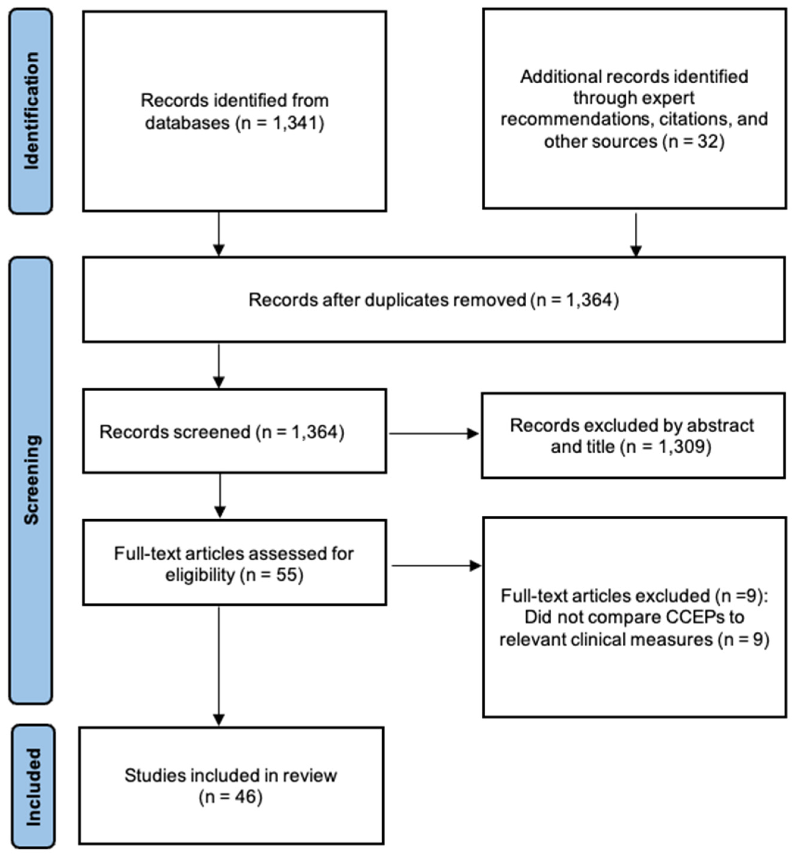
Figure 2.
Overview of cerebro-cerebral evoked potential studies. Summary of different stimulation and recording modalities used to evoke and measure CCEPs. Characteristic CCEP waveform examples are shown next to each experimental paradigm. Top-right: Categorization of 46 identified studies by clinical indication and stimulation/recording modality pair. IS: invasive stimulation. NS: non-invasive stimulation. IR: invasive recording. NR: non-invasive recording.
Figure 2.
Overview of cerebro-cerebral evoked potential studies. Summary of different stimulation and recording modalities used to evoke and measure CCEPs. Characteristic CCEP waveform examples are shown next to each experimental paradigm. Top-right: Categorization of 46 identified studies by clinical indication and stimulation/recording modality pair. IS: invasive stimulation. NS: non-invasive stimulation. IR: invasive recording. NR: non-invasive recording.
Disclaimer/Publisher’s Note: The statements, opinions and data contained in all publications are solely those of the individual author(s) and contributor(s) and not of MDPI and/or the editor(s). MDPI and/or the editor(s) disclaim responsibility for any injury to people or property resulting from any ideas, methods, instructions or products referred to in the content. |
© 2026 by the authors. Licensee MDPI, Basel, Switzerland. This article is an open access article distributed under the terms and conditions of the Creative Commons Attribution (CC BY) license.
Copyright: This open access article is published under a Creative Commons CC BY 4.0 license, which permit the free download, distribution, and reuse, provided that the author and preprint are cited in any reuse.


