

Submitted:
22 May 2026
Posted:
25 May 2026
You are already at the latest version
Abstract
Background and purpose Persistent Post-Concussive Symptoms (PPCS) develop in 15-20% of individuals following mild traumatic brain injury (mTBI), representing a global health burden. This open-label cohort study investigated the effects of individualized, algorithm-based intermittent hypoxic-hyperoxic conditioning (IHHC) on pain and health-related quality of life (QoL) in PPCS patients.
Method A total of 158 consecutive patients (70% female; mean age 40.8 years; mean symptom duration 28.5 ± 21.4 months) received IHHC protocols tailored to symptom duration, baseline pain, QoL, and demographics, and was followed up 6-weeks post-treatment initiation. The protocols consisted of a median of 7 sessions (mean 9.1, range 3-35), each comprising intermittent cycles of 3-8 minutes of hypoxia (FiO2 8.5-13%) and 1-3 minutes of hyperoxia (FiO2 34-36%). 44 of the 158 patients completed the 6-month questionnaire, constituting a 6-month follow-up cohort. Pain intensity (NRS) and QoL (SF-36) were assessed at baseline, 6 weeks, and 6 months.
Results Baseline SF-36 scores were 38.9-40.5% below the Danish population norms. SF-36 scores (8 domains) increased by 23.6% (p< 0.0001) at 6 weeks and 33.4% (p< 0.0001) at 6 months. Physical (PCS) and Mental (MCS) Component Summary scores improved sig-nificantly in both cohorts; the largest gain was MCS at 6 months (+8.06 points; p< 0.0001). At baseline, 69.0% and 77.2% of patients reported pain with an intensity NRS-score ≥ 3 during rest or activity, respectively, and headache was the predominant pain source (81.7% of patients with NRS ≥ 3). Pain intensity decreased by 24.0% and 21.0% (p< 0.001) at 6 weeks, and by 44.4% and 41.3% (p< 0.001) at 6 months during rest and activity, re-spectively.
Conclusion Individualized IHHC was associated with reduced headache-associated pain and improved QoL in PPCS patients. Controlled trials are warranted to confirm these findings.
Keywords:
persistent post-concussive symptoms
; mild traumatic brain injury
; mTBI
; PCS
; concussion
; intermittent hypoxic conditioning
; acute hypoxic conditioning
; long-term sequalae
; headache
; migraine
1. Introduction
Mild traumatic brain injury (mTBI), commonly referred to as concussion and commotio cerebri, is the most common neurological disorder worldwide, surpassing stroke, Alzheimer’s disease, and Parkinson’s disease in incidence [1]. While the prognosis for mTBI is generally favorable, a subset of patients develops persistent symptoms, often classified as persistent post-concussive symptoms (PPCS), post-concussion syndrome, or mild neurocognitive disorder (NCD) due to TBI [2,3,4,5]. A 2023 systematic review and meta-analysis estimated that the prevalence of PPCS after mTBI was 16% up to six months after initial presentation to emergency departments or trauma centers [6]. Additionally, a Swedish study reported that 12% of patients met the criteria for PPCS one-year post-injury [8]. Given that over half of the global population is projected to sustain at least one TBI in their lifetime [9,10], PPCS represents a significant public health challenge.
The symptomatic burden of PPCS can be substantial. The TRACK-TBI study reported a mean of nine symptoms at 12 months post-injury, as assessed by the Rivermead Post-Concussion Symptoms Questionnaire (RPQ) [11]. PPCS symptoms can be broadly categorized into physical (e.g. headaches, dizziness, nausea, sleep disturbances, fatigue, visual problems, and sensitivity to light and noise), cognitive (e.g. impaired memory, concentration, and slowed thinking), and mental (e.g. irritability, depression, anxiety, and frustration) [11,12]. The persistent symptomatic burden significantly reduces health-related quality of life (QoL) in both adults [13] and children [14], decreases life satisfaction [15], and impairs return to work [15,16,17,18], contributing to substantial personal and societal costs.
Development of persistent post-concussive symptoms (PPCS) has been proposed to be the result of biological, psychological, social, and environmental factors [1]. Of biological hypotheses axonal injury [19], neuro-inflammation [20], altered neurotransmission [21], oxidative stress [21,22], mitochondrial dysfunction [23], disruption of blood-brain barrier [24,25], and altered cerebral blood flow [26,27,28] have been proposed as underlying factors in different studies. These alterations might result in dysautonomia (especially during non-rest states) [29,30], pituitary dysfunction [31,32,33], impaired glymphatic drainage [34,35], neuronal loss [36], reduced brain functional connectivity [37], and altered neurometabolism [38]. Although the underlying mechanisms remain incompletely understood, the persistence of symptoms and high degree of treatment resistance following mTBI are likely driven by a heterogeneous and multifactorial pathophysiology.
Exposure to controlled sublethal levels of hypoxia, known as hypoxic conditioning, can increase resilience and promote recovery. In particular, interval-based hypoxia known as Intermittent Hypoxic Conditioning (IHC), which involves alternating periods of hypoxia (FiO2 = 9-13%) with normoxia (FiO2 = 21%) or hyperoxia (FiO2 = 30-40%) (defined as Intermittent Hypoxic Conditioning (IHHC)), has demonstrated therapeutic potential across various pathologies [39,40,41]. Clinical trials have explored the effects of IHC in human diseases of the nervous system [40]-[47], cardiovascular system [48]-[62], respiratory system [63]-[67], and musculoskeletal system [44,46,47,68]. While no other studies to our knowledge have investigated the effects of this therapy on patients with PPCS, IHC have shown potential in reducing migraines [44] and dizziness [45], and improving quality of life [44,56,59,77], fatigue and exercise tolerance [46,58,63,64,66,77], blood flow [48,50,53,56]-[58,60]-[62], cognitive performance [42,43], and autonomic nervous regulation [62,65] in several other patient groups.
The underlying mechanisms have been extensively studied in cellular and animal models, with evidence suggesting that IHC/IHHC exerts its effects through reducing inflammation, restoring mitochondrial function, improving redox balance, modulating autonomic regulation, enhancing vascular function, and stimulating stem cell activity and tissue regeneration [39,69]. Given the overlap between the proposed pathophysiological mechanisms of PPCS and the biological effects of IHC/IHHC, we hypothesize that these therapies may offer a promising therapeutic approach for addressing the underlying causes driving persistent PPCS.
An appropriate hypoxic stimulus may be crucial for achieving beneficial treatment outcomes. Intermittent hypoxic-hyperoxic conditioning (IHHC) can be individualized using algorithm-based protocols to deliver the optimal hypoxic stimulus for each patient. The aim of the present study was to evaluate the effect of individualized IHHC on quality of life and headache-associated pain in patients suffering from long-term sequelae of mTBI.
2. Methods
2.1. Study Design
This study was conducted as an open-label cohort study at five medical clinics across Denmark. SANA® is a medical company specializing in individualized intermittent hypoxic/hyperoxic conditioning (IHHC), and all treatments were designed and administered by their medical staff. Prior to treatment initiation, all patients underwent a structured medical consultation with a physician, either in person or by telephone, including a comprehensive review of medical history.
Eligible patients were those experiencing persistent symptoms following one or more episodes of mild traumatic brain injury (mTBI), with the most recent injury sustained at least three months before treatment initiation. Minors (<18 years) could be included following individualized evaluation and with written consent from a parent or legal guardian. Exclusion criteria included pregnancy or a current diagnosis of malignant disease.
2.2. Data Collection and Questionnaires
Patients completed an online pre-treatment questionnaire 1 to 10 days before their first treatment session. Collected data included demographics (age, sex, height, weight), occupation, work absence (yes/no), and clinical details such as symptomatology, symptom duration, and prior treatments. Pain levels were assessed using the Numeric Rating Scale (NRS) for both rest and activity over the previous week (0 = no pain, 10 = worst imaginable pain). Additionally, patients completed the Short Form-36 (SF-36) health-related quality-of-life questionnaire. The diagnosis of PPCS was determined by the physician based on medical history and symptomatology.
2.3. Treatment Protocol
The SANA® Therapy sessions were administered with patients seated in a reclining chair and breathing through a tightly fitted oxygen mask. The IHHC protocol was personalized via an algorithm (version 1.0) developed from data on 756 patients previously treated under physician supervision. The algorithm-tailored session parameters included the duration of intervals, the total hypoxic exposure, and the target oxygen saturation for each session, based on the patient-specific factors as symptom severity, baseline pain levels (NRS), and SF-36 scores.
Each session comprised hypoxic intervals (3–8 minutes) followed by hyperoxic intervals (1–3 minutes). Inspiratory oxygen concentrations were maintained at 8.5–13% during hypoxia and 34–36% during hyperoxia. A pulse oximeter continuously monitored oxygen saturation, targeting levels between 77% and 84% determined by the algorithm. Session durations ranged from 18 to 42 minutes, with the total number of sessions determined by symptom progression. The interval between sessions varied according to algorithmic recommendations, typically starting with an interval of 7 days between the first three sessions, then extending to 10 to 21 days between subsequent treatments. Treatments were administered using certified medical hypoxicators (Hypoxbreath®, INVATIO, Hannover, Germany and CellOxy, TUR, Rostock, Germany).
2.4. Patient Inclusion and Follow-Up
Data were sourced from the SANA® database, comprising all patients treated at SANA® Clinics in Denmark between January 1, 2020, and October 31, 2025, with a minimum follow-up period of six-months. Patients were either self-referred or referred by insurance companies for rehabilitation. Inclusion required a minimum symptom duration of three months. Patients who completed less than three sessions or whose symptoms lasted less than three months were excluded from the statistical analysis.
Follow-up questionnaires were sent and completed online at six weeks and six months post-treatment initiation. Each patient received a single questionnaire per time point, and no reminder emails were sent. Responses at these intervals were analyzed to assess treatment efficacy, and patients who completed the respective questionnaires were included in the statistical analysis for those time points.
2.5. Statistical Analysis
Data were analyzed using Prism 10 (Version 10.6.0, GraphPad Software LLC). Normal distribution was assessed via QQ-plots of residuals. Descriptive statistics summarized patient demographics, symptom duration, and baseline measures. Group comparisons of demographic variables were conducted using one-way ANOVA with Sidak’s multiple comparisons correction where applicable. The Chi-square test was used for categorical demographic comparisons (e.g., sex distribution).
Changes in health-related quality of life (SF-36 scores) were evaluated using paired t-tests for within-cohort change (baseline to follow-up) with Sidak’s correction across the eight SF-36 domains. As a HRQoL summary, Physical Component Summary (PCS) and Mental Component Summary (MCS) scores were calculated using the Ware 1994 algorithm with US 1990 norm-based factor weights [70], which has been shown to be highly equivalent to country-specific algorithms in Danish data[71]. As a secondary summary, the SF-36 total score was computed as the sum of the eight domains (health transition (HT) excluded, as HT is a single transition item rather than a domain). Between-cohort baseline comparisons used Welch’s two-sample t-test. Pain scores (NRS, 0–10) were analyzed for patients reporting an initial pain level of ≥3 at rest or during activity. One-way ANOVA with Sidak’s correction was applied for multiple comparisons within each cohort.
A significance threshold of P < 0.05 was applied to all statistical analyses. Data were reported as means with 95% confidence intervals (CI), expressed both as absolute values and percentages relative to baseline, to assess the precision of the estimates.
2.5. Ethics
The study was approved by the local ethics committee under the Danish National Committee of Research Ethics (#1-10-72-274-21). The study was conducted in accordance with the Declaration of Helsinki, and all participant data were handled in compliance with the EU General Data Protection Regulation (GDPR).
3. Results
3.1. Patients and Treatment
A total of 553 patients with persistent post-concussive symptoms (PPCS) were identified in the SANA® consecutive database in the inclusion period. 19 patients were excluded due to symptom duration of less than three months, and three patients were excluded due to receiving less than three treatments (Figure 1). The first criterion was included to investigate the effects of IHC on chronic sequalae of mTBIs. In the remaining cohort, 158 patients completed the 6-week follow-up questionnaire, and were included in this study, constituting a 6-week follow-up cohort, with a mean age of 40.8 years (range: 11–79 years). The female-to-male ratio was 70:28 (with 2 patients of unspecified sex), and the mean symptom duration was 28.5 months. Sick leave from work or school at baseline was reported by 49.4% of patients. The 6-month follow-up questionnaire was completed by 44 patients, yielding a response rate of 27.8%. This formed a 6-week follow-up cohort (n=158) and a 6-month follow-up cohort (n=44) for subsequent paired analyses (Figure 1).
Group demographics are presented in Table 1. There was no significant difference between groups in age, sex, anthropometrics, symptom duration, or number of prior treatments. Sick leave at baseline were the only measured parameter where there was significant difference between groups (49.4% vs. 31.8%). Patients received a mean of 9.07 treatment sessions in the 6-week follow-up cohort and 9.86 in the 6-month follow-up cohort (median 7, range 3–35); 61.4% received between 4 and 8 sessions.
Prior to SANA® therapy, 98.7% of patients in the 6-week follow-up cohort reported that they had tried other treatment modalities with an average of 5.23 treatment types (median 5, range 0-20). The most common attempted treatment was physiotherapy (75.9%) and non-prescription analgesics (69.0%). A list of the most common prior treatments is provided in Table 2.
The most frequently noted side effects were mild headaches and tiredness that persisted for 12–24 hours. These symptoms generally subsided after the first few sessions as patients became accustomed to the hypoxic exposure. No serious adverse reactions were observed.
3.2. Pain
In the 6-week follow-up cohort, 86.1% of patients reported any pain with an NRS-score of above 0 at baseline. Subsequent pain analysis included patients with a baseline Numeric Rating Scale (NRS) score of ≥3 at rest and/or during activity. At baseline, 109 patients (69.0%) in the 6-week follow-up cohort reported pain with NRS ≥3 at rest with a mean intensity of 5.41 [95% CI, 5.09; 5.74]. During activity, 122 patients (77.2%) reported pain with NRS ≥3 with a mean intensity of 6.05 [95% CI, 5.70; 6.39]. Pain levels were significantly higher during activity compared to rest at baseline in both the 6-week and the 6-month cohorts (p < 0.01). In the 6-week follow-up cohort at baseline, 81.7% reported headache as a pain source, 50.0% neck pain, and 6.7% shoulder pain. Regardless of NRS-score, 80.4% of patients reported headache as a post-concussive symptom.
From baseline to the 6-week follow-up, pain at rest decreased by 1.30 NRS-points [95% CI, 0.90; 1.71] (mean pain reduction of 24.0%; p < 0.001), and pain during activity decreased by 1.27 points [95% CI, 0.88; 1.66] (mean pain reduction of 21.0%; p < 0.001). At the 6-month follow-up, pain was reduced at rest by 2.30 NRS-points [95% CI, 1.50; 3.11] (mean pain reduction of 44.4%; p < 0.001; n = 33) and during activity by 2.57 points [95% CI, 1.69; 3.45] (mean pain reduction of 41.3%; p < 0.001; n = 35) (Figure 2). Pain reduction was significantly greater at the 6-month than at the 6-week follow-up at rest (Δ = –1.00 NRS-points; p = 0.035) and during activity (Δ = –1.30 NRS-points; p = 0.009).
3.3. Quality of Life
Quality of life was evaluated using the SF-36 health survey (Table 4), which consists of 36 questions that measure 8 health domains: physical functioning, role-physical, bodily pain, general health, vitality, social functioning, role-emotional, and mental health. An additional measure, self-reported health transition over the past year, was collected but not included in the total score. Scores for each domain range from 0 to 100, with higher scores indicating better health [56].
At baseline, values were generally low, with average total scores of 384 in the 6-week follow-up cohort and 394 in the 6-month follow-up cohort. For comparison, the total SF-36 score for the Danish background population has been estimated to be 645 [74]. The lowest baseline scores were observed in the domains role-physical (scores of 14.2 and 12.5 in the 6-week and 6-month cohort, respectively) and vitality (scores of 28.1 and 28.3, respectively). There were no significant differences between the 6-week and 6-month follow-up cohorts at baseline in any SF-36 domain (Welch’s two-sample t-test, all p > 0.05).
Significant improvements were observed in all domains from baseline to the 6-week follow-up. In the 6-month follow-up group, significant improvements were seen in all domains except for the domain general health (Figure 3). Using the total score as a surrogate for overall quality of life, improvements from baseline of 90.5 points (23.6%) (p=5.6x10⁻¹⁷) at 6 weeks post-treatment initiation and 131.7 points (33.4%) (p=9.0×10⁻⁷) at 6 months post-treatment initiation were observed.
To assess the patients’ self-perceived evolution in health status, the domain health transition was included. Health transition scores below 50 indicate a perceived decline in health compared to one year ago, a score of 50 signifies no change, and a score above 50 reflects an improvement in health over the past year. Health transition scores improved significantly from a score below 50 at baseline to above 50 at the 6-week and 6-month follow-ups (p < 0.001). The mean health transition score increased from 42.2 at baseline to 58.2 at the 6-week follow-up, with a mean increase of 16.0 points [11.9; 20.1] (p < 0.001). In the 6-month follow up cohort, the health transition score increased with 19.3 points [11.8;26.9] (p < 0.001) from 47.2 to 66.5.
3.4. Physical and Mental Component Summaries
To complement the per-domain SF-36 analysis, Physical Component Summary (PCS) and Mental Component Summary (MCS) scores were computed. PCS and MCS are weighted, summarized scores of the eight SF-36 domains and are interpreted relative to U.S. population norms of 50, with lower scores reflecting greater impairment [70]. The Danish population norms have been calculated to 51.8 and 56.1 for PCS and MCS, respectively [72]. A change of approximately 2–5 points in SF-36 PCS or MCS scores is generally considered clinically meaningful [75,76].
In the 6-week follow-up cohort, PCS improved from 39.05 ± 8.63 at baseline to 41.4 ± 8.81 at 6 weeks (mean change +2.39, 95% CI 1.23;3.55, p = 8.7×10⁻⁵). MCS improved from 38.6 ± 11.0 to 45.3 ± 10.5 (mean change +6.74, 95% CI 5.09;8.39, p = 2.6×10⁻¹³). In the 6-month follow-up cohort, PCS improved from 37.9 ± 7.83 to 43.1 ± 9.06 (mean change +5.26, 95% CI 3.27 to 7.25; p = 5.5×10⁻⁶). MCS improved from 41.0 ± 10.2 to 48.8 ± 10.1 (mean change +7.78, 95% CI 4.22 to 11.34; p = 1.0×10⁻⁴). At baseline, the 6-week cohort lay approximately 1.6 SD below the Danish PCS norm and 1.7 SD below the MCS norm, consistent with the reduced physical and mental health burden expected in this patient group.
4. Discussion
Each year, fifty-five million individuals are estimated to sustain a mild traumatic brain injury globally [10]. mTBIs have often been referred to as the “silent epidemic”, and while it has the highest incidence of neurological diseases worldwide [1], much remains unknown in the pathophysiological understanding and in the treatment of the disease and its sequelae. This study presents a potential treatment option for patients suffering from long-term sequelae after mild traumatic brain injuries. This current study investigated the effects of algorithm-based, personalized intermittent hypoxia-hyperoxia conditioning (IHHC) in a cohort of 158 PPCS-patients with a mean symptom duration of 28.5 months. At baseline, the cohort had a 40.5% decreased health-related quality-of-life score measured by SF-36, when compared to the general Danish population [74]. Six weeks and six months after a treatment initiation with IHHC, health-related quality of life was significantly increased by 23.6% and 33.4%, respectively. Notably, the SF-36 mental component (MCS) improved from a level corresponding to moderate-to-severe impairment at baseline to only mildly reduced function at the 6-month follow-up, a clinically meaningful shift that reflects a substantial change relative to the general population norm. At baseline, 69.0% of patient experienced pain with an NRS-score ≥3 either at rest or during activity with headache as the largely predominant source of pain. After the treatment regiment, pain intensity was reduced significantly by 19.5-44.4%.
Post-traumatic headache (PTH) is a common sequela of mTBI. In the current cohort, 81.7% of patients reported PTH with an intensity of NRS ≥3, which is higher than prior studies estimating a headache prevalence of 34-58% one year post-trauma [11,12,77]. This might reflect SANA®’s primary focus on pain patients. PTH most frequently presents as a migraine-like headache, followed by a tension-type phenotype [78,79], with evidence suggesting shared pathophysiological mechanisms between migraine and post-traumatic migraine-like headache [80,81]. The pathophysiology of PTH may involve a) impaired descending pain modulation, b) neurometabolic changes, c) cortical spreading depression, d) calcitonin gene-related peptide (CGRP)-dependent mechanisms, and e) neuroinflammation [81]. Imaging studies have revealed altered cerebral blood flow (CBF) following mTBIs, correlating with post-concussive symptoms [1,82,83,84]. These CBF changes may drive the functional remodeling, exacerbate the metabolic crises, and activate the trigeminal sensory system [81] leading to headache and other PPCS symptoms.
Early treatment may be crucial in preventing the chronification of symptoms. Persistent post-traumatic headache (lasting more than 3 months) is largely attributed to central sensitization, which arises from continuous transmission from first-order neurons to second- and third-order neurons within the central nervous system [85]. Interventions that reduce initial pain transmission may curb central sensitization. Notably, targeted therapies against CGRP signaling have been shown to prevent allodynia in concussed rodents [86]. CGRP is also believed to be central in the signaling of nausea, photo- and phonophobia [87,88,89]. A 2020 randomized controlled trial found that an 8-week IHC regimen reduced migraine frequency, pain, depression, and anxiety while improving quality of life in migraine patients [44]. This was associated with decreased CBF velocity in the middle cerebral and basilar arteries accompanied by lower VEGF and CGRP levels [44]. Together it can be hypothesized, that not only may IHC be a potential treatment of PPCS, but the use of IHC may also be beneficial in the early phases of traumatic brain injury (TBI) to mitigate persistent symptoms by reducing CGRP activity. In this current study, the mean symptom duration was 28.5 months, reflecting the chronic and persistent nature of symptoms in the investigated cohort. Future research should explore the potential of IHC in the early stages of mTBIs to prevent symptom chronification.
This study has several limitations. First, the findings rely exclusively on self-reported questionnaires, which carry inherent biases. However, in the context of long-term sequelae after mTBI – where validated objective biomarkers are lacking – patient-reported outcomes remain among the most relevant indicators of treatment efficacy. Another limitation is the lack of use of headache-specific questionnaires to quantify headache frequency, intensity, or migraine attacks. Future studies on IHC’s effects on post-concussive symptoms should therefore include headache-specific questionnaires as well as the Rivermead Post-Concussion Symptoms Questionnaire, as PPCS encompasses a broad range of symptoms beyond headache.
Second, the retrospective design precluded blinding and the inclusion of a placebo-controlled group, limiting causal inference and raising the possibility that the observed improvements may reflect placebo effects. However, it is noteworthy that the ‘health transition’ scores of the SF-36 evaluation in both cohorts were at or below 50, indicating respectively health stagnation or decline over the preceding year. Participants had a mean symptom duration of 28.5-33.2 months and had previously attempted an average of 5.16-5.23 treatment modalities before initiating SANA® Therapy, underscoring the chronic and treatment-resistant nature of symptoms in this cohort. All patients were either self-paying or referred by an insurance company with also may have attributed to placebo responses due to self-justification and expectations.
Finally, the low follow-up rates in the 6-week and 6-month cohorts could have been increased by sending reminders to participants, and it remains unknown if the respondents at follow-up disproportionately represented patients who improved or those who did not.
One of the main strengths of this study is its heterogeneous cohort, which enhances the external validity of the findings. By applying broad inclusion criteria, the study population reflects the diverse and multifaceted symptomatology of patients with PPCS, encompassing psychological, physical, and social dimensions. This real-world representativeness increases the generalizability of the results to the broader population of patients with persistent post-concussive symptoms. The symptom burden in PPCS likely arise from a complex interplay of genetic, physical, psychological, and social factors [1]. Consequently, complete resolution of PPCS may require individualized assessment and interdisciplinary treatment approaches [90] combining relevant elements such as physiotherapy [91], cognitive behavioral therapy and psychoeducation [92,93], reposition maneuvers for benign paroxysmal positional vertigo [94], aerobic training [91,95], or pharmacological treatment [80,96]. Future studies should investigate whether combining such modalities with IHHC provides greater benefit than IHHC alone. Both efficacy and cost-effectiveness of these treatments must be evaluated in order to resolve PPCS for the most patients possible.
5. Conclusions
In conclusion, this study demonstrates the potential of individualized, algorithm-based IHHC-treatment (SANA® Therapy) in reducing headache-associated pain and improving quality of life in this treatment-resistant PPCS-cohort with a symptom duration of 26.8 to 33.2 months of a stagnant or worsening nature. The findings indicate IHHC’s potential as a clinically relevant and cost-effective treatment-option for PPCS patients.
6. Patents
No patents are disclosed in this manuscript. However, the SANA® Therapy algorithms are proprietary, confidential, and remain the exclusive intellectual property of SANA Medical Systems ApS, Risskov, Denmark.
Author Contributions
Conceptualization, RS, BE and CBF; methodology, RS, BE, LAH, CBF; validation, LAH and CBF; formal analysis, CBF; investigation, RS, BE and CBF.; original draft writing and literature search, JSK.; review and editing, CBF and LAH.; visualization, JSK.; supervision CBF; project administration, CBF. All authors have read and agreed to the published version of the manuscript.
Funding
The project was partly supported by the independent Research Fund Denmark (grant# 10.46540/3165-00221B).
Informed Consent Statement
Written informed consent was obtained from all participants included in the study. For minors, a parental or legal guardian consent was required before treatment initiation.
Data Availability Statement
The datasets analyzed during the current study are available from the corresponding author on reasonable request.
Institutional Review Board Statement
This study was approved by the local ethics committee under the Danish National Committee of Research Ethics (# 1-10-72-274-21) on 24 Mach 2022. The study was conducted in accordance with the Declaration of Helsinki, and all participant data were handled in accordance with the EU General Data Protection Regulation (GDPR).
Acknowledgments
In this section, you can acknowledge any support given which is not covered by the author contribution or funding sections. This may include administrative and technical support, or donations in kind (e.g., materials used for experiments). Where GenAI has been used for purposes such as generating text, data, or graphics, or for study design, data collection, analysis, or interpretation of data, please add “During the preparation of this manuscript/study, the author(s) used [tool name, version information] for the purposes of [description of use]. The authors have reviewed and edited the output and take full responsibility for the content of this publication.”.
Conflicts of Interest
The authors CBF, RS, and BE are co-founders and shareholders of SANA Medical Systems ApS, Risskov, Denmark. JSK receives a salary from SANA Medical Systems for her work as clinical therapist and research assistant. LAH declare she has no financial interests.
Abbreviations
The following abbreviations are used in this manuscript:
| ANOVA | Analysis of Variance |
| BP | Bodily Pain (SF-36 domain) |
| CBF | Cerebral Blood Flow |
| CGRP | Calcitonin Gene-Related Peptide |
| CI | Confidence Interval |
| COPD | Chronic Obstructive Pulmonary Disease |
| DSM-V | Diagnostic and Statistical Manual of Mental Disorders, 5th edition |
| FiO2 | Fraction of Inspired Oxygen |
| GDPR | General Data Protection Regulation |
| GH | General Health (SF-36 domain) |
| HRQoL | Health-Related Quality of Life |
| HT | Health Transition (SF-36 domain) |
| ICD-11 | International Classification of Diseases, 11th Revision |
| IHC | Intermittent Hypoxic Conditioning |
| IHHC | Intermittent Hypoxic-Hyperoxic Conditioning |
| MCS | Mental Component Summary |
| MH | Mental Health (SF-36 domain) |
| mTBI | Mild Traumatic Brain Injury |
| NRS | Numeric Rating Scale |
| PCS | Physical Component Summary |
| PF | Physical Functioning (SF-36 domain) |
| PPCS | Persistent Post-Concussive Symptoms |
| PTH | Post-Traumatic Headache |
| QoL | Quality of Life |
| RE | Role Emotional (SF-36 domain) |
| RP | Role Physical (SF-36 domain) |
| RPQ | Rivermead Post-Concussion Symptoms Questionnaire |
| SD | Standard Deviation |
| SEM | Standard Error of the Mean |
| SF | Social Functioning (SF-36 domain) |
| SF-36 | Short Form-36 Health Survey |
| TBI | Traumatic Brain Injury |
| VEGF | Vascular Endothelial Growth Factor |
| V | Vitality (SF-36 domain) |
| WHO | World Health Organization |
| PCS | Physical Component Summary |
| PF | Physical Functioning (SF-36 domain) |
| PPCS | Persistent Post-Concussive Symptoms |
| PTH | Post-Traumatic Headache |
| QoL | Quality of Life |
| RE | Role Emotional (SF-36 domain) |
| RP | Role Physical (SF-36 domain) |
| RPQ | Rivermead Post-Concussion Symptoms Questionnaire |
| SD | Standard Deviation |
| SEM | Standard Error of the Mean |
| SF | Social Functioning (SF-36 domain) |
| SF-36 | Short Form-36 Health Survey |
| SPECT | Single Photon Emission Computed Tomography |
| TBI | Traumatic Brain Injury |
| VEGF | Vascular Endothelial Growth Factor |
| V | Vitality (SF-36 domain) |
| WHO | World Health Organization |
References
- Maas AIR, Menon DK, Manley GT, Abrams M, Åkerlund C, Andelic N, Aries M, Bashford T, Bell MJ, Bodien YG, et al. Traumatic brain injury: progress and challenges in prevention, clinical care, and research. Lancet Neurol. 2022 Nov;21(11):1004-1060. doi: 10.1016/S1474-4422(22)00309-X. Epub 2022 Sep 29. Erratum in: Lancet Neurol. 2022 Dec;21(12):e10. [CrossRef] [PubMed] [PubMed Central]
- National Academies of Sciences Engineering and Medicine. Traumatic brain injury: a roadmap for accelerating progress. National Academies of Sciences Engineering and Medicine Consensus Study Report, 2022. https://nap.nationalacademies.org/catalog/25394/traumatic-brain-injury-a-roadmap-for-acceleratingprogress (accessed Aug 8, 2025).
- Sørensen AN, [3] HM. Vidensrapport om let hovedtraume, herunder hjernerystelse – Voksne, fuld version. København: Dansk Center for Hjernerystelse; 2024 [cited 2025 Sep 26]. Available from: https://dcfh.dk/wp-content/uploads/2024/03/3k_107256_Fuld-version_Vidensrapport-om-let-hovedtraume-voksne_Finale_1.pdf.
- World Health Organization (WHO). The ICD-11 classification of mental and behavioural disorders: clinical descriptions and diagnostic guidelines. 2019.
- American Psychiatric Association (APA). Diagnostic and Statistical Manual of Mental Disorders, 5th edition (DSM-V). American Psychiatric Association; 2013.
- Cancelliere C, Verville L, Stubbs JL, Yu H, Hincapié CA, Cassidy JD, Wong JJ, Shearer HM, Connell G, Southerst D, Howitt S, Guist B, Silverberg ND. Post-Concussion Symptoms and Disability in Adults With Mild Traumatic Brain Injury: A Systematic Review and Meta-Analysis. J Neurotrauma. 2023 Jun;40(11-12):1045-1059. Epub 2023 Jan 18. [CrossRef] [PubMed]
- Silverberg ND, Iverson GL; ACRM Brain Injury Special Interest Group Mild TBI Task Force members:; Cogan A, Dams-O-Connor K, Delmonico R, Graf MJP, Iaccarino MA, Kajankova M, Kamins J, McCulloch KL, McKinney G, Nagele D, Panenka WJ, et al.. The American Congress of Rehabilitation Medicine Diagnostic Criteria for Mild Traumatic Brain Injury. Arch Phys Med Rehabil. 2023 Aug;104(8):1343-1355. Epub 2023 May 19. [CrossRef] [PubMed]
- Oldenburg C, Lundin A, Edman G, Deboussard CN, Bartfai A. Emotional reserve and prolonged post-concussive symptoms and disability: a Swedish prospective 1-year mild traumatic brain injury cohort study. BMJ Open. 2018 Jul 6;8(7):e020884. [CrossRef] [PubMed] [PubMed Central]
- Maas AIR, Menon DK, Adelson PD, Andelic N, Bell MJ, Belli A, Bragge P, Brazinova A, Büki A, Chesnut RM, et al.; InTBIR Participants and Investigators. Traumatic brain injury: integrated approaches to improve prevention, clinical care, and research. Lancet Neurol. 2017 Dec;16(12):987-1048. Epub 2017 Nov 6. [CrossRef] [PubMed]
- Dewan MC, Rattani A, Gupta S, Baticulon RE, Hung YC, Punchak M, Agrawal A, Adeleye AO, Shrime MG, Rubiano AM, Rosenfeld JV, Park KB. Estimating the global incidence of traumatic brain injury. J Neurosurg. 2018 Apr 27;130(4):1080-1097. [CrossRef] [PubMed]
- Machamer J, Temkin N, Dikmen S, Nelson LD, Barber J, Hwang P, Boase K, Stein MB, Sun X, Giacino J, McCrea MA, Taylor SR, Jain S, Manley G; TRACK-TBI Investigators. Symptom Frequency and Persistence in the First Year after Traumatic Brain Injury: A TRACK-TBI Study. J Neurotrauma. 2022 Mar;39(5-6):358-370. Epub 2022 Feb 9. [CrossRef] [PubMed] [PubMed Central]
- Theadom A, Parag V, Dowell T, McPherson K, Starkey N, Barker-Collo S, Jones K, Ameratunga S, Feigin VL; BIONIC Research Group. Persistent problems 1 year after mild traumatic brain injury: a longitudinal population study in New Zealand. Br J Gen Pract. 2016 Jan;66(642):e16-23. [CrossRef] [PubMed] [PubMed Central]
- Voormolen DC, Polinder S, von Steinbuechel N, Vos PE, Cnossen MC, Haagsma JA. The association between post-concussion symptoms and health-related quality of life in patients with mild traumatic brain injury. Injury. 2019 May;50(5):1068-1074. Epub 2018 Dec 7. [CrossRef] [PubMed]
- Novak Z, Aglipay M, Barrowman N, Yeates KO, Beauchamp MH, Gravel J, Freedman SB, Gagnon I, Gioia G, Boutis K, Burns E, Ledoux AA, Osmond MH, Zemek RL; Pediatric Emergency Research Canada Predicting Persistent Postconcussive Problems in Pediatrics (PERC 5P) Concussion Team. Association of Persistent Postconcussion Symptoms With Pediatric Quality of Life. JAMA Pediatr. 2016 Dec 5;170(12):e162900. Epub 2016 Dec 5. [CrossRef] [PubMed]
- Stålnacke BM. Community integration, social support and life satisfaction in relation to symptoms 3 years after mild traumatic brain injury. Brain Inj. 2007 Aug;21(9):933-42. [CrossRef] [PubMed]
- Chu SY, Tsai YH, Xiao SH, Huang SJ, Yang CC. Quality of return to work in patients with mild traumatic brain injury: a prospective investigation of associations among post-concussion symptoms, neuropsychological functions, working status and stability. Brain Inj. 2017;31(12):1674-1682. Epub 2017 Sep 5. [CrossRef] [PubMed]
- Graff HJ, Siersma V, Møller A, Kragstrup J, Andersen LL, Egerod I, Malá Rytter H. Premorbid risk factors influencing labour market attachment after mild traumatic brain injury: a national register study with long-term follow-up. BMJ Open. 2019 Apr 11;9(4):e027297. [CrossRef] [PubMed] [PubMed Central]
- Graff HJ, Siersma V, Møller A, Waldorff FB, Modin FA, Rytter HM. Labour market attachment dynamics in patients with concussion: a Danish nationwide register-based cohort study. BMC Public Health. 2023;23(1):2493. Published 2023 Dec 13. [CrossRef]
- Johnson VE, Stewart W, Smith DH. Axonal pathology in traumatic brain injury. Exp Neurol. 2013 Aug;246:35-43. Epub 2012 Jan 20. [CrossRef] [PubMed] [PubMed Central]
- Visser K, Koggel M, Blaauw J, van der Horn HJ, Jacobs B, van der Naalt J. Blood-based biomarkers of inflammation in mild traumatic brain injury: A systematic review. Neurosci Biobehav Rev. 2022 Jan;132:154-168. Epub 2021 Nov 23. [CrossRef] [PubMed]
- Joyce JM, Mercier LJ, Stokoe M, La PL, Bell T, Batycky JM, Debert CT, Harris AD. Glutamate, GABA and glutathione in adults with persistent post-concussive symptoms. Neuroimage Clin. 2022;36:103152. Epub 2022 Aug 18. [CrossRef] [PubMed] [PubMed Central]
- Cruz-Haces M, Tang J, Acosta G, Fernandez J, Shi R. Pathological correlations between traumatic brain injury and chronic neurodegenerative diseases. Transl Neurodegener. 2017 Jul 11;6:20. [CrossRef] [PubMed] [PubMed Central]
- Kim S, Han SC, Gallan AJ, Hayes JP. Neurometabolic indicators of mitochondrial dysfunction in repetitive mild traumatic brain injury. Concussion. 2017 Oct 4;2(3):CNC48. [CrossRef] [PubMed] [PubMed Central]
- Yoen H, Yoo RE, Choi SH, Kim E, Oh BM, Yang D, Hwang I, Kang KM, Yun TJ, Kim JH, Sohn CH. Blood-Brain Barrier Disruption in Mild Traumatic Brain Injury Patients with Post-Concussion Syndrome: Evaluation with Region-Based Quantification of Dynamic Contrast-Enhanced MR Imaging Parameters Using Automatic Whole-Brain Segmentation. Korean J Radiol. 2021 Jan;22(1):118-130. Epub 2020 Aug 11. [CrossRef] [PubMed] [PubMed Central]
- Yoo RE, Choi SH, Oh BM, Do Shin S, Lee EJ, Shin DJ, Jo SW, Kang KM, Yun TJ, Kim JH, Sohn CH. Quantitative dynamic contrast-enhanced MR imaging shows widespread blood-brain barrier disruption in mild traumatic brain injury patients with post-concussion syndrome. Eur Radiol. 2019 Mar;29(3):1308-1317. Epub 2018 Jul 31. [CrossRef] [PubMed]
- Brooks BL, Low TA, Plourde V, Virani S, Jadavji Z, MacMaster FP, Barlow KM, Lebel RM, Yeates KO. Cerebral blood flow in children and adolescents several years after concussion. Brain Inj. 2019;33(2):233-241. Epub 2018 Oct 31. [CrossRef] [PubMed]
- Barlow KM, Iyer K, Yan T, Scurfield A, Carlson H, Wang Y. Cerebral Blood Flow Predicts Recovery in Children with Persistent Post-Concussion Symptoms after Mild Traumatic Brain Injury. J Neurotrauma. 2021 Aug 15;38(16):2275-2283. Epub 2021 Feb 3. [CrossRef] [PubMed] [PubMed Central]
- Wong ET, Kapadia A, Krishnamurthy V, Mikulis DJ. Cerebrovascular Reactivity and Concussion. Neuroimaging Clin N Am. 2023 May;33(2):335-342. Epub 2023 Feb 26. [CrossRef] [PubMed]
- Mercier LJ, Batycky J, Campbell C, Schneider K, Smirl J, Debert CT. Autonomic dysfunction in adults following mild traumatic brain injury: A systematic review. NeuroRehabilitation. 2022;50(1):3-32. [CrossRef] [PubMed]
- Pelo R, Suttman E, Fino PC, McFarland MM, Dibble LE, Cortez MM. Autonomic dysfunction and exercise intolerance in concussion: a scoping review. Clin Auton Res. 2023 Apr;33(2):149-163. Epub 2023 Apr 10. [CrossRef] [PubMed] [PubMed Central]
- Eggertsdóttir Claessen LÓ, Kristjánsdóttir H, Jónsdóttir MK, Lund SH, Unnsteinsdóttir Kristensen I, Sigurjónsdóttir HÁ. Pituitary dysfunction following mild traumatic brain injury in female athletes. Endocr Connect. 2024 Jan 16;13(2):e230363. [CrossRef] [PubMed] [PubMed Central]
- Snook ML, Henry LC, Sanfilippo JS, Zeleznik AJ, Kontos AP. Association of Concussion With Abnormal Menstrual Patterns in Adolescent and Young Women. JAMA Pediatr. 2017 Sep 1;171(9):879-886. [CrossRef] [PubMed] [PubMed Central]
- Pavlovic D, Pekic S, Stojanovic M, Popovic V. Traumatic brain injury: neuropathological, neurocognitive and neurobehavioral sequelae. Pituitary. 2019 Jun;22(3):270-282. [CrossRef] [PubMed]
- Braun M, Sevao M, Keil SA, Gino E, Wang MX, Lee J, Haveliwala MA, Klein E, Agarwal S, Pedersen T, Rhodes CH, Jansson D, Cook D, Peskind E, Perl DP, Piantino J, Schindler AG, Iliff JJ. Macroscopic changes in aquaporin-4 underlie blast traumatic brain injury-related impairment in glymphatic function. Brain. 2024 Jun 3;147(6):2214-2229. [CrossRef] [PubMed] [PubMed Central]
- Zhuo J, Raghavan P, Li J, Roys S, Njonkou Tchoquessi RL, Chen H, Wickwire EM, Parikh GY, Schwartzbauer GT, Grattan LM, Wang Z, Gullapalli RP, Badjatia N. Longitudinal assessment of glymphatic changes following mild traumatic brain injury: Insights from perivascular space burden and DTI-ALPS imaging. Front Neurol. 2024 Aug 7;15:1443496. [CrossRef] [PubMed] [PubMed Central]
- Woodrow RE, Grossac J, Hong YT, Winzeck S, Geeraerts T, Shah SA, Peattie ARD, Manktelow AE, Outtrim JG, Karakatsanis NA, Schiff ND, Fryer TD, Menon DK, Coles JP, Stamatakis EA. Outcomes and Mechanisms Associated With Selective Thalamic Neuronal Loss in Chronic Traumatic Brain Injury. JAMA Netw Open. 2024 Aug 1;7(8):e2426141. [CrossRef] [PubMed] [PubMed Central]
- Nordin LE, Möller MC, Julin P, Bartfai A, Hashim F, Li TQ. Post mTBI fatigue is associated with abnormal brain functional connectivity. Sci Rep. 2016 Feb 16;6:21183. [CrossRef] [PubMed] [PubMed Central]
- Giza C, Greco T, Prins ML. Concussion: pathophysiology and clinical translation. Handb Clin Neurol. 2018;158:51-61. [CrossRef] [PubMed]
- Burtscher J, Citherlet T, Camacho-Cardenosa A, Camacho-Cardenosa M, Raberin A, Krumm B, Hohenauer E, Egg M, Lichtblau M, Müller J, Rybnikova EA, Gatterer H, Debevec T, Baillieul S, Manferdelli G, Behrendt T, Schega L, Ehrenreich H, Millet GP, Gassmann M, Schwarzer C, Glazachev O, Girard O, Lalande S, Hamlin M, Samaja M, Hüfner K, Burtscher M, Panza G, Mallet RT. Mechanisms underlying the health benefits of intermittent hypoxia conditioning. J Physiol. 2024 Nov;602(21):5757-5783. Epub 2023 Oct 20. [CrossRef] [PubMed]
- Zhang Q, Zhao W, Li S, Ding Y, Wang Y, Ji X. Intermittent Hypoxia Conditioning: A Potential Multi-Organ Protective Therapeutic Strategy. Int J Med Sci. 2023 Sep 18;20(12):1551-1561. [CrossRef] [PubMed] [PubMed Central]
- Behrendt T, Bielitzki R, Behrens M, Herold F, Schega L. Effects of Intermittent Hypoxia-Hyperoxia on Performance- and Health-Related Outcomes in Humans: A Systematic Review. Sports Med Open. 2022 May 31;8(1):70. [CrossRef] [PubMed] [PubMed Central]
- Bayer U, Likar R, Pinter G, Stettner H, Demschar S, Trummer B, Neuwersch S, Glazachev O, Burtscher M. Intermittent hypoxic-hyperoxic training on cognitive performance in geriatric patients. Alzheimers Dement (N Y). 2017 Feb 8;3(1):114-122. [CrossRef] [PubMed] [PubMed Central]
- Wang H, Shi X, Schenck H, Hall JR, Ross SE, Kline GP, Chen S, Mallet RT, Chen P. Intermittent Hypoxia Training for Treating Mild Cognitive Impairment: A Pilot Study. Am J Alzheimers Dis Other Demen. 2020 Jan-Dec;35:1533317519896725. [CrossRef] [PubMed] [PubMed Central]
- Bao X, Liu H, Liu HY, Long Y, Tan JW, Zhu ZM. The effect of intermittent hypoxia training on migraine: a randomized controlled trial. Am J Transl Res. 2020 Jul 15;12(7):4059-4065. [PubMed] [PubMed Central]
- Bao X, Tan JW, Long Y, Liu H, Liu HY. Effect of Intermittent Hypoxia Training for Dizziness: A Randomized Controlled Trial. J Sport Rehabil. 2019 Aug 1;28(6):540-543. [CrossRef] [PubMed]
- Zrzavy T, Pfitzner A, Flachenecker P, Rommer P, Zettl UK. Effects of normobaric hypoxic endurance training on fatigue in patients with multiple sclerosis: a randomized prospective pilot study. J Neurol. 2021 Dec;268(12):4809-4815. Epub 2021 May 18. [CrossRef] [PubMed] [PubMed Central]
- Sajjadi E, Seven YB, Ehrbar JG, Wymer JP, Mitchell GS, Smith BK. Acute intermittent hypoxia and respiratory muscle recruitment in people with amyotrophic lateral sclerosis: A preliminary study. Exp Neurol. 2022 Jan;347:113890. Epub 2021 Oct 6. [CrossRef] [PubMed] [PubMed Central]
- Bestavashvili A, Glazachev O, Bestavashvili A, Suvorov A, Zhang Y, Zhang X, Rozhkov A, Kuznetsova N, Pavlov C, Glushenkov D, Kopylov P. Intermittent Hypoxic-Hyperoxic Exposures Effects in Patients with Metabolic Syndrome: Correction of Cardiovascular and Metabolic Profile. Biomedicines. 2022 Feb 28;10(3):566. [CrossRef] [PubMed] [PubMed Central]
- Afina AB, Oleg SG, Alexander AB, Ines D, Alexander Yu S, Nikita VV, Denis ST, Daria GG, Zhang Y, Chavdar SP, Dmitriy VG, Elena AS, Irina VK, Philippe Yu K. The Effects of Intermittent Hypoxic-Hyperoxic Exposures on Lipid Profile and Inflammation in Patients With Metabolic Syndrome. Front Cardiovasc Med. 2021 Aug 27;8:700826. [CrossRef] [PubMed] [PubMed Central]
- Costalat G, Lemaitre F, Tobin B, Renshaw G. Intermittent hypoxia revisited: a promising non-pharmaceutical strategy to reduce cardio-metabolic risk factors? Sleep Breath. 2018 Mar;22(1):267-271. Epub 2017 Feb 2. [CrossRef] [PubMed]
- Duennwald T, Gatterer H, Groop PH, Burtscher M, Bernardi L. Effects of a single bout of interval hypoxia on cardiorespiratory control and blood glucose in patients with type 2 diabetes. Diabetes Care. 2013 Aug;36(8):2183-9. Epub 2013 Mar 27. [CrossRef] [PubMed] [PubMed Central]
- Serebrovska TV, Grib ON, Portnichenko VI, Serebrovska ZO, Egorov E, Shatylo VB. Intermittent Hypoxia/Hyperoxia Versus Intermittent Hypoxia/Normoxia: Comparative Study in Prediabetes. High Alt Med Biol. 2019 Dec;20(4):383-391. Epub 2019 Oct 7. [CrossRef] [PubMed]
- Serebrovska TV, Portnychenko AG, Drevytska TI, Portnichenko VI, Xi L, Egorov E, Gavalko AV, Naskalova S, Chizhova V, Shatylo VB. Intermittent hypoxia training in prediabetes patients: Beneficial effects on glucose homeostasis, hypoxia tolerance and gene expression. Exp Biol Med (Maywood). 2017 Sep;242(15):1542-1552. Epub 2017 Jul 31. [CrossRef] [PubMed] [PubMed Central]
- Serebrovska TV, Portnychenko AG, Portnichenko VI, Xi L, Egorov E, Antoniuk-Shcheglova I, Naskalova S, Shatylo VB. Effects of intermittent hypoxia training on leukocyte pyruvate dehydrogenase kinase 1 (PDK-1) mRNA expression and blood insulin level in prediabetes patients. Eur J Appl Physiol. 2019 Mar;119(3):813-823. Epub 2019 Jan 30. [CrossRef] [PubMed]
- Serebrovskaya TV, Xi L. Intermittent hypoxia training as non-pharmacologic therapy for cardiovascular diseases: Practical analysis on methods and equipment. Exp Biol Med (Maywood). 2016 Sep;241(15):1708-23. Epub 2016 Jul 12. [CrossRef] [PubMed] [PubMed Central]
- Glazachev O, Kopylov P, Susta D, Dudnik E, Zagaynaya E. Adaptations following an intermittent hypoxia-hyperoxia training in coronary artery disease patients: a controlled study. Clin Cardiol. 2017 Jun;40(6):370-376. Epub 2017 Mar 21. [CrossRef] [PubMed] [PubMed Central]
- del Pilar Valle M, García-Godos F, Woolcott OO, Marticorena JM, Rodríguez V, Gutiérrez I, Fernández-Dávila L, Contreras A, Valdivia L, Robles J, Marticorena EA. Improvement of myocardial perfusion in coronary patients after intermittent hypobaric hypoxia. J Nucl Cardiol. 2006 Jan-Feb;13(1):69-74. [CrossRef] [PubMed]
- Burtscher M, Pachinger O, Ehrenbourg I, Mitterbauer G, Faulhaber M, Pühringer R, Tkatchouk E. Intermittent hypoxia increases exercise tolerance in elderly men with and without coronary artery disease. Int J Cardiol. 2004 Aug;96(2):247-54. [CrossRef] [PubMed]
- Saeed O, Bhatia V, Formica P, Browne A, Aldrich TK, Shin JJ, Maybaum S. Improved exercise performance and skeletal muscle strength after simulated altitude exposure: a novel approach for patients with chronic heart failure. J Card Fail. 2012 May;18(5):387-91. Epub 2012 Mar 10. [CrossRef] [PubMed]
- Muangritdech N, Hamlin MJ, Sawanyawisuth K, Prajumwongs P, Saengjan W, Wonnabussapawich P, Manimmanakorn N, Manimmanakorn A. Hypoxic training improves blood pressure, nitric oxide and hypoxia-inducible factor-1 alpha in hypertensive patients. Eur J Appl Physiol. 2020 Aug;120(8):1815-1826. Epub 2020 Jun 10. [CrossRef] [PubMed]
- Lyamina NP, Lyamina SV, Senchiknin VN, Mallet RT, Downey HF, Manukhina EB. Normobaric hypoxia conditioning reduces blood pressure and normalizes nitric oxide synthesis in patients with arterial hypertension. J Hypertens. 2011 Nov;29(11):2265-72. [CrossRef] [PubMed]
- Panza GS, Puri S, Lin HS, Badr MS, Mateika JH. Daily Exposure to Mild Intermittent Hypoxia Reduces Blood Pressure in Male Patients with Obstructive Sleep Apnea and Hypertension. Am J Respir Crit Care Med. 2022 Apr 15;205(8):949-958. [CrossRef] [PubMed] [PubMed Central]
- Uzun AB, Iliescu M, Stanciu LE, Nedelcu AD, Petcu A, Popescu MN, Beiu C, Petcu LC, Tofolean DE. The Impact of Intermittent Hypoxia-Hyperoxia Therapy on Metabolism and Respiratory System in Obese Patients as Part of Comprehensive Medical Rehabilitation. Cureus. 2024 Oct 14;16(10):e71501. [CrossRef] [PubMed] [PubMed Central]
- Burtscher M, Haider T, Domej W, Linser T, Gatterer H, Faulhaber M, Pocecco E, Ehrenburg I, Tkatchuk E, Koch R, Bernardi L. Intermittent hypoxia increases exercise tolerance in patients at risk for or with mild COPD. Respir Physiol Neurobiol. 2009 Jan 1;165(1):97-103. Epub 2008 Nov 1. [CrossRef] [PubMed]
- Haider T, Casucci G, Linser T, Faulhaber M, Gatterer H, Ott G, Linser A, Ehrenbourg I, Tkatchouk E, Burtscher M, Bernardi L. Interval hypoxic training improves autonomic cardiovascular and respiratory control in patients with mild chronic obstructive pulmonary disease. J Hypertens. 2009 Aug;27(8):1648-54. [CrossRef] [PubMed]
- Zha S, Liu X, Yao Y, He Y, Wang Y, Zhang Q, Zhang J, Yi Y, Xiao R, Hu K. Short-term intermittent hypoxia exposure for dyspnea and fatigue in post-acute sequelae of COVID-19: A randomized controlled study. Respir Med. 2024 Oct;232:107763. Epub 2024 Aug 8. [CrossRef] [PubMed]
- Doehner W, Fischer A, Alimi B, Muhar J, Springer J, Altmann C, Schueller PO. Intermittent Hypoxic-Hyperoxic Training During Inpatient Rehabilitation Improves Exercise Capacity and Functional Outcome in Patients With Long Covid: Results of a Controlled Clinical Pilot Trial. J Cachexia Sarcopenia Muscle. 2024 Dec;15(6):2781-2791. Epub 2024 Nov 1. [CrossRef] [PubMed] [PubMed Central]
- Bogard AT, Pollet AK, Tan AQ. Intermittent hypoxia enhances voluntary activation and reduces performance fatigability during repeated lower limb contractions. J Neurophysiol. 2024 Dec 1;132(6):1717-1728. Epub 2024 Oct 23. [CrossRef] [PubMed] [PubMed Central]
- Yuan H, Liu J, Gu Y, Ji X, Nan G. Intermittent hypoxia conditioning as a potential prevention and treatment strategy for ischemic stroke: Current evidence and future directions. Front Neurosci. 2022 Nov 25;16:1067411. [CrossRef] [PubMed] [PubMed Central]
- Ware, J. E. (1994). SF-36 Physical and Mental Health Summary Scales: A User’s Manual. Boston, MA: The Health Institute, New England Medical Center Hospitals.
- Ware JE Jr, Gandek B, Kosinski M, et al. The equivalence of SF-36 summary health scores estimated using standard and country-specific algorithms in 10 countries: results from the IQOLA Project. International Quality of Life Assessment. J Clin Epidemiol. 1998;51(11):1167-1170. [CrossRef]
- Gundgaard J, Lauridsen J. Decomposition of sources of income-related health inequality applied on SF-36 summary scores: a Danish health survey. Health Qual Life Outcomes. 2006;4:53. Published 2006 Aug 22. [CrossRef]
- Lins L, Carvalho FM. SF-36 total score as a single measure of health-related quality of life: Scoping review. SAGE Open Med. 2016 Oct 4;4:2050312116671725. [CrossRef] [PubMed] [PubMed Central]
- Bjorner JB, Thunedborg K, Kristensen TS, Modvig J, Bech P. The Danish SF-36 Health Survey: translation and preliminary validity studies. J Clin Epidemiol. 1998 Nov;51(11):991-9. [CrossRef] [PubMed]
- Fu V, Weatherall M, McNaughton H. Estimating the minimal clinically important difference for the Physical Component Summary of the Short Form 36 for patients with stroke. J Int Med Res. 2021;49(12):3000605211067902. [CrossRef]
- Ogura K, Yakoub MA, Christ AB, et al. What Are the Minimum Clinically Important Differences in SF-36 Scores in Patients with Orthopaedic Oncologic Conditions?. Clin Orthop Relat Res. 2020;478(9):2148-2158. [CrossRef]
- Hoffman JM, Lucas S, Dikmen S, Temkin N. Clinical Perspectives on Headache After Traumatic Brain Injury. PM R. 2020 Oct;12(10):967-974. Epub 2020 Mar 2. [CrossRef] [PubMed]
- Ashina H, Iljazi A, Al-Khazali HM, Ashina S, Jensen RH, Amin FM, Ashina M, Schytz HW. Persistent post-traumatic headache attributed to mild traumatic brain injury: Deep phenotyping and treatment patterns. Cephalalgia. 2020 May;40(6):554-564. Epub 2020 Feb 2. [CrossRef] [PubMed]
- Ashina H, Iljazi A, Amin FM, Ashina M, Lipton RB, Schytz HW. Interrelations between migraine-like headache and persistent post-traumatic headache attributed to mild traumatic brain injury: a prospective diary study. J Headache Pain. 2020 Nov 19;21(1):134. [CrossRef] [PubMed] [PubMed Central]
- Ashina H, Eigenbrodt AK, Seifert T, Sinclair AJ, Scher AI, Schytz HW, Lee MJ, De Icco R, Finkel AG, Ashina M. Post-traumatic headache attributed to traumatic brain injury: classification, clinical characteristics, and treatment. Lancet Neurol. 2021 Jun;20(6):460-469. [CrossRef] [PubMed]
- Ashina H, Porreca F, Anderson T, Amin FM, Ashina M, Schytz HW, Dodick DW. Post-traumatic headache: epidemiology and pathophysiological insights. Nat Rev Neurol. 2019 Oct;15(10):607-617. Epub 2019 Sep 16. [CrossRef] [PubMed]
- Amen DG, Newberg A, Thatcher R, Jin Y, Wu J, Keator D, Willeumier K. Impact of playing American professional football on long-term brain function. J Neuropsychiatry Clin Neurosci. 2011 Winter;23(1):98-106. [CrossRef] [PubMed]
- Abdel-Dayem HM, Abu-Judeh H, Kumar M, Atay S, Naddaf S, El-Zeftawy H, Luo JQ. SPECT brain perfusion abnormalities in mild or moderate traumatic brain injury. Clin Nucl Med. 1998 May;23(5):309-17. [CrossRef] [PubMed]
- Stephens JA, Liu P, Lu H, Suskauer SJ. Cerebral Blood Flow after Mild Traumatic Brain Injury: Associations between Symptoms and Post-Injury Perfusion. J Neurotrauma. 2018 Jan 15;35(2):241-248. [CrossRef] [PubMed] [PubMed Central]
- Ashina H, Dodick DW. Post-traumatic Headache: Pharmacologic Management and Targeting CGRP Signaling. Curr Neurol Neurosci Rep. 2022 Feb;22(2):105-111. Epub 2022 Feb 9. [CrossRef] [PubMed]
- Navratilova E, Rau J, Oyarzo J, Tien J, Mackenzie K, Stratton J, Remeniuk B, Schwedt T, Anderson T, Dodick D, Porreca F. CGRP-dependent and independent mechanisms of acute and persistent post-traumatic headache following mild traumatic brain injury in mice. Cephalalgia. 2019 Dec;39(14):1762-1775. Epub 2019 Sep 24. [CrossRef] [PubMed]
- Goadsby PJ, Holland PR, Martins-Oliveira M, Hoffmann J, Schankin C, Akerman S. Pathophysiology of Migraine: A Disorder of Sensory Processing. Physiol Rev. 2017 Apr;97(2):553-622. [CrossRef] [PubMed] [PubMed Central]
- Ailani J, Kaiser EA, Mathew PG, McAllister P, Russo AF, Vélez C, Ramajo AP, Abdrabboh A, Xu C, Rasmussen S, Tepper SJ. Role of Calcitonin Gene-Related Peptide on the Gastrointestinal Symptoms of Migraine-Clinical Considerations: A Narrative Review. Neurology. 2022 Nov 7;99(19):841-853. [CrossRef] [PubMed] [PubMed Central]
- Edvinsson L, Ho TW. CGRP receptor antagonism and migraine. Neurotherapeutics. 2010 Apr;7(2):164-75. [CrossRef] [PubMed] [PubMed Central]
- Leddy JJ, Haider MN, Noble JM, Rieger B, Flanagan S, McPherson JI, Shubin-Stein K, Saleem GT, Corsaro L, Willer B. Management of Concussion and Persistent Post-Concussive Symptoms for Neurologists. Curr Neurol Neurosci Rep. 2021 Nov 24;21(12):72. [CrossRef] [PubMed]
- Quatman-Yates CC, Hunter-Giordano A, Shimamura KK, Landel R, Alsalaheen BA, Hanke TA, McCulloch KL. Physical Therapy Evaluation and Treatment After Concussion/Mild Traumatic Brain Injury. J Orthop Sports Phys Ther. 2020 Apr;50(4):CPG1-CPG73. [CrossRef] [PubMed]
- Chen CL, Lin MY, Huda MH, Tsai PS. Effects of cognitive behavioral therapy for adults with post-concussion syndrome: A systematic review and meta-analysis of randomized controlled trials. J Psychosom Res. 2020 Sep;136:110190. Epub 2020 Jul 17. [CrossRef] [PubMed]
- Caplain S, Chenuc G, Blancho S, Marque S, Aghakhani N. Efficacy of Psychoeducation and Cognitive Rehabilitation After Mild Traumatic Brain Injury for Preventing Post-concussional Syndrome in Individuals With High Risk of Poor Prognosis: A Randomized Clinical Trial. Front Neurol. 2019 Sep 4;10:929. [CrossRef] [PubMed] [PubMed Central]
- Hilton MP, Pinder DK. The Epley (canalith repositioning) manoeuvre for benign paroxysmal positional vertigo. Cochrane Database Syst Rev. 2014 Dec 8;2014(12):CD003162. [CrossRef] [PubMed] [PubMed Central]
- Leddy JJ, Haider MN, Ellis MJ, Mannix R, Darling SR, Freitas MS, Suffoletto HN, Leiter J, Cordingley DM, Willer B. Early Subthreshold Aerobic Exercise for Sport-Related Concussion: A Randomized Clinical Trial. JAMA Pediatr. 2019 Apr 1;173(4):319-325. [CrossRef] [PubMed] [PubMed Central]
- Barlow KM, Kirk V, Brooks B, Esser MJ, Yeates KO, Zemek R, Kirton A, Mikrogianakis A, MacMaster F, Nettel-Aguirre A, Hutchison J, Turley B, Cameron C, Hill M, Boyd R, Dewey D. Efficacy of Melatonin for Sleep Disturbance in Children with Persistent Post-Concussion Symptoms: Secondary Analysis of a Randomized Controlled Trial. J Neurotrauma. 2021 Apr 15;38(8):950-959. Epub 2020 Oct 23. [CrossRef] [PubMed]
- Malhotra AK, Jaffe RH, Shakil H, Mathieu F, Nathens AB, Kulkarni AV, Diep C, Yuan EY, Ladha KS, Coyte PC, Wilson JR, Wodchis WP, Witiw CD. Unemployment and Personal Income Loss After Traumatic Brain Injury. JAMA Surg. 2024 Dec 1;159(12):1415-1422. [CrossRef] [PubMed] [PubMed Central]
- Woodrow RE, Grossac J, Hong YT, Winzeck S, Geeraerts T, Shah SA, Peattie ARD, Manktelow AE, Outtrim JG, Karakatsanis NA, Schiff ND, Fryer TD, Menon DK, Coles JP, Stamatakis EA. Outcomes and Mechanisms Associated With Selective Thalamic Neuronal Loss in Chronic Traumatic Brain Injury. JAMA Netw Open. 2024 Aug 1;7(8):e2426141. [CrossRef] [PubMed] [PubMed Central]
- Kawai N, Maeda Y, Kudomi N, Yamamoto Y, Nishiyama Y, Tamiya T. Focal neuronal damage in patients with neuropsychological impairment after diffuse traumatic brain injury: evaluation using ¹¹C-flumazenil positron emission tomography with statistical image analysis. J Neurotrauma. 2010 Dec;27(12):2131-8. [CrossRef] [PubMed]
- Kang Y, Jamison K, Jaywant A, Dams-O’Connor K, Kim N, Karakatsanis NA, Butler T, Schiff ND, Kuceyeski A, Shah SA. Longitudinal alterations in gamma-aminobutyric acid (GABAA) receptor availability over ∼ 1 year following traumatic brain injury. Brain Commun. 2022 Jun 15;4(4):fcac159. [CrossRef] [PubMed] [PubMed Central]
- Hossain I, Marklund N, Czeiter E, Hutchinson P, Buki A. Blood biomarkers for traumatic brain injury: A narrative review of current evidence. Brain Spine. 2023 Dec 14;4:102735. [CrossRef] [PubMed] [PubMed Central]
- Hicks SD, Onks C, Kim RY, Zhen KJ, Loeffert J, Loeffert AC, Olympia RP, Fedorchak G, DeVita S, Gagnon Z, McLoughlin C, Madeira MM, Zuckerman SL, Lee T, Heller M, Monteith C, Campbell TR, Neville C, Fengler E, Dretsch MN. Refinement of saliva microRNA biomarkers for sports-related concussion. J Sport Health Sci. 2023 May;12(3):369-378. Epub 2021 Aug 28. [CrossRef] [PubMed] [PubMed Central]
- Frederiksen SD, Haanes KA, Warfvinge K, Edvinsson L. Perivascular neurotransmitters: Regulation of cerebral blood flow and role in primary headaches. J Cereb Blood Flow Metab. 2019 Apr;39(4):610-632. Epub 2017 Dec 18. [CrossRef] [PubMed] [PubMed Central]
- Ashina M, Lassen LH, Bendtsen L, Jensen R, Olesen J. Effect of inhibition of nitric oxide synthase on chronic tension-type headache: a randomised crossover trial. Lancet. 1999 Jan 23;353(9149):287-9. [CrossRef] [PubMed]
Figure 1.
Inclusion Flow-Chart and Study Design From our consecutive database, 553 patients were identified. Three patients were excluded for receiving less than three treatments, and 19 were excluded due to a symptom duration of less than 3 months. In the remaining group, 158 patients completed the 6-week questionnaire, constituting the 6-week follow-up cohort (analyzed at baseline and at 6 weejs post-treatment initiation). Of these, 44 patients completed the 6-month questionnaire and formed a 6-month follow-up cohort (analyzed at baseline and 6 months post-treatment initiation).
Figure 1.
Inclusion Flow-Chart and Study Design From our consecutive database, 553 patients were identified. Three patients were excluded for receiving less than three treatments, and 19 were excluded due to a symptom duration of less than 3 months. In the remaining group, 158 patients completed the 6-week questionnaire, constituting the 6-week follow-up cohort (analyzed at baseline and at 6 weejs post-treatment initiation). Of these, 44 patients completed the 6-month questionnaire and formed a 6-month follow-up cohort (analyzed at baseline and 6 months post-treatment initiation).
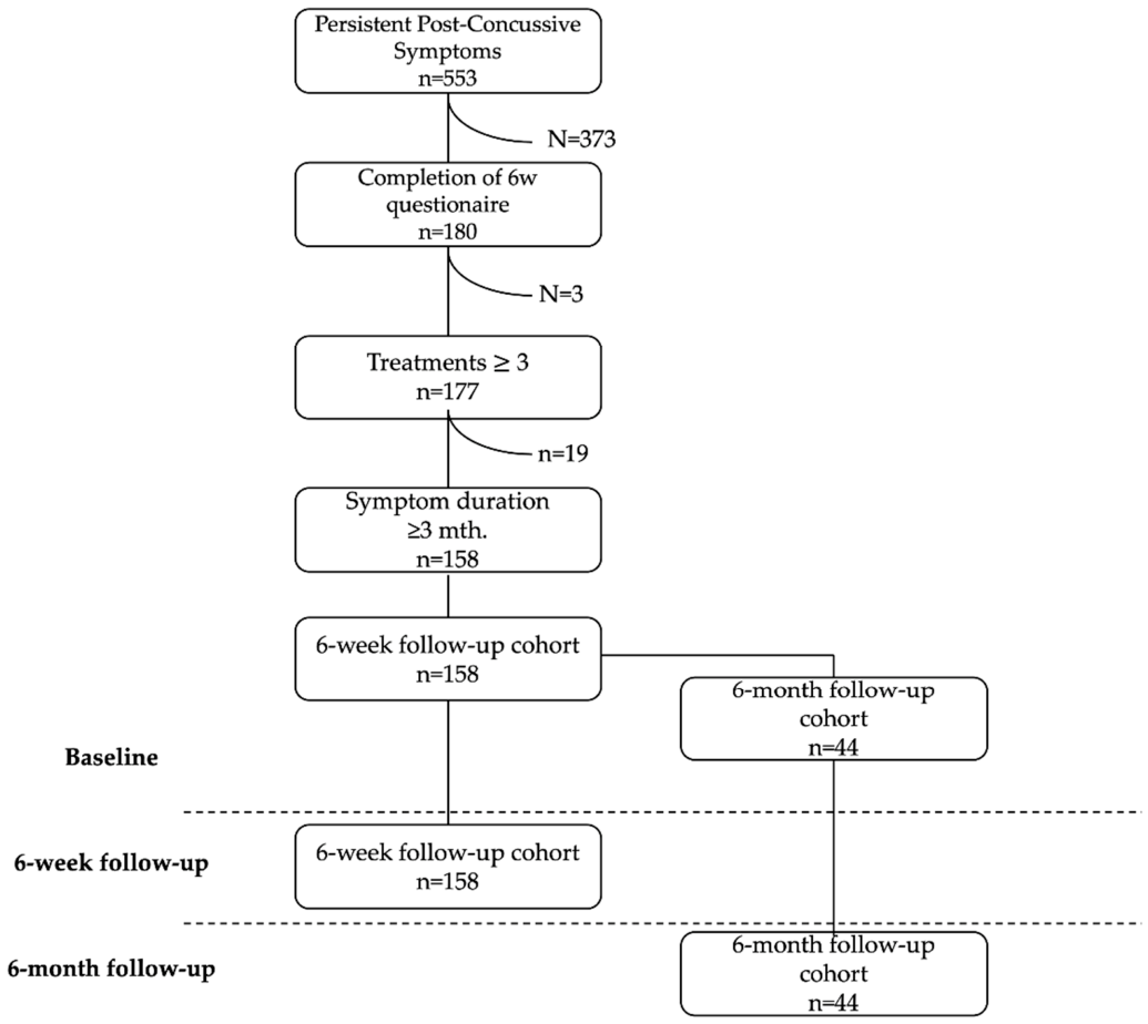
Figure 2.
NRS scores in the 6-week and 6-month follow-up cohorts before and after SANA® Therapy. Bar chart illustrating changes in pain scores (NRS, 0–10) at rest and during activity. Error bars represent SEM. The 6-week follow-up cohort is shown in green (baseline: light green; 6-week: dark green), and the 6-month follow-up cohort in blue (baseline: light blue; 6-month: dark blue). The analysis included patients with NRS ≥3 at rest or during activity at baseline. Significant reductions in pain were observed in both cohorts at rest and during activity, and pain reduction was greater at the 6-month than at the 6-week follow-up. Significance levels: *** p < 0.001.
Figure 2.
NRS scores in the 6-week and 6-month follow-up cohorts before and after SANA® Therapy. Bar chart illustrating changes in pain scores (NRS, 0–10) at rest and during activity. Error bars represent SEM. The 6-week follow-up cohort is shown in green (baseline: light green; 6-week: dark green), and the 6-month follow-up cohort in blue (baseline: light blue; 6-month: dark blue). The analysis included patients with NRS ≥3 at rest or during activity at baseline. Significant reductions in pain were observed in both cohorts at rest and during activity, and pain reduction was greater at the 6-month than at the 6-week follow-up. Significance levels: *** p < 0.001.
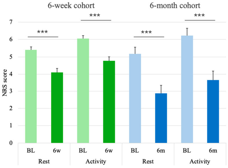
Figure 3.
SF-36 Quality of Life Evaluation at the 6-Week and 6-Month Follow-Up. Radar plots depict changes in quality of life measured by the SF-36. The 6-week cohort is shown in green (baseline: light green; 6-week: dark green), and the 6-month cohort in blue (baseline: light blue; 6-month: dark blue). The dashed line represents Danish reference values from [74]. Abbreviations: PF = Physical Functioning, RP = Role Physical, BP = Bodily Pain, GH = General Health, RE = Role Emotional, SF = Social Functioning, MH = Mental Health, V = Vitality. Significance levels: *p < 0.05, ***p < 0.001.
Figure 3.
SF-36 Quality of Life Evaluation at the 6-Week and 6-Month Follow-Up. Radar plots depict changes in quality of life measured by the SF-36. The 6-week cohort is shown in green (baseline: light green; 6-week: dark green), and the 6-month cohort in blue (baseline: light blue; 6-month: dark blue). The dashed line represents Danish reference values from [74]. Abbreviations: PF = Physical Functioning, RP = Role Physical, BP = Bodily Pain, GH = General Health, RE = Role Emotional, SF = Social Functioning, MH = Mental Health, V = Vitality. Significance levels: *p < 0.05, ***p < 0.001.
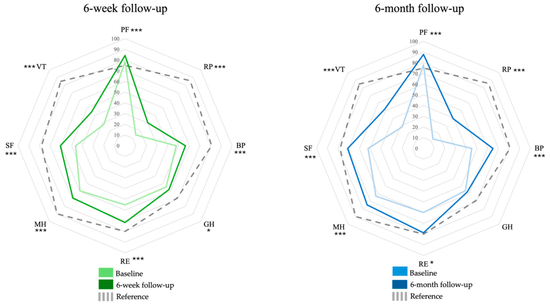

Table 1.
Demographics and characteristics of the 6-week and 6-month follow-up cohorts. Values are mean ± SD or n (%) as indicated. P-values are from Welch’s two-sample t-test for continuous variables and chi-square test for sex.
Table 1.
Demographics and characteristics of the 6-week and 6-month follow-up cohorts. Values are mean ± SD or n (%) as indicated. P-values are from Welch’s two-sample t-test for continuous variables and chi-square test for sex.
| 6w follow-up cohort |
6m follow-up cohort |
p | |
|---|---|---|---|
| Number of patients | 158 | 44 | |
| Age (years), mean [± SD] | 40.8 [± 14.1] | 41.5 [± 16.4] | 0.76 |
| Age, range | 11–79 | 11–70 | - |
| Sex, F:M (n) | 111:45 (2 unspecified) | 32:12 | 0.96 |
| Sex, female (%) | 70.3% | 72.7% | - |
| Height (cm), mean ± SD | 172.4 [± 9.3] | 171.6 [± 9.1] | 0.66 |
| Weight (kg), mean ± SD | 70.7 [± 14.3] | 71.7 [± 15.6] | 0.69 |
| Sick leave at baseline, n (%) | 78 (49.4%) | 14 (31.8%) | 0.03 |
| Symptom duration (months), mean [± SD] | 28.5 [± 21.4] | 32.2 [± 24.0] | 0.34 |
| Symptom duration, median (range) | 21 (4–60) | 30 (2–60) | |
| SANA sessions, mean ± SD | 9.07 [± 6.09] | 9.86 [± 6.86] | 0.48 |
| SANA sessions, median (range) | 7 (3–35) | 7 (4–35) | |
| Prior treatments, mean [± SD] | 5.23 [± 3.07] | 5.16 [± 3.51] | 0.90 |
| Prior treatments, range | 0–20 | 0–20 |
Table 2.
Prevalence of prior treatment modalities at baseline in the 6-weeks follow-up cohort (n = 158). 98.7% of patients had tried other treatment modalities with an average of 5.23 ± 3.07 treatment types (range 0–20).
Table 2.
Prevalence of prior treatment modalities at baseline in the 6-weeks follow-up cohort (n = 158). 98.7% of patients had tried other treatment modalities with an average of 5.23 ± 3.07 treatment types (range 0–20).
| Type of treatment | No. of patients | Prevalence |
|---|---|---|
| Physiotherapy | 120 | 75.9% |
| Analgesics — non-prescription | 109 | 69.0% |
| Osteopathy | 85 | 53.8% |
| Acupuncture | 75 | 47.5% |
| Analgesics — prescription | 68 | 43.0% |
| Chiropractor | 64 | 40.5% |
| Nerve block | 17 | 10.8% |
Table 4.
SF-36 domain evaluation. Quality-of-life scores were assessed by the nine SF-36 domains. The total score was used as a surrogate of overall quality of life. Reference values for the Danish population were obtained from [74]. Significance levels are marked as * = p < 0.05, ** = p<0.01, and *** p < 0.001.
Table 4.
SF-36 domain evaluation. Quality-of-life scores were assessed by the nine SF-36 domains. The total score was used as a surrogate of overall quality of life. Reference values for the Danish population were obtained from [74]. Significance levels are marked as * = p < 0.05, ** = p<0.01, and *** p < 0.001.
| SF-36 Domain | Ref. |
6-week follow-up cohort n=158 |
6-month follow-up cohort n=44 |
||||||
| BL | 6w | Δ | 95%CI | BL | 6m | Δ | 95%CI | ||
| Physical functioning | 75 | 78.4 | 84.3 | 5.85*** | 4.07; 7.64 | 78.1 | 87.8 | 9.77**** | 6.43; 13.1 |
| Role physical | 86 | 14.2 | 30.1 | 15.8*** | 9.19; 22.5 | 12.5 | 39.2 | 26.7*** | 14.9; 38.6 |
| Role emotional | 80 | 55.3 | 71.7 | 16.5*** | 8.81; 24.1 | 59.8 | 78.8 | 18.9* | 4.16; 33.7 |
| Social functioning | 78 | 46.3 | 60.5 | 14.2*** | 10.7; 17.7 | 51.8 | 71.0 | 19.2*** | 11.8; 26.5 |
| Bodily Pain | 81 | 47.8 | 56.5 | 8.64*** | 5.41; 11.9 | 45.1 | 65.0 | 19.9*** | 12.4; 27.5 |
| Vitality | 85 | 28.1 | 44.4 | 16.3*** | 13.1; 19.5 | 28.3 | 51.6 | 23.3*** | 16.2; 30.4 |
| Mental Health | 90 | 59.5 | 69.1 | 9.60*** | 7.46; 11.7 | 62.9 | 74.6 | 11.7*** | 6.68; 16.8 |
| General Health | 69 | 54.3 | 58.0 | 3.67* | 1.29; 6.05 | 55.6 | 57.7 | 2.16 | -3.30; 7.62 |
| Health transition | N/A | 42.2 | 58.2 | 16.0*** | 11.9; 20.1 | 47.2 | 66.5 | 19.3*** | 11.8; 26.9 |
| Total score | 645 | 384 | 475 | 90.5*** | 394 | 526 | 132*** | ||
Disclaimer/Publisher’s Note: The statements, opinions and data contained in all publications are solely those of the individual author(s) and contributor(s) and not of MDPI and/or the editor(s). MDPI and/or the editor(s) disclaim responsibility for any injury to people or property resulting from any ideas, methods, instructions or products referred to in the content. |
© 2026 by the authors. Licensee MDPI, Basel, Switzerland. This article is an open access article distributed under the terms and conditions of the Creative Commons Attribution (CC BY) license (http://creativecommons.org/licenses/by/4.0/).
Copyright: This open access article is published under a Creative Commons CC BY 4.0 license, which permit the free download, distribution, and reuse, provided that the author and preprint are cited in any reuse.


