

Submitted:
21 May 2026
Posted:
25 May 2026
You are already at the latest version
Abstract
The tumor microbiome has been implicated in pancreatic ductal adenocarcinoma (PDAC)’s poor response to treatment, demanding new methods for understanding host-microbe interactions in therapy. Traditional 2D systems, while widely used, fail to adequately recapitulate human PDAC due to insufficient representation of structural, immunological and stromal components. Differences in cancer-specific microbiomes, microbe-immune interactions, and the unique physiological and immunosuppressive features of PDAC have hindered the clinical translation of immune therapies. Reproducible 3D culture systems that integrate the human tumor–immune–microbe axis represent a promising avenue for treatment research, yet they remain underexplored in PDAC. In this narrative review, we discuss the key microbial determinants of therapy resistance in PDAC, explore the current 3D multi-cellular modelling approaches in other cancer types, and provide a path forward for similar integrative translational models in PDAC.
Keywords:
pancreatic cancer
; immune checkpoint inhibitor
; organoids
; spheroids
; microbiomes
1. Introduction
Pancreatic ductal adenocarcinoma (PDAC) is a solid tumor accounting for approximately 85% of pancreatic cancer cases [1]. It remains one of the most lethal malignancies with a five-year survival rate of just 13% worldwide [2]. Surgical resection is often unsuccessful in PDAC due to late diagnosis resulting in unresectable metastatic tumors and high recurrence rates post-resection, while both radiotherapy and first-line chemotherapy show limited treatment response [3]. A promising alternative therapeutic strategy is the use of immunotherapies which involves bolstering the host’s immune system towards recognizing cancer cells as a threat. Immune checkpoint inhibitor (ICI) therapy is a type of immunotherapy that blocks T-cell and cancer cell receptors, such as programmed cell death receptor (PD-1) and its ligand (PD-L1), responsible for bypassing cancer cell recognition and apoptosis.
Although ICI therapy has resulted in improved overall survival rates in other solid tumors like melanoma and lung cancers, pancreatic cancer has shown limited response to both monotherapies involving single target receptors and combination treatments with chemotherapy or antibiotics [4,5]. Reduced effectiveness of immunotherapies in PDAC is largely attributed to its fibrotic, or desmoplastic, stroma which forms a mechanical barrier and an immune suppressive tumor microenvironment (TME), hindering immune cell infiltration and function. This highlights the urgent need to alter this hostile immune microenvironment and to employ alternative immune-modulating approaches beyond conventional treatments [4,6].
Increasing evidence indicates that both commensal and tumor-associated microbiota play a significant role in immune modulation, tumor initiation and progression, as well as response and resistance to treatment of PDAC [3,7]. Considering the emerging significance of the microbiome, investigating its mechanistic influence on the immune and therapeutic landscape of PDAC is critical; it therefore demands physiologically relevant models. Two-dimensional (2D) culture models are widely used due to their affordability and reproducibility; however, their simplistic monolayer architecture fails to capture the phenotypic and structural complexity of tumors. Although animal and xenograft models are frequently utilized in PDAC research, their translational relevance to human systems is often limited due to physiological differences, lack of immune and stromal complexity, and high associated costs [8]. In contrast to 2D models, three-dimensional (3D) models more closely recapitulate tumor structures and stromal composition, while also being cost-effective relative to in vivo systems. Additionally, 3D systems can be flexibly cultured with other components such as drugs, immune cells, stromal cells [8], and microbes to investigate complex cross-interactions [9].
Despite increasing evidence that the microbiome profoundly shapes the immune landscape of tumors thus influencing responses to immunotherapy, integrative 3D culture models incorporating microbial and immune components remain exceedingly limited in PDAC. Organoid technology involving the immune-microbe axis has been widely adopted in colorectal and gastric cancer research, however, similar integration in PDAC organoids proved challenging [7], likely due to the tumor’s unique desmoplastic architecture and complex TME. A significant gap persists in the development of reproducible multi-culture, or heterotypic, 3D systems capable of recapitulating PDAC’s intricate stromal composition, structural heterogeneity, and dynamic immune–microbial interplay.
In this review, we summarize the microbial influences on immune modulation, oncogenesis, and therapy resistance in PDAC, as well as the translational capability of current pre-clinical models. We then critically evaluate recent advances in modelling the tumor–immune–microbe axis using heterotypic 3D systems across various cancer types. Collectively, this emphasizes the pressing necessity to develop integrative clinically translational models for future PDAC research to improve therapeutic outcomes.
2. The PDAC Tumor Microenvironment
PDAC is a lethal malignancy of solid tumors that accounts for approximately 90% of all pancreatic cancers [1]. Symptoms of PDAC are usually non-specific which means cases are often diagnosed late at advanced stages [10,11]. PDAC tumors are situated in the center of a complex system of immunosuppressive cells, fibrous barricades, collapsed blood vessels, and an overall unique tumor microenvironment (Figure 1).
The tumor microenvironment (TME) of PDAC consists of the cellular and molecular components surrounding tumor cells. These cellular components include stromal cells (e.g., pancreatic stellate cells (PSCs) and their activated form as cancer-associated fibroblasts (CAFs)), endothelial cells, and immune cells (e.g., macrophages, B-cells, T-cells). Additionally, there is an extracellular matrix (ECM) rich in growth factors and cytokines that supports tumor maintenance and progression. PDAC tumors in combination with this complex network form a dense and immunosuppressive stroma that drives disease progression and contributes to poor therapeutic outcomes and low survival rates [3,5,11].
Anti-inflammatory cytokines such as interleukin-10 (IL-10), pro-inflammatory cytokines including IL-1, IL-6, IL-17, and tumor necrosis factor-α (TNF-α), and the bidirectional immunomodulatory cytokine transforming growth factor-β (TGF-β) are critically involved in the pathophysiology of PDAC. They are dynamically modulated by interactions between tumor cells and infiltrating immune cells [12]. “Hot” tumors are defined by a strong presence of active, non-exhausted T-cells within the tumor microenvironment, particularly a high density of tumor-killing CD8+ T-cells [13]. On the contrary, PDAC possesses an immunologically “cold” TME, defined by the accumulation of immunosuppressive cells, such as Tumor-associated macrophages (TAMs) with a pro-tumorigenic M2 phenotype and regulatory T-cells (Tregs) that reside within the tumor, among other immunosuppressive factors which debilitate dendritic cells and suppress cytotoxic T-cells, all supporting PDAC progression and desensitizing tumors towards therapies [3,12,14].
The PDAC TME is considered a “desmoplastic environment”, referring to the dense, fibrotic stroma surrounding the tumor. Solid stress is the compressive forces generated by CAFs and the dense ECM within the TME. Stellate cell-derived CAFs, a heterogenous group of cells responsible for producing ECM components such as collagen, hyaluronic acid or fibronectin, are the main contributors to desmoplasia [10,11]. CAFs produce a cell surface fibroblast activation protein (FAP) which has been associated with oncogenic effects such as angiogenesis, tumorigenesis, metastasis and immunosuppression [5,10]. However, their role in PDAC appears to be more complex.
CAFs can reversibly differentiate into myofibroblastic or immune-mediating subtypes. Myofibroblastic CAFs (myCAFs), triggered by TGF-β, express high levels of α-smooth muscle actin (α-SMA) responsible for producing the ECM proteins primarily constituting the surrounding stroma [10,15]. Additionally, a positive correlation between large clones and their close proximity to myofibroblastic CAFs indicating that its secretions promote PDAC expansion [16]. The depletion of myCAFs using diphtheria toxin accumulated cytotoxic T-lymphocytes (CTLs) and enhanced PD-L1 therapy [17]. Cytotoxic or cell-killing T-cells, such as CD4+ and CD8+ T-cells, are collectively termed as CTLs. Contrarily, genetic deletion of myCAFs resulted in a more aggressive and invasive phenotype of pancreatic tumor cells [10,18,19]. Collectively, these findings suggest that myCAFs are not solely tumor-promoting but also possess a tumor-restraining role. Inflammatory CAFs are stimulated by IL-1 and encourage proliferative tumorigenic effects like PDAC solid stress through the production of hyaluronan. Inflammatory CAFs express low levels of α-SMA but high levels of the type I angiotensin II receptor responsible for vasoconstriction and increased blood pressure. The inhibition of this receptor results in decreased intratumoral solid stress resulting in improved infiltration and drug delivery [15,20]. On the other hand, another CAF subpopulation termed antigen-presenting CAFs, expresses major-histocompatibility complex (MHC) class II molecules capable of presenting antigens to CD4+ T-cells, a process important for antitumor immune responses [15,20].
In PDAC, CAFs are major drivers of immune evasion and tumor progression through both contact-dependent and soluble immunosuppressive mechanisms. As a primary role, CAFs are thought to play key roles that are pro-tumorigenic and immunosuppressive. For instance, by suppressing IL-2 production, CAFs significantly impair the proliferation and differentiation of CD8+ T-cell function. Furthermore, the upregulation of cell surface proteins like Fas ligands (FASL) and the PD-1 ligand PD-L2 on CAFs bind to their respective receptors (Fas and PD-1) causing chronic stimulation of T-cells leading to reduced proliferation, dysfunction and death of CD4+ and CD8+ T-cells, also known T-cell exhaustion, ultimately promoting tumor cell survival [21,22]. Furthermore, CAFs produce the soluble factor prostaglandin E2 (PGE2), and inhibition of PGE2 restored T-cell proliferation partially [22]. This implies that CAFs plays a significant role in maintaining the immunosuppressive TME in PDAC, which has ultimately rendered immunotherapies ineffective. However, it may not be so straight forward.
CAFs exhibit a complex and bidirectional role in immune modulation. Interestingly, exposure of CAFs to radiation induces a senescent phenotype, which in turn enhances their tumor-promoting and immunosuppressive functions. This is evidenced by increased surface expression of immune checkpoint and immunoregulatory receptors, as well as ectonucleotidases that convert extracellular ATP into immunosuppressive adenosine, thereby further reinforcing an immunosuppressive TME [19]. Taken together, these data show that CAFs in PDAC are not exclusively tumor-promoting, despite their predominant role in driving tumor progression. Certain CAF subpopulations can support antitumor immunity, while indiscriminate depletion of CAFs may be therapeutically detrimental. This highlights their heterogeneous and context-dependent role in tumorigenesis. Furthermore, radiotherapy may enhance CAF-mediated immunosuppressive remodeling of the TME, thereby contributing to the profound therapy resistance observed in PDAC.
Within the PDAC tumor, there are spatially isolated sub-regions with differentially expressed and phenotypically heterogenous characteristics. One sub-region is known as reactive sub-TME regions housing high levels of activated CAFs expressing FAPα and inflammatory cytokine IL-6, forming the intratumoral TME. These reactive regions are chemo-sensitive and establish an inflammatory TME enriched in T-cells, TAMs, and endothelial cells, collectively facilitating tumorigenesis through the above described contact-dependent and secretory mechanisms of CAFs. Contrastingly, other regions have minimally activated CAFs at lower concentrations, and show chemoprotective effects [23].
As part of the overall TME, TAMs and CAFs make up most of the cellular components of the stroma. They are often found in close proximity with one another and jointly contribute towards immunosuppressive desmoplasia [24]. TAMs and CAFs mutually reinforce each other’s immunosuppressive functions through a positive feedback loop within the TME. TAM-derived cytokines such as TGF-β and IL-1β promote CAF differentiation and activation, while CAF was shown to secrete IL-33, IL-6 and granulocyte–macrophage colony-stimulating factor (GM-CSF) drive macrophage polarization toward the pro-tumorigenic M2 phenotype in several cancer types [25,26,27]. Together, these bidirectional interactions maintain the immunosuppressive and tumor-supportive PDAC TME, while the substantial heterogeneity between TME subregions likely contributes to differential therapeutic responses, limiting the efficacy of uniform treatment approaches.
The density of the stroma further inhibits the host’s immune response to the tumor. It immobilizes infiltrating immunogenic cells such as monocytes, neutrophils, mast cells, as well as a small number of tumor-infiltrating lymphocytes (TILs), contributing to the immune “cold” phenotype of PDAC tumors [5]. It also leads to collapse of the pancreatic vasculature, exacerbating hypoxia and free radical accumulation known to cause DNA damage and cell death, which in turn promotes cancer cell aggressiveness and contributes to the establishment of an immunosuppressive microenvironment [5,28]. Such conditions oppose the survival of immune cells and limit the infiltration of CD8+ cytotoxic T-cells which are essential for antitumor immunity [13]. Hypoxia, alongside molecules produced by pancreatic cells like the cytokine CCL2 and GM-CSF, recruit an immunosuppressive environment containing Tregs, myeloid-derived suppressor cells (MDSCs), and differentiates TAMs into their pro-tumorigenic M2 phenotype [5,12].
An important component of the cold PDAC tumor are Tregs which assist with immune evasion in TMEs, promote progression, and are associated with unfavorable outcomes in many cancers. Weaver et al. (2022) reported that inhibiting CCR8, a chemokine receptor expressed on tumor-infiltrating Tregs using complementary monoclonal antibodies, resulted in Treg depletion and consequently an increase in CD8+ T-cell infiltration, as well as significantly improved anti-PD1 efficacy in combination. CCR8 inhibition has shown significant tumor regression in murine models of PDAC (Pan02) and colorectal carcinoma (CT26) [29,30]. In this context, CCR8 blockade induces an immunological shift toward antitumorigenic responses and may represent a promising therapeutic strategy in cancer treatment.
The main marker for Treg cells is the Forkhead Box Protein (FOXP3) transcription factor [31]. The presence of intratumoral Tregs expressing FOXP3 (FOXP3+ Tregs) in PDAC is associated with tumor progression, immune evasion, poor prognosis, and reduced intratumoral CD8+ T-cell abundance. High FOXP3+ Treg accumulation in regions surrounding the tumor may suppress local antitumor immunity by limiting CD8+ T-cell recruitment and function [32,33,34,35]. Consistently, pre-clinical targeting of FOXP3+ Tregs using anti-cytotoxic T Lymphocyte-associated-protein 4 (anti-CTLA-4), anti-CD25, and CCR5 inhibition reduced tumor growth and enhanced CD8+ T-cell-mediated antitumor responses [34]. Furthermore, Kiryu et al. (2021) showed that patients with an immune rich TME possessing a high density of TILs (CD3+, CD4+ and CD8+ T-cells) in the TME that were positive for PD-1 in surgically resected PDAC tissues had significantly better overall survival (OS: 1086 days with high CD3+ T-cells; 1360 days with high CD4+ T-cells; 1223 days with high CD8+ T-cells) and disease-free survival (DFS: 554 days; 554 days; 607 days) compared to those with PD-1- T-cells (OS: 699 days, DFS median: 351 days with high CD3+ T-cells). However, immune rich TMEs in the presence of FOXP3 showed opposing results; patient resections with high levels of intratumoral FOXP3+ T-cells in the presence PD-1+ T-cells showed significantly worse prognosis (OS = 692 days) as opposed to subgroups with low FOXP3+ (OS = 1420 days) [36]. Collectively, these findings highlight that the prognostic benefit of T-cell-rich PDAC microenvironments is highly context-dependent, as FOXP3+ Tregs not only interfere with effective CD8+ T-cell infiltration but may also functionally suppress infiltrating cytotoxic T-cells once present within the tumor. Consequently, even immune-infiltrated (“hot”) PDAC tumors may remain therapeutically resistant in the presence of high FOXP3+ Treg abundance. Therefore, strategies aimed at enhancing CTL infiltration while simultaneously depleting or inhibiting FOXP3+ Tregs are likely critical for improving antitumor immune responses in PDAC.
Independent of immune cells, FOXP3 gene expression has also been reported within PDAC cells [37]. Tumor-intrinsic FOXP3 can modulate cytokine expression (including effects on IL-6 and IL-8) and, in co-culture with naïve T-cells, FOXP3+ PDAC cells inhibit T-cell proliferation. These effects are partially reversed when cancer-cell FOXP3 is suppressed, supporting a role for cancer-derived FOXP3 in tumor immune evasion [31]. Wang et al. (2017) further discovered that FOXP3+ PDAC cells directly trans-activate the T cell chemotactic protein CCL5, promoting the recruitment of Tregs and contributing to immune evasion [38]. Additionally, tumor-associated neutrophils secrete abundant CCL5, which enhances cancer cell migration and invasion while negatively correlating with cytotoxic CD8+ T-cell infiltration, further suppressing anti-tumor immunity [39].
Overall, PDAC possesses multiple interconnected mechanisms that collectively maintain a highly immunosuppressive and poorly infiltrated TME. CAFs establish a dense desmoplastic stromal network that physically protects tumor cells while promoting immunosuppressive macrophage polarization, whereas FOXP3+ regulatory T-cells suppress effective intratumoral immune infiltration and anti-tumor responses. Compounding this, genetically distinct and heterogeneous sub-TME regions can generate diverse phenotypes, varying chemotherapy susceptibility and immune profiles within the same tumor. Additional factors, including hypoxia and inflammatory cytokines, further reinforce the immunosuppressive landscape. Consequently, PDAC remains exceptionally difficult to treat, with these features contributing to high mortality rates, poor prognosis and resistance to an array of therapies and drug activity. Beyond host-derived influences, emerging evidence suggests that microbial components further modulate PDAC progression and therapeutic response, making PDAC more challenging to treat and emphasizing the urgent need for more clinically translatable and physiologically representative disease models.
3. The Microbiome’s Influence on Cancer
3.1. Microbial Signatures Correlate with Survival Duration and Treatment Response
Emerging evidence indicates the patient gut, oral, and tumor-associated microbiome signatures are associated with oncogenesis, metastasis, and immune suppression in PDAC [40]. Alterations to healthy microbial gut and oral microbiome populations, known as dysbiosis, are frequently observed in PDAC patients [41]. Several studies have identified microbial patterns in oral and fecal samples unique to PDAC patients using shotgun and 16S rRNA sequencing. These studies reported that PDAC patients – in comparison to healthy controls – had enriched bacterial family populations of Akkermansia, Odoribacter [42], Veillonellaceae [41,42], Streptococcus [41], Bacteroidaceae, Lachnospiraceae G7, Enterobacteriaceae, and Staphylococcaceae [43], with reductions in Ruminococcaceae, Lachnospiraceae, Clostridiales [42], Faecalibacterium prausnitzii [41], Haemophilus [43], Streptococcus mitis and Neisseria elongata [44] in oral and fecal samples. In addition to changes to the commensal bacteria, pathogenic bacterial populations have been implicated in tumor initiation. Two studies linked periodontal disease-causing bacteria, such as Porphyromonas gingivalis and Aggregatibacter actinomycetemcomitans, to higher risk of PDAC development [45,46]. While there is consensus across these studies that bacterial genera in the gut and oral microbiomes are associated with PDAC and even its development, there is a lack of understanding around mechanism. Whether or not these associations are causal, and if so, how specific genera or species enact their effect, remains to be seen.
In addition to systemic influences of the oral and gut microbiomes, these communities impact PDAC directly as well through the pancreatic microbiome. The pancreatic microbiome is thought to be established by bacteria from the oral cavity [43] and gut [47], which translocate to the pancreas via the circulatory system or the biliary and pancreatic ducts. Translocation of these species into the pancreas may then promote inflammation and cause acute pancreatitis development [2,48], which can increase the risk of PDAC [45]. It was further suggested that the progressive inflammatory nature of the PDAC TME may also promote this translocation and facilitate microbial colonization within pancreatic tissue [49]. Compared to healthy participants, the microbiome of PDAC has reduced microbial diversity [48], fewer butyrate-producing taxa, and a concomitant increase in pathogenic lipopolysaccharide (LPS)-producing bacteria [50], likely a reflection of the dysbiotic state of the gut- and oral-microbiomes of the patient. Importantly, no microbial differences between malignant and benign human surgical resections have been identified [51], implying that the intratumoral microbiome’s composition is established before malignancy.
The microbial composition of PDAC tumors has been linked specifically to survival duration and therapeutic response, though these associations are clouded by the disruptive effects of chemotherapy and radiotherapy on the microbiome [6,52,53]. Specifically, intratumoral microbial composition correlates to PDAC survival: long-term survivors (LTS, >5 years) of PDAC harbor more diverse and compositionally distinct microbiomes compared to short-term survivors (STS), suggesting that intratumoral microbial diversity may contribute to differences in patient outcomes [54]. For LTS, these genera include Saccharopolyspora, Streptomyces, Pseudoxanthomonas, Bacillus clausii [54], Neorickettsia and Mediterraneibacter [55]. Further studies in a Chinese cohort also identified intratumoral Megasphaera, Enterococcus and Sphingomonas positively correlated to overall survival [56] which was dissimilar to the US cohort [54], possibly influenced by population-specific factors such as diet, environment and host background. Huang et al. (2022) found that Megasphaera sp.XA511 isolated from PDAC tumors was associated with LTS and improved antitumor efficacy of anti-PD1 therapy in a murine 4T1 breast cancer model [56]. STS tumors, on the other hand, were dominated by Clostridia [54], Bacteroides, Lactobacillus, Peptoniphilus [57], and Stenotrophomonas [55]. Additionally, other taxa such as Streptococcus infantis, Acinetobacter spp. (A. johnsonii, A. lwoffii), and Pseudomonas luteola have been associated with poor patient outcomes, further highlighting microbial contributions to prognosis [58]. Collectively, these insights point to tumor-associated microbiota as potential modulators of therapy response and/or prognostic markers.
3.2. Microbiome Influences on Immunity in PDAC
Building on evidence that sequencing studies have consistently associated microbial composition with survival duration in PDAC patients, immune–microbe interactions may help explain the observed correlations with patient prognosis, therapeutic responses and overall disease outcomes. A summary of these interactions in PDAC is illustrated in Figure 2. Research across different cancer types has shown that the level of CD8+ T-cells within a tumor serves as a key indicator for predicting the effectiveness of anti-PD-1/PD-L1 immunotherapy [13]. Riquelme et al. (2019) reported that LTS-associated bacteria from surgically resected PDAC samples, including Saccharopolyspora, Pseudoxanthomonas, and Streptomyces, correlated positively with intratumoral CD8+ T-cell and Granzyme B densities, suggesting that these microbes may enhance antitumor immune responses by fostering T-cell activation and infiltration [54]. In contrast, the presence of STS-related anaerobic genera in PDAC tumor samples such as Bacteroides, Lactobacillus, and Peptoniphilus correlated with reduced tumor-infiltrating CD4+, CD8+, and CD45RO+ T-cells and shorter overall patient survival [57].
Tumor-associated bacteria are associated with immune responses within the TME. In metastatic renal cell carcinoma, ICI treatment responders and non-responders showed notable differences in relative abundance of intratumoral microbes [59]. In another study, Gopalakrishnan et al. (2018) demonstrated that increased diversity in the gut microbiota can improve immunotherapy responses in metastatic melanoma by promoting T-helper cell-1 (Th1) polarization of CD4+ T-cells and enhancing the infiltration of cytotoxic CD8+ T-cells. In an in vitro system, PDAC spheroid models infected with the oral bacteria Fusobacterium nucleatum induced the secretion of pro-tumorigenic cytokines such as GM-CSF, CXCL1, IL-8, and MIP-3α which significantly enhanced PDAC cell proliferation and migration, with no observable effect in non-cancerous pancreatic epithelial cells. These tumorigenic effects were largely abrogated by GM-CSF blockade [60]. Taken together, the gut-, oral- and tumor-associated microbiota of PDAC patients likely affect the immune response. Though more research is needed to understand these processes, some mechanisms are understood.
Figure 2.
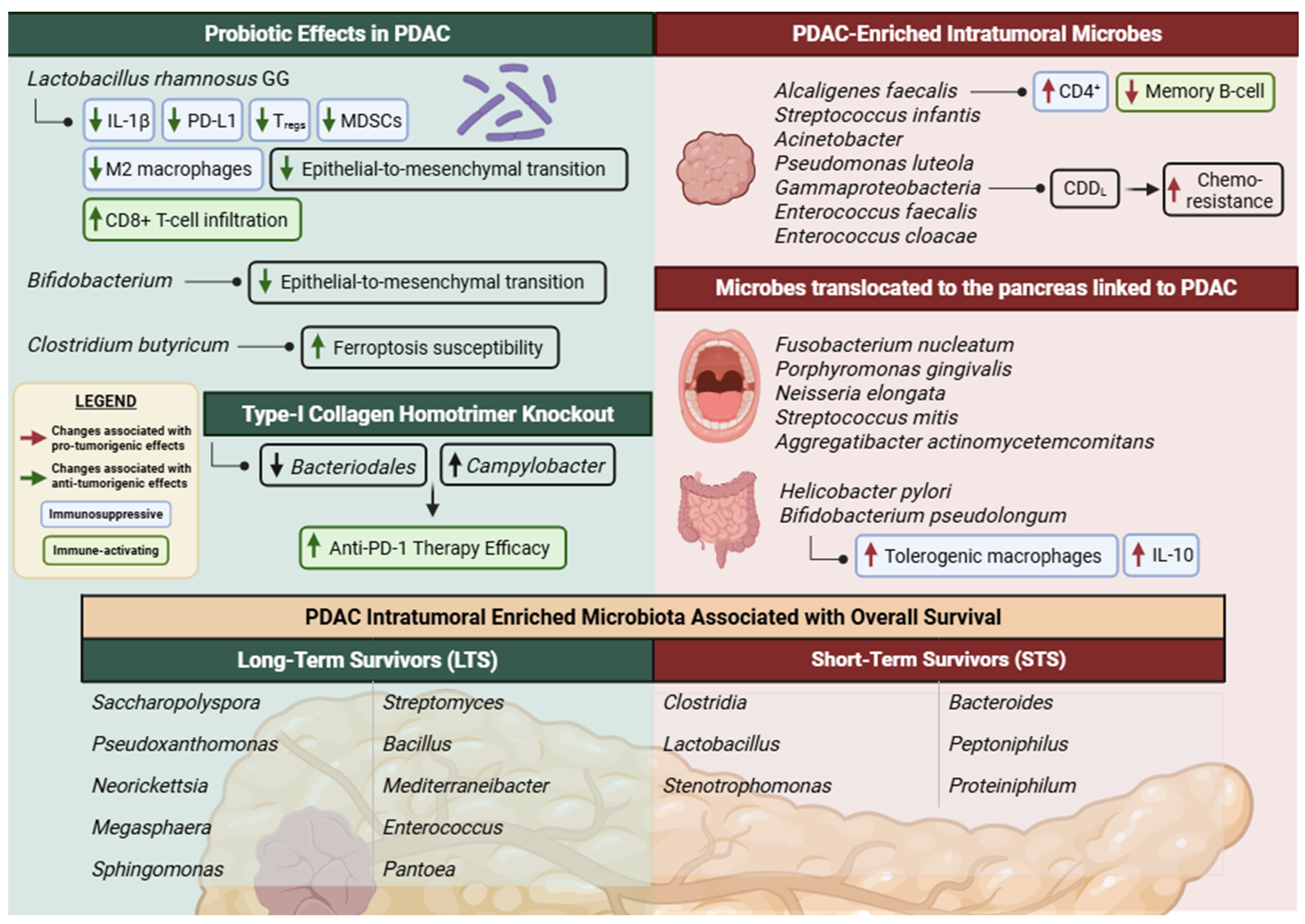
Influences of the intratumoral and translocated microbiome on PDAC. Beneficial bacteria (green panel) and their mechanisms associated with anti-tumorigenic effects, long-term survival and improved ICI efficacy. This panel also highlights immunotherapy enhancing probiotics and the type I collagen gene knockout along with linked bacterial changes. Pro-tumorigenic bacteria (red panel) and their mechanisms associated with short-term survival and immunosuppression. PDAC-Enriched Intratumoral Microbes lists bacteria with higher relative abundance in relevance to healthy controls and may be inclusive of some LTS bacteria. Adapted from [41,42,43,44,45,46,47,54,55,56,57,58,61,62,63,64].
Figure 2.
Influences of the intratumoral and translocated microbiome on PDAC. Beneficial bacteria (green panel) and their mechanisms associated with anti-tumorigenic effects, long-term survival and improved ICI efficacy. This panel also highlights immunotherapy enhancing probiotics and the type I collagen gene knockout along with linked bacterial changes. Pro-tumorigenic bacteria (red panel) and their mechanisms associated with short-term survival and immunosuppression. PDAC-Enriched Intratumoral Microbes lists bacteria with higher relative abundance in relevance to healthy controls and may be inclusive of some LTS bacteria. Adapted from [41,42,43,44,45,46,47,54,55,56,57,58,61,62,63,64].
3.2.1. Bacterial Metabolites
Accumulating evidence suggests that microbial-derived metabolites may be key mediators of the immunomodulatory effects of microbiota in PDAC. The intratumoral pancreatic microbiome can influence the TME and PDAC prognosis through these metabolic products, as well as other yet undefined mechanisms [56]. Microbial metabolites are small molecules produced by bacteria through primary or secondary metabolism, including bacterial cell-intrinsic components such as LPS, or cell-extrinsic products, like short chain fatty acids. These bioactive compounds can reshape the TME by modulating innate immune signaling, myeloid cell function, and altering T-cell infiltration, thereby impacting tumor progression, therapy resistance, and response to immunotherapy. Some microbes may also reduce treatment effectiveness by impairing immune responses or metabolizing therapeutic agents. Understanding these molecular mechanisms is critical for improving current cancer treatment strategies [65,66].
Interactions between the microbiome and targeted therapies beyond PDAC highlight the capacity of microbes and their metabolites to shape immunotherapy. This is further supported in other tumor types; for example, Muribaculum gordoncarteri and elevated levels of its metabolite urocanic acid, suppresses MDSC recruitment through inhibition of the CXCL1–CXCR2 signaling axis in colorectal tumor vascular endothelial cells. Notably, ICI responders displayed higher fecal urocanic acid concentrations and increased abundance of Muribaculum gordoncarteri compared with non-responders, underscoring the translational potential of microbiome–drug interactions in modulating antitumor immunity and highlighting both as potential predictive biomarkers for treatment response [67].
LPS is an outer membrane component of gram-negative bacteria, exerts bi-directional effects on tumor immunity, playing a pivotal role in pancreatic tumorigenesis. In PDAC-associated dysbiosis, the relative abundance of LPS-producing genera such as Prevotella, Hallella, and Enterobacter is elevated. Toll-like receptor (TLR)-signaling plays a major role in regulating inflammatory responses and is critically involved in tumorigenesis, immune suppression and therapy resistance [68]. LPS binds to and activates TLRs found on pancreatic cancer cells, such as TLR2, TLR4, TLR9 and the signaling adaptor MyD88, stimulating tumor cell activation and proliferation [69]. In support, Ikebe et al. (2009) demonstrate that LPS enhances invasive capacity in pancreatic cancer cell lines via activation of TLR4/MyD88/NF-κB inflammatory signaling pathway, with blockade of any element in this pathway effectively reducing LPS-induced tumor invasiveness [70]. This suggests that the inflammatory TME in PDAC is influenced by the microbiome and sustained through LPS-induced TLR signaling. Sustained TLR4 signaling further correlates with PD-L1 expression on tumor cells which binds to PD-1 receptors on T-cells, leading to functional inhibition and indicating that chronic LPS–TLR4 activation enhances immune checkpoint-mediated suppression [61]. Consistently, Yin et al. (2021) demonstrated that LPS signaling synergizes with anti-PD-L1 therapy to suppress pancreatic tumor growth [71].
In melanoma, clinical responders to anti-PD-1 therapy were enriched for LPS-producing bacteria encoding the immunostimulatory hexa-acylated LPS, which elicited stronger TLR4-mediated immune activation and enhanced anti-PD-1 treatment efficacy in murine models, whereas penta-acylated LPS inhibits this activation [72]. LPS also exerts time-dependent effects on the tumor immune microenvironment. Short-term LPS exposure promotes pro-inflammatory M1 macrophage polarization, whereas prolonged stimulation (>72 hours) expands immunosuppressive M2-like macrophages and MDSCs, limiting CD8+ T-cell infiltration and reinforcing the immunosuppressive TME [61].
Importantly, TLR activation is not restricted to LPS alone. For instance, Bifidobacterium pseudolongum has been shown to accelerate PDAC oncogenesis in a TLR-dependent manner, as blockade of downstream signaling (e.g., via TRAF6 inhibition) abrogates its tumor-promoting effects. Additionally, cell-free extracts from B. pseudolongum promote macrophage polarization toward a tolerogenic phenotype characterized by increased anti-inflammatory IL-10 production, further supporting a role for microbiome-driven TLR signaling in shaping an immunosuppressive tumor microenvironment [47].
Additionally, certain Lachnospiraceae produce MHC class I peptides via flagellin-related genes which are structurally homologous to tumor antigens, primed CD8+ T-cells and improved antitumor immunity against melanoma in ICI complete responders [73]. Other microbial metabolites, such as Bifidobacterium-derived inosine [74] and Enterococcus muropeptides [75] have also been implicated in improved responses to immune checkpoint inhibition in CRC and melanoma murine models, respectively.
Collectively, these findings highlight microbial-derived metabolites as key mediators of immune modulation and inflammation within the PDAC TME. The presence and functional activity of specific microbial taxa, such as LPS-producing bacteria, may be predictive of patient response to therapy and may also be useful as therapeutic adjuvants for PDAC immunotherapy.
3.2.2. Differing Effects of Microbes Hinders Clinical Translation
The microbiome is comprises of a diverse range of genera and species that are often shared across multiple tissue and cancer types; however, their functional impact is highly context dependent, with opposing roles in PDAC versus other cancers and tissues. This variability likely arises from the unique biological and immunological landscape of the PDAC microenvironment.
The bacterial genus Campylobacter is an enteric pathogen commonly associated with gastrointestinal infection and has been implicated in tumorigenesis in colorectal cancer (CRC), where it is enriched in primary tumors of metastatic patients. Mechanistically, Campylobacter jejuni promotes tumor progression through production of cytolethal distending toxin, which activates the pro-tumorigenic JAK2–STAT3–MMP9 signaling pathway in murine metastatic models, human CRC tissues, and patient-derived organoids [76]. In contrast, the role of Campylobacter in PDAC appears more context dependent. Chen et al. (2023) demonstrated that tumor cell-derived type I collagen homotrimers improved overall PDAC survival rates in murine models by shaping the intratumoral microbiome and immune-tumor interactions in PDAC. Gene knockout of collagen homotrimers was associated with enhanced CD8+ T-cell infiltration, improved response to anti-PD-1 immunotherapy, and reduced intratumoral hypoxia. This was accompanied by an intratumoral microbial shift characterized by a decrease in Bacteroidales, typically enriched in the hypoxic and immunosuppressive PDAC TME, and an increase in microaerophilic Campylobacterales populations. Depletion using broad spectrum antibiotics with collagen homotrimers reversed therapeutic effects [63], an observation contradicting several other studies [47,51] suggesting that therapeutic approaches of microbial depletion may be context-dependent as well. Interestingly, microbiome profiling using 16S rRNA amplicons indicated that Campylobacter abundance was less prevalent in PDAC as compared to healthy controls [77] and the adjacent bile duct malignancy, known as cholangiocarcinoma [78]. It is worth noting that this study has inconsistencies in their reports, however the graphical data presented proof of the unique relative reduction of Campylobacter in PDAC patients to healthy controls. Furthermore, compared to normal pancreatic tissue, matched benign pancreatic lesions also demonstrated lower relative abundance of Campylobacter [79]. Collectively, these findings indicate that Campylobacter does not exhibit a consistent tumor-promoting or tumor-suppressive role across cancer types, but rather displays context-dependent associations across pancreatic disease states.
The presence of specific microbial taxa has divergent immunomodulatory implications, influencing both T- and B-cell infiltration through BCR- and TCR-mediated signaling. In PDAC, B-cell infiltration has been associated with opposing clinical outcomes, contributing to favorable prognosis through antibody-mediated anti-tumor responses, while also promoting tumorigenesis and immune suppression when induced by IL-1β resulting in reduced survival [58,80]. The microbial context-dependent effect is further exemplified by the Alcaligenes genus. Alcaligenes spp. can stimulate immune responses by activating dendritic cells and naïve B-cells via the lipid A component of LPS within gut-associated lymphoid tissues. However, Alcaligenes faecalis in resected early-stage PDAC is associated with increased naïve CD4+ T-cells and reduced memory B-cells, an immune profile linked to poor overall survival [58].
By shaping adaptive immune pathways and producing metabolites or peptides that mimic tumor antigens or alter the TME, microbes can either promote or suppress antitumor immunity. Growing evidence indicates that the composition of a patient’s microbiome can serve not only as a predictor of disease but also as a therapeutic immune modulator to enhance the efficacy of PDAC immunotherapy. Integrating microbial signatures and employing microbial therapeutic strategies into clinical stratification may improve prognosis prediction and optimize therapeutic responses and survival of PDAC patients towards both immunotherapy and chemotherapy [49].
3.3. Microbial Therapeutic Strategies and Experimental Investigations
As the influence of the tumor and gut microbiomes on PDAC outcomes become more apparent, their therapeutic manipulation emerges as a new treatment avenue. There are three main types of microbial therapeutic approaches in PDAC which are commonly used in combination with each other and other treatments: first, the depletion of pro-tumorigenic bacteria using antibiotics; second, the administration of probiotics, prebiotics or their bioactive metabolites known as postbiotics; and third, fecal microbiota transplantation (FMT) from long term PDAC survivors or healthy donors. Table 1 shows a summary of experimental investigations carried out and their observations across these strategies.
3.3.1. Pro-tumorigenic Bacterial Depletion
The identification of pro-tumorigenic bacteria in PDAC tumors suggests the use of antibiotics for microbial ablation in experimental mouse models and for microbial reduction or depletion in clinical patients to assist with efficacy in other therapies like ICIs and chemotherapy. This route has shown moderate success in preclinical models.
In the wild-type BxPC3 and mutant L3.6pl variant of the Kras oncogene cell-line xenografts and genetically engineered KrasG12D/+;PTENlox/+;Pdx1-Cre (KPP) mouse models, antibiotic-mediated depletion of intestinal microbiota slowed PDAC tumor progression, showed a decrease in poorly differentiated tumors, and resulted in fewer malignant lobules relative to microbiota-intact mice. The xenograft tumors themselves, however, regardless of antibiotic treatment remained devoid of detectable intratumoral bacteria and did not establish them over time. It was suggested that gut microbiota or other commensals external to the tumor, rather than tumor-resident species alone, may influence disease trajectory. Intratumoral bacteria may therefore have more impactful roles associated to therapeutic response as opposed to tumor progression [51].
More strikingly, ablation of the gut and intratumoral bacteria was shown to overcome treatment resistance to both gemcitabine [64] and immune checkpoint inhibition [47] in CRC and PDAC models, respectively. Furthermore, antibiotic-mediated depletion of the gut microbiome protected against both preinvasive and invasive PDAC, whereas FMT from PDAC-bearing murine hosts, but not controls, reversed tumor protection. Bacterial ablation reprogrammed the immune environment with reduced accumulation of MDSCs, enhanced pro-inflammatory activation of macrophages (M1 polarization), and increased activation of CD4+ T-helper cells and CD8+ T-cells [47].
3.3.2. Pre-, Pro- and Postbiotics
As established by several studies, microbiota have varying influences on the TME. The identification of microbial communities in responders to ICI treatment has been useful as predictors of therapy response across multiple cancer types [81], which has opened avenues towards using microbes as probiotics in tandem with therapies in patients with poor treatment response [82]. Probiotics are the administration of live microorganisms in controlled doses that provide health benefits to the host. For instance, Xie et al. (2025) [83] introduced Clostridium butyricum as a probiotic in CRC murine models. C. butyricum is known to be an intratumoral bacterium of the human gut and is commonly in reduced abundance in fecal samples of PDAC patients [84]. The introduction of C. butyricum in germ-free and immunologically humanized mouse models resulted in the inhibition of the PI3K-AKT-NF-κB-IL-6 cascade signaling, associated with tumor progression and inflammation. This occurs by the interaction between the C. butyricum surface protein secD and the CRC receptor GRP78. The resulting reduction of IL-6 in combination with anti-PD1 further improved CD8+ T-cell infiltration and suppressed TAM infiltration in the murine models as well as heterotypic organoid models inclusive of matched stromal CAFs and immune cells [83]. Similarly, oral gavage supplementation of C. butyricum or its metabolite butyrate in PDAC murine models increased tumor susceptibility to ferroptosis, a form of programmed cell death, through enhanced intracellular oxidative stress and accumulation of intracellular lipids [85].
However, contradictory observations have been reported regarding the effect of Clostridiales in PDAC. Clostridium butyricum reduced tumor growth and enhanced immune responses in CRC models [83] and Clostridium sensu stricto 1 was associated with reduced risk of PDAC development [86]. However, this same species was also identified as highly abundant intratumorally in STS PDAC patients [56]. These differing observations, distinguished primarily by the location of the Clostridium spp., suggest that bacterial location and surrounding microenvironment may strongly influence microbial behavior. Within the gut, Clostridiales have access to fermentable substrates required for beneficial butyrate production, whereas within the devascularized and immunosuppressive PDAC TME, the same bacteria may adopt context-dependent pathogenic or tumor-supportive roles. This further suggests that microbial abundance alone, particularly from FMT stool profiling, may not fully reflect microbial function within the intratumoral environment. A similar phenomenon has also been reported with Enterococcus faecalis, where its muropeptides enhanced ICI efficacy in melanoma models [72]. However, when introduced to the hypoxic environments within PDAC spheroids, both E. faecalis and E. cloacae increased pathogenicity [9]. Whether direct supplementation of postbiotics could counteract these tumor-promoting effects in STS-associated PDAC models remains an important area for investigation.
In addition to Clostridium, the administration of Bifidobacterium- and Lactobacillus-enriched probiotic mixtures have shown promising results. In PDAC-xenografted mice, these mixtures attenuated epithelial-to-mesenchymal transition (EMT), thereby limiting tumor invasiveness [62,87]. Han et al. (2024) also investigated the probiotic potential of Lactobacillus rhamnosus GG for enhancing immunotherapy efficacy in PDAC murine models [61], which is discussed further in Section 3.4. These studies suggest that supplementation with different probiotic species may exert distinct anti-tumorigenic effects.
Prebiotics are commonly defined as substrates digestible by host microbes that confer health benefits. Non-digestible carbohydrates, such as inulin, are fermented by gut microbes producing short chain fatty acids (SCFAs), like butyrate and propionate, that play many roles in gut health, immune modulation and cancer dynamics [88]. Prebiotics attenuated tumor growth and in combination with inhibitor drug therapies, like MEK inhibitor therapy, reduced tumor resistance to treatments in melanoma and colon cancer [89]. The use of inulin as a prebiotic is sometimes used in combination with other pre- and probiotic regimens. Inulin with prebiotic mucin in melanoma and colon cancer [89], and probiotics such as Lactobacillus plantarum LS/07 in colon cancer [90] induced apoptosis, reduced intratumoral pro-inflammatory cytokines, and also increased dendritic cell, CD4+ and CD8+ T-cell infiltration in rat and murine models. Furthermore, addition of inulin enriched the growth of microbial taxa with antitumor potential such as Bacteroides, Akkermansia muciniphila, Parabacteroides and Barnesiella, as well as Clostridium spp. known to produce butyrate [89].
In a PDAC randomized clinical study, the combination of a probiotics group consisting of 10 strains of Streptococcus, Bifidobacterium, and Lactobacillus along with inulin prebiotics, collectively termed the synbiotic group, conferred anti-tumorigenic responses and significantly decreased adverse effects post-surgery compared to probiotic treatment alone or the placebo groups. Synbiotics resulted in significant increases in CD8+ T-cell infiltration and anti-tumorigenic interferon-γ (IFN-γ) expression, and exhibited a gradual decrease in inflammatory cytokines compared to both the probiotic and placebo groups [91]. Considering the above findings, supplementation with prebiotics is a simple non-invasive strategy to improve tumor immune microenvironment in combination with similar treatments like probiotics or conventional therapies and has already been supported with clinical application. Potential benefits observed with inulin may also extend to other prebiotic compounds and therefore warrant similarly rigorous investigation.
In addition to pre- and probiotic administration, emerging interest surrounds the use of postbiotics as supplemental treatments to chemotherapy and immunotherapy [88]. Postbiotics are non-viable microbial components and secreted bioactive compounds as a result of probiotics digesting prebiotics. Consequently, some of the therapeutic benefits associated with synbiotic or combined pre- and probiotic regimens may ultimately be mediated through postbiotic production. Among these are SCFAs, which are also associated with potent tumor-suppressive effects [87]. Donohoe et al. (2014) demonstrated that butyrate, the bacterial SCFA, functions as a nuclear histone deacetylases (HDAC) inhibitor in CRC, promoting histone acetylation, apoptosis, and tumor suppression in gnotobiotic mouse models under pre- and probiotic interventions [92]. Gnotobiotic BALB/c mice repopulated with 4 commensal bacteria from the altered Schaedler flora, and with or without butyrate-producing Butyrivibrio fibrisolvens (probiotic) were fed either low or high fiber diets (prebiotic). CRC was then induced with azoxymethane (AOM) and dextran sodium sulfate (DSS) after bacterial supplementation. Only when in combination with the high-fiber diet and the probiotic B. fibrisolvens, mice developed significantly lesser tumors compared to other treatment groups (1 vs. 3-4 tumors per mouse), with this protective effect remaining evident even under higher AOM/DSS doses (3 vs. 8-11 tumors per mouse) [92]. This data suggests that probiotics or prebiotics alone may not be sufficient to fully protect against tumorigenesis. However, this study model was limited to only four commensal bacterial species and therefore lacked the complexity of microbial crosstalk observed in the clinical gut environment.
Clinical translation of postbiotics remains constrained by limited mechanistic understanding and lack of standardization in therapeutic applications [87]. Nevertheless, similar observations have also been reported in PDAC. For instance, sodium butyrate was supplemented to PDAC cell lines and subcutaneous PDAC murine xenografts by oral gavage, and both were in combination with the chemotherapeutic drug Gemcitabine supplied intraperitoneally. The combined treatments conferred similar anti-tumorigenic results. In PDAC cells, addition of butyrate in combination with Gemcitabine inhibited cell growth and increased apoptosis. In subcutaneous PDAC murine xenografts, butyrate with or without Gemcitabine significantly decreased polarization of M2 macrophages and desmoplastic stroma. Butyrate also influenced increased abundance of SCFA-producing bacteria and reduced pro-inflammatory microbes in feces [93], as seen similarly in clinical PDAC patient fecal samples [94]. Using 16S rRNA gene sequencing on PDAC tumoral resections, Tavano et al. (2025) identified a decreased intratumoral abundance of the butyrate-producing genus Jeotgalicoccus in PDAC samples compared to matched normal pancreatic tissues [95]. Additionally, sequencing of extracellular microbial vesicles in blood and PDAC surgical resections, identified reduced abundance of Actinobacteria, a phylum associated with butyrate production and anti-inflammation [96]. These findings suggest that butyrate production may play an important role as a postbiotic in limiting tumorigenesis and improving therapy response. Beyond its anti-tumorigenic effects, butyrate also appears to contribute to broader SCFA homeostasis within the host by increasing other SCFA-producing bacteria in the gut, supporting overall microbial and physiological health.
Butyrate-producing species such as Faecalibacterium prausnitzii, Eubacterium rectale, and Roseburia intestinalis found in the mucosal gut environment have emerged as promising probiotic candidates due to their roles in maintaining intestinal immune balance and exerting butyrate-mediated anti-tumor effects [87]. These beneficial taxa are significantly reduced in the gut environment of PDAC patients relative to healthy controls, suggesting a link between microbial variation and disease progression [41,94]. Restoration of such communities or their metabolic outputs in PDAC may therefore serve as an effective therapeutic strategy.
3.3.3. Fecal Microbiota Transplantation (FMT)
The composition of gut microbial communities differs between healthy individuals and patients with PDAC and has been associated with variations in patient survival outcomes. As these microbial communities are transferable between individuals via fecal microbiota transplantation (FMT), a strategy that has shown therapeutic efficacy in gastrointestinal disorders, FMT has emerged as a potential approach for modulating the microbiome in PDAC. Riquelme et al. (2019) found that in antibiotic-treated tumor-bearing mice (C57BL/6 implanted orthotopically with KPC cell lines), FMT from human LTS or healthy donors not only slowed tumor progression but also enhanced immune activation of CD8+ T-cells. The same FMT treatments also reduced tumor infiltration by Treg cells, thereby shifting the immune balance towards an anti-tumor state. Mice that received FMT from STS patients had larger tumors compared to healthy FMT controls [54]. Furthermore, the FMT STS mouse group developed intratumoral microbial profiles enriched in Clostridiales, closely reflecting the donor tumor microbiota. Notably, Clostridiales was identified as the strongest microbial signature associated with PDAC STS patient tumors compared with LTS tumors, with the highest differential abundance [54].
While FMT-based murine models have provided valuable insight into microbiome–tumor interactions in PDAC, their ability to accurately recapitulate the human intratumoral microbiome remains limited. Although human-derived microbial communities can successfully but partially colonize the murine gut following FMT (~40% transferred from human donor), only a small fraction gut microbes directly translocate to the tumor (<5% of human origin and 20% of murine origin), with the majority of intratumoral bacteria arising from non-gut sources [54]. Despite minimal translocation, FMT still significantly altered the composition and diversity of the tumor microbiome, indicating that microbiome-driven effects may occur predominantly through indirect modulation rather than direct bacterial seeding into the tumor. This distinction suggests that FMT in murine models reflects a reshaping of the tumor-associated microbial ecosystem rather than faithful reconstruction of the human condition. Consequently, while these models are widely used to investigate microbiome-mediated immune and tumor responses, species-specific differences and incomplete representation of the clinical intratumoral microbiome may limit their translational relevance to PDAC progression and therapy response. This highlights a critical need for more representative and clinically translatable models that better capture the complexity and origin of the human intratumoral microbiome.
Even though human-derived fecal microbiota has shown only partial transferability and immune reconstitution in murine models, transgenic murine systems have more recently been experimentally utilized to investigate microbiome-driven PDAC progression through FMT. In one study, fecal microbiota from inflammation-induced, advanced-stage double-transgenic Kras/Ela-CreERT (Kras/Cre or KC) PDAC mice were transplanted into unstimulated recipient mice harboring the same Kras/Cre mutation. Recipient mice subsequently developed macroscopic tumors, exhibited progressive reductions in microbial diversity over time, and demonstrated distinct alterations in both stool and intratumoral microbial composition. These changes included increased abundance of Faecalibaculum, as well as Actinobacteriota and its genus Bifidobacterium mimicking microbiomes in advanced PDAC, alongside reductions in Lachnospiraceae, Roseburia, and uncultured Desulfovibrionaceae. Lachnospiraceae, Roseburia, and other Firmicutes taxa, which were reduced in advanced PDAC mice, are recognized producers of anti-inflammatory SCFAs, like butyrate. Reduced abundance of these microbial populations is consistent with microbial signatures previously associated with chronic pancreatitis and PDAC, highlighting the loss of anti-inflammatory microbial populations during disease progression [97]. Together, these findings support a contributory role for gut and intratumoral microbiota in driving PDAC development within genetically susceptible hosts.
Although FMT has demonstrated promising therapeutic effects in human clinical practice and murine models, there remain two main obstacles in cancer therapy, namely selection of “healthy” donors in human trials, and FMT delivery method. Healthy controls are the standard donor source, but defining “healthy” remains debatable, and outcomes are variable between individuals, with risks of rejection and low engraftment. Alternatives include autologous FMT, where patients’ own stools are banked prior to disease onset or using stool samples from patients who previously responded well to therapy, an approach that is gaining traction as an adjuvant in immunotherapy. Equally critical is the delivery route where traditional methods such as enema, colonoscopy, and endoscopy are increasingly complemented or replaced by oral capsules [98,99,100,101], with evidence that combining routes can improve microbiome engraftment rates [102].
In PDAC, FMT is particularly relevant as modulation of the gut microbiome has been shown to influence the tumor microbiome and disease course. Experimentally, however, species-specific differences in gut microbiota composition between human and murine donors may hinder the faithful translation of microbiome-mediated immune responses and tumorigenic effects in murine models [103]. While significant hurdles remain before FMT can be integrated into routine PDAC treatment, addressing donor criteria, delivery optimization, and integration with standard therapies could make FMT a promising future adjuvant strategy in PDAC [2].
3.4. Combination Therapies with ICI
Expanding on current understanding of the impact of bacteria and their metabolites on cancer immunotherapy, the field is beginning to explore using microbiota manipulation to improve ICI therapy in preclinical and clinical models. In colorectal CT26 tumor-bearing mice, FMT enhanced the efficacy of anti-PD-1 therapy, prolonging survival and improving tumor control relative to FMT or anti-PD1 treatments on their own. Bacteroides species, including B. thetaiotaomicron and B. fragilis, have been implicated as key contributors to enhanced anti-PD1 efficacy [104]. Mechanisms include the induced secretion of anti-inflammatory IL-10 in dendritic cells, intestinal homeostasis and inhibited CRC growth through its SCFA propionate. In the same study, FMT downregulated tumorigenic metabolites and pathways, while upregulating metabolites with anti-tumor effects and inhibiting tumor-promoting microbes. FMT in combination with anti-PD1 resulted in a higher proportion of altered plasma metabolite production compared to monotherapies (anti-PD-1 or FMT), and upregulated the metabolite kynurenic acid known to inhibit colon and renal cancer proliferation [104].
Furthermore, microbial-derived molecules including endotoxin and metabolic products have been shown to modulate ICI efficacy. Depletion of hexa-acylated LPS-producing bacteria using a narrow-spectrum antibiotic polymyxin B, which targets Gram-negative bacteria while also inhibiting LPS signaling, abolished anti-PD-1 efficacy. Supplementation with a low dose of hexa-acylated lipid A of LPS restored therapeutic benefit by enhancing CD8+ T-cell infiltration through TLR4 signaling. In contrast, penta-acylated LPS lacked such immunostimulatory effects. The lpxL and lpxM genes are responsible for the penta- and hexa-acylation of the lipid A component in LPS, resulting in differing number, length, and positioning of acyl chains that ultimately influence LPS immunostimulatory potency. Although overall LPS biosynthesis gene abundance was elevated in non-responders, only lpxM-mediated hexa-acylation showed a significant positive association with anti-PD-1 response, predominantly linked to Proteobacteria. Collectively, it was suggested that not just the presence of LPS, but its structural form critically determines whether microbial signaling promotes immune activation or contributes to immunotherapy resistance [72]. Bacteria-derived butyrate in a murine breast cancer organoid–immune cell co-culture system, significantly enhanced ICI-induced tumor cell apoptosis and upregulated CD8 mRNA and IFN-y immune expression, underscoring the systemic relevance of host–microbiome interactions in shaping immunotherapy responses [105].
Probiotic-based approaches have also emerged as a therapeutic avenue for improved therapeutic outcomes. To address PDAC resistance to immunotherapy associated with the modulation of intratumoral bacterial communities, Han et al. (2024) orally administered the tumor-targeting bacteria Lactobacillus rhamnosus GG with a gallium (Ga3+)-polyphenol network as probiotics in murine models of PDAC. Gallium disrupts bacterial iron metabolism and iron-associated survival mechanisms. The L. rhamnosus and Ga3+ behave as biological antagonists towards intratumoral Gram-negative and LPS-producing bacteria. This interaction consequently depleted the pro-tumorigenic Proteobacteria and LPS produced by bacteria and inhibiting tumoral TLR4/NF-κB signaling pathway activation. Pathway inactivation resulted in reduced tumor cell expression of immunosuppressive IL-1β and PD-L1 receptors and upregulated expression of anti-tumor cytokines like IFN-γ, TNF-α, IL-6, and IL-12. These shifts improved CD8+ T-cell infiltration and significantly decreased intratumoral Tregs, MDSCs and M2-like macrophages. Collectively the probiotics disrupted growth of the existing tumor, inhibited metastasis of PDAC tumors to the lungs, and reducing the LPS-producing intratumoral microbiome, and establishing an antitumoral immune microenvironment [61]. A major limitation of this study is that the enhanced efficacy of anti-PD-L1 therapy following depletion of LPS-producing bacteria was demonstrated using the antimicrobial peptide polymyxin B rather than the proposed probiotic itself. Although the probiotic was shown to reduce PD-L1 expression, its effects were not directly evaluated in combination with anti-PD-L1 therapy.
Expanding beyond single-strain probiotic interventions, Zhou et al. (2026) isolated and identified 15 bacterial species from the feces of non-small cell lung cancer patients that responded to the ICI anti-PD1 therapy. The 15-species community were orally administrated daily to specific-pathogen free (SPF) female mice that were subcutaneously injected with melanoma, CRC, non-small cell lung cancer (NSCLC), or primary squamous cell carcinoma cell lines. The multi-strain probiotic increased efficacy of anti-PD1 treatment in all tumor models through increased tumor infiltration and cytotoxicity of CD8+ T-cells, as opposed to anti-PD1 therapy alone. Using a similar experimental setup, NSCLC model mice were pre-treated with antibiotics and orally supplemented with FMT from non-responder patients with advanced NSCLC and exhibited resistance to anti-PD-1 therapy as anticipated. However, supplementing the 15 beneficial bacteria by oral gavage into the non-responder FMT murine group in combination with anti-PD1 reversed therapy resistance and significantly reduced tumor growth [106]. These results suggest that the identification and implementation of selective beneficial bacteria, either individually or as communities, may be used as probiotics to reshape the TME from an immunologically “cold” to “hot” phenotype through complex synergistic mechanisms. In doing so, probiotics have the potential to serve as minimally invasive adjuvants that enhance the efficacy of existing immunotherapies in clinical settings.
Beyond commensals, engineering or modifying the intratumor microbiome to directly kill or deliver antitumor molecules are being considered as a therapeutic tool, though studies remain limited. This is largely due to key gaps in understanding of the PDAC intratumoral microbiome, particularly in separating it from confounding contributions of oral and gut microbial translocation and distal manipulation. In addition, the highly desmoplastic PDAC TME and its effects on intratumoral microbial interactions, and function remains unclear, with microbes likely behaving very differently under such extreme conditions. Leveraging the genetic and molecular interactions between tumor cells and microbes may allow selective targeting of pathogenic species or augmentation of beneficial populations [107], offering a promising microbiome-based approach for PDAC therapy.

Table 1.
Summary of microbial therapeutic interventions conducted on human tissue samples, 3D and murine models in various cancer types.
Table 1.
Summary of microbial therapeutic interventions conducted on human tissue samples, 3D and murine models in various cancer types.
| Tumor | Model Type | Bacterial intervention |
Results | Reference |
|---|---|---|---|---|
| PDAC |
KrasG12D;Trp53R172H; Pdx1-Cre (KPC) implanted C57BL/6 mice |
FMT | Antitumor response and immune activation enhanced with FMT microbiome from LTS with no evidence of disease. FMT from LTS and healthy controls further decreased immunosuppressive Treg tumor infiltration. | [54] |
| PDAC | Double-transgenic Kras/Ela-CreERT (KC) mice stimulated with Cerulein | FMT | FMT from KC mice with stimulated advanced PDAC to unstimulated KC mice changed stool and intratumoral microbial diversity overtime and developed macroscopic tumors. | [97] |
| PDAC | Xenograft and the KrasG12D;PTENlox; Pdx1-Cre (KPP) genetic mouse |
Antibiotic treated | Microbial ablation slowed tumor progression resulting in lesser malignant lobules than microbiota-intact mice. Although xenograft cohorts had no intratumoral bacteria, PDAC tumor growth was sustained suggesting that intrapancreatic microbiota is not the sole driver of PDAC acceleration. | [51] |
| PDAC | Human fecal and tissues samples and C57BL/6 (H-2Kb) mice (KC, KPC, OT-I, OT-II, WT) | Antibiotic depletion, overcoming gemcitabine and ICI resistance in mice | Bacterial depletion enhanced ICI efficacy by upregulated PD-1 expression, led to reduced MDSCs and increased M1 macrophage differentiation resulting in enhanced Th1 differentiation of CD4+ T-cells and CD8+ T-cell activation. | [47] |
| CRC | Gnotobiotic mice, AOM/DSS tumor initiation | Pre- and probiotic effects | Butyrate collects in the nucleus as a tumor-suppressive metabolite functioning as an HDAC inhibitor stimulating histone acetylation, inhibiting proliferation and inducing apoptosis. | [92] |
| CRC | CRC organoid/CAF co-culture, germ-free mice, humanized mice | Probiotic | Enhance anti-PD1 efficacy by blocking pathway that secrete IL-6, improving CTL infiltration and suppressing TAMs. | [83] |
| Breast | 4T1 murine organoids with matched immune cell co-culture (iTO) | Bacterial metabolites & ICI (anti-CTLA4 and anti-PD1) | Significantly enhanced ICI-led tumor apoptosis and increased CD8 mRNA and IFNγ expression on T-cells. | [105] |
| CRC | CT26 inoculated mice | FMT & ICI | Combination therapy resulted in improved tumor control and survival rate compared to singular treatment. Tumor-bearing mice treated with ICI (anti-PD1) changed microbiota composition through FMT wherein, from the Bacteroides genus, increased levels of B. fragilis and B. thetaiotaomicron along with decreased levels of B. ovatus may have enhanced anti-PD-1 efficacy. Upregulated bacterial metabolites post-FMT in mouse plasma, suggested to promote anti-PD-1 response. |
[104] |
| CRC | C57BL/6 mice | Antibiotic treatment | Hexa-acylated bacterial-derived LPS enhanced ICI anti-PD-1 efficacy with TLR4 signaling, and increased tumor infiltrating CD8+ T-cells. Antibiotic-induced depletion of hexa-acylated LPS-bacteria inhibited anti-PD-1 tumor reduction. Penta-acylated LPS reduced immune activation and did not improve ICI therapy. |
[72] |
| Colon | MC-26 cell line subcutaneous xenograft in immunocompromised BALB/c mice | Antibiotic treatment with chemotherapy drug | Depletion of bacteria with cytidine deaminase enzyme, like Gammaproteobacteria, using antibiotic ciproflaxin and chemotherapeutic drug gemcitabine significantly reduced tumor size | [64] |
| PDAC | Orthotopic xenograft of C57BL/6 J mice using KPC1199 cell line | Pro- and postbiotic | Mice were supplemented with C. butyricum supernatant or butyrate. Enhanced susceptibility to ferroptosis via intracellular oxidative stress and lipid accumulation, thus increasing anti-tumor potential. | [85] |
| PDAC | Subcutaneous xenograft of BxPC-3 cell line into nude BALB/c mice | Postbiotics and chemotherapy drug | Butyrate with or without gemcitabine decreased desmoplastic fibrosis, significantly reduced tumor size, protected intestinal barrier. Shifted gut microbiome to have more butyrate-producing bacteria and less pro-inflammatory bacteria. | [93] |
| PDAC | Subcutaneous and orthotopic injection of PANC-02 cell lines into C57BL/6 mice | Probiotic and ICI | Supplementation with Lactobacillus rhamnosus GG embedded in a gallium-polyphenol network antagonistically competed with LPS reducing TAMs and MDSCs, and increased CD8+ T-cells, enhancing anti-PD-L1 efficacy. | [61] |
| Melanoma, Colon cancer |
Subcutaneous injection of cell lines into gnotobiotic C57BL/6 and C3H mice | Prebiotic | Inulin and mucin both inhibited melanoma growth. Inulin, but not mucin, limited colon tumor growth and enhanced MEK inhibitor treatment efficacy. They both altered gut microbiota composition towards phylotypes with anti-tumor potential. | [89] |
| Breast cancer | Sprague-Dawley rats intraperitoneally injected with N-methyl-N-nitrosourea | Probiotic, Prebiotic, melatonin | The probiotic Lactobacillus plantarum LS/07 and the prebiotic inulin in combination with melatonin enhanced CD4+ and CD8+ T-cell infiltration, but intratumoral CD25+FOXP3+ Tregs were also increased. | [90] |
| Melanoma, CRC, NSCLC, primary squamous cell carcinoma | Subcutaneous injection of cell lines into SPF female BALB/c and C57BL/6 mice | Probiotic community and ICI | Selective 15 bacteria consortium (RCom) increased tumor infiltration and cytotoxicity of CD8+ T-cells, compared to anti-PD1 therapy alone in all tumor types. NSCLC mice treated with FMT from non-responder ICI patients with exhibited resistance to anti-PD-1 therapy, but RCom supplementation reversed therapy resistance and significantly reduced tumor growth. | [106] |
Collectively, these findings position both gut and intratumoral microbiome modulation as a promising avenue to reshape tumor immunity, sensitize PDAC to existing therapies, and enhance treatment efficacy. Pushalkar et al. (2018) proposed that combining antibiotic-mediated microbial depletion with ICI therapy may improve therapeutic outcomes in humans, based on their observation of upregulated PD-1 expression on effector T-cells following antibiotic treatment in mice [47]. While early results are encouraging, most of these approaches remain based on murine models, which do not fully reflect human PDAC biology. A key limitation is the lack of robust, patient-relevant models that can simultaneously capture the intratumoral microbiome, heterogenic desmoplastic stroma, and dynamic immune interactions under physiologically relevant conditions. This makes it difficult to define causality, predict patient-specific responses, and identify reproducible microbial or metabolic targets. As a result, while microbiome-based therapeutic strategies in murine models show promise, their clinical translation remains constrained by variability across models and incomplete mechanistic resolution, highlighting the urgent need for clinically translatable pre-clinical models.
4. Translational Capability of Pre-Clinical Models
4.1. Two-Dimensional and Animal Models
Traditional two-dimensional (2D) models involve culturing tumor-derived cell lines as monolayers in defined growth media. These systems can also incorporate transwell-based PDAC migration and invasion assays, where cells are seeded into upper membrane chambers coated with ECM components such as collagen I or Matrigel, while chemoattractants or serum-containing media are placed in the lower chamber to induce directional migration. Migrated or invaded cells are then quantified, enabling investigation of PDAC cell motility, invasiveness, and responses to inhibitory compounds [108,109]. 2D models are cost-effective and highly manipulable, making them valuable experimental workhorses that have supported research for decades. However, when considering their physiological similarity to complex tumors like PDAC, 2D models lack key biological features. Of these absent features, the most critically missing are cell-type heterogeneity, oxygen gradients, ECM and stromal interactions, and tumor–microenvironment dynamics. These differences severely restrict 2D models’ predictive value for drug response and tumor biology studies [49,110,111].
In contrast, animal models provide a more physiologically relevant platform for studying pancreatic tumorigenesis and therapeutic responses than 2D models. Yet they have their own obstacles to clinical translation, as introduced above. Here, we will discuss the advantages and disadvantages of the two main types of animal models used in PDAC research, genetically engineered mouse models (GEMMs) and patient-derived xenograft (PDX) models. GEMMs recapitulate key aspects of PDAC tumor initiation and progression, possess a desmoplastic stroma, and acquire the impaired vascularization that restricts drug delivery [112]. PDX models, on the other hand, maintain the structure of the patient tumor including human-derived tumor and stromal cells, better recapitulating the relevance to drug efficacy [113,114,115,116]. Since PDX models lack an intact immune system, they cannot be used for the interrogation of tumor–immune interactions and immunotherapeutic strategies, while GEMMs can [117].
A commonly used GEMM of pancreatic cancer is the KPC model, which harbors pancreas-specific expression of KrasG12D and Trp53R172H mutations driven by the Pdx-1-Cre recombinase. KPC mice progress through all stages of PDAC and spontaneously develop invasive metastatic tumors, making them a widely used model of advanced disease [47,118]. Importantly, they recapitulate several hallmark features of human PDAC, including extensive desmoplasia, inter- and intratumoral TME heterogeneity, chromosomal instability, progressive adverse effects similar to cachexia such as lack of appetite and muscle and fat loss, hemorrhage in the abdominal cavity, and metastatic spread [118]. As such, KPC models are regarded as the most clinically representative murine models of advanced PDAC currently available. Another GEMM model are KC mice that express mutant Kras alone under Pdx1-Cre control without accompanying Trp53 mutation, resulting in slower tumor progression and predominantly pre-invasive pancreatic intraepithelial neoplasm-like lesions [47,118].
Germ-free KC and KPC mice have also become valuable tools for investigating intratumoral microbial shifts and influences on tumor progression compared to microbial-intact KC or KPC mice [47]. KPC mice can also be cross-bred with transgenic mouse strains for a range of experimental applications, including depletion of specific stromal components such as α-SMA to investigate their role within the PDAC microenvironment [18], or fluorescent labeling of Cre recombinase for tumor visualization studies [119]. Given that gut microbiota can translocate to the pancreas and influence the TME, FMT-based approaches have been used to assess gut microbial contributions to disease progression. Studies involving varying donor species such transplantation of human donor feces into KPC mice [54], advanced-stage KPC-derived feces into pre-invasive KC mice [47], and inflamed KC microbiota into KC recipients [97] demonstrated measurable shifts in both gut and intratumoral microbial composition alongside altered tumor progression.
While GEMM models provide histologically comparable representation of PDAC and can be modifiable and investigative using knockout and transgenic technologies [120,121,122], their utility is constrained by low experimental throughput, high costs, and the extended time required to establish and maintain these models [112,123]. Although FMT strategies allow manipulation of the gut microbiota in GEMMs, these models remain limited in their ability to faithfully reproduce the complex human intratumoral microbiome. As a result, investigations of intratumoral microbial contributions to PDAC are largely restricted to comparative analyses between germ-free and microbiota-intact murine systems rather than direct modeling of human intratumoral microbial composition. Therefore these models lack utility for understanding interactions in the human-derived tumor-microbiota-immune axis.
PDX models theoretically address some of these limitations by directly implanting human PDAC tissue into immunocompromised mice, either orthotopically or subcutaneously, potentially preserving characteristics of the donor tumor including the intratumoral microbiome. However, this assumption remains unconfirmed, as matched human donor-to-PDX 16S rRNA microbial profiling studies are currently lacking. It can be assumed that if physiological characteristics of the primary tumor are being replicated in PDX models, then the intratumoral microbiome would also be transplanted. However, Yu et al. (2021) reported that intratumoral microbiota present in human PDAC tissue samples had significantly higher inter-sample divergence and microbial diversity as compared to PDX models [124]. It is worth noting, however, that their reports do not explicitly mention if the intratumoral microbiome in PDX models were compared with their matched human donors. This possibility seems unlikely however as the dataset for each sample type was obtained from different sources. PDX mice are immunocompromised to avoid human engraftment rejection and severe immune reactions, meaning they will lack tumor-immune and microbiota-immune axes. Subcutaneous PDX models are minimally invasive and easy to monitor, whereas orthotopic models more closely recapitulate primary human PDAC and metastatic progression but are harder to assess during tumor development [110,125].
PDX models retain the genetic, histologic, and differentiation features of patient tumors in situ and provide reliable predictive value for patient-specific drug responses [110,114]. Delitto et al. (2015) reported histological characteristics such as glandular formation and tumor differentiation in subcutaneous murine xenografts were retained from the primary PDAC patient, with persistent desmoplastic stromal components and metastatic circulating potential through several passages into new mice. Similarly, orthotopic mice shared and maintained the histological architecture, stromal fibrosis and mucin production of their original human donors across multiple murine passages [113,116]. However, the stroma was found to progressively lose human stromal components and be replaced by murine stroma after implantation [115].
While PDXs can model metastatic potential and accurate histological representations of their human PDAC counterparts, these models are more resource-intensive than GEMMs, suffer from variable engraftment success, and quite importantly, are immunodeficient. Since PDX models rely on immunodeficient host mice that lack key functional immune components, their application in immunotherapy research is restricted [110,115], and therefore hinders the investigation of microbe–tumor–immune interactions within this system. Certain studies have attempted to “humanize” murine models by introducing immune components derived from human patients into immunocompromised PDX mice. While this partially improves clinical relevance, the murine host environment still limits compatibility of several immune interactions due to species-cross reactivity, and may contribute to rejection or systemic complications [126], thereby restricting the reliability of these models for immunotherapy investigations in PDAC.
Although donor tumor architecture is largely preserved in PDX models, successful engraftment remains a major limitation of these systems. Successful engraftment is dependent on preservation of primary tumor viability through minimization of post-resection ischemia time prior to engraftment. Immediate (<24 hours) subcutaneous engraftment of primary PDAC tumor resection to murine models provided a success rate of up to 66.7% [113,115,127]. Orthotopic engrafts also showed similar engraftment rates at 59% but increased with every new passage up to 90% by the third generation [116], suggesting a shift from the patient-derived stroma to a murine-derived one.
Interestingly, failure to engraft into murine models was associated with better median disease-free survival (DFS: 12.2 months) in their counterpart donor patients compared to successful engraftment groups (DFS: 6.2 months), which highlights a method of predicting recurrence post-surgery [114]. Failed xenografts predicted approximately 81% reduced risk of death in PDAC patients and is related to the expression of the tumor suppressing SMAD4 gene in failed xenografts. Successful engrafts presented less frequent Smad4 protein resulting in higher metastatic potential and worse median overall survival (OS: 299 days) compared to failed engrafts (OS: >800 days) [125]. The correlation of patient outcome with engraftment success was independent of clinical factors like lymph metastasis and size of the primary tumor being engrafted, suggesting that engraftment success may depend on internal characteristics of the primary tumor such as the viability of tumor cells, the ratio of tumor and stromal cells [114,125,128], and the genetic heterogeneity among individuals.
Collectively, GEMMs and PDXs offer complementary advantages in PDAC research. GEMMs are better suited for investigating tumor initiation and progression and, due to their intact immune systems, intrinsic immune interactions. PDXs more effectively preserve patient-specific tumor heterogeneity and clinical characteristics making them better suited to investigating intratumoral dynamics and drug delivery and efficacy. However, both models remain limited by long establishment times, restricted scalability, and incomplete representation of the intratumoral microbiome. PDXs, in particular, present distinct technical and biological constraints such as the absence of a functional immune system, variable engraftment success inciting extensive costs and human stroma replacement with murine elements over time.
Overall, the selective addition of microbes in murine models to replicate the intratumoral microbiome is limited to the addition of gut microbes which are not fully translocated to the pancreas and are not representative of the intratumoral microflora. To date, only a single study compared intratumoral microbial signatures between human tissue samples and PDX tissue [124]; therefore, a significant gap remains, as matched patient tumors and their corresponding PDX counterparts have not been comprehensively analyzed. To address this, future research must use 16S rRNA or shotgun sequencing to determine whether progressive microbiome loss or compositional shifts occur following xenograft implantation in PDAC.
A majority of current microbial therapeutic strategies (Section 3.3) rely on animal models. While these systems provide a valuable preliminary platform for studying microbe–tumor–immune interactions, as seen in GEMMs, both human- and bacterial-dependent differences in histology and TME architecture may result in inaccurate representation of human PDAC biology and treatment, and ultimately contribute to poor clinical translation of therapeutic outcomes. For instance, Thomas et al. (2018) [51] confirmed the presence of intratumoral microbiota in benign and malignant human surgical resections of PDAC using 16S rRNA sequencing and culture. However, the murine specimens used in this study exhibited intratumoral microbiota in only 50% of transgenic KrasG12D;PTENlox/+ mice and were devoid of intratumoral microbiota in Nod-SCID xenograft mice and germ-free mice. Germ-free mice housed in sterile conditions also did not acquire intrapancreatic microbial populations over time. These observed inconsistencies between complex clinical features in humans and murine models limit the translational capabilities of animal models to human in vivo conditions [51].
As previously discussed, the gut microbiome influences PDAC survival outcomes and can translocate to the pancreas, thereby shaping the tumor microenvironment (TME). FMT using microbiota derived from human STS PDAC patients promoted tumorigenesis and decreased effective cytotoxic CD8+ T-cell infiltration within tumors, compared to LTS and healthy controls. However, the immunological and tumor-related effects of FMT are highly dependent on the experimental procedure and donor origin. Specifically, whether the transplanted microbiota is derived from human or murine donors significantly influences immune reconstitution and tumor suppression in germ-free murine models. For instance, Chung et al. (2012) noted that transfer of human- or rat-derived microbiota into germ-free mice failed to fully restore murine immune characteristics, resulting in reduced CD4+ and CD8+ T-cell populations, diminished dendritic cell numbers, impaired T-cell expansion, and lower production of antimicrobial peptides by intestinal epithelial cells, as compared to murine microbiota. In contrast, supplementation with segmented filamentous bacteria and murine-derived cecal or fecal contents restored T-cell populations and immune maturation [103]. These findings suggest that although human-derived FMT can elicit partial immune responses in murine models, species-specific differences within the microbiota–tumor–immune axis limit the ability of these systems to faithfully recapitulate human immune responses and tumorigenesis.
The continued use of animal models raises not only concerns regarding clinical translatability, but also ethical and regulatory considerations. Increasing recognition of the translational limitations of animal models in diseases such as cancer and Alzheimer’s disease has prompted initiatives by organizations such as the National Institutes of Health to encourage transition toward more human-relevant systems [129]. Collectively, this shift suggests an increasing movement toward reducing reliance on animal models in future biomedical research. To address these shortcomings, three-dimensional (3D) culture models, especially spheroids and organoids, have gained prominence. These models strike a balance between the reductionism of 2D culture and the multifaceted integration of human stroma, immune cells and the microbiome seen within in vivo systems, enabling more physiologically relevant yet experimentally tractable studies of PDAC biology.
4.2. Three-Dimensional Models: Spheroids and Organoids
Three-dimensional models have been able to partially recapitulate the complex architecture of both the tumor and its microenvironment while maintaining its heterogeneity along with key genotypic and phenotypic traits. In this section, we discuss these 3D models, a term we use here to refer to tumor spheroids or organoids, tissue-based 3D bioprinting, air-liquid interface and microfluidic organ-on-a-chip culture models.
Spheroids are formed by the aggregation of cells from established cell lines in a scaffold-free environment, either through self-assembly or forced culture from single-cell suspensions without the use of ECMs. Heterospheroids are spheroid models composed of multiple cell types, enabling more complex cellular interactions and improved representation of the PDAC TME. In contrast organoids, sometimes referred to as tumoroids [76], are three-dimensional structures derived from primary patient tissues, embryonic stem cells, or induced pluripotent stem cells, and are cultured within artificial extracellular matrices such as Matrigel or collagen to support self-organization and replicate key in vivo features [130]. By preserving cell–cell and cell–ECM interactions, organoid systems overcome limitations of 2D monolayers and tumor cells embedded in ECM gels, reflecting in vivo tumor heterogeneity more faithfully [105]. For instance, Sharpe et al. (2024) developed a heterotypic system (assembloids) that enabled direct physical interactions between patient-derived esophageal adenocarcinoma organoids and stromal CAFs. This system preserved key features of the primary tumor including histology, differentiation, and tumor–stroma organization [131]. Another key feature of tumors is the expression of mutant alleles, which can be lost in 2D cultures but is maintained with 3D systems. For instance, prostate cancer mutations such as SPOP and FOXA1 were not expressed in 2D cell lines, whereas organoids expressed these critical mutations, alongside mutations in genomic instability regulatory genes, chromatin-associated enzymes, and tumor suppressor genes indicating that the organoid maintained the mutational landscape of the primary tumor [132,133].
3D PDAC models have provided a reproducible and structurally representative alternative to 2D and animal models, and although there are several advantages to its use, the validity of these models being translational to clinical responders and recapitulating PDAC in an experimental set up need further development. On their own, spheroids and organoids lack many components of the physiological TME such as fibroblasts, immune and endothelial cells [134]. The co-involvement of stromal cells and immune cells with spheroids or organoids may fill the gaps in these models providing a valuable platform for advancing both basic and translational research in immunotherapy for advanced-stage cancers [135].
One major advantage of incorporating stromal and immune components into 3D culture systems is the improved ability to recapitulate the dense and fibrotic PDAC microenvironment observed clinically; a defining feature of PDAC that strongly influences disease behavior and therapeutic response. Durymanov et al. (2019) developed 3D in vitro microtumors as a heterospheroid model with a combination of the PDAC cell line PANC-1 and the murine fibroblast NIH-3T3 cell line. The combined heterospheroid model exhibited improved ECM production in the microtumors including the presence of fibronectin, fibrillar collagen I, laminin, and hyaluronan [8], components representative of clinical PDAC relevance. The dense stroma prevented nanoparticle penetration, mimicking the impaired influx of interstitial fluid towards lymphatic capillaries as observed in clinical PDAC tumors and murine xenograft models [136]. Spheroids containing only PANC-1 lacked fibronectin production and in extension were unable to produce fibrillar collagen I network as exhibited in the heterospheroid model.
Hypoxia is another hallmark feature of solid PDAC tumors, where collapsed vasculature and dense cellular architecture create oxygen-deficient tumor cores. These hypoxic conditions inhibit CD8+ T-cell function and promote an immunosuppressive TME, contributing to the PDAC’s poor response to particularly immunotherapies. As hypoxia stimulates the quiescent-fibroblasts into α-SMA-expressing myofibroblasts, the subcutaneous injection of two-week-old heterospheroids with pre-existing hypoxic cores into murine models recapitulated the hyperplasia seen in the ductal epithelium of PDAC more closely. Additionally, the heterospheroids showed lower caspase activity after two weeks of growth compared to PANC-1 only homospheroids, underscoring the pro-survival effect of fibroblasts on tumorigenicity [8]. In extension, Pednekar et al. (2021) employed a unique method to mimic the spatial arrangement and dense fibrotic stroma of PDAC, termed as µtissues, by encapsulating a spheroid of PANC-1 cancer cells in a collagen hydrogel embedded with pancreatic stellate cells (PSCs). This model effectively recapitulated the desmoplastic architecture characteristic of PDAC, demonstrating a PDAC-relevant gene expression profile (upregulated COL1α1, POSTN, FN1, MMP2, αSMA, VCL, PDGFRβ, TGFβR, and VIM genes) comparable to transcriptomic patient data and facilitating the evaluation of anti-fibrotic therapies [11].
Collectively, these findings highlight the important role that fibroblasts and hypoxic cores play in generating the dense desmoplastic architecture and the immunosuppressive TME of PDAC, respectively. This reinforces their relevance in both clinical disease features and the development of more physiologically representative in vitro models. Heterospheroid systems have also supported immunological investigations in gastric, colorectal, and PDAC models through incorporation of immune components such as peripheral blood mononuclear cells (PBMCs) isolated from healthy donor blood [27,130,137]. However, this approach introduced complications such as human leukocyte antigen-mismatch and immune-mediated tumor cell rejection [128]. Additionally, while heterospheroids provide useful baseline platforms for exhaustive mechanistic testing, the generation of spheroids is commonly dependent on commercial immortalized cell lines or murine clonal cells and are therefore unable to replicate patient-specific heterogeneities. Patient-derived cell lines are not typically used in spheroid systems, as they lack the supporting ECM and stromal complexities required to maintain patient-specific features.
Using organoids in place of spheroids, on the other hand, can further expand on architectural complexity while preserving parent tumor characteristics and avoiding human leukocyte antigen (HLA)-associated cell rejection. Patient-derived organoids (PDOs), established from resected tumors or fine-needle biopsies [112], have been validated as predictive tools for chemotherapy sensitivity, and systematic manipulation of TME factors, such as investigating the effects of bacterial metabolites on ICI efficacy [49,105]. Pancreatic progenitor-organoids differentiated from human pluripotent stem cells retained the original phenotypic heterogeneity, histoarchitecture, and patient-specific epigenetic landscapes of the parental tumor. Importantly, they retained the hypoxic adaptation and variable response to targeted therapies as observed in the patient [138].
Aside from recapitulating the PDAC physiology and its TME, incorporating immune components into 3D culture models is important for understanding immunological mechanisms contributing to clinical therapy resistance. Co-culture systems are multicellular in vitro models that combine PDAC tumor cells with stromal and immune components to better recapitulate the native tumor microenvironment [49]. They have the potential to be valuable for the assessment of immunotherapeutics such as ICIs in the context of T-cell infiltration [139] as well as drug screening [138].
The utility of complex organoids using autologous components has been demonstrated outside of PDAC. For instance, Xie et al. (2025) established patient-derived CRC organoids incorporating matched CAFs and immune cells from the same patients, generating a more representative immunosuppressive TME model. Using this system, the authors demonstrated that the probiotic Clostridium butyricum enhanced anti-PD1 efficacy within the co-culture platform. In agreement of translational capability, C. butyricum fecal populations positively correlates with patients that respond to ICI therapy [83].
In PDAC, several studies have co-cultured PDOs with matched CAFs and autologous or allogenic immune cells [22,83,139]. Tsai et al. (2018) found that PDAC organoids formed from patient-derived tissue closely recapitulate in situ tumors compared to organoids formed from cell lines or xenografts. The same study showed the addition of T-cells to organoid media external to empty Matrigel domes exhibited no T-cell infiltration; all T-cells lined the perimeter of the dome. However, upon the addition of PDAC PDOs containing matched CAFs to the Matrigel domes, the T-cells penetrated the Matrigel domes and infiltrated the PDOs [139]. Similarly, Gorchs et al. (2019) used both allogeneic and autologous immune cells with patient matched PDAC organoids and CAFs and found similar immune reactions in both types of immune cells, suggesting that matched PBMCs are not a necessity for these models. The study also identified that CAFs induced the expression of co-inhibitory receptors on stimulated T-cells such as CTLA-4, LAG-3, TIM-3, and PD-1. The expression of PD-1, and co-expression of PD-1 and TIM-3 in the presence of matched CAFs were negatively associated with CD8+ T-cell proliferation. Furthermore, after being exposed to CAFs, T-cells that expressed the inhibitory molecules produced lower levels of IFN-y, CD107a and TNF-α. Blocking the immune checkpoint receptors PD-L1 and PD-L2 present on CAFs, resulted in restoration of CD8+ and CD4+ T-cell proliferation. When CAFs were added to unstimulated PBMCs, the proportion of CD4+FOXP3+ Tregs significantly increased [22].
Collectively, these results suggest that using PDOs cultured with immune and stromal cells allows CAFs to induce T-cell exhaustion and immunosuppressive T-cell expression, and prevent cytotoxic T-cell infiltration, all of which are attributable to clinical PDAC and thus shows translations capabilities. Regardless of the presence or absence of immune cells, several matched organoid-CAF co-culture models also demonstrated pro-tumorigenic inflammatory and EMT-related gene expression, and increased resistance to chemotherapeutic drugs [139,140,141].
Another 3D system involving patient-derived models, stromal cells and incorporated immune cells is the Interaction with Organoid-in-Matrix (InterOMax) model established by Lahusen et al. (2024). The InterOMax model consists of murine KPC clonal PDAC spheroids or human PDAC PDOs with stromal PSCs and isolated PBMCs from healthy donors on an agarose microwell chip array system. The PDOs in this platform preserved patient-specific heterogeneity within each organoid, while enabling investigation of the molecular mechanisms underlying T-cell responses within the PDAC TME. Organoids present in the center of commonly used scaffold or matrix domes (i.e., Matrigel matrix) are not equally exposed to T-cells as compared to organoids on the outer edges of the dome. In contrast to regular matrices, the uniform PDOs within the InterOMax system were evenly spaced and had a thin and flattened matrix layer, allowing a consistent rate of T-cell penetration into the PDOs. In the presence of PSCs, this model demonstrated an increase in FOXP3 marker for Tregs and a decrease in T-cell activation markers (IL-2, IFN-y, granzyme B). These observations signify that the PSCs inhibit cytotoxic T-cell functionality and is representative of clinical PDAC features. The limitations for this model included poor sensitivity of immunofluorescent staining of infiltrated T-cells, reliance on endpoint rather than real-time detection, a collagen-only matrix not fully representative of patient ECM complexity, and the use of a consistent and unmatched source of T-cells [142].
Microphysiological gradients are missing from many static 3D models. To address this omission, some researchers have incorporated microfluidics into their organoid-based models. Microfluidic devices contain microscopic channels, with multiple dimensions under 1 mm, that direct controlled microfluid flow through separate compartments housing different cell populations. This allows manipulation of individual compartments under defined conditions. The defined microchannel architecture and controlled fluid flow improve organoid homogeneity and allow tighter control over culture conditions, while laminar flow enables establishment of soluble factor gradients across the system. Droplet emulsion microfluidics support reproducible micro-organoid formation from low volume patient samples [143,144,145]. Microfluidic platforms have been used to investigate a wide range of cancer-related processes including tumor cell migration, inflammatory cytokine-driven metastasis, ICI responses, and broader TME interactions across multiple cancer types including melanoma, lung, breast, prostate, lymphatic, and pancreatic cancers [144].
Haque et al. (2022) developed an organoid system with microfluidic tumor-on-a-chip devices that incorporate the desmoplastic-initiating pancreatic stellate cells and macrophages to effectively recapitulate the desmoplastic and immune-rich milieu of PDAC. This advanced platform not only preserves cellular function and longevity but also enables the evaluation of therapeutic efficacy in a patient-specific manner, providing a more accurate representation of patient drug responses and enhancing the translatability of preclinical findings [146]. Tumor-on-a-chip models offer a highly customizable platform for specific experimental needs, but this flexibility also comes with trade-offs, as each new investigation often requires redesign and optimization of the system. As a result, these models can be resource-intensive and time-consuming to establish and adapt, particularly when adjusting configurations for different biological questions.
Both the tumor-on-a-chip and InterOMax models demonstrate the ability of advanced 3D in vitro systems to integrate tumor, stromal, and immune components while maintaining key PDAC features such as cell–cell and cell–matrix interactions and patient-specific intratumoral heterogeneity. These models provide more representative platforms for investigating immunotherapy responses and evaluating patient-specific drug responses with newer generations of 3D systems progressively addressing limitations identified in earlier models.
PDOs can be used to assess frontline treatment efficacy and identify new treatment sensitivities that emerge after chemotherapy or during tumor relapse, helping guide personalized second-line treatment strategies [49,140,147]. Nevertheless, PDOs present notable limitations. One of the main challenges associated with organoid models is the lack of standardization across studies. Variations in extracellular matrix selection, organoid formation protocols, and culture conditions can all influence organoid morphology and behavior. Achieving uniform organoid size is also difficult, as multiple organoids of varying diameters often develop within the same matrix, resulting in inconsistent cell-to-extracellular fluid ratios. There are differences in growth media components, leading to alteration of the transcriptional landscape and biases in the representation of specific tumor subtypes. PDOs dependence on growth factors may also apply selective pressure that enriches for organoids carrying PDAC driver mutations [49]. These challenges highlight the need for models that more accurately capture tumor heterogeneity and TME complexity.
Increasing evidence continues to uncover the influence of microbiota, immune populations, and stromal components on PDAC progression and tumorigenicity, yet a fully comprehensive model integrating all these factors has not yet been established. The primary gap is the lack of a standardized protocols for co-culturing PDOs with bacterial cultures, which complicates reproducibility and inter-study comparisons [7,49,148]. Furthermore, most existing platforms struggle to accommodate anaerobic species or to preserve spatially protected microbial niches, both of which are central to host–microbe dynamics in PDAC. Ongoing optimization of co-culture conditions, differentiation protocols, as well as incorporating immune and microbial compartments into organoid models, or otherwise known as heterotypic co-cultures will be essential to unlock the full potential of 3D models in both mechanistic studies and therapeutic development.
4.3. Heterotypic 3D Models in PDAC and Other Cancers
As 3D pancreatic models become more refined, an emerging frontier lies in their use to interrogate how microbial interactions shape tumor biology and therapeutic outcomes. While tumor–stroma and tumor–immune PDAC co-cultures have become established, the deliberate integration of microbes into these systems remains limited. Dissecting microbial effects on tumor immunity has been difficult ex vivo, often forcing reliance on in vivo murine models where direct interactions are challenging to analyze [49]. Incorporating microbes into immune-reactive organoid co-cultures provides a tractable system to study these dynamics in a physiologically relevant context. These heterotypic integrative models enable exploration of how tumor-resident microbes shape immune surveillance and in extension modulate responses to immunotherapy. A summary of pre-clinical 2D, 3D, and heterotypic 3D models in PDAC research is illustrated in Figure 3.
Heterotypic culture models incorporating tumor, stromal, immune, and microbial components are becoming increasingly established in colorectal and intestinal research. Heterotypic systems of varying complexities have been developed, ranging from direct supplementation of microbial and immune components into organoid media, to localized targeted approaches such as microinjection, and more advanced vertically compartmentalized tumor-on-a-chip platforms. While the majority of proof-of-concept studies have been performed using gut microbes on intestinal platforms, all of the above-mentioned approaches hold potential for incorporating intratumoral microbiota into PDAC in vitro investigations.
Starting with the simplest approach, direct addition of immune components and microbes to immune cells or organoid media has been widely used to investigate the influence of specific intratumoral microbes on ICI therapy response. In a CRC investigation, PDOs and autologous TILs were co-cultured to form the in vitro heterotypic culture system. Autologous TILs were exposed to the selected probiotic, Clostridium butyricum, in addition to human anti-PD1 or IgG supplements, prior to addition to the PDOs. C. butyricum suppressed IL-6 secretion and infiltration of TAMs, while consequently activating CD8+ T-cells, altering the immunosuppressive nature of CRC and improving anti-PD1 effectiveness [83]. Similarly, the immune reactive tumor organoid (iTO) model developed by Shelkey et al. (2022) combined murine tumor-derived breast cancer organoids with matched splenocytes and showed that bacterial metabolites enhanced ICI-induced tumor cell apoptosis, increased CD8+ T-cell survival, and upregulated IFN-γ and CD8 mRNA expression [105]. Together, these studies demonstrate that these models can be used to understand how microbiota and their metabolites can directly modulate immune responses and improve immunotherapy outcomes across different tumor systems.
In order to upgrade any of these models to include a microbial component, technical obstacles must be overcome. Addition of microbes via media external to the organoid requires the bacteria to first infiltrate the organoid structure. This is not a uniform or robust process and can only be done using aerotolerant bacteria. On the other hand, the more targeted approach of microinjection bypasses this step by introducing them directly into a defined niche that better reflects their native environment; however, it is technically challenging, inconsistent, and the ratio of bacteria to human cells cannot be precisely controlled [149]. Microbial microinjection is a favorable technique to consider for co-culturing anaerobic species with solid tumors like PDAC as it directly introduces the target microbes into the hypoxic luminal centers of the tumor organoid. In PDAC, this is particularly relevant as intratumoral bacteria are likely to localize within hypoxic tumor cores in situ. Microbial agents can be microinjected into the center of intact organoids, mirroring in vivo infections while maintaining human specificity. Developing non-cancerous intestinal organoids from human induced pluripotent stem cells (hIPSCs) allowed for patient-specific rapid recreation of host genetic variation within the models. Microinjection of hIPSC-derived organoids with commensal and pathogenic strains such as Salmonella enterica serovar Typhimurium [150], and Shigella toxin producing O157:H7 E. coli strain [151] induced cytokine production, innate immune activation, and broader transcriptional changes. Imaging further demonstrated epithelial barrier invasion, with S. Typhimurium localizing within Salmonella-containing vacuoles, while invasion-deficient invA mutants showed reduced epithelial penetration as would be expected. Furthermore, fluorescently labelled polymorphonuclear leukocytes introduced into the surrounding media allowed visualization of immune infiltration, showing accumulation at the organoid perimeter, migration through the tissue, and eventual localization within the lumen.[150,151].
While PDOs provide strong architectural relevance and allow interaction between tumor, stromal, and immune components, they offer limited control over spatial compartmentalisation and microenvironmental gradients. In this context, tumor-on-a-chip systems enable more precise modelling of the tumour–microbe axis within defined compartments. For instance, the intestine naturally exhibits distinct luminal and basal vascular regions with corresponding oxygen gradients, a feature that is also characteristic of the hypoxic and heterogeneous PDAC microenvironment. Reflecting this, the microbiota–intestine-on-a-chip model developed by De Gregorio et al. (2022) incorporated a 3D human intestinal organoid of Caco-2 epithelial cells and intestinal myofibroblasts within a porous gelatin scaffold, with commensal bacteria (Lactobacillus rhamnosus and Bifidobacterium longum) maintained in an anaerobic luminal chamber and PBMCs positioned in the basal compartment. This arrangement maintained a luminal–basal oxygen gradient and enabled vertical stratification of microaerophilic and obligate anaerobic bacteria, closely replicating the spatial and microbial organization of the intestinal environment, while helping elucidate the protective roles of the microbiota against epithelial injury and inflammation [152]. Adaptation of similar compartmentalized chip-based systems for PDAC research may provide a valuable approach for replicating hallmark features of the disease, including collapsed vasculature and hypoxic tumor cores generated through center-focused oxygen gradients. Such models could allow clearer investigation of oxygen-dependent cellular interactions within the PDAC TME. Dedicated microbial compartments may additionally provide controlled environments for culturing and introducing PDAC intratumoral microbes, potentially isolated directly from patient tumor samples, into different experimental configurations depending on the investigation.
While several studies have shown promising results using 3D models for interrogating the tumor-immune-microbiome axis, similar approaches are extremely limited in PDAC. Tajpara et al. (2025) focused on bacteria isolated from pancreatic intraductal papillary mucinous neoplasms (IPMNs) and established a cross-culture system involving heterospheroids and immune cells. 3D heterospheroids made of PANC-1 cells and murine pancreatic stellate cells were co-incubated with IPMN-derived bacteria prior to the addition of human mucosal-associated invariant T (MAIT) cells. MAIT cells can identify IPMN-associated patient isolated bacteria even in a 3D spheroid environment. Activation of MAIT cells were characterized by increased cytokine production, upregulation of the lymphocyte activation marker CD69 and concomitant TCR downregulation. The creation of the 3D heterospheroid model not only enabled the establishment of physiologically relevant hypoxic conditions but also allowed visualization of bacterial behavior in a tumor-like microenvironment. Notably, Enterococcus cloacae and Entercoccus faecalis exhibited enhanced pathogenicity under hypoxia. Additionally, bacterial species (Granulicatella adiacens, E. cloacae, and E. faecalis) isolated from IPMN patients with high-grade dysplasia exhibited greater invasive potential into pancreatic spheroids compared to E. cloacae, and E. faecalis isolated from low-grade dysplasia tumors. This observation in particular shows the complex effect of heterogeneity on microbial function within the TME. Collectively, these findings demonstrate that patient-derived intratumoral bacteria from IPMN exhibit context-dependent pathogenicity, disrupting pancreatic cell metabolism and inducing oncometabolic changes in 3D spheroid models while remaining recognizable to MAIT cells, which respond with immune activation. This highlights the metabolic reprogramming potential of tumor-resident microbes and their pathways as possible therapeutic vulnerabilities in pancreatic tumor biology [9]. It is encouraging to see functional PDAC heterotypic 3D systems that can capture microbe–tumour–immune interactions under hypoxic conditions and support appropriate immune recognition. It is important to note, however, that this model does not incorporate patient-derived organoids and therefore lacks some of the complexity and clinical specificity of PDAC. Nevertheless, it still represents an important confirmatory step in validating microbe–tumor–immune interactions in PDAC within a controlled 3D system.
Despite growing interest in heterotypic PDAC systems, most current studies still rely on animal models or on 3D models that capture only parts of the system, either PDAC with microbes or PDAC with immune components, rather than fully encompassing the complete microbe–tumor–immune axis. The model designed by Tajpara et al. (2025) represents a crucial first step toward pancreatic 3D models that capture the multidirectional interactions between tumor cells, immune populations, and microbes. Although Han et al. (2024) established a heterospheroid model containing PDAC and stellate cells, to test the infiltration and antibacterial capabilities of the Lactobacillus rhamnosus GG probiotic, the immunological cross-study was carried out in mouse models that were subcutaneously and orthotopically injected with PDAC cell lines [61]. Similarly, other research [8,54] used murine approaches, thereby foregoing the mechanistic precision and physiological relevance that 3D in vitro models can offer on the microbe-immune-tumor axis [152].
A promising step forward for PDAC is a recently published standardized protocol, revolving around the incorporation of immune cells into heterospheroids containing the immortalized PDAC cell line PANC-1 and CAFs [137]. Briefly, PANC-1/CAF heterospheroids were grown for 8 days at 37 °C and 5% CO2 wherein the option of adding PBMCs extracted from healthy individuals can be added to the heterospheroids at Day 4. While several attempts have been made thus far towards the formation of heterospheroids [8,9,11,61], they lacked standardization required for interpretation across studies. This newly validated and tested protocol is a favorable advance towards standardized immune-reactive 3D models to investigate microbial intervention in PDAC. The consideration of adding bacterial components to this standardized multi-cell model could be a potential solution for investigating cross-interactions. Co-culturing bacterial strains with isolated patient PBMCs were previously carried out to investigate the influence of live, heat-killed and supernatant bacterial fractions on immune responses [56,153]. The addition of these induced bacteria-immune populations could potentially be added into 3D heterotypic models to study the microbe-tumor-immune axis as a whole.
PDAC research has been conducted thus far using a range of models from the simplistic 2D monolayer of cells, to animal models and finally to the most representative 3D models of PDAC. Despite major advances, animal models still fail to fully recapitulate the heterogeneity and complex microenvironment of clinical PDAC, contributing to the growing shift toward 3D model systems. 3D PDAC models have been used in various setups incorporating tumor and immune components, and in some cases, probiotic effects on tumor organoids. However, there is a severe lack of initiatives incorporating the microbe-tumor-immune axis into 3D models despite their evidenced integrative roles in PDAC tumorigenesis and poor immunotherapeutic response. Developing an appropriate model can open avenues to investigate and further improve immunotherapies.
5. Future Considerations for PDAC
Intratumoral and gut microbiota directly and indirectly influence the cancer tumor microenvironment and potential responses to immunotherapies. In pancreatic cancer, intratumoral microbes may arise from the translocation of gut microbiota via the circulatory and biliary ducts. In addition to the microbial factor, pancreatic ductal adenocarcinoma (PDAC) has a uniquely immunosuppressive physiology further preventing efficient response to therapies. In order to more effectively investigate the complex network between the microbiome, immune system and PDAC tumor, a standardized and representative multifaceted, or heterotypic, three-dimensional system is suggested.
Approaches for investigating microbe influence on tumorigenesis, immune modulation and therapy resistance in organoid models have been widely and successfully applied in colorectal and gastric cancers [7,83,150,151,152]. However, microbial integration into PDAC research has been surprisingly limited despite the availability of replicative 3D models [8,11,61,137]. To date, the only documented attempt of a microbe-inclusive and immune-reactive 3D PDAC system was successfully performed by Tajpara et al. (2025) [9] involving PDAC/fibroblast heterospheroids incorporated with human mucosal-associated invariant T (MAIT) cells and bacterial species isolated from pancreatic intraductal papillary mucinous neoplasms. This work demonstrates a clear link between intratumoral microbiota and pathogenesis, and encourages further studies using these types of advanced 3D models.
Several studies have confirmed microbe association to immune modulation as well as PDAC prognosis and ICI treatment response. However, further research towards tunable heterotypic 3D systems revolving around the microbe-immune-tumor axis present opportunities in identifying causality. The lack of existing heterotypic studies in PDAC presents a major gap in advancing PDAC research and treatment development. The standardized immunoreactive 3D PDAC protocol provided by Thomas et al. (2026) [137] may be the first step towards inciting more microbe-driven PDAC investigations. Expanding such systems could provide valuable insights into the complex immuno-microbial dynamics that shape PDAC tumor biology and therapeutic response. Although 3D models can be more modifiable than 2D models, more affordable than animal models and better representative of disease as compared to both, it is also important to recognize that host–microbe interactions characterized in vitro may not fully mirror those observed in vivo, underscoring the need for contextual validation within physiologically representative models [154].
Author Contributions
Conceptualization, F.Z.O. and J.C.K.; writing—original draft preparation, F.Z.O.; writing—review and editing, F.Z.O. and J.C.K.; visualization, F.Z.O. All authors have read and agreed to the published version of the manuscript.
Funding
F.Z.O. is fully supported by the AUT Doctoral Scholarship.
Institutional Review Board Statement
Not applicable.
Informed Consent Statement
Not applicable.
Data Availability Statement
No new data were created or analyzed in this study. Data sharing is not applicable to this article.
Acknowledgments
During the preparation of this manuscript, F.Z.O. used ChatGPT-5.3-mini for the purpose of condensing and rephrasing author’s own compiled information on review content, as well as preliminary summaries of articles. The authors have reviewed and edited the output and take full responsibility for the content of this publication. All images were created in https://BioRender.com.
Conflicts of Interest
The authors declare no conflicts of interest.
Abbreviations
The following abbreviations are used in this manuscript:
| PDAC | Pancreatic ductal adenocarcinoma |
| PSCs | Pancreatic stellate cells |
| CAFs | Cancer-associated fibroblasts |
| ICI | Immune checkpoint inhibitor |
| PD-1 | Programmed cell death 1 |
| PD-L | Programmed cell death ligand 1 |
| CTLA-4 | Cytotoxic T Lymphocyte-associated-protein 4 |
| TME | Tumor microenvironment |
| PDOs | Patient-derived organoids |
| ECM | Extracellular matrix |
| IL | Interleukin |
| FAP | Fibroblast activation protein |
| FASL | Fas ligand |
| CD | Cluster of differentiation |
| TGF-β | Transforming growth factor beta |
| TNF-α | Tumor necrosis factor alpha |
| MHC | Major histocompatibility complex |
| TAMs | Tumor-associated macrophages |
| GM-CSF | Granulocyte-macrophage colony-stimulating factor |
| CCR8 | C-C motif chemokine receptor 8 |
| CCL | C-C motif chemokine ligand |
| TILs | Tumor-infiltrating lymphocytes |
| MDSCs | Myeloid-derived suppressor cells |
| PGE2 | Prostaglandin 2 |
| FOXP3 | Forkhead Box Protein 3 |
| MTI | Microbe-tumor-immune |
| IPMN | Intraductal Papillary Mucinous Neoplasm |
| Tregs | Regulatory T-cells |
| CTLs | Cytotoxic T-lymphocytes |
| TME | Tumor microenvironment |
| myCAFs | Myofibroblastic CAFs |
| α-SMA | Alpha-smooth muscle actin |
| CRC | Colorectal cancer |
| LPS | Lipopolysaccharide |
| LTS | Long term survivors |
| STS | Short term survivors |
| CXCL1 | C-X-C motif chemokine ligand 1 |
| MIP-3α | Macrophage Inflammatory Protein-3 alpha |
| CXCR2 | C-X-C motif chemokine receptor 2 |
| TLR- | Toll-like receptor |
| MyD88 | Myeloid differentiation primary response 88 |
| NF-κB | Nuclear factor kappa-light-chain-enhancer of activated B cells |
| BCR | B-cell receptor |
| TCR | T-cell receptor |
| FMT | Fecal microbiota transplantation |
| secD | Protein translocase subunit SecD |
| GRP78 | Glucose-regulated protein 78 |
| EMT | Epithelial-to-mesenchymal transition |
| SCFAs | Short chain fatty acids |
| MEK | Methyl ethyl ketone |
| IFN-γ | Interferon- γ |
| HDAC | Histone deacetylases |
| AOM | azoxymethane |
| DSS | Dextran sodium sulfate |
| SPF | Specific-pathogen free mice |
| NSCLC | Non-small cell lung cancer |
| Th1 | T-helper cell-1 |
| iTO | Immune-reactive tumor organoid |
| GEMMs | Genetically engineered mouse models |
| PDX | Patient-derived xenografts |
| DFS | Disease-free survival |
| OS | Overall-survival |
| LAG-3 | Lymphocyte-activation gene 3 |
| TIM-3 | T-cell immunoglobulin and mucin domain-containing molecule-3 |
| PBMCs | Peripheral blood mononuclear cells |
| HLA | Human leukocyte antigens |
| hPSCs | Human pancreatic stellate cells |
| MAIT | Mucosal-associated invariant T-cells |
| hIPSCs | Human induced pluripotent stem cells |
References
- Resell, M.; Rabben, H.-L.; Sharma, A.; Hagen, L.; Hoang, L.; Skogaker, N.T.; Aarvik, A.; Bjåstad, E.K.; Svensson, M.K.; Amrutkar, M.; et al. Proteomics Profiling of Research Models for Studying Pancreatic Ductal Adenocarcinoma. Sci. Data 2025, 12, 266. [Google Scholar] [CrossRef] [PubMed]
- Yamamura, R.; Sonoshita, M. Fecal Microbiota Transplantation as a Novel Therapeutic Strategy for Pancreatic Cancer. Transl. Regul. Sci. 2025, 7, 37–45. [Google Scholar] [CrossRef]
- Qian, J.; Zhang, X.; Wei, B.; Tang, Z.; Zhang, B. The Correlation between Gut and Intra-Tumor Microbiota and PDAC: Etiology, Diagnostics and Therapeutics. Biochimica et Biophysica Acta (BBA) - Reviews on Cancer 2023, 1878, 188943. [Google Scholar] [CrossRef]
- Marei, H.E.; Hasan, A.; Pozzoli, G.; Cenciarelli, C. Cancer Immunotherapy with Immune Checkpoint Inhibitors (ICIs): Potential, Mechanisms of Resistance, and Strategies for Reinvigorating T Cell Responsiveness When Resistance Is Acquired. Cancer Cell Int. 2023, 23, 64. [Google Scholar] [CrossRef]
- Cereda, V.; D’Andrea, M.R. Pancreatic Cancer: Failures and Hopes—a Review of New Promising Treatment Approaches. Explor Target Antitumor Ther. 2025, 6, 1002299. [Google Scholar] [CrossRef]
- Jiang, M.; Yang, Z.; Dai, J.; Wu, T.; Jiao, Z.; Yu, Y.; Ning, K.; Chen, W.; Yang, A. Intratumor Microbiome: Selective Colonization in the Tumor Microenvironment and a Vital Regulator of Tumor Biology. MedComm (2020) 2023, 4, e376. [Google Scholar] [CrossRef]
- de Castilhos, J.; Tillmanns, K.; Blessing, J.; Laraño, A.; Borisov, V.; Stein-Thoeringer, C.K. Microbiome and Pancreatic Cancer: Time to Think about Chemotherapy. Gut Microbes 2024, 16, 2374596. [Google Scholar] [CrossRef]
- Durymanov, M.; Kroll, C.; Permyakova, A.; O’Neill, E.; Sulaiman, R.; Person, M.; Reineke, J. Subcutaneous Inoculation of 3D Pancreatic Cancer Spheroids Results in Development of Reproducible Stroma-Rich Tumors. Transl. Oncol. 2019, 12, 180–189. [Google Scholar] [CrossRef]
- Tajpara, P.; Sobkowiak, M.J.; Healy, K.; Naud, S.; Gündel, B.; Halimi, A.; Khan, Z.A.; Gabarrini, G.; Le Guyader, S.; Imreh, G.; et al. Patient-Derived Pancreatic Tumor Bacteria Exhibit Oncogenic Properties and Are Recognized by MAIT Cells in Tumor Spheroids. Front Immunol. 2025, 16, 1553034. [Google Scholar] [CrossRef]
- Ho, W.J.; Jaffee, E.M.; Zheng, L. The Tumour Microenvironment in Pancreatic Cancer — Clinical Challenges and Opportunities. Nat. Rev. Clin. Oncol. 2020, 17, 527–540. [Google Scholar] [CrossRef] [PubMed]
- Pednekar, K.P.; Heinrich, M.A.; van Baarlen, J.; Prakash, J. Novel 3D Μtissues Mimicking the Fibrotic Stroma in Pancreatic Cancer to Study Cellular Interactions and Stroma-Modulating Therapeutics. Cancers 2021, 13, 5006. [Google Scholar] [CrossRef] [PubMed]
- Joseph, A.M.; Al Aiyan, A.; Al-Ramadi, B.; Singh, S.K.; Kishore, U. Innate and Adaptive Immune-Directed Tumour Microenvironment in Pancreatic Ductal Adenocarcinoma. Front Immunol. 2024, 15, 1323198. [Google Scholar] [CrossRef]
- Raskov, H.; Orhan, A.; Christensen, J.P.; Gögenur, I. Cytotoxic CD8+ T Cells in Cancer and Cancer Immunotherapy. Br. J. Cancer 2021, 124, 359–367. [Google Scholar] [CrossRef]
- Balsano, R.; Zanuso, V.; Pirozzi, A.; Rimassa, L.; Bozzarelli, S. Pancreatic Ductal Adenocarcinoma and Immune Checkpoint Inhibitors: The Gray Curtain of Immunotherapy and Spikes of Lights. Curr. Oncol. 2023, 30, 3871–3885. [Google Scholar] [CrossRef]
- Biffi, G.; Oni, T.E.; Spielman, B.; Hao, Y.; Elyada, E.; Park, Y.; Preall, J.; Tuveson, D.A. IL1-Induced JAK/STAT Signaling Is Antagonized by TGFβ to Shape CAF Heterogeneity in Pancreatic Ductal Adenocarcinoma. Cancer Discov. 2019, 9, 282–301. [Google Scholar] [CrossRef] [PubMed]
- Lodestijn, S.C.; Miedema, D.M.; Lenos, K.J.; Nijman, L.E.; Belt, S.C.; El Makrini, K.; Lecca, M.C.; Waasdorp, C.; van den Bosch, T.; Bijlsma, M.F.; et al. Marker-Free Lineage Tracing Reveals an Environment-Instructed Clonogenic Hierarchy in Pancreatic Cancer. Cell Rep. 2021, 37, 109852. [Google Scholar] [CrossRef]
- Yamashita, K.; Kumamoto, Y. CAFs-Associated Genes (CAFGs) in Pancreatic Ductal Adenocarcinoma (PDAC) and Novel Therapeutic Strategy. Int. J. Mol. Sci. 2024, 25, 6003. [Google Scholar] [CrossRef]
- Özdemir, B.C.; Pentcheva-Hoang, T.; Carstens, J.L.; Zheng, X.; Wu, C.-C.; Simpson, T.; Laklai, H.; Sugimoto, H.; Kahlert, C.; Novitskiy, S.V.; et al. Depletion of Carcinoma-Associated Fibroblasts and Fibrosis Induces Immunosuppression and Accelerates Pancreas Cancer with Diminished Survival. Cancer Cell 2014, 25, 719–734. [Google Scholar] [CrossRef]
- Berzaghi, R.; Gundersen, K.; Dille Pedersen, B.; Utne, A.; Yang, N.; Hellevik, T.; Martinez-Zubiaurre, I. Immunological Signatures from Irradiated Cancer-Associated Fibroblasts. Front Immunol. 2024, 15, 1433237. [Google Scholar] [CrossRef]
- Elyada, E.; Bolisetty, M.; Laise, P.; Flynn, W.F.; Courtois, E.T.; Burkhart, R.A.; Teinor, J.A.; Belleau, P.; Biffi, G.; Lucito, M.S.; et al. Cross-Species Single-Cell Analysis of Pancreatic Ductal Adenocarcinoma Reveals Antigen-Presenting Cancer-Associated Fibroblasts. Cancer Discov. 2019, 9, 1102–1123. [Google Scholar] [CrossRef] [PubMed]
- Lakins, M.A.; Ghorani, E.; Munir, H.; Martins, C.P.; Shields, J.D. Cancer-Associated Fibroblasts Induce Antigen-Specific Deletion of CD8 + T Cells to Protect Tumour Cells. Nat. Commun. 2018, 9, 948. [Google Scholar] [CrossRef]
- Gorchs, L.; Fernández Moro, C.; Bankhead, P.; Kern, K.P.; Sadeak, I.; Meng, Q.; Rangelova, E.; Kaipe, H. Human Pancreatic Carcinoma-Associated Fibroblasts Promote Expression of Co-Inhibitory Markers on CD4+ and CD8+ T-Cells. Front. Immunol. 2019, 10. [Google Scholar] [CrossRef]
- Grünwald, B.T.; Devisme, A.; Andrieux, G.; Vyas, F.; Aliar, K.; McCloskey, C.W.; Macklin, A.; Jang, G.H.; Denroche, R.; Romero, J.M.; et al. Spatially Confined Sub-Tumor Microenvironments in Pancreatic Cancer. Cell 2021, 184, 5577–5592.e18. [Google Scholar] [CrossRef] [PubMed]
- Raskov, H.; Orhan, A.; Gaggar, S.; Gögenur, I. Cancer-Associated Fibroblasts and Tumor-Associated Macrophages in Cancer and Cancer Immunotherapy. Front. Oncol. 2021, 11. [Google Scholar] [CrossRef]
- Andersson, P.; Yang, Y.; Hosaka, K.; Zhang, Y.; Fischer, C.; Braun, H.; Liu, S.; Yu, G.; Liu, S.; Beyaert, R.; et al. Molecular Mechanisms of IL-33–Mediated Stromal Interactions in Cancer Metastasis. JCI Insight 3 e122375. [CrossRef] [PubMed]
- Zhang, J.; Zhang, D.; Liu, X.; Xu, M.; He, J.; Ma, J.; Yang, Z. Dynamic Crosstalk between Tumor-Associated Macrophages and Cancer-Associated Fibroblasts in Pancreatic Ductal Adenocarcinoma. Crit. Rev. Oncol. 2025, 216, 104965. [Google Scholar] [CrossRef] [PubMed]
- Zhang, J.; Fu, L.; Yasuda-Yoshihara, N.; Yonemura, A.; Wei, F.; Bu, L.; Hu, X.; Akiyama, T.; Kitamura, F.; Yasuda, T.; et al. IL-1β Derived from Mixed-Polarized Macrophages Activates Fibroblasts and Synergistically Forms a Cancer-Promoting Microenvironment. Gastric Cancer 2023, 26, 187–202. [Google Scholar] [CrossRef]
- Zhang, C.; Cao, S.; Toole, B.P.; Xu, Y. Cancer May Be a Pathway to Cell Survival under Persistent Hypoxia and Elevated ROS: A Model for Solid-Cancer Initiation and Early Development. Int. J. Cancer 2015, 136, 2001–2011. [Google Scholar] [CrossRef]
- Wen, Y.; Xia, Y.; Yang, X.; Li, H.; Gao, Q. CCR8: A Promising Therapeutic Target against Tumor-Infiltrating Regulatory T Cells. Trends Immunol. 2025, 46, 153–165. [Google Scholar] [CrossRef]
- Weaver, J.D.; Stack, E.C.; Buggé, J.A.; Hu, C.; McGrath, L.; Mueller, A.; Wong, M.; Klebanov, B.; Rahman, T.; Kaufman, R.; et al. Differential Expression of CCR8 in Tumors versus Normal Tissue Allows Specific Depletion of Tumor-Infiltrating T Regulatory Cells by GS-1811, a Novel Fc-Optimized Anti-CCR8 Antibody. OncoImmunology 2022, 11, 2141007. [Google Scholar] [CrossRef]
- Hinz, S.; Pagerols-Raluy, L.; Oberg, H.-H.; Ammerpohl, O.; Grüssel, S.; Sipos, B.; Grützmann, R.; Pilarsky, C.; Ungefroren, H.; Saeger, H.-D.; et al. Foxp3 Expression in Pancreatic Carcinoma Cells as a Novel Mechanism of Immune Evasion in Cancer. Cancer Res. 2007, 67, 8344–8350. [Google Scholar] [CrossRef] [PubMed]
- Jia, H.; Qi, H.; Gong, Z.; Yang, S.; Ren, J.; Liu, Y.; Li, M.-Y.; Chen, G.G. The Expression of FOXP3 and Its Role in Human Cancers. Biochimica et Biophysica Acta (BBA) - Reviews on Cancer 2019, 1871, 170–178. [Google Scholar] [CrossRef]
- Hiraoka, N.; Onozato, K.; Kosuge, T.; Hirohashi, S. Prevalence of FOXP3+ Regulatory T Cells Increases During the Progression of Pancreatic Ductal Adenocarcinoma and Its Premalignant Lesions. Clin. Cancer Res. 2006, 12, 5423–5434. [Google Scholar] [CrossRef] [PubMed]
- Looi, C.-K.; Chung, F.F.-L.; Leong, C.-O.; Wong, S.-F.; Rosli, R.; Mai, C.-W. Therapeutic Challenges and Current Immunomodulatory Strategies in Targeting the Immunosuppressive Pancreatic Tumor Microenvironment. J. Exp. Clin. Cancer Res. 2019, 38, 162. [Google Scholar] [CrossRef]
- Tang, Y.; Xu, X.; Guo, S.; Zhang, C.; Tang, Y.; Tian, Y.; Ni, B.; Lu, B.; Wang, H. An Increased Abundance of Tumor-Infiltrating Regulatory T Cells Is Correlated with the Progression and Prognosis of Pancreatic Ductal Adenocarcinoma. PLoS ONE 2014, 9, e91551. [Google Scholar] [CrossRef]
- Kiryu, S.; Ito, Z.; Suka, M.; Bito, T.; Kan, S.; Uchiyama, K.; Saruta, M.; Hata, T.; Takano, Y.; Fujioka, S.; et al. Prognostic Value of Immune Factors in the Tumor Microenvironment of Patients with Pancreatic Ductal Adenocarcinoma. BMC Cancer 2021, 21, 1197. [Google Scholar] [CrossRef]
- Karanikas, V.; Speletas, M.; Zamanakou, M.; Kalala, F.; Loules, G.; Kerenidi, T.; Barda, A.K.; Gourgoulianis, K.I.; Germenis, A.E. Foxp3 Expression in Human Cancer Cells. J. Transl. Med. 2008, 6, 19. [Google Scholar] [CrossRef] [PubMed]
- Wang, X.; Lang, M.; Zhao, T.; Feng, X.; Zheng, C.; Huang, C.; Hao, J.; Dong, J.; Luo, L.; Li, X.; et al. Cancer-FOXP3 Directly Activated CCL5 to Recruit FOXP3+Treg Cells in Pancreatic Ductal Adenocarcinoma. Oncogene 2017, 36, 3048–3058. [Google Scholar] [CrossRef]
- Luo, H.; Ikenaga, N.; Nakata, K.; Higashijima, N.; Zhong, P.; Kubo, A.; Wu, C.; Tsutsumi, C.; Shimada, Y.; Hayashi, M.; et al. Tumor-Associated Neutrophils Upregulate Nectin2 Expression, Creating the Immunosuppressive Microenvironment in Pancreatic Ductal Adenocarcinoma. J. Exp. Clin. Cancer Res. 2024, 43, 258. [Google Scholar] [CrossRef]
- Chakladar, J.; Kuo, S.Z.; Castaneda, G.; Li, W.T.; Gnanasekar, A.; Yu, M.A.; Chang, E.Y.; Wang, X.Q.; Ongkeko, W.M. The Pancreatic Microbiome Is Associated with Carcinogenesis and Worse Prognosis in Males and Smokers. Cancers 2020, 12, 2672. [Google Scholar] [CrossRef]
- Nagata, N.; Nishijima, S.; Kojima, Y.; Hisada, Y.; Imbe, K.; Miyoshi-Akiyama, T.; Suda, W.; Kimura, M.; Aoki, R.; Sekine, K.; et al. Metagenomic Identification of Microbial Signatures Predicting Pancreatic Cancer From a Multinational Study. Gastroenterology 2022, 163, 222–238. [Google Scholar] [CrossRef]
- Half, E.; Keren, N.; Reshef, L.; Dorfman, T.; Lachter, I.; Kluger, Y.; Reshef, N.; Knobler, H.; Maor, Y.; Stein, A.; et al. Fecal Microbiome Signatures of Pancreatic Cancer Patients. Sci. Rep. 2019, 9, 16801. [Google Scholar] [CrossRef]
- Vogtmann, E.; Han, Y.; Caporaso, J.G.; Bokulich, N.; Mohamadkhani, A.; Moayyedkazemi, A.; Hua, X.; Kamangar, F.; Wan, Y.; Suman, S.; et al. Oral Microbial Community Composition Is Associated with Pancreatic Cancer: A Case-Control Study in Iran. Cancer Med. 2020, 9, 797–806. [Google Scholar] [CrossRef] [PubMed]
- Farrell, J.J.; Zhang, L.; Zhou, H.; Chia, D.; Elashoff, D.; Akin, D.; Paster, B.J.; Joshipura, K.; Wong, D.T.W. Variations of Oral Microbiota Are Associated with Pancreatic Diseases Including Pancreatic Cancer. 2012. [Google Scholar] [CrossRef]
- Michaud, D.S.; Izard, J.; Wilhelm-Benartzi, C.S.; You, D.-H.; Grote, V.A.; Tjønneland, A.; Dahm, C.C.; Overvad, K.; Jenab, M.; Fedirko, V.; et al. Plasma Antibodies to Oral Bacteria and Risk of Pancreatic Cancer in a Large European Prospective Cohort Study. Gut 2013, 62, 1764–1770. [Google Scholar] [CrossRef]
- Fan, X.; Alekseyenko, A.V.; Wu, J.; Peters, B.A.; Jacobs, E.J.; Gapstur, S.M.; Purdue, M.P.; Abnet, C.C.; Stolzenberg-Solomon, R.; Miller, G.; et al. Human Oral Microbiome and Prospective Risk for Pancreatic Cancer: A Population-Based Nested Case-Control Study. Gut 2018, 67, 120–127. [Google Scholar] [CrossRef] [PubMed]
- Pushalkar, S.; Hundeyin, M.; Daley, D.; Zambirinis, C.P.; Kurz, E.; Mishra, A.; Mohan, N.; Aykut, B.; Usyk, M.; Torres, L.E.; et al. The Pancreatic Cancer Microbiome Promotes Oncogenesis by Induction of Innate and Adaptive Immune Suppression. Cancer Discov. 2018, 8, 403. [Google Scholar] [CrossRef] [PubMed]
- del Castillo, E.; Meier, R.; Chung, M.; Koestler, D.C.; Chen, T.; Paster, B.J.; Charpentier, K.P.; Kelsey, K.T.; Izard, J.; Michaud, D.S. The Microbiomes of Pancreatic and Duodenum Tissue Overlap and Are Highly Subject Specific but Differ between Pancreatic Cancer and Non-Cancer Subjects. Cancer Epidemiol. Biomark. Prev. 2019, 28, 370–383. [Google Scholar] [CrossRef]
- Perelló-Reus, C.M.; Rubio-Tomás, T.; Cisneros-Barroso, E.; Ibargüen-González, L.; Segura-Sampedro, J.J.; Morales-Soriano, R.; Barceló, C. Challenges in Precision Medicine in Pancreatic Cancer: A Focus in Cancer Stem Cells and Microbiota. Front. Oncol. 2022, 12, 995357. [Google Scholar] [CrossRef]
- Ren, Z.; Jiang, J.; Xie, H.; Li, A.; Lu, H.; Xu, S.; Zhou, L.; Zhang, H.; Cui, G.; Chen, X.; et al. Gut Microbial Profile Analysis by MiSeq Sequencing of Pancreatic Carcinoma Patients in China. Oncotarget 2017, 8, 95176–95191. [Google Scholar] [CrossRef]
- Thomas, R.M.; Gharaibeh, R.Z.; Gauthier, J.; Beveridge, M.; Pope, J.L.; Guijarro, M.V.; Yu, Q.; He, Z.; Ohland, C.; Newsome, R.; et al. Intestinal Microbiota Enhances Pancreatic Carcinogenesis in Preclinical Models. Carcinogenesis 2018, 39, 1068–1078. [Google Scholar] [CrossRef]
- Liang, Y.; Li, Q.; Liu, Y.; Guo, Y.; Li, Q. Awareness of Intratumoral Bacteria and Their Potential Application in Cancer Treatment. Discov. Oncol. 2023, 14, 57. [Google Scholar] [CrossRef]
- Wang, W.; Fan, J.; Zhang, C.; Huang, Y.; Chen, Y.; Fu, S.; Wu, J. Targeted Modulation of Gut and Intra-Tumor Microbiota to Improve the Quality of Immune Checkpoint Inhibitor Responses. Microbiol. Res. 2024, 282, 127668. [Google Scholar] [CrossRef] [PubMed]
- Riquelme, E.; Zhang, Y.; Zhang, L.; Montiel, M.; Zoltan, M.; Dong, W.; Quesada, P.; Sahin, I.; Chandra, V.; San Lucas, A.; et al. Tumor Microbiome Diversity and Composition Influence Pancreatic Cancer Outcomes. Cell 2019, 178, 795–806.e12. [Google Scholar] [CrossRef] [PubMed]
- Leng, J.; Xu, H.; Liu, X.; Yang, Y.; Ning, C.; Sun, L.; Qu, J.; Ke, X.; Lan, X. Intratumoral Microbiota of Pancreatic Ductal Adenocarcinoma Impact Patient Prognosis by Influencing Tumor Microenvironment. Discov. Onc 2024, 15, 443. [Google Scholar] [CrossRef]
- Huang, Y.; Zhu, N.; Zheng, X.; Liu, Y.; Lu, H.; Yin, X.; Hao, H.; Tan, Y.; Wang, D.; Hu, H.; et al. Intratumor Microbiome Analysis Identifies Positive Association Between Megasphaera and Survival of Chinese Patients With Pancreatic Ductal Adenocarcinomas. Front Immunol. 2022, 13, 785422. [Google Scholar] [CrossRef] [PubMed]
- Abe, S.; Masuda, A.; Matsumoto, T.; Inoue, J.; Toyama, H.; Sakai, A.; Kobayashi, T.; Tanaka, T.; Tsujimae, M.; Yamakawa, K.; et al. Impact of Intratumoral Microbiome on Tumor Immunity and Prognosis in Human Pancreatic Ductal Adenocarcinoma. J. Gastroenterol. 2024, 59, 250–262. [Google Scholar] [CrossRef]
- Meng, Y.; Wang, C.; Usyk, M.; Kwak, S.; Peng, C.; Hu, K.S.; Oberstein, P.E.; Krogsgaard, M.; Li, H.; Hayes, R.B.; et al. Association of Tumor Microbiome with Survival in Resected Early-Stage PDAC. mSystems 2025, 10, e01229-24. [Google Scholar] [CrossRef]
- Meza, L.A.; Choi, Y.; Govindarajan, A.; Dizman, N.; Zengin, Z.B.; Hsu, J.; Salgia, N.; Salgia, S.; Malhotra, J.; Chawla, N.S.; et al. Association of Intra-Tumoral Microbiome and Response to Immune Checkpoint Inhibitors (ICIs) in Patients with Metastatic Renal Cell Carcinoma (mRCC). JCO 2022, 40, 372–372. [Google Scholar] [CrossRef]
- Udayasuryan, B.; Ahmad, R.N.; Nguyen, T.T.D.; Umaña, A.; Roberts, L.M.; Sobol, P.; Jones, S.D.; Munson, J.M.; Slade, D.J.; Verbridge, S.S. Fusobacterium Nucleatum Induces Proliferation and Migration in Pancreatic Cancer Cells through Host Autocrine and Paracrine Signaling. Sci. Signal 2022, 15, eabn4948. [Google Scholar] [CrossRef]
- Han, Z.-Y.; Fu, Z.-J.; Wang, Y.-Z.; Zhang, C.; Chen, Q.-W.; An, J.-X.; Zhang, X.-Z. Probiotics Functionalized with a Gallium-Polyphenol Network Modulate the Intratumor Microbiota and Promote Anti-Tumor Immune Responses in Pancreatic Cancer. Nat. Commun. 2024, 15, 7096. [Google Scholar] [CrossRef] [PubMed]
- Panebianco, C.; Pisati, F.; Ulaszewska, M.; Andolfo, A.; Villani, A.; Federici, F.; Laura, M.; Rizzi, E.; Potenza, A.; Latiano, T.P.; et al. Tuning Gut Microbiota through a Probiotic Blend in Gemcitabine-treated Pancreatic Cancer Xenografted Mice. Clin. Transl. Med. 2021, 11, e580. [Google Scholar] [CrossRef]
- Chen, Y.; Yang, S.; Tavormina, J.; Tampe, D.; Zeisberg, M.; Wang, H.; Mahadevan, K.; Wu, C.-J.; Sugimoto, H.; Chang, C.-C.; et al. An Oncogenic Collagen I Homotrimer from Cancer Cells Binds to A3β1 Integrin and Impacts Tumor Microbiome and Immunity to Promote Pancreatic Cancer. Cancer Cell 2022, 40, 818–834.e9. [Google Scholar] [CrossRef]
- Geller, L.T.; Barzily-Rokni, M.; Danino, T.; Jonas, O.H.; Shental, N.; Nejman, D.; Gavert, N.; Zwang, Y.; Cooper, Z.A.; Shee, K.; et al. Potential Role of Intratumor Bacteria in Mediating Tumor Resistance to the Chemotherapeutic Drug Gemcitabine. Science 2017, 357, 1156–1160. [Google Scholar] [CrossRef] [PubMed]
- Wang, M.; Yu, F.; Li, P. Intratumor Microbiota in Cancer Pathogenesis and Immunity: From Mechanisms of Action to Therapeutic Opportunities. Front. Immunol. 2023, 14. [Google Scholar] [CrossRef] [PubMed]
- Griffin, M.E.; Hang, H.C. Microbial Mechanisms to Improve Immune Checkpoint Blockade Responsiveness. Neoplasia 2022, 31, 100818. [Google Scholar] [CrossRef]
- Zhang, M.; Wei, Z.; Wei, B.; Lai, C.; Zong, G.; Tao, E.; Fan, M.; Pan, Y.; Zhou, B.; Shen, L.; et al. Microbiota-Derived Urocanic Acid Triggered by Tyrosine Kinase Inhibitors Potentiates Cancer Immunotherapy Efficacy. Cell Host Microbe 2025, 33, 915–931.e9. [Google Scholar] [CrossRef]
- Grimmig, T.; Moench, R.; Kreckel, J.; Haack, S.; Rueckert, F.; Rehder, R.; Tripathi, S.; Ribas, C.; Chandraker, A.; Germer, C.T.; et al. Toll Like Receptor 2, 4, and 9 Signaling Promotes Autoregulative Tumor Cell Growth and VEGF/PDGF Expression in Human Pancreatic Cancer. Int. J. Mol. Sci. 2016, 17, 2060. [Google Scholar] [CrossRef]
- Kiss, B.; Mikó, E.; Sebő, É.; Toth, J.; Ujlaki, G.; Szabó, J.; Uray, K.; Bai, P.; Árkosy, P. Oncobiosis and Microbial Metabolite Signaling in Pancreatic Adenocarcinoma. Cancers 2020, 12, 1068. [Google Scholar] [CrossRef]
- Ikebe, M.; Kitaura, Y.; Nakamura, M.; Tanaka, H.; Yamasaki, A.; Nagai, S.; Wada, J.; Yanai, K.; Koga, K.; Sato, N.; et al. Lipopolysaccharide (LPS) Increases the Invasive Ability of Pancreatic Cancer Cells through the TLR4/MyD88 Signaling Pathway. J. Surg. Oncol. 2009, 100, 725–731. [Google Scholar] [CrossRef]
- Yin, H.; Pu, N.; Chen, Q.; Zhang, J.; Zhao, G.; Xu, X.; Wang, D.; Kuang, T.; Jin, D.; Lou, W.; et al. Gut-Derived Lipopolysaccharide Remodels Tumoral Microenvironment and Synergizes with PD-L1 Checkpoint Blockade via TLR4/MyD88/AKT/NF-κB Pathway in Pancreatic Cancer. Cell Death Dis. 2021, 12, 1033. [Google Scholar] [CrossRef]
- Sardar, P.; Beresford-Jones, B.S.; Xia, W.; Shabana, O.; Suyama, S.; Ramos, R.J.F.; Soderholm, A.T.; Tourlomousis, P.; Kuo, P.; Evans, A.C.; et al. Gut Microbiota-Derived Hexa-Acylated Lipopolysaccharides Enhance Cancer Immunotherapy Responses. Nat. Microbiol. 2025, 10, 795–807. [Google Scholar] [CrossRef]
- Macandog, A.D.G.; Catozzi, C.; Capone, M.; Nabinejad, A.; Nanaware, P.P.; Liu, S.; Vinjamuri, S.; Stunnenberg, J.A.; Galiè, S.; Jodice, M.G.; et al. Longitudinal Analysis of the Gut Microbiota during Anti-PD-1 Therapy Reveals Stable Microbial Features of Response in Melanoma Patients. Cell Host Microbe 2024, 32, 2004–2018.e9. [Google Scholar] [CrossRef] [PubMed]
- Mager, L.F.; Burkhard, R.; Pett, N.; Cooke, N.C.A.; Brown, K.; Ramay, H.; Paik, S.; Stagg, J.; Groves, R.A.; Gallo, M.; et al. Microbiome-Derived Inosine Modulates Response to Checkpoint Inhibitor Immunotherapy. Science 2020, 369, 1481–1489. [Google Scholar] [CrossRef] [PubMed]
- Griffin, M.E.; Espinosa, J.; Becker, J.L.; Luo, J.-D.; Carroll, T.S.; Jha, J.K.; Fanger, G.R.; Hang, H.C. Enterococcus Peptidoglycan Remodeling Promotes Checkpoint Inhibitor Immunotherapy. Science 2021, 373, 1040–1046. [Google Scholar] [CrossRef]
- He, Z.; Yu, J.; Gong, J.; Wu, J.; Zong, X.; Luo, Z.; He, X.; Cheng, W.M.; Liu, Y.; Liu, C.; et al. Campylobacter Jejuni-Derived Cytolethal Distending Toxin Promotes Colorectal Cancer Metastasis. Cell Host Microbe 2024, 32, 2080–2091.e6. [Google Scholar] [CrossRef] [PubMed]
- Wei, A.-L.; Li, M.; Li, G.-Q.; Wang, X.; Hu, W.-M.; Li, Z.-L.; Yuan, J.; Liu, H.-Y.; Zhou, L.-L.; Li, K.; et al. Oral Microbiome and Pancreatic Cancer. World J. Gastroenterol. 2020, 26, 7679–7692. [Google Scholar] [CrossRef]
- Mizutani, H.; Fukui, S.; Oosuka, K.; Ikeda, K.; Kobayashi, M.; Shimada, Y.; Nakazawa, Y.; Nishiura, Y.; Suga, D.; Moritani, I.; et al. Biliary Microbiome Profiling via 16 S rRNA Amplicon Sequencing in Patients with Cholangiocarcinoma, Pancreatic Carcinoma and Choledocholithiasis. Sci. Rep. 2025, 15, 16966. [Google Scholar] [CrossRef]
- Zhu, Y.; Liang, X.; Li, F.; Xu, J.; Chen, C.; Tang, H.; Wang, Y.; Li, L.; Xing, L.; Dong, Y.; et al. Integrative Analysis of Pancreatic Microbiota and Metabolome in Patients with Pancreatic Ductal Adenocarcinoma. Sci. Rep. 2025, 15, 44199. [Google Scholar] [CrossRef]
- Takahashi, R.; Macchini, M.; Sunagawa, M.; Jiang, Z.; Tanaka, T.; Valenti, G.; Renz, B.W.; White, R.A.; Hayakawa, Y.; Westphalen, C.B.; et al. Interleukin-1β-Induced Pancreatitis Promotes Pancreatic Ductal Adenocarcinoma via B Lymphocyte-Mediated Immune Suppression. Gut 2021, 70, 330–341. [Google Scholar] [CrossRef]
- Huang, X.; Hu, M.; Sun, T.; Li, J.; Zhou, Y.; Yan, Y.; Xuan, B.; Wang, J.; Xiong, H.; Ji, L.; et al. Multi-Kingdom Gut Microbiota Analyses Define Bacterial-Fungal Interplay and Microbial Markers of Pan-Cancer Immunotherapy across Cohorts. Cell Host Microbe 2023, 31, 1930–1943.e4. [Google Scholar] [CrossRef]
- Kern, L.; Tofield, A.; Frame, J.; Elinav, E. Next-Generation Probiotics: An Outlook into Current Applications and Future Developments. Nat. Rev. Microbiol. 2026, 1–20. [Google Scholar] [CrossRef] [PubMed]
- Xie, M.; Yuan, K.; Zhang, Y.; Zhang, Y.; Zhang, R.; Gao, J.; Wei, W.; Jiang, L.; Li, T.; Ding, Y.; et al. Tumor-Resident Probiotic Clostridium Butyricum Improves aPD-1 Efficacy in Colorectal Cancer Models by Inhibiting IL-6-Mediated Immunosuppression. Cancer Cell 2025, 43, 1885–1901.e10. [Google Scholar] [CrossRef] [PubMed]
- Yan, Q.; Jia, L.; Wen, B.; Wu, Y.; Zeng, Y.; Wang, Q. Clostridium Butyricum Protects Against Pancreatic and Intestinal Injury After Severe Acute Pancreatitis via Downregulation of MMP9. Front Pharmacol. 2022, 13, 919010. [Google Scholar] [CrossRef]
- Yang, X.; Zhang, Z.; Shen, X.; Xu, J.; Weng, Y.; Wang, W.; Xue, J. Clostridium Butyricum and Its Metabolite Butyrate Promote Ferroptosis Susceptibility in Pancreatic Ductal Adenocarcinoma. Cell Oncol. (Dordr) 2023, 46, 1645–1658. [Google Scholar] [CrossRef]
- Daniel, N.; Farinella, R.; Chatziioannou, A.C.; Jenab, M.; Mayén, A.-L.; Rizzato, C.; Belluomini, F.; Canzian, F.; Tavanti, A.; Keski-Rahkonen, P.; et al. Genetically Predicted Gut Bacteria, Circulating Bacteria-Associated Metabolites and Pancreatic Ductal Adenocarcinoma: A Mendelian Randomisation Study. Sci. Rep. 2024, 14, 25144. [Google Scholar] [CrossRef]
- Cruz, M.S.; Tintelnot, J.; Gagliani, N. Roles of Microbiota in Pancreatic Cancer Development and Treatment. Gut Microbes 2024, 16, 2320280. [Google Scholar] [CrossRef]
- Jagwani, S.; Musumeci, L.; Flores, L.; Mackenzie, G.G.; Amiji, M.M. Strategic Modulation of the Gastrointestinal Microbiome to Enhance Pancreatic Cancer Immunotherapy. Drug Discov. Today 2025, 30, 104528. [Google Scholar] [CrossRef] [PubMed]
- Li, Y.; Elmén, L.; Segota, I.; Xian, Y.; Tinoco, R.; Feng, Y.; Fujita, Y.; Segura Muñoz, R.R.; Schmaltz, R.; Bradley, L.M.; et al. Prebiotic-Induced Anti-Tumor Immunity Attenuates Tumor Growth. Cell Rep. 2020, 30, 1753–1766.e6. [Google Scholar] [CrossRef]
- Kassayová, M.; Bobrov, N.; Strojný, L.; Orendáš, P.; Demečková, V.; Jendželovský, R.; Kubatka, P.; Kisková, T.; Kružliak, P.; Adamkov, M.; et al. Anticancer and Immunomodulatory Effects of Lactobacillus Plantarum LS/07, Inulin and Melatonin in NMU-Induced Rat Model of Breast Cancer. Anticancer Res. 2016, 36, 2719–2728. [Google Scholar]
- Maher, S.; Elmeligy, H.A.; Aboushousha, T.; Helal, N.S.; Ossama, Y.; Rady, M.; Hassan, A.M.A.; Kamel, M. Synergistic Immunomodulatory Effect of Synbiotics Pre- and Postoperative Resection of Pancreatic Ductal Adenocarcinoma: A Randomized Controlled Study. Cancer Immunol. Immunother. 2024, 73, 109. [Google Scholar] [CrossRef] [PubMed]
- Donohoe, D.R.; Holley, D.; Collins, L.B.; Montgomery, S.A.; Whitmore, A.C.; Hillhouse, A.; Curry, K.P.; Renner, S.W.; Greenwalt, A.; Ryan, E.P.; et al. A Gnotobiotic Mouse Model Demonstrates That Dietary Fiber Protects Against Colorectal Tumorigenesis in a Microbiota- and Butyrate–Dependent Manner. Cancer Discov. 2014, 4, 1387–1397. [Google Scholar] [CrossRef]
- Panebianco, C.; Villani, A.; Pisati, F.; Orsenigo, F.; Ulaszewska, M.; Latiano, T.P.; Potenza, A.; Andolfo, A.; Terracciano, F.; Tripodo, C.; et al. Butyrate, a Postbiotic of Intestinal Bacteria, Affects Pancreatic Cancer and Gemcitabine Response in in Vitro and in Vivo Models. Biomed. Pharmacother. 2022, 151, 113163. [Google Scholar] [CrossRef] [PubMed]
- Zhou, W.; Zhang, D.; Li, Z.; Jiang, H.; Li, J.; Ren, R.; Gao, X.; Li, J.; Wang, X.; Wang, W.; et al. The Fecal Microbiota of Patients with Pancreatic Ductal Adenocarcinoma and Autoimmune Pancreatitis Characterized by Metagenomic Sequencing. J. Transl. Med. 2021, 19, 215. [Google Scholar] [CrossRef] [PubMed]
- Tavano, F.; Napoli, A.; Gioffreda, D.; Palmieri, O.; Latiano, T.; Tardio, M.; di Mola, F.F.; Grottola, T.; Büchler, M.W.; Gentile, M.; et al. Could the Microbial Profiling of Normal Pancreatic Tissue from Healthy Organ Donors Contribute to Understanding the Intratumoral Microbiota Signature in Pancreatic Ductal Adenocarcinoma? Microorganisms 2025, 13, 452. [Google Scholar] [CrossRef]
- Kim, J.R.; Han, K.; Han, Y.; Kang, N.; Shin, T.-S.; Park, H.J.; Kim, H.; Kwon, W.; Lee, S.; Kim, Y.-K.; et al. Microbiome Markers of Pancreatic Cancer Based on Bacteria-Derived Extracellular Vesicles Acquired from Blood Samples: A Retrospective Propensity Score Matching Analysis. Biology 2021, 10, 219. [Google Scholar] [CrossRef]
- Świdnicka-Siergiejko, A.; Daniluk, J.; Miniewska, K.; Daniluk, U.; Guzińska-Ustymowicz, K.; Pryczynicz, A.; Dąbrowska, M.; Rusak, M.; Ciborowski, M.; Dąbrowski, A. Inflammatory Stimuli and Fecal Microbiota Transplantation Accelerate Pancreatic Carcinogenesis in Transgenic Mice, Accompanied by Changes in the Microbiota Composition. Cells 2025, 14, 361. [Google Scholar] [CrossRef]
- Youngster, I.; Mahabamunuge, J.; Systrom, H.K.; Sauk, J.; Khalili, H.; Levin, J.; Kaplan, J.L.; Hohmann, E.L. Oral, Frozen Fecal Microbiota Transplant (FMT) Capsules for Recurrent Clostridium Difficile Infection. BMC Med. 2016, 14, 134. [Google Scholar] [CrossRef]
- Staley, C.; Hamilton, M.J.; Vaughn, B.P.; Graiziger, C.T.; Newman, K.M.; Kabage, A.J.; Sadowsky, M.J.; Khoruts, A. Successful Resolution of Recurrent Clostridium Difficile Infection Using Freeze-Dried, Encapsulated Fecal Microbiota; Pragmatic Cohort Study. Am. J. Gastroenterol. 2017, 112, 940–947. [Google Scholar] [CrossRef]
- Kao, D.; Roach, B.; Silva, M.; Beck, P.; Rioux, K.; Kaplan, G.G.; Chang, H.-J.; Coward, S.; Goodman, K.J.; Xu, H.; et al. Effect of Oral Capsule– vs Colonoscopy-Delivered Fecal Microbiota Transplantation on Recurrent Clostridium Difficile Infection. JAMA 2017, 318, 1985–1993. [Google Scholar] [CrossRef]
- Ng, S.C.; Kamm, M.A.; Yeoh, Y.K.; Chan, P.K.S.; Zuo, T.; Tang, W.; Sood, A.; Andoh, A.; Ohmiya, N.; Zhou, Y.; et al. Scientific Frontiers in Faecal Microbiota Transplantation: Joint Document of Asia-Pacific Association of Gastroenterology (APAGE) and Asia-Pacific Society for Digestive Endoscopy (APSDE). Gut 2020, 69, 83–91. [Google Scholar] [CrossRef] [PubMed]
- Ianiro, G.; Punčochář, M.; Karcher, N.; Porcari, S.; Armanini, F.; Asnicar, F.; Beghini, F.; Blanco-Míguez, A.; Cumbo, F.; Manghi, P.; et al. Variability of Strain Engraftment and Predictability of Microbiome Composition after Fecal Microbiota Transplantation across Different Diseases. Nat. Med. 2022, 28, 1913–1923. [Google Scholar] [CrossRef]
- Chung, H.; Pamp, S.J.; Hill, J.A.; Surana, N.K.; Edelman, S.M.; Troy, E.B.; Reading, N.C.; Villablanca, E.J.; Wang, S.; Mora, J.R.; et al. Gut Immune Maturation Depends on Colonization with a Host-Specific Microbiota. Cell 2012, 149, 1578–1593. [Google Scholar] [CrossRef]
- Huang, J.; Zheng, X.; Kang, W.; Hao, H.; Mao, Y.; Zhang, H.; Chen, Y.; Tan, Y.; He, Y.; Zhao, W.; et al. Metagenomic and Metabolomic Analyses Reveal Synergistic Effects of Fecal Microbiota Transplantation and Anti-PD-1 Therapy on Treating Colorectal Cancer. Front. Immunol. 2022, 13. [Google Scholar] [CrossRef]
- Shelkey, E.; Oommen, D.; Stirling, E.R.; Soto-Pantoja, D.R.; Cook, K.L.; Lu, Y.; Votanopoulos, K.I.; Soker, S. Immuno-Reactive Cancer Organoid Model to Assess Effects of the Microbiome on Cancer Immunotherapy. Sci. Rep. 2022, 12, 9983. [Google Scholar] [CrossRef]
- Zhou, H.; Sun, R.; Nie, X.; Xia, L.; Dong, H.; Liu, Y.; Hou, S.; Dong, W.; Zhu, X.; Yao, Y.; et al. A Clinic-Responder-Derived Defined Microbial Consortium Enhances Anti-PD-1 Immunotherapy Efficacy in Mice. Nat. Microbiol. 2026, 11, 993–1007. [Google Scholar] [CrossRef]
- Jiang, M.; Yang, Z.; Dai, J.; Wu, T.; Jiao, Z.; Yu, Y.; Ning, K.; Chen, W.; Yang, A. Intratumor Microbiome: Selective Colonization in the Tumor Microenvironment and a Vital Regulator of Tumor Biology. MedComm (2020) 2023, 4, e376. [Google Scholar] [CrossRef]
- Liu, X.; Li, T.; Huang, X.; Wu, W.; Li, J.; Wei, L.; Qian, Y.; Xu, H.; Wang, Q.; Wang, L. DEPDC1B Promotes Migration and Invasion in Pancreatic Ductal Adenocarcinoma by Activating the Akt/GSK3β/Snail Pathway. Oncol. Lett. 2020, 20, 146. [Google Scholar] [CrossRef]
- Roy, I.; McAllister, D.M.; Gorse, E.; Dixon, K.; Piper, C.T.; Zimmerman, N.P.; Getschman, A.E.; Tsai, S.; Engle, D.D.; Evans, D.B.; et al. Pancreatic Cancer Cell Migration and Metastasis Is Regulated by Chemokine-Biased Agonism and Bioenergetic Signaling. Cancer Res. 2015, 75, 3529–3542. [Google Scholar] [CrossRef]
- Garcia, P.L.; Miller, A.L.; Yoon, K.J. Patient-Derived Xenograft Models of Pancreatic Cancer: Overview and Comparison with Other Types of Models. Cancers 2020, 12, 1327. [Google Scholar] [CrossRef] [PubMed]
- Heinrich, M.A.; Mostafa, A.M.R.H.; Morton, J.P.; Hawinkels, L.J.A.C.; Prakash, J. Translating Complexity and Heterogeneity of Pancreatic Tumor: 3D in Vitro to in Vivo Models. Adv. Drug Deliv. Rev. 2021, 174, 265–293. [Google Scholar] [CrossRef]
- Boj, S.F.; Hwang, C.-I.; Baker, L.A.; Chio, I.I.C.; Engle, D.D.; Corbo, V.; Jager, M.; Ponz-Sarvise, M.; Tiriac, H.; Spector, M.S.; et al. Organoid Models of Human and Mouse Ductal Pancreatic Cancer. Cell 2015, 160, 324–338. [Google Scholar] [CrossRef]
- Kim, M.P.; Evans, D.B.; Wang, H.; Abbrusseze, J.L.; Fleming, J.B.; Gallick, G.E. Orthotopic and Heterotopic Generation of Murine Pancreatic Cancer Xenografts. Nat. Protoc. 2009, 4, 1670–1680. [Google Scholar] [CrossRef]
- Chen, Q.; Wei, T.; Wang, J.; Zhang, Q.; Li, J.; Zhang, J.; Ni, L.; Wang, Y.; Bai, X.; Liang, T. Patient-Derived Xenograft Model Engraftment Predicts Poor Prognosis after Surgery in Patients with Pancreatic Cancer. Pancreatology 2020, 20, 485–492. [Google Scholar] [CrossRef] [PubMed]
- Delitto, D.; Pham, K.; Vlada, A.C.; Sarosi, G.A.; Thomas, R.M.; Behrns, K.E.; Liu, C.; Hughes, S.J.; Wallet, S.M.; Trevino, J.G. Patient-Derived Xenograft Models for Pancreatic Adenocarcinoma Demonstrate Retention of Tumor Morphology through Incorporation of Murine Stromal Elements. Am. J. Pathol. 2015, 185, 1297–1303. [Google Scholar] [CrossRef] [PubMed]
- Pérez-Torras, S.; Vidal-Pla, A.; Miquel, R.; Almendro, V.; Fernández-Cruz, L.; Navarro, S.; Maurel, J.; Carbó, N.; Gascón, P.; Mazo, A. Characterization of Human Pancreatic Orthotopic Tumor Xenografts Suitable for Drug Screening. Cell Oncol. (Dordr) 2011, 34, 511–521. [Google Scholar] [CrossRef] [PubMed]
- Vishwanath, K.; Choi, H.; Gupta, M.; Zhou, R.; Sorace, A.G.; Yankeelov, T.E.; Lima, E.A.B.F. Modeling Tumor Dynamics and Predicting Response to Therapies in a Murine Pancreatic Cancer Model. npj Syst. Biol. Appl. 2025, 11, 123. [Google Scholar] [CrossRef]
- Gopinathan, A.; Morton, J.P.; Jodrell, D.I.; Sansom, O.J. GEMMs as Preclinical Models for Testing Pancreatic Cancer Therapies. Dis. Model Mech. 2015, 8, 1185–1200. [Google Scholar] [CrossRef]
- Norberg, K.J.; Liu, X.; Fernández Moro, C.; Strell, C.; Nania, S.; Blümel, M.; Balboni, A.; Bozóky, B.; Heuchel, R.L.; Löhr, J.M. A Novel Pancreatic Tumour and Stellate Cell 3D Co-Culture Spheroid Model. BMC Cancer 2020, 20, 475. [Google Scholar] [CrossRef]
- Rowley, M.; Ohashi, A.; Mondal, G.; Mills, L.; Yang, L.; Zhang, L.; Sundsbak, R.; Shapiro, V.; Muders, M.; Smyrk, T.; et al. Inactivation of Brca2 Promotes Trp53-Associated but Inhibits KrasG12D-Dependent Pancreatic Cancer Development in Mice. Gastroenterology 2011, 140, 1303–1313.e3. [Google Scholar] [CrossRef]
- Moustakas, A.; Löhr, J.M.; Heuchel, R.L. Cellular Heterogeneity in Pancreatic Cancer: The Different Faces of Gremlin Action. Sig Transduct. Target Ther. 2022, 7, 364. [Google Scholar] [CrossRef]
- Mucciolo, G.; Curcio, C.; Roux, C.; Li, W.Y.; Capello, M.; Curto, R.; Chiarle, R.; Giordano, D.; Satolli, M.A.; Lawlor, R.; et al. IL17A Critically Shapes the Transcriptional Program of Fibroblasts in Pancreatic Cancer and Switches on Their Protumorigenic Functions. Proc. Natl. Acad. Sci. U S A 2021, 118, e2020395118. [Google Scholar] [CrossRef] [PubMed]
- Darrigrand, J.-F.; Isaacson, A.; Spagnoli, F.M. Generation of Human iPSC-Derived Pancreatic Organoids to Study Pancreas Development and Disease 2025.
- Yu, D.; Wang, T.; Liang, D.; Mei, Y.; Zou, W.; Guo, S. The Landscape of Microbial Composition and Associated Factors in Pancreatic Ductal Adenocarcinoma Using RNA-Seq Data. Front Oncol. 2021, 11, 651350. [Google Scholar] [CrossRef] [PubMed]
- Garrido-Laguna, I.; Uson, M.; Rajeshkumar, N.V.; Tan, A.C.; de Oliveira, E.; Karikari, C.; Villaroel, M.C.; Salomon, A.; Taylor, G.; Sharma, R.; et al. Tumor Engraftment in Nude Mice and Enrichment in Stroma-Related Gene Pathways Predicts Poor Survival and Resistance to Gemcitabine in Patients with Pancreatic Cancer. Clin. Cancer Res. 2011, 17, 5793–5800. [Google Scholar] [CrossRef]
- Park, C.-K.; Khalil, M.; Pham, N.-A.; Wong, S.; Ly, D.; Sacher, A.; Tsao, M.-S. Humanized Mouse Models for Immuno-Oncology Research: A Review and Implications in Lung Cancer Research. JTO Clin. Res. Rep. 2025, 6, 100781. [Google Scholar] [CrossRef]
- Mattie, M.; Christensen, A.; Chang, M.S.; Yeh, W.; Said, S.; Shostak, Y.; Capo, L.; Verlinsky, A.; An, Z.; Joseph, I.; et al. Molecular Characterization of Patient-Derived Human Pancreatic Tumor Xenograft Models for Preclinical and Translational Development of Cancer Therapeutics. Neoplasia 2013, 15, 1138–1150. [Google Scholar] [CrossRef] [PubMed]
- Behrens, D.; Walther, W.; Fichtner, I. Pancreatic Cancer Models for Translational Research. Pharmacol. Ther. 2017, 173, 146–158. [Google Scholar] [CrossRef]
- NIH to Prioritize Human-Based Research Technologies | National Institutes of Health (NIH). Available online: https://www.nih.gov/news-events/news-releases/nih-prioritize-human-based-research-technologies (accessed on 15 May 2026).
- Courau, T.; Bonnereau, J.; Chicoteau, J.; Bottois, H.; Remark, R.; Assante Miranda, L.; Toubert, A.; Blery, M.; Aparicio, T.; Allez, M.; et al. Cocultures of Human Colorectal Tumor Spheroids with Immune Cells Reveal the Therapeutic Potential of MICA/B and NKG2A Targeting for Cancer Treatment. J. Immunother. Cancer 2019, 7, 74. [Google Scholar] [CrossRef]
- Sharpe, B.P.; Nazlamova, L.A.; Tse, C.; Johnston, D.A.; Thomas, J.; Blyth, R.; Pickering, O.J.; Grace, B.; Harrington, J.; Rajak, R.; et al. Patient-Derived Tumor Organoid and Fibroblast Assembloid Models for Interrogation of the Tumor Microenvironment in Esophageal Adenocarcinoma. Cell Rep. Methods 2024, 4. [Google Scholar] [CrossRef]
- Gao, D.; Vela, I.; Sboner, A.; Iaquinta, P.J.; Karthaus, W.R.; Gopalan, A.; Dowling, C.; Wajala, J.N.; Undvall, E.A.; Arora, V.K.; et al. Organoid Cultures Derived from Patients with Advanced Prostate Cancer. Cell 2014, 159, 176–187. [Google Scholar] [CrossRef] [PubMed]
- Yang, H.; Zhang, N.; Liu, Y.-C. An Organoids Biobank for Recapitulating Tumor Heterogeneity and Personalized Medicine. Chin. J. Cancer Res. 2020, 32, 408–413. [Google Scholar] [CrossRef]
- Yao, J.; Yang, M.; Atteh, L.; Liu, P.; Mao, Y.; Meng, W.; Li, X. A Pancreas Tumor Derived Organoid Study: From Drug Screen to Precision Medicine. Cancer Cell Int. 2021, 21, 398. [Google Scholar] [CrossRef] [PubMed]
- Xu, J.; Pham, M.D.; Corbo, V.; Ponz-Sarvise, M.; Oni, T.; Öhlund, D.; Hwang, C.-I. Advancing Pancreatic Cancer Research and Therapeutics: The Transformative Role of Organoid Technology. Exp. Mol. Med. 2025, 57, 50–58. [Google Scholar] [CrossRef]
- Diop-Frimpong, B.; Chauhan, V.P.; Krane, S.; Boucher, Y.; Jain, R.K. Losartan Inhibits Collagen I Synthesis and Improves the Distribution and Efficacy of Nanotherapeutics in Tumors. Proc. Natl. Acad. Sci. 2011, 108, 2909–2914. [Google Scholar] [CrossRef]
- Thomas, B.; Nadia, V.-V.; Clara, F.; Nelly, P.; Nathalie, B.; Celine, G.; Christel, L.; Laurent, G. Protocol for Analyzing Immune Cell Infiltration in 3D Pancreatic Heterotypic Spheroids Composed of Tumor Cells and Cancer-Associated Fibroblasts. STAR Protoc. 2026, 7, 104383. [Google Scholar] [CrossRef]
- Huang, L.; Holtzinger, A.; Jagan, I.; BeGora, M.; Lohse, I.; Ngai, N.; Nostro, C.; Wang, R.; Muthuswamy, L.B.; Crawford, H.C.; et al. Ductal Pancreatic Cancer Modeling and Drug Screening Using Human Pluripotent Stem Cell and Patient-Derived Tumor Organoids. Nat. Med. 2015, 21, 1364–1371. [Google Scholar] [CrossRef]
- Tsai, S.; McOlash, L.; Palen, K.; Johnson, B.; Duris, C.; Yang, Q.; Dwinell, M.B.; Hunt, B.; Evans, D.B.; Gershan, J.; et al. Development of Primary Human Pancreatic Cancer Organoids, Matched Stromal and Immune Cells and 3D Tumor Microenvironment Models. BMC Cancer 2018, 18, 335. [Google Scholar] [CrossRef] [PubMed]
- Go, Y.-H.; Choi, W.H.; Bae, W.J.; Jung, S.-I.; Cho, C.-H.; Lee, S.A.; Park, J.S.; Ahn, J.M.; Kim, S.W.; Lee, K.J.; et al. Modeling Pancreatic Cancer with Patient-Derived Organoids Integrating Cancer-Associated Fibroblasts. Cancers 2022, 14, 2077. [Google Scholar] [CrossRef]
- Schuth, S.; Le Blanc, S.; Krieger, T.G.; Jabs, J.; Schenk, M.; Giese, N.A.; Büchler, M.W.; Eils, R.; Conrad, C.; Strobel, O. Patient-Specific Modeling of Stroma-Mediated Chemoresistance of Pancreatic Cancer Using a Three-Dimensional Organoid-Fibroblast Co-Culture System. J. Exp. Clin. Cancer Res. 2022, 41, 312. [Google Scholar] [CrossRef]
- Lahusen, A.; Cai, J.; Schirmbeck, R.; Wellstein, A.; Kleger, A.; Seufferlein, T.; Eiseler, T.; Lin, Y.-N. A Pancreatic Cancer Organoid-in-Matrix Platform Shows Distinct Sensitivities to T Cell Killing. Sci. Rep. 2024, 14, 9377. [Google Scholar] [CrossRef]
- Mu, P.; Zhou, S.; Lv, T.; Xia, F.; Shen, L.; Wan, J.; Wang, Y.; Zhang, H.; Cai, S.; Peng, J.; et al. Newly Developed 3D in Vitro Models to Study Tumor–Immune Interaction. J. Exp. Clin. Cancer Res. 2023, 42, 1–16. [Google Scholar] [CrossRef]
- Xie, H.; Appelt, J.W.; Jenkins, R.W. Going with the Flow: Modeling the Tumor Microenvironment Using Microfluidic Technology. Cancers 2021, 13, 6052. [Google Scholar] [CrossRef]
- Ding, S.; Hsu, C.; Wang, Z.; Natesh, N.R.; Millen, R.; Negrete, M.; Giroux, N.; Rivera, G.O.; Dohlman, A.; Bose, S.; et al. Patient-Derived Micro-Organospheres Enable Clinical Precision Oncology. Cell Stem Cell 2022, 29, 905–917.e6. [Google Scholar] [CrossRef]
- Haque, M.R.; Wessel, C.R.; Leary, D.D.; Wang, C.; Bhushan, A.; Bishehsari, F. Patient-Derived Pancreatic Cancer-on-a-Chip Recapitulates the Tumor Microenvironment. Microsyst. Nanoeng. 2022, 8, 36. [Google Scholar] [CrossRef] [PubMed]
- Peschke, K.; Jakubowsky, H.; Schäfer, A.; Maurer, C.; Lange, S.; Orben, F.; Bernad, R.; Harder, F.N.; Eiber, M.; Öllinger, R.; et al. Identification of Treatment-induced Vulnerabilities in Pancreatic Cancer Patients Using Functional Model Systems. EMBO Mol. Med. 2022, 14, e14876. [Google Scholar] [CrossRef] [PubMed]
- Puschhof, J.; Pleguezuelos-Manzano, C.; Martinez-Silgado, A.; Akkerman, N.; Saftien, A.; Boot, C.; de Waal, A.; Beumer, J.; Dutta, D.; Heo, I.; et al. Intestinal Organoid Cocultures with Microbes. Nat. Protoc. 2021, 16, 4633–4649. [Google Scholar] [CrossRef] [PubMed]
- Aguilar, C.; Alves da Silva, M.; Saraiva, M.; Neyazi, M.; Olsson, I.A.S.; Bartfeld, S. Organoids as Host Models for Infection Biology – a Review of Methods. Exp. Mol. Med. 2021, 53, 1471–1482. [Google Scholar] [CrossRef]
- Forbester, J.L.; Goulding, D.; Vallier, L.; Hannan, N.; Hale, C.; Pickard, D.; Mukhopadhyay, S.; Dougan, G. Interaction of Salmonella Enterica Serovar Typhimurium with Intestinal Organoids Derived from Human Induced Pluripotent Stem Cells. Infect. Immun. 2015, 83, 2926–2934. [Google Scholar] [CrossRef]
- Karve, S.S.; Pradhan, S.; Ward, D.V.; Weiss, A.A. Intestinal Organoids Model Human Responses to Infection by Commensal and Shiga Toxin Producing Escherichia Coli. PLoS ONE 2017, 12, e0178966. [Google Scholar] [CrossRef]
- De Gregorio, V.; Sgambato, C.; Urciuolo, F.; Vecchione, R.; Netti, P.A.; Imparato, G. Immunoresponsive Microbiota-Gut-on-Chip Reproduces Barrier Dysfunction, Stromal Reshaping and Probiotics Translocation under Inflammation. Biomaterials 2022, 286, 121573. [Google Scholar] [CrossRef] [PubMed]
- Lee, S.-H.; Cho, S.-Y.; Yoon, Y.; Park, C.; Sohn, J.; Jeong, J.-J.; Jeon, B.-N.; Jang, M.; An, C.; Lee, S.; et al. Bifidobacterium Bifidum Strains Synergize with Immune Checkpoint Inhibitors to Reduce Tumour Burden in Mice. Nat. Microbiol. 2021, 6, 277–288. [Google Scholar] [CrossRef] [PubMed]
- Ferguson, M.; Foley, E. Microbial Recognition Regulates Intestinal Epithelial Growth in Homeostasis and Disease. FEBS J. 2022, 289, 3666–3691. [Google Scholar] [CrossRef] [PubMed]
Figure 1.
Schematic diagram of the desmoplastic and immunosuppressive TME of an in vivo PDAC tumor. Collagen, fibronectin, and hyaluronic acid produced by the CAFs form a barrier preventing cytotoxic immune cells like CD4+ and CD8+ T-cells from entering the tumor. CAFs additionally suppress differentiation and promote dysfunctions, exhaustion, and death of CD8+ T-cells via suppressed IL-2, and upregulation of PD-1, PD-L1, FAS/FASL and 3. Immunosuppressive cells like Tregs and MDSCs hinder T-cell activation and contribute to an immunosuppressive tumor microenvironment through increased CCR8 and FOXP3 expression. This in turn increases CCL5 suppressing anti-tumor activity. Abbreviations are available at the end.
Figure 1.
Schematic diagram of the desmoplastic and immunosuppressive TME of an in vivo PDAC tumor. Collagen, fibronectin, and hyaluronic acid produced by the CAFs form a barrier preventing cytotoxic immune cells like CD4+ and CD8+ T-cells from entering the tumor. CAFs additionally suppress differentiation and promote dysfunctions, exhaustion, and death of CD8+ T-cells via suppressed IL-2, and upregulation of PD-1, PD-L1, FAS/FASL and 3. Immunosuppressive cells like Tregs and MDSCs hinder T-cell activation and contribute to an immunosuppressive tumor microenvironment through increased CCR8 and FOXP3 expression. This in turn increases CCL5 suppressing anti-tumor activity. Abbreviations are available at the end.
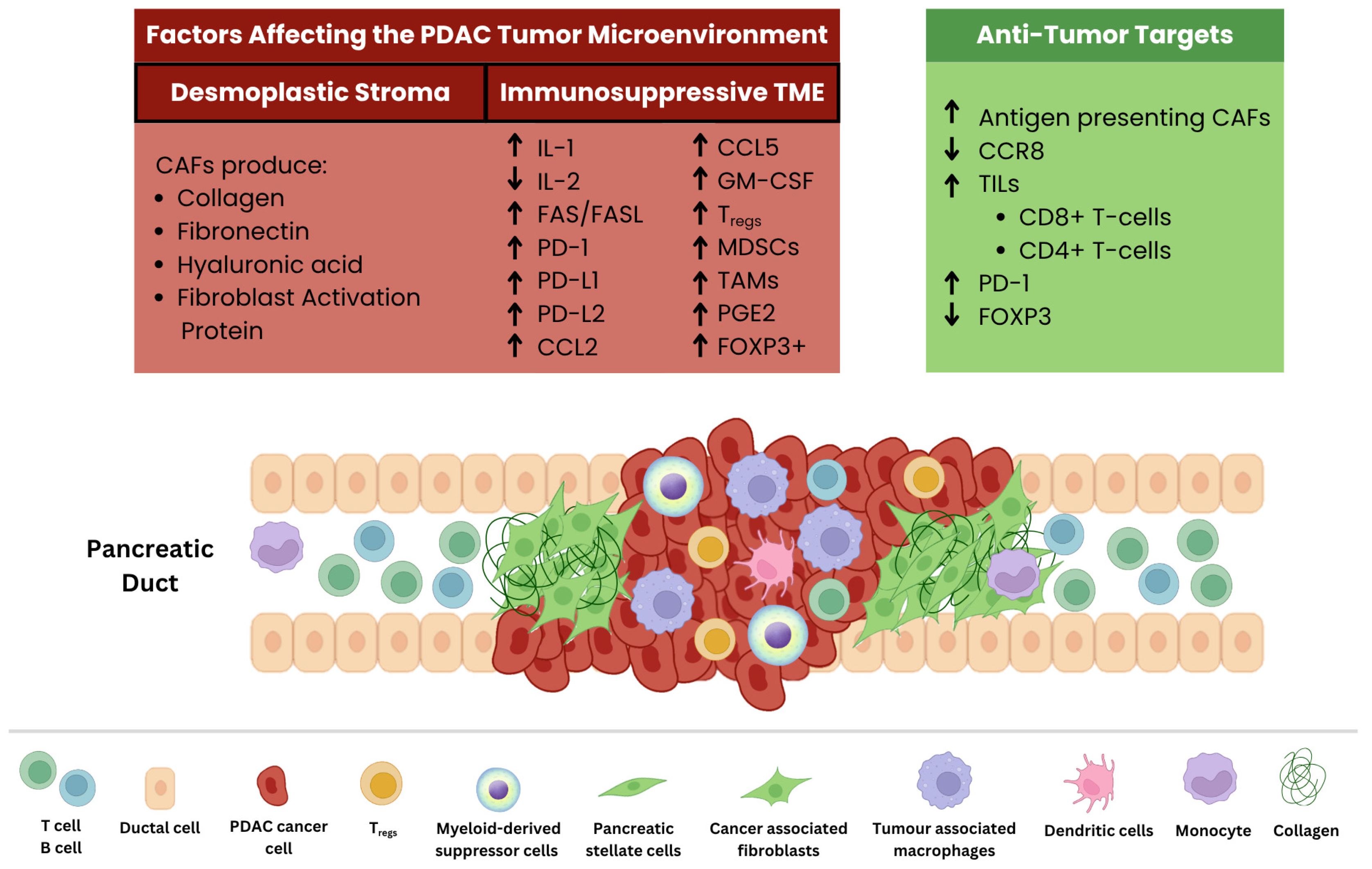
Figure 3.
An overview of pre-clinical models used in PDAC research. Two-dimensional or monolayer cultures are commonly used for simplistic direct assays. Adherent monolayer cultures display morphological characteristics that may help improve understanding of specific cell types. Animal models include GEMMs and PDX murine models; GEMMs are formed from genetic knock-ins, whereas PDX are established by injecting either commercially available immortalized human cell lines (2D) or dissociated cells from patient tumor resections. Injections are either done under the skin (subcutaneous) or into the pancreas (orthotopic). 3D models include multicellular aggregates called heterospheroids or organoids formed from patient tissue samples (PDOs). These models were then used in experimental systems involving multiple biological niches or heterotypic approaches; tumor-immune and microbe-tumor-immune (MTI). Under heterotypic tumor-immune established systems in PDAC, the InterOMax model and microfluidic tumor-on-a-chip both include PDOs, stromal cells like human PSCs or hPSCs (InterOMax used primary PSCs from patients, whereas tumor-on-a-chip used human cell lines) and immune cells. The MTI model used heterospheroids in combination with bacteria isolated from patients with Intraductal Papillary Mucinous Neoplasm (IPMN). After co-incubation, Mucosal-associated invariant T-cells (MAIT) were added to the heterotypic culture. Adapted from [8,9,11,54,61,115,142,146].
Figure 3.
An overview of pre-clinical models used in PDAC research. Two-dimensional or monolayer cultures are commonly used for simplistic direct assays. Adherent monolayer cultures display morphological characteristics that may help improve understanding of specific cell types. Animal models include GEMMs and PDX murine models; GEMMs are formed from genetic knock-ins, whereas PDX are established by injecting either commercially available immortalized human cell lines (2D) or dissociated cells from patient tumor resections. Injections are either done under the skin (subcutaneous) or into the pancreas (orthotopic). 3D models include multicellular aggregates called heterospheroids or organoids formed from patient tissue samples (PDOs). These models were then used in experimental systems involving multiple biological niches or heterotypic approaches; tumor-immune and microbe-tumor-immune (MTI). Under heterotypic tumor-immune established systems in PDAC, the InterOMax model and microfluidic tumor-on-a-chip both include PDOs, stromal cells like human PSCs or hPSCs (InterOMax used primary PSCs from patients, whereas tumor-on-a-chip used human cell lines) and immune cells. The MTI model used heterospheroids in combination with bacteria isolated from patients with Intraductal Papillary Mucinous Neoplasm (IPMN). After co-incubation, Mucosal-associated invariant T-cells (MAIT) were added to the heterotypic culture. Adapted from [8,9,11,54,61,115,142,146].
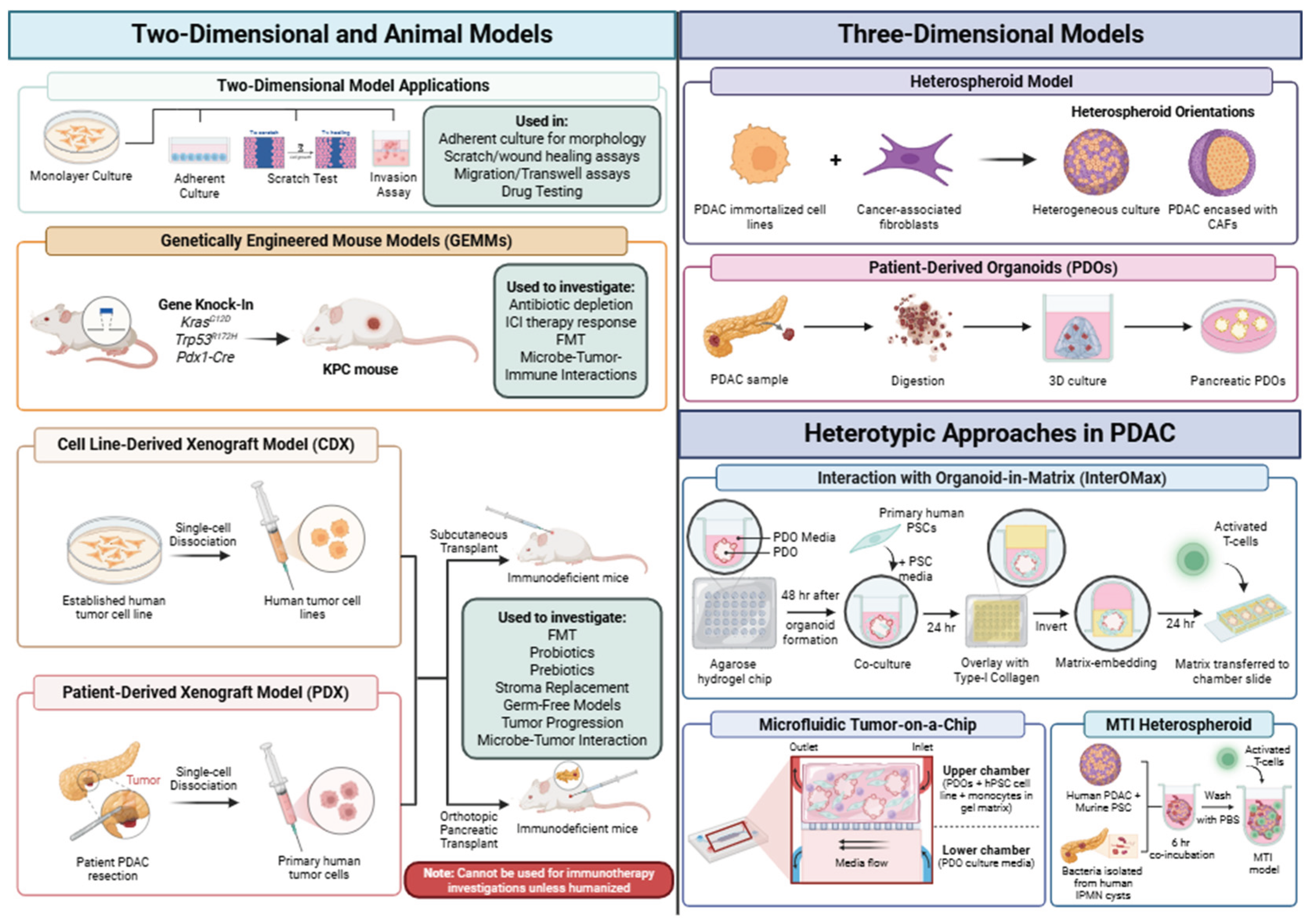
Disclaimer/Publisher’s Note: The statements, opinions and data contained in all publications are solely those of the individual author(s) and contributor(s) and not of MDPI and/or the editor(s). MDPI and/or the editor(s) disclaim responsibility for any injury to people or property resulting from any ideas, methods, instructions or products referred to in the content. |
© 2026 by the authors. Licensee MDPI, Basel, Switzerland. This article is an open access article distributed under the terms and conditions of the Creative Commons Attribution (CC BY) license (http://creativecommons.org/licenses/by/4.0/).
Copyright: This open access article is published under a Creative Commons CC BY 4.0 license, which permit the free download, distribution, and reuse, provided that the author and preprint are cited in any reuse.