

Submitted:
20 May 2026
Posted:
20 May 2026
You are already at the latest version
Abstract
The World Health Organization recognizes mosquito-borne viruses as a global public health threat with pandemic potential and has called on countries to prepare for the next pandemic. ZIKV is an important mosquito-borne virus with pandemic potential. Its unique neurotropism and vertical transmission capacity constitute special pathogenic and transmission risks. The prevention and control of ZIKV faces significant challenges due to the lack of vaccines and therapeutics. ZIKV entry into host cells is the initial step in establishing infection, and research on its entry mechanisms provides the fundamental theoretical basis for the development of anti-ZIKV entry inhibitory drugs. This review will summarize recent research findings from the perspectives of ZIKV receptor selection, membrane fusion mechanisms, and entry inhibitor research, aiming to provide concise information for a deeper understanding of ZIKV entry mechanisms.
Keywords:
zika virus
; receptor
; membrane fusion
1. Introduction
The introduction should briefly place the study in a broad context and highlight why it is important. ZIKV was first identified in 1947 through a yellow fever surveillance network in arhesus monkey from the Zika Forest of Uganda. It is primarily transmitted by Aedes mosquitoes. The natural reservoir of ZIKV remains unclear, with evidence indicating that the virus mainly circulates among wild primates and Aedes mosquitoes. In May 2015, Brazil confirmed its first cases of ZIKV infection, and the virus quickly spread from Brazil to numerous countries[1]. That same year, the WHO declared the ZIKV outbreak a Public Health Emergency of International Concern. The incubation period for ZIKV infection is not completely defined but is generally estimated to be 3-12 days. Approximately 80% of infections are asymptomatic, while about 20% present with mild symptoms[1]. In addition to mosquito-borne transmission, ZIKV can also be spread through blood transfusion, sexual contact, and mother-to-child transmission, making its prevention and control particularly challenging[2].
ZIKV infection can lead to severe neurological disorders such as Guillain-Barré syndrome. The virus can cross the blood—placental barrier, infect the fetus, and disrupt embryonic development, potentially resulting in fetal death or microcephaly in newborns. During the 2015-2017 outbreak in South America, thousands of microcephaly cases were reported in newborns within less than a year[3]. ZIKV can cross the blood-testis barrier, infect the male reproductive tract, and persist for prolonged periods. Due to high viral loads found in semen, ZIKV has the potential to be transmitted to females via sexual contact[4].
The molecular mechanisms by which ZIKV enters host cells are not yet fully understood, posing a major bottleneck in the study of ZIKV infection mechanisms and the development of specific entry inhibitors. The attachment of ZIKV to host cell receptors is a critical initial step in viral entry. The entire process, from virion contact with the cell surface to membrane fusion, requires coordinated involvement of multiple host molecules[1]. ZIKV adhesion receptors typically mediate weak initial interactions between the virion and the cell surface, after which the virus forms stronger bonds with other receptor molecules. Within the unique environment of the endosome, ZIKV particles may further engage endosomal receptors, adopting structural states that facilitate membrane fusion. Studies have shown that various host molecules, including ITGB4, AXL, DC-SIGN, and TIM-1, mediate ZIKV entry in different cell types. This review summarizes the current understanding of the molecular mechanisms of ZIKV entry and discusses existing challenges and future research directions in this field.
2. Structure of ZIKV
The ZIKV virion is a small spherical particle approximately 50 nanometers in diameter. Its structure from the inside out consists of: core RNA genome,C protein, prM and E protein (Figure 1a). ZIKV contains a single-stranded, positive-sense RNA genome, which can be immediately utilized by the protein synthesis machinery[5].
prM and E protein form regular dimeric structures on the viral surface and are responsible for receptor binding on the host cell surface. Under appropriate conditions, the E protein undergoes conformational changes that mediate the fusion of the viral envelope with the host cell membrane. As the major viral antigen: The E protein is the primary target recognized and attacked by the host immune system (particularly neutralizing antibodies)[6]. Its variations can affect viral virulence and transmission efficiency.
The E protein is a type I transmembrane glycoprotein with a molecular weight of approximately 53 kDa. It exists on the viral surface as head-to-tail homodimers. Each virion has approximately 180 copies of the E protein. These dimers are arranged in a specific geometric pattern, forming the smooth surface of the virion17. Structurally, the E protein monomer can be divided into three distinct domains (Figure 1b), highly similar to other flaviviruses but with its unique characteristics:
EDI—Central domain: Serves as the structural backbone, connecting the other two domains, and contains a unique N-linked glycosylation site (at Asn154). This glycosylation site is crucial for the ZIKV neurotropism and vertical transmission[7]. It might mediate virus entry into neural precursor cells and placental cells by interacting with specific receptors on the host cell surface[7].
EDII—Dimerization domain: Mediates the formation of E protein dimers and contains the fusion loop. This is an elongated, finger-like structure ending in a highly conserved, hydrophobic amino acids. The fusion loop is involved in the virus entry process. During the virus entry process, the fusion loop inserts into the inner membrane of the host cell, initiating the membrane fusion. This region is the target of many cross-reactive antibodies.However, because its sequence is relatively conserved among flaviviruses, the resulting neutralizing antibodies are generally not very potent and can sometimes even lead to ADE[8].
EDIII—Receptor-binding domain: This is an immunoglobulin-like domain located at the C-terminal of E protein and is responsible for the attachment to target cells[9]. This is the receptor-binding domain and the primary target of the most potent neutralizing antibodies[6]. It features a typical immunoglobulin-like fold[9]. This domain is highly variable among flaviviruses and determines the serotype specificity of the virus[6]. Antibodies targeting EDIII usually exhibit high neutralizing potency and strong specificity, and are less likely to cause ADE[6].
3. ZIKV Entry Pathways
ZIKV employs diverse endocytic pathways to enter host cells, a critical first step in establishing infection. The specific entry mechanism can vary depending on the host cell type, which contributes to ZIKV’s broad tropism and pathogenicity.
ZIKV primarily enters host cells via clathrin-mediated endocytosis (Figure 1c)[10]. During the infection stage in human microglial cells (CHME3) and fibroblast cells (HT1080), ZIKV binds to cell receptors and is transported to clathrin-coated pit regions. Chlorpromazine prevents ZIKV entry into human glioblastoma cells T98G by inhibiting the formation of clathrin-coated pits on the cell surface[10]. Knocking down the clathrin heavy chain in T98G cells or CHME3 cells significantly reduces the Zika virus infection rate[11]. With the involvement of molecules like dynamin, clathrin invaginates and promotes the pinching off of the plasma membrane, forming endocytic vesicles. These vesicles further develop into early endosomes. Within endosomes, as the pH decreases, the E envelope protein undergoes conformational changes. The E protein dimers expose their membrane fusion peptide located in domain II; this fusion peptide inserts into the endosomal membrane, further leading to membrane fusion and the release of the viral genome, thus completing entry.
Furthermore, ZIKV can also enter T98G cells via a non-clathrin pathway, namely caveola-mediated endocytosis (Figure 1c)[10]. Studies have shown that both siRNA targeting caveolin-1 and the overexpression of a dominant-negative caveolin-1 can inhibit caveolae-mediated endocytosis and significantly reduce ZIKV’s ability to enter cells[10].
4. Receptor Selection of ZIKV
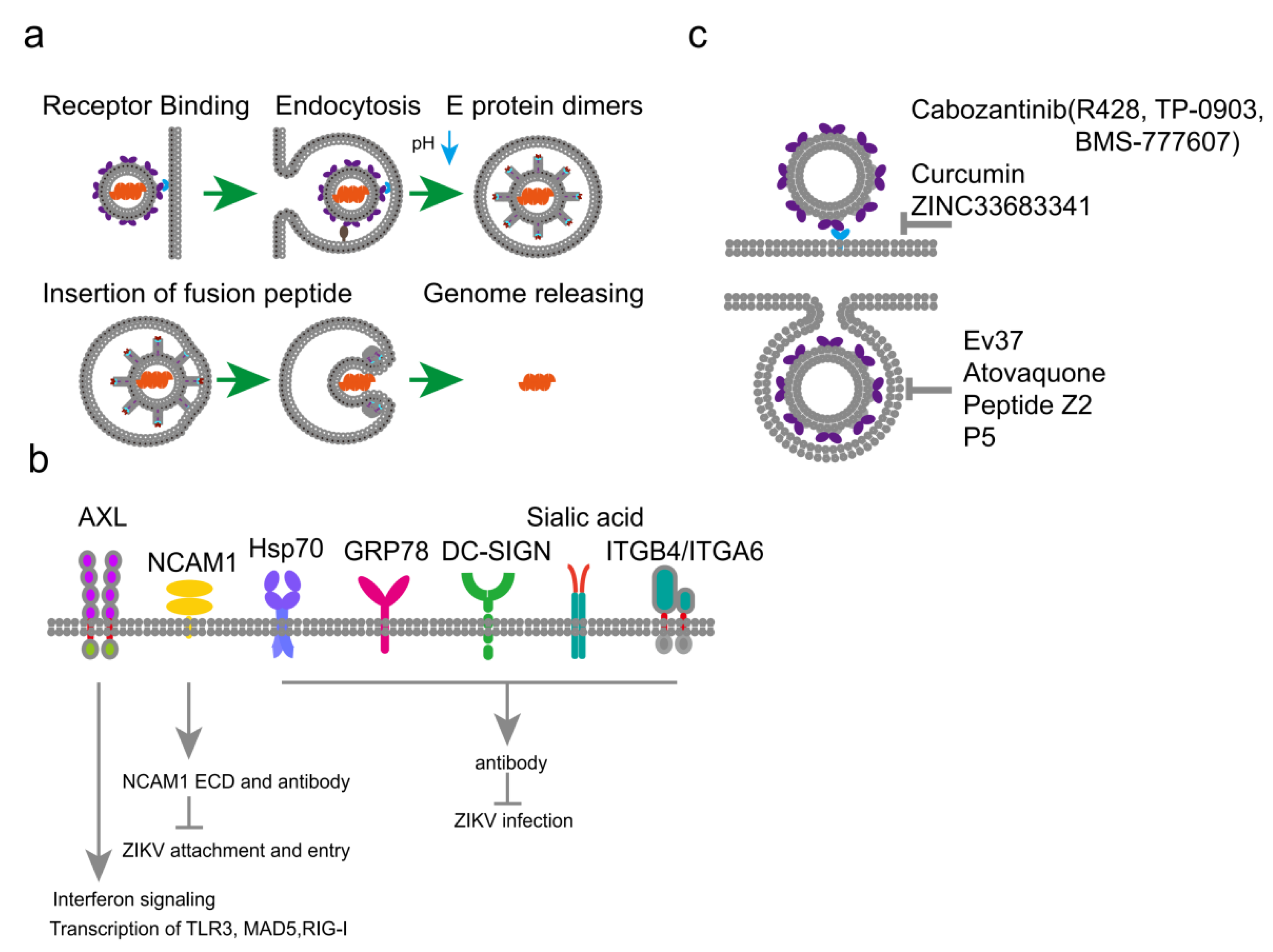
The entry process was conducted by the coordination of multiple receptor molecules. ZIKV adhesion receptors are typically responsible for the initial weak interaction binding between the virion and the cell surface, after which the virion forms tighter bonds with other receptor molecules. Within the unique environment of the endosome, ZIKV virion forms structural features conducive to membrane fusion. Research has shown that various molecules such as AXL, DC-SIGN and integrins mediate ZIKV entry in different tissue cells(Figure 2a).
TAM Receptor AXL
Expression profile analysis of neural cells revealed a positive correlation between AXL expression levels and the tropism of ZIKV for different types of neural cells, leading to the hypothesis that AXL might be a cellular receptor for ZIKV[12] . Related cytological experiments have also confirmed the role of Axl and its ligand Gas6 in mediating ZIKV infection in various neural cells, including glioblastoma cells, human glial cells, and astrocytes[13]. However, Recent research by Wells et al. showed that effective ZIKV infection still occurred in neural precursor cells after AXL gene knockout[14]. AXL might primarily influence ZIKV replication by regulating the interferon pathway rather than acting solely as the essential entry receptor[15].
Axl indirectly binds ZIKV via the Gas6 bridge and mediates virus entry through the clathrin-mediated endocytosis pathway. The ZIKV/Gas6 complex downregulates the interferon signaling pathway by activating Axl kinase activity, thereby promoting viral infection. Studies indicate that in skin cells, Axl acts as a cell surface attachment factor for ZIKV, and both endocytosis and acidic pH are required to establish effective infection. Axl also promotes the formation of autophagosomes in skin cells, a process associated with enhanced ZIKV infection.
In pregnant mice, there were no significant differences in ZIKV RNA levels in the brains and spleens among wild-type, Axl knockout, and Mertk knockout mice[16]. Similarly, non-pregnant Axl knockout female mice treated with the IFNAR-blocking antibody also showed same brain viral RNA levels comparable to wild-type mice[16]. Similar patterns of viral RNA distribution was observed in the placentas of wild-type, Axl knockout, and Axl/Mertk double knockout mice[16] . These results indicate that in IFNAR-blocked mice, Axl and Mertk are not essential receptors for ZIKV infection[16]. Even in the absence of Axl or Mertk receptors, transplacental transmission can still occur, and the virus can replicate in placental and fetal tissues[16].
NCAM1
NCAM1 is highly expressed in the brain. Its overexpression in HEK293T cells promotes ZIKV attachment and entry[17]. Knocking down NCAM1 in glioblastoma cells significantly reduces ZIKV infection[17]. Co-immunoprecipitation experiments showed that NCAM1 can interact with ZIKV E envelope protein[17]. Both the NCAM1 extracellular domain and anti-NCAM1antibodies can competitively inhibit the binding activity between ZIKV and NCAM1, significantly suppressing ZIKV binding and entry into U-251 MG cells[17].
Hsp70
Hsp70 plays a key role in multiple stages of ZIKV replication[18]. Hsp70 is distributed both on the cell surface and inside the cell. During the ZIKV entry stage, cell surface Hsp70 interacts with ZIKV and enhances the efficiency of ZIKV infection and entry[18]. Anti-Hsp70 antibodies inhibit ZIKV infection rates and reduce virion production[18]. Furthermore, co-incubating ZIKV with recombinant human Hsp70 protein reduces the copy number of virus particles in the supernatant[18]. Inside infected cells, ZIKV double-stranded RNA and Hsp70 show co-localization, and Hsp70 is part of the viral replication complex[18]. In summary, Hsp70is involved in both the entry and replication processes of ZIKV[18].
GRP78
An interaction between GRP78 and the ZIKV E envelope protein was detected in human A549 cells[19]. A monoclonal antibody targeting the N-terminus of GRP78 was able to hinder ZIKV entry into host cells, significantly reducing the viral infection rate and replication levels[20]. Knocking down GRP78 expression similarly reduced ZIKV entry, indicating that GRP78 is involved in ZIKV binding to cells[20]. Furthermore, the absence of GRP78 affects ZIKV’s ability to impair host cell translation and alters the localization of viral replication factories, but does not affect viral RNA synthesis[19]. These findings suggest that GRP78 is an important host factor in the ZIKV infection process, potentially involved in coordinating the construction of viral replication factories[19].
Sialic Acid
Sialic acid on the host cell surface plays a significant role in ZIKV entry[21]. In Vero cells and human induced pluripotent stem cell-derived neural precursor cells, removing cell surface sialic acid with neuraminidase significantly reduced the ZIKV infection rate[21]. Additionally, although Vero cells lacking sialic acid remained susceptible to ZIKV, the infection efficiency of both the African strain (MR766) and the Asian strain (PF13) was significantly reduced[21]. In 2,3-linked sialic acid-deficient Huh7 cells, the ZIKV infection rate was also significantly reduced[21]. At 4 °C, there was no significant difference in the attachment amount of ZIKV particles between neuraminidase-treated and untreated cells, confirming that sialic acid is not essential for initial ZIKV attachment; however, when cells were incubated at 37 °C (to promote internalization) and then treated with pronase to remove surface-attached but non-internalized virus particles, a significant reduction in viral RNA content was observed in neuraminidase-treated Vero cells[21].
C-type Lectin Receptor DC-SIGN
DC-SIGN is directly involved in the binding and transmission process of Zika virus[22]. DC-SIGN is abundantly expressed on the surface of immature macrophages and dendritic cells. Compared to parental Raji cells or Raji cells expressing Langerin, Raji cells expressing DC-SIGN efficiently bound Zika virus[22]. This binding could be blocked by DC-SIGN specific antibodies, but not by isotype control antibodies[22]. Compared to Langerin-Raji cells, DC-SIGN-Raji cells successfully transmitted the virus to target cells after 4 hours of co-culture with Zika virus[22]. A polysaccharide that blocks C-type lectin receptors and a DC-SIGN specific antibody both inhibited DC-SIGN-Raji cell-mediated ZIKV infection[22].
Besides ZIKV, DC-SIGN mediates the entry of various other flaviviruses including DENV, WNV, and JEV[22]. The CRD domain of DC-SIGN can bind to the envelope proteins of multiple flaviviruses[22]. The CRD domain in is crucial for DC-SIGN’s function as a viral receptor[22].
Glycosaminoglycans
Negatively charged glycosaminoglycans can mediate ZIKV entry[23]. Placental-derived glycosaminoglycans can bind to the ZIKV E protein, mediating adhesion between ZIKV and cells[23]. Glycosaminoglycans mediate adhesion for various viruses beyond flaviviruses, such as influenza virus, SARS-CoV-2, and herpes simplex virus, suggesting that glycosaminoglycans function as broad-spectrum adhesion molecules rather than specific receptors for ZIKV.
Integrins
Integrin αvβ5 internalizes Zika virus during neural stem cells infection[24]. The cell surface integrin α6β4 (ITGA6/ ITGB4) can bind to the ZIKV E protein and mediate viral entry into various cells. Antibodies targeting ITGB4 prevented ZIKV infection and damage in placental tissue, reduced ZIKV titers in embryos, and increased embryo survival rates. The ITGB4/ITGA6 heterodimer is expressed on the surface of various central nervous system cells such as glial cells, Schwann cells, and neural stem cells, mediating neural stem cell differentiation and enhancing the strength of the blood-brain barrier by anchoring astrocytes to the vascular basement membrane[25].
5. ZIKV Membrane Fusion Mechanisms
Given the high structural similarity between ZIKV virions and DENV, it is inferred that their membrane fusion mechanisms are similar (Figure 2b). The fusion process is triggered by the acidic environment of the endosome as the virion is internalized by the host cell. This drop in pH induces a dramatic conformational change in the E proteins. Protons bind to specific histidine residues, destabilizing the dimeric interactions. The E protein dimers dissociate into monomers. This dissociation is crucial as it exposes the previously buried fusion loops and liberates the domains to undergo large-scale rearrangements.
Following dissociation, the monomers rapidly reassociate into homotrimers. This is a key transitional state. During trimer formation, the E proteins extend away from the viral surface, projecting the now-exposed hydrophobic fusion loops at the tip of EDII towards the endosomal membrane. The fusion loops directly insert into the lipid bilayer of the host membrane. At this stage, the virus is essentially stapled to the host membrane via the E protein trimers, with one end in the viral membrane and the fusion loop end in the host membrane. EDIII remains largely associated with the core of this extended trimer.
The most dramatic and energetically favorable step is the “jack-knife” refolding of EDIII. In the extended trimer, EDIII and the C-terminal stem swing upwards by over 100A, folding back towards the viral membrane and the trimer core. This refolding motion is like closing a hinge[26]. It physically pulls the host membrane, into which the fusion loops are embedded, and the viral membrane, to which the protein is anchored, into close apposition. The energy released during this refolding from a high-energy intermediate to a stable post-fusion hairpin is used to overcome the repulsive forces between the two lipid bilayers. The immense force and proximity generated by this action destabilize the outer leaflets of both membranes, leading to hemifusion. In hemifusion, the outer leaflets have merged, forming a hemifusion stalk, but the inner leaflets and aqueous contents remain separate.
The hemifusion stalk is an unstable intermediate. The continued reorganization of the lipids, driven by the stable, low-energy post-fusion hairpin structure of the E protein trimer, progresses to the fusion of the inner membrane leaflets. This opens a small, aqueous fusion pore. The pore then rapidly expands, allowing the viral nucleocapsid to be released into the cytoplasm, thereby completing the infection process. In its final, stable post-fusion state, the E protein trimer resembles a hairpin, with DIII and the stem packed tightly against the central trimer core formed by EDI and EDII, locking the fusion process in an irreversible state (Figure 5) [27].
6. ZIKV Entry Inhibitors
Given that host cell entry represents the initial stage of Zika virus infection, targeting the entry process mediated by the viral surface E protein has become a central strategy in antiviral therapy. Currently, ZIKV entry inhibitors at the preclinical stage mainly fall into two categories: antibody-based therapeutics that functioned by neutralizing viral infectivity, and antiviral agents designed to block viral attachment and other essential entry steps.
Small Molecule Compounds Inhibiting ZIKV Entry:
The majority of currently reported ZIKV entry inhibitors function by binding to the viral E protein to block viral entry (Figure 2c). For instance, EGCG, a polyphenolic compound present in green tea exhibits antiviral activity against various ZIKV strains[28]. This compound exerts its inhibitory effect by binding to the EDI and EDII domains of the E protein, thereby preventing the conformational changes in EDIII required for viral entry. Despite its antiviral properties, the catechol group in EGCG may cause non-specific inhibition of multiple cellular targets. Digitonin, an amphiphilic steroidal saponin classified as a terpenoid, displays antiviral activity against multiple ZIKV strains by inhibiting the interaction between E protein and host cells[29]. Gossypol, a polyphenolic compound, potently inhibits different ZIKV strains through high-affinity binding to the E protein, particularly the EDIII domain[30]. Its derivative, ST087010, operates via the same mechanism but exhibits more potent inhibition[30]. Atranorin, protects human glioblastoma cells from ZIKV infection by directly targeting the E protein, thereby disrupting viral entry and reducing infectivity[31]. Tannic acid directly binds to the E protein and induces conformational changes in several residues critical for dimerization and membrane fusion, thus preventing ZIKV infection[29].
Curcumin inhibits ZIKV at the initial phase of infection by interacting with the E protein to block virus binding to cell-surface receptors[32]. It suppresses ZIKV infection in a dose-dependent manner across various human cell lines, including HeLa, BHK-12, and Vero 6 cells[32]. Palmatine, a protoberberine alkaloid, also acts during early infection stages by interacting with the E protein to affect ZIKV binding to host receptors, as well as viral entry and stability[29]. ZINC23845959, ZINC23400466, and ZINC12415353 were identified as inhibitors that bind to the E protein via a β-octyl glucoside binding site, significantly suppressing ZIKV infection in Vero cells[33].
Structure-based virtual screening revealed that the small molecule F1065-0358 effectively inhibits ZIKV replication in Vero cells[29]. All-atom molecular dynamics simulations verified its stable binding to conserved residues in the EDI (His144, Phe183) and EDIII (Lys301, Tyr305, Asn362) domains, preventing E protein trimerization during membrane fusion[29]. This molecule shows potential for inhibiting other flaviviruses[29]. Cyano hydrazone compounds 3-110-22, 3-110-2, and 3-149-3 target a pocket at the EDI-EDII interface of the ZIKV E protein, which is essential for low pH-induced conformational changes[29].
Screening of two compound libraries using a competitive amplified luminescent proximity homogeneous assay identified LAS52154459[29]. It functions by inhibiting E protein-mediated membrane fusion but with low antiviral activity. A pyrimidine derivative designated compound 16a, discovered via structure-based computer-aided drug design, also inhibits ZIKV infection[29]. It acts specifically during virus internalization and post-internalization steps, likely by blocking E-mediated membrane fusion. Screening an FDA-approved drug library identified atovaquone as an inhibitor of ZIKV and four distinct serotypes of DENV, suggesting its potential as a pan-flavivirus entry inhibitor[29].
Monoclonal Antibodies Inhibiting ZIKV Entry:
Neutralizing antibodies can be used for the prevention and treatment of ZIKV infection. Researchers have isolated several specific/cross-reactive monoclonal antibodies. Structural biology studies reveal that, for instance, the C10 monoclonal antibody binds to specific residues at the intradimer interface of the E protein, locking the E protein structure under acidic conditions and thereby hindering the conformational changes of the E protein required for membrane fusion. The anti-ZIKV antibody 2A10G6 recognizes the fusion loop and effectively neutralizes ZIKV both in vitro and in mouse models[34]. Five ZIKV-neutralizing monoclonal antibodies isolated from E80-immunized mice specifically bound to and neutralized Asian lineage ZIKV strains. They all recognize a novel linear epitope located on the glycan loop of E protein domain. Among them, the 5F8 mAb primarily inhibits early post-attachment entry steps.
Specific human monoclonal antibodies isolated from a single ZIKV-infected patient, Z23 and Z3L1, exhibited potent ZIKV-specific neutralizing activity in vitro[6]. The human mAb ZIKV-117 neutralizes ZIKV strains belonging to both African and Asian-American lineages[35]. It significantly reduced ZIKV infection of the placenta and fetus and decreased mortality in mice[35]. A Zika virus-specific IgM termed DH1017, isolated from a ZIKV-infected pregnant woman, demonstrated ultrapotent neutralization dependent on its IgM isotype and conferred protection in mice[36]. Furthermore, antibodies from dengue patients with prior exposure to Japanese encephalitis virus can exhibit broadly neutralizing activity against ZIKV[37].
Characterization of highly specific monoclonal antibodies targeting the glycan loop of the Zika virus envelope protein, such as clones A11 and A42, has been reported[38]. The oligomeric state of the ZIKV E protein is critical for defining protective immune responses, with stable recombinant homodimers inducing neutralizing antibodies in mice that recognize epitopes similar to those targeted by human antibodies from ZIKV-infected individuals[38]. The development of neutralizing antibodies against Zika virus often leverages structural information on the envelope protein.
The efficacy of many human mAbs has been tested in animal models. For example, the therapeutic efficacy of ZIKV-117 was evaluated in type I IFN-deficient pregnant mice, where the mAb treatment markedly reduced tissue pathology, placental and fetal infection, and mortality. A cytotoxic-skewed immune set point has been identified as a predictor for low neutralizing antibody levels after Zika virus infection. The isolation of a human inferred germline antibody that binds to an immunodominant epitope and neutralizes Zika virus has also been described[39].
Antibodies from dengue patients with prior exposure to JEV are broadly neutralizing against Zika virus, and a rare class of ZIKV-cross-reactive human monoclonal antibodies with increased somatic hypermutation and broad neutralization against multiple flaviviruses has been identified. Furthermore, a subset of human monoclonal antibodies isolated from subjects previously infected with ZIKV recognize diverse epitopes on the E protein and exhibit potent neutralizing activity[40]. One study recorded longitudinal antibody responses against ZIKV during natural infection, showing that mAbs isolated during early infection were largely EDI/EDII specific and weakly neutralizing, whereas DIII-specific neutralizing activity increased over time[6].
7. Discussion
ZIKV entry into host cells is a critical determinant of its pathogenesis, tropism, and the severe clinical outcomes associated with infection. Despite significant advances, the precise mechanisms governing viral receptor engagement, internalization, and membrane fusion remain incompletely elucidated, presenting substantial challenges and directing future research works.
A challenge in ZIKV research is the identification of a specific, ubiquitously expressed receptor. The lack of a consensus primary receptor obscures a comprehensive understanding of its cell tropism. ZIKV likely utilizes cell-type-specific entry factors, enabling broad tissue infectivity. This receptor redundancy may represent an adaptive strategy for effective infection but complicates therapeutic strategies targeting viral entry at the receptor level. In addition, the precise molecular mechanisms governing fusion remain incompletely resolved. While low pH is required to induce conformational changes in the E glycoprotein into a fusogenic state, the kinetics and regulation of the actual fusion event may involve additional host factors.
Future investigations should focus on several critical avenues. First, there is a need to synthesize disparate receptor studies into a cohesive model explaining how ZIKV selects entry portals across different tissues in vivo. Second, advanced structural biology approaches, particularly cryo-electron tomography, should be applied to visualize the dynamics of fusion in situ within native cellular contexts. Third, combining AI-driven drug design with deeper mechanistic insights into viral entry may accelerate the development of novel broad-spectrum antivirals and next-generation vaccine platforms effective against ZIKV.
Author Contributions
Conceptualization, H.C. and X.H.; investigation, W.Z.; resources, H.C.and X.Z.; writing—original draft preparation, X.H., W.L. and R.L.; writing—review and editing, H.C.; supervision, W.Z.; project administration, X.X.; funding acquisition, H.C. and X.Z; All authors have read and agreed to the published version of the manuscript.
Funding
This research was funded by Basic Scientific Research Foundation of the Chinese Academy of Quality and Inspection & Testing, grant number 2025JK035, Hainan Provincial Natural Science Foundation of China, grant number 324MS134 and 325MS154, Basic Scientific Research Funds for Inner Mongolia Autonomous Region Government-Directly Affiliated Universities- “Four Prestigious Journals” Landmark Papers “Open Bidding and Leadership Initiative” Project , grant number BR251404.
Acknowledgments
We would like to express our sincere gratitude to all those who have contributed to this work.
Conflicts of Interest
The authors declare no conflicts of interest.
Abbreviations
The following abbreviations are used in this manuscript:
| ZIKV | Zika virus |
| WHO | World Health Organization |
| ADE | Antibody dependent enhancement |
| DENV | Dengue Virus |
| JEV | Japanese Encephalitis Virus |
| WNV | West Nile Virus |
| FDA | Food and Drug Administration |
References
- Zanluca, C.; Melo, V.C.; Mosimann, A.L.; Santos, G.I.; Santos, C.N.; Luz, K. First report of autochthonous transmission of Zika virus in Brazil. Mem. Inst. Oswaldo Cruz 2015, 110, 569–572. [Google Scholar] [CrossRef] [PubMed]
- Musso, D.; Roche, C.; Robin, E.; Nhan, T.; Teissier, A.; Cao-Lormeau, V.M. Potential sexual transmission of Zika virus. Emerg. Infect. Dis. 2015, 21, 359–361. [Google Scholar] [CrossRef] [PubMed]
- de Oliveira, W.K.; de França, G.V.A.; Carmo, E.H.; Duncan, B.B.; de Souza Kuchenbecker, R.; Schmidt, M.I. Infection-related microcephaly after the 2015 and 2016 Zika virus outbreaks in Brazil: a surveillance-based analysis. Lancet 2017, 390, 861–870. [Google Scholar] [CrossRef]
- Mead, P.S.; Duggal, N.K.; Hook, S.A.; Delorey, M.; Fischer, M.; Olzenak McGuire, D.; et al. Zika virus shedding in semen of symptomatic infected men. N. Engl. J. Med. 2018, 378, 1377–1385. [Google Scholar] [CrossRef]
- Shang, Z.; Song, H.; Shi, Y.; Qi, J.; Gao, G.F. Crystal structure of the capsid protein from Zika virus. J. Mol. Biol. 2018, 430, 948–962. [Google Scholar] [CrossRef]
- Stettler, K.; Beltramello, M.; Espinosa, D.A.; Graham, V.; Cassotta, A.; Bianchi, S.; et al. Specificity, cross-reactivity, and function of antibodies elicited by Zika virus infection. Science 2016, 353, 823–826. [Google Scholar] [CrossRef]
- Fontes-Garfias, C.R.; Shan, C.; Luo, H.; Muruato, A.E.; Medeiros, D.B.A.; Mays, E.; et al. Functional analysis of glycosylation of Zika virus envelope protein. Cell Rep. 2017, 21, 1180–1190. [Google Scholar] [CrossRef]
- Dejnirattisai, W.; Supasa, P.; Wongwiwat, W.; Rouvinski, A.; Barba-Spaeth, G.; Duangchinda, T.; et al. Dengue virus sero-cross-reactivity drives antibody-dependent enhancement of infection with Zika virus. Nat. Immunol. 2016, 17, 1102–1108. [Google Scholar] [CrossRef]
- Rey, F.A.; Heinz, F.X.; Mandl, C.; Kunz, C.; Harrison, S.C. The envelope glycoprotein from tick-borne encephalitis virus at 2 Å resolution. Nature 1995, 375, 291–298. [Google Scholar] [CrossRef]
- Li, M.; Zhang, D.; Li, C.; Zheng, Z.; Fu, M.; Ni, F.; et al. Characterization of Zika virus endocytic pathways in human glioblastoma cells. Front. Microbiol. 2020, 11, 242. [Google Scholar] [CrossRef] [PubMed]
- Yu, W.; Tao, J.; Cao, H.; Zheng, W.; Zhang, B.; Zhang, Y.; et al. The HAVCR1-centric host factor network drives Zika virus vertical transmission. Cell Rep. 2025, 44, 115464. [Google Scholar] [CrossRef]
- Retallack, H.; Di Lullo, E.; Arias, C.; Knopp, K.A.; Laurie, M.T.; Sandoval-Espinosa, C.; et al. Zika virus cell tropism in the developing human brain and inhibition by azithromycin. Proc. Natl. Acad. Sci. USA 2016, 113, 14408–14413. [Google Scholar] [CrossRef]
- Komarasamy, T.V.; Adnan, N.A.A.; James, W.; Balasubramaniam, V. Zika virus neuropathogenesis: the different brain cells, host factors and mechanisms involved. Front. Immunol. 2022, 13, 773191. [Google Scholar] [CrossRef]
- Wells, M.F.; Salick, M.R.; Wiskow, O.; Ho, D.J.; Worringer, K.A.; Ihry, R.J.; et al. Genetic ablation of AXL does not protect human neural progenitor cells and cerebral organoids from Zika virus infection. Cell Stem Cell 2016, 19, 703–708. [Google Scholar] [CrossRef]
- Chen, J.; Yang, Y.F.; Yang, Y.; Zou, P.; Chen, J.; He, Y.; et al. AXL promotes Zika virus infection in astrocytes by antagonizing type I interferon signalling. Nat. Microbiol. 2018, 3, 302–309. [Google Scholar] [CrossRef]
- Hastings, A.K.; Yockey, L.J.; Jagger, B.W.; Hwang, J.; Uraki, R.; Gaitsch, H.F.; et al. TAM receptors are not required for Zika virus infection in mice. Cell Rep. 2017, 19, 558–568. [Google Scholar] [CrossRef]
- Mayank, S.; Ying, Z.; Jian, C.; Devika, S.; Andrew, S.M.; Yang, Z.; et al. Chemical proteomics tracks virus entry and uncovers NCAM1 as Zika virus receptor. Nat. Commun. 2020, 11(1), 3896. [Google Scholar] [CrossRef] [PubMed]
- Taguwa, S.; Yeh, M.T.; Rainbolt, T.K.; Nayak, A.; Shao, H.; Gestwicki, J.E.; et al. Zika virus dependence on host Hsp70 provides a protective strategy against infection and disease. Cell Rep. 2019, 26, 906–920.e3. [Google Scholar] [CrossRef] [PubMed]
- Royle, J.; Ramírez-Santana, C.; Akpunarlieva, S.; Donald, C.L.; Gestuveo, R.J.; Anaya, J.M.; et al. Glucose-regulated protein 78 interacts with Zika virus envelope protein and contributes to a productive infection. Viruses 2020, 12(5), 524. [Google Scholar] [CrossRef]
- Khongwichit, S.; Sornjai, W.; Jitobaom, K.; Greenwood, M.; Greenwood, M.P.; Hitakarun, A.; et al. A functional interaction between GRP78 and Zika virus E protein. Sci. Rep. 2021, 11(1), 393. [Google Scholar] [CrossRef] [PubMed]
- Tan, C.W.; Huan Hor, C.H.; Kwek, S.S.; Tee, H.K.; Sam, I.C.; Goh, E.L.K.; et al. Cell surface α2,3-linked sialic acid facilitates Zika virus internalization. Emerg. Microbes Infect. 2019, 8, 426–437. [Google Scholar] [CrossRef]
- Carbaugh, D.L.; Baric, R.S.; Lazear, H.M. Envelope protein glycosylation mediates Zika virus pathogenesis. J. Virol. 2019, 93(12), e00113-19. [Google Scholar] [CrossRef]
- Pomin, V.H. Sulfated glycans and related digestive enzymes in the Zika virus infectivity: potential mechanisms of virus–host interaction and perspectives in drug discovery. Interdiscip. Perspect. Infect. Dis. 2017, 2017, 4894598. [Google Scholar] [CrossRef]
- Wang, S.; Zhang, Q.; Tiwari, S.K.; Lichinchi, G.; Yau, E.H.; Hui, H.; et al. Integrin αvβ5 internalizes Zika virus during neural stem cells infection and provides a promising target for antiviral therapy. Cell Rep. 2020, 30, 969–983.e4. [Google Scholar] [CrossRef]
- Wagner, S.; Tagaya, M.; Koziol, J.A.; Quaranta, V.; del Zoppo, G.J. Rapid disruption of an astrocyte interaction with the extracellular matrix mediated by integrin alpha 6 beta 4 during focal cerebral ischemia/reperfusion. Stroke 1997, 28, 858–865. [Google Scholar] [CrossRef]
- Zhang, Y.; Zhang, W.; Ogata, S.; Clements, D.; Strauss, J.H.; Baker, T.S.; et al. Conformational changes of the flavivirus E glycoprotein. Structure 2004, 12, 1607–1618. [Google Scholar] [CrossRef] [PubMed]
- Zhang, X.; Ge, P.; Yu, X.; Brannan, J.M.; Bi, G.; Zhang, Q.; et al. Cryo-EM structure of the mature dengue virus at 3.5-Å resolution. Nat. Struct. Mol. Biol. 2013, 20, 105–110. [Google Scholar] [CrossRef] [PubMed]
- Vázquez-Calvo, Á.; Jiménez de Oya, N.; Martín-Acebes, M.A.; Garcia-Moruno, E.; Saiz, J.C. Antiviral properties of the natural polyphenols delphinidin and epigallocatechin gallate against the flaviviruses West Nile virus, Zika virus, and dengue virus. Front. Microbiol. 2017, 8, 1314. [Google Scholar] [CrossRef]
- Roy, A.; Liu, Q.; Yang, Y.; Debnath, A.K.; Du, L. Envelope protein-targeting Zika virus entry inhibitors. Int. J. Mol. Sci. 2024, 25(17), 9424. [Google Scholar] [CrossRef]
- Gao, Y.; Tai, W.; Wang, X.; Jiang, S.; Debnath, A.K.; Du, L.; et al. A gossypol derivative effectively protects against Zika and dengue virus infection without toxicity. BMC Biol. 2022, 20(1), 143. [Google Scholar] [CrossRef] [PubMed]
- Huang, G.G.; Wang, H.Y.; Wang, X.H.; Yang, T.; Zhang, X.M.; Feng, C.L.; et al. Atranorin inhibits Zika virus infection in human glioblastoma cell line SNB-19 via targeting Zika virus envelope protein. Phytomedicine 2024, 125, 155343. [Google Scholar] [CrossRef]
- Mounce, B.C.; Cesaro, T.; Carrau, L.; Vallet, T.; Vignuzzi, M. Curcumin inhibits Zika and chikungunya virus infection by inhibiting cell binding. Antivir. Res. 2017, 142, 148–157. [Google Scholar] [CrossRef]
- Telehany, S.M.; Humby, M.S.; McGee, T.D., Jr.; Riley, S.P.; Jacobs, A.; Rizzo, R.C. Identification of Zika virus inhibitors using homology modeling and similarity-based screening to target glycoprotein E. Biochemistry 2020, 59, 3709–3724. [Google Scholar] [CrossRef] [PubMed]
- Zhao, H.; Fernandez, E.; Dowd, K.A.; Speer, S.D.; Platt, D.J.; Gorman, M.J.; et al. Structural basis of Zika virus-specific antibody protection. Cell 2016, 166, 1016–1027. [Google Scholar] [CrossRef] [PubMed]
- Sapparapu, G.; Fernandez, E.; Kose, N.; Bin, C.; Fox, J.M.; Bombardi, R.G.; et al. Neutralizing human antibodies prevent Zika virus replication and fetal disease in mice. Nature 2016, 540, 443–447. [Google Scholar] [CrossRef]
- Singh, T.; Hwang, K.K.; Miller, A.S.; Jones, R.L.; Lopez, C.A.; Dulson, S.J.; et al. A Zika virus-specific IgM elicited in pregnancy exhibits ultrapotent neutralization. Cell 2022, 185, 4826–4840.e17. [Google Scholar] [CrossRef] [PubMed]
- Salem, G.M.; Galula, J.U.; Wu, S.R.; Liu, J.H.; Chen, Y.H.; Wang, W.H.; et al. Antibodies from dengue patients with prior exposure to Japanese encephalitis virus are broadly neutralizing against Zika virus. Commun. Biol. 2024, 7, 15. [Google Scholar] [CrossRef]
- McLaury, A.R.; Haun, B.K.; To, A.; Mayerlen, L.; Medina, L.O.; Lai, C.Y.; et al. Characterization of two highly specific monoclonal antibodies targeting the glycan loop of the Zika virus envelope protein. Viral Immunol. 2024, 37, 167–175. [Google Scholar] [CrossRef]
- Magnani, D.M.; Silveira, C.G.T.; Rosen, B.C.; Ricciardi, M.J.; Pedreño-Lopez, N.; Gutman, M.J.; et al. A human inferred germline antibody binds to an immunodominant epitope and neutralizes Zika virus. PLoS Negl. Trop. Dis. 2017, 11, e0005655. [Google Scholar] [CrossRef]
- Rogers, T.F.; Goodwin, E.C.; Briney, B.; Sok, D.; Beutler, N.; Strubel, A.; et al. Zika virus activates de novo and cross-reactive memory B cell responses in dengue-experienced donors. Sci. Immunol. 2017, 2(14), eaan6809. [Google Scholar] [CrossRef]
Figure 1.
(a): Structure of the ZIKV virion, with the E protein homodimers on its surface highlighted in the white box. The E proteins (PDB:5JHM) are arranged in a head-to-tail manner, forming a stable structure. The transmembrane domains of E and M (PDB:5IRE) are embedded into the viral envelope. (b): Amino acid positions of the different domains within the E protein. Its EDIII and EDII domains are connected by the EDI domain. The EDIII domain contains the fusion loop (red), which becomes exposed in the post-fusion conformation of the E protein for insertion into the host cell membrane. GL 145-164 represents the N-linked glycosylation site (yellow). TM: transmembrane domain (brown). (c): ZIKV primarily enters host cells via clathrin-mediated and caveolae-mediated endocytosis. The binding of the virus to the receptor induces the invagination of the plasma membrane, which eventually gets pinched off to form a vesicle. During receptor-mediated endocytosis, endocytic proteins, including the AP2 complex and clathrin triskelion (green) or caveolae (yellow), coat the budding vesicle to form a spherical structure known as a clathrin-coated pit; simultaneously, dynamin (brown) is recruited to the coated pit. Subsequently, dynamin disengages from the pit, leading to the formation of an endosome.
Figure 1.
(a): Structure of the ZIKV virion, with the E protein homodimers on its surface highlighted in the white box. The E proteins (PDB:5JHM) are arranged in a head-to-tail manner, forming a stable structure. The transmembrane domains of E and M (PDB:5IRE) are embedded into the viral envelope. (b): Amino acid positions of the different domains within the E protein. Its EDIII and EDII domains are connected by the EDI domain. The EDIII domain contains the fusion loop (red), which becomes exposed in the post-fusion conformation of the E protein for insertion into the host cell membrane. GL 145-164 represents the N-linked glycosylation site (yellow). TM: transmembrane domain (brown). (c): ZIKV primarily enters host cells via clathrin-mediated and caveolae-mediated endocytosis. The binding of the virus to the receptor induces the invagination of the plasma membrane, which eventually gets pinched off to form a vesicle. During receptor-mediated endocytosis, endocytic proteins, including the AP2 complex and clathrin triskelion (green) or caveolae (yellow), coat the budding vesicle to form a spherical structure known as a clathrin-coated pit; simultaneously, dynamin (brown) is recruited to the coated pit. Subsequently, dynamin disengages from the pit, leading to the formation of an endosome.
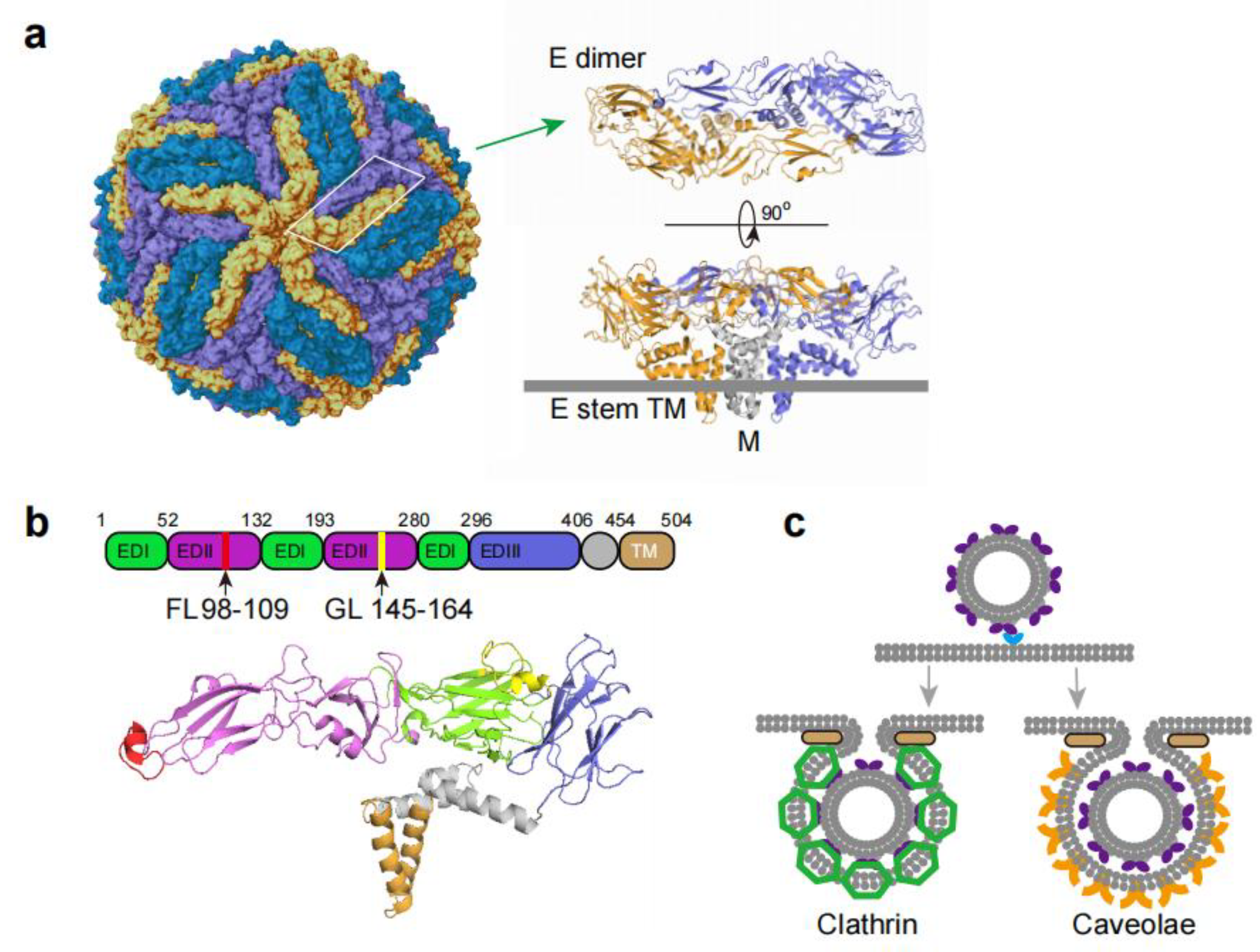
Figure 2.
(a): ZIKV receptors identified across different cell lines. AXL: Astrocytes; Endothelial cells; Glial cells; Neural progenitor cells; Microglial cells; Sertoli cells. NCAM1: HEK293T; Glioblastoma cells; U-251 MG cells. Hsp70: Huh7.5 cells. GRP78: A549 cells. Sialic acid: Vero cells, hiPSC-derived NPCs, Huh7 cells. DC-SIGN: Vero Cells, SNB-19 Cells. ITGB4: LLC-MK2, Vero cells, A549 cells, Hun-7 cells. The ECD of NCAM1 and its corresponding antibodies can inhibit ZIKV adhesion and entry. Antibodies targeting HSP70, GRP78, DC-SIGN, Sialic acid, and ITGB4 have been shown to effectively block ZIKV entry.(b): Hypothetical model of ZIKV cell entry based on current studies. ZIKV attaches to receptors (blue) and is internalized via clathrin/ caveola -mediated endocytosis. Endosome acidification triggers E protein (deep violet) conformational changes: dimers reorganize into trimmers (lavender), exposing the fusion peptide. This peptide inserts into the endosomal membrane. Further acidification drives trimer refolding into a hairpin structure, forcing viral and endosomal membranes to fuse, thereby releasing the viral genome into the cytoplasm. (c): Small Molecule Compounds Inhibiting ZIKV Entry.
Figure 2.
(a): ZIKV receptors identified across different cell lines. AXL: Astrocytes; Endothelial cells; Glial cells; Neural progenitor cells; Microglial cells; Sertoli cells. NCAM1: HEK293T; Glioblastoma cells; U-251 MG cells. Hsp70: Huh7.5 cells. GRP78: A549 cells. Sialic acid: Vero cells, hiPSC-derived NPCs, Huh7 cells. DC-SIGN: Vero Cells, SNB-19 Cells. ITGB4: LLC-MK2, Vero cells, A549 cells, Hun-7 cells. The ECD of NCAM1 and its corresponding antibodies can inhibit ZIKV adhesion and entry. Antibodies targeting HSP70, GRP78, DC-SIGN, Sialic acid, and ITGB4 have been shown to effectively block ZIKV entry.(b): Hypothetical model of ZIKV cell entry based on current studies. ZIKV attaches to receptors (blue) and is internalized via clathrin/ caveola -mediated endocytosis. Endosome acidification triggers E protein (deep violet) conformational changes: dimers reorganize into trimmers (lavender), exposing the fusion peptide. This peptide inserts into the endosomal membrane. Further acidification drives trimer refolding into a hairpin structure, forcing viral and endosomal membranes to fuse, thereby releasing the viral genome into the cytoplasm. (c): Small Molecule Compounds Inhibiting ZIKV Entry.
Disclaimer/Publisher’s Note: The statements, opinions and data contained in all publications are solely those of the individual author(s) and contributor(s) and not of MDPI and/or the editor(s). MDPI and/or the editor(s) disclaim responsibility for any injury to people or property resulting from any ideas, methods, instructions or products referred to in the content. |
© 2026 by the authors. Licensee MDPI, Basel, Switzerland. This article is an open access article distributed under the terms and conditions of the Creative Commons Attribution (CC BY) license (http://creativecommons.org/licenses/by/4.0/).
Copyright: This open access article is published under a Creative Commons CC BY 4.0 license, which permit the free download, distribution, and reuse, provided that the author and preprint are cited in any reuse.


