Submitted:
18 May 2026
Posted:
20 May 2026
You are already at the latest version
Abstract
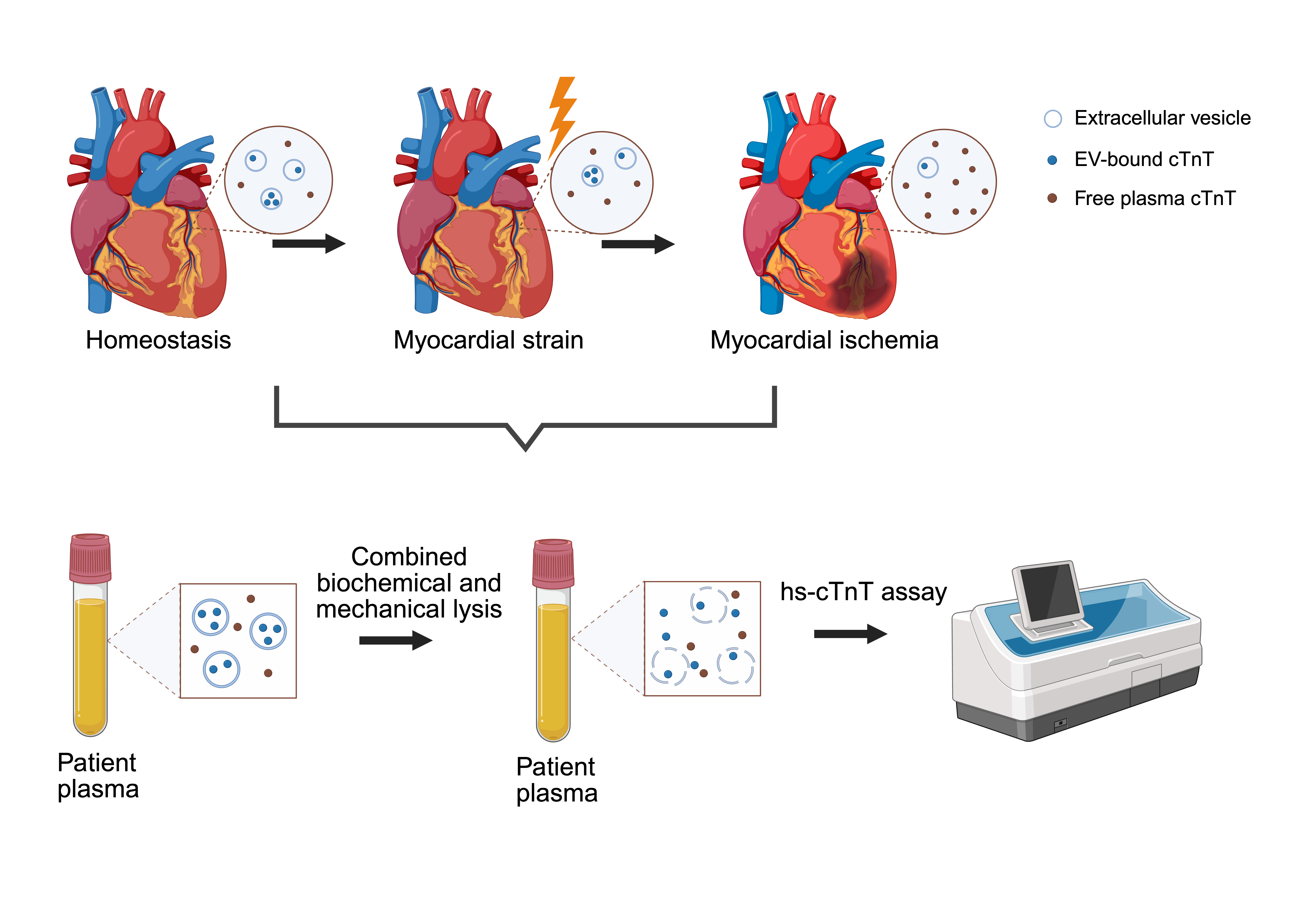
Background/Objectives: High-sensitivity cardiac troponin (hs-cTn) assays are used in routine diagnostics to detect myocardial injury. However, a fraction of circulating cardiac troponin T (cTnT) enclosed within extracellular vesicles (EVs) goes widely undetected. This study introduces a combined lysis- and sonication-based protocol to release and quantify EV-bound cTnT in a time-efficient manner using a state-of-the-art hs-cTnT immunoassay. Methods: Plasma samples from patients with non-ST-segment elevation myocardial infarction (NSTEMI), unstable angina, pulmonary embolism, decompensated aortic stenosis, atrial fibrillation, myocarditis, and healthy controls were treated with lysis buffer and subsequently sonicated. Treated and untreated samples were assessed and compared to a conventional EV isolation method. Results: Following combined lysis and sonication, cTnT levels were significantly higher compared to native, unprocessed samples across all cohorts. Median increase post-processing ranged from 10% in decompensated aortic stenosis to 34% in healthy controls. In NSTEMI, EV-bound cTnT accounted for 15% of plasma cTnT and remained stable over 72 hours. EV cTnT/plasma cTnT ratios were comparable between the combined lysis and sonication approach and the conventional EV isolation method. Processing time prior to cTnT measurement was reduced from approximately 2.5 hours to approximately 10 minutes using combined lysis and sonication compared to the established EV isolation method. Conclusions: Our method allows for rapid liberation of a previously inaccessible EV-bound fraction of cTnT without the need for time-consuming and resource-intensive EV isolation workflows. The resulting total cTnT signatures indicate differential cTnT compartmentation depending on the underlying myocardial pathophysiology, enabling early differentiation of the mechanisms underlying troponin elevation. This approach is readily implementable alongside standard hs-cTnT testing at minimal additional time expense and may improve diagnostic sensitivity and specificity in acute clinical settings.
Keywords:
extracellular vesicles
; cardiac troponin T
; hs-cTnT assay
; myocardial injury
; myocardial ischemia
; sonication
; troponin compartmentation
; cardiac biomarker
Copyright: This open access article is published under a Creative Commons CC BY 4.0 license, which permit the free download, distribution, and reuse, provided that the author and preprint are cited in any reuse.


