

Submitted:
17 May 2026
Posted:
18 May 2026
You are already at the latest version
Abstract
Titanium-based dental implants have evolved significantly, with the development of binary alloys like Ti-15Zr (Roxolid™) representing a pivotal advancement in mechanical performance. Current research focuses on biomimetic surface engineering to further accelerate osseointegration and optimize bone regeneration, particularly in clinically compromised sites. This review constitutes a narrative synthesis of how these strategies replicate the bone extracellular matrix (ECM) through a holistic framework of architectural, mechanical, and biochemical integration. A structured literature search across PubMed, Scopus, and Web of Science (2010–2026) identified relevant studies focusing on the synergy between Ti-15Zr substrates and surface modifications. Evidence confirms that the high fatigue strength of Roxolid™ alloys provides an ideal foundation for advanced, hierarchical surface engineering without compromising structural integrity. This strategy utilizes macro-topography for primary stability, nano-topography for protein adsorption, and bio-functionalization (e.g., RGD peptides, osteogenic ions) to direct mesenchymal stem cell (MSC) differentiation. This synergy accelerates the transition from passive to active osseointegration, effectively bridging the "biological gap" during early healing. Biomimetic engineering transforms implants into instructive biological platforms, improving outcomes for patients with compromised bone quality and facilitating predictable immediate loading protocols.
Keywords:
Ti-15Zr
; Roxolid™
; biomimetic surface engineering
; osseointegration
; osteoinstruction
; bone regeneration
1. Introduction
Osseointegration represents the fundamental cornerstone of modern implant dentistry, defining long-term clinical success through a direct structural and functional connection between living bone and the load-bearing implant surface [1,2]. Since its formal description in the 1960s, the clinical predictability of implant procedures has achieved remarkable levels, as retrospective studies have reported survival rates exceeding 95% over 10-year follow-up periods. Specifically, surface characteristics play a crucial role in maintaining both primary and secondary stability over time [3].
The technological trajectory of implant surfaces has evolved from early machined (smooth) surfaces to increasingly sophisticated controlled-roughness topographies, marking a turning point in achieving faster and more predictable osseointegration. This paradigm shift was catalyzed by the recognition that surface microtopography profoundly influences the biological response at the bone-implant interface (BII). As extensively analyzed by Wennerberg and Albrektsson, neither surface topography alone nor chemistry in isolation determines osseointegration quality; it is their synergistic combination that modulates the cascade of molecular and cellular events leading to successful bone apposition [4,5].
Contemporary implant surfaces are therefore no longer conceived as biologically inert components but as active biointerfaces capable of directing protein adsorption, platelet activation, and stem cell fate [6]. From a biological standpoint, osseointegration is a complex temporally and spatially orchestrated process involving a cascade of molecular and cellular events, from initial homeostasis to the formation of lamellar bone [7]. Mavrogenis et al. underscored that the biology of the BII depends strictly on the surface’s ability to interact productively with mesenchymal stem cells (MSCs) and osteoblast precursors, promoting differentiation, osteocalcin expression, and bone matrix synthesis [8]. This biological understanding has driven the development of high-performance alloys such as RoxolidTM, which combine reduced-diameter implants with enhanced tensile strength and excellent biocompatibility, paving the way for less invasive procedures and early loading protocols [9,10].
The primary aim of this comprehensive review is to analyze current scientific evidence on biomimetic strategies applied to dental implant surfaces, with a particular focus on Roxolid technology and its evolutions (such as the SLActive® hydrophilic surface modification). Secondary objectives include: (i) examining how physicochemical surface modifications and the integration of bioactive factors can accelerate osseointegration kinetics; (ii) appraising clinical evidence on implant stability in demanding scenarios; and (iii) providing clinicians with an updated evidence-based synthesis of current biotechnological innovations and their translational implications.
2. Materials and Methods
A structured literature search was conducted to provide a comprehensive overview of the current status of biomimetic surface engineering on Roxolid™ implants. This format provides a critical and integrative synthesis of the available evidence, combining data from preclinical, translational, and clinical studies. The methodology focused on identifying relevant studies published between January 2010 and March 2026, ensuring coverage of the evolution of the field from early mechanical modifications to current bio-instructive surface technologies. The search was performed across PubMed, Scopus, and Web of Science, using the following search string:
(“Ti-15Zr” OR “Roxolid”) AND (“Biocompatibility” OR “Cell Viability”) AND (“Surface treatment” OR “SLA ACTIVE” OR “SLA” OR “Biomimetic Surface” OR “Surface Engineering”)
Additional hand-searching was performed on reference lists of included articles and leading implantology journals to ensure comprehensiveness. Boolean operators were used to refine the search and combine the selected concepts appropriately. No language restrictions were applied during the initial search.
2.1. Eligibility Criteria
Inclusion Criteria: (1) peer-reviewed articles focusing on Ti-Zr (Roxolid™) implants; (2) studies investigating surface modifications (physical, chemical, or biological, e.g., anodization, functionalization, peptide coating); (3) articles discussing the impact of surface engineering on cellular response (e.g., osteoblast differentiation, protein adsorption) or bone regeneration; (4) studies comparing Ti-Zr alloys to CP Grade 4 Titanium; (5) both in vitro (cell-based) and in vivo (animal/clinical) studies.
Exclusion Criteria: (1) studies focusing solely on standard titanium (Grade 4) without comparative relevance to Ti-Zr alloys; (2) grey literature (e.g., conference abstracts without full-text documentation); (3) research with insufficient details on surface characterization methodologies.
2.2. Study Selection and Data Extraction
The search yielded an initial pool of 954 articles. Following screening of titles, abstracts, and full texts against the eligibility criteria, 53 studies met the inclusion criteria for this review. Selected full-text articles were analysed to extract qualitative data regarding surface modification techniques (e.g., SLA, anodization) and characterization (SEM, XPS, contact angle); mechanical-biological synergy (cell viability, differentiation markers—RUNX2, ALP—and in vivo bone-to-implant contact percentages); and clinical outcomes. This comprehensive approach allowed for a critical narrative synthesis of the literature, highlighting both established evidence and future challenges in the field of biomimetic surface engineering.
2.3. Methodological Quality Assessment
The methodological rigor of the included studies was evaluated qualitatively on the basis of study design, reproducibility of surface characterization, suitability of the outcome measures, and consistency of the reported findings. Particular attention was paid to the translational relevance of the evidence, with clear distinction made between mechanistic in vitro investigations, animal studies, and clinical observations. This approach enabled the integration of heterogeneous evidence while preserving scientific rigor and interpretative coherence.
3. Relevant Sections
The field of oral implantology is undergoing a fundamental transition from passive, bioinert structural replacement toward biomimetic, osteoinstructive interfaces that replicate the hierarchical architecture and physicochemical properties of the native dentoalveolar complex. This shift is motivated by the inherent limitations of conventional osteoconductive surfaces, which function as inert scaffolds permitting cell migration and adhesion without actively modulating cellular behaviour [11]. Osseointegration in this model depends entirely on the proximity and viability of host osteogenic cells, establishing merely a permissive—rather than instructive—biological milieu [12].
In contrast, osteoinstructive biomimetic implants are engineered to assume a proactive biological function [13,14]. By emulating the architecture and biochemical signature of the native bone extracellular matrix (ECM), these interfaces provide precise molecular and structural cues—via nanotopography and localized ion or bioactive molecule release—that actively drive the recruitment, proliferation, and differentiation of mesenchymal stem cells (MSCs) into mature, functional osteoblasts [15]. This molecular dialogue enables the host to recognize the implant as endogenous tissue rather than a foreign substrate, transforming the interface from a static contact zone into a dynamic site of accelerated bone regeneration [16]. The clinical relevance of this transition is particularly pronounced in high-risk cohorts—including geriatric patients and individuals with systemic comorbidities such as diabetes or osteoporosis—in whom compromised bone metabolism substantially reduces the predictability of conventional implant therapy [17,18].
3.1. The Roxolid™ Implant System
Introduced by Straumann in 2009, Roxolid™ represents a significant paradigm evolution in implantology. This material was engineered to overcome the mechanical limitations of commercially pure titanium (cpTi) while maintaining superior biocompatibility. Roxolid™ is a binary, single-phase α-alloy composed of approximately 85% titanium (Ti) and 15% zirconium (Zr). Zirconium acts as a solid-solution strengthener, significantly increasing the mechanical properties of the alloy without introducing cytotoxic elements such as aluminum or vanadium, which have been associated with local irritation and potential systemic concerns in Ti-6Al-4V alloys [19,20,21].
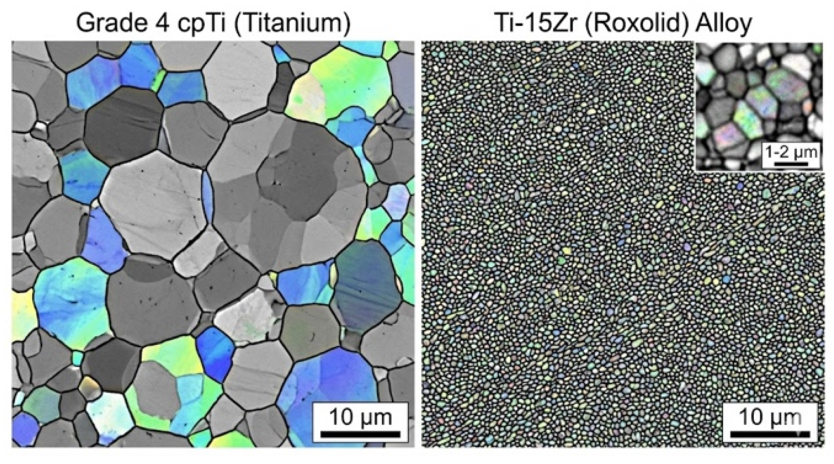
The unique microstructural configuration of Ti-15Zr is central to its performance. Traditional Grade 4 cpTi exhibits grain sizes of 20–30 µm, with larger and more equiaxed (polygonal) morphology characteristic of fully recrystallized titanium. In Ti-15Zr (Roxolid™), grain refinement is dramatic: grains are ultra-fine (approximately 1–2 µm), elongated, and densely packed, forming microbands as a direct result of the intensive cold-working process. This process creates a very high dislocation density and a pronounced crystallographic texture, with basal planes oriented parallel to the wire axis—a configuration that confers increased mechanical strength without sacrificing toughness or ductility [22].
3.2. Mechanical Performance
This microstructural refinement translates into significant mechanical advantages. Roxolid™ exhibits a tensile strength 10–15% higher than Grade 4 cpTi, with yield strength values of approximately 799 MPa (SLA surface) and 784 MPa (machined surface), and ultimate tensile strength values of approximately 968 MPa and 987 MPa, respectively [23]. The alloy demonstrates a fatigue endurance limit approximately 30% higher than Grade 4 cpTi (560 MPa for machined and 500 MPa for SLA surfaces), with ISO 14801 testing confirming fatigue strength improvements of 11–38% over Grade 4 counterparts depending on implant design [22]. Fracture toughness remains slightly superior (~22.34 J/cm² vs. ~19.30 J/cm²), with maintained uniform elongation ductility of ~6.0–6.2% [22].
Critically, Roxolid™ features a lower elastic modulus than cpTi, which helps mitigate the stress shielding effect—a mismatch between implant and bone stiffness that leads to peri-implant bone resorption [24]. The addition of zirconium improves the stability of the protective oxide layer, rendering Ti-Zr alloys more resistant to pitting corrosion in biological fluids [24,25,26]. This “mechanical surplus” is clinically decisive: it enables the development of narrow-diameter implants (Ø 2.9–3.5 mm) that maintain structural integrity under high masticatory loads, frequently obviating the need for invasive bone augmentation procedures in sites with limited bone volume [9,27,28].
Figure 1.
Visualization of the orientations of individual crystal grains within the metal structure of Roxolid™ (right) compared to cpTitanium Grade 4 (left).
Figure 1.
Visualization of the orientations of individual crystal grains within the metal structure of Roxolid™ (right) compared to cpTitanium Grade 4 (left).
3.3. Osseointegration: Biological Cascade and Molecular Mechanisms
Osseointegration constitutes the fundamental biological prerequisite for the long-term clinical success of endosseous implants. It is defined as the direct structural and functional connection between living, organized bone and the surface of a load-bearing implant, established and maintained without the interposition of fibrous connective tissue. This process is not a single event, but a temporally and spatially coordinated biological sequence that unfolds across overlapping phases, each governed by a distinct cellular and molecular program [17].
The cascade is initiated immediately upon surgical implant placement, when disruption of the cortical and medullary vasculature triggers the coagulation cascade and the formation of a provisional fibrin clot at the implant interface. This clot serves as a three-dimensional provisional scaffold enriched with growth factors—including platelet-derived growth factor (PDGF) and transforming growth factor-beta (TGF-β)—that orchestrate the subsequent recruitment of immune and progenitor cells. Concurrently, plasma proteins adsorb rapidly onto the implant oxide surface within seconds of blood contact, establishing a proteinaceous conditioning film that determines the nature of subsequent cell-surface interactions [8,29].
The acute inflammatory phase (0–3 days) is characterized by the sequential infiltration of neutrophils and monocyte-derived macrophages. Rather than representing a merely defensive response, this phase plays an indispensable instructive role: macrophages adopting the pro-inflammatory M1 phenotype secrete cytokines—including IL-6, IL-1β, and TNF-α—that amplify the local inflammatory signal and prime the regenerative microenvironment [30]. Critically, the surface physicochemical properties of the implant directly modulate macrophage activation state, establishing an immunological set-point that influences all subsequent healing events [31,32].
As the inflammatory phase resolves, the proliferative phase (~days 4–14) is marked by the transition of macrophages toward the anti-inflammatory, reparative M2 phenotype—a shift essential for the establishment of a pro-osteogenic milieu [17,33]. MSCs, recruited from the periosteum, endosteum, and circulating blood, undergo proliferation and osteoblastic commitment under the influence of Bone Morphogenetic Proteins (BMPs) and Wnt/β-catenin signaling. Concurrently, VEGF-driven angiogenesis restores the local oxygen and nutrient supply, a prerequisite for the energetically demanding process of mineral matrix deposition. Activated osteoblasts begin secreting an unmineralized collagenous osteoid directly onto the implant surface, initiating the process of contact osteogenesis [17,34,35].
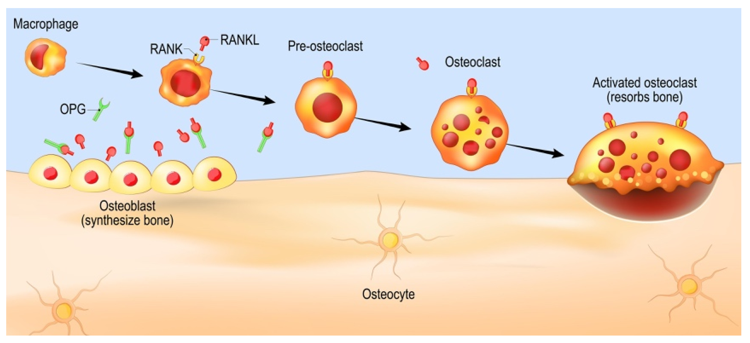
The maturation and mineralization phase (days 14–21) is defined by the progressive calcification of the osteoid matrix, evidenced biochemically by upregulation of Alkaline Phosphatase (ALP) activity and Osteocalcin (OC) expression [3,33,36,37]. The final remodeling phase, extending over months to years, is governed by the RANKL/RANK/OPG signaling axis, which maintains the homeostatic equilibrium between osteoclast-mediated resorption and osteoblast-mediated apposition. Through iterative cycles of bone turnover, the initial woven bone is progressively replaced by organized lamellar bone with a mature Haversian architecture, increasing the BIC ratio and ensuring long-term biomechanical competence [17,35,38].
Figure 2.
Activation and Polarization of the Macrophage.
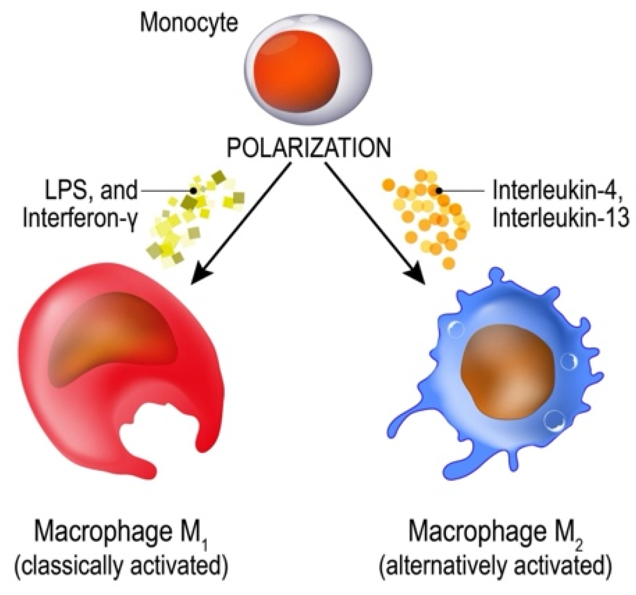
Figure 3.
Bone Biology and Remodeling: Role of RANK, RANKL and OPG.
3.4. Surface Treatments and Biomimetic Functionalization
The surface of a dental implant constitutes the primary determinant of the biological events governing osseointegration. Surface modification strategies are therefore fundamental engineering parameters that define the immunological, proteomic, and osteogenic trajectory of the healing response. These strategies are conventionally classified into three mechanistically distinct categories—subtractive (physical), additive (chemical and coating-based), and biomolecular (biofunctional)—each operating at a distinct stage of the biological cascade [39,40].
Figure 4.
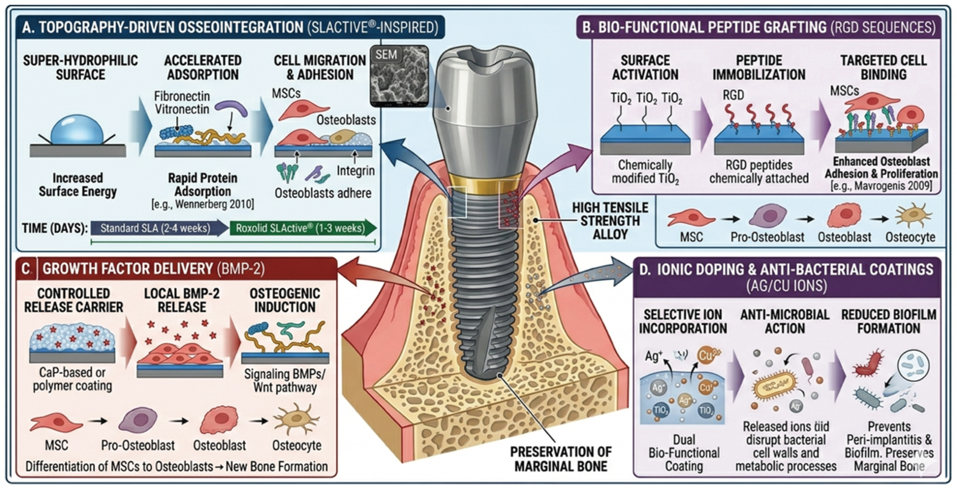
Biomimetic Advancements in RoxolidTM Implant Surface Modifications.
3.4.1. Subtractive Modifications: SLA and SLActive®
Subtractive surface modifications operate by selectively removing material from the implant surface to generate controlled topographic features. Sandblasting with large-grit alumina or silicon carbide particles (typically 250–500 µm) creates macro-scale surface irregularities (20–40 µm) that enhance mechanical interlocking with host bone. Subsequent acid etching—most commonly with hot HCl/H₂SO₄ mixtures—superimposes a finer micro-scale texture (2–5 µm pits) that promotes fibrin network entrapment, platelet activation, and osteoblast adhesion. The resulting bimodal SLA® topography represents the current clinical gold standard [17,40,41].
In the context of Roxolid™ implants, the SLActive® (modSLA) surface represents a further evolution. Produced by storing the acid-etched implant under nitrogen in isotonic saline to prevent hydrocarbon contamination, it introduces superhydrophilicity (contact angle ≈0°) and spontaneously formed nanostructures not present on conventional SLA surfaces, as revealed by high-magnification SEM imaging. These nanostructures catalyze fibrin network formation within 15 minutes of blood contact, reducing the standard healing period from 6–12 weeks to approximately 3–4 weeks and enabling earlier prosthetic loading [17,19,42]. Furthermore, SLA treatment on Ti-Zr substrates modulates the proteomic adsorption profile of proteins from saliva and plasma—enriching functionally relevant species such as serum albumin and fibronectin—thereby influencing initial cell adhesion and bacterial biofilm formation dynamics [17,43].
3.4.2. Additive Modifications: Anodization and Calcium Phosphate Coatings
Additive surface modifications introduce new chemical phases or topographic features onto the implant surface. Anodic oxidation (anodization) is the most widely employed electrochemical technique, producing a thickened, crystalline TiO₂ layer enriched with a self-organized nanotubular architecture. These nanotubes—with diameters tunable between 20 and 200 nm depending on electrolyte composition and applied voltage—dramatically increase the surface-to-volume ratio, enhance protein adsorption kinetics, and serve as reservoirs for the controlled local release of pharmacological agents [39,44]. Hydroxyapatite (HA) and calcium phosphate (CaP) coatings, deposited by plasma spraying, biomimetic precipitation, or pulsed laser deposition, introduce a mineral phase chemically analogous to the inorganic component of bone, establishing a direct osteoconductive interface. The susceptibility of plasma-sprayed HA coatings to delamination under cyclic mechanical loading has driven the development of thin-film nanocrystalline HA deposition techniques that preserve adhesive strength while maintaining osteogenic bioactivity [35,43].
3.4.3. Biofunctional Modifications: The Five Biomimetic Axes
The most advanced category encompasses biofunctional surface modifications, which transition the implant from a passive scaffold into an active biological signaling platform. The biological rationale derives from the native bone ECM, which integrates a hierarchical nano- to microscale architecture—primarily composed of type I collagen fibrils and carbonated hydroxyapatite crystallites—with a biochemical repertoire of adhesion ligands, sequestered growth factors, and mechanotransductive signals that collectively govern cell adhesion, migration, proliferation, and differentiation [39]. Contemporary biomimetic strategies are organized along five principal axes:
Axis 1: ECM-mimetic coatings. Structural proteins such as collagen and glycosaminoglycans like hyaluronic acid are deposited on the implant surface to recreate the adhesive and viscoelastic properties of the pericellular matrix, promoting osteoprogenitor cell attachment and directional migration.
Axis 2: Molecular and ionic functionalization. At the peptide level, the covalent or adsorptive immobilization of RGD (Arg-Gly-Asp) and related sequences such as GRGD directly engages α₅β₁ and αvβ₃ integrins on MSC membranes, activating FAK/ERK and PI3K/Akt signaling cascades that converge on osteogenic transcription factors, most notably RUNX2. The P-15 peptide—a synthetic analog of the cell-binding domain of type I collagen—further accelerates early osseointegration [45]. At the ionic level, incorporation of Sr²⁺ and Mg²⁺ within the surface oxide layer modulates bone remodeling: Sr²⁺ simultaneously promotes osteoblast-mediated formation and downregulates osteoclastic resorption through modulation of the RANKL/OPG ratio and activation of the Wnt/β-catenin pathway, while Mg²⁺ acts as a cofactor for integrin-mediated cell adhesion and stimulates peri-implant angiogenesis via VEGF upregulation [46].
Axis 3: Growth factor tethering. Controlled-release incorporation of Bone Morphogenetic Proteins (BMP-2, BMP-4, BMP-7) activates SMAD-dependent pathways to drive MSC commitment toward the osteoblastic lineage; PDGF and VEGF coordinate the angiogenic response essential for sustaining the metabolic demands of active bone formation [47].
Axis 4: Bioactive ceramic functionalization. Nanocrystalline hydroxyapatite establishes direct chemical continuity with the mineral phase of host bone, promoting rapid interfacial bonding and reducing the critical lag period between implant placement and functional loading [48,49].
Axis 5: Antibacterial functionalization. Since bacterial biofilm formation at the transmucosal interface is the primary etiological driver of peri-implantitis, surface strategies incorporating drug-coated platforms (antibiotics, bisphosphonates, statins), antimicrobial peptides such as GL13K, and stimuli-responsive systems—including zinc or silver ion release and pH-responsive drug delivery—represent an integrated approach that simultaneously addresses osseointegration and infection prevention [48,50].
The ideal biomimetic surface modification should be resorbable at a physiologically controlled rate, immunologically inert, mechanically stable under functional loading, and capable of providing spatiotemporally appropriate biological cues without inducing ectopic or dysregulated tissue responses [49].
3.5. Hierarchical Multi-Scale Engineering of the Tissue-Implant Interface
Modern endosseous implantology is characterized by the hierarchical, multi-scale engineering of the tissue-implant interface. Surface design is conceptualized as a stratified framework in which each level of modification optimizes the biological response at a distinct stage of the healing cascade—from initial mechanical anchorage to long-term molecular osseointegration [51].

Table 1.
Hierarchical design principles for advanced endosseous implants and their corresponding biological and clinical applications.
Table 1.
Hierarchical design principles for advanced endosseous implants and their corresponding biological and clinical applications.
| Hierarchical Level | Design Principle | Technique | Clinical/Biological Application |
|---|---|---|---|
| Macro/Micro | Mechanical Interlocking | Sandblasting / Acid-Etching (SLA) | Primary stability; bone-to-implant contact (BIC) |
| Nanostructural | ECM Emulation | Anodization (TiO₂ nanotubes) | Protein adsorption; MSC recruitment and adhesion |
| Molecular | Biochemical Signaling | Biomolecular grafting (RGD, BMP-2, Mg²⁺/Sr²⁺) | Osteo-instructive signaling; accelerated mineralization |
| Mechanical | Compliance Matching | Ti-Zr alloy / PEEK composites | Young’s modulus optimization; stress shielding mitigation |
3.5.1. Macro- and Micro-Topography
Macro-topography—defined by thread geometry and overall implant morphology—ensures primary mechanical interlocking with the surrounding bone, constituting a prerequisite for immediate and early loading protocols [52]. At the macroscale (10 µm to mm), implant geometry governs primary mechanical stability through physical interlocking with surrounding bone; at the microscale (1–10 µm), surface features maximize BIC and promote mechanical interdigitation with mineralized tissue [53]. The SLA process creates a bimodal topography: large-grit sandblasting (corundum/Al₂O₃) generates macro-roughness (20–40 µm), while subsequent hot acid etching (HCl/H₂SO₄) superimposes micro-pits (2–5 µm) that trap fibrin networks and promote osteoblast adhesion [41].
Surface roughness, quantified by Sa (arithmetic mean height) and Sz (maximum peak-to-valley height), is a principal modulator of the host cellular response [54]. Moderately rough surfaces (Sa 1–2 µm) are currently considered optimal for the osteogenic response. Although increased roughness has been theoretically associated with higher susceptibility to bacterial colonization, prospective clinical trials have not demonstrated a significantly elevated incidence of peri-implantitis for implants within the moderately rough range [55]. Surface wettability—expressed as the water contact angle—governs protein adsorption kinetics and cell–surface interactions; hydrophilic surfaces promote the rapid adsorption of fibronectin and vitronectin, accelerating early tissue healing and osteoblast differentiation [6,56].
3.5.2. Nano-Topography: Protein Adsorption and MSC Recruitment
Nano-topographic features (1–100 nm) serve as the primary interface for early biological signaling. By mimicking the nanostructure of the native ECM, these modifications enhance the adsorption of key serum proteins—principally fibronectin and vitronectin—which act as adhesion anchors for MSCs, triggering intracellular signaling cascades that direct osteoblastic differentiation [16]. The most clinically relevant technique for generating nanostructures on Ti-Zr alloys is anodization, wherein controlled electrolytic oxidation with strong acids (H₂SO₄, H₃PO₄, or HF) produces a dense, self-organized array of TiO₂ nanotubes. Advanced chemical etching with H₂SO₄/H₂O₂ has been used to create hierarchical Micro-/Submicro-/Nanostructured (MSN) surfaces on Ti-Zr alloys, demonstrating significantly higher hydrophilicity and osteoblast attachment compared to standard machined or mono-scale etched surfaces [42,57].
3.5.3. Molecular Functionalization: Active Osteoinduction
The apex of the hierarchical framework involves the bio-functionalization of nanostructured surfaces with bioactive molecules. Peptide grafting: immobilization of RGD or GRGD peptides directly targets integrin receptors on MSC membranes. Implants functionalized via an innovative dip-coating technique (IDCT) with 1% GRGD exhibit a nano-porous surface with a Young’s modulus (~28.7 GPa) approaching that of human cortical bone, along with enhanced red blood cell accumulation and fibrin formation [15,58]. Ion incorporation: Sr²⁺ and Mg²⁺ within the TiO₂ nanostructure modulate local bone remodeling. In the Ti-Zr context, SLA treatment selectively enriches serum albumin and fibronectin from saliva and plasma, simultaneously enhancing osteoprogenitor cell recognition and attenuating bacterial biofilm formation [17,19,43]. Growth factors and drug delivery: BMP-2 incorporation activates the SMAD signaling pathway, upregulating RUNX2 and bone sialoprotein (BSP) expression. Surfaces can additionally be functionalized with antibiotics (e.g., gentamicin), analgesics, Ca-phosphate, or bisphosphonates for localized delivery, enhancing infection control and accelerating bone cell proliferation without systemic cytotoxic effects [47,50,59].
3.5.4. Biomechanical Optimization: Stress Shielding and Structural Compliance
A critical limitation of conventional titanium implants is the mechanical mismatch with host bone. The higher stiffness of metallic substrates relative to cortical bone alters local strain distributions—a phenomenon termed stress shielding—leading to peri-implant bone resorption [26,35,58]. The biomimetic approach addresses this through: (1) optimization of the implant’s Young’s modulus using low-modulus alloys (such as Ti-15Zr) or PEEK-based composites; and (2) the implementation of 3D-printed, porous lattice structures that permit deep tissue infiltration and physiological load transfer [58]. By mirroring the hierarchical porosity of native trabecular bone, these architectures ensure long-term structural stability while preserving the integrity of the surrounding host environment [60,61].
The frontier of surface engineering further involves stimuli-responsive “smart” materials capable of dynamic modulation of surface chemistry in real time. pH-responsive antibacterial release and toggling between antimicrobial and osteogenic surface states represent the next generation of implant design [62,63].
3.6. The Ti-15Zr (Roxolid™) Alloy: Mechanical and Biological Rationale
The binary Ti-15Zr alloy (Roxolid™) exemplifies the integration of biomechanical superiority with advanced surface engineering capability [24,64,65,66]. By retaining the α-phase crystalline structure of cpTi while incorporating 15% zirconium, the alloy achieves a ~40% increase in tensile strength and fatigue resistance without compromising biocompatibility. This mechanical robustness enables the use of narrow-diameter implants (NDIs, Ø 3.3 mm) in anatomically challenging sites, frequently obviating the need for invasive bone grafting [28,67].
In pure titanium, aggressive treatments such as deep acid etching or heavy grit-blasting may introduce micro-cracks or compromise surface integrity; the improved fatigue resistance of Ti-15Zr permits more sophisticated surface engineering without such risks [68]. Furthermore, the native zirconium oxide (ZrO₂) layer that forms on the alloy surface provides enhanced chemical stability and a superior base for biomimetic functionalization—including hydroxyapatite deposition, TiO₂ nanotube growth, and bioactive peptide immobilization [44,69,70].
Roxolid™ is compatible with both SLA® and SLActive® surface technologies. The SLActive® surface introduces superhydrophilicity (contact angle ≈0°) and spontaneously formed surface nanostructures [36,71], catalyzing fibrin network formation within 15 minutes and reducing the standard healing period from 6–12 weeks to 3–4 weeks [17,19,36]. Histomorphometric data from animal models confirm that modSLA Roxolid™ implants achieve higher BIC ratios and improved marginal bone level (MBL) stability, particularly in defect-grafted sites [19,72].
3.7. Biological Response: Immunomodulation, Osteoblast Activity, and Proteomic Profile
3.7.1. Macrophage Polarization and Immunomodulation
The biological response to Roxolid™ surfaces is deeply governed by immunomodulatory mechanisms. Research demonstrates that Ti-Zr modSLA surfaces drive macrophage polarization toward the M2 phenotype more effectively than traditional titanium surfaces, suppressing chronic pro-inflammatory cytokine release (IL-6, TNF-α, IL-1β) and upregulating anti-inflammatory mediators (IL-10, TGF-β1), establishing a conducive microenvironment that allows osseointegration to commence earlier [19,26,28,73]. A related immunomodulatory strategy involves the coating of Roxolid™ surfaces with S53P4 bioactive glass, whose dissolution products significantly reduce pro-inflammatory cytokine expression in human gingival fibroblasts and osteoblasts, preventing excessive bone resorption and promoting a stable biological seal [20,42,73].
3.7.2. Osteoblast Adhesion, Differentiation, and Proteomic Profile
In vitro studies confirm that osteoblast-like cells (MC3T3-E1) attach in significantly higher numbers to Ti-Zr surfaces than to cpTi, demonstrating increased ALP activity and elevated OC expression, indicative of mature mineralized matrix production [19,74]. These effects are potentiated by zirconium ions, which exert direct osteoinductive activity on osteoblast maturation. At the molecular level, SLA treatment on Ti-Zr results in a unique proteomic adsorption pattern from saliva, including 14 exclusive proteins—among them serum albumin—which mediate initial cell adhesion and may concomitantly reduce bacterial biofilm formation [43]. Collagen- and hyaluronic acid-based ECM coatings further enhance soft-tissue sealing and MSC recruitment, while RGD peptide grafting onto the Roxolid™ surface significantly improves BIC and removal torque values [20,22,26].
3.8. Clinical Performance of Ti-15Zr Narrow-Diameter Implants
Systematic reviews and meta-analyses consistently confirm the high clinical predictability of Roxolid™ 3.3 mm NDIs, establishing them as a reliable alternative to bone grafting in atrophic bone regions. Reported survival rates exceed 98.4% at 1 year and 97.7% at 2 years, with MBL averaging 0.41 mm after 2 years—values comparable to, or exceeding, those of standard-diameter titanium implants [28,72,75]. No implant body fractures were reported across primary studies, validating the fatigue strength advantages of the Ti-Zr alloy [17,19,26,35]. Long-term follow-up data further confirm that Ti-Zr SLA surfaces demonstrate significantly greater soft-tissue attachment compared to machined or ceramic surfaces, ensuring a robust biological seal against oral pathogens and reduced bleeding on probing (BoP) [76,77,78,79,80].
Table 2.
Clinical advantages associated with the transition to osteoinstructive biomimetic surfaces.
Table 2.
Clinical advantages associated with the transition to osteoinstructive biomimetic surfaces.
| Clinical Parameter | Description |
|---|---|
| Enhanced Healing Kinetics | Increased bioactivity reduces the latency period between implant placement and prosthetic loading, optimizing the surgical workflow. |
| Biological Integrity | A robust, biologically integrated interface reduces the risk of early and late implant failure. |
| Proactive Defence | Nanostructured surfaces inhibit biofilm formation, providing a physiological defence against peri-implantitis and soft tissue recession. |
Table 3.
Summary of clinical outcomes for Ti-15Zr (Roxolid™) narrow-diameter implants reported in systematic reviews and clinical studies.
Table 3.
Summary of clinical outcomes for Ti-15Zr (Roxolid™) narrow-diameter implants reported in systematic reviews and clinical studies.
| Clinical Outcome | Evidence |
|---|---|
| Survival Rate (1 yr) | ≥98.4% for Ti-Zr 3.3 mm narrow-diameter implants (NDIs) |
| Survival Rate (2 yr) | ≥97.7%; marginal bone loss (MBL) ≈ 0.41 mm |
| Fracture Resistance | No implant body fractures reported in primary studies despite narrow diameter |
| Soft Tissue Integration | Superior attachment vs. machined or ceramic surfaces; robust biological seal |
| Immunomodulation | M2 macrophage polarization; reduced IL-6, TNF-α, IL-1β; upregulated IL-10 and TGF-β1 |
| Osteoblast Activity | Increased ALP activity and Osteocalcin (OC) expression vs. cpTi |
3.9. Biomimetic Surface Engineering of Roxolid™ in Bone Regeneration: From Guided Protocols to Instructive Scaffolds
The biomimetic potential of Ti-15Zr (Roxolid™) extends beyond the immediate implant–bone interface to encompass the broader biological domain of bone regeneration, rendering it a uniquely versatile osteoinstructive platform in anatomically and systemically compromised clinical scenarios. This regenerative capacity is grounded in two converging material properties: a lower elastic modulus (approximately 96–99 GPa) relative to commercially pure titanium, which mitigates stress shielding by transferring mechanical loads more uniformly to the surrounding bone and thereby prevents disuse atrophy while sustaining healthy remodeling [14,58]; and a hierarchically engineered surface that actively modulates the sequential biological cascade—hemostasis, inflammation, proliferation, and remodeling—governing peri-implant bone formation [17,19].
A cornerstone of this osteoinstructive capacity is immunomodulation. The physicochemical properties of the Ti-Zr surface, particularly when combined with superhydrophilic SLActive® treatment, accelerate macrophage polarization from the pro-inflammatory M1 phenotype toward the reparative M2 phenotype, promoting the secretion of anti-inflammatory mediators—principally IL-10 and TGF-β1—that suppress chronic inflammation and establish a pro-osteogenic microenvironment essential for MSC recruitment and osteoblast commitment [19,20,73]. Concurrently, the interaction of the Roxolid™ surface with biological fluids generates a distinctive proteomic adsorption profile: the presence of zirconium selectively enriches proteins implicated in cell adhesion and coagulation cascade initiation—the primary biological triggers of the healing cascade—thereby priming the interface for accelerated tissue integration [43].
Biomolecular functionalization further transforms the Ti-15Zr surface into an active regenerative platform. Grafting of RGD and GRGD peptide sequences directly engages α₅β₁ and αvβ₃ integrin receptors on MSC membranes, enhancing cellular adhesion, directional migration, and osteoblastic commitment [15,45]. Incorporation of BMP-2 activates the SMAD signaling pathway—upregulating RUNX2 and bone sialoprotein (BSP)—while concomitant Wnt/β-catenin signaling synergistically amplifies osteoprogenitor differentiation [40,47]. At the ionic level, Sr²⁺ doping simultaneously promotes osteoblast-mediated bone formation and suppresses osteoclastic resorption through modulation of the RANKL/OPG ratio, while Mg²⁺ acts as a cofactor for integrin-mediated adhesion and stimulates peri-implant angiogenesis via VEGF upregulation [46]. Notably, copper (Cu²⁺)-doped Ti-Zr alloys confer intrinsic antibacterial properties—demonstrating up to 89% inhibition of adherent Porphyromonas gingivalis—without compromising cytocompatibility, representing a compelling multifunctional strategy that unifies osseointegration, angiogenesis, and infection control within a single surface architecture [50,65].
These surface-level properties translate directly into measurable regenerative advantages in both guided bone regeneration (GBR) protocols and scaffold-based strategies. In GBR applications, the mechanical surplus of Roxolid™ narrow-diameter implants enables simultaneous placement with resorbable or non-resorbable membranes and bone substitutes in sites of severe ridge atrophy, sustaining functional loads in grafted, low-density bone without compromising structural integrity [28,67]. The hierarchical surface engineering synergizes with the regenerative milieu established by the membrane barrier, accelerating MSC recruitment into the newly forming bone compartment; preclinical minipig models confirm that superhydrophilic Roxolid™ implants achieve significantly higher bone-to-implant contact (BIC) and faster bone apposition compared to standard SLA titanium controls, even in circumferential defects grafted with bone substitutes [10,19]. At the scaffold level, three-dimensional-printed, patient-specific Ti-Zr porous lattice structures—designed to replicate the hierarchical porosity of native trabecular bone (pore sizes 300–600 µm, interconnectivity >80%)—enable deep vascular ingrowth, physiologically compliant load transfer, and direct surface functionalization with hydroxyapatite or bioactive peptides on the scaffold struts [60,61,69]. The native ZrO₂-enriched oxide layer characteristic of Ti-15Zr further provides a chemically superior substrate for biomimetic calcium phosphate precipitation and BMP-2 tethering relative to cpTi, yielding significantly higher bone volume fraction (BV/TV) and earlier mineralization onset in critical-size defect models [44,47,70].
Clinical translation corroborates these preclinical findings. Survival rates for 3.3 mm Roxolid™ implants exceed 98.4% at one year and 97.7% at two years, with superhydrophilic surfaces reaching maximum secondary stability approximately twice as rapidly as conventional SLA surfaces—critically compressing the vulnerability window of the stability dip at weeks 2–4 of healing [28,72]. Beyond bone, Ti-Zr SLActive® surfaces demonstrate superior soft-tissue attachment relative to machined or ceramic counterparts, establishing a robust transmucosal biological seal that protects the underlying regenerated bone from bacterial infiltration and the sequelae of peri-implantitis [76,77,78]. Collectively, the integration of stress-shielding mitigation, immunomodulatory surface chemistry, multifunctional biomolecular functionalization, and scaffold-level architectural biomimicry positions Ti-15Zr as a uniquely comprehensive osteoinstructive platform—one whose regenerative potential extends well beyond conventional osseointegration to address the full biological complexity of bone regeneration in the most demanding clinical environments, including post-oncological defects and patients with systemic conditions compromising bone metabolism [17,18].
4. Discussion
The current findings underscore a fundamental transition in oral implantology, where the convergence of subtractive and additive surface engineering represents the frontier of biomimetic design. The integration of both modalities is essential for transitioning from bioinert, osteoconductive substrates to highly osteoinstructive interfaces that actively modulate the host response, particularly in patients with compromised metabolic or regenerative capacity. The clinical success of Roxolid™ implants in challenging scenarios is not solely attributable to mechanical strength, but to a sophisticated synergy between material science and biomimetic surface engineering.
While standard Grade 4 titanium implants often require a specific diameter threshold to ensure structural reliability, the superior fatigue strength of Ti-15Zr provides a “mechanical surplus” that facilitates advanced surface engineering without risking mechanical failure. Recent studies confirm that aggressive modifications—such as electrochemical anodization to create nanotubes or the application of nano-coatings—do not compromise the structural stability of the Ti-Zr body, unlike in pure Ti implants [68].
This hierarchical modification strategy operates across three distinct tiers: the macro/micro-level, where SLA remains the foundational anchor for mechanical interlocking; the nano-level, where nanofeatures mimic the natural architecture of the bone ECM to enhance serum protein adsorption and MSC recruitment; and the molecular-level, where functionalization with RGD peptides, osteogenic ions (Ca²⁺, Mg²⁺, Sr²⁺), and growth factors actively instructs osteoblasts. Mg²⁺ acts as a cofactor for integrin-mediated cell adhesion, while Sr²⁺ exerts a dual-action effect of increasing osteoblastic activity while simultaneously downregulating osteoclastic resorption through RANKL/OPG ratio modulation. This osteoinstructive potential effectively compensates for host regenerative deficiencies in compromised environments such as atrophic or D4-type bone, transforming previously unfavorable sites into responsive biological beds by stimulating Wnt/β-catenin and MAPK signaling pathways [15,20,46].
A primary debate in the literature concerns the risk of “over-engineering” the surface. However, it is demonstrated that the superior fatigue limits of Roxolid™ provide a significant safety margin for highly advanced bio-functionalization without compromising long-term stability [19,22,68]. Functionalized Ti-Zr surfaces significantly shorten the “biological gap” between initial stabilization and functional integration, representing a paradigm shift from mechanical to biological loading protocols that may eventually allow for immediate functional loading regardless of bone quality, provided the surface is sufficiently “instructed.”
Despite the considerable advances described, several limitations warrant acknowledgment. Much of the evidence for osteo-instructive surfaces is derived from in vitro and animal models, and the stability of bioactive coatings under cyclic masticatory forces remains an area where robust clinical validation is still needed. The current body of clinical evidence exhibits significant heterogeneity in surface characterization protocols, cell culture conditions, and trial methodologies, complicating direct inter-study comparisons and rendering the establishment of a universally accepted “gold standard” surface specification elusive. Future research should prioritize the standardization of surface characterization parameters—particularly Sa, Sz, contact angle, and protein adsorption profiles—alongside prospective randomized controlled trials with extended follow-up periods and harmonized outcome measures, with particular attention to systemically compromised patient populations. The clinical translation of stimuli-responsive smart surfaces, dynamic drug delivery systems, and AI-assisted implant design represents the most promising frontier for the next generation of biomimetic implantology [50,81].
5. Conclusions
The integration of biomimetic surface engineering on Ti-15Zr (Roxolid™) substrates marks a pivotal evolution in modern implant dentistry. This transition from passive, bio-inert devices to active, osteo-instructive platforms redefines the biological dialogue at the implant-bone interface. The inherent mechanical superiority of Roxolid™ serves as an essential foundation, providing the structural surplus required to implement complex, hierarchical surface modifications—spanning from macro-mechanical anchoring to molecular bio-functionalization—without compromising long-term fatigue resistance or implant longevity. This hierarchical synergy effectively accelerates the healing cascade and improves clinical outcomes, particularly in patients with compromised bone quality.
The biomimetic approach holds significant promise for improving outcomes in patients with compromised bone quality and facilitating predictable immediate loading protocols. Clinical survival rates exceed 98.4% at one year and 97.7% at two years, with marginal bone loss averaging 0.41 mm, validating the biomechanical and biological rationale of the Ti-15Zr system. By unifying hierarchical topography, biomechanical compliance, and dynamic biochemical signaling, clinicians can generate a peri-implant environment recognized as “self” by the host tissue, promoting favorable immunomodulation and superior clinical success rates exceeding 98% [63]. Ultimately, the integration of high-strength alloys with superhydrophilic nanostructured surfaces is poised to become the new gold standard, facilitating less invasive surgical interventions in atrophic bone and enabling more efficient, patient-centered immediate loading protocols across diverse and challenging clinical scenarios.
6. Future Directions
The field is rapidly evolving toward the development of “smart surfaces.” Emerging strategies, including the use of TiO₂ nanotubes for controlled local drug-delivery systems, laser-structuring for optimized osteocyte interaction and antimicrobial resistance, and 3D-printed patient-specific Ti-Zr scaffolds, represent the next frontier in oral fixed rehabilitation [50,81,82]. As molecular-level modifications and smart coatings continue to evolve, the predictability and longevity of Roxolid™ implants are expected to reach even higher standards. Future research should prioritize long-term clinical validation of bio-functional coatings under masticatory loading and the development of stimuli-responsive smart surfaces. Notwithstanding, future research must prioritize longitudinal clinical trials to validate the stability of bioactive coatings under sustained functional loads and to refine current osteo-instructive protocols, as the current literature remains heterogeneous and lacks definitive clinical evidence supporting the absolute superiority of any single specific surface design.
Funding
This research received no external funding.
Acknowledgments
The authors acknowledge the use of Gemini (Google) for the generation and conceptual design of the scientific figures.
Informed Consent Statement
Not applicable.
Data Availability Statement
No new data were created or analyzed in this study.
Conflicts of Interest
The authors declare no conflicts of interest.
Abbreviations
The following abbreviations are used in this manuscript:
| ALP | Alkaline Phosphatase |
| BII | Bone-Implant Interface |
| BIC | Bone-to-Implant Contact |
| BMP | Bone Morphogenetic Protein |
| BMP-2 | Bone Morphogenetic Protein-2 |
| BMP-4 | Bone Morphogenetic Protein-4 |
| BMP-7 | Bone Morphogenetic Protein-7 |
| BoP | Bleeding on Probing |
| BSP | Bone Sialoprotein |
| CaP | Calcium Phosphate |
| cpTi | Commercially Pure Titanium |
| ECM | Extracellular Matrix |
| ERK | Extracellular signal-Regulated Kinase |
| FAK | Focal Adhesion Kinase |
| GRGD | Gly-Arg-Gly-Asp (peptide sequence) |
| HA | Hydroxyapatite |
| IDCT | Innovative Dip-Coating Technique |
| IL-1β | Interleukin-1 beta |
| IL-6 | Interleukin-6 |
| IL-10 | Interleukin-10 |
| MAPK | Mitogen-Activated Protein Kinase |
| MBL | Marginal Bone Loss |
| modSLA | Modified Sand-blasted Large-grit Acid-etched surface |
| MSC | Mesenchymal Stem Cell |
| MSN | Micro-/Submicro-/Nanostructured (surface) |
| NDI | Narrow-Diameter Implant |
| OC | Osteocalcin |
| OPG | Osteoprotegerin |
| PDGF | Platelet-Derived Growth Factor |
| PEEK | Polyether Ether Ketone |
| PI3K | Phosphoinositide 3-Kinase |
| RANKL | Receptor Activator of Nuclear factor Kappa-B Ligand |
| RGD | Arg-Gly-Asp (peptide sequence) |
| RUNX2 | Runt-related transcription factor 2 |
| Sa | Arithmetic Mean Height (surface roughness parameter) |
| SEM | Scanning Electron Microscopy |
| SLA | Sand-blasted Large-grit Acid-etched (surface) |
| SMAD | Suppressor of Mothers Against Decapentaplegic (signaling proteins) |
| Sr²⁺ | Strontium ion |
| Mg²⁺ | Magnesium ion |
| Sz | Maximum Peak-to-Valley Height (surface roughness parameter) |
| TGF-β | Transforming Growth Factor-beta |
| TGF-β1 | Transforming Growth Factor-beta 1 |
| Ti-Zr | Titanium-Zirconium (alloy) |
| TiO₂ | Titanium Dioxide |
| TNF-α | Tumor Necrosis Factor-alpha |
| VEGF | Vascular Endothelial Growth Factor |
| XPS | X-ray Photoelectron Spectroscopy |
| ZrO₂ | Zirconium Dioxide |
References
- Brånemark, P.I.; Adell, R.; Breine, U.; Hansson, B.O.; Lindström, J.; Ohlsson, A. Intra-osseous anchorage of dental prostheses. I. Experimental studies. Scand. J. Plast. Reconstr. Surg. 1969, 3, 81–100. [Google Scholar] [CrossRef]
- Albrektsson, T.; Brånemark, P.I.; Hansson, H.A.; Lindström, J. Osseointegrated titanium implants: Requirements for ensuring a long-lasting, direct bone anchorage in man. Acta Orthop. Scand. 1981, 52, 155–170. [Google Scholar] [CrossRef] [PubMed]
- Balshe, A.A.; Assad, D.A.; Eckert, S.E.; Koka, S.; Weaver, A.L. A Retrospective Study of the Survival of Smooth- and Rough-Surface Dental Implants. Int. J. Oral. Maxillofac. Implant. 2009, 24, 1113–1118. [Google Scholar]
- Wennerberg, A.; Albrektsson, T. Effects of titanium surface topography on bone integration: A systematic review. Clin. Oral. Implant. Res. 2009, 20 (Suppl. 4), 172–184. [Google Scholar] [CrossRef] [PubMed]
- Albrektsson, T.; Wennerberg, A. The impact of oral implants—past and future, 1966–2042. J. Can. Dent. Assoc. 2005, 71, 327. [Google Scholar]
- Gittens, R.A.; Scheideler, L.; Rupp, F.; Hyzy, S.L.; Geis-Gerstorfer, J.; Schwartz, Z.; Boyan, B.D. A Review on the Wettability of Dental Implant Surfaces II: Biological and Clinical Aspects. Acta Biomater. 2014, 10, 2907–2918. [Google Scholar] [CrossRef]
- Davies, J.E. Mechanisms of endosseous integration. Int. J. Prosthodont. 1998, 11, 391–401. [Google Scholar]
- Mavrogenis, A.F.; Dimitriou, R.; Parvizi, J.; Babis, G.C. Biology of Implant Osseointegration. J. Musculoskelet. Neuronal Interact. 2009, 9, 61–71. [Google Scholar]
- Chiapasco, M.; Casentini, P.; Zaniboni, M.; Corsi, E.; Anello, T. Titanium–Zirconium Alloy Narrow-Diameter Implants (Straumann Roxolid®) for the Rehabilitation of Horizontally Deficient Edentulous Ridges: Prospective Study on 18 Consecutive Patients. Clin. Oral. Implant. Res. 2012, 23, 1136–1141. [Google Scholar] [CrossRef]
- Gottlow, J.; Dard, M.; Kjellson, F.; Obrecht, M.; Sennerby, L. Evaluation of a new titanium-zirconium dental implant: a biomechanical and histological comparative study in the mini pig. Clin. Implant Dent. Relat. Res. 2012, 14(4), 538–545. [Google Scholar] [CrossRef]
- Wang, Z.; Wang, J.; Wu, R.; Wei, J. Construction of Functional Surfaces for Dental Implants to Enhance Osseointegration. Front. Bioeng. Biotechnol. 2023, 11, 1320307. [Google Scholar] [CrossRef]
- Cooper, L.F.; Shirazi, S. Osseointegration—The Biological Reality of Successful Dental Implant Therapy: A Narrative Review. Front. Oral. Maxillofac. Med. 2021, 4, 37. [Google Scholar] [CrossRef]
- Miron, R.J.; Zhang, Y.F. Osteoinduction: A Review of Old Concepts with New Standards. J. Dent. Res. 2012, 91, 736–744. [Google Scholar] [CrossRef]
- Bakitian, F.A. A Comprehensive Review of the Contemporary Methods for Enhancing Osseointegration and the Antimicrobial Properties of Titanium Dental Implants. Cureus 2024, 16, e68720. [Google Scholar] [CrossRef] [PubMed]
- Dalby, M.J.; Gadegaard, N.; Oreffo, R.O.C. Harnessing Nanotopography and Integrin–Matrix Interactions to Influence Stem Cell Fate. Nat. Mater. 2014, 13, 558–569. [Google Scholar] [CrossRef] [PubMed]
- Luo, Y.; Dolder, C.K.; Walker, J.M.; Misra, R.; Dean, D.; Becker, M.L. The Influence of Nanotopography on Cell Behaviour through Interactions with the Extracellular Matrix. Biomolecules 2022, 12, 429. [Google Scholar] [CrossRef] [PubMed]
- Łosiewicz, B.; Osak, P.; Nowińska, D.; Maszybrocka, J. Developments in Dental Implant Surface Modification. Coatings 2025, 15, 109. [Google Scholar] [CrossRef]
- Deng, J.; Van Duyn, C.; Cohen, D.J.; Schwartz, Z.; Boyan, B.D. Strategies for Improving Impaired Osseointegration in Compromised Animal Models. J. Dent. Res. 2024, 103, 467–476. [Google Scholar] [CrossRef]
- Grandin, H.; Berner, S.; Dard, M. A Review of Titanium Zirconium (TiZr) Alloys for Use in Endosseous Dental Implants. Materials 2012, 5, 1348–1360. [Google Scholar] [CrossRef]
- Hotchkiss, K.M.; Ayad, N.B.; Hyzy, S.L.; Boyan, B.D.; Olivares-Navarrete, R. Dental Implant Surface Chemistry and Energy Alter Macrophage Activation In Vitro. Clin. Oral. Implant. Res. 2017, 28, 414–423. [Google Scholar] [CrossRef]
- Yu, F.; Addison, O.; Davenport, A. A Synergistic Effect of Albumin and H₂O₂ Accelerates Corrosion of Ti6Al4V. Acta Biomater. 2015, 26, 355–365. [Google Scholar] [CrossRef] [PubMed]
- Medvedev, A.E.; Molotnikov, A.; Lapovok, R.; Zeller, R.; Berner, S.; Habersetzer, P.; Dalla Torre, F. Microstructure and Mechanical Properties of Ti-15Zr Alloy Used as Dental Implant Material. J. Mech. Behav. Biomed. Mater. 2016, 62, 384–398. [Google Scholar] [CrossRef] [PubMed]
- El-Hady, A.I.A.; Eid, H.I.; Mohamed, S.L.; Fadl, S.M. Influence of Titanium and Titanium-Zirconium Alloy as Implant Materials on Implant Stability of Maxillary Implant Retained Overdenture: A Randomized Clinical Trial. BMC Oral. Health 2024, 24, 902. [Google Scholar] [CrossRef] [PubMed]
- Sharma, A.; Waddell, J.N.; Li, K.C.; Sharma, L.A.; Prior, D.J.; Duncan, W.J. Is Titanium-Zirconium Alloy a Better Alternative to Pure Titanium for Oral Implant? Composition, Mechanical Properties, and Microstructure Analysis. Saudi Dent. J. 2021, 33, 546–553. [Google Scholar] [CrossRef]
- Akimoto, T.; Ueno, T.; Tsutsumi, Y.; Doi, H.; Hanawa, T.; Wakabayashi, N. Evaluation of Corrosion Resistance of Implant-Use Ti-Zr Binary Alloys with a Range of Compositions. J. Biomed. Mater. Res. B Appl. Biomater. 2018, 106, 73–79. [Google Scholar] [CrossRef]
- Ou, P.; Zhang, T.; Wang, J.; Li, C.; Shao, C.; Ruan, J. Bone Response In Vivo of Ti-45Zr Alloy as Dental Implant Material. J. Mater. Sci. Mater. Med. 2022, 33, 47. [Google Scholar] [CrossRef]
- Barter, S.; Stone, P.; Brägger, U. A Pilot Study to Evaluate the Success and Survival Rate of Titanium–Zirconium Implants in Partially Edentulous Patients: Results after 24 Months of Follow-Up. Clin. Oral. Implant. Res. 2012, 23, 873–881. [Google Scholar] [CrossRef]
- Altuna, P.; Lucas-Taulé, E.; Gargallo-Albiol, J.; Figueras-Álvarez, O.; Hernández-Alfaro, F.; Nart, J. Clinical Evidence on Titanium-Zirconium Dental Implants: A Systematic Review and Meta-Analysis. Int. J. Oral. Maxillofac. Surg. 2016, 45, 842–850. [Google Scholar] [CrossRef]
- Apostu, D.; Lucaciu, O.; Lucaciu, G.D.O.; Crisan, B.; Crisan, L.; Baciut, M.; Onisor, F.; Baciut, G.; Câmpian, R.S.; Bran, S. Systemic Drugs That Influence Titanium Implant Osseointegration. Drug Metab. Rev. 2017, 49, 92–104. [Google Scholar] [CrossRef]
- Yadav, S.; Nath, A.; Suresh, R.; Kurumathur Vasudevan, A.; Balakrishnan, B.; Rajan Peter, M. Advances in Implant Surface Technology: A Biological Perspective. Cureus 2025, 17, e78264. [Google Scholar] [CrossRef]
- Jemat, A.; Ghazali, M.J.; Razali, M.; Otsuka, Y. Surface Modifications and Their Effects on Titanium Dental Implants. BioMed Res. Int. 2015, 2015, 791725. [Google Scholar] [CrossRef]
- John, A.A.; Jaganathan, S.K.; Supriyanto, E.; Manikandan, A. Surface Modification of Titanium and Its Alloys for the Enhancement of Osseointegration in Orthopaedics. Curr. Sci. 2016, 111, 1003–1015. [Google Scholar] [CrossRef]
- Bragdon, C.R.; Jasty, M.; Greene, M.; Rubash, H.E.; Harris, W.H. Biologic Fixation of Total Hip Implants: Insights Gained from a Series of Canine Studies. J. Bone Jt. Surg. Am. 2004, 86, 105–117. [Google Scholar] [CrossRef]
- Nuss, K.M.R.; von Rechenberg, B. Biocompatibility Issues with Modern Implants in Bone—A Review for Clinical Orthopaedics. Open Orthop. J. 2008, 2, 66–78. [Google Scholar] [CrossRef] [PubMed]
- Ou, P.; Hao, C.; Liu, J.; He, R.; Wang, B.; Ruan, J. Cytocompatibility of Ti-xZr Alloys as Dental Implant Materials. J. Mater. Sci. Mater. Med. 2021, 32, 50. [Google Scholar] [CrossRef] [PubMed]
- Sartoretto, S.C.; Alves, A.T.N.N.; Resende, R.F.B.; Calasans-Maia, J.A.; Granjeiro, J.M.; Calasans-Maia, M.D. Early Osseointegration Driven by the Surface Chemistry and Wettability of Dental Implants. J. Appl. Oral. Sci. 2015, 23, 279–287. [Google Scholar] [CrossRef]
- Wennerberg, A.; Albrektsson, T. On Implant Surfaces: A Review of Current Knowledge and Opinions. Int. J. Oral. Maxillofac. Implant. 2010, 25, 63–74. [Google Scholar]
- Abdulghafor, M.A.; Mahmood, M.K.; Tassery, H.; Tardivo, D.; Falguiere, A.; Lan, R. Biomimetic Coatings in Implant Dentistry: A Quick Update. J. Funct. Biomater. 2024, 15, 15. [Google Scholar] [CrossRef]
- Stróż, A.; Gawlikowski, M.; Balin, K.; Osak, P.; Kubisztal, J.; Zubko, M.; Maszybrocka, J.; Dudek, K.; Łosiewicz, B. Biological Activity and Thrombogenic Properties of Oxide Nanotubes on the Ti-13Nb-13Zr Biomedical Alloy. J. Funct. Biomater. 2023, 14, 375. [Google Scholar] [CrossRef]
- Syam, S.; Wu, C.-J.; Lan, W.-C.; Ou, K.-L.; Huang, B.-H.; Lin, Y.-Y.; Saito, T.; Tsai, H.-Y.; Chuo, Y.-C.; Yen, M.-L.; et al. The Potential of a Surface-Modified Titanium Implant with Tetrapeptide for Osseointegration Enhancement. Appl. Sci. 2021, 11, 2616. [Google Scholar] [CrossRef]
- Sayin Ozel, G.; Inan, O.; Secilmis Acar, A.; Alniacik Iyidogan, G.; Dolanmaz, D.; Yildirim, G. Stability of Dental Implants with Sandblasted and Acid-Etched (SLA) and Modified (SLActive) Surfaces during the Osseointegration Period. J. Dent. Res. Dent. Clin. Dent. Prospect. 2021, 15, 226–231. [Google Scholar] [CrossRef]
- Wennerberg, A.; Galli, S.; Albrektsson, T. Current Knowledge about the Hydrophilic and Nanostructured SLActive Surface. Clin. Cosmet. Investig. Dent. 2011, 3, 59–67. [Google Scholar] [CrossRef]
- Souza, J.G.S.; Bertolini, M.; Costa, R.C.; Lima, C.V.; Barão, V.A.R. Proteomic Profile of the Saliva and Plasma Protein Layer Adsorbed on Ti–Zr Alloy: The Effect of Sandblasted and Acid-Etched Surface Treatment. Biofouling 2020, 36, 428–441. [Google Scholar] [CrossRef]
- Konatu, R.T.; Domingues, D.D.; Escada, A.L.A.; Chaves, J.A.M.; Netipanyj, M.F.D.; Nakazato, R.Z.; Popat, K.C.; Grandini, C.R.; Alves Claro, A.P.R. Synthesis and Characterization of Self-Organized TiO₂ Nanotubes Grown on Ti-15Zr Alloy Surface to Enhance Cell Response. Surf. Interfaces 2021, 26, 101439. [Google Scholar] [CrossRef]
- Shariati, K.; Bedar, M.; Huang, K.X.; Moghadam, S.; Mirzaie, S.; LaGuardia, J.S.; Chen, W.; Kang, Y.; Ren, X.; Lee, J.C. Biomaterial Cues for Regulation of Osteoclast Differentiation and Function in Bone Regeneration. Adv. Ther. 2025, 8, 2400296. [Google Scholar] [CrossRef]
- Zhu, Q.; Gao, P.; Guo, Y.; Deng, B.; Xu, C. Strontium Functionalized in Biomaterials for Bone Tissue Engineering: A Prominent Role in Osteoimmunomodulation. Front. Bioeng. Biotechnol. 2022, 10, 928799. [Google Scholar] [CrossRef]
- Salhotra, A.; Shah, H.N.; Levi, B.; Longaker, M.T. Mechanisms of Bone Development and Repair. Nat. Rev. Mol. Cell Biol. 2020, 21, 696–711. [Google Scholar] [CrossRef]
- Erol-Taygun, M.; Zheng, K.; Boccaccini, A.R. Nanoscale Bioactive Glasses in Medical Applications. Int. J. Appl. Glass Sci. 2013, 4, 136–148. [Google Scholar] [CrossRef]
- Cruz, M.B.; Silva, N.; Marques, J.F.; Mata, A.; Silva, F.S.; Caramês, J. Biomimetic Implant Surfaces and Their Role in Biological Integration—A Concise Review. Biomimetics 2022, 7, 74. [Google Scholar] [CrossRef]
- Meng, F.; Yin, Z.; Ren, X.; Geng, Z.; Su, J. Construction of Local Drug Delivery System on Titanium-Based Implants to Improve Osseointegration. Pharmaceutics 2022, 14, 1069. [Google Scholar] [CrossRef]
- Murugan, J.; Durairajan, R.; Sivakumar, S.; Kartik, R.; Manickam, A.; Srinivasan, G.; Mohan, S. A Critical Review on Multifunctional Surface Strategies for Titanium Alloys in Bioactive and Durable Implants. Next Bioeng. 2025, 1, 100009. [Google Scholar] [CrossRef]
- Menini, M.; Bagnasco, F.; Calimodio, I.; Di Tullio, N.; Delucchi, F.; Baldi, D.; Pera, F. Influence of Implant Thread Morphology on Primary Stability: A Prospective Clinical Study. BioMed Res. Int. 2020, 2020, 6974050. [Google Scholar] [CrossRef]
- Ogawa, T.; Shibata, R.; Komatsu, K.; Matsuura, T.; Chao, D.; Park, W.; Hirota, M. Bridging the Missing Middle in Osseointegration: Meso-Scale Topography between Macro Design and Microroughness. Int. J. Implant Dent. 2025, 11, 68. [Google Scholar] [CrossRef]
- Kim, D.S.; Cho, W.T.; Heo, S.C.; Huh, J.B. Comparison of Osseointegration in Commercial SLA-Treated Dental Implants with Different Surface Roughness: A Pilot Study in Beagle Dogs. J. Adv. Prosthodont. 2024, 16, 348–357. [Google Scholar] [CrossRef]
- Saulacic, N.; Schaller, B. Prevalence of Peri-Implantitis in Implants with Turned and Rough Surfaces: A Systematic Review. J. Oral. Maxillofac. Res. 2019, 10, e1. [Google Scholar] [CrossRef]
- Rupp, F.; Liang, L.; Geis-Gerstorfer, J.; Scheideler, L.; Hüttig, F. Surface Characteristics of Dental Implants: A Review. Dent. Mater. 2018, 34, 40–57. [Google Scholar] [CrossRef]
- Zhang, Z.; Xu, R.; Yang, Y.; Liang, C.; Yu, X.; Liu, Y.; Wang, T.; Yu, Y.; Deng, F. Micro/Nano-Textured Hierarchical Titanium Topography Promotes Exosome Biogenesis and Secretion to Improve Osseointegration. J. Nanobiotechnol. 2021, 19, 78. [Google Scholar] [CrossRef]
- Niinomi, M.; Nakai, M. Titanium-Based Biomaterials for Preventing Stress Shielding between Implant Devices and Bone. Int. J. Biomater. 2011, 2011, 836587. [Google Scholar] [CrossRef]
- Jiang, T.; Wu, H.; Fu, S.; Chen, X.; Liu, P. Recent Progress in Manufacturing Technology, Element Addition and Surface Modification of TiZr Materials in Dental Implants. J. Mater. Res. Technol. 2025, 37, 5491–5513. [Google Scholar] [CrossRef]
- Zhang, T.; Wei, Q.; Zhou, H.; Jing, Z.; Liu, X.; Zheng, Y.; Cai, H.; Wei, F.; Jiang, L.; Yu, M.; et al. Three-Dimensional-Printed Individualized Porous Implants: A New “Implant-Bone” Interface Fusion Concept for Large Bone Defect Treatment. Bioact. Mater. 2021, 6, 3659–3670. [Google Scholar] [CrossRef]
- Jiao, J.; Hong, Q.; Zhang, D.; Wang, M.; Tang, H.; Yang, J.; Qu, X.; Yue, B. Influence of Porosity on Osteogenesis, Bone Growth and Osteointegration in Trabecular Tantalum Scaffolds Fabricated by Additive Manufacturing. Front. Bioeng. Biotechnol. 2023, 11, 1117954. [Google Scholar] [CrossRef]
- Han, J.; Ma, Q.; An, Y.; Wu, F.; Zhao, Y.; Wu, G.; Wang, J. The Current Status of Stimuli-Responsive Nanotechnologies on Orthopedic Titanium Implant Surfaces. J. Nanobiotechnol. 2023, 21, 277. [Google Scholar] [CrossRef]
- Liu, X.; Feng, Z.; Ran, Z.; Zeng, Y.; Cao, G.; Li, X.; Ye, H.; Wang, M.; Liang, W.; He, Y. External Stimuli-Responsive Strategies for Surface Modification of Orthopedic Implants: Killing Bacteria and Enhancing Osteogenesis. ACS Appl. Mater. Interfaces 2024, 16, 67028–67044. [Google Scholar] [CrossRef]
- Hulka, I.; Uţu, I.-D.; Brito-Garcia, S.; Verdu-Vazquez, A.; Mirza-Rosca, J.C. Electrochemical Study and Mechanical Properties of Ti-Zr Alloy for Biomedical Applications. Crystals 2024, 14, 493. [Google Scholar] [CrossRef]
- Shi, N.; Li, D.; Tan, J.; et al. Ti-15Zr-5.5Cu: A Dental Titanium Alloy with Robust Antibacterial Activity and Superior Corrosion Resistance. iScience 2026, 29, 115499. [Google Scholar] [CrossRef]
- Haimov, E.; Sarikov, R.; Haimov, H.; Juodzbalys, G. Differences in Titanium, Titanium-Zirconium, Zirconia Implants Treatment Outcomes: A Systematic Literature Review and Meta-Analysis. J. Oral. Maxillofac. Res. 2023, 14, e1. [Google Scholar] [CrossRef] [PubMed]
- Coskunses, F.M.; Tak, Ö. Clinical Performance of Narrow-Diameter Titanium–Zirconium Implants in Immediately Loaded Fixed Full-Arch Prostheses: A 2-Year Clinical Study. Int. J. Implant Dent. 2021, 7, 30. [Google Scholar] [CrossRef] [PubMed]
- Brizuela-Velasco, A.; Pérez-Pevida, E.; Jiménez-Garrudo, A.; Gil-Mur, F.J.; Manero, J.M.; Punset-Fuste, M.; Chávarri-Prado, D.; Diéguez-Pereira, M.; Monticelli, F. Mechanical Characterisation and Biomechanical and Biological Behaviours of Ti-Zr Binary-Alloy Dental Implants. BioMed Res. Int. 2017, 2017, 2785863. [Google Scholar] [CrossRef]
- Micksch, T.; Liebelt, N.; Scharnweber, D.; Schwenzer, B. Investigation of the Peptide Adsorption on ZrO₂, TiZr, and TiO₂ Surfaces as a Method for Surface Modification. ACS Appl. Mater. Interfaces 2014, 6, 7408–7416. [Google Scholar] [CrossRef] [PubMed]
- Nahum, E.Z.; Lugovskoy, A.; Lugovskoy, S.; Sobolev, A. Surface Properties of Ti65Zr Alloy Modified with TiZr Oxide and Hydroxyapatite. Nanomaterials 2024, 14, 15. [Google Scholar] [CrossRef]
- Blumenthal, N.C.; Cosma, V. Inhibition of Apatite Formation by Titanium and Vanadium Ions. J. Biomed. Mater. Res. 1989, 23, 13–22. [Google Scholar] [CrossRef]
- Peña-Cardelles, J.F.; Akhondi, S.; Markovic, J.; Pala, K.; Lanis, A.; Gallucci, G.O. Survival Rate and Marginal Bone Loss in Titanium vs. Titanium-Zirconia Single-Unit Narrow Diameter Implants: A Systematic Review and Meta-Analysis of Prospective Studies. Med. Oral. Patol. Oral. Cir. Bucal 2025, 30, e414–e422. [Google Scholar] [CrossRef]
- Hotchkiss, K.M.; Ayad, N.B.; Hyzy, S.L.; Boyan, B.D.; Olivares-Navarrete, R. A Systematic Review Comparing the Macrophage Inflammatory Response to Hydrophobic and Hydrophilic SLA Titanium or Titanium–Zirconium Surfaces During In Vitro Studies. Clin. Oral. Implant. Res. 2023, 34, 541–557. [Google Scholar] [CrossRef]
- Sista, S.; Wen, C.; Hodgson, P.D.; Pande, G. The Influence of Surface Energy of Titanium-Zirconium Alloy on Osteoblast Cell Functions In Vitro. J. Biomed. Mater. Res. A 2011, 97, 27–36. [Google Scholar] [CrossRef]
- Cao, R.; Chen, B.; Xu, H.; Fan, Z. Clinical Outcomes of Titanium-Zirconium Alloy Narrow-Diameter Implants for Single-Crown Restorations: A Systematic Review and Meta-Analysis. Br. J. Oral. Maxillofac. Surg. 2023, 61, 403–410. [Google Scholar] [CrossRef]
- Barker, E.; AlQobaly, L.; Shaikh, Z.; Franklin, K.; Moharamzadeh, K. Implant Soft-Tissue Attachment Using 3D Oral Mucosal Models—A Pilot Study. Dent. J. 2020, 8, 72. [Google Scholar] [CrossRef]
- Jin, S.; Yu, Y.; Zhang, T.; Xie, D.; Zheng, Y.; Wang, C.; Liu, Y.; Xia, D. Surface Modification Strategies to Reinforce the Soft Tissue Seal at Transmucosal Region of Dental Implants. Bioact. Mater. 2024, 42, 404–432. [Google Scholar] [CrossRef]
- Rausch, M.A.; Shokoohi-Tabrizi, H.; Wehner, C.; Pippenger, B.E.; Wagner, R.S.; Ulm, C.; Moritz, A.; Chen, J.; Andrukhov, O. Impact of Implant Surface Material and Microscale Roughness on the Initial Attachment and Proliferation of Primary Human Gingival Fibroblasts. Biology 2021, 10, 356. [Google Scholar] [CrossRef]
- Benic, G.I.; Gallucci, G.O.; Mokti, M.; Hämmerle, C.H.; Weber, H.P.; Jung, R.E. Titanium-Zirconium Narrow-Diameter versus Titanium Regular-Diameter Implants for Anterior and Premolar Single Crowns: 1-Year Results of a Randomized Controlled Clinical Study. J. Clin. Periodontol. 2013, 40, 1052–1061. [Google Scholar] [CrossRef]
- Tolentino, L.; Sukekava, F.; Garcez-Filho, J.; Tormena, M.; Lima, L.A.; Araújo, M.G. One-Year Follow-Up of Titanium/Zirconium Alloy × Commercially Pure Titanium Narrow-Diameter Implants Placed in the Molar Region of the Mandible: A Randomized Controlled Trial. Clin. Oral. Implant. Res. 2016, 27, 393–398. [Google Scholar] [CrossRef]
- Park, J.; Cimpean, A.; Tesler, A.B.; Mazare, A. Anodic TiO₂ Nanotubes: Tailoring Osteoinduction via Drug Delivery. Nanomaterials 2021, 11, 2359. [Google Scholar] [CrossRef]
- Veiko, V.; Karlagina, Y.; Zernitckaia, E.; Egorova, E.; Radaev, M.; Yaremenko, A.; Chernenko, G.; Romanov, V.; Shchedrina, N.; Ivanova, E.; et al. Laser-Induced µ-Rooms for Osteocytes on Implant Surface: An In Vivo Study. Nanomaterials 2022, 12, 4229. [Google Scholar] [CrossRef]
Disclaimer/Publisher’s Note: The statements, opinions and data contained in all publications are solely those of the individual author(s) and contributor(s) and not of MDPI and/or the editor(s). MDPI and/or the editor(s) disclaim responsibility for any injury to people or property resulting from any ideas, methods, instructions or products referred to in the content. |
© 2026 by the authors. Licensee MDPI, Basel, Switzerland. This article is an open access article distributed under the terms and conditions of the Creative Commons Attribution (CC BY) license.
Copyright: This open access article is published under a Creative Commons CC BY 4.0 license, which permit the free download, distribution, and reuse, provided that the author and preprint are cited in any reuse.


