

Submitted:
13 May 2026
Posted:
15 May 2026
You are already at the latest version
Abstract
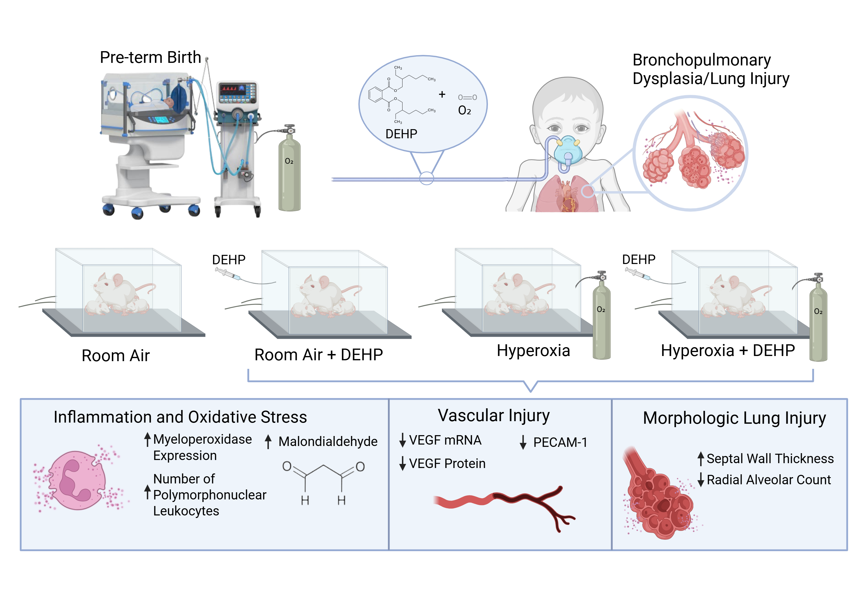
Chronic lung disease of prematurity (CLD) is a common complication of preterm birth with a com-plex pathology. Recent epidemiologic studies have identified a link between neonatal exposure to di(2-ethylhexyl) phthalate (DEHP), frequently used in medical equipment, and the development of CLD. We hypothesize that DEHP exposure in the early neonatal period contributes to lung injury in newborn rats. Newborn rat pups were raised in one of the following environments: room air (RA), RA + DEHP, hyperoxia (60% oxygen), and hyperoxia + DEHP. Ambient DEHP was inhaled in a dose of 25mg/m3 for 6 hours daily for 14 days. Lung tissue and blood samples were collected on day 14 of life. Independent exposure to DEHP and hyperoxia resulted in thicker pulmonary septal walls, fewer alveoli, increased pulmonary polymorphonuclear leukocytes and myeloperoxidase (MPO) activity and decreased expression of CD31 on endothelial cells in lung tissue. Additionally, DEHP-exposed rats showed higher serum malondialdehyde (MDA) levels and reduced vascular endothelial growth factor (VEGF) mRNA and protein levels compared to controls. Our experiments demonstrate that inhaled DEHP, with or without hyperoxia, resulted in a similar pattern of morphological lung injury and inflammation characteristic of CLD, and a causative link to CLD of prematurity.
Keywords:
phthalates
; environmental toxin
; bronchopulmonary dysplasia
; chronic lung disease
; lung injury
; prematurity
; animal model
1. Introduction
Preterm birth is associated with significant morbidity and mortality [1], largely attributed to the disruption of second- and third-trimester development of multiple organs, prominently the lungs. Chronic lung disease related to prematurity (CLD), also referred to as bronchopulmonary dysplasia (BPD), is a multifactorial disorder. The development of CLD in preterm infants has been linked to volutrauma and barotrauma due to mechanical ventilation [2], infection and non-infectious inflammation [3], and exposure to hyperoxia [2,3,4]. However, these factors alone do not predict the development of CLD with high sensitivity or specificity [5,6,7,8]. It is therefore likely that other factors contribute to, and perhaps even precipitate, CLD of prematurity. As improvements in neonatal intensive care continue to reduce known contributors to CLD (barotrauma, volutrauma, infection-driven inflammation) without significantly decreasing disease incidence [9], a search for additional modifiable factors that contribute to CLD pathobiology is imperative.
A recent epidemiologic study identified an association between NICU-based exposure to inhaled environmental phthalates and the development of CLD in a large, multi-site cohort of preterm infants [10]. Phthalate diesters, the most ubiquitous of which is di(2-ethylhexyl) phthalate (DEHP), are commonly used organic chemical adjuvants that make plastic materials soft and pliable [11,12]. Exposure to specific phthalates via the respiratory circuit is associated with adverse clinical outcomes in preterm infants [13]. Both in vitro and animal studies indicate that phthalates activate inflammation and dysregulate cellular pathways [14,15] that have been implicated in the development of CLD [16,17,18]. The contribution of phthalate exposure via inhalation to the pathogenesis and pathobiology of CLD has not been specifically examined. Prior studies in a rat model have demonstrated that DEHP exposure through various routes including intravenous and gavage feeding during gestation and early life correlates with adverse outcomes including overall poor growth, delayed lung development, impaired cell proliferation, and histological abnormalities in lung tissue [19,20]. Preterm infants requiring respiratory support are commonly exposed to significant levels of DEHP daily during early life, primarily via inhalation. However, its specific role in the pathogenesis of chronic lung disease of prematurity remains largely unexplored.
To address this gap in knowledge we designed a system that delivers aerosolized DEHP to neonatal rats in a manner that mimics the inhalational exposure experienced by preterm infants receiving respiratory support. Unlike the clinical correlate, our free breathing animal model is not confounded by barotrauma or volutrauma from positive pressure respiratory support, a significant advance in the study of the impact of phthalate exposure on lung development. In the data detailed here, we report on the use of this system to investigate the contribution of DEHP, administered with or without supplemental oxygen, to the pathogenesis and mechanistic pathobiology of neonatal lung injury in a rat model of CLD.
2. Materials and Methods
All procedures and protocols were approved by the Institutional Animal Care and Use Committees at The Feinstein Institutes for Medical Research (Protocol # 24-0631) and conformed to ARRIVE guidelines [21,22].
2.1. Animal Model
We adapted an established model of CLD due to prematurity in which a sealed chamber that allows continuous delivery of specific oxygen concentrations to induce CLD in newborn rat pups [23]. Our modified chamber allows for the introduction of aerosolized DEHP. The system, which contained two separate cages for rat pups and their mother, is equipped with a measurement device “Micro Dust Pro” for DEHP concentration monitoring (CH Technologies, Westwood, NJ, USA), and Oxygen analyzer AMI for oxygen concentrations in the ambient air (Advanced Micro Instruments Inc 225 paularino Avenue,Costa Mesa,CA 92626).
2.2. DEHP Preparation
DEHP (CAS No. 117-81-7, 0.985 g/mL, Sigma-Aldrich Corp., St. Louis, MO) was passed through an aerosol generator (4-jet refillable Blam nebulizer, CH Technologies, Westwood, NJ) and continuously administered.
Based on a prior study [24] that demonstrated toxic effects of DEHP in rats we chose an inhalation dose of 25 mg/m³ of DEHP to study its effect on newborn rat lung.
2.3. Experimental conditions
Pregnant female Sprague-Dawley rats (Charles River, Wilmington, MA) were maintained in a biosafety level 2 (BSL2) room at the animal facility of the Feinstein Institutes for Medical Research. Normal day/night light cycles were maintained. After delivery and recovery, the mother and newborn rat pups were allocated to one of four groups. Pups and dams were maintained in the chamber for 14 days. Animals were exposed to an ambient air mixture, as specified in Table 1. On day 14, all pups were euthanized; blood was obtained by cardiac puncture, and serum was isolated and stored at -80 °C. The lungs were extracted en bloc, lung tissue was homogenized, and RNA and protein fractions were isolated as described below. For histopathology, the lungs were inflated and fixed with a 1:1 mixture of 4% paraformaldehyde in phosphate buffered saline and optimal cutting temperature compound (OCT). This solution was infused through the trachea, using a fixed volume of 1mL, as a slow infusion over 2-3 minutes. Inflation was volume dependent and not pressure dependent. We did not use constant pressure to avoid barotrauma on the newborn lung. Using a fixed volume with slow infusion rate is an acceptable method for lung inflation for histopathological evaluations [25]. The right lung was embedded in paraffin and cut into 6 µm slices. Lung sections were stained with hematoxylin and eosin (H&E). The left lung was frozen and cryosectioned into 10 µm slices for immunofluorescence staining (IF).
2.4. Histological Preparation
Paraffin-embedded H&E-stained lung sections were examined under light microscopy (Zeiss Axio observer 7). For each animal, non-contiguous fields were evaluated on representative slides to determine average septal thickness and polymorphonuclear cell (PMN) count at 40X magnification, and radial alveolar count (RAC) at 10X magnification.
2.4.1. Septal Thickness
The mean thickness of 100 septa on ten non-contiguous 40X fields per animal was determined as previously described [26].
2.4.2. Radial Alveolar Count (RAC)
RAC measure, an index of alveologenesis [27,28],was determined by 1) identifying a terminal respiratory bronchiole, 2) drawing a vertical line from the center of a terminal respiratory bronchiole to the nearest acinar edge (either intra-alveolar septum or pleura), and 3) counting the number of alveoli cut by this line a sample measurement can be found in Appendix Figure A1. Radial alveolar count was measured under 10X by averaging the count from ten or fewer randomly chosen fields per animal.
2.4.3. Polymorphonuclear (PMN) Count
Each non-contiguous 40X field was divided into a 3x3 grid. The number of PMN cells in each of the 9 grid spaces was counted, and the mean was taken for the field. Means from 10 such fields per slide were used to generate PMN counts for the entire lung. Images were analyzed using ImageJ software (National Institutes of Health).
2.5. Immunofluorescence Preparation
Cryosections of lung tissues were stained by immunofluorescence for target antigens and were counterstained with 4’6-diamidino-2-phenylindole (DAPI) mounting media (Vector Laboratories, H-1200-10) for nuclear visualization. For each target, non-overlapping fields of view were randomly acquired using a confocal microscope (Zeiss LSM880). Confocal settings (gain, laser power, and pinhole size) were optimized for each individual target and then held constant for all subsequent imaging of that target. Target-positive area was quantified by thresholding using ImageJ software (National Institutes of Health). For each target, the threshold was determined from no-secondary antibody control and subsequently held constant for all images analyzed. Target-positive cells were counted and identified as DAPI-positive nuclei colocalizing with the target-positive signal. The target-positive area per target-positive cell was then calculated for each image. For each animal, values from all acquired images were averaged for statistical analysis.
2.5.1. Myeloperoxidase (MPO)
For myeloperoxidase (MPO) staining, cryosections were incubated with Rabbit Anti-MPO primary antibody (ABCAM, AB208670, 1:200) followed by an Alexa Fluor Plus 594 conjugated goat anti-rabbit secondary antibody (Invitrogen, A3653401, 1:500). 10 random non-overlapping fields of view were acquired per rat at 20X magnification. MPO expression was defined as the mean ratio of MPO-positive area per MPO-positive cell for all fields of view per animal.
2.5.2. CD31/PECAM-1
For platelet endothelial cell adhesion molecule-1 (PECAM-1/CD31) staining, cryosections were incubated with Goat anti-CD31 primary antibody (R&D Systems, AF3628, 1:250) followed by an Alexa Fluor 594 conjugated donkey anti-goat secondary antibody (Invitrogen, A11058, 1:500). 3 to 8 random non-overlapping fields of view were acquired per rat at 40X magnification. Prior to quantification, mean intensity projections were generated from full z-stacks for each field using Zen Black software (Carl Zeiss). CD31 expression was defined as the mean ratio of CD31-positive area per CD31-positive cell for all fields of view per animal.
2.6. RNA Isolation and cDNA synthesis
Total RNA was isolated from a sample of lung homogenate weighing 20-30 mg (RNeasy Mini Kit, Cat. 74104, Qiagen, Germantown, MD) per manufacturer instructions. Concentration was determined spectrophotometrically (Nanodrop Technologies, Wilmington, DE). A 260/280 ratio of 2.0 was accepted as “pure” for RNA [29]. Complementary DNA (cDNA) was created from total RNA (1 µg) via reverse transcription (iScript gDNA Clear cDNA Synthesis Kit, Bio-Rad Life Science, Cat. 1725034, Hercules, CA) per manufacturer instructions. Concentration was determined spectrophotometrically. A 260/230 ratio of 1.8 was accepted as “pure” for DNA [29].
2.7. RT-PCR
RT-PCR was performed using TaqMan technology (TaqMan 7900HT Fast Real-Time PCR System, Applied Biosystems, USA) per manufacturer’s instructions. PCR primers and probes for VEGF (Rn01511602_m1) and β-actin (Rn00667869_m1) were obtained from Thermo Fisher Scientific (Waltham, MA, USA). Both template cDNA (positive) and/or RNase-free water (negative) served as controls. The PCR cycling protocol used involved 40 cycles of 1) denaturing (95 °C for 10 seconds), 2) primer annealing and extension (60 °C for 1 minute), followed by fluorescent labeling. The relative gene expression was calculated according to the comparative Ct method (2-ΔΔCt or ΔΔCt method) [30]. cDNA concentration was normalized to the concentration of β-actin cDNA in the same sample.
2.8. Western Blotting
Total protein was extracted from frozen lung homogenate via radioimmunoprecipitation (Lysis Buffer System, Cat. sc-24948, Santa Cruz Biotechnology, Inc., Dallas, TX) in the presence of phosphatase (Pierce, Cat. PIA32957, Thermo Fisher Scientific, Waltham, MA) and protease (Pierce, Cat. PIA32955, Thermo Fisher Scientific, Waltham, MA) inhibitors. Total concentration was determined using the BCA Protein Assay Kit (Thermo Fisher Scientific, Cat. PI23225, Waltham, MA) per manufacturer instructions. 20 µg of denatured protein was suspended in 12 µL total volume (water + sample loading buffer) and subjected to sodium dodecyl sulfate-polyacrylamide gel electrophoresis (SDS-PAGE) on a gradient (4%-15%) precast gel (Mini-PROTEAN® TGX™ Precast Protein Gels, Cat. 4561084, Bio-Rad Life Science, Hercules, CA) at 150 V for 45 minutes. The gel was transferred onto polyvinylidene fluoride (PVDF) membranes and blocked with bovine serum albumin for 1 hour. Membranes were then incubated overnight at 4 °C with primary monoclonal antibodies to VEGF (Novus Biologicals, Cat. NB100-664, Centennial, CO, diluted 1:1000), VEGFR1 (Abcam, Cat. ab32152, Waltham, MA, diluted 1:2000), or VEGFR2 (Abcam, Cat. ab221679, Waltham, MA, diluted 1:2000). β-actin (Abcam, Cat. mAbcam8226, Waltham, MA, diluted 1:1000) was also determined as a loading control. Membranes were treated with secondary detection antibodies: goat anti-mouse immunoglobulin G (IgG) (Abcam, Cat. ab205719, Waltham, MA) diluted 1:2000 or goat anti-rabbit IgG (Abcam, Cat. ab205718, Waltham, MA) diluted 1:2000 and subjected to detection via enhanced chemiluminescence (Thermo Fisher Scientific, Cat. 32106, Waltham, MA). The intensities of protein bands were quantified by Image Lab software (Bio-Rad, Hercules, CA). Each homogenate sample was assessed in triplicate; if an individual value differed from the other two by >33%, the process was repeated. Mean values from each individual animal were used to determine the mean and standard deviation (SD) for the group.
2.9. ELISA
ELISA determination of MDA (Abcam, Cat. ab238537, Waltham, MA) levels in serum were performed per manufacturer instructions.
2.10. Statistical Analysis
All data are presented as the mean ± standard deviation (SD). Statistical analyses were performed using GraphPad Prism 9 software (GraphPad Software, San Diego, CA). Normality of distribution of residuals was assessed using D’Agostino-Pearson omnibus, Anderson-Darling, Shapiro-Wilk, and Kolmogorov-Smirnov tests and by examination of Q: Q plots. If residuals were normally distributed, we applied one-way analysis of variance (ANOVA) followed by the Tukey correction for multiple comparisons (P ≤ 0.05).
3. Results
We studied seven dams and their offspring. Pup numbers per experimental group were as follows: Room Air (RA) (n=13), DEHP (n=8), hyperoxia (n=14), and hyperoxia+DEHP (n=14). A significant reduction in average body weight (P ≤ 0.0001) was observed on the 14th day of life in the DEHP, hyperoxia and hyperoxia+DEHP groups compared to the RA (Appendix Figure A2 & Table A1). Combined exposure to hyperoxia+DEHP resulted in the largest decrement in body weight. In postmortem human specimens, chronic lung disease of prematurity is characterized by 1) morphologic or histologic evidence of parenchymal injury (distorted, malformed, or absent alveoli, edematous alveolar septa), 2) local or systemic inflammation (infiltrate of polymorphonuclear [PMN] cells in the lung tissue, 3) serum markers of oxidative injury, and 4) aberrant or inadequate vascularization (low levels of expression of angiogenic growth factors) [31].
3.1. Morphologic Lung Injury
Representative H&E-stained sections from animals in each of the four groups are provided in Figure 1A. These images qualitatively demonstrate the key features of lung injury: distorted, malformed (yellow circle) or absent alveoli; septal edema/thickening (black arrow); and an interstitial PMN infiltrate (yellow arrows).
Measurements of septal thickness are quantified in Figure 1B. This data demonstrates that septal thickness was significantly greater than baseline following exposure to either DEHP or hyperoxia alone (P ≤ 0.001). Septal thickness in rats exposed to hyperoxia+DEHP, in turn, was significantly greater than baseline (P ≤ 0.001) and was also more pronounced than the difference detected following exposure to each individual agent (P ≤ 0.001). Indeed, the increase over baseline in mean septal thickness after exposure to both agents was roughly equal to the sum of increase over baseline of septa exposed to each individual agent in isolation.
Figure 1C details the quantification of RAC, a measure of alveolar development [28,32]. RAC declined significantly following exposure to either DEHP or hyperoxia alone (P ≤ 0.001). Once again, the difference from baseline following exposure to a combination of hyperoxia and DEHP was roughly equal to the sum of the differences from baseline for each individual agent. This data suggests that both DEHP and hyperoxia alone induce lung injury. The additive nature of the degree of injury caused by the two in combination, as opposed to either less profound damage or a synergistic response, suggests an element of independence in the underlying mechanisms.
3.2. Inflammation and Oxidative Stress
Data assessing the effects of DEHP, hyperoxia, or hyperoxia+DEHP on PMN accumulation are depicted in Figure 2A. We found that the number of PMNs/40X magnification field was higher than baseline under all three experimental conditions. There were more PMNs following DEHP exposure than following hyperoxia. PMN counts after exposure to both hyperoxia and DEHP, while significantly higher than those noted following hyperoxia alone, could not be distinguished from numbers determined following exposure to DEHP alone. These findings suggest that PMN accumulation after either hyperoxia or DEHP involves a similar underlying mechanism.
MPO is a heme-containing peroxidase enzyme present inside the neutrophil. Representative sections showing MPO are depicted in Figure 2B and Figure 2C, confirming PMN presence in the lung tissue. Results showed a significant increase in MPO expression in the DEHP group as compared to RA (P ≤ 0.001). The hyperoxia group also showed an increase in MPO expression but the effect was less pronounced than the DEHP alone group (P ≤ 0.01). Exposure to both hyperoxia and DEHP again shows a significant increase in MPO expression as compared to the RA (P ≤ 0.001).
Hyperoxia induces lipid peroxidation and the production of reactive oxygen species that are converted to MDA. Thus, the MDA concentration in the blood has been used as a marker of oxidative injury [33,34]. Serum levels of MDA in our three experimental groups are graphically depicted in Figure 3. This data shows that DEHP, hyperoxia, and hyperoxia+ DEHP, increased the serum MDA concentration as compared to RA.
3.3. Vascular Injury
One prominent characteristic of CLD is a reduced and dysmorphic vascular bed [35]. CD31/PECAM-1 is highly expressed at endothelial cell intercellular junctions and vascular cells and is best known as a marker for vascular endothelium to identify blood vessels. The expression of CD31 in the DEHP group was significantly decreased relative to RA (P ≤ 0.01). CD31 expression after exposure to hyperoxia and hyperoxia+DEHP was lower (P ≤ 0.001) than in the RA group (Figure 4A and Figure 4B).
Vascular endothelial growth factor (VEGF) is a potent contributor to angiogenesis during embryonic development [36] and has been implicated in the pathogenesis of hyperoxic lung injury [37] We first examined VEGF abundance in lung homogenate. Figure 5A depicts a representative immunoblot of VEGF and a β-actin loading control, while quantification is presented in Figure 5B. This data indicates that the abundance of VEGF protein is significantly less than baseline following exposure to either hyperoxia or DEHP. Levels post-exposure to hyperoxia+DEHP were significantly lower than base line but were indistinguishable from values of either agent alone. Because VEGF levels are in part transcriptionally controlled [38], we assessed levels of the mRNA encoding VEGF in our four exposure groups (Figure 5C). Abundance in all three intervention groups was significantly lower than abundance at baseline. These findings suggest that intervention interferes with VEGF transcription or mRNA degradation. The effects of exposure to hyperoxia+DEHP do not indicate an additive or synergistic process. These findings suggest that overlapping mechanisms lower DEHP and hyperoxia-mediated expression of VEGF.
The effects of VEGF are mediated by binding to two receptors, VEGFR1 and VEGFR2. We therefore examined the abundance of these receptors in our three treatment groups. The difference in VEGFR1 abundance was not statistically different between the groups (Figure 6). As detailed in Figure 7, exposure to hyperoxia as well as combined exposure to hyperoxia+DEHP decreases the expression of VEGR2 as compared to DEHP and RA alone. However, the abundance of VEGFR2 in all three treatment groups was not statistically different from baseline.
4. Discussion
Phthalate diesters are common industrial organic chemicals found in a wide array of medical equipment commonly used in NICUs [39]. These compounds have well-documented effects on endocrine responses [40,41]. Multiple epidemiologic studies have demonstrated an association between exposure during the extremely vulnerable early-life developmental window and chronic dysfunction in several organ systems [42]. To date, mechanistic information about how inhalational exposure to DEHP during the neonatal period affects lung development has been lacking. Our animal experiments show that early-life exposure to DEHP is associated with distorted alveologenesis, decreased angiogenesis and enhanced inflammatory response to oxidative stress. These findings are similar to changes found in CLD in human populations [3,43].
This is the first animal model developed to assess the direct effect of DEHP exposure via inhalation on early neonatal lung development with and without associated exposure to hyperoxia. Both DEHP exposure alone and coincident exposure to hyperoxia and DEHP showed poor growth manifested as low body weight and histopathologic findings similar to those seen in CLD. DEHP-associated inflammatory response, distorted alveologenesis, and reduced endothelial cell are notable findings that perhaps provide a key to the pathogenesis of CLD in infants who have a relatively benign NICU course.
Infants born prematurely spend a critical period of development in the chemically intensive NICU environment [13]. Phthalates are ubiquitous chemical adjuvants used to improve the flexibility and durability of common medical equipment. Phthalates, including DEHP, leach from the underlying plastic scaffold under conditions of heat and humidity [13]. Preterm infants are at high risk of toxicity due to immature pulmonary architecture [44], immature antioxidant [45] and anti-inflammatory [46] mechanisms, decreased and immature gas exchange surface area [44,47] and decreased capability to metabolize and excrete toxins [48].
It has previously been shown that DEHP exposure promotes an inflammatory response through immunological mediators that lead to pathological changes in the lung tissues of adult mice [49]. In this study, we adapted an established animal model of prematurity-related CLD and found that exposure to DEHP in the early neonatal period leads to an inflammatory response and contributes to or exacerbates the development of histologic changes in lung tissue seen in CLD of prematurity. The characteristic hallmarks of CLD are a large and simplified alveolar structure, a reduced and dysmorphic vascular bed, and septal wall fibrosis [50,51] In our model, lung injury and inflammation were confirmed by histology and immunofluorescence staining. Our study shows increased accumulation of PMN and MPO expression, increased septal wall thickness, and poor alveolar growth, manifested as reduced RAC along with reduced expression of CD31 and VEGF suggestive of poor vascular bed in pups exposed to DEHP in the early neonatal period. Molecular studies showed increased inflammatory markers in DEHP-exposed pups. As in prior studies, oxygen exposure also led to exaggerated inflammatory changes and poor angiogenesis and alveolar growth in DEHP-exposed pups.
Exposure to hyperoxia has been described as impaired growth in animal model [52]. The phthalate effect on growth is complex and depends on timing of exposure. Infants with BPD also shows impaired growth in established disease [53]. Our study shows decreased body weight in the DEHP, hyperoxia, hyperoxia + DEHP exposed groups as compared to alone. Body weight is an indirect measure of growth, but in the absence of birthweight and other parameters, it is difficult to suggest its association with phthalate exposure.
Histological examination of lung tissue shows scanty blood vessels with distorted alveolar growth in pups exposed to DEHP. Concomitant hyperoxia and DEHP exposure showed exacerbation of these changes and lung damage in early neonatal rat pups, similar to what is seen on histological examination of lung tissue taken from preterm infants with CLD. Our data showed a significant decrease in RAC in the DEHP-exposed group, while coincident exposure to both hyperoxia and DEHP demonstrated a further decrease in RAC.
In addition to exploring histological changes, our immunoflourescence staining shows reduced expression of CD31 after exposure to DEHP, hyperoxia and Hyperoxia+DEHP. CD31 expression indicates the presence of blood vessels, as it is highly expressed at endothelial cell intercellular junctions and serves as a widely used endothelial marker in immunohistochemistry [54,55].
We were able to measure markers of angiogenesis known to impact pulmonary development [18,36,56]. Our experiments showed that neonatal pups exposed to DEHP in early postnatal life demonstrate significantly lower expression of VEGF. As pulmonary vascular growth precedes tissue growth, lower VEGF expression may be the primary factor associated with poor alveolar growth in infants developing CLD related to DEHP exposure. This finding highlights a possible role of DEHP in lung injury in early life, even in normoxia. VEGF regulates angiogenesis by activating its receptors VEGFR1 (sFlt-1) and VEGFR2 (KDR/Flk1). VEGFR1 plays a negative role in vasculogenesis, as it does not have any mitogenic effect on endothelial cells [57]. VEGFR1 is also described as an antiangiogenic factor, as it binds extracellularly to VEGF and decreases its availability to VEGFR2 [58]. VEGFR2 is a major signal transducer for angiogenesis, promoting migration, differentiation, and proliferation of endothelial cells [59] and the formation of vascular tubes [60]. Our study shows that DEHP exposure in the early neonatal period is associated with decreased expression of VEGF without significant change in expression of VEGFR1 /VEGFR2. Thus, the effects of hyperoxia, and hyperoxia+DEHP, on the abundance of elements in the VEGF pathway appear to be limited to the biosynthesis of VEGF itself.
MDA is a marker of oxidative damage that is associated with the development of CLD of prematurity [61]. Our experiments demonstrated a significant elevation of MDA levels in pups exposed to DEHP, again indicating that DEHP may contribute to oxidative damage in newborn rat pups.Significant increase in MDA level after exposure to DEHP as compared to hyperoxia and hyperoxia+DEHP suggesting a contribution of DEHP to inflammation-mediated lung damage.
Overall, our experiments demonstrate that DEHP exposure is associated with inflammatory lung damage and enhanced oxidative stress, reduced vasculature, leading to histological changes in pulmonary architecture in a neonatal rat model. Our data support the hypothesis drawn from epidemiological studies [10,13] that DEHP exposure may be an important contributor to the development of CLD. As DEHP exposure is a potentially modifiable factor in the preterm infant hospital environment, there is an urgent need to further explore both this pathological link and the possible replacement of DEHP in common NICU respiratory circuits with less toxic chemical plasticizers.
A key feature of our animal model is that it allows disentanglement of the role of volutrauma, barotrauma, and hyperoxia from the impact of DEHP exposure—a feat that would be virtually impossible in patient-based studies at the present time. In clinical populations, significant inhalational DEHP exposure is derived from respiratory support equipment [13], which also confers some degree of pressure and/or volume support to the developing lung. In our model, rats breathe naturally in an unsupported manner, eliminating any contribution from volutrauma or barotrauma. Newborn rat pups are at the saccular stage of lung development at birth, akin to preterm infants at the highest risk for CLD born at 24–26 weeks of gestational age [23]. They, therefore, provide a unique avenue to examine the developmental impact of various inhalational environmental exposures.
Our study does have limitations. Rat models do not perfectly mimic human biology; thus, there may be differences in disease progression in animals compared to humans such that our findings may not perfectly represent human pathology. Variability by sex may play a role in the biological impact of DEHP, a known endocrine disruptor associated with sexually dimorphic outcomes in humans [62]. Our study was not powered to explore sex variability in the pathogenesis of BPD. Additionally, we used high concentrations of DEHP in our experiments; although studies of preterm infant DEHP exposure indicate similarly high levels of exposure [13,63,64,65,66], it is possible that hospitalized preterm infants do not experience this level of exposure in all circumstances.
Traditionally, neonatology research has not evaluated the impact of environmental exposures on CLD development. Our study builds on the existing animal and human epidemiological literature to demonstrate the link between inhalational phthalate exposure and lung damage that is histopathologically similar to what is seen in human CLD. Human studies on the clinical impact of environmental toxicants, including DEHP, are challenging. Epidemiological observational human studies of inhalational DEHP exposure via respiratory support equipment are universally subject to confounding by indication, as the source of DEHP exposure is the respiratory support equipment that also generates barotrauma and volutrauma. Studying DEHP both as a single toxicant and as a mixture with hyperoxia is critical as both RA and oxygen therapy via the respiratory circuit are common in NICU care. Our animal model allows isolation of these exposures and specific mixtures through the ambient free breathing environment, eliminating confounding by indication.
Chronic lung disease of prematurity is a complex and multifactorial disease associated with significant morbidity and mortality [7] As such, it is a central driver of the cost of care of preterm infants: the median hospitalization cost of a preterm infant with CLD is almost double that of an otherwise comparable preterm infant without CLD [67] Our study highlights that DEHP, a major component of the NICU environment despite recent efforts at exposure mitigation, may contribute to adverse pulmonary outcomes among preterm infants. Therefore, decreasing or eliminating the use of DEHP in the production of neonatal respiratory support circuits could positively impact preterm infant lung development, reduce costs of care, and reduce significant suffering in the preterm population.
5. Conclusions
In our novel animal model, inhaled DEHP induced lung inflammation, impaired angiogenesis and alveolarization, and histological abnormalities similar to what seen in pathological specimens taken from preterm infant with severe CLD. DEHP and hyperoxia exposure separately and together elicited morphological lung injury and inflammation characteristic of CLD.
DEHP exposure in the NICU may be toxic to the developing lungs of preterm infants and thus may be a preventable contributor to CLD. Our study was designed to identify and characterize the impact of DEHP exposure on preterm lung injury, demonstrating its significance and justifying the need for further investigation. Unraveling the complete molecular cascade is a complex endeavor that often requires a multi-stage approach. This current work serves as an essential foundation, pointing us towards the specific biological questions that now need to be addressed to develop comprehensive mitigation strategies.
Author Contributions
Shahana Perveen was involved in conceptualization, data curation, funding acquisition, investigation, analysis and supervision. Shahana Perveen wrote the first draft and edited the manuscript. Li Lou was involved in data curation, investigation and analysis. Sohini Alim was involved in investigations, data curation, analysis and manuscript editing, Abigail Akselrod was involved in investigations, analysis and data curation, Chunfang Zhao was involved with data curation, investigation. Namita Sen, was involved with investigation, data curation and analysis of data. Clifford S. Deutschman was involved in supervision, data analysis and manuscript editing. Annemarie Stroustrup was involved in conceptualization, funding acquisition, analysis, supervision and final manuscript editing. All authors have read and agreed to the published version of the manuscript.
Funding
This study was funded by the National Institute of Environmental Health Sciences R01ES034018. Additional funding from the Liling Family Neonatal Research Laboratory, Division of Neonatology, Cohen Children Medical Center.
Institutional Review Board Statement
Data Availability Statement
The data sets generated during and/or analyzed during the current study are available from the corresponding author on reasonable request.
Conflicts of Interest
Dr. Deutschman declares the following potential conflicts of interest: 1. Scientific Editor, Critical Care Medicine honorarium of $1500.00/year, ongoing. 2. Siemens Inc, Consultant on product for sepsis, past, ended May 2022, received approx. $3000. 3. Stock options for Enlivex, Jerusalem Israel. 4. La Jolla Pharmaceuticals, now owned by Innoviva, has supplied our lab with angiotensin II for use in experiments, last received in 2021. 5. Royalties from Elsevier for textbook “Evidence-Based Practice of Critical Care, 1st, 2nd and 3rd Editions”. 6. Complementary registration at Society of Critical Care Medicine Annual meeting as Past-President. 7. Reimbursement by Shock Society of travel expenses to Rockville MD for Strategic Planning meeting, Nov 2023. The funders had no role in the design of the study; in the collection, analyses, or interpretation of data; in the writing of the manuscript; or in the decision to publish the results. All other authors declare they have no conflicts of interest related to this work to disclose.
Abbreviations
The following abbreviations are used in this manuscript:
| CLD | Chronic lung disease of prematurity |
| DEHP | di(2-ethylhexyl) phthalate |
| RA | Room Air |
| MPO | Myeloperoxidase |
| MDA | Malondialdehyde |
| VEGF | Vascular endothelial growth factor |
| BPD | Bronchopulmonary dysplasia |
| NICU | Neonatal Intensive Care Unit |
| IACUC | Institutional Animal Care and Use Committees |
| ARRIVE | Animal Research: Reporting of In Vivo Experiments |
| BSL2 | Biosafety level 2 |
| H&E | Hematoxylin and Eosin |
| OCT | Optimal Cutting Temperature compound |
| IF | Immunofluorescence |
| PMN | Polymorphonuclear leukocytes |
| RAC | Radial Alveolar Count |
| DAPI | 4’6-diamidino-2-phenylindole |
| CD31/PECAM-1 | Platelet Endothelial Cell Adhesion Molecule-1 / CD31 |
| cDNA | Complementary DNA |
| SDS-PAGE | Sodium Dodecyl Sulfate-Polyacrylamide Gel Electrophoresis |
| PVDF | Polyvinylidene Fluoride |
| IgG | Immunoglobulin G |
| SD | Standard Deviation |
| ANOVA | Analysis of Variance |
Appendix A
Appendix A.1
Figure A1.
Representative 10x magnification fields showing lines measuring Radial Alveolar Count (RAC) in neonatal rat lungs in room air and following inhaled DEHP, hyperoxia, or hyperoxia + DEHP. Neonatal Sprague Dawley Rats breathing the mixture noted for 14 days. Lines connect a terminal bronchiole to the nearest acinar edge, with alveolar intersections counted to determine RAC.
Figure A1.
Representative 10x magnification fields showing lines measuring Radial Alveolar Count (RAC) in neonatal rat lungs in room air and following inhaled DEHP, hyperoxia, or hyperoxia + DEHP. Neonatal Sprague Dawley Rats breathing the mixture noted for 14 days. Lines connect a terminal bronchiole to the nearest acinar edge, with alveolar intersections counted to determine RAC.
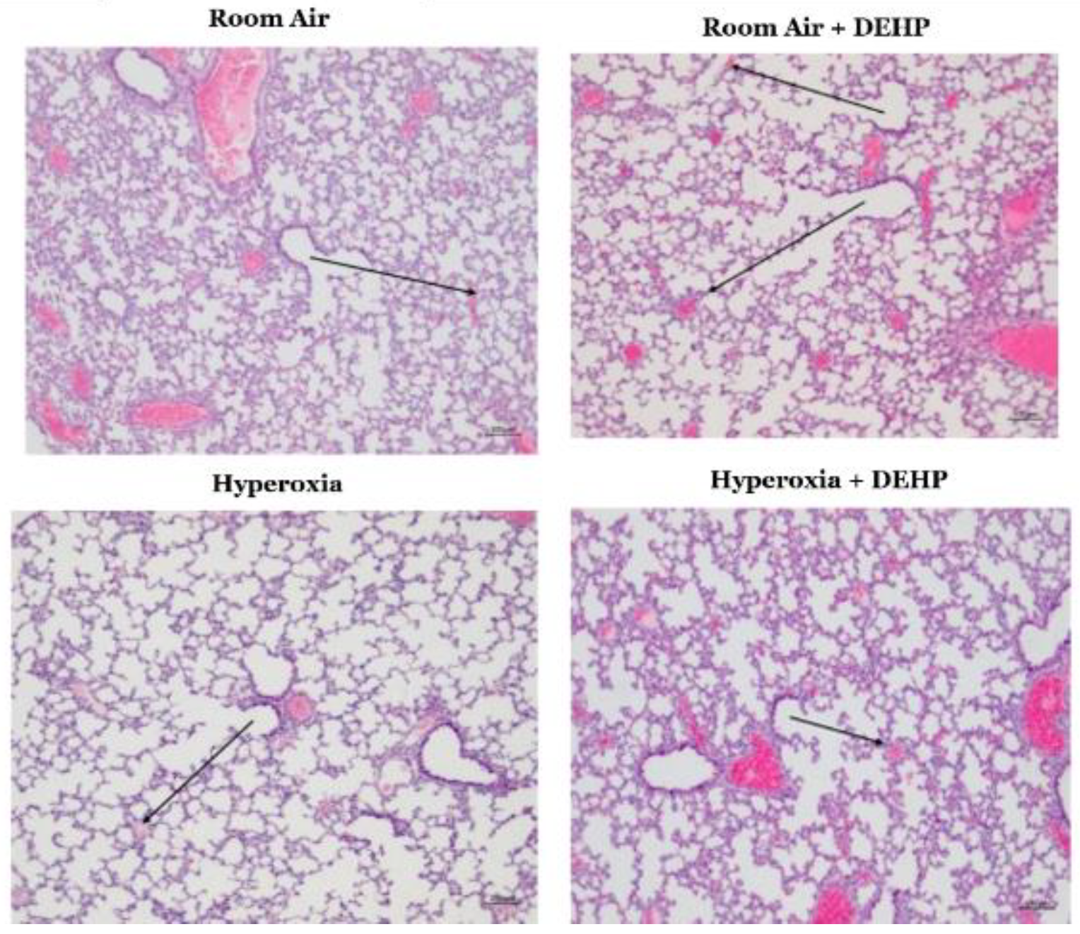
Figure A2.
Weight of neonatal rats pups in room air and following inhaled DEHP, hyperoxia, or hyperoxia + DEHP. Neonatal Sprague Dawley Rats breathing the mixture noted for 14 days. n= 8-14 animals/intervention. Data is shown as a box and whisker plot with whiskers indicating the minimum and maximum values in each group. Significance determined using one-way ANOVA with Tukey’s correction. * P ≤ 0.05, ** P ≤ 0.01, *** P ≤ 0.001.
Figure A2.
Weight of neonatal rats pups in room air and following inhaled DEHP, hyperoxia, or hyperoxia + DEHP. Neonatal Sprague Dawley Rats breathing the mixture noted for 14 days. n= 8-14 animals/intervention. Data is shown as a box and whisker plot with whiskers indicating the minimum and maximum values in each group. Significance determined using one-way ANOVA with Tukey’s correction. * P ≤ 0.05, ** P ≤ 0.01, *** P ≤ 0.001.
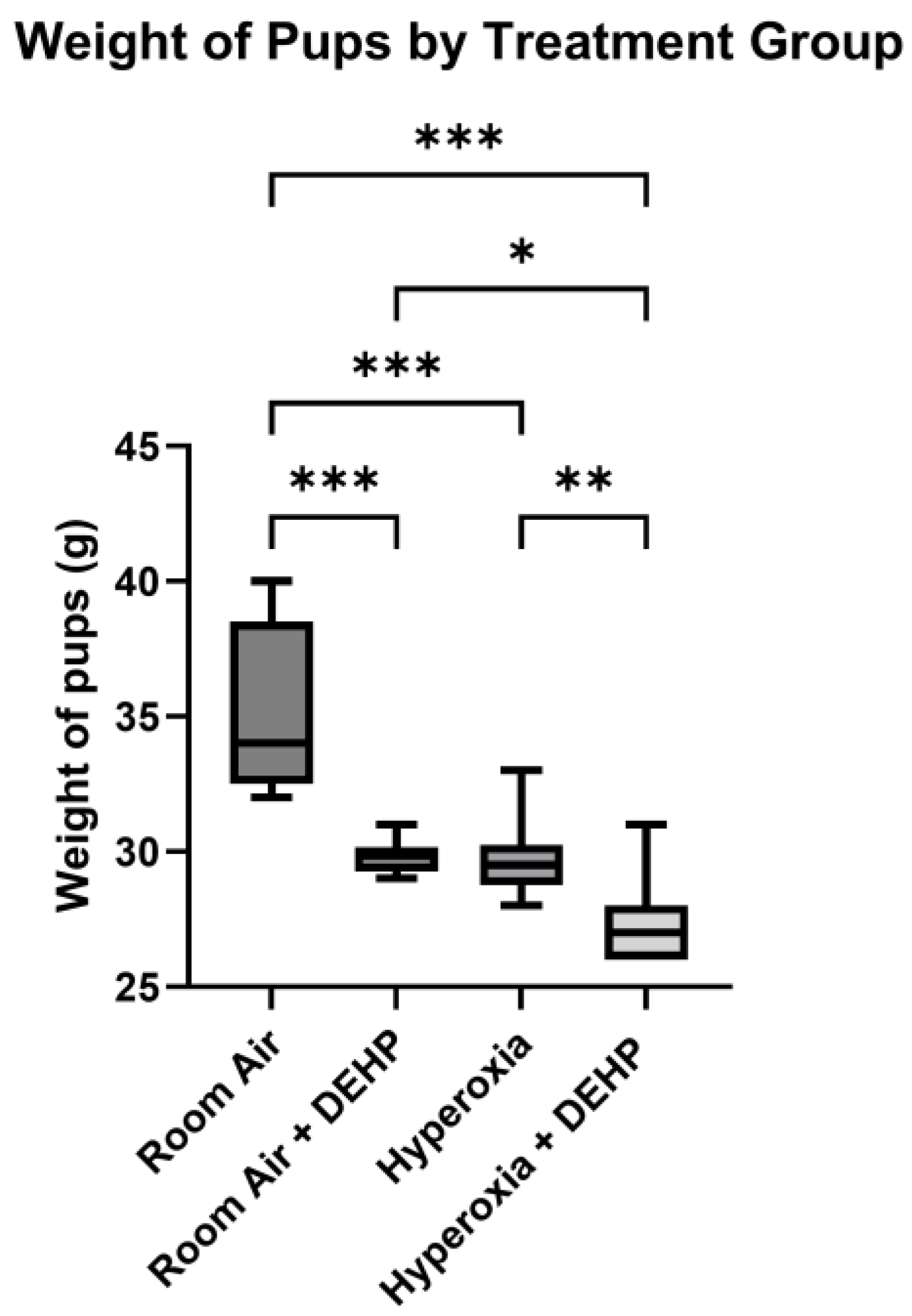

Table A1.
Number of values, minimum, maximum, range, and mean of weight of rat pups in each intervention group.
Table A1.
Number of values, minimum, maximum, range, and mean of weight of rat pups in each intervention group.
| Room Air | Room Air + DEHP | Hyperoxia | Hyperoxia + DEHP | |
|---|---|---|---|---|
| Number of values | 13 | 8 | 14 | 14 |
| Minimum | 32.00 | 29.00 | 28.00 | 26.00 |
| Maximum | 40.00 | 31.00 | 33.00 | 31.00 |
| Range | 8.000 | 2.000 | 5.000 | 5.000 |
| Mean | 35.42 | 29.88 | 29.71 | 27.29 |
References
- Patel, R.M. Short- and Long-Term Outcomes for Extremely Preterm Infants. Am. J. Perinatol. 2016, 33(3), 318–28. [Google Scholar] [CrossRef]
- Dankhara, N.; Holla, I.; Ramarao, S.; Kalikkot Thekkeveedu, R. Bronchopulmonary Dysplasia: Pathogenesis and Pathophysiology. J. Clin. Med. 2023, 12(13). [Google Scholar] [CrossRef] [PubMed]
- Jobe, A.H. Mechanisms of Lung Injury and Bronchopulmonary Dysplasia. Am. J. Perinatol. 2016, 33(11), 1076–8. [Google Scholar] [CrossRef]
- Dong, Y.; Leidner, A.; Marega, M.; Rivetti, S.; Bellusci, S. When prenatal infection meets postnatal hyperoxia: Better models for bronchopulmonary dysplasia and its therapeutic approaches. Chin. Med. J. Pulm. Crit. Care Med. 2024, 2(4), 289–292. [Google Scholar] [CrossRef]
- Northway, W.H.; Rosan, R.C.; Porter, D.Y. Pulmonary disease following respirator therapy of hyaline-membrane disease. Bronchopulmonary dysplasia. N Engl. J. Med. 1967, 276(7), 357–68. [Google Scholar] [CrossRef]
- Bland, R.D.; Albertine, K.H.; Carlton, D.P.; et al. Chronic lung injury in preterm lambs: abnormalities of the pulmonary circulation and lung fluid balance. Pediatr. Res. 2000, 48(1), 64–74. [Google Scholar] [CrossRef] [PubMed]
- Davidson, L.M.; Berkelhamer, S.K. Bronchopulmonary Dysplasia: Chronic Lung Disease of Infancy and Long-Term Pulmonary Outcomes. J. Clin. Med. 2017, 6(1). [Google Scholar] [CrossRef]
- Greenough, A. Long term respiratory outcomes of very premature birth (<32 weeks). Semin Fetal Neonatal Med. 2012, 17(2), 73–6. [Google Scholar] [CrossRef]
- Jeon, G.W.; Oh, M.; Chang, Y.S. Increased bronchopulmonary dysplasia along with decreased mortality in extremely preterm infants. Sci. Rep. 2025, 15(1), 8720. [Google Scholar] [CrossRef]
- Stroustrup, A.; Zhang, X.; Spear, E.; et al. Phthalate exposure in the neonatal intensive care unit is associated with development of bronchopulmonary dysplasia. Env. Int. 2023, 178, 108117. [Google Scholar] [CrossRef]
- Stroustrup, A.; Bragg, J.B.; Spear, E.A.; et al. Cohort profile: the Neonatal Intensive Care Unit Hospital Exposures and Long-Term Health (NICU-HEALTH) cohort, a prospective preterm birth cohort in New York City. BMJ Open 2019, 9(11), e032758. [Google Scholar] [CrossRef] [PubMed]
- Foong, R.E.; Franklin, P.; Sanna, F.; et al. Longitudinal effects of prenatal exposure to plastic-derived chemicals and their metabolites on asthma and lung function from childhood into adulthood. Respirology 2022. [Google Scholar] [CrossRef]
- Stroustrup, A.; Bragg, J.B.; Busgang, S.A.; et al. Sources of clinically significant neonatal intensive care unit phthalate exposure. J. Expo. Sci. Env. Epidemiol. 2020, 30(1), 137–148. [Google Scholar] [CrossRef]
- Palacios-Arreola, M.I.; Morales-Montor, J.; Cazares-Martinez, C.J.; Gomez-Arroyo, S.; Nava-Castro, K.E. Environmental pollutants: an immunoendocrine perspective on phthalates. Front Biosci. (Landmark Ed) 2021, 26(3), 401–430. [Google Scholar] [CrossRef]
- Tseng, H.H.; Li, C.Y.; Wu, S.T.; et al. Di-(2-ethylhexyl) Phthalate Promotes Allergic Lung Inflammation by Modulating CD8α. Front Immunol. 2022, 13, 581854. [Google Scholar] [CrossRef]
- Surate Solaligue, D.E.; Rodríguez-Castillo, J.A.; Ahlbrecht, K.; Morty, R.E. Recent advances in our understanding of the mechanisms of late lung development and bronchopulmonary dysplasia. Am. J. Physiol. Lung Cell Mol. Physiol. 2017, 313(6), L1101–L1153. [Google Scholar] [CrossRef]
- Rodríguez-Castillo, J.A.; Pérez, D.B.; Ntokou, A.; Seeger, W.; Morty, R.E.; Ahlbrecht, K. Understanding alveolarization to induce lung regeneration. Respir. Res. 2018, 19(1), 148. [Google Scholar] [CrossRef]
- Bhatt, A.J.; Pryhuber, G.S.; Huyck, H.; Watkins, R.H.; Metlay, L.A.; Maniscalco, W.M. Disrupted pulmonary vasculature and decreased vascular endothelial growth factor, Flt-1, and TIE-2 in human infants dying with bronchopulmonary dysplasia. Am. J. Respir. Crit. Care Med. 2001, 164 10 Pt 1, 1971–80. [Google Scholar] [CrossRef]
- Liang, Z.J.; Wu, Q.P.; Chen, B.T.; Lin, Z.L.; Lin, J.; Chen, S.Q. Postnatal hyperoxia or DEHP exposure leads to growth restriction and delayed lung development in newborn rats. Pediatr. Neonatol. 2018, 59(1), 24–30. [Google Scholar] [CrossRef] [PubMed]
- Rosicarelli, B.; Stefanini, S. DEHP effects on histology and cell proliferation in lung of newborn rats. Histochem Cell Biol. 2009, 131(4), 491–500. [Google Scholar] [CrossRef] [PubMed]
- Percie du Sert, N.; Ahluwalia, A.; Alam, S.; et al. Reporting animal research: Explanation and elaboration for the ARRIVE guidelines 2.0. PLoS Biol. 2020, 18(7), e3000411. [Google Scholar] [CrossRef]
- Percie du Sert, N.; Hurst, V.; Ahluwalia, A.; et al. The ARRIVE guidelines 2.0: Updated guidelines for reporting animal research. PLoS Biol. 2020, 18(7), e3000410. [Google Scholar] [CrossRef]
- O’Reilly, M.; Thébaud, B. Animal models of bronchopulmonary dysplasia. The term rat models. Am. J. Physiol. Lung Cell Mol. Physiol. 2014, 307(12), L948–58. [Google Scholar] [CrossRef]
- Ma, M.; Kondo, T.; Ban, S.; et al. Exposure of prepubertal female rats to inhaled di(2-ethylhexyl)phthalate affects the onset of puberty and postpubertal reproductive functions. Toxicol. Sci. 2006, 93(1), 164–71. [Google Scholar] [CrossRef]
- Morton, J.; Snider, T.A. Guidelines for collection and processing of lungs from aged mice for histological studies. Pathobiol. Aging Age Relat. Dis. 2017, 7(1), 1313676. [Google Scholar] [CrossRef]
- Perveen, S.; Ayasolla, K.; Zagloul, N.; et al. MIF inhibition enhances pulmonary angiogenesis and lung development in congenital diaphragmatic hernia. Pediatr. Res. 2019, 85(5), 711–718. [Google Scholar] [CrossRef]
- Cooney, T.P.; Thurlbeck, W.M. The radial alveolar count method of Emery and Mithal: a reappraisal 1--postnatal lung growth. Thorax 1982, 37(8), 572–9. [Google Scholar] [CrossRef]
- Cherian, D.M.; Shalini, C.; Andrews, C.; Maheswari, U. D. P. Evaluation of Pulmonary Hypoplasia in Various Congenital Anomalies with a Comparison of Two Conventional Methods of Assessment: Radial Alveolar Count (RAC) and Lung Weight: Body Weight Ratio (LBW). Turk. Patoloji Derg. 2021, 37(2), 154–160. [Google Scholar] [CrossRef]
- Wilfinger, W.W.; Mackey, K.; Chomczynski, P. Effect of pH and ionic strength on the spectrophotometric assessment of nucleic acid purity. Biotechniques 1997, 22(3), 474-6, 478-81. [Google Scholar] [CrossRef] [PubMed]
- Regier, N.; Frey, B. Experimental comparison of relative RT-qPCR quantification approaches for gene expression studies in poplar. BMC Mol. Biol. 2010, 11, 57. [Google Scholar] [CrossRef] [PubMed]
- Niedermaier, S.; Hilgendorff, A. Bronchopulmonary dysplasia—an overview about pathophysiologic concepts. Mol. Cell Pediatr. 2015, 2(1), 2. [Google Scholar] [CrossRef]
- Catozzi, C.; Modena, A.; Storti, M.; Ricci, F.; Villetti, G.; De Luca, D. Quantitative lung ultrasound findings correlate with radial alveolar count in experimental bronchopulmonary dysplasia. Ultrasound J. 2024, 16(1), 46. [Google Scholar] [CrossRef] [PubMed]
- Aydemir, D.; Karabulut, G.; Barlas, N.; Ulusu, N.N. DEHP impairs the oxidative stress response and disrupts trace element and mineral metabolism within the mitochondria of detoxification organs. Toxicol. Ind. Health 2025, 41(2), 108–121. [Google Scholar] [CrossRef]
- Del Rio, D.; Stewart, A.J.; Pellegrini, N. A review of recent studies on malondialdehyde as toxic molecule and biological marker of oxidative stress. Nutr. Metab. Cardiovasc Dis. 2005, 15(4), 316–28. [Google Scholar] [CrossRef]
- Baker, C.D.; Abman, S.H. Impaired pulmonary vascular development in bronchopulmonary dysplasia. Neonatology 2015, 107(4), 344–51. [Google Scholar] [CrossRef]
- Perrone, S.; Manti, S.; Buttarelli, L.; et al. Vascular Endothelial Growth Factor as Molecular Target for Bronchopulmonary Dysplasia Prevention in Very Low Birth Weight Infants. Int. J. Mol. Sci. 2023, 24(3). [Google Scholar] [CrossRef]
- Oak, P.; Hilgendorff, A. The BPD trio? Interaction of dysregulated PDGF, VEGF, and TGF signaling in neonatal chronic lung disease. Mol. Cell Pediatr. 2017, 4(1), 11. [Google Scholar] [CrossRef]
- Hanson, J.; Gorman, J.; Reese, J.; Fraizer, G. Regulation of vascular endothelial growth factor, VEGF, gene promoter by the tumor suppressor, WT1. Front Biosci. 2007, 12, 2279–90. [Google Scholar] [CrossRef]
- Pak, V.M.; Nailon, R.E.; McCauley, L.A. Controversy: neonatal exposure to plasticizers in the NICU. MCN Am. J. Matern Child. Nurs. 2007, 32(4), 244–9. [Google Scholar] [CrossRef] [PubMed]
- Zeng, F.; Zhang, L.; Deng, F.; Lou, S. Early-life exposure to di (2-ethyl-hexyl) phthalate: Role in children with endocrine disorders. Front Cell Dev. Biol. 2023, 11, 1115229. [Google Scholar] [CrossRef] [PubMed]
- Li, X.; Xiao, C.; Liu, J.; et al. Association of Di(2-ethylhexyl) Phthalate Exposure with Reproductive Hormones in the General Population and the Susceptible Population: A Systematic Review and Meta-Analysis. Env. Health (Wash) 2024, 2(11), 750–765. [Google Scholar] [CrossRef]
- Braun, J.M.; Sathyanarayana, S.; Hauser, R. Phthalate exposure and children’s health. Curr. Opin. Pediatr. 2013, 25(2), 247–54. [Google Scholar] [CrossRef] [PubMed]
- Coalson, J.J. Pathology of bronchopulmonary dysplasia. Semin Perinatol. 2006, 30(4), 179–84. [Google Scholar] [CrossRef] [PubMed]
- Baker, C.D.; Alvira, C.M. Disrupted lung development and bronchopulmonary dysplasia: opportunities for lung repair and regeneration. Curr. Opin. Pediatr. 2014, 26(3), 306–14. [Google Scholar] [CrossRef]
- Georgeson, G.D.; Szony, B.J.; Streitman, K.; et al. Antioxidant enzyme activities are decreased in preterm infants and in neonates born via caesarean section. Eur. J. Obstet. Gynecol. Reprod. Biol. 2002, 103(2), 136–9. [Google Scholar] [CrossRef] [PubMed]
- Choi, C.W.; Lee, J.; Oh, J.Y.; Lee, S.H.; Lee, H.J.; Kim, B.I. Protective effect of chorioamnionitis on the development of bronchopulmonary dysplasia triggered by postnatal systemic inflammation in neonatal rats. Pediatr. Res. 2016, 79(2), 287–94. [Google Scholar] [CrossRef]
- Williams, E.E.; Gareth Jones, J.; McCurnin, D.; et al. Functional morphometry: non-invasive estimation of the alveolar surface area in extremely preterm infants. Pediatr. Res. 2023, 94(5), 1707–1713. [Google Scholar] [CrossRef]
- Stroustrup, A.; Teitelbaum, S.L.; Aschner, J.L. The Value of Preterm Infant Environmental Health Cohorts: The Canary in the Coal Mine. JAMA Pediatr. 2017, 171(12), 1139–1140. [Google Scholar] [CrossRef]
- Guo, J.; Han, B.; Qin, L.; et al. Pulmonary toxicity and adjuvant effect of di-(2-exylhexyl) phthalate in ovalbumin-immunized BALB/c mice. PLoS ONE 2012, 7(6), e39008. [Google Scholar] [CrossRef]
- Kalikkot Thekkeveedu, R.; Guaman, M.C.; Shivanna, B. Bronchopulmonary dysplasia: A review of pathogenesis and pathophysiology. Respir. Med. 2017, 132, 170–177. [Google Scholar] [CrossRef]
- Berger, J.; Bhandari, V. Animal models of bronchopulmonary dysplasia. The term mouse models. Am. J. Physiol. Lung Cell Mol. Physiol. 2014, 307(12), L936–47. [Google Scholar] [CrossRef]
- Woeller, C.F.; Lim, S.A.; Roztocil, E.; et al. Neonatal hyperoxia impairs adipogenesis of bone marrow-derived mesenchymal stem cells and fat accumulation in adult mice. Free Radic. Biol. Med. 2021, 167, 287–298. [Google Scholar] [CrossRef]
- Miller, A.N.; Curtiss, J.; Taylor, S.N.; Backes, C.H.; Kielt, M.J. A review and guide to nutritional care of the infant with established bronchopulmonary dysplasia. J. Perinatol. 2023, 43(3), 402–410. [Google Scholar] [CrossRef]
- Woodfin, A.; Voisin, M.B.; Nourshargh, S. PECAM-1: a multi-functional molecule in inflammation and vascular biology. Arterioscler. Thromb. Vasc. Biol. 2007, 27(12), 2514–23. [Google Scholar] [CrossRef]
- Privratsky, J.R.; Newman, P.J. PECAM-1: regulator of endothelial junctional integrity. Cell Tissue Res. 2014, 355(3), 607–19. [Google Scholar] [CrossRef]
- Moradi-Gharibvand, N.; Hashemibeni, B. The Effect of Stem Cells and Vascular Endothelial Growth Factor on Cancer Angiogenesis. Adv. BioMed Res. 2023, 12, 124. [Google Scholar] [CrossRef]
- Shibuya, M. Differential roles of vascular endothelial growth factor receptor-1 and receptor-2 in angiogenesis. J. Biochem Mol. Biol. 2006, 39(5), 469–78. [Google Scholar] [CrossRef]
- Ikeda, T.; Sun, L.; Tsuruoka, N.; et al. Hypoxia down-regulates sFlt-1 (sVEGFR-1) expression in human microvascular endothelial cells by a mechanism involving mRNA alternative processing. Biochem J. 2011, 436(2), 399–407. [Google Scholar] [CrossRef]
- Takahashi, H.; Shibuya, M. The vascular endothelial growth factor (VEGF)/VEGF receptor system and its role under physiological and pathological conditions. Clin. Sci. (Lond) 2005, 109(3), 227–41. [Google Scholar] [CrossRef]
- Koch, S.; Claesson-Welsh, L. Signal transduction by vascular endothelial growth factor receptors. Cold Spring Harb. Perspect. Med. 2012, 2(7), a006502. [Google Scholar] [CrossRef]
- Thompson, A.; Bhandari, V. Pulmonary Biomarkers of Bronchopulmonary Dysplasia. Biomark. Insights 2008, 3, 361–373. [Google Scholar] [CrossRef]
- Palanza, P.; Paterlini, S.; Brambilla, M.M.; et al. Sex-biased impact of endocrine disrupting chemicals on behavioral development and vulnerability to disease: Of mice and children. Neurosci. Biobehav Rev. 2021, 121, 29–46. [Google Scholar] [CrossRef]
- Calafat, A.M.; Needham, L.L.; Silva, M.J.; Lambert, G. Exposure to di-(2-ethylhexyl) phthalate among premature neonates in a neonatal intensive care unit. Research Support, U.S. Gov’t, Non-P.H.S. Research Support, U.S. Gov’t, P.H.S. Pediatrics. 2004, 113(5), e429-34. [Google Scholar]
- Al-Saleh, I.; Elkhatib, R.; Alnuwaysir, H.; et al. The cumulative risk assessment of phthalates exposure in preterm neonates. Int. J. Hyg. Env. Health 2023, 248, 114112. [Google Scholar] [CrossRef]
- Panneel, L.; Cleys, P.; Poma, G.; et al. Ongoing exposure to endocrine disrupting phthalates and alternative plasticizers in neonatal intensive care unit patients. Env. Int. 2024, 186, 108605. [Google Scholar] [CrossRef]
- Mallow, E.B.; Fox, M.A. Phthalates and critically ill neonates: device-related exposures and non-endocrine toxic risks. J. Perinatol. 2014, 34(12), 892–7. [Google Scholar] [CrossRef]
- Lai, K.C.; Lorch, S.A. Healthcare Costs of Major Morbidities Associated with Prematurity in US Children’s Hospitals. J. Pediatr. 2023, 256, 53–62.e4. [Google Scholar] [CrossRef]
Figure 1A.
Responses of neonatal lungs in room air and following inhaled DEHP, hyperoxia, and hyperoxia + DEHP. Representative H&E-stained sections, 40X magnification. Neonatal Sprague Dawley rats breathing the mixture noted for 14 days. Description in the text. Arrows—neutrophils.
Figure 1A.
Responses of neonatal lungs in room air and following inhaled DEHP, hyperoxia, and hyperoxia + DEHP. Representative H&E-stained sections, 40X magnification. Neonatal Sprague Dawley rats breathing the mixture noted for 14 days. Description in the text. Arrows—neutrophils.
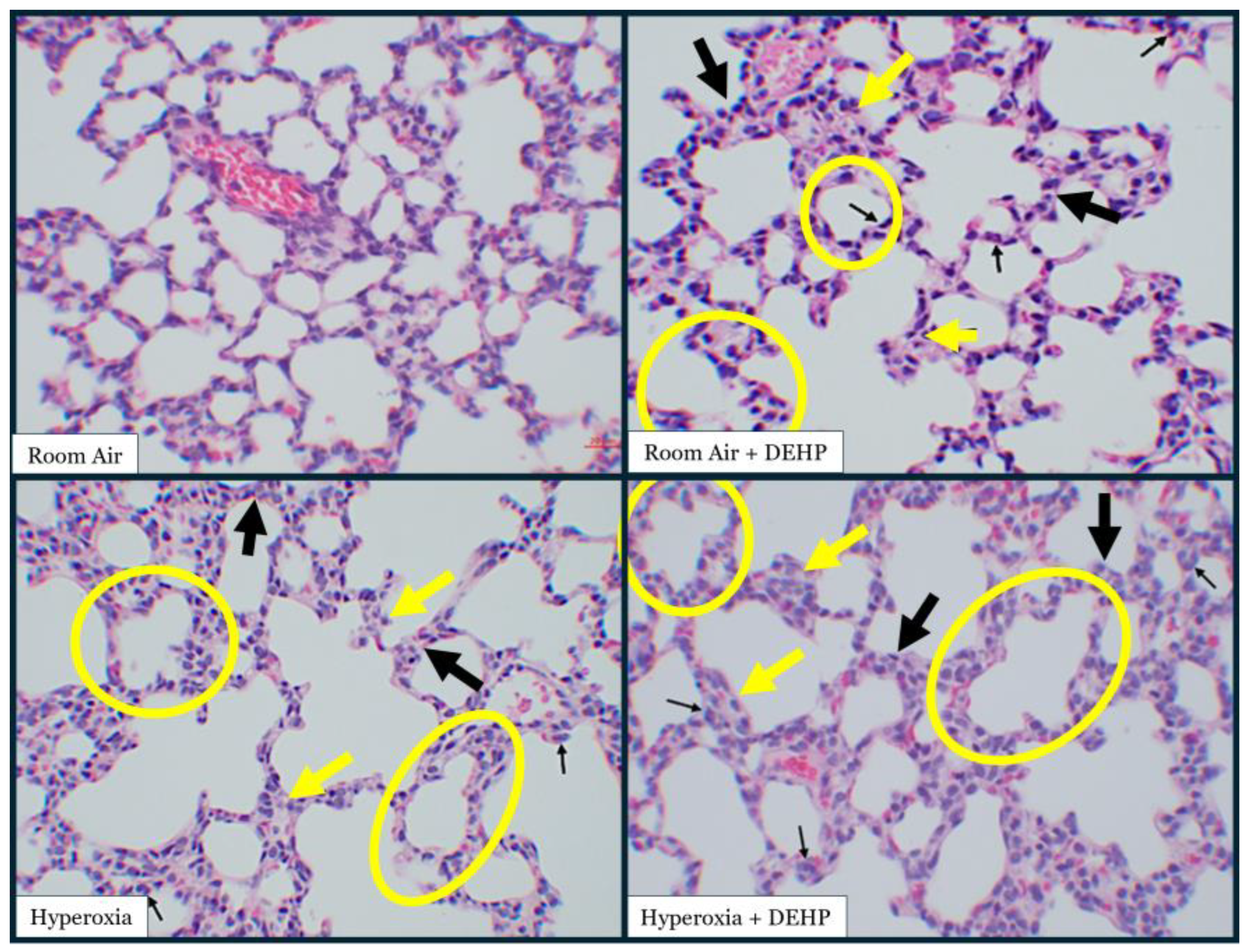
Figure 1B.
Septal Thickness in neonatal rat lungs in room air and following inhaled DEHP, hyperoxia, or hyperoxia + DEHP. Neonatal Sprague Dawley Rats breathing the mixture noted for 14 days. Y-axis—mean thickness in µm. Measurements averaged from counts on 10 non-contiguous 40X magnification fields/animal, n = 5-7 animals/intervention. Data is shown as Mean ± SD. Significance determined using one-way ANOVA with Tukey’s correction. ** = P ≤ 0.01, *** = P ≤ 0.001.
Figure 1B.
Septal Thickness in neonatal rat lungs in room air and following inhaled DEHP, hyperoxia, or hyperoxia + DEHP. Neonatal Sprague Dawley Rats breathing the mixture noted for 14 days. Y-axis—mean thickness in µm. Measurements averaged from counts on 10 non-contiguous 40X magnification fields/animal, n = 5-7 animals/intervention. Data is shown as Mean ± SD. Significance determined using one-way ANOVA with Tukey’s correction. ** = P ≤ 0.01, *** = P ≤ 0.001.
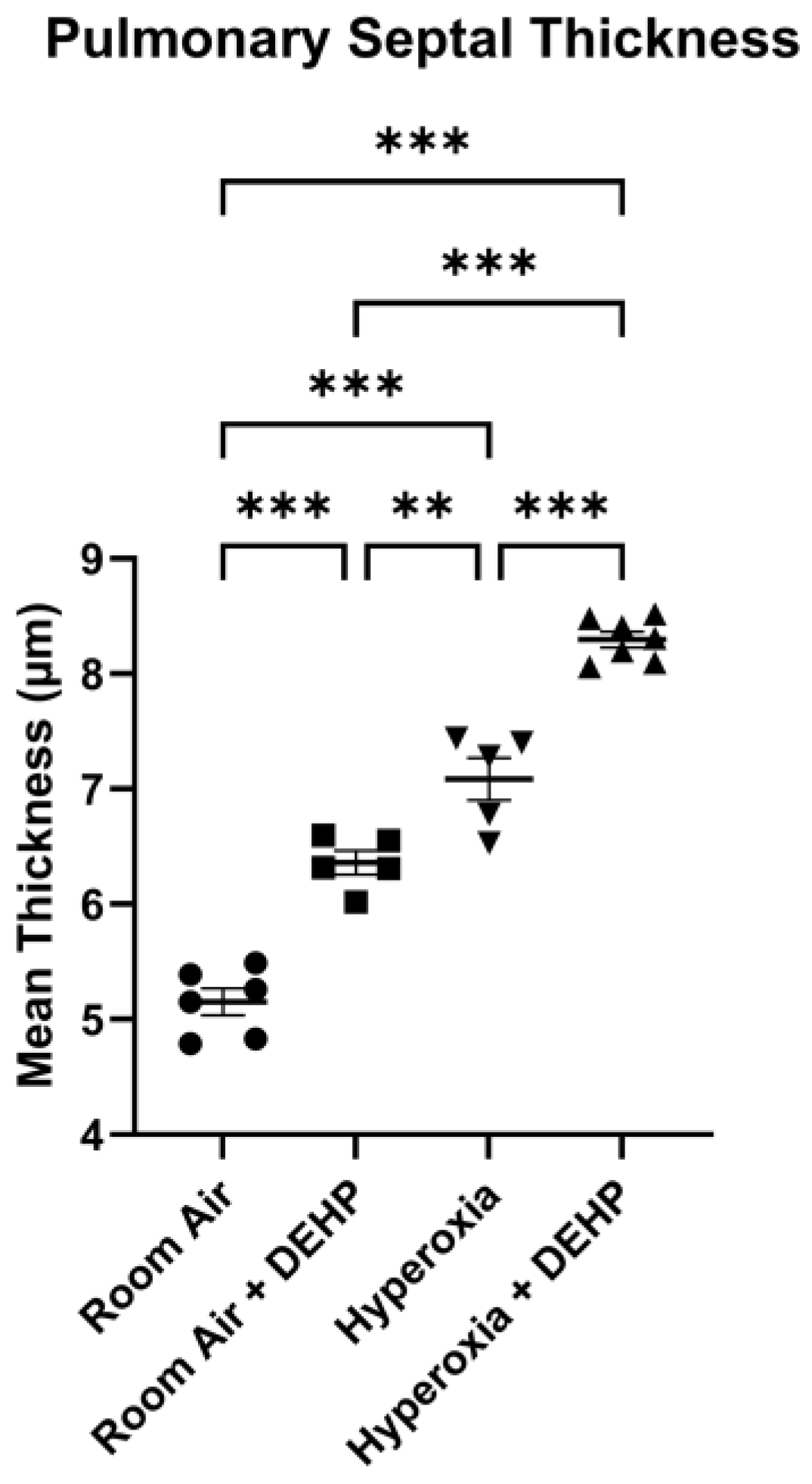
Figure 1C.
Radial Alveolar Count (RAC) in neonatal rat lungs in room air and following inhaled DEHP, hyperoxia, or hyperoxia + DEHP. Neonatal Sprague Dawley Rats breathing the mixture noted for 14 days. Y-axis—number of alveoli crossed by a line connecting a terminal bronchiole to the nearest acinar edge. Measurements averaged from counts on 10 non-contiguous 10X magnification fields/animal, n = 5-6 animals/intervention. Data is shown as Mean ± SD. Significance determined using one-way ANOVA with Tukey’s correction. * = P ≤ 0.05, *** = P ≤ 0.001.
Figure 1C.
Radial Alveolar Count (RAC) in neonatal rat lungs in room air and following inhaled DEHP, hyperoxia, or hyperoxia + DEHP. Neonatal Sprague Dawley Rats breathing the mixture noted for 14 days. Y-axis—number of alveoli crossed by a line connecting a terminal bronchiole to the nearest acinar edge. Measurements averaged from counts on 10 non-contiguous 10X magnification fields/animal, n = 5-6 animals/intervention. Data is shown as Mean ± SD. Significance determined using one-way ANOVA with Tukey’s correction. * = P ≤ 0.05, *** = P ≤ 0.001.
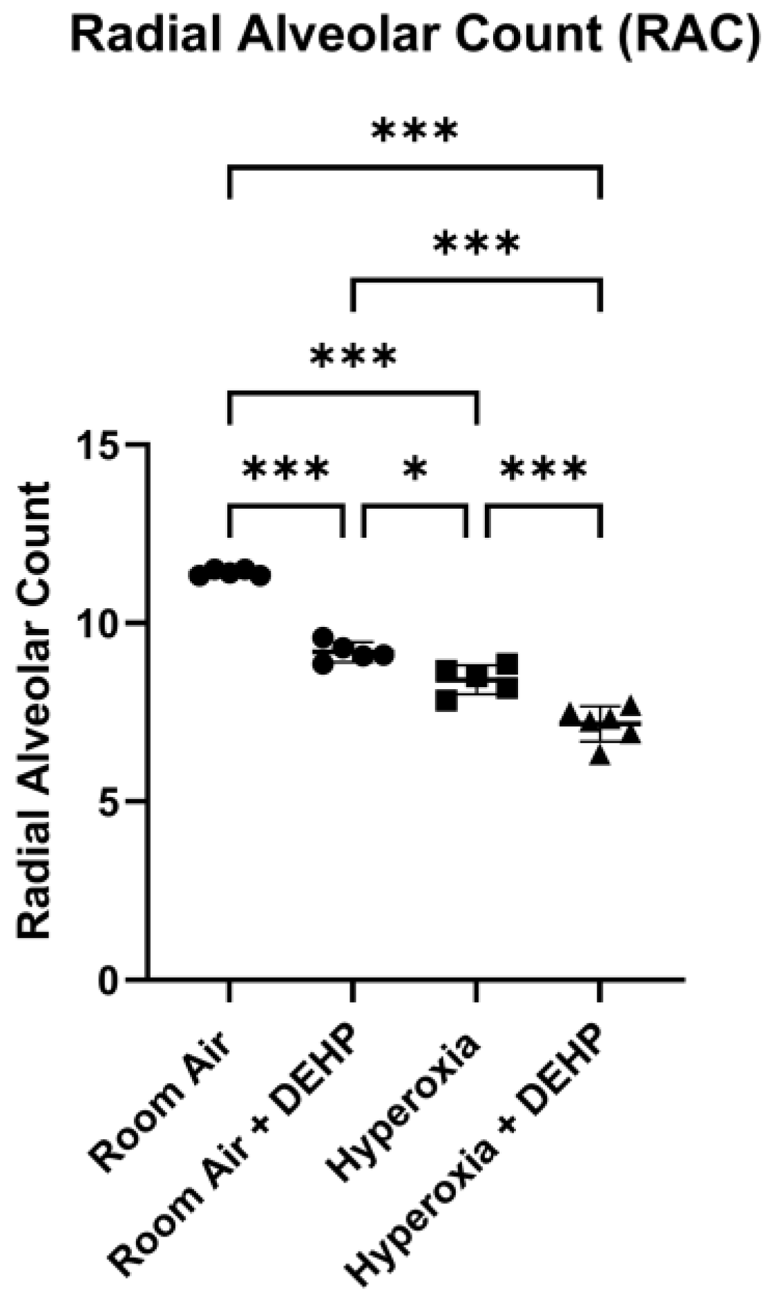
Figure 2A.
Mean number of PMNs/40X field in neonatal rat lungs in room air and following inhaled DEHP, hyperoxia, or hyperoxia + DEHP. Neonatal Sprague Dawley Rats breathing the mixture noted for 14 days. Measurements obtained as described in Methods. n = 5-7 animals/intervention. Data is shown as Mean ± SD. Significance determined using one-way ANOVA with Tukey’s correction. **** = P ≤ 0.0001.
Figure 2A.
Mean number of PMNs/40X field in neonatal rat lungs in room air and following inhaled DEHP, hyperoxia, or hyperoxia + DEHP. Neonatal Sprague Dawley Rats breathing the mixture noted for 14 days. Measurements obtained as described in Methods. n = 5-7 animals/intervention. Data is shown as Mean ± SD. Significance determined using one-way ANOVA with Tukey’s correction. **** = P ≤ 0.0001.
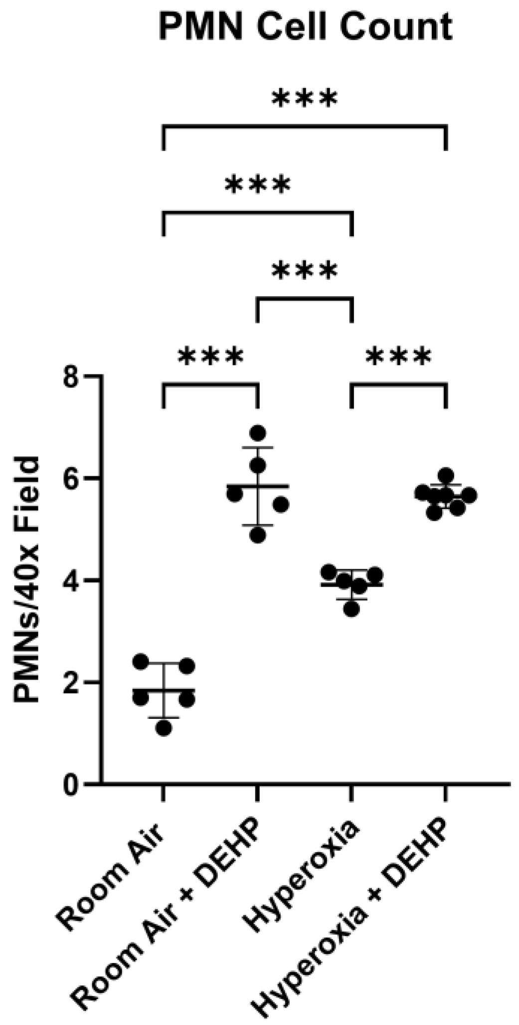
Figure 2B.
Mean MPO+ Area/MPO+ Cell in neonatal rat lungs in room air and following inhaled DEHP, hyperoxia, or hyperoxia + DEHP. Neonatal Sprague Dawley Rats breathing the mixture noted for 14 days. Y-axis—mean area of MPO+ immunofluorescence staining per MPO+ cell (myeloperoxidase-positive neutrophils). Measurements obtained as described in Methods, n = 4-5 animals/intervention. Data is shown as Mean ± SD. Significance determined using one-way ANOVA with Tukey’s correction. ** = P ≤ 0.01, *** = P ≤ 0.001.
Figure 2B.
Mean MPO+ Area/MPO+ Cell in neonatal rat lungs in room air and following inhaled DEHP, hyperoxia, or hyperoxia + DEHP. Neonatal Sprague Dawley Rats breathing the mixture noted for 14 days. Y-axis—mean area of MPO+ immunofluorescence staining per MPO+ cell (myeloperoxidase-positive neutrophils). Measurements obtained as described in Methods, n = 4-5 animals/intervention. Data is shown as Mean ± SD. Significance determined using one-way ANOVA with Tukey’s correction. ** = P ≤ 0.01, *** = P ≤ 0.001.
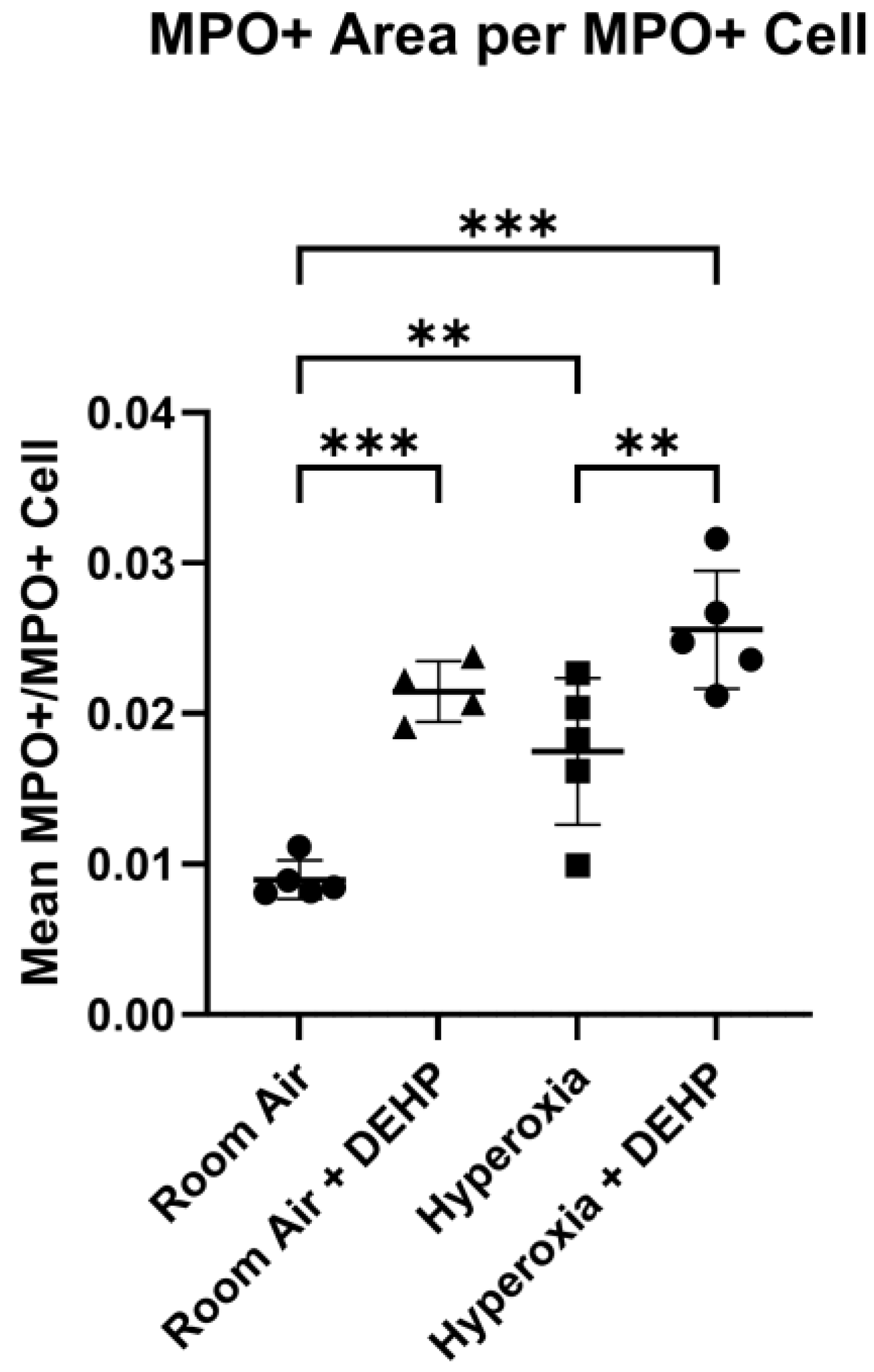
Figure 2C.
Representative 20X fields of MPO and DAPI staining in neonatal rat lungs in room air and following inhaled DEHP, hyperoxia, or hyperoxia + DEHP.
Figure 2C.
Representative 20X fields of MPO and DAPI staining in neonatal rat lungs in room air and following inhaled DEHP, hyperoxia, or hyperoxia + DEHP.
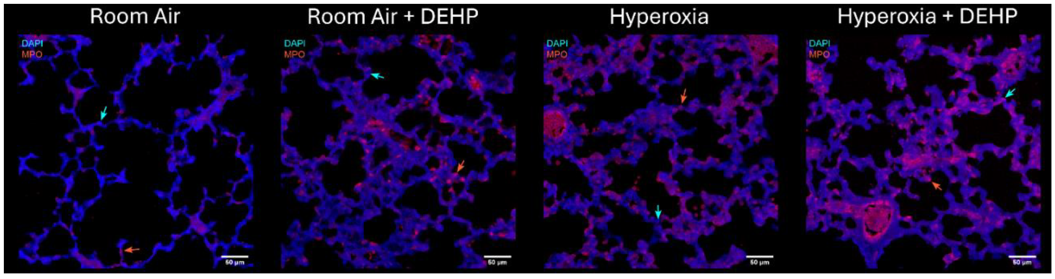
Figure 3.
Malondialdehyde (MDA) concentration in serum. Neonatal Sprague Dawley Rats breathing the mixture noted for 14 days. Measurements obtained as described in Methods. n = 5-12 animals/intervention. Data is shown as Mean ± SD. Significance determined using one-way ANOVA with Tukey’s correction. * = P ≤ 0.05, ** = P ≤ 0.01, *** = P ≤ 0.001.
Figure 3.
Malondialdehyde (MDA) concentration in serum. Neonatal Sprague Dawley Rats breathing the mixture noted for 14 days. Measurements obtained as described in Methods. n = 5-12 animals/intervention. Data is shown as Mean ± SD. Significance determined using one-way ANOVA with Tukey’s correction. * = P ≤ 0.05, ** = P ≤ 0.01, *** = P ≤ 0.001.
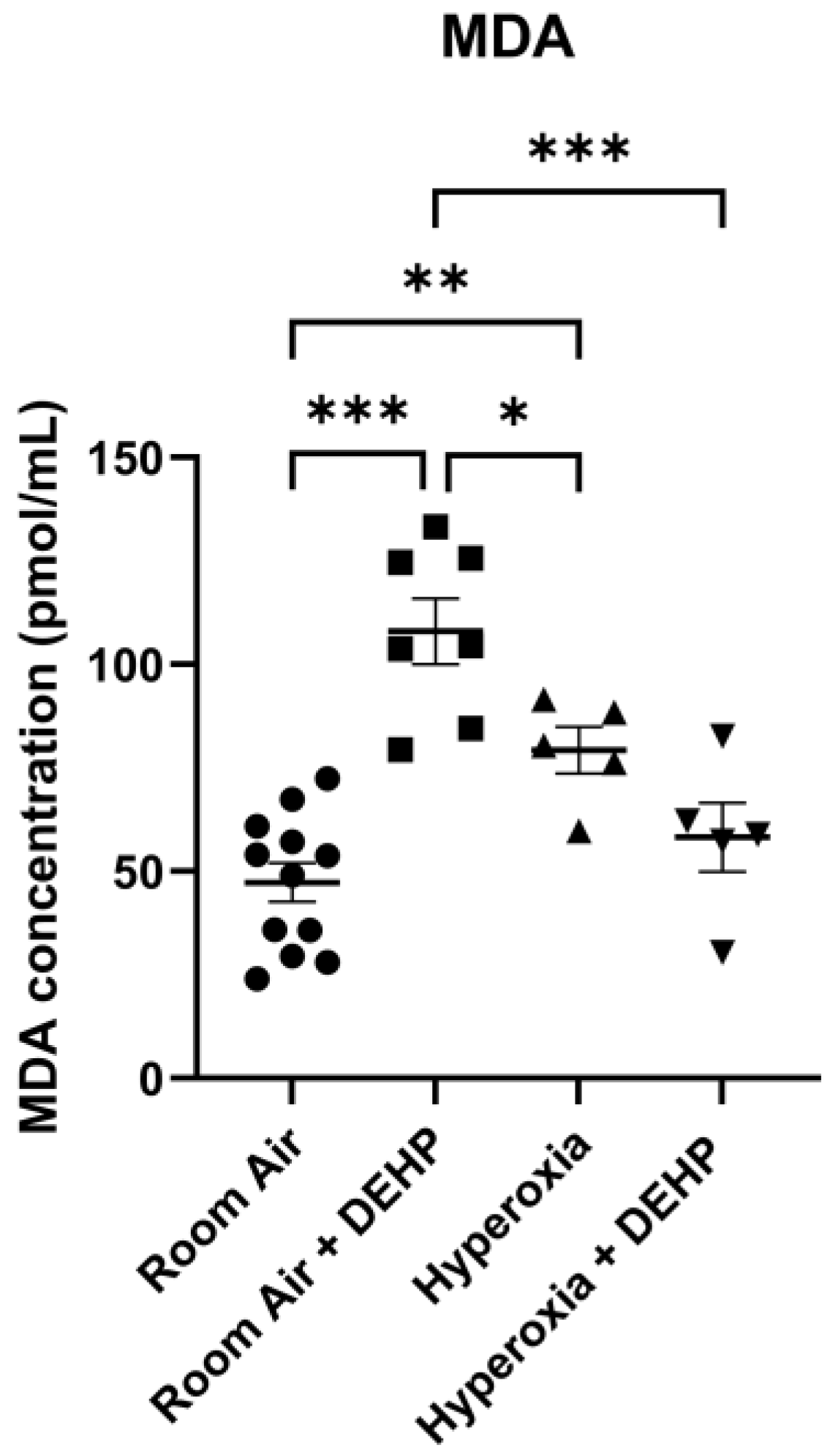
Figure 4A.
Mean CD31+ Area/CD31+ Cell in neonatal rat lungs at baseline and following inhaled DEHP, hyperoxia, or hyperoxia + DEHP. Neonatal Sprague Dawley Rats breathing the mixture noted for 14 days. Y-axis—mean area of CD31+ immunofluorescence staining per CD31+ cell. Measurements obtained as described in Methods, n = 4-5 animals/intervention. Data is shown as Mean ± SD. Significance determined using one-way ANOVA with Tukey’s correction. ** = P ≤ 0.01, *** = P ≤ 0.001.
Figure 4A.
Mean CD31+ Area/CD31+ Cell in neonatal rat lungs at baseline and following inhaled DEHP, hyperoxia, or hyperoxia + DEHP. Neonatal Sprague Dawley Rats breathing the mixture noted for 14 days. Y-axis—mean area of CD31+ immunofluorescence staining per CD31+ cell. Measurements obtained as described in Methods, n = 4-5 animals/intervention. Data is shown as Mean ± SD. Significance determined using one-way ANOVA with Tukey’s correction. ** = P ≤ 0.01, *** = P ≤ 0.001.
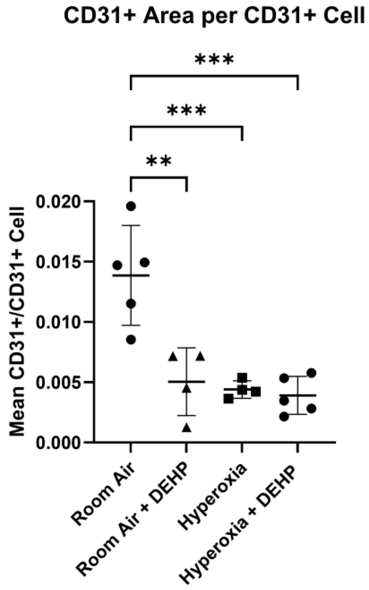
Figure 4B.
Representative 40X mean intensity projections of CD31 and DAPI staining in neonatal rat lungs in room air and following inhaled DEHP, hyperoxia, or hyperoxia + DEHP.
Figure 4B.
Representative 40X mean intensity projections of CD31 and DAPI staining in neonatal rat lungs in room air and following inhaled DEHP, hyperoxia, or hyperoxia + DEHP.
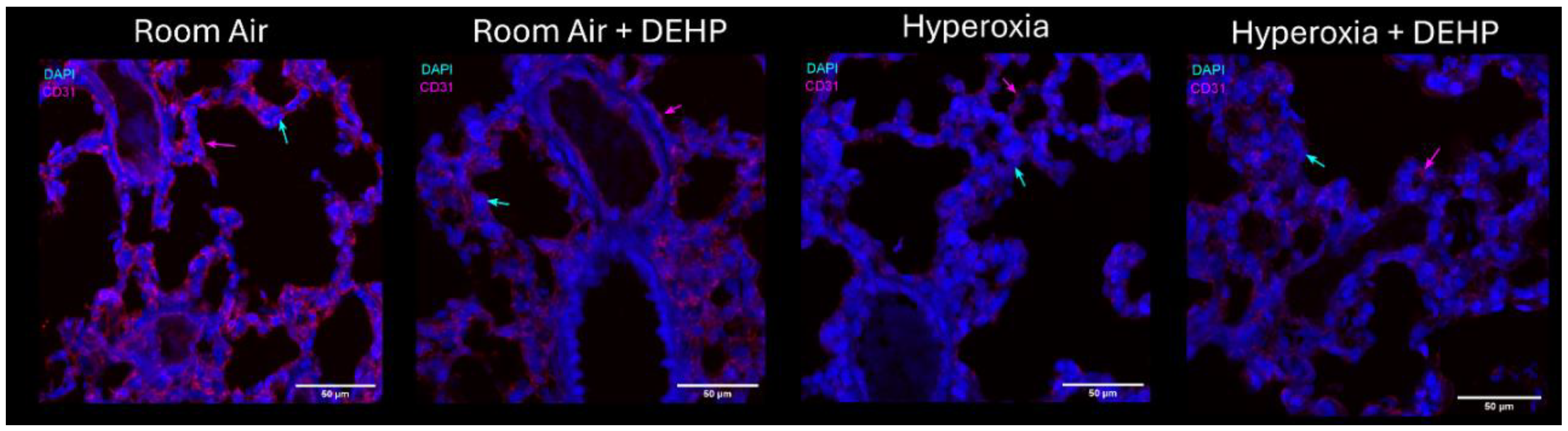
Figure 5.
Relative Abundance of VEGF in Lung Homogenate. Neonatal Sprague Dawley Rats breathing the mixture noted for 14 days. Data is shown as Mean ± SD. Significance determined using one-way ANOVA with Tukey’s correction. *** = P ≤ 0.001. (a) Representative immunoblot. Signal generated by VEGF normalized to signal generated by β-actin in the same sample. Intensity from each animal assessed in triplicate; mean of these was used for quantification. (b) Quantification of fluorescence intensity on immunoblots. Mean signals from each animal used to generate Mean ± SD for each group. n = 6-8 animals/intervention. (c) Data generated using RT-PCR as described in Methods. Intensity determined using ΔΔCt method normalized to signal from mRNA encoding β-Actin in same sample. n = 6-8 animals/intervention.
Figure 5.
Relative Abundance of VEGF in Lung Homogenate. Neonatal Sprague Dawley Rats breathing the mixture noted for 14 days. Data is shown as Mean ± SD. Significance determined using one-way ANOVA with Tukey’s correction. *** = P ≤ 0.001. (a) Representative immunoblot. Signal generated by VEGF normalized to signal generated by β-actin in the same sample. Intensity from each animal assessed in triplicate; mean of these was used for quantification. (b) Quantification of fluorescence intensity on immunoblots. Mean signals from each animal used to generate Mean ± SD for each group. n = 6-8 animals/intervention. (c) Data generated using RT-PCR as described in Methods. Intensity determined using ΔΔCt method normalized to signal from mRNA encoding β-Actin in same sample. n = 6-8 animals/intervention.
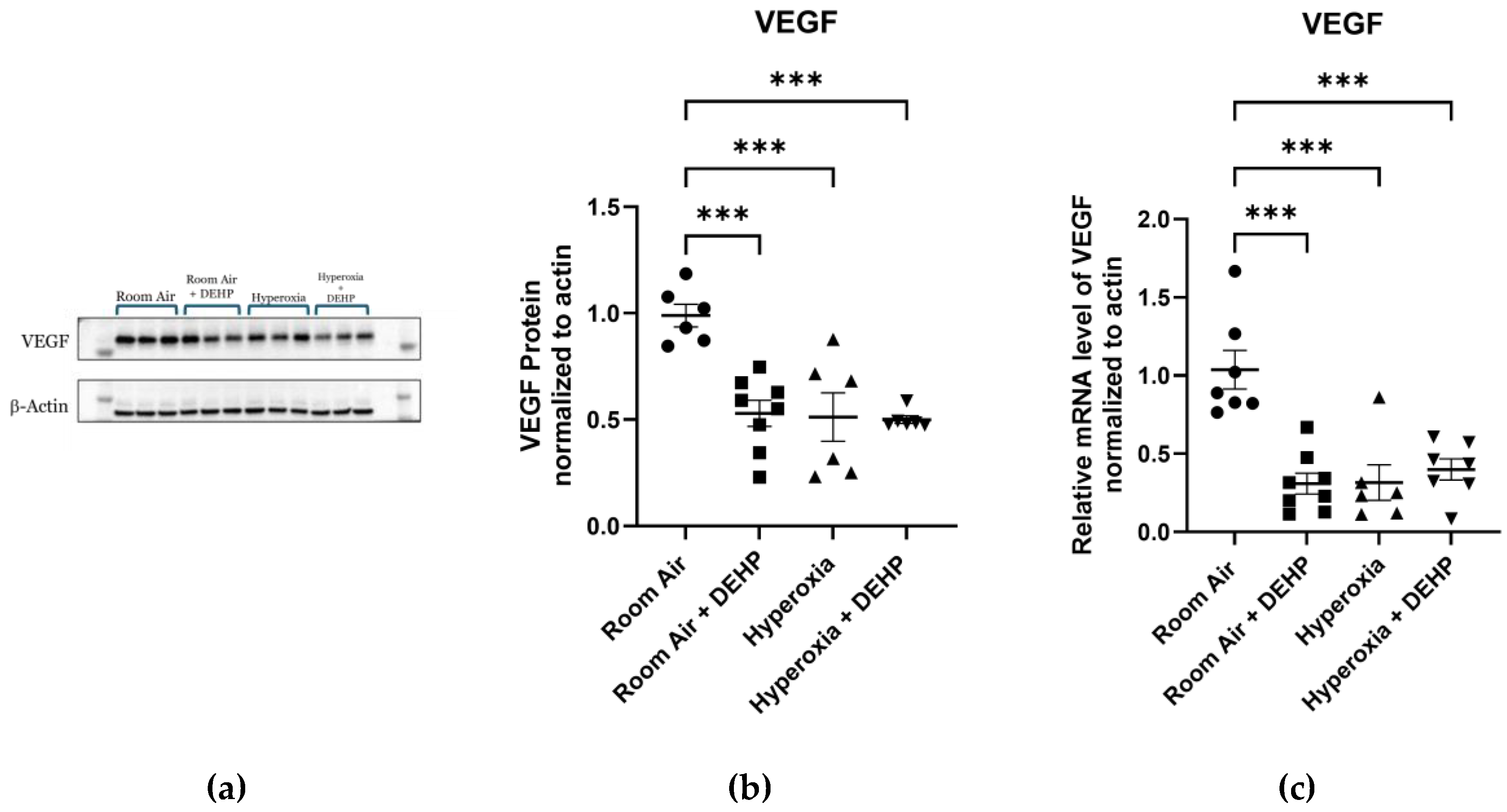
Figure 6.
Relative Abundance of VEGFR1 in Lung Homogenate. Neonatal Sprague Dawley Rats breathing the mixture noted for 14 days. Quantification of fluorescence intensity on immunoblots. Mean signals from each animal used to generate Mean ± SD for each group. Data is shown as Mean ± SD. n = 4-7 animals/intervention. Significance determined using one-way ANOVA with Tukey’s correction.
Figure 6.
Relative Abundance of VEGFR1 in Lung Homogenate. Neonatal Sprague Dawley Rats breathing the mixture noted for 14 days. Quantification of fluorescence intensity on immunoblots. Mean signals from each animal used to generate Mean ± SD for each group. Data is shown as Mean ± SD. n = 4-7 animals/intervention. Significance determined using one-way ANOVA with Tukey’s correction.
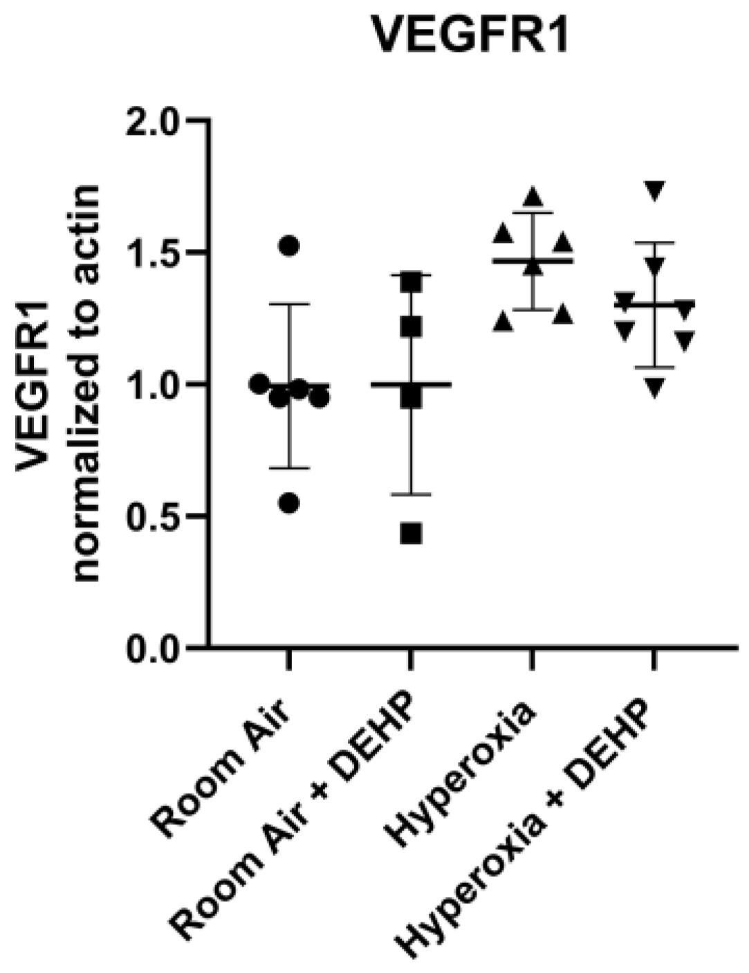
Figure 7.
Relative Abundance of VEGFR2 in Lung Homogenate. Neonatal Sprague Dawley Rats breathing the mixture noted for 6hrs/day for 14 days. Significance determined using one-way ANOVA with Tukey’s correction. * = P ≤ 0.05, ** = P ≤ 0.01. (a) Representative immunoblot. Signal generated by VEGFR2 normalized to signal generated by β-actin in the same sample. Intensity from each animal assessed in triplicate; mean of these was used for quantification. (b) Quantification of fluorescence intensity on immunoblots. Mean signals from each animal used to generate Mean ± SD for each group. n = 4-7 animals/intervention.
Figure 7.
Relative Abundance of VEGFR2 in Lung Homogenate. Neonatal Sprague Dawley Rats breathing the mixture noted for 6hrs/day for 14 days. Significance determined using one-way ANOVA with Tukey’s correction. * = P ≤ 0.05, ** = P ≤ 0.01. (a) Representative immunoblot. Signal generated by VEGFR2 normalized to signal generated by β-actin in the same sample. Intensity from each animal assessed in triplicate; mean of these was used for quantification. (b) Quantification of fluorescence intensity on immunoblots. Mean signals from each animal used to generate Mean ± SD for each group. n = 4-7 animals/intervention.
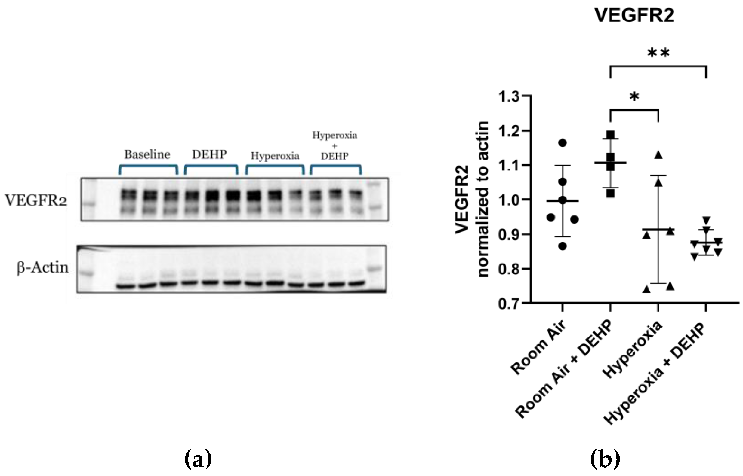
Table 1.
Environmental exposure to experimental groups. Newborn Sprague-Dawley rat pups were divided into four groups based on concentration of inhaled oxygen and/or DEHP.
Table 1.
Environmental exposure to experimental groups. Newborn Sprague-Dawley rat pups were divided into four groups based on concentration of inhaled oxygen and/or DEHP.
| Group | Number of Pups | O2 Concentration (%) | DEHP Concentration (mg/m3) |
|---|---|---|---|
| Room Air | 13 | 21 | 0 |
| Room Air + DEHP | 8 | 21 | 25 |
| Hyperoxia | 14 | 60 | 0 |
| Hyperoxia + DEHP | 14 | 60 | 25 |
Disclaimer/Publisher’s Note: The statements, opinions and data contained in all publications are solely those of the individual author(s) and contributor(s) and not of MDPI and/or the editor(s). MDPI and/or the editor(s) disclaim responsibility for any injury to people or property resulting from any ideas, methods, instructions or products referred to in the content. |
© 2026 by the authors. Licensee MDPI, Basel, Switzerland. This article is an open access article distributed under the terms and conditions of the Creative Commons Attribution (CC BY) license (http://creativecommons.org/licenses/by/4.0/).
Copyright: This open access article is published under a Creative Commons CC BY 4.0 license, which permit the free download, distribution, and reuse, provided that the author and preprint are cited in any reuse.