
Submitted:
13 May 2026
Posted:
14 May 2026
You are already at the latest version
Abstract
Alzheimer’s disease (AD) is a multifactorial neurodegenerative disorder driven by complex interactions between protein aggregation, oxidative stress, neuroinflammation, and cellular dysfunction. Among plant-derived compounds, curcumin has emerged as one of the most extensively studied polyphenols due to its broad spectrum of biological activities. This review provides a critical synthesis of mechanistic, preclinical, and clinical evidence on curcumin in AD. Experimental studies consistently demonstrate that curcumin modulates key pathogenic processes, including neuroinflammatory signaling, oxidative stress, and amyloid-β aggregation, with more limited evidence for effects on tau pathology. While in vitro studies offer detailed mechanistic insights, in vivo models provide more integrated evidence, including improvements in cognitive performance and reductions in pathological markers. Despite this strong preclinical foundation, clinical evidence remains limited and inconsistent. Randomized controlled trials have not demonstrated clear therapeutic efficacy, with outcomes strongly influenced by formulation, bioavailability, and study design. Poor solubility, rapid metabolism, and limited brain exposure remain key translational barriers. In response, increasing attention has been directed toward formulation strategies and structurally related compounds. Emerging curcuminoids, such as bisdemethoxycurcumin (BDMC), are discussed as potential next-generation candidates. Preliminary evidence suggests that BDMC may modulate oxidative stress, autophagy, astrocyte senescence, and amyloid-related processes, although data remain largely preclinical. Overall, curcumin represents a mechanistically rich and preclinically promising multi-target compound, but with unresolved translational limitations. Future research should prioritize pharmacokinetic optimization, formulation-dependent validation, and exploration of novel curcuminoid strategies to bridge the gap between experimental findings and clinical application in AD.
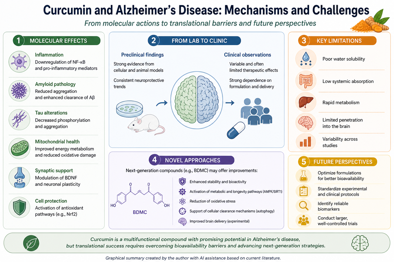
Keywords:
curcumin
; Alzheimer’s disease
; neuroinflammation
; oxidative stress
; amyloid-β
; tau pathology
; mitochondrial dysfunction
; bioavailability
; formulation strategies
; bisdemethoxycurcumin
; curcuminoids
; translational research
1. Introduction
Alzheimer's disease is the most prevalent neurodegenerative disorder with a multifactorial etiology and one of the main causes of dementia worldwide. At the neuropathological level, AD is characterized by the presence of extracellular amyloid-β (Aβ)-containing plaques and intracellular neurofibrillary tangles (NFTs) composed of abnormal hyperphosphorylated tau, but its clinical and pathological expression involves a much more complex disease process than the classical amyloid–tau framework [1,2]. More recent models propose AD as a multifactorial disorder with interacting disturbances in proteostasis, lysosomal and endosomal trafficking, synaptic homeostasis, mitochondrial function, oxidative balance, and innate immune signaling [3,4], and therapeutic efforts have increasingly shifted from single-target concepts to strategies able to modulate multiple pathogenic processes simultaneously [3,5]. Among the numerous interacting mechanisms implicated in AD, oxidative stress and neuroinflammation have emerged as central and interdependent drivers of disease progression [6,7,8]. Oxidative stress is characterized by an imbalance between reactive oxygen species (ROS) production and antioxidant defenses, resulting in lipid, protein, and nucleic acid damage, and contributing to synaptic dysfunction and neuronal loss [9,10]. Simultaneously, inflammatory responses mediated by brain-resident glial cells are now recognized as central to AD pathogenesis, rather than just secondary reactions to neuronal injury [11,12]. Importantly, oxidative stress and neuroinflammation reinforce each other, creating a self-perpetuating pathogenic loop that may accelerate neurodegeneration [13,14,15].
This conceptual shift toward a multifactorial and network-based understanding of AD is particularly relevant for the evaluation of plant polyphenols as potential neuroprotective agents. Rather than acting only as direct radical scavengers, polyphenols are widely discussed as pleiotropic modulators of interconnected cellular networks, influencing signaling pathways, neuroinflammation, mitochondrial function, and the brain–gut axis [16,17]. More recent reviews also emphasize that their relevance to brain health cannot be reduced to antioxidant activity alone and that their biological effects may involve transcriptional, metabolic, and immunomodulatory mechanisms [17,18,19]. This broader framework supports the rationale for focusing on selected polyphenols as multi-target therapeutic candidates in Alzheimer’s disease.
2. Neuroinflammation in Alzheimer’s Disease
It is now commonly acknowledged that neuroinflammation plays a key role in the pathophysiology of Alzheimer's disease, contributing to both the onset and progression of the illness. Inflammatory processes are increasingly seen as active drivers of disease dynamics, closely interacting with Aβ accumulation, tau pathology, and synaptic dysfunction, rather than as a secondary response to neuronal degeneration [7,13]. Genetic, transcriptomic, and experimental data demonstrating the crucial role of innate immune mechanisms in AD have provided compelling evidence for this paradigm shift [20,21]. As the innate immune system was shown to play a causal role in neurodegeneration, this paradigm shift sparked a surge in interest in microglia biology. Meanwhile, microglial activation, which had already been shown in the initial AD neuropathology studies, was thought to be a component of an inflammatory response that followed neuron damage [21].
Microglia, the resident immune cells of the central nervous system, play a central role in coordinating neuroinflammatory responses. Under physiological conditions, they contribute to the maintenance of brain homeostasis through continuous surveillance, phagocytosis, and the secretion of trophic factors. In the early stages of Alzheimer’s disease, microglial activation may exert protective effects by promoting amyloid-β clearance and limiting plaque formation [13,22].
However, prolonged exposure to pathological stimuli drives microglia toward dysfunctional activation states characterized by reduced phagocytic capacity and increased production of pro-inflammatory mediators, including tumor necrosis factor-α (TNF-α), interleukin-1β (IL-1β), and interleukin-6 (IL-6) [7,23]. These changes are associated with neuronal damage and synaptic dysfunction.
The traditional binary classification of microglia into M1 (pro-inflammatory) and M2 (anti-inflammatory) phenotypes has been increasingly challenged, with emerging evidence supporting a continuum of activation states [24]. In this context, the concept of disease-associated microglia (DAM) has emerged as a more accurate representation of microglial responses in neurodegeneration. Single-cell transcriptomic studies have demonstrated that DAM exhibit distinct transcriptional profiles and are closely associated with amyloid plaques. Importantly, both protective and detrimental roles have been described depending on disease stage and microenvironmental context, highlighting the dynamic and context-dependent nature of microglial function [25].
Additional genetic evidence further supports the role of microglia in Alzheimer’s disease. Variants of the triggering receptor expressed on myeloid cells 2 (TREM2) gene have been identified as significant risk factors for late-onset AD. TREM2 plays a key role in regulating microglial metabolism, survival, and phagocytic activity, particularly in response to lipid and amyloid-related signals [26,27]. Impaired TREM2 signaling has been associated with defective plaque compaction and altered microglial responses, underscoring its importance in immune pathways linked to disease progression [28,29].
Astrocytes, together with microglia, are central components of the neuroinflammatory environment in AD. Reactive astrocytes display considerable heterogeneity and may adopt both protective and neurotoxic phenotypes. Notably, activated microglia can induce a neurotoxic astrocytic state through the release of inflammatory mediators, leading to neuronal dysfunction and loss of synaptic support [30,31,32]. Bidirectional communication between microglia and astrocytes represents a key mechanism that amplifies inflammatory signaling and sustains chronic neuroinflammation [13,30,33].
At the molecular level, neuroinflammatory responses in AD are regulated by multiple signaling pathways. Among these, the nuclear factor kappa B (NF-κB) pathway plays a central role in promoting pro-inflammatory gene expression [7]. In parallel, amyloid-β accumulation has been linked to activation of the NLRP3 inflammasome, resulting in the maturation of IL-1β and IL-18. Experimental studies indicate that NLRP3 activation contributes to AD pathology, whereas its inhibition may attenuate neuroinflammation and improve disease outcomes in animal models [34,35,36,37].
Importantly, neuroinflammation is closely interconnected with other pathogenic mechanisms, particularly oxidative stress. Inflammatory mediators exacerbate oxidative damage and mitochondrial dysfunction, while reactive oxygen species can further activate inflammatory pathways such as NF-κB and inflammasome signaling. This mutually reinforcing cycle accelerates neuronal injury and disease progression [6,7,20].
Neuroinflammation also contributes to the development of core Alzheimer’s disease pathologies. Inflammatory mediators have been shown to influence the processing of amyloid precursor protein, promote amyloid-β aggregation, and facilitate tau hyperphosphorylation and propagation [7,13]. Accordingly, immune dysregulation not only represents a response to ongoing pathology but also actively contributes to its progression [38]
Neuroinflammation can therefore be viewed as a multifactorial process involving coordinated interactions between intracellular signaling pathways, glial cells, and genetic risk factors. This complexity supports the rationale for therapeutic strategies targeting multiple components of the inflammatory cascade [39].
3. Oxidative Stress in Alzheimer’s Disease
Oxidative stress is widely recognized as a major contributor to the pathogenesis of Alzheimer’s disease and plays a significant role in neuronal dysfunction and degeneration [9,10]. It arises from an imbalance between the production of reactive oxygen species and the capacity of cellular antioxidant defense systems, leading to damage to lipids, proteins, and nucleic acids. The brain is particularly vulnerable to oxidative stress due to its high oxygen consumption and lipid-rich composition, making redox imbalance a critical factor in the disruption of neuronal homeostasis and synaptic function [6].
Multiple sources contribute to ROS overproduction in AD, among which mitochondrial dysfunction is one of the most prominent. Impairments in mitochondrial respiratory chain activity compromise neuronal energy metabolism, resulting in increased electron leakage, enhanced ROS generation, and reduced ATP production [44,45]. Notably, mitochondrial abnormalities have been observed early in the course of AD, suggesting that oxidative stress may actively contribute to disease onset rather than merely representing a consequence of neurodegeneration [46,47].
In addition to mitochondrial sources, amyloid-β can directly induce oxidative stress. Aβ aggregation has been shown to enhance oxidative damage through free radical generation and interactions with redox-active metal ions such as copper and iron [48]. This oxidative environment promotes lipid peroxidation and protein oxidation, ultimately contributing to neuronal loss and synaptic dysfunction. Moreover, oxidative stress can further enhance Aβ production and aggregation, creating a self-amplifying pathogenic loop [6,48].
Oxidative damage is also closely associated with tau pathology. Elevated ROS levels can promote tau hyperphosphorylation through the activation of stress-related kinases, leading to microtubule destabilization and neurofibrillary tangle formation [49,50]. These findings indicate that oxidative stress not only contributes to neuronal injury but also actively modulates core pathological features of AD [51,52].
At the cellular level, antioxidant defense systems play a critical role in maintaining redox homeostasis. Among these, the nuclear factor erythroid 2-related factor 2 (Nrf2) pathway is a key regulator of antioxidant gene expression. Under physiological conditions, Nrf2 activation promotes the transcription of genes involved in redox balance and detoxification. However, accumulating evidence suggests that Nrf2 signaling is impaired in AD, thereby increasing susceptibility to oxidative damage. Consequently, restoration of Nrf2 activity has been proposed as a potential therapeutic strategy [53,54].
Oxidative stress is closely interconnected with neuroinflammation. Reactive oxygen species can activate inflammatory signaling pathways, including NF-κB, while inflammatory mediators further enhance ROS production, forming a self-perpetuating cycle of damage. This interaction highlights the need to consider oxidative stress and neuroinflammation as integrated components of AD pathophysiology rather than independent processes [6,13].
Overall, oxidative stress represents a complex and dynamic contributor to AD, affecting cellular signaling pathways, protein aggregation, and mitochondrial function. This complexity underscores the need for therapeutic approaches capable of targeting multiple aspects of redox imbalance. In this context, plant-derived polyphenols are of particular interest due to their ability to modulate redox-sensitive signaling pathways, support mitochondrial function, and act as antioxidants [16,19,40,55].
4. Curcumin as a Modulator of Neuroinflammation and Oxidative Stress in Alzheimer’s Disease
4.1. General Rationale for Curcumin in AD
Curcumin, the principal polyphenolic component of Curcuma longa, remains one of the most extensively studied phytochemicals in the context of Alzheimer’s disease. Its reported biological activities include modulation of neuroinflammation, oxidative stress, amyloid-β aggregation, tau-related pathology, mitochondrial dysfunction, and cell-survival signaling pathways. Recent reviews consistently characterize curcumin as a multi-target compound rather than a simple antioxidant [56,57,58,59,60,61]. The complexity and interdependence of the pathogenic processes in AD make this multi-target profile especially pertinent. Curcumin's neuroprotective potential is supported by a wealth of preclinical data, but its clinical translation is still limited. This is primarily because of its poor oral bioavailability, quick metabolism, and restricted brain exposure, all of which continue to be significant obstacles to clinical application [62,63,64,65].
4.2. Anti-Inflammatory Effects
Attenuation of inflammatory signaling is a consistently reported feature of curcumin activity. In Alzheimer’s disease, chronic activation of astrocytes and microglia is increasingly recognized as an active driver of disease progression rather than merely a response to neuronal damage. [7,13,66]
In this context, curcumin has been widely reported to modulate neuroinflammatory pathways, particularly those involving NF-κB signaling, cytokine production, oxidative stress–inflammation crosstalk, and glial activation [67,68,69]
At the molecular level, its anti-inflammatory effects are most commonly linked to suppression of NF-κB-associated signaling. NF-κB is a key transcriptional regulator of pro-inflammatory gene expression, driving the production of mediators such as tumor necrosis factor-α (TNF-α), interleukin-1β (IL-1β), interleukin-6 (IL-6), inducible nitric oxide synthase (iNOS), and cyclooxygenase-2 (COX-2). Reviews consistently report that curcumin can attenuate NF-κB-dependent inflammatory responses, although the strength of this evidence remains largely preclinical [69,70]
This mechanism is supported by direct in vitro evidence in microglia-like systems. In BV-2 mouse microglial cells stimulated with lipoteichoic acid (LTA), curcumin reduced the expression of inducible nitric acid synthase (iNOS) and cyclooxygenase-2 (COX-2) and inhibited the release of inflammatory mediators, including tumor necrosis factor-alpha (TNF-α), prostaglandin E2, and nitric oxide. These effects were associated with activation of the antioxidative HO-1/Nrf2/ARE pathway and inhibition of p38 mitogen-activated protein kinase (MAPK) signaling, suggesting coordinated regulation of inflammatory and redox-sensitive pathways rather than a single-target mechanism [69,71]
Additional in vitro studies indicate that curcumin can modulate microglial inflammatory responses through alternative signaling pathways. For example, modulation of the Janus kinase/signal transducer and activator of transcription/suppressor of cytokine signaling (JAK/STAT/SOCS) axis has been reported in BV-2 cells, further supporting a multi-target mode of action [72] However, given the variability in experimental models and conditions, these findings should be interpreted with caution.
Stronger integrative evidence comes from in vivo models. A study in an Aβ1–42-induced Alzheimer’s disease mouse model reported that curcumin improved behavioral outcomes and reduced inflammatory and oxidative stress markers, including TNF-α, IL-6, IL-1β, superoxide dismutase, and malondialdehyde, and was associated with activation of the AMPK signaling pathway [73] These findings are particularly relevant as they integrate inflammatory, oxidative, and metabolic pathways within a single experimental context.
Additional animal studies support the anti-neuroinflammatory profile of curcumin. In a p25 transgenic mouse model, curcumin attenuated glial activation, reduced the production of pro-inflammatory mediators, and improved cognitive outcomes, although such models do not fully recapitulate the complexity of human Alzheimer’s disease[74].
Taken together, these findings indicate that curcumin acts as a preclinically active modulator of neuroinflammatory signaling. Mechanistic support is strongest in in vitro systems, whereas integrated effects are more consistently demonstrated in animal models. However, direct clinical evidence for anti-neuroinflammatory efficacy in AD remains limited, and translation to human disease has yet to be established.
4.3. Impact on Redox Homeostasis and Oxidative Stress
Oxidative stress represents one of the most consistently reported mechanistic features of curcumin in dementia research. A recent systematic review focusing on preclinical models reported that curcumin treatment was consistently associated with reductions in malondialdehyde levels, alongside improvements in antioxidant defenses and cognitive outcomes [75]. Although this analysis was not restricted to Alzheimer’s disease, it provides strong support for the broader conclusion that modulation of oxidative stress is a robust and reproducible preclinical effect of curcumin.
Importantly, curcumin’s antioxidant activity is not limited to direct radical scavenging but also involves modulation of redox-sensitive signaling pathways, including Nrf2-mediated responses [76]. This broader mechanistic profile aligns with the current understanding of polyphenols as regulators of cellular signaling rather than simple chemical antioxidants.
Evidence from meta-analyses further indicates that curcumin reduces oxidative stress markers such as malondialdehyde while enhancing endogenous antioxidant defenses, including superoxide dismutase and glutathione-related systems [77].
Preclinical evidence also highlights a strong interplay between oxidative stress and inflammation in curcumin-treated models. Reductions in oxidative stress markers are frequently accompanied by decreased inflammatory activity and improved cognitive performance in rodent AD-like systems. These findings are consistent with the broader framework of AD pathophysiology, in which reactive oxygen species and neuroinflammatory processes form a self-amplifying pathogenic loop.
4.4. Effects on Amyloid-β Pathology
The anti-amyloid activity of curcumin represents one of the most extensively studied aspects of its biological profile. Experimental studies consistently demonstrate that curcumin can directly interact with amyloid-β, inhibit fibril formation, and destabilize pre-formed aggregates [78,79]. In addition to these direct interactions, curcumin has been shown to influence aggregation dynamics and reduce amyloid burden in vivo, including decreased plaque deposition in transgenic mouse models [80,81]
However, more recent literature adopts a more cautious perspective compared to earlier reports. Rather than describing a uniform inhibition of Aβ fibrillization, current evidence suggests that curcumin exerts context- and model-dependent effects on aggregation pathways, assembly states, and aggregate toxicity [82]. This shift reflects a broader recognition that amyloid modulation is a complex, dynamic process rather than a single-target mechanism.
Evidence from both in vitro and in vivo studies supports this interpretation. Curcumin has long been shown to bind amyloidogenic molecules[83], and ongoing research continues to explore derivatives and analogs designed to improve potency and activity at lower concentrations. In animal models, particularly those employing bioavailable formulations, curcumin has been associated with improvements in amyloid-related pathological readouts and behavioral outcomes, although these effects are often accompanied by parallel changes in inflammatory and oxidative stress markers [84,85,86].
Despite robust preclinical findings, translation to human disease remains limited. Clinical studies in patients with Alzheimer’s disease have not demonstrated clear anti-amyloid efficacy, and outcomes have been largely constrained by pharmacokinetic limitations and study design factors [87]. The strongest human imaging study reported changes in PET-derived measures related to amyloid and tau in non-demented individuals receiving a bioavailable curcumin formulation; however, these findings do not constitute evidence of disease-modifying efficacy in AD [88].
Taken together, the available evidence supports a well-established anti-amyloid profile of curcumin in experimental systems, while highlighting the absence of convincing clinical confirmation. This discrepancy underscores the importance of distinguishing between mechanistic potential and translational efficacy when evaluating curcumin as a candidate therapeutic agent in Alzheimer’s disease[87,89].
4.5. Effects on tau Pathology
Curcumin's effects on tau have drawn more attention in recent years, in part because research on Alzheimer's disease has moved beyond a framework that is solely focused on amyloid. Curcumin should be considered in relation to tau hyperphosphorylation, tau aggregation, and tau-associated neurotoxicity in addition to its effects on amyloid-β, according to a specialized review on curcumin and tau pathology [90]. This viewpoint is pertinent because tau pathology is strongly associated with neurodegeneration and cognitive decline in AD, making tau-directed mechanisms a crucial part of any discussion about multi-target therapy. In vitro aggregation studies provide the most compelling direct evidence of curcumin's tau-directed effects. It has been demonstrated that curcumin binds tau and prevents tau fibrillization and aggregation in vitro. Curcumin was found to have a direct impact on tau assembly and aggregate stability by inhibiting the formation of β-sheets, reducing the formation of tau fibrils, and disintegrating preformed tau filaments [91]. Although these results should be viewed as biochemical evidence rather than proof of therapeutic efficacy in vivo, they offer significant mechanistic support for the notion that curcumin can disrupt tau aggregation pathways. The potential of curcumin to modulate tau is further supported by more recent formulation-based and derivative studies. The anti-tau activity of curcumin or curcumin-containing systems may be enhanced by formulation strategies, as a curcumin–artemisinin co-amorphous system was found to inhibit recombinant tau aggregation in vitro and modulate tau phosphorylation-related readouts [92]. Similarly, in cellular models such as human-derived neuroblastoma (SH-SY5Y) cells and primary cortical neurons, newly synthesized curcumin derivatives have been demonstrated to modify toxic tau oligomer aggregation pathways and transform toxic tau oligomers into less toxic aggregate species [93]. These studies are especially helpful because they imply that tau modulation may entail both remodeling of toxic tau species and inhibition of aggregation.
Curcumin may also affect tau hyperphosphorylation, according to evidence from animal models, though this evidence is more sparse and inconsistent than that found in the literature on oxidative stress and neuroinflammation. Curcumin decreased tau hyperphosphorylation and downregulated glycogen synthase kinase 3 (GSK3) and cyclin dependent kinase 5 (CDK5) linked to aberrant tau phosphorylation, in a scopolamine-induced AD-like rat model [94]. Curcumin treatment decreased p25-mediated tau hyperphosphorylation in a p25 transgenic mouse model, according to a review of tau-focused curcumin studies [74]. This suggests that curcumin may indirectly influence tau pathology through kinase-related and stress-response pathways [95].
Despite these results, claims about tau should be presented more carefully than claims about the anti-inflammatory or antioxidant properties of curcumin. A large portion of the direct tau evidence is derived from cellular models, simplified aggregation assays, or purified protein systems, which are useful for identifying mechanisms but do not accurately replicate the intricacy of tau pathology in the human brain. Furthermore, it is challenging to identify tau-specific mechanisms because in vivo studies frequently evaluate tau-related endpoints alongside Aβ, oxidative stress, inflammation, or behavioral outcomes. When considered collectively, the evidence suggests that curcumin has the ability to modulate tau in preclinical systems, especially by inhibiting tau aggregation, remodeling toxic tau assemblies, and potentially controlling tau phosphorylation pathways [91]. Nevertheless, the majority of this evidence is still preclinical and mechanistic. There is currently no solid clinical proof that curcumin alters tau pathology in AD patients. Therefore, rather than being a clinically proven anti-tau treatment, curcumin should be characterized as a promising tau-modulating substance in experimental models [91,95].
4.6. Mitochondrial Function, Apoptosis, and Cellular Resilience
The concept of cellular resilience, which encompasses mitochondrial function, apoptosis regulation, oxidative stress responses, and neuronal survival pathways, is increasingly being used to discuss curcumin. This viewpoint is especially pertinent to Alzheimer's disease, where the production of reactive oxygen species, decreased bioenergetics, synaptic failure, and activation of apoptotic pathways are all strongly associated with mitochondrial dysfunction. In this regard, curcumin is regarded as both an antioxidant and a modulator of downstream stress-response mechanisms that affect the survival and vulnerability of neurons [96]. Curcumin may offer protection against mitochondrial dysfunction linked to AD-like pathology, according to experimental research. For instance, attenuation of mitochondrial dysfunction and apoptosis has been associated with curcumin-mediated neuroprotection against Aβ-induced toxicity, supporting the idea that mitochondrial stabilization may contribute to its neuroprotective profile [97].
Furthermore, a more recent study found that curcumin slowed the progression of AD-like conditions by modifying mitochondrial stress responses via the axis of Jumonji domain-containing protein 3 (JMJD3)-trimethylated lysine 27 on histone H3 (H3K27me3)-brain derived neurotrophic factor (JMJD3–H3K27me3–BDNF) [98]. This suggests that curcumin's effects might go beyond direct redox activity to include regulation of epigenetic and mitochondrial stress responses. Additionally significant is the connection between apoptosis and mitochondrial defense. Mitochondrial damage can increase cytochrome c release, caspase activation, and neuronal death in AD-related models. In an Aβ1–42-induced AD mouse model, curcumin has been shown to improve cognition, decrease inflammatory cytokine levels, decrease malondialdehyde, and increase superoxide dismutase activity in addition to reducing apoptosis-associated brain tissue damage [73]. Because they link oxidative and inflammatory readouts with mitochondrial/apoptotic injury within the same experimental framework, these findings are especially pertinent.
Curcumin may play a part in controlling damage caused by endoplasmic reticulum stress and mitochondria, according to cell-based research. Curcumin has been shown to prevent thapsigargin-induced cell damage and apoptosis in SH-SY5Y cells, with mitofusin-2-related mitochondrial dysfunction involved [99]. Because endoplasmic reticulum stress, mitochondrial dysfunction, and apoptosis are interrelated processes linked to neuronal injury, this model is mechanistically relevant even though it is not specifically an AD model. Therefore, rather than providing concrete proof of disease modification in AD, such studies should be interpreted as mechanistic support. According to the available data, curcumin may promote cellular resilience by reducing apoptosis-related damage, regulating mitochondrial stress, and affecting signaling pathways linked to survival. Nevertheless, these results are frequently assessed in conjunction with alterations in oxidative stress, inflammation, Aβ burden, or behavioral performance. Therefore, rather than being separate therapeutic mechanisms, curcumin's mitochondrial and anti-apoptotic effects should be understood as a component of an interconnected neuroprotective network [89,100,101].
4.7. In Vitro Studies
In vitro studies provide the foundation for mechanistic understanding of curcumin activity; however, their translational relevance is inherently limited. The literature on curcumin in AD is mechanistically rich and has been crucial in identifying its possible molecular targets. However, the evidence base is methodologically heterogeneous. Curcumin's effects on Aβ aggregation, tau protein aggregation, oxidative stress, inflammatory mediator release, mitochondrial dysfunction, and neuronal viability have been studied in both cellular and cell-free systems. These investigations are important because they enable the controlled investigation of mechanistic questions, such as whether curcumin can directly interact with amyloidogenic proteins, modify redox-sensitive pathways, or affect glial inflammatory signaling. Some of the most convincing mechanistic evidence comes from in vitro research on Aβ. Curcumin has been demonstrated to directly interact with amyloidogenic species by inhibiting Aβ aggregation and destabilizing preformed fibrils in biochemical systems [81,85,102].
The entire cellular and extracellular environment of the human AD brain cannot be replicated by these assays, which usually use simplified protein systems. Redox-modulating and anti-inflammatory mechanisms are also supported by in vitro research. Curcumin decreased the release of inflammatory mediators and altered p38 MAPK signaling and HO-1/Nrf2/ARE pathways in BV-2 microglial cells [71]. The idea that curcumin can affect inflammatory signaling through a variety of pathways is further supported by additional research in BV-2 cells that revealed modulation of the Janus kinase/signal transducer and activator of transcription/suppressor of cytokine signaling (JAK/STAT/SOCS) axis [72].
Although these findings support curcumin's mechanistic plausibility as an anti-neuroinflammatory substance, they still depend on the type of cell, stimulus, concentration, and duration of exposure. Tau-directed effects have also been better understood thanks to in vitro research. Research employing cellular models or purified tau indicates that curcumin and its derivatives may disrupt tau aggregation or alter harmful tau oligomeric species [69,91,93]. These results should not be taken as proof of clinical anti-tau efficacy, but they are mechanistically instructive, particularly for comprehending how curcumin-like compounds may impact protein assembly. The fact that curcumin concentrations used in experiments frequently surpass levels attainable in human plasma or brain tissue following traditional oral administration is a major drawback of in vitro research. Given curcumin's low systemic exposure, quick metabolism, and poor aqueous solubility, this problem is particularly crucial. Because of this, in vitro results should not be viewed as independent proof of therapeutic relevance but rather as evidence of biological plausibility and mechanism discovery. As a result, validation in animal models and formulation techniques intended to enhance pharmacokinetic performance are becoming more and more important in contemporary curcumin research.
4.8. In Vivo Studies
In vivo models offer greater translational relevance than in vitro systems because they integrate pharmacokinetics, tissue exposure, behavioral outcomes, neuropathological changes, oxidative stress, inflammation, and neuronal injury within a living organism. Although preclinical and inconsistent, the in vivo research on curcumin in AD and dementia models is generally positive. The idea that curcumin has repeatable positive effects in rodent dementia models is supported by recent systematic research[74,80]. In addition to summarizing improvements in antioxidant defenses and cognitive outcomes, a systematic review of 29 rodent studies found that curcumin reduced oxidative stress markers, with all studies evaluating malondialdehyde demonstrating significant reductions [75]. This review offers compelling evidence for oxidative stress modulation as one of the most reliable preclinical effects of curcumin, even though it was not limited solely to AD. This profile is further supported by in vivo studies that are specific to AD. Curcumin enhanced cognitive and spatial memory outcomes, decreased oxidative stress markers and inflammatory cytokines, attenuated pathological damage and apoptosis in brain tissue, and was linked to AMPK pathway activation in an Aβ1–42-induced AD mouse model [34,74,80,84].
Additional research on animals emphasizes the significance of formulation. The long-standing issue of low bioavailability was specifically addressed in a study that used a bioavailable micellar curcumin formulation in an AD mouse model. Following micellar encapsulation, the formulation was given in drinking water, and the study assessed neuropathological effects in an AD model [103]. Such research is crucial because it changes the focus from whether curcumin has biological activity to whether the brain can be sufficiently exposed to make those mechanisms therapeutically meaningful.
Since they demonstrate that curcumin's effects are rarely limited to a single pathway, in vivo studies are also important. Improvements in oxidative stress, inflammatory markers, Aβ burden, tau-related endpoints, apoptosis, or mitochondrial function frequently coexist with improvements in cognition in animal models. Curcumin's multi-target interpretation is supported by this pattern, but it also poses a methodological challenge: it can be challenging to identify whether redox modulation, anti-inflammatory effects, anti-amyloid activity, mitochondrial protection, or a combination of these mechanisms are the main drivers of cognitive improvement.
Animal-related literature is still quite varied. Aβ peptide injection paradigms, transgenic mice, chemically induced dementia-like models, diet-related models, and models of aging-related cognitive impairment are among the models. Construct validity, disease stage, endpoint selection, dosage schedule, formulation, administration route, and behavioral testing techniques vary among these models. Therefore, even though there is encouraging and frequently directionally consistent animal evidence for curcumin, it is not consistent enough to support strong clinical conclusions without qualification . Overall, curcumin's status as a preclinically active multi-target compound in AD-like pathology is strongly supported by in vivo data [104]. The most compelling evidence relates to improvements in oxidative stress, inflammatory markers, and cognitive outcomes; effects on apoptosis, mitochondrial stress, and proteinopathy-related endpoints are also supported. However, these results are still preclinical, and the degree to which each model replicates human AD pathology, formulation, dose, and exposure all have a significant impact on their translational relevance. Although generally supportive, the heterogeneity of models and experimental conditions limits direct translation to human disease.
4.9. Clinical Studies and Translational Relevance
Compared to the preclinical literature, the clinical evidence for curcumin in Alzheimer's disease is still scarce and significantly weaker. A 24-week randomized, double-blind, placebo-controlled study in patients with mild-to-moderate AD, followed by an open-label extension, is the most well-known dedicated clinical trial in diagnosed AD. Although oral Curcumin C3 Complex was generally well tolerated in this study, there was no discernible clinical or biomarker efficacy during the placebo-controlled period [105]. Because it tested curcumin directly in an AD population, this trial is still significant. However, it also highlights the discrepancy between high experimental activity and low efficacy following traditional oral administration.
An 18-month randomized, double-blind, placebo-controlled trial of bioavailable Theracurmin in middle-aged and older non-demented adults provides a different kind of human evidence. This study examined brain signals related to tau and amyloid using 2-(1-{6-[(2-[F-18]fluoroethyl)(methyl)amino]-2-naphthyl}ethylidene)malononitrile positron emission tomography (FDDNP-PET) and reported improvements in memory and attention. The authors came to the conclusion that daily oral Theracurmin may enhance memory function and that PET results were consistent with decreased tau and amyloid buildup in specific brain regions [86]. However, because the participants in this study were not patients with established AD, it should be interpreted with caution when considering AD therapy. As a result, it offers a biologically intriguing translational signal but no proof that curcumin can effectively treat AD.
The possibility that bioavailable formulations could result in quantifiable cognitive benefits has been investigated in more recent clinical research. CurQfen, a curcumin–galactomannan complex, was found to improve a number of cognitive and functional outcomes when compared to placebo and unformulated standard curcumin in a randomized, double-blind, placebo-controlled study of people with moderate dementia caused by AD [106]. Because it directly addresses formulation-dependent efficacy, this study is pertinent; however, it should be carefully considered in light of the limited clinical evidence base and the requirement for independent replication. A cautious interpretation is also supported by systematic evidence. Both in vivo AD animal models and randomized controlled trials involving human subjects were included in a systematic review and meta-analysis on cognitive aging. Curcumin was found to consistently improve cognition in preclinical animal studies, but the evidence for a strong clinical benefit was not as strong in pooled human data. This supports the more general conclusion that there is a glaring disparity between promising preclinical results and less certain clinical efficacy in the curcumin field [56]. All things considered, recent research on humans indicates that curcumin is generally safe and, depending on the formulation, may result in signals related to cognition or biomarkers. Nevertheless, there is still insufficient, inconsistent, and limited clinical evidence to support curcumin as a successful treatment for AD. Inconsistent results are probably caused by variations in formulation, dose, duration, participant population, disease stage, and outcome measures. Curcumin is therefore best characterized as a formulation-sensitive, mechanistically promising candidate whose clinical relevance in AD is still being studied.
4.10. Bioavailability and Formulation Challenges
One of the main translational obstacles in curcumin research is still poor bioavailability. After oral administration, native curcumin usually results in low systemic exposure, extensive metabolism, and low aqueous solubility [107]. These restrictions are especially significant for AD, as therapeutic relevance would necessitate adequate exposure to biologically active curcumin species in the central nervous system in addition to gastrointestinal absorption and systemic circulation. The distinction between total curcuminoid exposure, conjugated metabolites, and pharmacologically significant unconjugated curcumin is becoming more and more important in contemporary curcumin research.
According to the independent crossover pharmacokinetic study comparing a number of commercial formulations, plasma concentrations of unconjugated curcumin remained extremely low even when formulations increased uptake and produced higher levels of conjugated curcumin. The authors contended that rather than poorly membrane-permeable conjugated metabolites, bioavailability claims should concentrate on biologically significant unconjugated curcumin [108,109,110]. This result implies that greater bioavailability does not always imply therapeutically significant brain exposure and supports a more circumspect interpretation of formulation studies. However, because traditional curcumin exposure is pharmacokinetically unfavorable, formulation strategies are still very important. Numerous strategies intended to enhance absorption are described in reviews of clinical curcumin formulations, such as micelles, phospholipid complexes, lipid-based carriers, nanoparticles, amorphous dispersions, and curcumin–fiber or curcumin–polysaccharide complexes [108,110,111,112,113,114,115,116,117,118,119,120,121,122,123].
Several generations of delivery systems have been developed to improve bioavailability and clinical performance, according to a review of clinical trials using curcumin formulations. It also noted that formulation claims need to be carefully evaluated rather than assuming that all enhanced formulations are equivalent [64]. Because the blood–brain barrier (BBB) separates the central nervous system from systemic circulation, formulation science is particularly crucial for AD. Improved formulation may make it more feasible to target AD-relevant pathology, according to preclinical research employing delivery-enhanced curcumin preparations. For instance, a 2024 study evaluated neuropathological outcomes and directly addressed the bioavailability issue using a bioavailable micellar curcumin formulation in an AD mouse model [124]. Similar to this, research employing curcumin-loaded nanoparticles in AD models has shown better pathological and cognitive results when compared to free curcumin, indicating the potential benefit of delivery-based strategies while staying well within the preclinical evidence base [125]. The interpretation of mechanistic studies is also influenced by this formulation-centered viewpoint.
Due to curcumin's low bioavailability and quick metabolism, many in vitro studies employ concentrations that might not be possible in human plasma or brain tissue following traditional oral administration. Therefore, unless pharmacokinetic data shows achievable exposure in relevant tissues, biological effects seen in cell culture should be regarded as mechanistic evidence. The same caution applies to animal studies: when formulation, dose, route of administration, and tissue exposure are clearly reported, positive results are more translationally informative [64]. Therefore, current formulation research is crucial to figuring out whether curcumin's multi-target mechanisms can become clinically meaningful, not just a technical supplement to curcumin biology. Future research should evaluate whether a formulation improves exposure to pharmacologically relevant curcumin species in the brain and results in repeatable biomarker changes in addition to whether it raises circulating curcuminoid levels [126]. Curcumin should be viewed as a promising but formulation-limited compound with significant preclinical support and unresolved clinical translational potential until such evidence becomes available.
A representative overview of experimental and clinical studies evaluating curcumin in Alzheimer’s disease and related dementia models is presented in Table 1.
4.11. Overall Assessment
When considered collectively, the literature indicates that curcumin is one of the plant-derived polyphenols that has been studied the most in relation to Alzheimer's disease. Its biological activity encompasses a number of interrelated domains, such as mitochondrial function, cellular stress-response pathways, neuroinflammation modulation, oxidative stress regulation, interference with amyloid-β aggregation, and more speculative effects on tau pathology [83,91,92,95].
Preclinical and mechanistic research provide the best evidence base. Curcumin's interactions with inflammatory mediators, redox-sensitive signaling pathways, and amyloidogenic proteins are thoroughly explained by in vitro research. In vivo studies, which more reliably show integrated biological effects, such as improvements in cognitive function along with decreases in oxidative stress and neuroinflammatory markers across various experimental models, support these findings [69,73,75]. Heterogeneity in model systems, experimental setup, and outcome measures, however, continues to be a significant constraint. Clinical evidence, on the other hand, is still sparse, inconsistent, and heavily influenced by formulation. Despite acceptable safety profiles, early randomized controlled trials in AD patients did not show clear therapeutic efficacy [105].
Curcumin may have quantifiable biological or cognitive effects in humans, according to more recent research employing bioavailability-enhanced formulations, but these results are still conflicting and insufficient to prove clinical efficacy. This disparity is indicative of a larger translational gap between strong preclinical activity and weak or equivocal results in human research. Crucially, pharmacokinetic limitations continue to be a major problem. The interpretation of both mechanistic and clinical findings is complicated by poor solubility, rapid metabolism, and limited systemic exposure. These factors emphasize the significance of formulation in determining biological relevance [139,140].
Overall, curcumin can be considered a well-characterized multi-target compound with promising preclinical evidence and strong mechanistic support; however, there is insufficient and formulation-dependent clinical data to support firm conclusions about its therapeutic efficacy in AD. Therefore, curcumin is best positioned as a promising lead compound whose relevance lies in its ability to modulate multiple interconnected pathogenic pathways rather than as a validated anti-AD therapy. Its main unresolved challenge is the translation of preclinical findings into robust and repeatable clinical benefit.
5. Bisdemethoxycurcumin as an Emerging Curcuminoid Candidate
Curcuma longa contains a naturally occurring curcuminoid called bisdemethoxycurcumin (also called BDC in some formulation studies), which shares structural similarities with curcumin. BDMC is still far less studied than curcumin, which predominates in the literature on Alzheimer's disease. This makes it especially intriguing in the context of this review: BDMC can be positioned as an emerging curcuminoid that might help advance the field beyond traditional curcumin-centered approaches, rather than as a curcumin substitute.
5.1. Rationale for Considering BDMC in AD
There are two primary justifications for taking BDMC into account in AD. First, BDMC and curcumin have a number of biologically significant characteristics in common, such as documented neuroprotective, anti-inflammatory, and antioxidant effects [141,142]. Second, the research on AD that is currently available indicates that BDMC may activate processes that are particularly pertinent to the biology of AD today, such as oxidative stress regulation, SIRT1/AMPK signaling, autophagy, astrocyte senescence, amyloid-β clearance, and formulation-dependent brain delivery [143,144]. This is significant because the field of curcumin is moving away from straightforward antioxidant narratives and toward a more complex translational framework that includes glial biology, cellular resilience, proteostasis, and drug delivery. Because recent research has looked at BDMC in connection to autophagy-mediated reduction of senescence and amyloid pathology rather than just as a general antioxidant, it fits nicely into this more recent framework.
5.2. Mechanistic Pathways, Cellular Targets, and Translational Considerations of BDMC
One important mechanistic study used human neuroblastoma cells exposed to Aβ1–42 to study BDMC in an in vitro AD-related model [141]. In this model, BDMC enhanced antioxidant capacity, including glutathione-related activity and superoxide dismutase, and increased cell survival. Additionally, the study found that silent information regulator 1 (SIRT1) expression and AMP-activated protein kinase (AMPK) phosphorylation were elevated, and that BDMC's protective effects were diminished when AMPK or SIRT1 were pharmacologically inhibited. These findings lend credence to the hypothesis that BDMC protects against Aβ-associated cellular stress through AMPK/SIRT1-related mechanisms rather than through direct antioxidant activity alone.
Additionally, BDMC has been tested in vivo in the double-transgenic mouse model for Alzheimer’s disease research (APP/PS) [143]. In the study, BDMC was given intracerebroventricularly to the mice, and then it was looked into whether SIRT1 signaling was involved in the effects. The study found that while treatment with the SIRT1 inhibitor (EX527) attenuated these effects, BDMC reduced oxidative stress-related pathology and improved AD-related outcomes. This suggests that SIRT1 plays a part in the neuroprotective effects of BDMC in an animal model relevant to AD. Because this paper employs the AD mouse model, it offers more robust disease-context support than cell-based research. However, intracerebroventricular administration circumvents the typical pharmacokinetic barriers that restrict oral or systemic delivery, which is a significant translational limitation.
The relationship between autophagy, astrocyte senescence, and Aβ clearance is a particularly intriguing new avenue. A recent study examined whether BDMC could pharmacologically induce autophagy and decrease senescence-associated pathology [144]. The study found that AD pathology is linked to increased senescence and decreased autophagy in the brain. The authors discovered that BDMC promoted autophagy in primary astrocytes and made it easier for externally administered Aβ to be eliminated. BDMC has been shown to cross the blood–brain barrier, increase autophagy-related proteins, lower senescence-associated secretory phenotype markers, lower hippocampal Aβ load, and enhance working memory and neuromuscular coordination in 3xTg-AD transgenic mice. Because it shifts the conversation away from neurons and toward astrocytes as active participants in AD pathology, this study is particularly valuable. It also fits in nicely with the growing theory that neurodegeneration is caused by cellular senescence and impaired autophagy.
Similar to curcumin, BDMC has significant formulation and delivery issues. BDC is nearly insoluble in water, poorly absorbed, and quickly degraded, according to a study on BDMC-loaded H-ferritin nanocages [142]. In order to improve solubility, stability, and delivery, the authors created an H-ferritin nanocage formulation. Peripheral blood mononuclear cells from AD patients showed transcriptomic alterations as a result of the formulation crossing an in vitro BBB model; pathway analysis revealed effects on chemokines and macrophage activation. While stressing that in vivo confirmation is still necessary, the authors concluded that BDC-HFn enhanced drug stability and modulated inflammation-related gene expression. This study provides preliminary evidence supporting the potential importance of formulation in determining the translational relevance of curcuminoids, suggesting that delivery may play a more integral role than previously considered. In addition, it highlights a possible connection between delivery strategies, AD-related inflammatory pathways, and curcuminoid chemistry. However, these findings should be interpreted with caution, as the study relied on an in vitro BBB model and PBMC transcriptomic analysis rather than clinical outcomes, and therefore represents early translational evidence rather than confirmation of therapeutic efficacy.
5.3. Translational Positioning of BDMC in Alzheimer’s Disease
Instead of being positioned as a proven therapeutic agent for AD, BDMC should be considered an emerging curcuminoid candidate. The available literature suggests several potentially relevant mechanisms, including protection against Aβ-induced cellular stress, modulation of oxidative stress via AMPK/SIRT1-related pathways, reduction of senescence-associated astrocyte dysfunction, stimulation of autophagy, facilitation of Aβ clearance, and formulation-dependent anti-inflammatory effects.
These mechanisms align with the overarching theme of this review, particularly the concept that plant-derived compounds may act through interconnected glial, redox, neuroinflammatory, and proteostatic pathways. In addition, formulation-focused studies suggest that delivery strategies may influence the translational potential of BDMC, potentially linking curcuminoid chemistry with AD-related inflammatory pathways. However, such findings should be interpreted with caution, as they are primarily based on in vitro BBB models and transcriptomic analyses rather than clinical outcomes, and therefore represent early translational observations rather than definitive evidence of therapeutic efficacy.
Despite these promising mechanistic insights, the overall body of evidence remains limited. Compared with curcumin, BDMC has been investigated far less extensively, lacks clinical validation in AD, and much of the available data is restricted to cell-based systems, animal models, or formulation-focused studies. Its primary value in the present manuscript is therefore conceptual and strategic: BDMC illustrates how exploration of structurally related curcuminoids with distinct mechanistic profiles and potentially improved formulation characteristics may help advance the field beyond canonical curcumin.
Overall, BDMC represents a promising but still early-stage candidate in AD research. Its emerging profile—linking curcuminoid biology to autophagy, astrocyte senescence, Aβ clearance, oxidative stress regulation, and formulation-dependent brain delivery—highlights its potential relevance. Nevertheless, it should currently be regarded as a mechanistically supported preclinical compound rather than a clinically validated therapeutic option.
The multifaceted mechanisms potentially responsible for the neuroprotective activity of BDMC are summarized in Figure 1.
6. Conclusions and Future Perspectives
Alzheimer’s disease is a complex, multifactorial neurodegenerative disorder driven by interconnected processes, including neuroinflammation, oxidative stress, protein aggregation, mitochondrial dysfunction, and impaired cellular homeostasis. This complexity has shifted therapeutic strategies away from single-target approaches toward compounds capable of modulating multiple pathogenic pathways simultaneously.
In this context, curcumin remains one of the most extensively studied plant-derived polyphenols in AD research. The available evidence consistently demonstrates a broad spectrum of biological activity, including modulation of neuroinflammatory signaling, regulation of oxidative stress, interference with amyloid-β aggregation, and, to a lesser extent, effects on tau-related processes. These findings are strongly supported by mechanistic and preclinical studies, positioning curcumin as a prototypical multi-target compound within the AD research landscape.
Despite this strong experimental foundation, clinical translation remains limited. Human studies have yielded modest or inconclusive results, and therapeutic efficacy in AD has not been definitively established. A major contributor to this discrepancy is poor bioavailability, characterized by low aqueous solubility, rapid metabolism, and limited systemic and brain exposure. Consequently, curcumin exemplifies a compound with substantial mechanistic promise but unresolved translational challenges.
The increasing emphasis on formulation science has further refined the interpretation of curcumin’s therapeutic potential. Advances in delivery systems, including nanoparticles, micelles, and phospholipid complexes, have demonstrated improved pharmacokinetic profiles in preclinical and early clinical studies. However, it remains unclear whether these improvements translate into clinically meaningful outcomes. Future research should therefore focus on establishing whether enhanced formulations achieve therapeutically relevant brain concentrations and produce reproducible effects on validated disease biomarkers.
Within this evolving framework, structurally related curcuminoids, such as bisdemethoxycurcumin, have emerged as potential next-generation candidates. Preliminary evidence suggests that BDMC may modulate oxidative stress, Aβ-related toxicity, autophagy, and astrocyte senescence, while also benefiting from emerging formulation strategies. However, the current evidence base remains largely preclinical, and its clinical relevance has yet to be established.
Taken together, the available literature supports a reframing of curcumin not as a clinically validated therapeutic agent, but as a mechanistically rich benchmark compound that has contributed to defining the multi-target paradigm in AD research. Its primary value lies in illustrating how plant-derived molecules can interact with interconnected pathological networks, while its limitations highlight key barriers to translation, including pharmacokinetics, formulation, and study design.
Future research priorities should include: (i) the development of delivery systems capable of achieving therapeutically relevant brain exposure; (ii) the identification and validation of biomarkers reflecting target engagement and disease modification; (iii) the design of well-powered, rigorously controlled clinical trials using standardized formulations; and (iv) the exploration of emerging curcuminoids and related compounds with improved pharmacological profiles.
In summary, curcumin represents both the promise and the limitations of plant-derived multi-target compounds in AD. While it has significantly advanced mechanistic understanding, its clinical potential remains constrained. Bridging this gap will require a shift from descriptive mechanistic studies toward integrative, translational research. In this context, emerging curcuminoid strategies, including BDMC, represent a promising but still exploratory direction for future investigation.
Institutional Review Board Statement
Not applicable.
Informed Consent Statement
Not applicable.
Data Availability Statement
Data sharing is not applicable to this article.
Conflicts of Interest
The author declares no conflict of interest.
References
- Gouilly, D.; Rafiq, M.; Nogueira, L.; et al. Beyond the amyloid cascade: An update of Alzheimer’s disease pathophysiology. Rev. Neurol. 2023, 179(8), 812–30. [Google Scholar] [CrossRef]
- DeTure, M.A.; Dickson, D.W. The neuropathological diagnosis of Alzheimer’s disease. Mol. Neurodegener. 2019, 14(1), 32. [Google Scholar] [CrossRef] [PubMed]
- Knopman, D.S.; Amieva, H.; Petersen, R.C.; et al. Alzheimer disease. Nat. Rev. Dis. Prim. 2021, 7(1), 33. [Google Scholar] [CrossRef]
- Hein, Z.M.; Karikalan, B.; Gopalakrishna, P.K.; et al. Toward a Unified Framework in Molecular Neurobiology of Alzheimer’s Disease: Revisiting the Pathophysiological Hypotheses. Mol. Neurobiol. 2026, 63(1), 282. [Google Scholar] [CrossRef] [PubMed]
- Bajinka, O.; Jallow, L.; Ozdemir, G. A multi-target therapeutic framework for Alzheimer’s disease: an integrative mechanistic review. Neuroscience 2026, 596, 143–57. [Google Scholar] [CrossRef] [PubMed]
- Butterfield, D.A.; Halliwell, B. Oxidative stress, dysfunctional glucose metabolism and Alzheimer disease. Nat. Rev. Neurosci. 2019, 20(3), 148–60. [Google Scholar] [CrossRef]
- Heneka, M.T.; Carson, M.J.; El, Khoury J; et al. Neuroinflammation in Alzheimer’s disease. Lancet Neurol. 2015, 14(4), 388–405. [Google Scholar] [CrossRef]
- Puranik, N.; Song, M. Oxidative Stress and the Role of Immune Cells in Alzheimer’s Disease: Therapeutic Implications and Future Perspectives. CNS Neurol. Disord. Drug Targets 2025, 24(9), 685–700. [Google Scholar] [CrossRef]
- Trofin, D.M.; Sardaru, D.P.; Trofin, D.; et al. Oxidative Stress in Brain Function. Antioxidants 2025, 14(3), 297. [Google Scholar] [CrossRef]
- Li, J.; O, W.; Li, W.; et al. Oxidative Stress and Neurodegenerative Disorders. Int. J. Mol. Sci. 2013, 14(12), 24438–75. [Google Scholar] [CrossRef]
- Gleichman, A.J.; Carmichael, S.T. Glia in neurodegeneration: Drivers of disease or along for the ride? Neurobiol. Dis. 2020, 142, 104957. [Google Scholar] [CrossRef] [PubMed]
- Bogus, K.; Marchesi, N.; Campagnoli, L.I.M.; et al. Glial Cells as Key Mediators in the Pathophysiology of Neurodegenerative Diseases. Int. J. Mol. Sci. 2026, 27(2), 884. [Google Scholar] [CrossRef]
- Leng, F.; Edison, P. Neuroinflammation and microglial activation in Alzheimer disease: where do we go from here? Nat. Rev. Neurol. 2021, 17(3), 157–72. [Google Scholar] [CrossRef] [PubMed]
- Dmytriv, T.R.; Duve, K. V.; Storey, K.B.; et al. Vicious cycle of oxidative stress and neuroinflammation in pathophysiology of chronic vascular encephalopathy. Front Physiol. 2024, 15. [Google Scholar] [CrossRef] [PubMed]
- Fischer, R.; Maier, O. Interrelation of Oxidative Stress and Inflammation in Neurodegenerative Disease: Role of TNF. Oxid. Med. Cell Longev. 2015, 2015, 1–18. [Google Scholar] [CrossRef] [PubMed]
- Kaluza, M.; Ksiazek-Winiarek, D.; Szpakowski, P.; et al. Polyphenols in the Central Nervous System: Cellular Effects and Liposomal Delivery Approaches. Int. J. Mol. Sci. 2025, 26(13), 6477. [Google Scholar] [CrossRef]
- Jalouli, M.; Rahman, M.A.; Biswas, P.; et al. Targeting natural antioxidant polyphenols to protect neuroinflammation and neurodegenerative diseases: a comprehensive review. Front Pharmacol. 2025, 16. [Google Scholar] [CrossRef]
- Hunt, T.; Pontifex, M.G.; Vauzour, D. (Poly)phenols and brain health – beyond their antioxidant capacity. FEBS Lett. 2024, 598(24), 2949–62. [Google Scholar] [CrossRef]
- Arias-Sánchez, R.A.; Torner, L.; Fenton Navarro, B. Polyphenols and Neurodegenerative Diseases: Potential Effects and Mechanisms of Neuroprotection. Molecules 2023, 28(14), 5415. [Google Scholar] [CrossRef]
- Heppner, F.L.; Ransohoff, R.M.; Becher, B. Immune attack: the role of inflammation in Alzheimer disease. Nat. Rev. Neurosci. 2015, 16(6), 358–72. [Google Scholar] [CrossRef]
- Schwabe, T.; Srinivasan, K.; Rhinn, H. Shifting paradigms: The central role of microglia in Alzheimer’s disease. Neurobiol. Dis. 2020, 143, 104962. [Google Scholar] [CrossRef]
- Keren-Shaul, H.; Spinrad, A.; Weiner, A.; et al. A Unique Microglia Type Associated with Restricting Development of Alzheimer’s Disease. Cell 2017, 169(7), 1276–1290.e17. [Google Scholar] [CrossRef] [PubMed]
- Qin, J.; Ma, Z.; Chen, X.; et al. Microglia activation in central nervous system disorders: A review of recent mechanistic investigations and development efforts. Front Neurol. 2023, 14. [Google Scholar] [CrossRef] [PubMed]
- Xiong, X.Y.; Liu, L.; Yang, Q.W. Functions and mechanisms of microglia/macrophages in neuroinflammation and neurogenesis after stroke. Prog. Neurobiol. 2016, 142, 23–44. [Google Scholar] [CrossRef] [PubMed]
- Cheng, Y.H.; Ho, M.S. Disease-associated microglia in neurodegenerative diseases: Friend or foe? PLoS Biol. 2025, 23(10), e3003426. [Google Scholar] [CrossRef]
- Ulland, T.K.; Colonna, M. TREM2 — a key player in microglial biology and Alzheimer disease. Nat. Rev. Neurol. 2018, 14(11), 667–75. [Google Scholar] [CrossRef]
- Ulland, T.K.; Song, W.M.; Huang, S.C.C.; et al. TREM2 Maintains Microglial Metabolic Fitness in Alzheimer’s Disease. Cell 2017, 170(4), 649–663.e13. [Google Scholar] [CrossRef]
- Wang, T.; Liu, X.; Wang, X.; et al. TREM2 and microglial immunity in Alzheimer’s disease: mechanisms, genetics, and therapeutic opportunities. Front Immunol. 2026, 17. [Google Scholar] [CrossRef]
- Qi, X.; Zhu, K.; Ke, W.; et al. Roles of TREM2 in Alzheimer’s disease. Transl. Neurodegener. 2025, 14(1), 55. [Google Scholar] [CrossRef]
- Liddelow, S.A.; Guttenplan, K.A.; Clarke, L.E.; et al. Neurotoxic reactive astrocytes are induced by activated microglia. Nature 2017, 541(7638), 481–7. [Google Scholar] [CrossRef]
- Huang, M.; Long, A.; Hao, L.; et al. Astrocyte in Neurological Disease: Pathogenesis and Therapy. MedComm 2025, 6(8). [Google Scholar] [CrossRef] [PubMed]
- Linnerbauer, M.; Wheeler, M.A.; Quintana, F.J. Astrocyte Crosstalk in CNS Inflammation. Neuron 2020, 108(4), 608–22. [Google Scholar] [CrossRef] [PubMed]
- Lawrence, J.M.; Schardien, K.; Wigdahl, B.; et al. Roles of neuropathology-associated reactive astrocytes: a systematic review. Acta Neuropathol. Commun. 2023, 11(1), 42. [Google Scholar] [CrossRef]
- Heneka, M.T.; Kummer, M.P.; Stutz, A.; et al. NLRP3 is activated in Alzheimer’s disease and contributes to pathology in APP/PS1 mice. Nature 2013, 493(7434), 674–8. [Google Scholar] [CrossRef] [PubMed]
- Jha, D.; Bakker, E.N.T.P.; Kumar, R. Mechanistic and therapeutic role of NLRP3 inflammasome in the pathogenesis of Alzheimer’s disease. J Neurochem. published online. 9 Mar 2023. [CrossRef]
- Mustafa, M.A.; Bansal, P.; Pallavi, M.; et al. Exploring the Role of NLRP3 in Neurodegeneration: Cutting-Edge Therapeutic Strategies and Inhibitors. Dev. Neurobiol. 2025, 85(3). [Google Scholar] [CrossRef]
- Barczuk, J.; Siwecka, N.; Lusa, W.; et al. Targeting NLRP3-Mediated Neuroinflammation in Alzheimer’s Disease Treatment. Int. J. Mol. Sci. 2022, 23(16), 8979. [Google Scholar] [CrossRef]
- Song, F.; Qian, Y.; Peng, X.; et al. Perturbation of the transcriptome: implications of the innate immune system in Alzheimer’s disease. Curr. Opin. Pharmacol. 2016, 26, 47–53. [Google Scholar] [CrossRef]
- Liu, C.Y.; Yang, Y.; Ju, W.N.; et al. Emerging Roles of Astrocytes in Neuro-Vascular Unit and the Tripartite Synapse With Emphasis on Reactive Gliosis in the Context of Alzheimer’s Disease. Front Cell Neurosci. 2018, 12. [Google Scholar] [CrossRef]
- Del, Rio D; Costa, L.G.; Lean, M.E.J.; et al. Polyphenols and health: What compounds are involved? Nutr. Metab. Cardiovasc. Dis. 2010, 20(1), 1–6. [Google Scholar] [CrossRef]
- Hunt, T.; Pontifex, M.G.; Vauzour, D. (Poly)phenols and brain health – beyond their antioxidant capacity. FEBS Lett. 2024, 598(24), 2949–62. [Google Scholar] [CrossRef]
- Vauzour, D. Dietary Polyphenols as Modulators of Brain Functions: Biological Actions and Molecular Mechanisms Underpinning Their Beneficial Effects. Oxid. Med. Cell Longev. 2012, 2012, 1–16. [Google Scholar] [CrossRef] [PubMed]
- Grabska-Kobyłecka, I.; Szpakowski, P.; Król, A.; et al. Polyphenols and Their Impact on the Prevention of Neurodegenerative Diseases and Development. Nutrients 2023, 15(15), 3454. [Google Scholar] [CrossRef] [PubMed]
- Li, X.; Wu, Z.; Si, X.; et al. The role of mitochondrial dysfunction in the pathogenesis of Alzheimer’s disease and future strategies for targeted therapy. Eur. J. Med. Res. 2025, 30(1), 434. [Google Scholar] [CrossRef] [PubMed]
- D’Alessandro, M.C.B.; Kanaan, S.; Geller, M.; et al. Mitochondrial dysfunction in Alzheimer’s disease. Ageing Res. Rev. 2025, 107, 102713. [Google Scholar] [CrossRef]
- Misrani, A.; Tabassum, S.; Yang, L. Mitochondrial Dysfunction and Oxidative Stress in Alzheimer’s Disease. Front Aging Neurosci. 2021, 13. [Google Scholar] [CrossRef]
- Cenini, G.; Voos, W. Mitochondria as Potential Targets in Alzheimer Disease Therapy: An Update. Front Pharmacol. 2019, 10. [Google Scholar] [CrossRef]
- Cheignon, C.; Tomas, M.; Bonnefont-Rousselot, D.; et al. Oxidative stress and the amyloid beta peptide in Alzheimer’s disease. Redox Biol. 2018, 14, 450–64. [Google Scholar] [CrossRef]
- Iqbal, K.; del, C. Alonso A; Chen, S.; et al. Tau pathology in Alzheimer disease and other tauopathies. Biochim. Et. Biophys. Acta (BBA) -Mol. Basis Dis. 2005, 1739(2–3), 198–210. [Google Scholar] [CrossRef]
- Wang, J.Z.; Xia, Y.Y.; Grundke-Iqbal, I.; et al. Abnormal Hyperphosphorylation of Tau: Sites, Regulation, and Molecular Mechanism of Neurofibrillary Degeneration. J. Alzheimer’s Dis. 2012, 33(s1), S123–39. [Google Scholar] [CrossRef]
- Pappolla, M.A.; Martins, R.N.; Poeggeler, B.; et al. Oxidative Stress in Alzheimer’s Disease: The Shortcomings of Antioxidant Therapies. J. Alzheimer’s Dis. 2024, 101(s1), S155–78. [Google Scholar] [CrossRef]
- Alkhalifa, A.E.; Alkhalifa, O.; Durdanovic, I.; et al. Oxidative Stress and Mitochondrial Dysfunction in Alzheimer’s Disease: Insights into Pathophysiology and Treatment. J. Dement. Alzheimer’s Dis. 2025, 2(2), 17. [Google Scholar] [CrossRef]
- Ma, Q. Role of Nrf2 in Oxidative Stress and Toxicity. Annu Rev. Pharmacol. Toxicol. 2013, 53(1), 401–26. [Google Scholar] [CrossRef] [PubMed]
- Bono, S.; Feligioni, M.; Corbo, M. Impaired antioxidant KEAP1-NRF2 system in amyotrophic lateral sclerosis: NRF2 activation as a potential therapeutic strategy. Mol. Neurodegener. 2021, 16(1), 71. [Google Scholar] [CrossRef]
- Vauzour, D. Polyphenols and brain health. OCL 2017, 24(2), A202. [Google Scholar] [CrossRef]
- Yu, L.; Li, N.; Li, B.; et al. Targeting cognitive aging with curcumin supplementation: A systematic review and meta-analysis. J. Prev. Alzheimers Dis. 2025, 12(8), 100248. [Google Scholar] [CrossRef] [PubMed]
- Kehinde, S.A.; Lin, W.P.; Lay, B.B.; et al. Curcumin and Dementia: A Systematic Review of Its Effects on Oxidative Stress and Cognitive Outcomes in Animal Models. Int. J. Mol. Sci. 2025, 26(14), 7026. [Google Scholar] [CrossRef]
- Tsai, I.C.; Hsu, C.W.; Chang, C.H.; et al. The Effect of Curcumin Differs on Individual Cognitive Domains across Different Patient Populations: A Systematic Review and Meta-Analysis. Pharmaceuticals 2021, 14(12), 1235. [Google Scholar] [CrossRef]
- Hewlings, S.; Kalman, D. Curcumin: A Review of Its Effects on Human Health. Foods 2017, 6(10), 92. [Google Scholar] [CrossRef]
- Hamaguchi, T.; Ono, K.; Yamada, M. REVIEW: Curcumin and Alzheimer’s Disease. CNS Neurosci. Ther. 2010, 16(5), 285–97. [Google Scholar] [CrossRef]
- Nelson, K.M.; Dahlin, J.L.; Bisson, J.; et al. The Essential Medicinal Chemistry of Curcumin. J. Med. Chem. 2017, 60(5), 1620–37. [Google Scholar] [CrossRef]
- Bučević Popović, V.; Karahmet Farhat, E.; Banjari, I.; et al. Bioavailability of Oral Curcumin in Systematic Reviews: A Methodological Study. Pharmaceuticals 2024, 17(2), 164. [Google Scholar] [CrossRef]
- El-Saadony, M.T.; Saad, A.M.; Mohammed, D.M.; et al. Curcumin, an active component of turmeric: biological activities, nutritional aspects, immunological, bioavailability, and human health benefits - a comprehensive review. Front Immunol. 2025, 16. [Google Scholar] [CrossRef]
- Hegde, M.; Girisa, S.; BharathwajChetty, B.; et al. Curcumin Formulations for Better Bioavailability: What We Learned from Clinical Trials Thus Far? ACS Omega 2023, 8(12), 10713–46. [Google Scholar] [CrossRef]
- Anand, P.; Kunnumakkara, A.B.; Newman, R.A.; et al. Bioavailability of Curcumin: Problems and Promises. Mol. Pharm. 2007, 4(6), 807–18. [Google Scholar] [CrossRef]
- Heneka, M.T.; Flier, W.M.; van der; Jessen, F.; et al. Neuroinflammation in Alzheimer disease. Nat. Rev. Immunol. 2025, 25(5), 321–52. [Google Scholar] [CrossRef]
- Abdul-Rahman, T.; Awuah, W.A.; Mikhailova, T.; et al. Antioxidant, anti-inflammatory and epigenetic potential of curcumin in Alzheimer’s disease. BioFactors 2024, 50(4), 693–708. [Google Scholar] [CrossRef] [PubMed]
- Azzini, E.; Peña-Corona, S.I.; Hernández-Parra, H.; et al. Neuroprotective and anti-inflammatory effects of curcumin in Alzheimer’s disease: Targeting neuroinflammation strategies. Phyther. Res. 2024, 38(6), 3169–89. [Google Scholar] [CrossRef] [PubMed]
- Zhou, B.; Hu, B. Anti-inflammatory effect of curcumin on neurological disorders: a narrative review. Front Pharmacol. 2025, 16. [Google Scholar] [CrossRef] [PubMed]
- Esmaealzadeh, N.; Miri, M.S.; Mavaddat, H.; et al. The regulating effect of curcumin on NF-κB pathway in neurodegenerative diseases: a review of the underlying mechanisms. Inflammopharmacology 2024, 32(4), 2125–51. [Google Scholar] [CrossRef]
- Yu, Y.; Shen, Q.; Lai, Y.; et al. Anti-inflammatory Effects of Curcumin in Microglial Cells. Front Pharmacol. 2018, 9. [Google Scholar] [CrossRef]
- Porro, C.; Cianciulli, A.; Trotta, T.; et al. Curcumin Regulates Anti-Inflammatory Responses by JAK/STAT/SOCS Signaling Pathway in BV-2 Microglial Cells. Biology 2019, 8(3), 51. [Google Scholar] [CrossRef]
- Shao, S.; Ye, X.; Su, W.; et al. Curcumin alleviates Alzheimer’s disease by inhibiting inflammatory response, oxidative stress and activating the AMPK pathway. J. Chem. Neuroanat. 2023, 134, 102363. [Google Scholar] [CrossRef] [PubMed]
- Sundaram, J.R.; Poore, C.P.; Sulaimee, N.H.; Bin; et al. Curcumin Ameliorates Neuroinflammation, Neurodegeneration, and Memory Deficits in p25 Transgenic Mouse Model that Bears Hallmarks of Alzheimer’s Disease. J. Alzheimer’s Dis. 2017, 60(4), 1429–42. [Google Scholar] [CrossRef] [PubMed]
- Kehinde, S.A.; Lin, W.P.; Lay, B.B.; et al. Curcumin and Dementia: A Systematic Review of Its Effects on Oxidative Stress and Cognitive Outcomes in Animal Models. Int. J. Mol. Sci. 2025, 26(14), 7026. [Google Scholar] [CrossRef] [PubMed]
- Cui, J.; Li, H.; Zhang, T.; et al. Research progress on the mechanism of curcumin anti-oxidative stress based on signaling pathway. Front Pharmacol. 2025, 16. [Google Scholar] [CrossRef] [PubMed]
- Kavyani, Z.; Najafi, K.; Naghsh, N.; et al. The effects of curcumin supplementation on biomarkers of inflammation, oxidative stress, and endothelial function: A meta-analysis of meta-analyses. Prostaglandins Other Lipid Mediat 2024, 174, 106867. [Google Scholar] [CrossRef]
- Ono, K.; Hasegawa, K.; Naiki, H.; et al. Curcumin has potent anti-amyloidogenic effects for Alzheimer’s β-amyloid fibrils in vitro. J. Neurosci. Res. 2004, 75(6), 742–50. [Google Scholar] [CrossRef]
- Zhao, L.N.; Chiu, S.W.; Benoit, J.; et al. The Effect of Curcumin on the Stability of Aβ Dimers. J. Phys. Chem. B 2012, 116(25), 7428–35. [Google Scholar] [CrossRef]
- Lim, G.P.; Chu, T.; Yang, F.; et al. The Curry Spice Curcumin Reduces Oxidative Damage and Amyloid Pathology in an Alzheimer Transgenic Mouse. J. Neurosci. 2001, 21(21), 8370–7. [Google Scholar] [CrossRef]
- Yang, F.; Lim, G.P.; Begum, A.N.; et al. Curcumin Inhibits Formation of Amyloid β Oligomers and Fibrils, Binds Plaques, and Reduces Amyloid in Vivo. J. Biol. Chem. 2005, 280(7), 5892–901. [Google Scholar] [CrossRef]
- Iteire, K.; Uwejigho, R.; Okonofua, G. Curcumin Attenuates Lipopolysaccharide-Induced Neuroinflammation and Memory Deficiency by Inhibiting Microglia Activation in Mice Hippocampus. Galician Med. J. 2022, 29(4), E202245. [Google Scholar] [CrossRef]
- Radbakhsh, S.; Barreto, G.E.; Bland, A.R.; et al. Curcumin: A small molecule with big functionality against amyloid aggregation in neurodegenerative diseases and type 2 diabetes. BioFactors 2021, 47(4), 570–86. [Google Scholar] [CrossRef]
- Su, I.J.; Chang, H.Y.; Wang, H.C.; et al. A Curcumin Analog Exhibits Multiple Biologic Effects on the Pathogenesis of Alzheimer’s Disease and Improves Behavior, Inflammation, and β-Amyloid Accumulation in a Mouse Model. Int. J. Mol. Sci. 2020, 21(15), 5459. [Google Scholar] [CrossRef] [PubMed]
- Lo, Cascio F; Marzullo, P.; Kayed, R.; et al. Curcumin as Scaffold for Drug Discovery against Neurodegenerative Diseases. Biomedicines 2021, 9(2), 173. [Google Scholar] [CrossRef] [PubMed]
- Small, G.W.; Siddarth, P.; Li, Z.; et al. Memory and Brain Amyloid and Tau Effects of a Bioavailable Form of Curcumin in Non-Demented Adults: A Double-Blind, Placebo-Controlled 18-Month Trial. Am. J. Geriatr. Psychiatry 2018, 26(3), 266–77. [Google Scholar] [CrossRef]
- Goozee, K.G.; Shah, T.M.; Sohrabi, H.R.; et al. Examining the potential clinical value of curcumin in the prevention and diagnosis of Alzheimer’s disease. Br. J. Nutr. 2016, 115(3), 449–65. [Google Scholar] [CrossRef] [PubMed]
- Chen, M.; Du, Z.Y.; Zheng, X.; et al. Use of curcumin in diagnosis, prevention, and treatment of Alzheimer’s disease. Neural Regen. Res. 2018, 13(4), 742. [Google Scholar] [CrossRef]
- Lehoczki, A.; Fekete, M.; Jarecsny, T.; et al. The Neuroprotective Role of Curcumin: From Molecular Pathways to Clinical Translation—A Narrative Review. Nutrients 2025, 17(17), 2884. [Google Scholar] [CrossRef]
- Sivanantharajah, L.; Mudher, A. Curcumin as a Holistic Treatment for Tau Pathology. Front Pharmacol. 2022, 13. [Google Scholar] [CrossRef]
- Rane, J.S.; Bhaumik, P.; Panda, D. Curcumin Inhibits Tau Aggregation and Disintegrates Preformed Tau Filaments in vitro. J. Alzheimer’s Dis. 2017, 60(3), 999–1014. [Google Scholar] [CrossRef]
- Dubey, T.; Sonawane, S.K.; Mannava, M.C.; et al. The inhibitory effect of Curcumin-Artemisinin co-amorphous on Tau aggregation and Tau phosphorylation. Colloids Surf. B Biointerfaces 2023, 221, 112970. [Google Scholar] [CrossRef]
- Lo, Cascio F; Puangmalai, N.; Ellsworth, A.; et al. Toxic Tau Oligomers Modulated by Novel Curcumin Derivatives. Sci. Rep. 2019, 9(1), 19011. [Google Scholar] [CrossRef]
- Das, T.K.; Jana, P.; Chakrabarti, S.K.; et al. Curcumin Downregulates GSK3 and Cdk5 in Scopolamine-Induced Alzheimer’s Disease Rats Abrogating Aβ 40/42 and Tau Hyperphosphorylation. J. Alzheimers Dis. Rep. 2019, 3(1), 257–67. [Google Scholar] [CrossRef]
- Sivanantharajah, L.; Mudher, A. Curcumin as a Holistic Treatment for Tau Pathology. Front Pharmacol. 2022, 13. [Google Scholar] [CrossRef] [PubMed]
- Bagheri, H.; Ghasemi, F.; Barreto, G.E.; et al. Effects of curcumin on mitochondria in neurodegenerative diseases. BioFactors 2020, 46(1), 5–20. [Google Scholar] [CrossRef]
- Huang, H.C.; Xu, K.; Jiang, Z.F. Curcumin-Mediated Neuroprotection Against Amyloid-β-Induced Mitochondrial Dysfunction Involves the Inhibition of GSK-3β. J. Alzheimer’s Dis. 2012, 32(4), 981–96. [Google Scholar] [CrossRef] [PubMed]
- Li, J.; Wang, S.; Zhang, S.; et al. Curcumin slows the progression of Alzheimer’s disease by modulating mitochondrial stress responses via JMJD3-H3K27me3-BDNF axis. Am. J. Transl. Res. 2021, 13(12), 13380–93. [Google Scholar]
- Zhou, H.Y.; Sun, Y.Y.; Chang, P.; et al. Curcumin Inhibits Cell Damage and Apoptosis Caused by Thapsigargin-Induced Endoplasmic Reticulum Stress Involving the Recovery of Mitochondrial Function Mediated by Mitofusin-2. Neurotox. Res. 2022, 40(2), 449–60. [Google Scholar] [CrossRef]
- Mukherjee, S.; Mishra, A.K.; Peer, G.D.G.; et al. The Interplay of the Unfolded Protein Response in Neurodegenerative Diseases: A Therapeutic Role of Curcumin. Front Aging Neurosci. 2021, 13. [Google Scholar] [CrossRef]
- Sarker, M.R.; Franks, S.F. Efficacy of curcumin for age-associated cognitive decline: a narrative review of preclinical and clinical studies. Geroscience 2018, 40(2), 73–95. [Google Scholar] [CrossRef] [PubMed]
- Sallaberry, C.A.; Voss, B.J.; Stone, W.B.; et al. Curcumin Reduces Amyloid Beta Oligomer Interactions with Anionic Membranes. ACS Chem. Neurosci. 2023, 14(22), 4026–38. [Google Scholar] [CrossRef]
- Cade, S.; Prestidge, C.; Zhou, X.; et al. The effects of a bioavailable curcumin formulation on Alzheimer’s disease pathologies: A potential risk for neuroinflammation. Ibrain 2024, 10(4), 500–18. [Google Scholar] [CrossRef]
- Huang, Y.; Liu, L.; Wu, S.; et al. Curcumin as a Multi-Target Bioactive Molecule: Mechanistic Insights and Translational Perspectives. Int. J. Mol. Sci. 2026, 27(4), 1824. [Google Scholar] [CrossRef]
- Ringman, J.M.; Frautschy, S.A.; Teng, E.; et al. Oral curcumin for Alzheimer’s disease: tolerability and efficacy in a 24-week randomized, double blind, placebo-controlled study. Alzheimers Res. Ther. 2012, 4(5), 43. [Google Scholar] [CrossRef]
- Das, S.S.; Gopal, P.M.; Thomas, J. V.; et al. Influence of CurQfen®-curcumin on cognitive impairment: a randomized, double-blinded, placebo-controlled, 3-arm, 3-sequence comparative study. Front. Dement. 2023, 2. [Google Scholar] [CrossRef]
- Smail, S.W.; Bergsten, P.; Taha, K.O.; et al. Curcumin: biochemistry, pharmacology, advanced drug delivery systems, and its epigenetic role in combating cancer. Front Pharmacol. 2025, 16. [Google Scholar] [CrossRef]
- Kroon, M.A.G.M.; Laarhoven, H.W.M.; van; Swart, E.L.; et al. A pharmacokinetic study and critical reappraisal of curcumin formulations enhancing bioavailability. IScience 2025, 28(6), 112575. [Google Scholar] [CrossRef]
- Kumar, D.; Jacob, D.; PS, S.; et al. Enhanced bioavailability and relative distribution of free (unconjugated) curcuminoids following the oral administration of a food-grade formulation with fenugreek dietary fibre: A randomised double-blind crossover study. J. Funct. Foods 2016, 22, 578–87. [Google Scholar] [CrossRef]
- Vareed, S.K.; Kakarala, M.; Ruffin, M.T.; et al. Pharmacokinetics of Curcumin Conjugate Metabolites in Healthy Human Subjects. Cancer Epidemiol. Biomark. Prev. 2008, 17(6), 1411–7. [Google Scholar] [CrossRef]
- Kanai, M.; Imaizumi, A.; Otsuka, Y.; et al. Dose-escalation and pharmacokinetic study of nanoparticle curcumin, a potential anticancer agent with improved bioavailability, in healthy human volunteers. Cancer Chemother. Pharmacol. 2012, 69(1), 65–70. [Google Scholar] [CrossRef]
- Sasaki, H.; Sunagawa, Y.; Takahashi, K.; et al. Innovative Preparation of Curcumin for Improved Oral Bioavailability. Biol. Pharm. Bull. 2011, 34(5), 660–5. [Google Scholar] [CrossRef]
- Gopi, S.; Jacob, J.; Varma, K.; et al. Comparative Oral Absorption of Curcumin in a Natural Turmeric Matrix with Two Other Curcumin Formulations: An Open-label Parallel-arm Study. Phyther. Res. 2017, 31(12), 1883–91. [Google Scholar] [CrossRef]
- SUNAGAWA, Y.; HIRANO, S.; KATANASAKA, Y.; et al. Colloidal Submicron-Particle Curcumin Exhibits High Absorption Efficiency—A Double-Blind, 3-Way Crossover Study&mdash. J. Nutr. Sci. Vitaminol. 2015, 61(1), 37–44. [Google Scholar] [CrossRef]
- Antony, B.; Merina, B.; Iyer, V.; et al. A pilot cross-over study to evaluate human oral bioavailability of BCM-95 ® CG (Biocurcumax TM ), a novel bioenhanced preparation of curcumin. Indian J. Pharm. Sci. 2008, 70(4), 445. [Google Scholar] [CrossRef]
- Stohs, S.J.; Ji, J.; Bucci, L.R.; et al. A Comparative Pharmacokinetic Assessment of a Novel Highly Bioavailable Curcumin Formulation with 95% Curcumin: A Randomized, Double-Blind, Crossover Study. J. Am. Coll. Nutr. 2018, 37(1), 51–9. [Google Scholar] [CrossRef]
- Briskey, D.; Sax, A.; Mallard, A.R.; et al. Increased bioavailability of curcumin using a novel dispersion technology system (LipiSperse®). Eur. J. Nutr. 2019, 58(5), 2087–97. [Google Scholar] [CrossRef]
- Cuomo, F.; Perugini, L.; Marconi, E.; et al. Enhanced Curcumin Bioavailability through Nonionic Surfactant/Caseinate Mixed Nanoemulsions. J. Food Sci. 2019, 84(9), 2584–91. [Google Scholar] [CrossRef]
- Gota, V.S.; Maru, G.B.; Soni, T.G.; et al. Safety and Pharmacokinetics of a Solid Lipid Curcumin Particle Formulation in Osteosarcoma Patients and Healthy Volunteers. J. Agric. Food Chem. 2010, 58(4), 2095–9. [Google Scholar] [CrossRef]
- Douglass, B.J.; Clouatre, D.L. Beyond Yellow Curry: Assessing Commercial Curcumin Absorption Technologies. J. Am. Coll. Nutr. 2015, 34(4), 347–58. [Google Scholar] [CrossRef]
- SUNAGAWA, Y.; HIRANO, S.; KATANASAKA, Y.; et al. Colloidal Submicron-Particle Curcumin Exhibits High Absorption Efficiency—A Double-Blind, 3-Way Crossover Study&mdash. J. Nutr. Sci. Vitaminol. 2015, 61(1), 37–44. [Google Scholar] [CrossRef]
- Stohs, S.J.; Chen, O.; Ray, S.D.; et al. Highly Bioavailable Forms of Curcumin and Promising Avenues for Curcumin-Based Research and Application: A Review. Molecules 2020, 25(6), 1397. [Google Scholar] [CrossRef] [PubMed]
- Tabanelli, R.; Brogi, S.; Calderone, V. Improving Curcumin Bioavailability: Current Strategies and Future Perspectives. Pharmaceutics 2021, 13(10), 1715. [Google Scholar] [CrossRef] [PubMed]
- Cade, S.; Prestidge, C.; Zhou, X.; et al. The effects of a bioavailable curcumin formulation on Alzheimer’s disease pathologies: A potential risk for neuroinflammation. Ibrain 2024, 10(4), 500–18. [Google Scholar] [CrossRef]
- Kanika, P.; Patel, A.; Tripathy, B. Curcuma longa-Derived Curcumin Nanoparticles in Experimental Models of Alzheimer’s Disease: A Formulation-Based Approach. Probecell Sci. 2026, 52. [Google Scholar] [CrossRef]
- Kroon, M.A.G.M.; Laarhoven, H.W.M.; van; Swart, E.L.; et al. A pharmacokinetic study and critical reappraisal of curcumin formulations enhancing bioavailability. IScience 2025, 28(6), 112575. [Google Scholar] [CrossRef] [PubMed]
- Frautschy, S. Phenolic anti-inflammatory antioxidant reversal of AÎ2-induced cognitive deficits and neuropathology. Neurobiol. Aging 2001, 22(6), 993–1005. [Google Scholar] [CrossRef]
- Kim, D.S.H.L.; Park, S.Y.; Kim, J.Y. Curcuminoids from Curcuma longa L. (Zingiberaceae) that protect PC12 rat pheochromocytoma and normal human umbilical vein endothelial cells from βA(1–42) insult. Neurosci. Lett. 2001, 303(1), 57–61. [Google Scholar] [CrossRef]
- Zhang, L.; Fiala, M.; Cashman, J.; et al. Curcuminoids enhance amyloid-β uptake by macrophages of Alzheimer’s disease patients. J. Alzheimer’s Dis. 2006, 10(1), 1–7. [Google Scholar] [CrossRef]
- Garcia-Alloza, M.; Borrelli, L.A.; Rozkalne, A.; et al. Curcumin labels amyloid pathology in vivo, disrupts existing plaques, and partially restores distorted neurites in an Alzheimer mouse model. J. Neurochem 2007, 102(4), 1095–104. [Google Scholar] [CrossRef]
- Begum, A.N.; Jones, M.R.; Lim, G.P.; et al. Curcumin Structure-Function, Bioavailability, and Efficacy in Models of Neuroinflammation and Alzheimer’s Disease. J. Pharmacol. Exp. Ther. 2008, 326(1), 196–208. [Google Scholar] [CrossRef]
- Baum, L.; Lam, C.W.K.; Cheung, S.K.K.; et al. Six-Month Randomized, Placebo-Controlled, Double-Blind, Pilot Clinical Trial of Curcumin in Patients With Alzheimer Disease. J. Clin. Psychopharmacol. 2008, 28(1), 110–3. [Google Scholar] [CrossRef]
- Masoumi, A.; Goldenson, B.; Ghirmai, S.; et al. 1α,25-dihydroxyvitamin D3 Interacts with Curcuminoids to Stimulate Amyloid-β Clearance by Macrophages of Alzheimer’s Disease Patients. J. Alzheimer’s Dis. 2009, 17(3), 703–17. [Google Scholar] [CrossRef] [PubMed]
- Ahmed, T.; Enam, S.A.; Gilani, A.H. Curcuminoids enhance memory in an amyloid-infused rat model of Alzheimer’s disease. Neuroscience 2010, 169(3), 1296–306. [Google Scholar] [CrossRef]
- Fiala, M.; Mahanian, M.; Rosenthal, M.; et al. MGAT3 mRNA: A Biomarker for Prognosis and Therapy of Alzheimer’s Disease by Vitamin D and Curcuminoids. J. Alzheimer’s Dis. 2011, 25(1), 135–44. [Google Scholar] [CrossRef] [PubMed]
- Zhang, L.; Fang, Y.; Xu, Y.; et al. Curcumin Improves Amyloid β-Peptide (1-42) Induced Spatial Memory Deficits through BDNF-ERK Signaling Pathway. PLoS ONE 2015, 10(6), e0131525. [Google Scholar] [CrossRef] [PubMed]
- Gagliardi, S.; Franco, V.; Sorrentino, S.; et al. Curcumin and Novel Synthetic Analogs in Cell-Based Studies of Alzheimer’s Disease. Front Pharmacol. 2018, 9. [Google Scholar] [CrossRef]
- Lamichhane, G.; Liu, J.; Lee, S.J.; et al. Curcumin Mitigates the High-Fat High-Sugar Diet-Induced Impairment of Spatial Memory, Hepatic Metabolism, and the Alteration of the Gut Microbiome in Alzheimer’s Disease-Induced (3xTg-AD) Mice. Nutrients 2024, 16(2), 240. [Google Scholar] [CrossRef]
- Veselá, K.; Kejík, Z.; Abramenko, N.; et al. Investigating antibacterial and anti-inflammatory properties of synthetic curcuminoids. Front Med. 2024, 11. [Google Scholar] [CrossRef]
- Narcisi, L.M.; de R de, Chennevière A; Bourez, A.; et al. Exploring the phytochemical complexity of herbal based products by untargeted metabolomics with turmeric and hawthorn as case analyses. Food Chem. Adv. 2026, 11, 101292. [Google Scholar] [CrossRef]
- Xu, C.; Xiao, Z.; Wu, H.; et al. BDMC protects AD in vitro via AMPK and SIRT1. Transl. Neurosci. 2020, 11(1), 319–27. [Google Scholar] [CrossRef]
- Gagliardi, S.; Truffi, M.; Tinelli, V.; et al. Bisdemethoxycurcumin (BDC)-Loaded H-Ferritin-Nanocages Mediate the Regulation of Inflammation in Alzheimer’s Disease Patients. Int. J. Mol. Sci. 2022, 23(16), 9237. [Google Scholar] [CrossRef]
- Xu, Y.; Hu, R.; He, D.; et al. Bisdemethoxycurcumin inhibits oxidative stress and antagonizes Alzheimer’s disease by up-regulating SIRT1. Brain Behav. 2020, 10(7). [Google Scholar] [CrossRef]
- Khajuria, P.; Kour, D.; Sharma, K.; et al. Bisdemethoxycurcumin mitigates Alzheimer disease pathology through autophagy-mediated reduction of senescence and amyloid beta. Preprint 2025. [Google Scholar] [CrossRef]
Figure 1.
Mechanistic overview of the potential neuroprotective effects of bisdemethoxycurcumin in Alzheimer’s disease.
Figure 1.
Mechanistic overview of the potential neuroprotective effects of bisdemethoxycurcumin in Alzheimer’s disease.
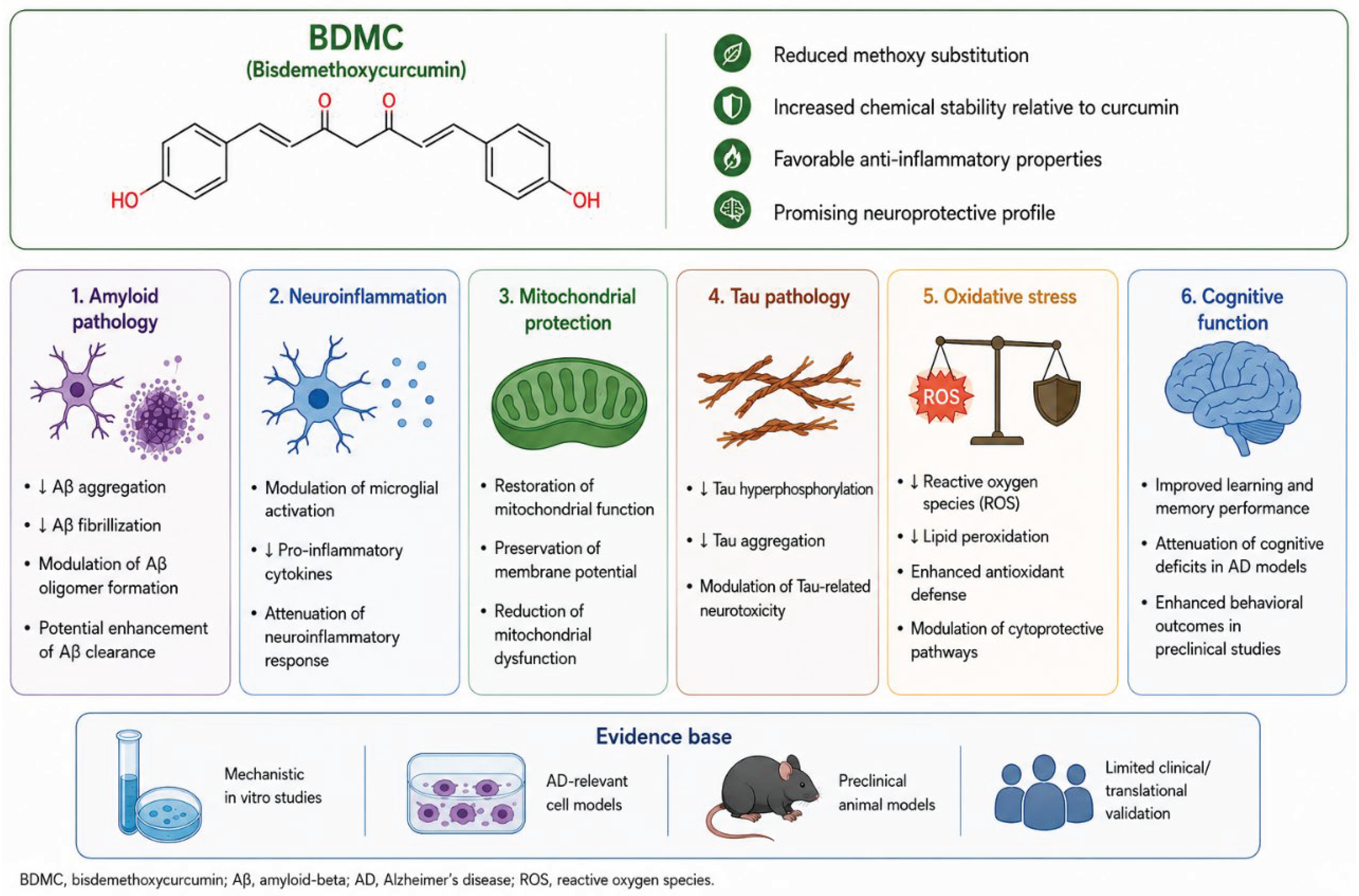

Table 1.
Representative studies on curcumin in Alzheimer’s disease and related dementia models.
| Reference | Type of evidence | Main finding(s) | Main mechanism(s) domain |
|---|---|---|---|
| [80] | In vivo, APPSw Tg+ and Tg− mice | Reduced oxidative damage, IL-1β and amyloid pathology | Anti-inflammatory; antioxidant; anti-amyloid |
| [127] | In vivo, Aβ-infused rat | Attenuated Aβ-induced oxidative/inflammatory damage | Anti-inflammatory; antioxidant |
| [128] | In vitro, PC12 rat pheochromocytoma cells | Protected neuronal cells against Aβ-induced toxicity | Neuroprotection; antioxidant |
| [78] | In vitro, β-amyloid fibrils | Inhibited Aβ aggregation and destabilized fibrils | Anti-amyloid aggregation |
| [81] | In vitro model of Aβ fibrillization + in vivo Tg2576 mice | Inhibited Aβ oligomers/fibrils, bound plaques, reduced amyloid burden | Anti-amyloid; plaque binding |
| [129] | Ex vivo, human AD macrophages | Curcuminoids enhanced Aβ uptake by macrophages | Immunomodulation; Aβ clearance |
| [130] | In vivo imaging, APPswe/PS1dE9 mice | Curcumin labeled amyloid deposits in vivo | Amyloid binding/imaging |
| [131] | In vivo Tg2576 APP sw mice | Curcumin reduced neuroinflammation and AD-like pathology | Anti-inflammatory; anti-amyloid |
| [132] | Clinical pilot randomized controlled trial (RCT), AD patients | Generally safe, but no clear clinical efficacy signal | Clinical tolerability; bioavailability limitation |
| [133] | Ex vivo, human AD macrophages | Curcuminoids + vitamin D enhanced Aβ phagocytosis in selected patients | Innate immunity; Aβ clearance |
| [134] | In vivo, amyloid-β peptide-infused rats | Curcuminoids improved memory and synaptic protein expression | Synaptic plasticity; cognition |
| [135] | Ex vivo, human AD macrophages | β-1,4-mannosyl-glycoprotein 4-β-N-acetylglucosaminyltransferase (MGAT3) response may distinguish curcuminoid-responsive subgroups | Biomarker; immunomodulation |
| [105] | Clinical RCT, AD patients | Oral curcumin was tolerated but showed limited efficacy, likely due to low bioavailability | Clinical translation; bioavailability |
| [136] | In vivo, Aβ1–42 Sprague-Dawley rats | Chronic curcumin improved spatial memory dose-dependently | Cognition; BDNF/synaptic pathways |
| [74] | In vivo, p25 mouse model | Reduced neuroinflammation, neurodegeneration and memory decline | Neuroinflammation; neurodegeneration; synaptic dysfunction; cognition |
| [86] | Clinical RCT, non-demented adults | Theracurmin improved memory/attention and reduced PET amyloid/tau signals | Amyloid/tau; cognition |
| [137] | Cell-based AD patient studies | Curcumin analogues modulated AD-related inflammatory and amyloidogenic pathways | Anti-inflammatory; amyloidogenesis |
| [73] | In vivo, Aβ1–42 hippocampal injection mouse model | Curcumin improved cognition and reduced inflammation, oxidative stress, neuronal damage and Aβ deposition | Neuroinflammation; oxidative stress; AMPK signaling; anti-apoptotic effects |
| [103] | In vivo, APPswe/PS1dE9 mice on a C57BL/6J background | A bioavailable curcumin formulation reduced some AD-related pathological features but was associated with increased neuroinflammatory responses | Neuroinflammation; amyloid pathology; bioavailability-related effects |
| [102] | In vitro biophysical study | Curcumin attenuated Aβ oligomer binding to anionic lipid membranes, potentially reducing membrane-associated toxicity | Anti-amyloid effects; membrane interaction modulation |
| [92] | In vitro, Tau aggregation/phosphorylation model | The co-amorphous curcumin–artemisinin formulation attenuated Tau aggregation and hyperphosphorylation | Tau aggregation; Tau phosphorylation; neuroprotection |
| [138] | In vivo, 3xTg-AD model | Curcumin improved AD-related pathological and cognitive alterations in a metabolically stressed AD model | Metabolic stress; neuroinflammation; amyloid/tau |
Disclaimer/Publisher’s Note: The statements, opinions and data contained in all publications are solely those of the individual author(s) and contributor(s) and not of MDPI and/or the editor(s). MDPI and/or the editor(s) disclaim responsibility for any injury to people or property resulting from any ideas, methods, instructions or products referred to in the content. |
© 2026 by the authors. Licensee MDPI, Basel, Switzerland. This article is an open access article distributed under the terms and conditions of the Creative Commons Attribution (CC BY) license (http://creativecommons.org/licenses/by/4.0/).
Copyright: This open access article is published under a Creative Commons CC BY 4.0 license, which permit the free download, distribution, and reuse, provided that the author and preprint are cited in any reuse.


