

Submitted:
12 May 2026
Posted:
13 May 2026
You are already at the latest version
Abstract
Rel/SpoT family enzymes participate in controlling the cellular levels of the alarmone (p)ppGpp, thereby activating the stringent response and promoting survival under stress conditions. These proteins contain an N-terminal catalytic domain and a C-terminal regulatory domain. They catalyze both the synthesis of ppGpp/pppGpp from ATP and GDP/GTP and their hydrolysis to GDP/GTP and pyrophosphate. Here, we report the crystal structure of the N-terminal domain of Rel from Streptococcus equisimilis (RelSeq385) in complex with pppGpp at 3.2 Å resolution. The asymmetric unit contains a dimer with asymmetric ligation, in which pppGpp occupies only the synthetase site in one monomer, whereas it is observed in both the hydrolase and synthetase sites in the other. Molecular dynamics simulations supported this binding arrangement for the monomer with both sites occupied and revealed additional probable transient binding sites that may contribute to alarmone binding.
Keywords:
RelSeq
; bifunctional enzyme
; RelA/SpoT homolog
; stringent response
; alarmone
; pppGpp
; crystal structure
; molecular dynamics
; nanoDSF
1. Introduction
Bacteria rapidly sense and adapt to environmental changes through complex strategies such as the stringent response, a pleiotropic process that broadly reshapes cellular metabolism and gene expression [1,2,3,4,5,6]. It is triggered by the intracellular accumulation of the alarmone signal molecules (p)ppGpp – guanosine 3’-diphosphate 5’-triphosphate (pppGpp) and guanosine 3’,5’-bisdiphosphate (ppGpp) – which are GTP/GDP derivatives bearing pyrophosphate groups at the ribose 3’-hydroxyl position [7]. Cellular (p)ppGpp levels are controlled by the RelA/SpoT homologue (RSH) family of proteins, as well as by small alarmone synthetases (SAS) and small alarmone hydrolases (SAH), which exclusively catalyze (p)ppGpp synthesis and hydrolysis in response to diverse stresses respectively [2,3,4,5,8,9,10]. Biochemically, Rel-like synthetases primarily generate pppGpp by transferring a pyrophosphate group from ATP to GTP, while ppGpp is formed either directly from GDP or via subsequent hydrolysis of pppGpp. The predominant RSH enzymes in bacteria are bifunctional, ribosome-associated “long” Rel proteins, among which the Rel homologue from Streptococcus equisimilis (RelSeq) is a representative that comprises an N-terminal catalytic domain (NTD) and a C-terminal regulatory domain (CTD) [11,12,13,14,15]. Deletion of the C-domain shifts the catalytic balance dramatically—activating synthetase activity ~12-fold while suppressing hydrolase activity ~150-fold—underscoring the critical regulatory role of the CTD in tuning the enzyme’s functional mode [8,12,15,16]. For this enzyme, when GTP is present in excess over GDP, it predominantly produces pppGpp, indicating a preference for GTP as the pyrophosphate acceptor and rendering the pentaphosphate, compared to ppGpp, a physiologically relevant immediate reaction product [11]. In our recent experiments, we found that NTD of RelSeq (RelSeq385) is conformationally most stable in its alarmone-bound form and exhibits a twofold higher affinity for pppGpp than for ppGpp [12]. Taking these observations together, we were led to the rather bold hypothesis idea that preferential stabilization of the alarmone-bound state may help keep (p)ppGpp within a range that supports proper cellular homeostasis, balancing the need for stringent-response signaling against the toxicity of alarmone overaccumulation [13,14,15].
Here, using an enzymatically active RelSeq385, we determined its crystal structure in complex with pppGpp. This allowed us to visualize the architecture of the synthetase and hydrolase active sites and to identify key residues directly involved in pppGpp coordination. Furthermore, given that the RelSeq fragment under study is a two-domain protein with inherent flexibility, we carried out molecular dynamic experiments to simulate the formation of the RelSeq385•pppGpp complex.
2. Results
2.1. Conformational Stability of RelSeq385
Experiments with full-length RelSeq in vitro are often hindered by its limited solubility, as the protein displays a strong propensity to aggregate and precipitate in response to even small deviations from a narrow set of optimal buffer conditions, whereas truncation of the CTD markedly improves its solubility while preserving catalytic activity [17,27]. In our standard purification protocol, the storage buffer contained glycerol, which is commonly used to enhance protein solubility and prevent aggregation or precipitation during isolation and storage [18]. However, under crystallization conditions the presence of glycerol reproducibly led to the formation of precipitates instead of well-ordered crystals, most likely due to interference of glycerol with crystal nucleation and growth. There, for preparation of RelSeq385 for crystallographic analysis, we therefore modified the storage buffer by omitting glycerol from the final composition. The conformational stability and aggregation state of the protein was assessed by nanodifferential scanning fluorimetry (nanoDSF) as described previously [18], after storage at 4 °C or 25 °C for up to one week, as well as following multiple freeze–thaw cycles and storage at -80 °C. Although the absence of glycerol led to a decrease in conformational stability, as reflected by a reduction in the melting temperature by approximately 4–6 °C, the shape of the unfolding curve, baseline fluorescence, and overall transition amplitude remained unchanged, indicating preservation of the overall protein fold and confirming that the protein remained suitable for subsequent structural analysis under these conditions. Importantly, monitoring of light scattering during nanoDSF measurements revealed no detectable aggregation under any of the tested storage conditions throughout the entire observation period. During the observation period of one week, the protein retained its conformational integrity when stored at 4 °C or 25 °C, as well as upon storage at -80 °C with multiple freeze-thaw cycles using liquid nitrogen (Figure S1).
2.2. RelSeq385 Catalytic Activities in (p)ppGpp Turnover
To avoid trapping RelSeq385 in an inactive conformation during crystallization with pppGpp, it is essential to ensure that the protein retains catalytic activity, thereby reflecting a functionally competent state consistent with its physiological role in alarmone metabolism. Structural and functional studies of RSH proteins have shown that active-site conformation and the positioning of key catalytic elements depend on the presence of substrate or product and can differ substantially between active and inactive enzyme states [19,28]. We assessed the catalytic activity of the enzyme under varying concentrations of magnesium and manganese ions, which play key roles in alarmone synthesis and hydrolysis, respectively.
2.2.1. Synthesis Activity
By coordinating the phosphate groups of the substrates, Mg2+ stabilizes their reaction competent configuration and promotes activation of the 3′-OH group of the acceptor nucleotide [29,30]. Structural and functional studies of RSH enzymes, uncluding RelSeq and RelMtbfrom Mycobacterium tuberculosis, have shown that (p)ppGpp synthetase activity depends on the balance between the total nucleotide substrate concentration and Mg2+ levels, with the optimum approximately matching the combined concentration of nucleotide substrates [17,30]. It has been shown that RelSeq exhibits maximal pppGpp synthetase activity at 3–7 mM MgCl2 when the total nucleotide substrate concentration is around 4 mM, whereas increasing the total substrate concentration to 12 mM shifts the activity optimum to 10–15 mM MgCl2, closely matching the combined concentration of ATP and GTP. This is consistent with a model in which the enzyme acts on the Mg2+•ATP and Mg2+• GTP complexes, and it is the concentration of these Mg–nucleotide species, rather than the total ATP/GTP levels, that determines the effective rate of (p)ppGpp synthesis in vitro. Excess Mg2+ beyond this optimal range leads to a reduction in synthesis rates, likely due to subtle changes in Mg2+•ATP coordination and active site conformation [13,17,30]. In our experiments, RelSeq385 synthesized pppGpp in the presence of 10 mM ATP and 4 mM GTP over a range of Mg2+ concentrations (Figure 1A) and across different temperatures and incubation times (Figure S2B). At higher Mg2+ concentrations of 11 mM and 26 mM, the synthetase activity of RelSeq385 in our experiments remained unchanged, indicating that the enzyme did not lose activity under these conditions and the excess Mg2+ was present in ATP-bound and, probably, AMP-bound forms (Figure S2C) [31,32,33].
2.2.2. Hydrolysis Activity
In long Rel/SpoT-like enzymes, 3′-pyrophosphohydrolase activity is Mn2+-dependent. Quantitative assays with the purified N-terminal domain of RelSeq have demonstrated Mn2+-dependent hydrolysis of (p)ppGpp at millimolar Mn2+ concentrations, with no detectable activity toward (p)ppApp, indicating a clear guanosine specificity of the hydrolase reaction [17,34].
The presence of Mg2+ in the concentration range required for synthetase activity (5–10 mM) does not suppress Mn2+-dependent hydrolase activity, indicating that Mg2+ at these levels does not interfere with alarmone hydrolysis by the enzyme [34]. Under our experimental conditions with 14 mM Mg2+, the hydrolase activity of RelSeq385 was strongly Mn2+-dependent, with only low residual activity detectable in the absence of Mn2+ (Figure 1B). We observed rapid degradation of pre-synthesized (p)ppGpp and free GTP, including Mg2+•GTP form (Figure S2D). These observations are consistent with a model in which Mg2+ supports the synthetase center, whereas Mn2+ activates the hydrolase domain of RelSeq [17,34].
Biochemical characterization of purified RelSeq385 demonstrated that its catalytic activity remained high throughout all purification steps, storage, and buffer adjustments, thereby ensuring that enzyme can be used for structural analysis of alarmone complexes that faithfully represent a physiologically active state of the enzyme. In addition, our experiments showed that an excess of Mg2+ ions did not interfere with the catalytic activity of the enzyme.
2.3. Stability Characterization of the RelSeq∙pppGpp Complex
To assess the conformational stability of the RelSeq385•pppGpp complex under temperature and time conditions relevant for crystallization, we incubated the protein and its pppGpp complex at 25 °C and monitored time-dependent conformational changes by analyzing their thermal denaturation profiles (Figure 2). The conformational stability of the protein remained unchanged over the entire observation period (7 days), as evidenced by an invariant melting temperature and overall unfolding profile (Figure 2A). The RelSeq385•pppGpp complex exhibited subtle but detectable changes in its thermal denaturation behavior over time (Figure 2B). While the overall shape of the unfolding curve and the melting temperature of the complex remained unchanged, the total amplitude of the fluorescence signal change associated with the conformational transition progressively decreased, consistent with a possible reduction in the fraction of protein (or protein•alarmone complexes) undergoing a cooperative transition, including potential alterations in complex stoichiometry [18,35,36].
2.4. Crystallographic Structure of the RelSeq385•pppGpp Complex
The structure of RelSeq385 in complex with pppGpp was solved and refined at 3.2 Å resolution by the molecular replacement method using monomer A of the 1VJ7 structure as a search model. The crystal belongs to C 1 2 1 space group with unit cell parameters a, b, c = 173.75, 44.98, 126.43 (Å).
The crystallographic asymmetric unit contains a RelSeq385 dimer, revealing two monomers captured in distinct conformational states (Figure 3). In monomer A, the alarmone pppGpp binds exclusively to the synthetase domain, whereas monomer B exhibits pppGpp bound in both the synthetase and hydrolase domains. Analysis of the crystallographic RelSeq385 dimer interface using the PDBePisa server did not predict it to be a physiologically relevant dimerization interface, suggesting that it likely arises from crystal packing.
While the protein backbone was well fitted into the electron density map, certain side chains were poorly resolved due to local weak density. Several loop regions were invisible in the density and thus omitted from modeling (monomer A: residues 113–123, 154–159, 344–360; monomer B: residues 111–130, 260–264, 342–362).
The overall structure of the RelSeq385 monomer consists of the hydrolase (residues 1–159) and synthetase (residues 176–372) domains joined by an overlapping central 3-helix bundle (residues 135–195).
2.4.1. The pppGpp-Hydrolase Active Site of RelSeq385
The hydrolase domain of RelSeq385 consists of α-helices 1–7 and shows a structurally conserved fold (Figure 3B, C).
The binding pocket for the substrate (p)ppGpp is surrounded by the extended α2/α3 loop (residues 40–50) that connects helices α2 and α3, helix α3 (including the His53 side chain), the conserved HD motif between helices α4 and α5 (residues 77–78), and helix α8 of the central 3-helix bundle.
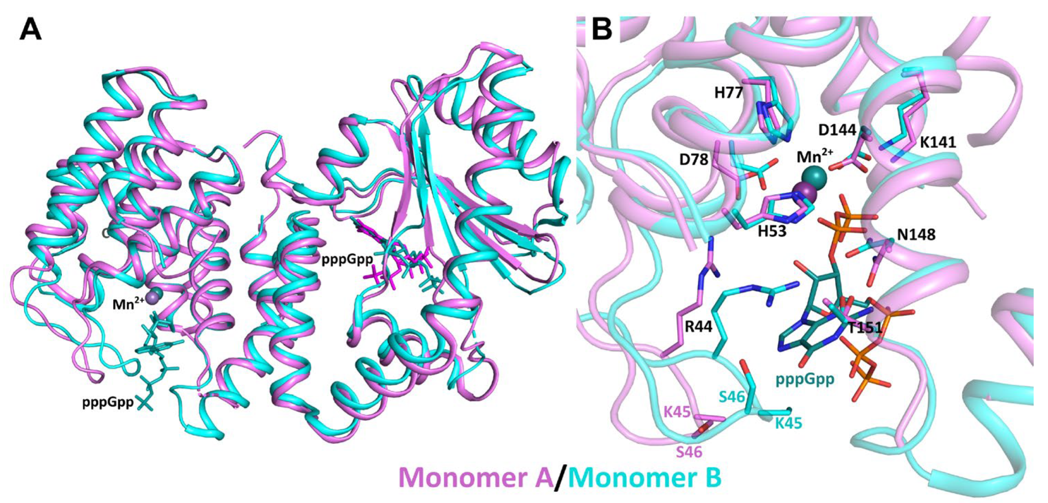
In the unoccupied HD pocket of monomer A, a divalent metal ion (Mn2+) is coordinated by the side chain atoms of His53, His77, and Asp144 (Figure 4A). As observed in monomer A, the α8/α9 loop fragment (residues 154–159) is disordered in the electron density maps in the ligand-free state. Upon pppGpp binding, the flexible loops surrounding the binding pocket undergo pronounced conformational changes (Figure 4C, 5). Specifically, in monomer B the α8/α9 loop becomes ordered, accompanied by coordinated movements of the α2/α3 and α8/α9 loops, which together form a cap beneath the entrance to the binding cleft, acting as steric gates. The α2/α3 loop is displaced and shifts proximally toward the substrate-binding pocket. This repositioning leads to local displacements of residues Arg44, Lys45 and Ser46. As a result, Arg44 loses its salt bridge with Asp78 and instead forms hydrogen bonds with the side chain atoms of Asn148 and interacts with the guanine base of pppGpp, while Lys45 and Ser46 also engage in interactions with the alarmone. This disruption of the salt bridge releases Asp78, allowing it to approach the Mn2+ ion. Consistent with the proposed role of Arg44 and Asp78 in coordinating Mn2+ to the 3′-diphosphate of (p)ppGpp [13], the observed rearrangement of Arg44 upon ligand binding underscores its dual role: facilitating substrate binding and contributing to the proper coordination of the metal ion essential for hydrolase activity.
The guanine base is coordinated within the HD active center through hydrogen bond interactions involving the main-chain amides of Lys45 (to N7 of pppGpp) and Ser46 (to O-6), as well as the main-chain carbonyl of Thr151 (to N-1) and Asn148 (to N2). Notably, the side chain of Lys45 was not resolved in the electron density and was therefore omitted from the model (atoms CG, CD, CE, and NZ in monomer A and atoms CD, CE, and NZ in monomer B). Additional stabilization of pppGpp is provided by interactions with the side chains of Arg44 and Asn148.
Turning to the phosphate moieties, the α-phosphate of the 3′-diphosphate group points Asn148 side chain (O1C – ND2), as well as with the essential manganese ion cofactor Mn2+ (O2C – Mn2+). In contrast, the β-phosphate is oriented toward the solvent and does not exhibit strong restraining interactions with nearby atoms. Finally, the 5’-triphosphate group of pppGpp is exposed to the bulk solvent.
Figure 4.
Active site architecture and pppGpp binding in the HD (cyan) and SYN (gold) domains of the bifunctional enzyme RelSeq385. In monomer A, the HD active site is unoccupied (panel A), whereas the SYN domain harbors a bound pppGpp molecule (panel B). In monomer B, pppGpp is present in both the HD (panel C) and SYN (panel D) active sites. HD and SYN domains of the two monomers of RelSeq385 are shown and colored in cyan and gold, respectively. The alarmone pppGpp is depicted as a stick model with atoms color-coded: carbon (green), oxygen (red), nitrogen (blue), and phosphorus (orange). Secondary structure elements are labeled in cyan (HD) and orange (SYN). Interatomic contacts are shown as gray dashed lines. The corresponding 2mFo-DFc electron density maps (contoured at 1σ) for pppGpp in the HD and SYN domains of both monomers are provided in the Supplemental Data (Figure S3).
Figure 4.
Active site architecture and pppGpp binding in the HD (cyan) and SYN (gold) domains of the bifunctional enzyme RelSeq385. In monomer A, the HD active site is unoccupied (panel A), whereas the SYN domain harbors a bound pppGpp molecule (panel B). In monomer B, pppGpp is present in both the HD (panel C) and SYN (panel D) active sites. HD and SYN domains of the two monomers of RelSeq385 are shown and colored in cyan and gold, respectively. The alarmone pppGpp is depicted as a stick model with atoms color-coded: carbon (green), oxygen (red), nitrogen (blue), and phosphorus (orange). Secondary structure elements are labeled in cyan (HD) and orange (SYN). Interatomic contacts are shown as gray dashed lines. The corresponding 2mFo-DFc electron density maps (contoured at 1σ) for pppGpp in the HD and SYN domains of both monomers are provided in the Supplemental Data (Figure S3).
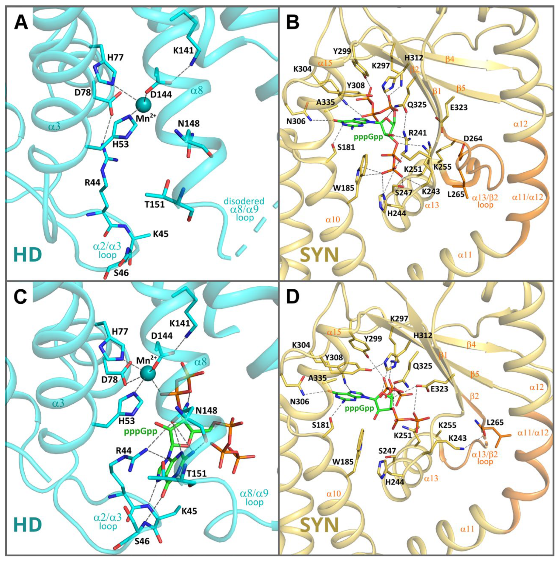
Figure 5.
Structural comparison of the two RelSeq385 conformations. (A) A cartoon representation of superimposed monomer A (violet) and monomer B (cyan) structures. pppGpp molecules are shown as sticks colored according to the monomer color. (B) Structural alignment of the HD active sites of the monomer A (violet) and monomer B (cyan), pppGpp bound in HD of the monomer B is shown as sticks.
Figure 5.
Structural comparison of the two RelSeq385 conformations. (A) A cartoon representation of superimposed monomer A (violet) and monomer B (cyan) structures. pppGpp molecules are shown as sticks colored according to the monomer color. (B) Structural alignment of the HD active sites of the monomer A (violet) and monomer B (cyan), pppGpp bound in HD of the monomer B is shown as sticks.
2.4.2. The pppGpp-Hydrolase Active Site of RelSeq385
The C-terminal synthetase domain (residues 176–371) consists of 5-stranded mixed β-sheet surrounded by five α-helices (α11−α15) in a sandwich-like arrangement, which forms a solvent accessible cavity (Figure 3B, C and Figure 4B, D).
Interestingly, the SYN domains of both monomers in the asymmetric unit are occupied by pppGpp. However, the binding mode of the alarmone differs slightly between monomer A and monomer B. While the guanine moiety adopts a conserved position and forms hydrogen bonds with Ser181, Asn306, and Ala335 in both monomers, Lys304 shows monomer-specific behavior: it contacts the guanine ring in monomer B (Figure 4D), but interacts with the 5′-triphosphate group of pppGpp in monomer A (Figure 4B).
The 3′-diphosphate and 5′-triphosphate groups exhibit distinct spatial arrangements in the two monomers, leading to a somewhat different pattern of hydrogen-bonding interactions with the same set of surrounding residues, including Trp185, Arg241, Lys243, His244, Ser247, Lys251, Lys297, Tyr299, His312, and Gln325, which are involved in binding and stabilizing pppGpp.
Beyond these differences in the bound alarmone’s orientation, the two monomers exhibit pronounced conformational heterogeneity in the surrounding structural elements. In monomer A, the catalytic loop α13/β2 (residues 254–266) is stabilized and ordered through van der Waals interactions with the side chains of the helical fragment comprising residues 209–217 (α11/α12) (Figure 4B). Conversely, in monomer B the α11/α12 fragment shows partial disorder, as evidenced by poorly defined side-chain electron density. This loss of rigidity weakens the interactions with the catalytic loop, thereby destabilizing the α13/β2 loop (Figure 4D). Thus, while in monomer A the catalytically essential residue Asp264 is clearly visible in the electron density and forms a salt bridge with the side chain of Lys255, in monomer B this interaction is lost due to disorder of the α13/β2 loop, which renders these residues poorly defined in the density. Consequently, the side chain of Lys255 is not resolved, and Asp264 is not visible at all.
Hogg and colleagues identified Asp264 and Glu323 as the catalytically essential carboxylate residues in the synthetase active site, a finding supported by mutagenesis: the D264G and E323Q mutations eliminate detectable synthetase activity [13]. They proposed that Glu323 coordinates Mg2+ and serves as the GTP/GDP 3′-OH proton acceptor during the (p)ppGpp-synthetase reaction. In our structure, however, the side chain of Glu323 in monomer A exhibits fragmented electron density, suggesting mobility and a tendency to form ionic interactions with the side chains of Lys243 or Lys251. In monomer B, by contrast, Glu323 becomes more ordered and appears to form the interactions with the bound pppGpp.
Furthermore, Lys243, which was earlier implicated as a probable ATP-phosphate binding residue [13], in monomer B undergoes a concomitant rearrangement away from the active site to interact with the backbone carbonyl of Leu265.
Taken together, our data suggest that the conformational state of the synthetase active site depends on the integrity of a structural framework that coordinates coupled and concerted movements of the loops surrounding the binding pocket.
2.5. Molecular Dynamics Simulations of RelSeq385•pppGpp
RSH enzymes are increasingly recognized as conformationally dynamic regulators, in which relative domain rearrangements and flexible linkers play a key role in tuning (p)ppGpp metabolism. Structural and bioinformatic studies of RSH proteins have highlighted that this plasticity is an intrinsic feature of the superfamily and is closely linked to alarmone binding and allosteric control [14,28,37,38]. RelSeq385 conforms to this paradigm: despite being a truncated construct, it comprises two domains connected by a flexible linker and thus samples multiple relative domain orientations, a view consistent with our prior assumptions about the enzyme’s stability in the alarmone-bound state [18]. Motivated by these considerations and by prior computational work on (p)ppGpp–protein systems, we complemented our crystallographic analysis of the RelSeq385•pppGpp complex with molecular dynamics (MD) simulations to characterize its conformational landscape in solution and to identify putative alarmone binding hotspots [12,29].
Throughout the simulations, RelSeq385 largely preserved its secondary structure and overall fold, whereas the magnitude and temporal pattern of backbone root mean square deviation (RMSD) pointed to pronounced interdomain motions within the NTD, consistent with the intrinsic flexibility described for Rel/RelA N-terminal domains [14,28].
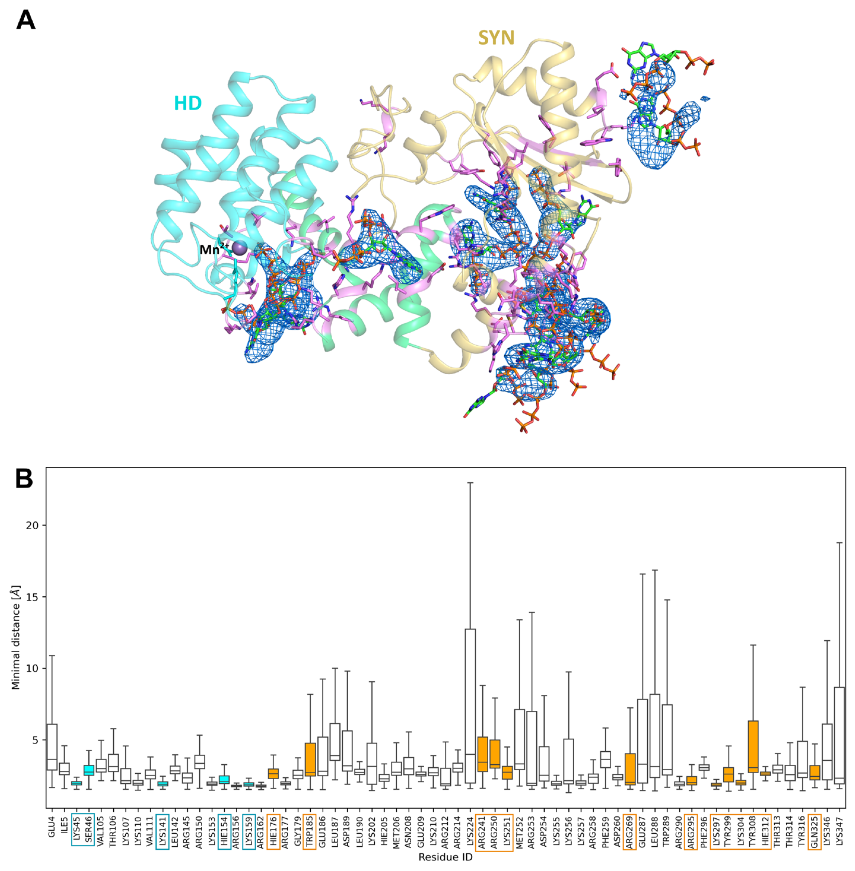
To identify preferred alarmone interaction regions, we extracted pppGpp–protein contacts along the trajectories using minimal distances between pppGpp and protein residues and constructed probability weighted contact maps. For each monomer in the crystallographic dimer, residue wise contact frequencies with pppGpp were calculated, highlighting hotspots of recurrent interaction. In monomer B, this analysis revealed several high probability patches, including positions that spatially overlapped with the pppGpp binding sites observed in our crystal structure, thereby supporting the functional relevance of the crystallographic binding sites (Figure 6).
In addition to the hydrolase and synthetase sites, however, the MD simulations also revealed additional zones of probable pppGpp binding. One notable such patch was localized in the linker connecting the HD and SYN domains, encompassing segments of α-helices α9 and α10. This region has previously been shown to act as a transmission core that mediates coupling between the SYN and HD domains [15]. Furthermore, an allosteric (p)ppGpp binding site was identified in E. coli RelA at the interface between the SYN and pseudo-HD domains where pppGpp binding promotes SYN activity by antagonistic allosteric cross-talk between the two NTD domains [39]. In our MD simulations, we observed a similarly located hotspot in RelSeq385, comprising residues Arg150, Lys153, His154, Glu186, Leu187, Asp189, and Leu190 (Figure S4), suggesting that a comparable allosteric mechanism may also operate in this enzyme. Additional MD identified binding areas included a region within α-helix α11 and the loop following α-helix α14 (residues 287–290).
MD simulations of monomer A showed a similar binding pattern for the HD and SYN domains but did not reveal linker occupancy. This may reflect the difference in starting monomer conformations and probably the transient nature of alarmone binding in the linker (data not shown).
3. Discussion
RelSeq from S. equisimilis is a bifunctional long RSH enzyme that catalyzes both (p)ppGpp synthesis and hydrolysis, playing a central role in stringent response. In this work, we focus on the N-terminal 1–385 fragment (RelSeq385), which corresponds to the catalytic core harboring HD and SYN subdomains. Our biochemical assays confirmed that RelSeq385 exhibits robust catalytic activities: (p)ppGpp synthesis via ATP–GTP/GDP 3′-pyrophosphotransferase and Mn2+-dependent 3′-pyrophosphohydrolase activity. As a continuation of our recent study showing that increasing concentrations of (p)ppGpp enhance the conformational stability of RelSeq385 [18], we now report its crystal structure in complex with pppGpp.
The RelSeq385•pppGpp structure (PDB 24IO) reveals remarkable similarities to the previously reported RelSeq385•GDP complex (PDB 1VJ7), in which the asymmetric unit likewise contains two monomers with distinct domain orientations and ligation states [13]. In 1VJ7, GDP occupies the synthetase domains of both monomers, with monomer 2 additionally harboring the unusual GDP derivative ppG2′:3′p in the hydrolase site. In our structure, pppGpp is bound in the synthetase domain of monomer A and in both the hydrolase and synthetase sites of monomer B, mirroring the dual-ligation pattern observed in 1VJ7 monomer 2 (structural superposition is shown in Figure S5). Despite the different ligands, key catalytic residues in the corresponding binding pockets exhibit similar orientations, underscoring the structural integrity of the active sites. This binding mode is further supported by an unpublished Staphylococcus aureus Rel (Sa-Rel) structure in complex with pppGpp (PDB 7OIW, chain B; data not published), where the alarmone also occupies the synthetase active site. Both RelSeq and Sa-Rel share a conserved long-RSH architecture with N-terminal catalytic domain and a C-terminal regulatory region. Sa-Rel is essential for stringent response initiation under amino acid limitation and stress, and loss of its hydrolase activity (e.g., in clinical Rel mutants) leads to (p)ppGpp overaccumulation, attenuated growth of the cells, and altered their antibiotic tolerance [40,41]. The 7OIW structure shows a similar binding pattern, with pppGpp bound to both the HD and SYN domains simultaneously. Structural superposition revealed that the hydrolase domain of RelSeq385 monomer B and that of 7OIW are nearly identical, exhibiting very similar coordination of the pppGpp molecule and identical positions of the catalytic residues (Figure S7A,B). In contrast, superposition of the synthetase domains showed that the conformation of 7OIW more closely resembles that of monomer A of RelSeq385 than that of monomer B (Cα RMSD over the SYN domain are 1.04 Å and 1.41 Å, respectively) (Figure S6A,C). The alarmone binding mode in the synthetase pocket is virtually identical between RelSeq385 and 7OIW, except for the position of the 5′-triphosphate group of pppGpp, which is exposed to the solvent, explaining the observed flexibility of this tail and the corresponding minor deviations in coordinates. Together, these data support a model in which pppGpp can occupy both catalytic sites in a subset of Rel proteins.
Despite the absence of the CTD, the truncated RelSeq385 enzyme retains significant conformational flexibility because its N-terminal catalytic core comprises two subdomains (HD and SYN) connected by a mobile linker region. To explore the binding profile of pppGpp within this conformational landscape, we employed molecular dynamics simulations. The MD simulations confirmed the dual occupancy pattern observed in monomer B. Among the dynamic elements, the linker region connecting the HD and SYN domains warranted particular attention, as the MD calculations indicated high-probability binding of the alarmone in this interdomain interface. This is consistent with reports that (p)ppGpp can occupy allosteric sites between catalytic domains to mediate antagonistic coupling and conformational switching in RSH enzymes [39,42].
Although we did not observe alarmone binding in the linker region in our crystal structure, this discrepancy likely reflects the different conditions captured by the two methods: crystallography provides a static view under packing constraints, whereas MD simulates a solvated dynamic ensemble. Thus, (p)ppGpp-binding in the linker region may be transient or dependent on a conformational state not sufficiently populated in the crystal lattice. Overall, MD captures an ensemble of accessible states, whereas crystallography provides a static snapshot of a single structure. This underscores that RelSeq385 does not exist as a single rigid conformation but rather behaves as a flexible equilibrium modulated by ligand occupancy and metal coordination.
This notion of conformational flexibility as a determinant of catalytic output is consistent with broader structural insights into the RSH enzyme family, in which nucleotide-driven conformational switching has emerged as a key regulatory mechanism. For example, in Rel from Thermus thermophilus (RelTt), GDP binding to the synthetase domain stabilizes an open, synthetase-active state, whereas ppGpp binding to the HD domain induces a closed, hydrolase-active conformation that prevents futile catalytic cycles. In line with this, distinct τ-shaped (hydrolase-active) and extended (synthetase-active) conformations of long RSH enzymes are further stabilized by Mn2+, which maintains the active-site architecture [20].
Our MD-derived evidence for a high probability of alarmone binding in the linker region of RelSeq385 deserves separate attention. Together with the observation that RelSeq385 binds pppGpp with significantly higher affinity than ppGpp [18] — similar to what has been shown for RelA [39] — this finding suggests that this interaction may represent an additional allosteric regulatory mechanism, analogous to the product-dependent positive feedback already described for long RSH enzymes of the RelA/Rel lineage [39]. Consistent with this view, pppGpp acts as a more potent activator than ppGpp in RelA, binding to an allosteric site at the SYNTH–pseudo-HD interface, stabilizing an open, catalytically competent conformation that relieves intramolecular inhibition and enhances alarmone synthesis [39,42,43,44].
Based on these findings, we cautiously consider two mechanistic hypotheses for RelSeq385: (1) transient (p)ppGpp sequestration as a “depot” for rapid stress response, and (2) dual-site autoregulation to avoid futile cycling. Such mechanisms might stabilize ordered conformations and buffer alarmone levels, though further work is needed to clarify allosteric coupling between its domains [22,23].
In summary, our data suggest that pppGpp binding to the catalytic domains of RelSeq385 can contribute to buffering intracellular alarmone levels and limiting futile ATP/GTP consumption. The pronounced conformational flexibility of the enzyme, together with the potential existence of additional transient binding sites, points to a more complex regulatory landscape than previously appreciated. However, the precise molecular mechanisms underlying these effects remain to be fully delineated in future studies. Given the central role of Rel/SpoT family enzymes in bacterial stress adaptation and virulence, and the lack of close homologs in mammalian cells, these proteins are increasingly recognized as attractive, pathogen-selective antibacterial targets. Structural and mechanistic insight into the function of their individual domains therefore provides an important foundation for future structure-guided inhibitor design [21,24,25,26].
4. Materials and Methods
4.1. Protein Expression and Purification
Protein expression and purification were performed essentially as described previously [45], with minor modifications detailed below. The N-terminal domain of RelSeq (residues 1–385, RelSeq385) bearing a C-terminal hexahistidine tag was produced in Escherichia coli BL21 (DE3) carrying a pET21-based expression plasmid and purified as described above. Transformed cells were grown in LB medium with 100 μg/mL ampicillin at 37 °C to OD600 ≈ 0.6, then induced with 1 mM IPTG for 3 h at 37 °C. Cells were harvested by centrifugation at 6,000 rpm for 20 min (JLA 8.1 rotor) and resuspended in lysis buffer (20 mM Tris-HCl pH 7.9, 300 mM KCl, 5 mM MgCl2, 20% (v/v) glycerol, 10 mM imidazole, 5 mM β- mercaptoethanol, 280 μg/mL lysozyme, 0.1 mg/mL DNase I (Sigma-Aldrich), and one protease inhibitor cocktail tablet (Roche) per 50 mL). Cell disruption was carried out using an EmulsiFlex C3 homogenizer (Avestin), and insoluble material was removed by centrifugation at 45,000 rpm for 30 min (Ti50.2 rotor).
The clarified lysate was loaded onto a 5-mL HisTrap FF column (17531901, GE Healthcare) equilibrated with buffer HT (20 mM Tris-HCl pH 7.9, 5 mM MgCl2, 20% (v/v) glycerol, 5 mM β-mercaptoethanol) containing 300 mM KCl and 10 mM imidazole. RelSeq385 was eluted by a stepwise increase in imidazole concentration from 10 mM to 200 mM, and the pooled fractions were diluted with buffer HT containing 10 mM KCl to adjust the final KCl concentration to 50 mM. For further purification, the sample was applied to a 5-mL HiTrap Q anion-exchange column (GE Healthcare) pre-equilibrated in 20 mM Tris-HCl pH 9.5, 50 mM KCl, 5 mM MgCl2, 20% (v/v) glycerol, and 5 mM β-mercaptoethanol. Bound protein was eluted using a linear KCl gradient from 50 mM to 1.5 M. Fractions containing RelSeq385, as assessed by 10% SDS-PAGE (Figure S8A), were pooled, aliquoted, and flash-frozen in liquid nitrogen.
4.2. Conformational Stability of RelSeq385 and RelSeq385•pppGpp Complexes by nanoDSF
Conformational stability of RelSeq385 was assessed essentially as described in [18]. In all experiments, samples were subjected to a temperature ramp from 20 °C to 95 °C at a heating rate of 1 °C/min. Protein stability was evaluated when stored in a standard buffer of 20 mM Tris-HCl, pH 8.0, 1 M NaCl, 5 mM MgCl2, 1 mM DTT without glycerol and in the same buffer with 20% glycerol. The protein concentration in both cases was 130 μM. The protein was kept at 4 °C, 25 °C, and –80 °C with three freeze–thaw cycles. Conformational stability of the protein and its complex with 2 mM pppGpp at 25 °C was monitored over one week by sampling at multiple time points (0 hours, 1, 2, 3, 4, 7 days). Samples (130 μM) were analyzed in standard storage buffer without glycerol.
For all measurements, reaction mixtures were loaded into glass capillaries (NanoTemper Technologies GmbH) with a volume of 10 μL per capillary, and nanoDSF experiments were conducted on a Prometheus NT.48 instrument (NanoTemper Technologies GmbH). Thermal unfolding and aggregation profiles were recorded, and melting temperatures were extracted using the PR.ThermControl software package (NanoTemper Technologies GmbH). Subsequent data analysis and visualization were performed in GraphPad Prism (GraphPad Software).
4.3. Synthesis Activity of RelSeq385
Alarmone nucleotide synthesis was performed essentially as described previously [45], with minor modifications. Reactions for pppGpp used buffer B (30 mM Tris-HCl pH 8.0, 100 mM NaCl) containing 10 mM ATP, 4 mM GTP, 50 μM RelSeq385, and MgCl2 at concentrations yielding final [Mg2+] of 1, 3, 5, 11, or 26 mM. The mixture was incubated for 40 min at 37 °C and was applied to a Mono Q 5/50 GL (1 mL) anion exchange column (GE Healthcare), pppGpp was eluted using a linear gradient of LiCl from 50 mM to 300 mM in buffer C (25 mM Tris-HCl pH 8.0, 0.5 mM EDTA).
For preparative synthesis of both pppGpp and ppGpp, reactions were carried out under the same conditions using 10 mM MgCl2, with ppGpp produced by substituting GDP for GTP as the guanine nucleotide substrate. Fractions containing the desired alarmone species were pooled and precipitated by addition of 1.5 M LiCl and two volumes of absolute ethanol, followed by centrifugation at 16,100 × g for 20 min. The pellet was resuspended in water, and nucleotide concentration was determined by UV at 253 nm using a molar extinction coefficient of 13,700 M−1 cm−1. The sample was aliquoted and stored at –20 °C until use.
4.4. Hydrolysis Activity of RelSeq385
Analytical hydrolysis of pppGpp was performed essentially as described in [34], with minor modifications. Reactions contained 5 mM pppGpp, 50 mM Tris-HCl pH 8.0, 250 mM NaCl, 14 mM MgCl2, RelSeq385 at 22.7 μM, and MnCl2 at final concentrations of 0, 1, 3, or 6.5 mM. Mixtures were incubated for 40 min at 37 °C and applied to a Mono Q 5/50 GL (1 mL) anion-exchange column (GE Healthcare). (p)ppGpp species were eluted using a linear gradient of LiCl from 50 mM to 300 mM in buffer C (25 mM Tris-HCl pH 8.0, 0.5 mM EDTA).
4.5. Protein Crystallization, Data Collection and Structure Determination
For crystallographic experiments the protein RelSeq385 was dialyzed against 20 mM Tris-HCl, pH 7.9, 650 mM NaCl, 2.5 mM MgCl2, 1 mM DTT and concentrated to 6 mg/ml using 10 kDa cut off concentrator (Millipore). The protein sample was then supplemented with 2 mM pppGpp, and this mixture was used for crystallization experiments. Crystals were grown at 298 K using the hanging-drop method with a 1:1 volume ratio of protein and precipitant solution containing 0.1 M Tris-HCl, pH 8.5, 24% polyethylene glycol 8000 and 1 M NaCl. The resulting crystals (Figure S8B) were harvested and immersed in the mother liquor supplemented with 20% glycerol as a cryoprotectant before being flash-frozen in liquid nitrogen. The best crystal diffracted to a resolution of 3 Å. Diffraction data were collected at 100 K using XtaLAB Synergy-S diffractometer equipped with PhotonJet-S Cu microfocus X-ray source and HyPix-6000HE detector (Rigaku Oxford Diffraction, Oxford, UK) at the Center for XRD studies, St. Petersburg State University. Indexing and integration were performed using autoPROC [46]. The high resolution of the data was truncated to 3.2 Å to improve data quality metrics. For estimate anisotropic correction, scaling was completed using STARANISO web server [47]. Elliptically truncated data were used for molecular replacement with phenix.phaser [48].
The structure was solved by molecular replacement using the N-terminal domain of Rel/Spo homolog from S. equisimilis as a search model (PDB 1VJ7, monomer A) [13].
Iterative refinement, model building, and validation were performed using phenix.refine [49,50]. Data collection and refinement statistics are presented in Table S1. The final structure has been deposited in the Protein Data Bank with the accession code 24IO.
Protein structure visualizations were prepared using PyMOL (Version 2.5, Schrödinger, LLC) and VMD (Version 1.9.2 a52).
4.6. Bioinformatics
4.7. MD Simulations
Since solved crystal structure has missing residues we build them using MODELLER [54]. GROMACS compatible topology for pppGpp was prepared using acpype [55] and antechamber [56,57]. We build systems consisting of one RelSeq385 monomer and 32 pppGpp molecules placed in random positions inside 90x90x90A periodic box. MD simulations were performed using GROMACS 2026 with the AMBER19sb [58] force field and the OPC3 [59] water model. The system was neutralized by adding 100 mM NaCl to achieve zero net charge, and energy minimized. Equilibration was performed in two stages: first, positional restraints (posres) were applied to all heavy atoms of the protein, keeping them at their initial positions, and particle velocities were assigned from the Maxwell-Boltzmann distribution at 310 K; the system was equilibrated for 5 ns using a 2 fs integration step and the V-rescale [60] thermostat (coupling time 1 ps) and C-rescale [61] barostat (coupling time 1 ps). In the second stage, restraints were removed and the system was equilibrated for 10 ns using the Nose-Hoover thermostat [62,63] (coupling time 2 ps) and Parinello-Rhaman barostat [64]. The final equilibrated state was used for 250 ns of MD simulations with the same parameters as in the second stage.
Supplementary Materials
The following supporting information can be downloaded at the website of this paper posted on Preprints.org.
Author Contributions
Conceptualization, S.A.K., D.S.V.; methodology, S.A.K., Z.A.S., P.S.K., A.V.S.; validation, S.A.K., D.S.V., A.V.S., A.P., A.A.K., A.L.K.; formal analysis, S.A.K., D.S.V., A.V.S.; investigation, S.A.K., Z.A.S., P.S.K., A.V.S.; resources, D.S.V., V.V.G., A.A.K., A.L.K.; data curation, S.A.K., D.S.V., A.P., A.L.K.; writing—original draft preparation, S.A.K., D.S.V.; writing—review and editing, S.A.K., D.S.V., A.V.S., A.P., A.A.K., A.L.K.; visualization, S.A.K., A.V.S., D.S.V.; supervision, S.A.K., D.S.V.; project administration, D.S.V.; funding acquisition, D.S.V. All authors have read and agreed to the published version of the manuscript.
Funding
This work was supported by the Russian Science Foundation 23-74-10088 of D.S.V. XRD studies had been prepared at the X-ray Diffraction Centre of St. Petersburg State University within the framework of project 126022017738-9.
Institutional Review Board Statement
Not applicable.
Informed Consent Statement
Not applicable.
Data Availability Statement
Coordinates and structure factors of RelSeq385 have been deposited in the PDB under accession code 24IO [https://www.rcsb.org/structure/24IO]. X-ray diffraction dataset is available in the Zenodo repository at [https://zenodo.org/records/18876971].
Acknowledgments
MD simulations were performed using the computational resources of the Supercomputer Center “Polytechnic” of Peter the Great Saint-Petersburg Polytechnic University.
Conflicts of Interest
The authors declare no conflicts of interest.
References
- Potrykus, K.; Cashel, M. (p)ppGpp: Still Magical? Annu Rev. Microbiol. 2008, 62, 35–51. [Google Scholar] [CrossRef]
- Gallant, J.; Palmer, L.; Pao, C.C. Anomalous synthesis of ppGpp in growing cells. Cell. 1977, 11, 181–185. [Google Scholar] [CrossRef] [PubMed]
- Potrykus, K.; Murphy, H.; Philippe, N.; Cashel, M. ppGpp is the major source of growth rate control in E. coli. Env. Microbiol. 2011, 13, 563–575. [Google Scholar] [CrossRef]
- Vinella, D.; Albrecht, C.; Cashel, M.; D’Ari, R. Iron limitation induces SpoT-dependent accumulation of ppGpp in Escherichia coli. Mol. Microbiol. 2005, 56, 958–970. [Google Scholar] [CrossRef] [PubMed]
- Battesti, A.; Bouveret, E. Acyl carrier protein/SpoT interaction, the switch linking SpoT-dependent stress response to fatty acid metabolism. Mol. Microbiol. 2006, 62, 1048–1063. [Google Scholar] [CrossRef]
- Haseltine, W.A.; Block, R. Synthesis of guanosine tetra- and pentaphosphate requires the presence of a codon-specific, uncharged transfer ribonucleic acid in the acceptor site of ribosomes. Proc. Natl. Acad. Sci. U S A 1973, 70, 1564–1568. [Google Scholar] [CrossRef]
- Hauryliuk, V.; Atkinson, G.C.; Murakami, K.S.; Tenson, T.; Gerdes, K. Recent functional insights into the role of (p)ppGpp in bacterial physiology. Nat. Rev. Microbiol. 2015, 13, 298–309. [Google Scholar] [CrossRef]
- Atkinson, G.C.; Tenson, T.; Hauryliuk, V. The RelA/SpoT Homolog (RSH) Superfamily: Distribution and Functional Evolution of ppGpp Synthetases and Hydrolases across the Tree of Life. In PLoS ONE; Stiller, J.W., Ed.; 2011; Volume 6. [Google Scholar] [CrossRef]
- Jimmy, S.; Saha, C.K.; Stavropoulos, C.; Oliveira, S.R.A.; Kurata, T.; Koh, A.; et al. A widespread toxin-antitoxin system exploiting growth control via alarmone signalling; Microbiology, 2019. [Google Scholar] [CrossRef]
- Nanamiya, H.; Kasai, K.; Nozawa, A.; Yun, C.-S.; Narisawa, T.; Murakami, K.; et al. Identification and functional analysis of novel (p)ppGpp synthetase genes in Bacillus subtilis. Mol. Microbiol. 2008, 67, 291–304. [Google Scholar] [CrossRef] [PubMed]
- Agirrezabala, X.; Fernández, I.S.; Kelley, A.C.; Cartón, D.G.; Ramakrishnan, V.; Valle, M. The ribosome triggers the stringent response by RelA via a highly distorted tRNA. EMBO Rep. 2013, 14, 811–816. [Google Scholar] [CrossRef]
- Arenz, S.; Abdelshahid, M.; Sohmen, D.; Payoe, R.; Starosta, A.L.; Berninghausen, O.; et al. The stringent factor RelA adopts an open conformation on the ribosome to stimulate ppGpp synthesis. Nucleic Acids Res. 2016, 44, 6471–6481. [Google Scholar] [CrossRef]
- Hogg, T.; Mechold, U.; Malke, H.; Cashel, M.; Hilgenfeld, R. Conformational antagonism between opposing active sites in a bifunctional RelA/SpoT homolog modulates (p)ppGpp metabolism during the stringent response [corrected]. Cell. 2004, 117, 57–68. [Google Scholar] [CrossRef]
- Pausch, P.; Abdelshahid, M.; Steinchen, W.; Schäfer, H.; Gratani, F.L.; Freibert, S.-A.; et al. Structural Basis for Regulation of the Opposing (p)ppGpp Synthetase and Hydrolase within the Stringent Response Orchestrator Rel. Cell Rep. 2020, 32, 108157. [Google Scholar] [CrossRef] [PubMed]
- Tamman, H.; Van Nerom, K.; Takada, H.; Vandenberk, N.; Scholl, D.; Polikanov, Y.; et al. A nucleotide-switch mechanism mediates opposing catalytic activities of Rel enzymes. Nat. Chem. Biol. 2020, 16, 834–840. [Google Scholar] [CrossRef]
- Sinha, A.K.; Winther, K.S. The RelA hydrolase domain acts as a molecular switch for (p)ppGpp synthesis. Commun. Biol. 2021, 4, 434. [Google Scholar] [CrossRef]
- Mechold, U.; Murphy, H.; Brown, L.; Cashel, M. Intramolecular Regulation of the Opposing (p)ppGpp Catalytic Activities of RelSeq, the Rel/Spo Enzyme from Streptococcus equisimilis. J. Bacteriol. 2002, 184, 2878–2888. [Google Scholar] [CrossRef]
- Vinogradova, D.; Spiridonova, Z.; Antysheva, T.; Sidorova, Z.; Kasatsky, P.; Tolstyko, E.; et al. (p)ppGpp Binding to the Bifunctional Enzyme RelSeq Increases Its Conformational Stability. Nanobiotechnology Rep. 2026, 20, 903–915. [Google Scholar] [CrossRef]
- Xiao, H.; Kalman, M.; Ikehara, K.; Zemel, S.; Glaser, G.; Cashel, M. Residual guanosine 3’,5’-bispyrophosphate synthetic activity of relA null mutants can be eliminated by spoT null mutations. J. Biol. Chem. 1991, 266, 5980–5990. [Google Scholar] [CrossRef]
- Tamman, H.; Ernits, K.; Roghanian, M.; Ainelo, A.; Julius, C.; Perrier, A.; et al. Structure of SpoT reveals evolutionary tuning of catalysis via conformational constraint. Nat. Chem. Biol. 2023, 19, 334–345. [Google Scholar] [CrossRef]
- Pacios, O.; Blasco, L.; Bleriot, I.; Fernandez-Garcia, L.; Ambroa, A.; López, M.; et al. (p)ppGpp and Its Role in Bacterial Persistence: New Challenges. Antimicrob. Agents Chemother. 2020, 64, e01283-20. [Google Scholar] [CrossRef]
- Gaca, A.O.; Kajfasz, J.K.; Miller, J.H.; Liu, K.; Wang, J.D.; Abranches, J.; et al. Basal levels of (p)ppGpp in Enterococcus faecalis: the magic beyond the stringent response. mBio 2013, 4, e00646-00613. [Google Scholar] [CrossRef] [PubMed]
- Hallez, R.; Delaby, M.; Sanselicio, S.; Viollier, P.H. Hit the right spots: cell cycle control by phosphorylated guanosines in alphaproteobacteria. Nat. Rev. Microbiol. 2017, 15, 137–148. [Google Scholar] [CrossRef] [PubMed]
- Hobbs, J.K.; Boraston, A.B. (p)ppGpp and the Stringent Response: An Emerging Threat to Antibiotic Therapy. ACS Infect. Dis. 2019, 5, 1505–1517. [Google Scholar] [CrossRef] [PubMed]
- Wexselblatt, E.; Katzhendler, J.; Saleem-Batcha, R.; Hansen, G.; Hilgenfeld, R.; Glaser, G.; et al. ppGpp analogues inhibit synthetase activity of Rel proteins from Gram-negative and Gram-positive bacteria. Bioorg Med. Chem. 2010, 18, 4485–4497. [Google Scholar] [CrossRef]
- Syal, K.; Flentie, K.; Bhardwaj, N.; Maiti, K.; Jayaraman, N.; Stallings, C.L.; et al. Synthetic (p)ppGpp Analogue Is an Inhibitor of Stringent Response in Mycobacteria. Antimicrob. Agents Chemother. 2017, 61, e00443-17. [Google Scholar] [CrossRef] [PubMed]
- Jain, V.; Saleem-Batcha, R.; Chatterji, D. Synthesis and hydrolysis of pppGpp in mycobacteria: a ligand mediated conformational switch in Rel. Biophys. Chem. 2007, 127, 41–50. [Google Scholar] [CrossRef]
- Patil, P.R.; Vithani, N.; Singh, V.; Kumar, A.; Prakash, B. A revised mechanism for (p)ppGpp synthesis by Rel proteins: The critical role of the 2’-OH of GTP. J. Biol. Chem. 2020, 295, 12851–12867. [Google Scholar] [CrossRef]
- Kushwaha, G.S.; Patra, A.; Bhavesh, N.S. Structural Analysis of (p)ppGpp Reveals Its Versatile Binding Pattern for Diverse Types of Target Proteins. Front Microbiol. 2020, 11, 575041. [Google Scholar] [CrossRef]
- Sinha, S.K.; Rs, N.; Devarakonda, Y.; Rathi, A.; Reddy Regatti, P.; Batra, S.; et al. Tale of Twin Bifunctional Second Messenger (p)ppGpp Synthetases and Their Function in Mycobacteria. ACS Omega 2023, 8, 32258–32270. [Google Scholar] [CrossRef]
- Buelens, F.P.; Leonov, H.; de Groot, B.L.; Grubmüller, H. ATP-Magnesium Coordination: Protein Structure-Based Force Field Evaluation and Corrections. J. Chem. Theory Comput. 2021, 17, 1922–1930. [Google Scholar] [CrossRef]
- Glonek, T. 31P NMR of Mg-ATP in dilute solutions: complexation and exchange. Int. J. Biochem. 1992, 24, 1533–1559. [Google Scholar] [CrossRef]
- Mudryk, K.; Lee, C.; Tomaník, L.; Malerz, S.; Trinter, F.; Hergenhahn, U.; et al. How Does Mg2+(aq) Interact with ATP(aq)? Biomolecular Structure through the Lens of Liquid-Jet Photoemission Spectroscopy. J. Am. Chem. Soc. 2024, 146, 16062–16075. [Google Scholar] [CrossRef]
- Potrykus, K.; Thomas, N.E.; Bruhn-Olszewska, B.; Sobala, M.; Dylewski, M.; James, T.; et al. Estimates of RelSeq, Mesh1, and SAHMex Hydrolysis of (p)ppGpp and (p)ppApp by Thin Layer Chromatography and NADP/NADH Coupled Assays. Front Microbiol. 2020, 11, 581271. [Google Scholar] [CrossRef]
- Lisina, S.; Inam, W.; Huhtala, M.; Howaili, F.; Zhang, H.; Rosenholm, J.M. Nano Differential Scanning Fluorimetry as a Rapid Stability Assessment Tool in the Nanoformulation of Proteins. Pharmaceutics 2023, 15, 1473. [Google Scholar] [CrossRef]
- Gorny, H.; Mularoni, A.; Delcros, J.-G.; Freton, C.; Preto, J.; Krimm, I. Combining nano-differential scanning fluorimetry and microscale thermophoresis to investigate VDAC1 interaction with small molecules. J. Enzym. Inhib. Med. Chem. 2023, 38, 2121821. [Google Scholar] [CrossRef] [PubMed]
- Loveland, A.B.; Bah, E.; Madireddy, R.; Zhang, Y.; Brilot, A.F.; Grigorieff, N.; et al. Ribosome•RelA structures reveal the mechanism of stringent response activation. eLife 2016, 5, e17029. [Google Scholar] [CrossRef] [PubMed]
- Ronneau, S.; Hallez, R. Make and break the alarmone: regulation of (p)ppGpp synthetase/hydrolase enzymes in bacteria. FEMS Microbiol. Rev. 2019, 43, 389–400. [Google Scholar] [CrossRef]
- Roghanian, M.; Van Nerom, K.; Takada, H.; Caballero-Montes, J.; Tamman, H.; Kudrin, P.; et al. (p)ppGpp controls stringent factors by exploiting antagonistic allosteric coupling between catalytic domains. Mol. Cell. 2021, 81, 3310–3322.e6. [Google Scholar] [CrossRef] [PubMed]
- Gao, W.; Chua, K.; Davies, J.K.; Newton, H.J.; Seemann, T.; Harrison, P.F.; et al. Two novel point mutations in clinical Staphylococcus aureus reduce linezolid susceptibility and switch on the stringent response to promote persistent infection. PLoS Pathog. 2010, 6, e1000944. [Google Scholar] [CrossRef]
- Racki, L.R.; Tocheva, E.I.; Dieterle, M.G.; Sullivan, M.C.; Jensen, G.J.; Newman, D.K. Polyphosphate granule biogenesis is temporally and functionally tied to cell cycle exit during starvation in Pseudomonas aeruginosa. Proc. Natl. Acad. Sci. 2017, 114. [Google Scholar] [CrossRef]
- Shyp, V.; Tankov, S.; Ermakov, A.; Kudrin, P.; English, B.P.; Ehrenberg, M.; et al. Positive allosteric feedback regulation of the stringent response enzyme RelA by its product. EMBO Rep. 2012, 13, 835–839. [Google Scholar] [CrossRef]
- Kudrin, P.; Dzhygyr, I.; Ishiguro, K.; Beljantseva, J.; Maksimova, E.; Oliveira, S.R.A.; et al. The ribosomal A-site finger is crucial for binding and activation of the stringent factor RelA. Nucleic Acids Res. 2018, 46, 1973–1983. [Google Scholar] [CrossRef]
- Mechold, U.; Potrykus, K.; Murphy, H.; Murakami, K.S.; Cashel, M. Differential regulation by ppGpp versus pppGpp in Escherichia coli. Nucleic Acids Res. 2013, 41, 6175–6189. [Google Scholar] [CrossRef] [PubMed]
- Vinogradova, D.S.; Zegarra, V.; Maksimova, E.; Nakamoto, J.A.; Kasatsky, P.; Paleskava, A.; et al. How the initiating ribosome copes with ppGpp to translate mRNAs. PLoS Biol. 2020, 18, e3000593. [Google Scholar] [CrossRef]
- Vonrhein, C.; Flensburg, C.; Keller, P.; Sharff, A.; Smart, O.; Paciorek, W.; et al. Data processing and analysis with the autoPROC toolbox. Acta Crystallogr. D. Biol. Crystallogr. 2011, 67, 293–302. [Google Scholar] [CrossRef]
- STARANISO anisotropy & Bayesian estimation server. n.d. Available online: https://staraniso.globalphasing.org/cgi-bin/staraniso.cgi.
- McCoy, A.J.; Grosse-Kunstleve, R.W.; Adams, P.D.; Winn, M.D.; Storoni, L.C.; Read, R.J. Phaser crystallographic software. J. Appl. Crystallogr. 2007, 40, 658–674. [Google Scholar] [CrossRef]
- Afonine, P.V.; Grosse-Kunstleve, R.W.; Echols, N.; Headd, J.J.; Moriarty, N.W.; Mustyakimov, M.; et al. Towards automated crystallographic structure refinement with phenix.refine. Acta Crystallogr. D. Biol. Crystallogr. 2012, 68, 352–367. [Google Scholar] [CrossRef] [PubMed]
- Emsley, P.; Lohkamp, B.; Scott, W.G.; Cowtan, K. Features and development of Coot. Acta Crystallogr. D. Biol. Crystallogr. 2010, 66, 486–501. [Google Scholar] [CrossRef] [PubMed]
- Krissinel, E.; Henrick, K. Inference of macromolecular assemblies from crystalline state. J. Mol. Biol. 2007, 372, 774–797. [Google Scholar] [CrossRef]
- Krissinel, E.; Henrick, K. Secondary-structure matching (SSM), a new tool for fast protein structure alignment in three dimensions. Acta Crystallogr. D. Biol. Crystallogr. 2004, 60, 2256–2268. [Google Scholar] [CrossRef]
- Altschul, S. Gapped BLAST and PSI-BLAST: a new generation of protein database search programs. Nucleic Acids Res. 1997, 25, 3389–3402. [Google Scholar] [CrossRef]
- Webb, B.; Sali, A. Comparative Protein Structure Modeling Using MODELLER. Curr. Protoc. Bioinform. 2016, 54, 5.6.1–5.6.37. [Google Scholar] [CrossRef] [PubMed] [PubMed Central]
- Sousa Da Silva, A.W.; Vranken, W.F. ACPYPE - AnteChamber PYthon Parser interfacE. BMC Res. Notes 2012, 5, 367. [Google Scholar] [CrossRef]
- Wang, J.; Wang, W.; Kollman, P.A.; Case, D.A. Automatic atom type and bond type perception in molecular mechanical calculations. J. Mol. Graph Model. 2006, 25, 247–260. [Google Scholar] [CrossRef]
- Wang, J.; Wolf, R.M.; Caldwell, J.W.; Kollman, P.A.; Case, D.A. Development and testing of a general amber force field. J. Comput Chem. 2004, 25, 1157–1174. [Google Scholar] [CrossRef]
- Tian, C.; Kasavajhala, K.; Belfon, K.A.A.; Raguette, L.; Huang, H.; Migues, A.N.; et al. ff19SB: Amino-Acid-Specific Protein Backbone Parameters Trained against Quantum Mechanics Energy Surfaces in Solution. J. Chem. Theory Comput. 2020, 16, 528–552. [Google Scholar] [CrossRef]
- Izadi, S.; Onufriev, A.V. Accuracy limit of rigid 3-point water models. J. Chem. Phys. 2016, 145, 074501. [Google Scholar] [CrossRef] [PubMed]
- Bussi, G.; Donadio, D.; Parrinello, M. Canonical sampling through velocity rescaling. J. Chem. Phys. 2007, 126, 014101. [Google Scholar] [CrossRef] [PubMed]
- Bernetti, M.; Bussi, G. Pressure control using stochastic cell rescaling. J. Chem. Phys. 2020, 153, 114107. [Google Scholar] [CrossRef]
- Nosé, S. A molecular dynamics method for simulations in the canonical ensemble. Mol. Phys. 1984, 52, 255–268. [Google Scholar] [CrossRef]
- Hoover, W.G. Canonical dynamics: Equilibrium phase-space distributions. Phys. Rev. Gen. Phys. 1985, 31, 1695–1697. [Google Scholar] [CrossRef] [PubMed]
- Parrinello, M.; Rahman, A. Polymorphic transitions in single crystals: A new molecular dynamics method. J. Appl. Phys. 1981, 52, 7182–7190. [Google Scholar] [CrossRef]
Figure 1.
Catalytic activities of RelSeq385. (A) Synthetase activity as a function of Mg2+ concentration. (B) Hydrolase activity as a function of Mn2+ concentration.
Figure 1.
Catalytic activities of RelSeq385. (A) Synthetase activity as a function of Mg2+ concentration. (B) Hydrolase activity as a function of Mn2+ concentration.
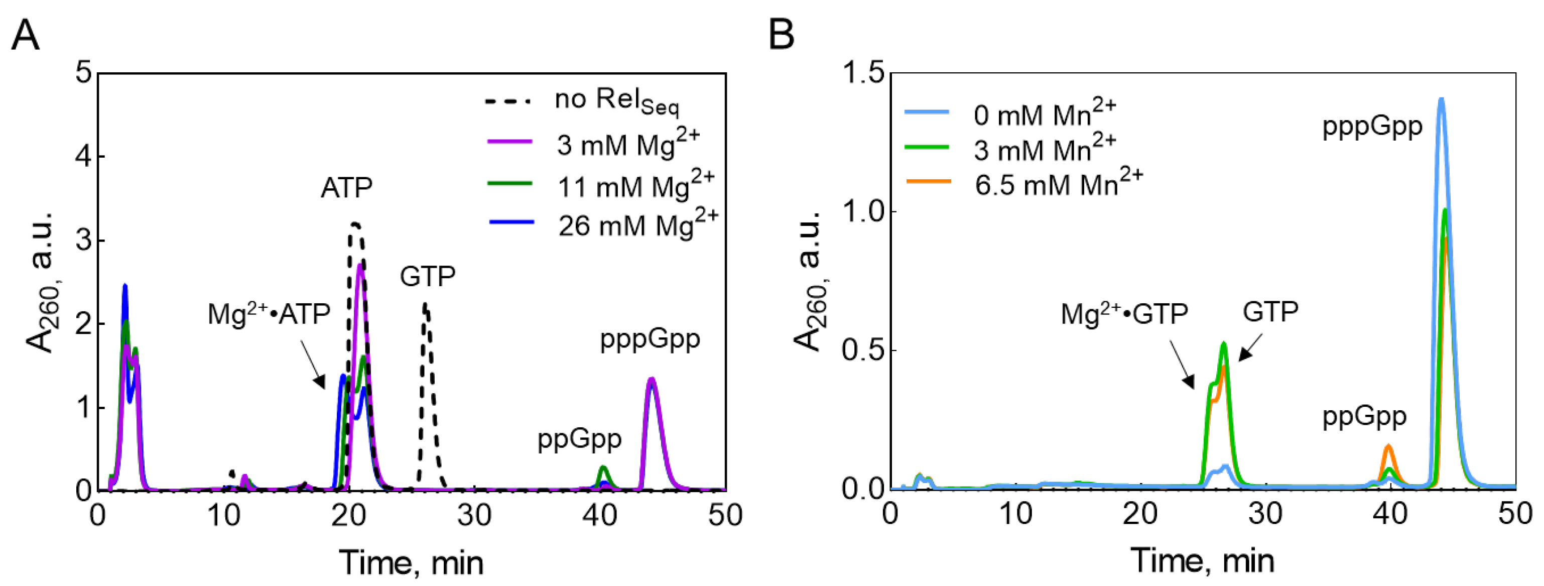
Figure 2.
Melting curves of RelSeq385 (A) and RelSeq385•pppGpp complex (B) during 7 days of incubation at 25 °C.
Figure 2.
Melting curves of RelSeq385 (A) and RelSeq385•pppGpp complex (B) during 7 days of incubation at 25 °C.
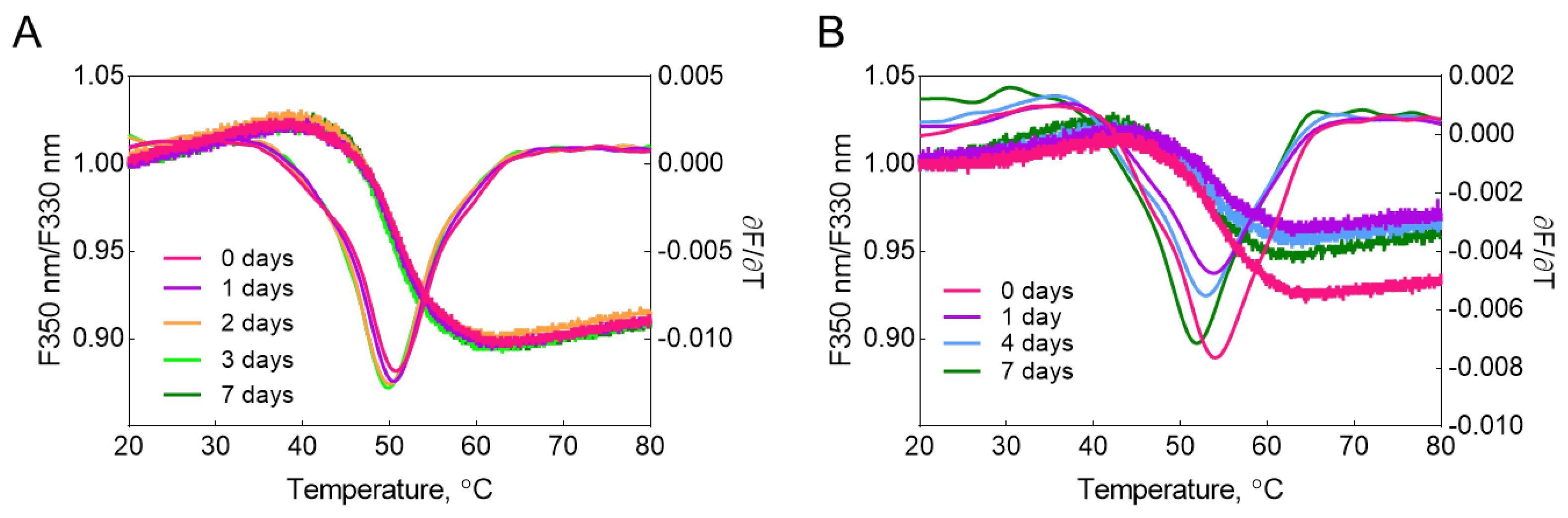
Figure 3.
(A) The overall view of the RelSeq385•pppGpp crystal structure in an asymmetric unit. The hydrolase domain (HD) is colored cyan, the central 3-helix bundle is colored light green and the synthetase domain (SYN) is colored gold. (B) Monomer A in complex with Mn2+ bound in HD and pppGpp bound in SYN. (C) Monomer B in complex with Mn2+ in HD and pppGpp bound in both HD and SYN. pppGpp molecules (sticks) are color-coded with carbon (green), oxygen (red), nitrogen (blue), and phosphorus (orange). Mn2+ ion in HD is shown as a purple sphere. For monomers A and B, the secondary structure elements of RelSeq385 are shown, with α-helices (α1–α16) and β-strands (β1–β5) numbered.
Figure 3.
(A) The overall view of the RelSeq385•pppGpp crystal structure in an asymmetric unit. The hydrolase domain (HD) is colored cyan, the central 3-helix bundle is colored light green and the synthetase domain (SYN) is colored gold. (B) Monomer A in complex with Mn2+ bound in HD and pppGpp bound in SYN. (C) Monomer B in complex with Mn2+ in HD and pppGpp bound in both HD and SYN. pppGpp molecules (sticks) are color-coded with carbon (green), oxygen (red), nitrogen (blue), and phosphorus (orange). Mn2+ ion in HD is shown as a purple sphere. For monomers A and B, the secondary structure elements of RelSeq385 are shown, with α-helices (α1–α16) and β-strands (β1–β5) numbered.
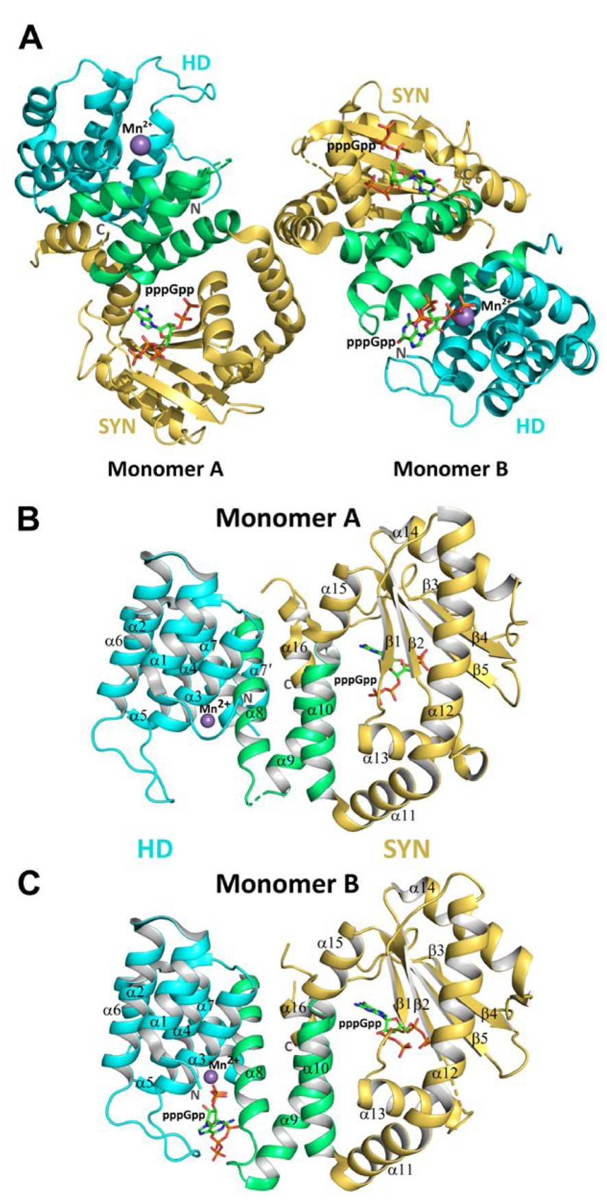
Figure 6.
MD simulations of RelSeq385 with 32 molecules of pppGpp in periodic water box with dimensions of 90x90x90A. (A) Probability maps for pppGpp positions shown on the central structure of the most probable RelSeq385 conformation based on conformational clustering. Domain coloring follows Figure 3. Residues with a minimal contact distance ≤ 4 Å are shown as violet sticks. (B) Box plot showing minimal distances between pppGpp and each protein residue (with median of minimal distance below 4 Å) observed during MD simulations. Residues whose contact distances are consistent with the crystallographic binding sites are highlighted: boxes for residues belonging to the HD and SYN active sites are colored cyan and orange, respectively; the same colors are used to box the residue labels on the x-axis.
Figure 6.
MD simulations of RelSeq385 with 32 molecules of pppGpp in periodic water box with dimensions of 90x90x90A. (A) Probability maps for pppGpp positions shown on the central structure of the most probable RelSeq385 conformation based on conformational clustering. Domain coloring follows Figure 3. Residues with a minimal contact distance ≤ 4 Å are shown as violet sticks. (B) Box plot showing minimal distances between pppGpp and each protein residue (with median of minimal distance below 4 Å) observed during MD simulations. Residues whose contact distances are consistent with the crystallographic binding sites are highlighted: boxes for residues belonging to the HD and SYN active sites are colored cyan and orange, respectively; the same colors are used to box the residue labels on the x-axis.
Disclaimer/Publisher’s Note: The statements, opinions and data contained in all publications are solely those of the individual author(s) and contributor(s) and not of MDPI and/or the editor(s). MDPI and/or the editor(s) disclaim responsibility for any injury to people or property resulting from any ideas, methods, instructions or products referred to in the content. |
© 2026 by the authors. Licensee MDPI, Basel, Switzerland. This article is an open access article distributed under the terms and conditions of the Creative Commons Attribution (CC BY) license (http://creativecommons.org/licenses/by/4.0/).
Copyright: This open access article is published under a Creative Commons CC BY 4.0 license, which permit the free download, distribution, and reuse, provided that the author and preprint are cited in any reuse.


