

Submitted:
26 April 2026
Posted:
28 April 2026
You are already at the latest version
Abstract
RNA-based therapeutics are reshaping the treatment landscape for skeletal muscle disorders by enabling modulation of RNA processing or direct correction of disease-causing alleles. In Duchenne muscular dystrophy (DMD), four antisense oligonucleotides—eteplirsen, golodirsen, viltolarsen, and casimersen—have received FDA approval; these phosphorodiamidate morpholino oligomers (PMOs) induce exon skipping to restore the reading frame and enable expression of internally truncated dystrophin. Beyond splice switching, RNA therapeutics include RNase H–active gapmers and steric-blocking antisense oligonucleotides (ASOs), small interfering RNAs (siRNAs) that mediate post-transcriptional gene silencing, and RNA-guided gene-modifying technologies such as CRISPR systems that can reframe or repair endogenous alleles. Despite major progress in DMD, broader clinical impact remains constrained by inefficient delivery to skeletal and especially cardiac muscle, the need for repeat administration for most modalities, and safety considerations that limit dose escalation and durability. Next-generation approaches aim to overcome these barriers through peptide- or antibody-conjugated oligonucleotides that enhance cellular uptake and tissue distribution, alternative chemistries with improved stability and potency, and viral or non-viral platforms for durable splice modulation. In parallel, CRISPR-based strategies—including base and prime editing—offer the prospect of one-time correction, while raising important questions regarding delivery, immunogenicity, editing specificity, and long-term safety. This review synthesizes recent advances in antisense and gene-modifying strategies for skeletal muscle and highlights practical priorities for translation, including improved muscle/heart delivery, controllable safety mechanisms, scalable manufacturing, and standardized biomarker-to-clinical outcome relationships.
Keywords:
antisense oligonucleotide
; phosphorodiamidate morpholino oligonucleotides
; small interfering RNA
; clustered regularly interspaced short palindromic repeats and CRISPR-associated protein system
; cell-penetrating peptides
; antibody–oligonucleotide conjugates
; adeno-associated viruses vector
; Duchenne muscular dystrophy
; myotonic dystrophy type 1
; facioscapulohumeral muscular dystrophy
1. Introduction of RNA-Based Therapeutics
RNA-based therapeutics comprise a broad spectrum of molecular mechanisms that enable precise modulation of gene expression at multiple levels [1]. These approaches range from antisense oligonucleotides that alter RNA processing or induce targeted degradation, to small interfering RNAs that silence transcripts through the RNA interference pathway, and RNA-guided editing systems that directly modify genomic DNA [2]. Although these modalities differ in their mechanisms, durability, and molecular targets, they collectively provide a versatile toolkit for correcting or compensating for pathogenic gene expression in skeletal muscle disorders [3].
1.1. Antisense Oligonucleotides (ASO)
ASOs are single-stranded nucleic acids that bind to RNA and modulate its function [4]. The molecular mechanism of an ASO depends on its chemical modifications and the target RNA site. RNase H–dependent ASOs, also known as “gapmers,” bind to target mRNA and induce its degradation through RNase H, which specifically cleaves DNA/RNA duplexes [5]. Another major class is RNase H–independent splice-switching oligonucleotides (SSOs), which bind to pre-mRNA and modulate splicing [6]. SSOs can incorporate artificial nucleic acids, such as phosphorodiamidate morpholino oligonucleotides (PMOs) [7]. PMOs are synthetic molecules designed to bind specific RNA sequences and block their processing or translation [8]. Unlike DNA or RNA, PMOs have an uncharged backbone, which confers high stability and resistance to nucleases. They bind strongly only to nearly perfectly complementary sequences and exhibit minimal activation of innate immune responses, limited nonspecific protein binding, and overall low toxicity [9,10].
1.2. Small Interfering RNAs (siRNA)
siRNAs are short, double-stranded RNA molecules, typically 21–23 nucleotides in length [11]. They consist of a sense strand and an antisense strand. The antisense strand is incorporated into the RNA-induced silencing complex (RISC), which guides the complex to complementary target mRNA. Once bound, RISC cleaves the target mRNA, leading to its degradation and effective gene silencing through the RNA interference (RNAi) pathway [1]. Chemical modifications and delivery strategies are often used to enhance siRNA stability, reduce off-target effects, and improve cellular uptake [12].
1.3. RNA-Guided Gene-Modifying Technologies
RNA-guided genome-editing technologies have also emerged as potential therapeutic approaches that enable direct modification of genomic DNA [13]. The clustered regularly interspaced short palindromic repeats (CRISPR) and CRISPR-associated protein system (CRISPR–Cas) is an RNA-guided technology that introduces double-strand breaks (DSBs) at target DNA sequences complementary to a guide RNA [14]. Following DSB formation, cellular DNA repair pathways can modify the genome through non-homologous end joining (NHEJ) or homology-directed repair (HDR) [15]. In the presence of a donor DNA template, HDR enables the precise insertion or correction of genetic sequences [16].
More recently developed technologies, such as base editing and prime editing, utilize engineered CRISPR systems to introduce precise genetic modifications without generating DSBs [17]. Base editors enable single-nucleotide substitutions, whereas prime editors allow targeted small insertions, deletions, or base substitutions with improved precision [18].
Adenosine deaminases acting on RNA (ADAR) are endogenous enzymes that mediate adenosine-to-inosine (A-to-I) RNA editing within double-stranded RNA structures [19]. Inosine is interpreted as guanosine during translation, thereby altering the encoded protein sequence [20]. By harnessing this endogenous system, guide RNA–directed approaches have been developed to recruit ADAR to target mRNAs and induce site-specific A-to-I RNA editing [21].
2. Delivery Strategies for RNA-Based Therapeutics
One of the challenges of RNA-based therapeutics targeting skeletal muscle is efficient delivery [22]. Systemically administered RNA molecules are rapidly cleared from circulation and show limited accumulation in target tissues, reducing therapeutic efficacy [23]. To overcome this limitation, various drug delivery strategies have been developed [8] (Figure 1).
2.1. Chemical Conjugation
Chemical conjugation is a key approach to enhance delivery [10,24]. For example, N-acetylgalactosamine (GalNAc), an amino sugar derivative, acts as a ligand for the asialoglycoprotein receptor, enabling selective uptake of ASOs and siRNAs into hepatocytes [25]. Peptides and antibodies are also commonly used for conjugation. In particular, PMOs are uncharged and poorly taken up by cells; conjugation with cell-penetrating peptides (PPMOs) markedly improves cellular delivery, although safety concerns remain [10,26]. Antibody–oligonucleotide conjugates (AOCs) also enable tissue-specific delivery such as antibodies targeting skeletal and cardiac muscle via Transferrin receptor 1 (TfR1)-mediated transcytosis [27,28].
2.2. Lipid Nanoparticles (LNPs)
LNPs are nanoscale vesicles that efficiently deliver nucleic acid therapeutics, as demonstrated by the successful delivery of mRNA vaccines against COVID-19 [29]. LNP-based platforms are now among the most promising for RNA therapeutics [30]. Notably, patisiran, an LNP-formulated siRNA targeting TTR mRNA for the treatment of ATTR amyloidosis, has received regulatory approval [31]. LNP-based delivery strategies are also being actively investigated for other RNA modalities, including antisense oligonucleotides [32].
2.3. Adeno-Associated Viruses (AAV) Vector
AAV vectors are widely used and relatively safe viral delivery systems that enable efficient transfer of genetic material into the nuclei of target cells, resulting in long-term transgene expression [33]. This sustained expression, often lasting for years, makes AAV vectors particularly suitable for therapeutic strategies requiring durable gene modulation [34]. In this context, RNA-guided gene-modifying technologies can be effectively delivered using AAV vectors by encoding the necessary components, such as guide RNAs and associated proteins, to enable long-term genome editing in target tissues [17].
2.4. U7 Small Nuclear RNA (snRNA)
snRNAs are components of small nuclear ribonucleoproteins (snRNPs), which are essential for pre-mRNA processing [35]. Among them, U7 snRNA is a specialized snRNA that functions in the 3′-end processing of replication-dependent histone pre-mRNAs [36]. Engineered U7 snRNA can be modified by replacing its natural histone-binding sequence and Sm-binding motif with antisense sequences targeting pre-mRNA [37]. In this form, U7 snRNP can modulate splicing by promoting exon skipping or inclusion, depending on the design of the antisense sequence. The main advantages of U7 snRNA-based approaches include its small genetic size, efficient nuclear localization, and generally low immunogenicity and cytotoxicity in cells [38].
3. Disease Case Studies
Emerging studies on RNA therapeutics targeting skeletal muscle diseases are rapidly expanding [39]. In particular, for Duchenne muscular dystrophy (DMD), four antisense oligonucleotides—eteplirsen, golodirsen, viltolarsen, and casimersen—have received FDA approval [40]. For other skeletal muscular disorders, such as myotonic dystrophy type 1 (DM1) and facioscapulohumeral muscular dystrophy (FCMD), RNA-based therapeutics are currently under clinical investigation [27,41].
Despite these advances, several bottlenecks remain in clinical development, including limited therapeutic efficacy, delivery challenges, toxicity concerns, and the lack of robust clinical endpoints and biomarkers [42]. In addition, therapeutic efficiency often varies depending on tissue distribution and disease context [39]. This section summarizes the current status of RNA drug development in skeletal muscle diseases and highlights emerging strategies to overcome these limitations.
3.1. Duchenne Muscular Dystrophy (DMD)
DMD is an X-linked recessive neuromuscular disorder that affects approximately 1 in 4,000–5,000 male births worldwide, with a global prevalence of ~3.6 per 100,000 individuals [43,44]. DMD is caused by mutations in the DMD gene, which encodes dystrophin, a key structural protein that stabilizes the sarcolemma during muscle contraction [45]. The absence of functional dystrophin leads to progressive muscle degeneration throughout the body, resulting in loss of ambulation typically by 10–12 years of age and premature mortality due to respiratory or cardiac failure [46]. Notably, cardiomyopathy remains a leading cause of death in patients with DMD [47].
To date, more than 7,000 distinct mutations in the DMD gene have been identified [48]. Mutation hotspots are located in exons 3–9 and 45–55 of the 79 exons, with deletions being the most common [49]. These mutations frequently disrupt the reading frame, leading to premature termination codons and loss of functional dystrophin expression [50]. RNA-based therapeutics have emerged as promising approaches for DMD because they can directly target disease-causing mutations at the RNA level. In particular, antisense oligonucleotides (ASOs) targeting pre-mRNA can modulate splicing by inducing exon skipping, thereby restoring the reading frame, and enabling the production of a truncated but partially functional dystrophin protein [51]. A schematic illustration of this mechanism is shown in Figure 2.
To date, four phosphorodiamidate morpholino oligonucleotides (PMOs) have been approved by the FDA for DMD, all based on exon-skipping mechanisms but targeting different exons: eteplirsen (exon 51), golodirsen (exon 53), viltolarsen (exon 53), and casimersen (exon 45) [52,53,54,55] (Table 1). These PMOs are administered as unconjugated (“naked”) oligonucleotides via weekly intravenous injection [40]. They act by sterically blocking splice sites on pre-mRNA, thereby inducing exon skipping to restore the reading frame and enable production of a truncated but partially functional dystrophin protein [56,57]. In clinical studies, these therapies have generally been well tolerated [58]. However, dystrophin restoration levels are typically low (often in the single-digit percentage range), and clinical benefits remain modest, with limited impact on disease progression [53,58]. One major limitation is inefficient delivery: due to their uncharged backbone, PMOs exhibit poor cellular uptake in skeletal muscle [59]. In particular, delivery to cardiac muscle is minimal, resulting in little or no therapeutic effect on cardiomyopathy, a leading cause of mortality in DMD patients [22].
Several strategies have been developed to enhance the therapeutic efficacy of PMOs in DMD. Dose escalation represents a straightforward approach; higher doses of eteplirsen (e.g., 100–200 mg/kg compared to earlier clinical doses of 30–50 mg/kg) are currently being evaluated to improve dystrophin restoration while assessing safety [43]. Another strategy involves the use of multiple PMOs targeting a single exon to enhance steric blockade of splicing regulatory elements and improve exon-skipping efficiency. For example, brogidirsen, a dual-targeting PMO designed for exon 44 skipping, is under phase 1/2 clinical evaluation and has demonstrated increased dystrophin expression, reaching approximately 10–20% in early studies (NCT05135663) [60].
Conjugation-based approaches have also shown promise. Peptide-conjugated PMOs (PPMOs) and antibody–oligonucleotide conjugates (AOC-PMOs), particularly those with antibodies against the transferrin receptor 1 (TfR1), enhance delivery to skeletal and cardiac muscle, addressing one of the major limitations of naked PMOs [28,61,62]. Several of these candidates are currently in clinical development. However, safety concerns remain, particularly for PPMOs [63]. For instance, SRP-5051 demonstrated hypomagnesemia associated with renal toxicity, leading to a temporary halt in clinical trials [9].
RNA-guided genome-editing technologies have also been investigated for the treatment of DMD. Guide RNA (gRNA)-directed recruitment of ADAR has been shown to enable targeted RNA editing and partial restoration of the reading frame [64]. In particular, ADAR-mediated editing can convert a premature stop codon (e.g., UAG) into a sense codon (e.g., UGG), thereby enabling translation of a partially functional dystrophin protein [65]. This approach has been demonstrated as a proof-of-concept in the mdx mouse model of DMD [19]. Another study has reported that embedding guide RNAs into U7 snRNA scaffolds can enhance intracellular delivery, stability, and overall efficiency of ADAR-mediated RNA editing [66].
3.2. Myotonic Dystrophy Type 1 (DM1)
DM1 is an autosomal dominant, adult-onset neuromuscular disorder with an estimated global prevalence of approximately 1 in 8,000 individuals [67]. DM1 is caused by an abnormal expansion of CTG trinucleotide repeats in the 3′ untranslated region of the DMPK gene, leading to the production of toxic CUG-repeat–containing RNA transcripts [68]. These mutant RNAs sequester RNA-binding proteins, particularly muscleblind-like (MBNL) proteins, resulting in widespread dysregulation of alternative splicing across multiple tissues [69].
Clinically, DM1 is a multisystem disorder characterized by progressive skeletal muscle weakness, cardiac conduction abnormalities, respiratory insufficiency, endocrine dysfunction, cognitive and behavioral impairments, and early-onset cataracts [70]. Among these manifestations, cardiac arrhythmias and conduction defects are major contributors to morbidity and mortality [71]. Currently, no disease-modifying therapies have been approved for DM1.
Clinical development of RNA-based therapeutics targeting the DMPK gene is ongoing. The first antisense oligonucleotide (ASO), the gapmer baliforsen, was well tolerated in patients with DM1; however, it failed to achieve sufficient concentrations in skeletal muscle, highlighting delivery as a major limitation [72]. To address this challenge, next-generation delivery approaches are being explored. Antibody–oligonucleotide conjugates (AOCs), particularly transferrin receptor 1 (TfR1)–targeting gapmer ASOs (ACHIEVE trial, NCT05481879), are currently under clinical evaluation. In addition, TfR1-targeting siRNA conjugates have been assessed in clinical trials (MARINA trial, NCT05027269), demonstrating reductions in mutant DMPK mRNA levels, increased availability of functional MBNL proteins, and partial correction of disease-associated mis-splicing in a dose-dependent manner, with trends toward improved muscle function [41].
CRISPR–Cas9–based approaches have also been investigated as therapeutic strategies for DM1 by directly targeting the expanded CTG repeats in the DMPK gene [73]. For example, in transgenic mouse models, delivery of CRISPR–Cas9 via adeno-associated virus (AAV) vectors, together with two single guide RNAs (sgRNAs) flanking the repeat region, enables excision of the expanded CTG tract [74]. This results in improvements in both molecular and functional phenotypes in skeletal and cardiac muscle [74]. In addition to repeat excision, alternative strategies have been explored to suppress DMPK expression. Targeting the DMPK promoter using CRISPR-based approaches has been reported to reduce transcription by up to 80% [75]. Although these genome-editing modalities show considerable promise, their clinical translation remains at an early stage, and further studies are required to establish their safety, delivery efficiency, and long-term effects in patients [76].
3.3. Facioscapulohumeral Muscular Dystrophy (FCMD)
FSHD is an autosomal dominant, adult-onset muscular disorder and one of the most common forms of muscular dystrophy, affecting an estimated 45,000–87,000 individuals in the United States and Europe [77]. FSHD is caused by aberrant expression of the transcription factor double homeobox 4 (DUX4), which activates a cascade of germline and pro-apoptotic genes, leading to toxicity in skeletal muscle [78]. In healthy individuals, DUX4 expression is epigenetically silenced. In FSHD type 1, contraction of the D4Z4 macrosatellite repeat array, and in FSHD type 2, mutations in epigenetic regulators such as structural maintenance of chromosomes flexible hinge domain containing 1 (SMCHD1), lead to chromatin relaxation and inappropriate DUX4 expression [79]. Although disease onset typically occurs in adolescence or early adulthood with progressive weakness of facial, scapular, and humeral muscles, approximately 7–15% of patients present in childhood with a more severe phenotype, often accompanied by hearing loss and retinal vasculopathy [80]. Currently, no disease-modifying therapies have been approved.
Antisense oligonucleotides (ASOs) targeting DUX4 mRNA have demonstrated preclinical efficacy. Gapmer ASOs have shown robust suppression of DUX4 expression in patient-derived FSHD myotubes and mouse models [81,82,83]. PMOs have also been reported to reduce DUX4 expression by sterically blocking the polyadenylation signal or coding regions of the transcript, resulting in improved pathological features in animal models [84].
To enhance delivery, antibody–oligonucleotide conjugate (AOC) strategies, particularly transferrin receptor 1 (TfR1)-targeted antibody–siRNA conjugates such as AOC 1020, have been investigated [77]. This approach reduced DUX4-regulated gene expression by approximately 75% in patient-derived myotubes and prevented muscle weakness and fibrosis following a single systemic dose in mouse models [77]. The safety and efficacy of these strategies are currently being evaluated in clinical trials.
4. Translational Priorities
RNA-based therapeutics for skeletal muscle disorders share several cross-cutting translational challenges that influence their clinical development, regardless of modality (ASO, siRNA, RNA editing), delivery strategy (peptide-conjugate, AOC, LNP, AAV, U7), or disease target (DMD, DM1, FSHD). These include the selection of predictive animal models, the establishment of robust biomarkers and clinical endpoints, the management of safety liabilities, and the development of scalable manufacturing and regulatory frameworks [85]. Addressing these priorities is essential for advancing RNA therapeutics from preclinical proof-of-concept to certain durable clinical benefit.
4.1. Animal Models
In addition to cell model, rodent models have been indispensable for mechanistic studies and early proof-of-concept in the treatment for skeletal muscle diseases [86]. However, their ability to predict human responses remains limited. For example, mdx mice exhibit mild pathology, rapid metabolism, and efficient muscle regeneration, which obscure long-term degenerative trajectories [87]. Moreover, systemic delivery of ASOs or siRNAs in mice does not accurately reflect human biodistribution challenges, particularly for cardiac and diaphragm uptake [86]. These discrepancies often lead to overestimation of therapeutic efficacy and underestimation of toxicity [88].
Large-animal models provide an essential translational bridge for RNA therapeutics. Canine DMD models (GRMD, CXMDJ) closely recapitulate human disease progression—including muscle weakness, respiratory decline, and cardiomyopathy—and have been widely used to evaluate exon-skipping ASOs and gene therapies [89,90]. Complementing canine models, the genome-edited microminipig with DMD exon 23 mutation (DMD-MMP) exhibits early locomotor deficits, progressive myocardial fibrosis, and declining left-ventricular ejection fraction, enabling longitudinal assessment of systemic dosing, imaging, and tissue biopsies [91]. Together, dogs and microminipigs provide complementary strengths for evaluating delivery, biodistribution, and chronic safety [92]. Beyond DMD, expanding large-animal resources—including non-human primates (NHPs)—is critical for assessing pharmacokinetics, immunogenicity, and tissue distribution of oligonucleotide therapeutics [93]. NHPs offer the closest approximation to human receptor biology and immune responses, making them indispensable for evaluating advanced modalities such as AOCs, LNPs, and AAV vectors [77,94].
4.2. Endpoints and Biomarkers
The development of sensitive and disease-relevant biomarkers is essential for translating RNA-based therapeutics in skeletal muscle disorders [95,96,97]. Molecular biomarkers such as exon-skipping efficiency, target transcript reduction, and correction of disease-related RNA splicing or transcriptional abnormalities provide direct measures of target engagement and RNA repair [98]. Quantification of dystrophin or other muscle proteins by Western blot or mass spectrometry remains a regulatory-accepted pharmacodynamic endpoint in DMD [99]. Circulating biomarkers, including serum creatine kinase and cardiac troponins, are widely used, while emerging markers such as myomiRs (miR-1, miR-133, miR-206) offer non-invasive readouts of muscle injury and regeneration [100]. Imaging biomarkers, particularly quantitative MRI, enable sensitive detection of muscle degeneration and fibrosis and are increasingly incorporated into clinical trials [101]. Functional endpoints such as the 6-minute walk test, North Star Ambulatory Assessment (NSAA), reachable workspace (FSHD), and myotonia/respiratory assessments (DM1) remain essential for linking molecular effects to clinical benefit [102,103,104,105]. Standardization across trials and harmonization of biomarker panels will be critical for regulatory acceptance.
4.3. Safety Considerations
Safety remains a central determinant of clinical translation for RNA therapeutics [106]. PMOs are generally well tolerated but require high systemic doses due to limited tissue uptake [52]. PPMOs improve delivery but have shown renal toxicity and hypomagnesemia in clinical studies, highlighting the need for careful monitoring [107]. Gapmer ASOs and siRNAs carry risks of unintended off-target gene silencing or toxicity related to protein interaction [108,109]. Delivery platforms introduce additional risks: AOCs may cause immunogenicity or receptor saturation; LNPs can induce complement activation or infusion reactions; and AAV vectors raise concerns regarding long-term expression, integration, and immune responses [110,111,112]. Because skeletal muscle diseases require chronic administration over years, long-term safety—particularly in cardiac and diaphragm tissues—must be evaluated in predictive large-animal models such as canine DMD and microminipigs [89,92].
4.4. Manufacturing and Regulatory Considerations
Manufacturing RNA therapeutics at clinical scale requires stringent control of chemical purity, stereochemistry, backbone integrity, and impurity profiles, particularly for PMOs, gapmers, and siRNAs [113]. Conjugated modalities such as AOCs and PPMOs demand reproducible linker chemistry and consistent drug–antibody ratios, complicating scale-up [27]. Regulatory agencies increasingly require mechanistic justification, robust biodistribution and toxicology data in two species, and standardized assays for target RNA or protein modulation [114]. Off-target assessment using genomic profiling and RNA-sequencing has become increasingly essential for evaluating the specificity and safety of RNA-based therapeutics [115,116]. Chemistry, Manufacturing, and Controls (CMC) frameworks must ensure identity, potency, stability, and batch-to-batch consistency [117]. As hybrid platforms (AOC-PMO, AOC-siRNA, AAV-RNA editors) expand, regulatory pathways must adapt to accommodate their distinct pharmacology and safety profiles.
5. Conclusions
RNA-based therapeutics have emerged as a clinically validated strategy for treating skeletal muscle disorders, supported by advances in RNA degradation, exon skipping, RNA interference, and RNA-editing technologies. Yet their successful translation requires bridging the gap between molecular efficacy and clinically meaningful outcomes. Rodent models remain indispensable for mechanistic discovery, but large-animal systems such as canine DMD models and genome-edited microminipigs provide more predictive platforms for evaluating delivery, biodistribution, and long-term safety. Progress in sensitive biomarkers, quantitative imaging, and standardized functional assessments is strengthening the ability to measure target engagement and therapeutic benefit. At the same time, careful attention to off-target effects, delivery-related toxicities, and evolving manufacturing and regulatory expectations is essential. Together, these advances are laying the foundation for safer, more effective, and more durable RNA-based therapies that can ultimately transform care for patients with neuromuscular disease.
Author Contributions
Conceptualization, T.K. and T.Y.; writing—original draft preparation, T.K.; writing—review and editing, T.K. and T.Y. All authors have read and agreed to the published version of the manuscript.
Funding
This research received no external funding. T.Y. is supported by the Friends of Garrett Cumming Research Fund, Muscular Dystrophy Canada, the Canadian Institute of Health Research (CIHR), the Women and Children’s Health Research Institute (WCHRI), the HM Toupin Neurological Science Research Fund, Alberta Innovates: Health Solutions (AIHS), Jesse’s Journey, the U.S. Department of Defense, and the Heart and Stroke Foundation Canada. T.K. is supported by research funding scholarships from Japanese Society of Neurology and Eli Lilly and Company.
Institutional Review Board Statement
Not applicable.
Informed Consent Statement
Not applicable.
Data Availability Statement
No new data were created or analyzed in this study. Data sharing is not applicable to this article.
Acknowledgments
We thank the University of Alberta Faculty of Medicine and Dentistry, the University of Alberta Faculty of Graduate and Postdoctoral Studies, the Friends of Garrett Cumming Research Fund, Muscular Dystrophy Canada, the Canadian Institute of Health Research (CIHR), the Women and Children’s Health Research Institute (WCHRI), the HM Toupin Neurological Science Research Fund, Alberta Innovates: Health Solutions (AIHS), Jesse’s Journey, and the Heart and Stroke Foundation Canada for their support.
Conflicts of Interest
T.Y. is a cofounder and shareholder of OligomicsTx Inc., which aims to commercialize antisense technology. T.K. declares that the research was conducted in the absence of any commercial or financial relationships that could be construed as a potential conflict of interest.
References
- Sparmann, A.; Vogel, J. RNA -based Medicine: From Molecular Mechanisms to Therapy. EMBO J. 2023, 42. [Google Scholar] [CrossRef] [PubMed]
- Miao, Y.; Fu, C.; Yu, Z.; Yu, L.; Tang, Y.; Wei, M. Current Status and Trends in Small Nucleic Acid Drug Development: Leading the Future. Acta Pharm. Sin. B 2024, 14, 3802–3817. [Google Scholar] [CrossRef]
- Egli, M.; Manoharan, M. Chemistry, Structure and Function of Approved Oligonucleotide Therapeutics. Nucleic Acids Res. 2023, 51, 2529–2573. [Google Scholar] [CrossRef] [PubMed]
- Bennett, C.F.; Krainer, A.R.; Cleveland, D.W. Antisense Oligonucleotide Therapies for Neurodegenerative Diseases. 2019. [Google Scholar] [CrossRef] [PubMed]
- Sardone, V.; Zhou, H.; Muntoni, F.; Ferlini, A.; Falzarano, M.S. Antisense Oligonucleotide-Based Therapy for Neuromuscular Disease. Molecules 2017, 22. [Google Scholar] [CrossRef]
- Havens, M.A.; Hastings, M.L. Splice-Switching Antisense Oligonucleotides as Therapeutic Drugs. Nucleic Acids Res. 2016, 44, 6549–6563. [Google Scholar] [CrossRef]
- Zhu, A.; Chiba, S.; Shimizu, Y.; Kunitake, K.; Okuno, Y.; Aoki, Y.; Yokota, T. Ensemble-Learning and Feature Selection Techniques for Enhanced Antisense Oligonucleotide Efficacy Prediction in Exon Skipping. Pharmaceutics 2023, 15. [Google Scholar] [CrossRef]
- Aslesh, T.; Maruyama, R.; Yokota, T. Skipping Multiple Exons to Treat DMD-Promises and Challenges. Biomedicines 2018, 6. [Google Scholar] [CrossRef]
- Sheikh, O.; Yokota, T. Pharmacology and Toxicology of Eteplirsen and SRP-5051 for DMD Exon 51 Skipping: An Update. Arch. Toxicol. 2022, 96. [Google Scholar] [CrossRef]
- Sabrina Haque, U.; Kohut, M.; Yokota, T. Comprehensive Review of Adverse Reactions and Toxicology in ASO-Based Therapies for Duchenne Muscular Dystrophy: From FDA-Approved Drugs to Peptide-Conjugated ASO. Curr. Res. Toxicol. 2024, 7. [Google Scholar] [CrossRef]
- Ebenezer, O.; Oyebamiji, A.K.; Olanlokun, J.O.; Tuszynski, J.A.; Wong, G.K.S. Recent Update on SiRNA Therapeutics. Int. J. Mol. Sci. 2025, 26. [Google Scholar] [CrossRef] [PubMed]
- Moazzam, M.; Zhang, M.; Hussain, A.; Yu, X.; Huang, J.; Huang, Y. The Landscape of Nanoparticle-Based SiRNA Delivery and Therapeutic Development. Mol. Ther. 2024, 32, 284–312. [Google Scholar] [CrossRef] [PubMed]
- Park, S.J.; Lee, G.E.; Cho, S.M.; Choi, E.H. Recent Applications, Future Perspectives, and Limitations of the CRISPR-Cas System. Mol. Ther. Nucleic Acids 2025, 36. [Google Scholar] [CrossRef]
- Jinek, M.; Chylinski, K.; Fonfara, I.; Hauer, M.; Doudna, J.A.; Charpentier, E. A Program. Dual-RNA-Guid. DNA Endonuclease Adapt. Bact. Immun.
- Lieber, M.R. The Mechanism of Human Nonhomologous DNA End Joining. J. Biol. Chem. 2008, 283, 1–5. [Google Scholar] [CrossRef]
- Mali, P.; Yang, L.; Esvelt, K.M.; Aach, J.; Guell, M.; DiCarlo, J.E.; Norville, J.E.; Church, G.M. RNA-Guided Human Genome Engineering via Cas9. Science (1979) . 2013, 339, 823–826. [Google Scholar] [CrossRef]
- Kantor, A.; McClements, M.E.; Maclaren, R.E. Crispr-Cas9 Dna Base-Editing and Prime-Editing. Int. J. Mol. Sci. 2020, 21, 1–22. [Google Scholar] [CrossRef] [PubMed]
- Anzalone, A. V.; Koblan, L.W.; Liu, D.R. Genome Editing with CRISPR–Cas Nucleases, Base Editors, Transposases and Prime Editors. Nat. Biotechnol. 2020, 38, 824–844. [Google Scholar] [CrossRef]
- Katrekar, D.; Chen, G.; Meluzzi, D.; Ganesh, A.; Worlikar, A.; Shih, Y.R.; Varghese, S.; Mali, P. In Vivo RNA Editing of Point Mutations via RNA-Guided Adenosine Deaminases. Nat. Methods 2019, 16, 239–242. [Google Scholar] [CrossRef]
- Cohen-Fultheim, R.; Twersky, I.; Krupkin, H.; Roth, S.H.; Levanon, E.Y.; Eisenberg, E. A Cytoplasmic Index for Quantifying Immune-Related A-to-I RNA Editing 2025.
- Booth, B.J.; Nourreddine, S.; Katrekar, D.; Savva, Y.; Bose, D.; Long, T.J.; Huss, D.J.; Mali, P. RNA Editing: Expanding the Potential of RNA Therapeutics. Mol. Ther. 2023, 31, 1533–1549. [Google Scholar] [CrossRef]
- Echigoya, Y.; Nakamura, A.; Nagata, T.; Urasawa, N.; Lim, K.R.Q.; Trieu, N.; Panesar, D.; Kuraoka, M.; Moulton, H.M.; Saito, T.; et al. Effects of Systemic Multiexon Skipping with Peptide-Conjugated Morpholinos in the Heart of a Dog Model of Duchenne Muscular Dystrophy. Proc. Natl. Acad. Sci. U. S. A. 2017, 114, 4213–4218. [Google Scholar] [CrossRef]
- Paunovska, K.; Loughrey, D.; Dahlman, J.E. Drug Delivery Systems for RNA Therapeutics. Nat. Rev. Genet. 2022, 23, 265–280. [Google Scholar] [CrossRef] [PubMed]
- Debacker, A.J.; Voutila, J.; Catley, M.; Blakey, D.; Habib, N. Delivery of Oligonucleotides to the Liver with GalNAc: From Research to Registered Therapeutic Drug. Mol. Ther. 2020, 28, 1759–1771. [Google Scholar] [CrossRef]
- Springer, A.D.; Dowdy, S.F. GalNAc-SiRNA Conjugates: Leading the Way for Delivery of RNAi Therapeutics. Nucleic Acid. Ther. 2018, 28, 109–118. [Google Scholar] [CrossRef]
- Leckie, J.; Yokota, T. Potential of Cell-Penetrating Peptide-Conjugated Antisense Oligonucleotides for the Treatment of SMA. Molecules 2024, 29. [Google Scholar] [CrossRef]
- Malecova, B.; Burke, R.S.; Cochran, M.; Hood, M.D.; Johns, R.; Kovach, P.R.; Doppalapudi, V.R.; Erdogan, G.; Arias, J.D.; Darimont, B.; et al. Targeted Tissue Delivery of RNA Therapeutics Using Antibody-Oligonucleotide Conjugates (AOCs). Nucleic Acids Res. 2023, 51, 5901–5910. [Google Scholar] [CrossRef]
- Etxaniz, U.; Marks, I.; Albin, T.; Diaz, M.; Bhardwaj, R.; Anderson, A.; Tyaglo, O.; Hoang, T.; Missinato, M.A.; Svensson, K.; et al. AOC 1044 Induces Exon 44 Skipping and Restores Dystrophin Protein in Preclinical Models of Duchenne Muscular Dystrophy. Nucleic Acids Res. 2025, 53. [Google Scholar] [CrossRef]
- Schoenmaker, L.; Witzigmann, D.; Kulkarni, J.A.; Verbeke, R.; Kersten, G.; Jiskoot, W.; Crommelin, D.J.A. MRNA-Lipid Nanoparticle COVID-19 Vaccines: Structure and Stability. Int. J. Pharm. 2021, 601. [Google Scholar] [CrossRef] [PubMed]
- Navid Talemi, M.; Ramezani Farani, M.; Alipour Eskandani, N.; Mirzaee, D.; Alipourfard, I.; Huh, Y.S. Programmable Lipid Nanoparticles for RNA Therapeutics: Design Principles and Clinical Translation. Mater. Today Bio 2026, 37, 102774. [Google Scholar] [CrossRef] [PubMed]
- Adams, D.; Gonzalez-Duarte, A.; O’Riordan, W.D.; Yang, C.-C.; Ueda, M.; Kristen, A. V.; Tournev, I.; Schmidt, H.H.; Coelho, T.; Berk, J.L.; et al. Patisiran, an RNAi Therapeutic, for Hereditary Transthyretin Amyloidosis. N. Engl. J. Med. 2018, 379, 11–21. [Google Scholar] [CrossRef]
- Jung, H.N.; Lee, S.Y.; Lee, S.; Youn, H.; Im, H.J. Lipid Nanoparticles for Delivery of RNA Therapeutics: Current Status and the Role of in Vivo Imaging. Theranostics 2022, 12, 7509–7531. [Google Scholar] [CrossRef]
- Pozdniakova, N.; Generalov, E.; Shevelev, A.; Tarasova, O. RNA Therapeutics: Delivery Problems and Solutions—A Review. Pharmaceutics 2025, 17. [Google Scholar] [CrossRef]
- Wang, J.H.; Gessler, D.J.; Zhan, W.; Gallagher, T.L.; Gao, G. Adeno-Associated Virus as a Delivery Vector for Gene Therapy of Human Diseases. Signal Transduct. Target. Ther. 2024, 9. [Google Scholar] [CrossRef] [PubMed]
- Gadgil, A.; Raczyńska, K.D. U7 SnRNA: A Tool for Gene Therapy. J. Gene Med. 2021, 23. [Google Scholar] [CrossRef]
- Ideue, T.; Adachi, S.; Naganuma, T.; Tanigawa, A.; Natsume, T.; Hirose, T. U7 Small Nuclear Ribonucleoprotein Represses Histone Gene Transcription in Cell Cycle-Arrested Cells. Proc. Natl. Acad. Sci. U. S. A. 2012, 109, 5693–5698. [Google Scholar] [CrossRef] [PubMed]
- Lesman, D.; Rodriguez, Y.; Rajakumar, D.; Wein, N. U7 SnRNA, a Small RNA with a Big Impact in Gene Therapy. Hum. Gene Ther. 2021, 32, 1317–1329. [Google Scholar] [CrossRef]
- Vulin, A.; Barthélémy, I.; Goyenvalle, A.; Thibaud, J.L.; Beley, C.; Griffith, G.; Benchaouir, R.; Le Hir, M.; Unterfinger, Y.; Lorain, S.; et al. Muscle Function Recovery in Golden Retriever Muscular Dystrophy after AAV1-U7 Exon Skipping. Mol. Ther. 2012, 20, 2120–2133. [Google Scholar] [CrossRef]
- Bubenik, J.L.; Scotti, M.M.; Swanson, M.S. Therapeutic Targeting of RNA for Neurological and Neuromuscular Disease. Genes. Dev. 2024, 38, 698–717. [Google Scholar] [CrossRef] [PubMed]
- Eser, G.; Topaloğlu, H. Current Outline of Exon Skipping Trials in Duchenne Muscular Dystrophy. Genes . 2022, 13. [Google Scholar] [CrossRef]
- Kwan, T.T.; Meng, Q.; Delos Santos, N.; Johnson, N.E.; Thornton, C.A.; Malecova, B.; Burke, R.; Tai, L.J.; Levin, A.A.; Younis, H.S.; et al. Delpacibart Etedesiran Improves the Molecular Pathology of Myotonic Dystrophy Type 1 in the Phase 1/2 MARINA Study. Mol. Ther. 2026. [Google Scholar] [CrossRef] [PubMed]
- De Serres-Bérard, T.; Ait Benichou, S.; Jauvin, D.; Boutjdir, M.; Puymirat, J.; Chahine, M. Recent Progress and Challenges in the Development of Antisense Therapies for Myotonic Dystrophy Type 1. Int. J. Mol. Sci. 2022, 23. [Google Scholar] [CrossRef] [PubMed]
- Chassin, A.; Ono, H.; Ashida, Y.; Imamura, M.; Aoki, Y. RNA Therapeutics for Duchenne Muscular Dystrophy: Exon Skipping, RNA Editing, and Translational Insights from Genome-Edited Microminipig Models. Int. J. Mol. Sci. 2026, 27. [Google Scholar] [CrossRef]
- Salari, N.; Fatahi, B.; Valipour, E.; Kazeminia, M.; Fatahian, R.; Kiaei, A.; Shohaimi, S.; Mohammadi, M. Global Prevalence of Duchenne and Becker Muscular Dystrophy: A Systematic Review and Meta-Analysis. J. Orthop. Surg. Res. 2022, 17. [Google Scholar] [CrossRef]
- Hoffman, E.P.; Brown, R.H.; Kunkel, L.M. Dystmphin: The Protein Product of the Duchenne Muscular Dystrophy Locus; 1987; Vol. 51. [Google Scholar]
- Birnkrant, D.J.; Bushby, K.; Bann, C.M.; Apkon, S.D.; Blackwell, A.; Brumbaugh, D.; Case, L.E.; Clemens, P.R.; Hadjiyannakis, S.; Pandya, S.; et al. Diagnosis and Management of Duchenne Muscular Dystrophy, Part 1: Diagnosis, and Neuromuscular, Rehabilitation, Endocrine, and Gastrointestinal and Nutritional Management. Lancet Neurol. 2018, 17, 251–267. [Google Scholar] [CrossRef] [PubMed]
- D’Ambrosio, E.S.; Mendell, J.R. Evolving Therapeutic Options for the Treatment of Duchenne Muscular Dystrophy. Neurotherapeutics 2023, 20, 1669–1681. [Google Scholar] [CrossRef]
- Bladen, C.L.; Salgado, D.; Monges, S.; Foncuberta, M.E.; Kekou, K.; Kosma, K.; Dawkins, H.; Lamont, L.; Roy, A.J.; Chamova, T.; et al. The TREAT-NMD DMD Global Database: Analysis of More than 7,000 Duchenne Muscular Dystrophy Mutations. Hum. Mutat. 2015, 36, 395–402. [Google Scholar] [CrossRef]
- Leckie, J.; Zia, A.; Yokota, T. An Updated Analysis of Exon-Skipping Applicability for Duchenne Muscular Dystrophy Using the UMD-DMD Database. Genes . 2024, 15. [Google Scholar] [CrossRef]
- Bez Batti Angulski, A.; Hosny, N.; Cohen, H.; Martin, A.A.; Hahn, D.; Bauer, J.; Metzger, J.M. Duchenne Muscular Dystrophy: Disease Mechanism and Therapeutic Strategies. Front. Physiol. 2023, 14. [Google Scholar] [CrossRef]
- Takeda, S.; Clemens, P.R.; Hoffman, E.P. Exon-Skipping in Duchenne Muscular Dystrophy. J. Neuromuscul. Dis. 2021, 8, S343–S358. [Google Scholar] [CrossRef]
- Mendell, J.R.; Rodino-Klapac, L.R.; Sahenk, Z.; Roush, K.; Bird, L.; Lowes, L.P.; Alfano, L.; Gomez, A.M.; Lewis, S.; Kota, J.; et al. Eteplirsen for the Treatment of Duchenne Muscular Dystrophy. Ann. Neurol. 2013, 74, 637–647. [Google Scholar] [CrossRef]
- Frank, D.E.; Schnell, F.J.; Akana, C.; El-Husayni, S.H.; Desjardins, C.A.; Morgan, J.; Charleston, J.S.; Sardone, V.; Domingos, J.; Dickson, G.; et al. Increased Dystrophin Production with Golodirsen in Patients with Duchenne Muscular Dystrophy. Neurology 2020, 94, E2270–E2282. [Google Scholar] [CrossRef]
- Harper, A.D.; Topaloglu, H.; Mercuri, E.; Suslov, V.; Wu, L.; Ayanoglu, C.Y.; Tansey, M.; Previtera, M.L.; Crozier, R.A.; Magnus, L.; et al. Safety and Efficacy of Viltolarsen in Ambulatory and Nonambulatory Males with Duchenne Muscular Dystrophy. Sci. Rep. 2024, 14. [Google Scholar] [CrossRef]
- Assefa, M.; Gepfert, A.; Zaheer, M.; Hum, J.M.; Skinner, B.W. Casimersen (AMONDYS 45TM): An Antisense Oligonucleotide for Duchenne Muscular Dystrophy. Biomedicines 2024, 12. [Google Scholar] [CrossRef]
- Torres-Masjoan, L.; Aguti, S.; Zhou, H.; Muntoni, F. Clinical Applications of Exon-Skipping Antisense Oligonucleotides in Neuromuscular Diseases. Mol. Ther. 2025, 33, 2689–2704. [Google Scholar] [CrossRef] [PubMed]
- Arechavala-Gomeza, V.; López-Martínez, A.; Aartsma-Rus, A. Antisense RNA Therapies for Muscular Dystrophies. J. Neuromuscul. Dis. 2025. [Google Scholar] [CrossRef] [PubMed]
- Clemens, P.R.; Rao, V.K.; Connolly, A.M.; Harper, A.D.; Mah, J.K.; Smith, E.C.; McDonald, C.M.; Zaidman, C.M.; Morgenroth, L.P.; Osaki, H.; et al. Safety, Tolerability, and Efficacy of Viltolarsen in Boys with Duchenne Muscular Dystrophy Amenable to Exon 53 Skipping: A Phase 2 Randomized Clinical Trial. JAMA Neurol. 2020, 77, 982–991. [Google Scholar] [CrossRef]
- Wang, B.; Cao, J.; Wu, J.; Zhao, Y.; Zhang, Y.; Abendroth, F.; Lin, C.; Zhong, L.; Yu, H.; Seow, Y.; et al. Cardiac and Skeletal Muscle Delivery of Biotherapeutics with a Blood Vessel Epicardial Substance-Targeting Peptide. Biomaterials 2026, 329. [Google Scholar] [CrossRef]
- Komaki, H.; Takeshita, E.; Kunitake, K.; Ishizuka, T.; Shimizu-Motohashi, Y.; Ishiyama, A.; Sasaki, M.; Yonee, C.; Maruyama, S.; Hida, E.; et al. Phase 1/2 Trial of Brogidirsen: Dual-Targeting Antisense Oligonucleotides for Exon 44 Skipping in Duchenne Muscular Dystrophy. Cell. Rep. Med. 2025, 6. [Google Scholar] [CrossRef]
- Gan, L.; Wu, L.C.L.; Wood, J.A.; Yao, M.; Treleaven, C.M.; Estrella, N.L.; Wentworth, B.M.; Hanson, G.J.; Passini, M.A. A Cell-Penetrating Peptide Enhances Delivery and Efficacy of Phosphorodiamidate Morpholino Oligomers in Mdx Mice. Mol. Ther. Nucleic Acids 2022, 30, 17–27. [Google Scholar] [CrossRef]
- Shah, M.N.A.; Wilton-Clark, H.; Haque, F.; Powell, B.; Sutanto, L.E.; Maradiya, R.; Zhabyeyev, P.; Roshmi, R.R.; Anwar, S.; Aslesh, T.; et al. DG9 Boosts PMO Nuclear Uptake and Exon Skipping to Restore Dystrophic Muscle and Cardiac Function. Nat. Commun. 2025, 16. [Google Scholar] [CrossRef] [PubMed]
- Haque, U.S.; Yokota, T. Enhancing Antisense Oligonucleotide-Based Therapeutic Delivery with DG9, a Versatile Cell-Penetrating Peptide. Cells 2023, 12. [Google Scholar] [CrossRef] [PubMed]
- Finkel, R.S. Read-through Strategies for Suppression of Nonsense Mutations in Duchenne/Becker Muscular Dystrophy: Aminoglycosides and Ataluren (PTC124). J. Child. Neurol. 2010, 25, 1158–1164. [Google Scholar] [CrossRef]
- Chen, G.; Katrekar, D.; Mali, P. RNA-Guided Adenosine Deaminases: Advances and Challenges for Therapeutic RNA Editing. Biochemistry 2019, 58, 1947–1957. [Google Scholar] [CrossRef]
- Byrne, S.M.; Burleigh, S.M.; Fragoza, R.; Jiang, Y.; Savva, Y.; Pabon, R.; Kania, E.; Rainaldi, J.; Portell, A.; Mali, P.; et al. An Engineered U7 Small Nuclear RNA Scaffold Greatly Increases ADAR-Mediated Programmable RNA Base Editing. Nat. Commun. 2025, 16. [Google Scholar] [CrossRef]
- Liao, Q.; Zhang, Y.; He, J.; Huang, K. Global Prevalence of Myotonic Dystrophy: An Updated Systematic Review and Meta-Analysis. Neuroepidemiology 2022, 56, 163–173. [Google Scholar] [CrossRef]
- Takahashi, M.P. Update on the Clinical and Therapeutic Aspects of Myotonic Dystrophy Type 1. Curr. Opin. Neurol. 2025, 38, 531–537. [Google Scholar] [CrossRef]
- Nezu, Y.; Kino, Y.; Sasagawa, N.; Nishino, I.; Ishiura, S. Expression of MBNL and CELF MRNA Transcripts in Muscles with Myotonic Dystrophy. Neuromuscul. Disord. 2007, 17, 306–312. [Google Scholar] [CrossRef]
- la Fontaine, L.A.; Bruijnes, J.E.; Smulders, F.H.; Gorissen-Brouwers, C.; Karnebeek, I.E.; Braakman, H.M.; Klinkenberg, S.; Mul, K.; ‘t Hoen, P.B.A.; van Kuijk, S.M.; et al. Comprehensive Four-Year Disease Progression Assessment of Myotonic Dystrophy Type 1. Neuromuscul. Disord. 2024, 43, 44–52. [Google Scholar] [CrossRef]
- Ginjupalli, V.K.M.; Reisqs, J.B.; Cupelli, M.; Chahine, M.; Boutjdir, M. Cardiac Involvement in Myotonic Dystrophy Type 1: Mechanisms, Clinical Perspectives, and Emerging Therapeutic Strategies. Int. J. Mol. Sci. 2025, 26. [Google Scholar] [CrossRef] [PubMed]
- Thornton, C.A.; Moxley, R.T.; Eichinger, K.; Heatwole, C.; Mignon, L.; Arnold, W.D.; Ashizawa, T.; Day, J.W.; Dent, G.; Tanner, M.K.; et al. Antisense Oligonucleotide Targeting DMPK in Patients with Myotonic Dystrophy Type 1: A Multicentre, Randomised, Dose-Escalation, Placebo-Controlled, Phase 1/2a Trial. Lancet Neurol. 2023, 22, 218–228. [Google Scholar] [CrossRef]
- Raaijmakers, R.H.L.; Ripken, L.; Ausems, C.R.M.; Wansink, D.G. CRISPR/Cas Applications in Myotonic Dystrophy: Expanding Opportunities. Int. J. Mol. Sci. 2019, 20. [Google Scholar] [CrossRef]
- Izzo, M.; Battistini, J.; Golini, E.; Voellenkle, C.; Provenzano, C.; Orsini, T.; Strimpakos, G.; Scavizzi, F.; Raspa, M.; Baci, D.; et al. Muscle-specific Gene Editing Improves Molecular and Phenotypic Defects in a Mouse Model of Myotonic Dystrophy Type 1. Clin. Transl. Med. 2025, 15. [Google Scholar] [CrossRef]
- Porquet, F.; Weidong, L.; Jehasse, K.; Gazon, H.; Kondili, M.; Blacher, S.; Massotte, L.; Di Valentin, E.; Furling, D.; Gillet, N.A.; et al. Specific DMPK-Promoter Targeting by CRISPRi Reverses Myotonic Dystrophy Type 1-Associated Defects in Patient Muscle Cells. Mol. Ther. Nucleic Acids 2023, 32, 857–871. [Google Scholar] [CrossRef]
- Laurent, M.; Geoffroy, M.; Pavani, G.; Guiraud, S. CRISPR-Based Gene Therapies: From Preclinical to Clinical Treatments. Cells 2024, 13. [Google Scholar] [CrossRef] [PubMed]
- Malecova, B.; Sala, D.; Melikian, G.M.; Johns, R.; Erdogan, G.; Hartmann, M.; Jordan, M.; Arias, J.D.; Bhattacharya, A.; Meng, Q.; et al. Development of a DUX4-Targeting Antibody Oligonucleotide Conjugate as a Therapy for FSHD. Nucleic Acids Res. 2026, 54. [Google Scholar] [CrossRef]
- Geng, L.N.; Yao, Z.; Snider, L.; Fong, A.P.; Cech, J.N.; Young, J.M.; vanderMaarel, S.M.; Ruzzo, W.L.; Gentleman, R.C.; Tawil, R.; et al. DUX4 Activates Germline Genes, Retroelements, and Immune Mediators: Implications for Facioscapulohumeral Dystrophy. Dev. Cell. 2012, 22, 38–51. [Google Scholar] [CrossRef] [PubMed]
- Statland, J.; Tawil, R. Facioscapulohumeral Muscular Dystrophy. Neurol. Clin. 2014, 32, 721–728. [Google Scholar] [CrossRef] [PubMed]
- Chen, T.H.; Wu, Y.Z.; Tseng, Y.H. Early-Onset Infantile Facioscapulohumeral Muscular Dystrophy: A Timely Review. Int. J. Mol. Sci. 2020, 21, 1–17. [Google Scholar] [CrossRef]
- Zhang, A.; Lim, K.R.Q.; Chen, Z.; Yokota, T.; Chen, Y.-W. DUX4 Reduction and Muscle Function Improvement by Subcutaneous Delivery of Gapmer Antisense Oligonucleotides. Mol. Ther. Nucleic Acids 2026, 37, 102791. [Google Scholar] [CrossRef]
- Rowel, K.; Lim, Q.; Maruyama, R.; Echigoya, Y.; Nguyen, Q.; Zhang, A.; Khawaja, H.; Chandra, S.; Jones, T.; Jones, P.; et al. Inhibition of DUX4 Expression with Antisense LNA Gapmers as a Therapy for Facioscapulohumeral Muscular Dystrophy. [CrossRef]
- Bouwman, L.F.; den Hamer, B.; van den Heuvel, A.; Franken, M.; Jackson, M.; Dwyer, C.A.; Tapscott, S.J.; Rigo, F.; van der Maarel, S.M.; de Greef, J.C. Systemic Delivery of a DUX4-Targeting Antisense Oligonucleotide to Treat Facioscapulohumeral Muscular Dystrophy. Mol. Ther. Nucleic Acids 2021, 26, 813–827. [Google Scholar] [CrossRef]
- Lu-Nguyen, N.; Malerba, A.; Herath, S.; Dickson, G.; Popplewell, L. Systemic Antisense Therapeutics Inhibiting DUX4 Expression Ameliorates FSHD-like Pathology in an FSHD Mouse Model. Hum. Mol. Genet. 2021, 30, 1398–1412. [Google Scholar] [CrossRef]
- Makkar, S.K. Advances in RNA-Based Therapeutics: Current Breakthroughs, Clinical Translation, and Future Perspectives. Front. Genet. 2025, 16. [Google Scholar] [CrossRef]
- McGreevy, J.W.; Hakim, C.H.; McIntosh, M.A.; Duan, D. Animal Models of Duchenne Muscular Dystrophy: From Basic Mechanisms to Gene Therapy. DMM Dis. Model. Mech. 2015, 8, 195–213. [Google Scholar] [CrossRef]
- Canonico, F.; Chirivi, M.; Maiullari, F.; Milan, M.; Rizzi, R.; Arcudi, A.; Galli, M.; Pane, M.; Gowran, A.; Pompilio, G.; et al. Focus on the Road to Modelling Cardiomyopathy in Muscular Dystrophy. Cardiovasc. Res. 2022, 118, 1872–1884. [Google Scholar] [CrossRef]
- Partridge, T.A. The Mdx Mouse Model as a Surrogate for Duchenne Muscular Dystrophy. FEBS J. 2013, 280, 4177–4186. [Google Scholar] [CrossRef]
- Kornegay, J.N.; Bogan, J.R.; Bogan, D.J.; Childers, M.K.; Li, J.; Nghiem, P.; Detwiler, D.A.; Larsen, C.A.; Grange, R.W.; Bhavaraju-Sanka, R.K.; et al. Canine Models of Duchenne Muscular Dystrophy and Their Use in Therapeutic Strategies. Mamm. Genome 2012, 23, 85–108. [Google Scholar] [CrossRef]
- Nakamura, A.; Takeda, S. Mammalian Models of Duchenne Muscular Dystrophy: Pathological Characteristics and Therapeutic Applications. J. Biomed. Biotechnol. 2011. [Google Scholar] [CrossRef] [PubMed]
- Otake, M.; Imamura, M.; Enya, S.; Kangawa, A.; Shibata, M.; Ozaki, K.; Kimura, K.; Ono, E.; Aoki, Y. Severe Cardiac and Skeletal Manifestations in DMD-Edited Microminipigs: An Advanced Surrogate for Duchenne Muscular Dystrophy. Commun. Biol. 2024, 7. [Google Scholar] [CrossRef] [PubMed]
- Echigoya, Y.; Trieu, N.; Duddy, W.; Moulton, H.M.; Yin, H.; Partridge, T.A.; Hoffman, E.P.; Kornegay, J.N.; Rohret, F.A.; Rogers, C.S.; et al. A Dystrophin Exon-52 Deleted Miniature Pig Model of Duchenne Muscular Dystrophy and Evaluation of Exon Skipping. Int. J. Mol. Sci. 2021, 22. [Google Scholar] [CrossRef]
- Jafar-Nejad, P.; Powers, B.; Soriano, A.; Zhao, H.; Norris, D.A.; Matson, J.; Debrosse-Serra, B.; Watson, J.; Narayanan, P.; Chun, S.J.; et al. The Atlas of RNase H Antisense Oligonucleotide Distribution and Activity in the CNS of Rodents and Non-Human Primates Following Central Administration. Nucleic Acids Res. 2021, 49, 657–673. [Google Scholar] [CrossRef]
- Kondratov, O.; Kondratova, L.; Mandel, R.J.; Coleman, K.; Savage, M.A.; Gray-Edwards, H.L.; Ness, T.J.; Rodriguez-Lebron, E.; Bell, R.D.; Rabinowitz, J.; et al. A Comprehensive Study of a 29-Capsid AAV Library in a Non-Human Primate Central Nervous System. Mol. Ther. 2021, 29, 2806–2820. [Google Scholar] [CrossRef]
- Molinaro, M.; Torrente, Y.; Villa, C.; Farini, A. Advancing Biomarker Discovery and Therapeutic Targets in Duchenne Muscular Dystrophy: A Comprehensive Review. Int. J. Mol. Sci. 2024, 25. [Google Scholar] [CrossRef]
- Coenen-Stass, A.M.L.; Mcclorey, G.; Manzano, R.; Betts, C.A.; Blain, A.; Saleh, A.F.; Gait, M.J.; Lochmüller, H.; Wood, M.J.A.; Roberts, T.C. Identification of Novel, Therapy-Responsive Protein Biomarkers in a Mouse Model of Duchenne Muscular Dystrophy by Aptamer-Based Serum Proteomics. Sci. Rep. 2015, 5. [Google Scholar] [CrossRef]
- Coenen-Stass, A.M.L.; Sork, H.; Gatto, S.; Godfrey, C.; Bhomra, A.; Krjutškov, K.; Hart, J.R.; Westholm, J.O.; O’Donovan, L.; Roos, A.; et al. Comprehensive RNA-Sequencing Analysis in Serum and Muscle Reveals Novel Small RNA Signatures with Biomarker Potential for DMD. Mol. Ther. Nucleic Acids 2018, 13, 1–15. [Google Scholar] [CrossRef] [PubMed]
- Fortunato, F.; Ferlini, A. Biomarkers in Duchenne Muscular Dystrophy: Current Status and Future Directions. J. Neuromuscul. Dis. 2023, 10, 987–1002. [Google Scholar] [CrossRef] [PubMed]
- From The Dubowitz Neuromuscular Centre, ( K A Dystrophin Quantification Biological and Translational Research Implications; 2014.
- Siracusa, J.; Koulmann, N.; Banzet, S. Circulating MyomiRs: A New Class of Biomarkers to Monitor Skeletal Muscle in Physiology and Medicine. J. Cachexia Sarcopenia Muscle 2018, 9, 20–27. [Google Scholar] [CrossRef]
- Andersen, G.; Dahlqvist, J.R.; Vissing, C.R.; Heje, K.; Thomsen, C.; Vissing, J. MRI as Outcome Measure in Facioscapulohumeral Muscular Dystrophy: 1-Year Follow-up of 45 Patients. J. Neurol. 2017, 264, 438–447. [Google Scholar] [CrossRef] [PubMed]
- Yoon, D.Y.; Daniels, M.J.; Willcocks, R.J.; Triplett, W.T.; Morales, J.F.; Walter, G.A.; Rooney, W.D.; Vandenborne, K.; Kim, S. Five Multivariate Duchenne Muscular Dystrophy Progression Models Bridging Six-Minute Walk Distance and MRI Relaxometry of Leg Muscles. J. Pharmacokinet. Pharmacodyn. 2024, 51, 671–683. [Google Scholar] [CrossRef]
- Mcdonald, C.M.; Henricson, E.K.; Abresch, R.T.; Florence, J.; Eagle, M.; Gappmaier, E.; Glanzman, A.M.; Spiegel, R.; Barth, J.; Elfring, G.; et al. The 6-Minute Walk Test and Other Clinical Endpoints in Duchenne Muscular Dystrophy: Reliability, Concurrent Validity, and Minimal Clinically Important Differences from a Multicenter Study. Muscle Nerve 2013, 48, 357–368. [Google Scholar] [CrossRef] [PubMed]
- Han, J.J.; De Bie, E.; Nicorici, A.; Abresch, R.T.; Bajcsy, R.; Kurillo, G. Reachable Workspace Reflects Dynamometer-Measured Upper Extremity Strength in Facioscapulohumeral Muscular Dystrophy. Muscle Nerve 2015, 52, 948–955. [Google Scholar] [CrossRef]
- Heatwole, C.; Bode, R.; Johnson, N.E.; Dekdebrun, J.; Dilek, N.; Eichinger, K.; Hilbert, J.E.; Logigian, E.; Luebbe, E.; Martens, W.; et al. Myotonic Dystrophy Health Index: Correlations with Clinical Tests and Patient Function. Muscle Nerve 2016, 53, 183–190. [Google Scholar] [CrossRef]
- Alhamadani, F.; Zhang, K.; Parikh, R.; Wu, H.; Rasmussen, T.P.; Bahal, R.; Zhong, X.B.; Manautou, J.E. Adverse Drug Reactions and Toxicity of the Food and Drug Administration–Approved Antisense Oligonucleotide Drugs. Drug. Metab. Dispos. 2022, 50, 879–887. [Google Scholar] [CrossRef]
- Sheikh, O.; Yokota, T. Pharmacology and Toxicology of Eteplirsen and SRP-5051 for DMD Exon 51 Skipping: An Update. Arch. Toxicol. 2022, 96. [Google Scholar] [CrossRef]
- Shen, W.; De Hoyos, C.L.; Migawa, M.T.; Vickers, T.A.; Sun, H.; Low, A.; Bell, T.A.; Rahdar, M.; Mukhopadhyay, S.; Hart, C.E.; et al. Chemical Modification of PS-ASO Therapeutics Reduces Cellular Protein-Binding and Improves the Therapeutic Index. Nat. Biotechnol. 2019, 37, 640–650. [Google Scholar] [CrossRef] [PubMed]
- Jackson, A.L.; Bartz, S.R.; Schelter, J.; Kobayashi, S. V.; Burchard, J.; Mao, M.; Li, B.; Cavet, G.; Linsley, P.S. Expression Profiling Reveals Off-Target Gene Regulation by RNAi. Nat. Biotechnol. 2003, 21, 635–637. [Google Scholar] [CrossRef] [PubMed]
- Ranjbar, S.; Zhong, X. bo; Manautou, J.; Lu, X. A Holistic Analysis of the Intrinsic and Delivery-Mediated Toxicity of SiRNA Therapeutics. Adv. Drug. Deliv. Rev. 2023, 201. [Google Scholar] [CrossRef]
- Dézsi, L.; Mészáros, T.; Kozma, G.; H-Velkei, M.; Oláh, C.Z.; Szabó, M.; Patkó, Z.; Fülöp, T.; Hennies, M.; Szebeni, M.; et al. A Naturally Hypersensitive Porcine Model May Help Understand the Mechanism of COVID-19 MRNA Vaccine-Induced Rare (Pseudo) Allergic Reactions: Complement Activation as a Possible Contributing Factor. Geroscience 2022, 44, 597–618. [Google Scholar] [CrossRef] [PubMed]
- Mingozzi, F.; High, K.A. Immune Responses to AAV Vectors: Overcoming Barriers to Successful Gene Therapy. Blood 2013, 122, 23–36. [Google Scholar] [CrossRef]
- Singh, A.; Tekade, M.; Nagaraja, S.; Bharti, A.; Tekade, R.K. Advancements in RNA-Based Therapies from Bench to Bedside. Npj Drug. Discov. 2026, 3, 4. [Google Scholar] [CrossRef]
- Hammond, S.M.; Aartsma-Rus, A.; Alves, S.; Borgos, S.E.; Buijsen, R.A.M.; Collin, R.W.J.; Covello, G.; Denti, M.A.; Desviat, L.R.; Echevarría, L.; et al. Delivery of Oligonucleotide-based Therapeutics: Challenges and Opportunities. EMBO Mol. Med. 2021, 13. [Google Scholar] [CrossRef]
- Michel, S.; Schirduan, K.; Shen, Y.; Klar, R.; Tost, J.; Jaschinski, F. Using RNA-Seq to Assess Off-Target Effects of Antisense Oligonucleotides in Human Cell Lines. Mol. Diagn. Ther. 2021, 25, 77–85. [Google Scholar] [CrossRef]
- Ruan, H.; Dou, D.; Lu, J.; Xiao, X.; Gong, X.; Zhang, X. Off-Target Effects of Oligonucleotides and Approaches of Preclinical Assessments. SLAS Discov. 2025, 35. [Google Scholar] [CrossRef] [PubMed]
- Lemaitre, M.M. Individualized Antisense Oligonucleotide Therapies: How to Approach the Challenge of Manufacturing These Oligos from a Chemistry, Manufacturing, and Control-Regulatory Standpoint. Nucleic Acid. Ther. 2022, 32, 101–110. [Google Scholar] [CrossRef] [PubMed]
Figure 1.
Overview of major RNA-based therapeutic modalities and delivery strategies. RNA therapeutics include ASOs that induce RNA degradation or splice switching, siRNAs that mediate post-transcriptional gene silencing, and RNA-guided genome-modifying technologies. These modalities can be paired with diverse delivery platforms—including chemical conjugation such as peptide or antibody ligands, lipid nanoparticles (LNPs), adeno-associated virus (AAV) vectors, and U7 small nuclear RNA systems—to enhance tissue uptake, stability, and therapeutic activity. Figures were created with BioRender.com.
Figure 1.
Overview of major RNA-based therapeutic modalities and delivery strategies. RNA therapeutics include ASOs that induce RNA degradation or splice switching, siRNAs that mediate post-transcriptional gene silencing, and RNA-guided genome-modifying technologies. These modalities can be paired with diverse delivery platforms—including chemical conjugation such as peptide or antibody ligands, lipid nanoparticles (LNPs), adeno-associated virus (AAV) vectors, and U7 small nuclear RNA systems—to enhance tissue uptake, stability, and therapeutic activity. Figures were created with BioRender.com.
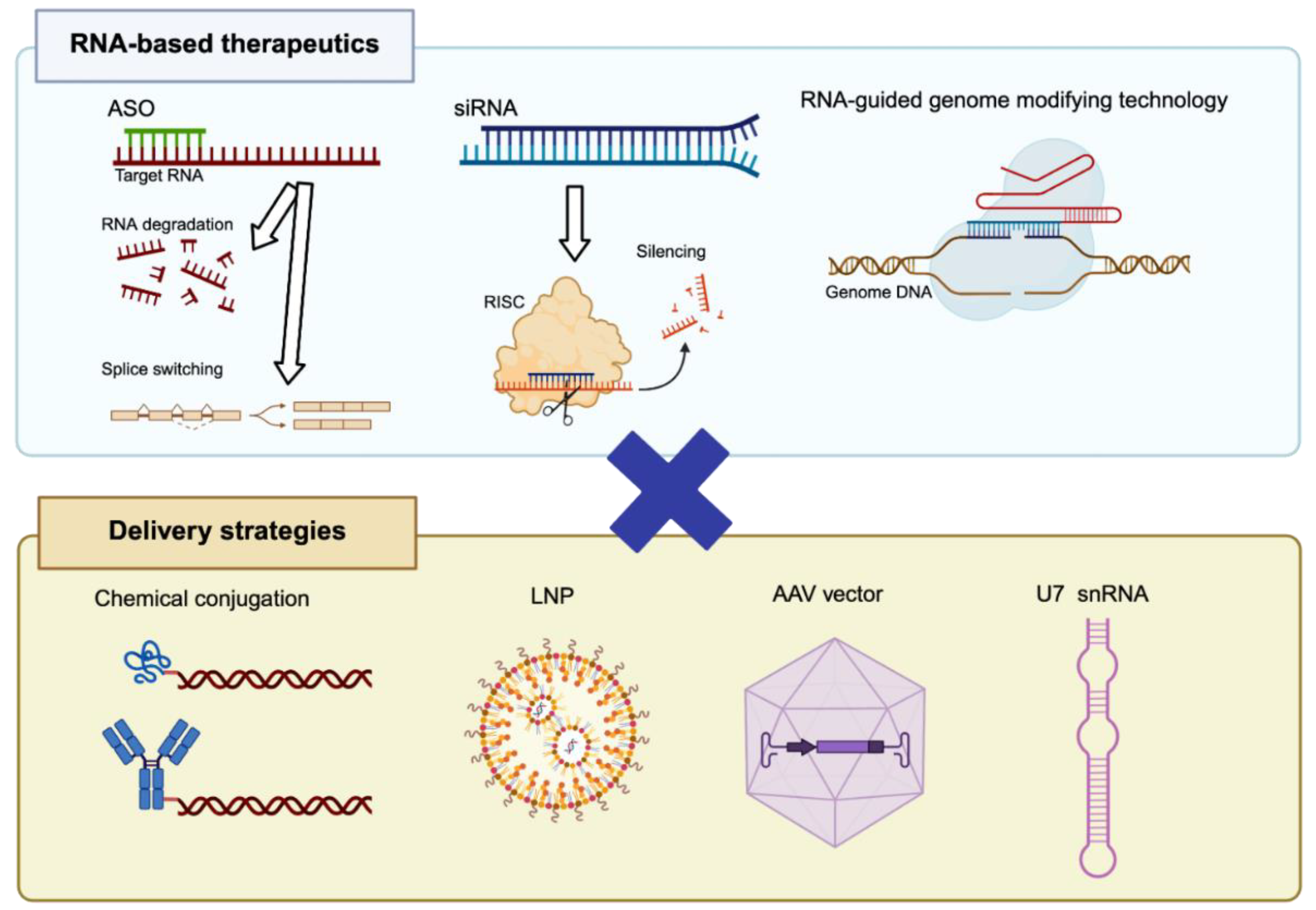
Figure 2.
Splicing-switching ASO restores the dystrophin reading frame in Duchenne muscular dystrophy. In the wild-type dystrophin gene, exons are spliced in-frame to produce full-length functional dystrophin. In DMD, deletion of exon 45 disrupts the reading frame, generating a premature stop codon and resulting in non-functional dystrophin. ASO–mediated exon 44 skipping removes exon 44, restores the reading frame, and enables production of a truncated but partially functional dystrophin protein. Figures were created with BioRender.com.
Figure 2.
Splicing-switching ASO restores the dystrophin reading frame in Duchenne muscular dystrophy. In the wild-type dystrophin gene, exons are spliced in-frame to produce full-length functional dystrophin. In DMD, deletion of exon 45 disrupts the reading frame, generating a premature stop codon and resulting in non-functional dystrophin. ASO–mediated exon 44 skipping removes exon 44, restores the reading frame, and enables production of a truncated but partially functional dystrophin protein. Figures were created with BioRender.com.
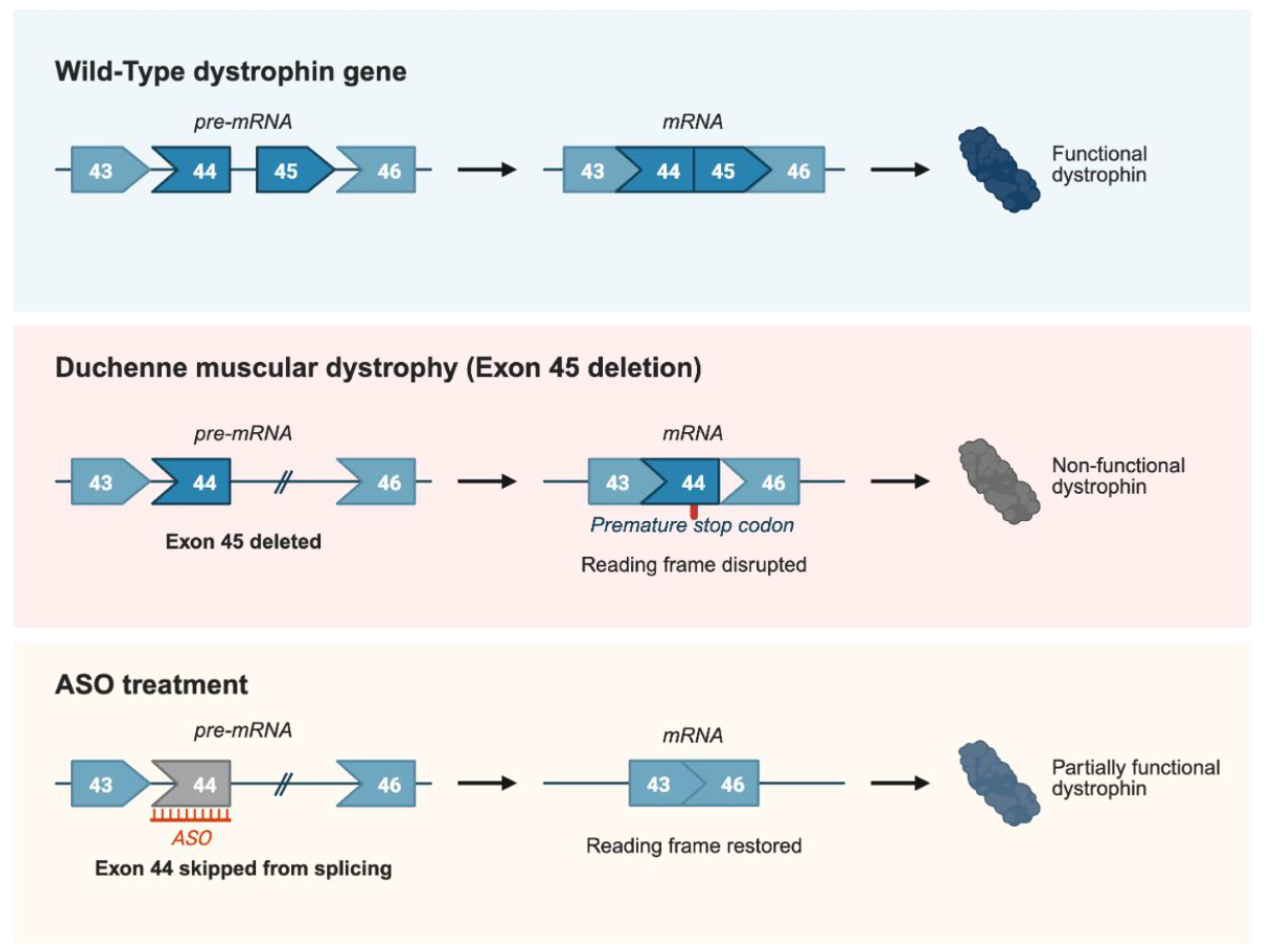

Table 1.
FDA-approved ASOs for DMD. All are naked PMOs that target specific exons of dystrophin pre-mRNA to induce exon skipping, restore the reading frame, and enable production of a truncated but partially functional dystrophin protein.
Table 1.
FDA-approved ASOs for DMD. All are naked PMOs that target specific exons of dystrophin pre-mRNA to induce exon skipping, restore the reading frame, and enable production of a truncated but partially functional dystrophin protein.
| ASO | Target exon | Chemistry | FDA approval | Administration |
| Eteplirsen | Exon 51 | PMO | 2016 | Intravenous injection |
| Golodirsen | Exon 53 | PMO | 2019 | Intravenous injection |
| Viltolarsen | Exon 53 | PMO | 2020 | Intravenous injection |
| Casimersen | Exon 45 | PMO | 2021 | Intravenous injection |
Disclaimer/Publisher’s Note: The statements, opinions and data contained in all publications are solely those of the individual author(s) and contributor(s) and not of MDPI and/or the editor(s). MDPI and/or the editor(s) disclaim responsibility for any injury to people or property resulting from any ideas, methods, instructions or products referred to in the content. |
© 2026 by the authors. Licensee MDPI, Basel, Switzerland. This article is an open access article distributed under the terms and conditions of the Creative Commons Attribution (CC BY) license (http://creativecommons.org/licenses/by/4.0/).
Copyright: This open access article is published under a Creative Commons CC BY 4.0 license, which permit the free download, distribution, and reuse, provided that the author and preprint are cited in any reuse.


