Submitted:
25 March 2026
Posted:
23 April 2026
You are already at the latest version
Abstract
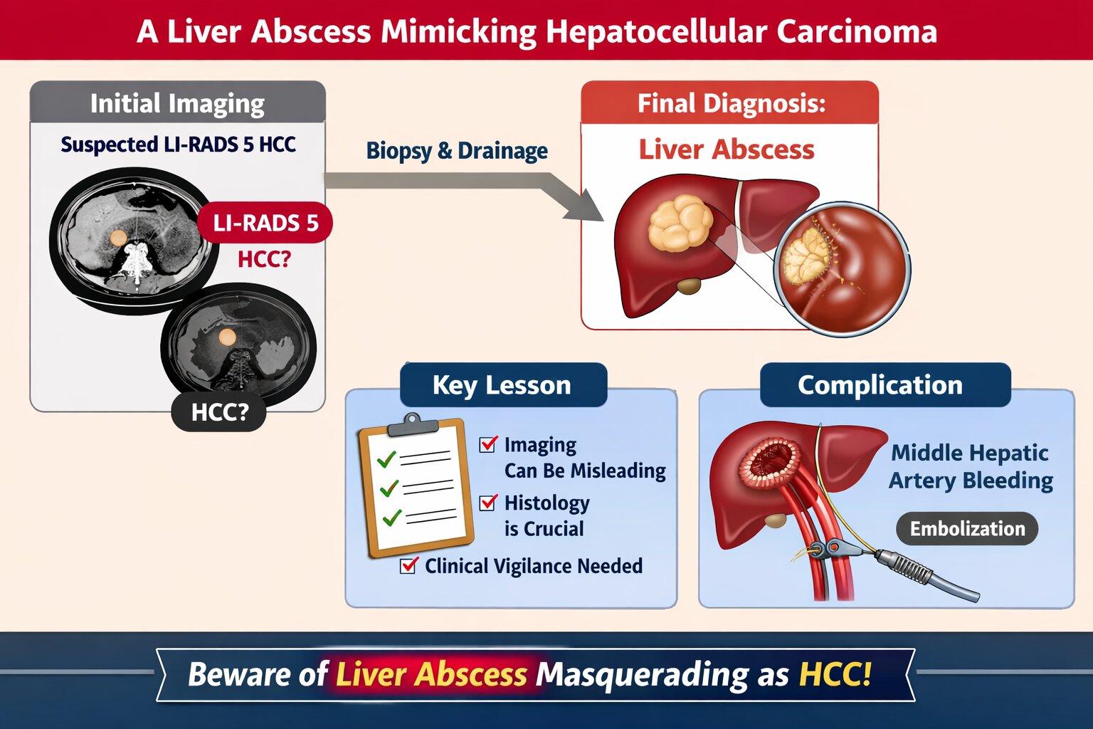
Background: Liver abscesses represent an atypical yet potentially life-threatening complication of bacterial, fungal, protozoal, and helminthic infections. Frequently, the clinical findings associated with liver abscesses are nonspecific, necessitating a reliance on imaging for diagnosis. It is uncommon for a liver abscess to radiographically resemble a malignant liver tumor such as hepatocellular carcinoma (HCC). Here, we present the case of a 45-year-old male who was initially diagnosed with HCC (BCLC C) but was subse-quently found to have a liver abscess following biopsy.
Case Presentation: A male patient, 45, presented with stiffness and pain in the right upper abdomen. He complained of nausea and vomiting since 10 days before admission as well. All supportive imaging suggested a diagnosis of HCC. A liver abscess was detected during a biopsy. A liver ultrasound-guided FNAB showcased chronic, suppurative in-flammation with negative acid-fast bacilli on Ziehl-Neelsen staining. The patient sub-sequently developed a complication of middle hepatic artery bleeding and underwent immediate embolization.
Discussion: In fact, a liver abscess can be the initial manifestation of HCC. Patients tend to have a poorer prognosis because the diagnosis of a liver abscess often delays the discovery of the underlying HCC. Radiographically, liver abscesses range from well-circumscribed cystic lesions with an enhancing rim to heterogeneously enhancing mass-like lesions, which are sometimes indistinguishable from liver neoplasms. However, it is so scarce that a liver abscess may radiographically mimic HCC.
Conclusion: Assessing liver abscess is somewhat complicated since the symptoms vary a lot. Therefore, a correct and exact diagnosis entail a combination of more comprehensive clinical and supporting examinations.
Keywords:
liver abscess
; hepatocellular carcinoma
; hepatoma
; HCC
1. Introduction
A liver abscess can be defined as a collection of suppurative material enclosed within the liver parenchyma [1]. Liver abscesses are an infrequent yet life-threatening complication of bacterial, fungal, protozoan, and helminthic infections [2]. Amoebic liver abscesses and pyogenic liver abscesses are the two main etiologies. In certain cases, liver abscesses can be caused by fungi, mycobacteria, and other atypical organisms [3]. Although the frequency of liver abscesses varies by region, the overall incidence is quite low, ranging from 2.3 cases per 100,000 hospitalizations in North America to 275.4 per 100,000 in Taiwan. In the early 1900s, the mortality rate was 75%–80%, while at the present, it has declined dramatically to 10%–40% due to the improvements in antibiotics administration and interventional procedures [1].
Liver abscesses are primarily caused by Entamoeba histolytica, with main transmission occurring through the fecal-oral route [3]. During the early 1900s, pylephlebitis secondary to appendicitis was the leading cause of liver abscess. By the late 1900s, biliary tract disease had become the predominant cause, a trend that continues in the present day. Additionally, the incidence of liver abscesses associated with malignancy and its treatment has escalated, including cases resulting from liver metastases and complications of transarterial chemoembolization (TACE) or radiofrequency ablation (RFA) [1].
The clinical course, diagnosis, and management of liver abscesses have progressed significantly due to advances in imaging modalities, antibiotics, and therapeutic techniques. While liver abscesses may present with diverse imaging characteristics depending on their stage of maturation and internal composition, it is uncommon for a liver abscess to radiographically mimic a malignant liver tumor such as hepatocellular carcinoma (HCC). Consequently, distinguishing a liver abscess from HCC in these instances can be challenging [4].
Liver abscesses present diagnostic challenges due to variable symptoms and frequently nonspecific objective findings. As a result, diagnosis often depends on imaging modalities [1]. Accurate identification of liver abscess is essential, as it significantly influences prognosis and treatment, given the lesion's treatable nature and the substantial morbidity and mortality associated with undiagnosed or untreated cases [5]. Despite improvements in mortality rates, they remain elevated, underscoring the importance of early diagnosis for optimal clinical outcomes [1]. Herein, we report a case of a 45-year-old male patient with an initial diagnosis of HCC (BCLC C), then later diagnosed with a liver abscess after biopsy.
2. Case Presentation
A 45-year-old man presented with abdominal rigidity and pain localized to the right upper quadrant. He also reported persistent nausea and vomiting for 10 days before admission. The patient had previously been treated at a private hospital, where he was diagnosed with hepatocellular carcinoma (HCC) classified as BCLC stage C. Abdominal ultrasonography performed at the referring hospital revealed hepatomegaly with multiple solid masses in the right hepatic lobe, the largest measuring 11.3 × 7.7 cm, causing inferior compression of the right kidney and interpreted as hepatoma (Figure 1). Contrast-enhanced abdominal computed tomography at the same institution demonstrated hepatomegaly with multiple solid, lobulated, exophytic masses in hepatic segments 2, 3, 4a, and 4b, with the largest lesion measuring 9.8 × 12.7 × 9.2 cm. These lesions showed arterial phase hyperenhancement and washout in the venous and delayed phases, findings interpreted as consistent with HCC and classified as LI-RADS 5. Splenomegaly with homogeneous parenchyma and left pleural effusion were also noted (Figure 2).
On admission to the referral hospital, the patient appeared moderately ill. His body weight was 61 kg and height 165 cm. Vital signs were as follows: blood pressure, 136/77 mmHg; pulse, 107 beats/min; respiratory rate, 18 breaths/min; oxygen saturation, 96% on room air; and axillary temperature, 37.8 °C. Initial laboratory evaluation showed hemoglobin 9.3 g/dL, hematocrit 27.8%, leukocyte count 18,660/μL, platelet count 403,000/μL, and differential count 0.1/0.1/91.4/4.0/4.4. The erythrocyte sedimentation rate was 99 mm/h. Coagulation studies showed prothrombin time 11.1 s, activated partial thromboplastin time 34.8 s, and international normalized ratio 1.2. Liver chemistry demonstrated direct bilirubin 4.78 mg/dL, total bilirubin 6.67 mg/dL, aspartate aminotransferase 41.5 U/L, alanine aminotransferase 28.6 U/L, alkaline phosphatase 306 U/L, gamma-glutamyl transferase 309.5 U/L, albumin 2.36 g/dL, and total protein 6.19 g/dL. Carcinoembryonic antigen was 0.9 ng/mL. Chest radiography showed cardiomegaly.
Based on the available imaging studies, the initial working diagnosis remained HCC BCLC stage C. The patient received maintenance fluids and empirical therapy, including intravenous ondansetron 8 mg three times daily, omeprazole 40 mg twice daily, ceftriaxone 1 g twice daily, N-acetylcysteine 5 g once daily, and paracetamol 1 g three times daily, as well as oral probiotics, nutritional supplementation, silybin-phospholipid, ambroxol, and ursodeoxycholic acid.
A repeat abdominal CT scan at the referral hospital again demonstrated hepatomegaly with multiple exophytic, lobulated, solid masses in segments 2, 3, 4a, and 4b, accompanied by left pleural effusion and splenomegaly with homogeneous parenchyma (Figure 3). During hospitalization, the patient continued to experience intermittent abdominal pain, bloating, nausea, and a pulling sensation in the abdomen. Follow-up laboratory testing showed hemoglobin 7.3 g/dL, hematocrit 21.4%, leukocyte count 17,830/μL, platelet count 447,000/μL, differential count 0.1/0.2/83.5/8.2/8.0, erythrocyte sedimentation rate 65/116 mm, direct bilirubin 3.34 mg/dL, total bilirubin 4.84 mg/dL, alanine aminotransferase 34.6 U/L, aspartate aminotransferase 26 U/L, albumin 2.62 g/dL, sodium 132 mmol/L, potassium 3.94 mmol/L, and chloride 100 mmol/L. Chest radiography showed bilateral pleural effusion. Doripenem 500 mg three times daily and oral attapulgite were initiated.
On the following day, the patient developed abdominal firmness, reduced appetite, and ascites, with a higher axillary temperature. Urinalysis showed urobilinogen +1, while blood cultures remained sterile. He received one unit of packed red blood cells and albumin transfusion. By hospital day 8, he continued to report abdominal stiffness, although laboratory values had partially improved: hemoglobin 10.1 g/dL, hematocrit 30.2%, leukocyte count 10,470/μL, platelet count 400,000/μL, direct bilirubin 2.52 mg/dL, total bilirubin 4.07 mg/dL, aspartate aminotransferase 20.2 U/L, alanine aminotransferase 27.1 U/L, albumin 3.26 g/dL, blood urea nitrogen 12 mg/dL, and serum creatinine 0.67 mg/dL. Hepatitis B surface antigen and anti-HIV were nonreactive. Esophagogastroduodenoscopy showed no esophageal varices or congestive gastropathy but demonstrated gastroesophageal reflux disease with a loose gastroesophageal flap valve (Figure 4). Echocardiography findings were unremarkable.
By hospital day 12, abdominal stiffness recurred. Physical examination revealed a palpable solid mass with tenderness in the right upper abdomen. Magnetic resonance imaging of the abdomen demonstrated a multilobulated liver mass in segments 2, 3, and 4 with diffusion restriction and heterogeneous hyperintensity on T2-weighted imaging, with the largest dimension measuring 11.6 × 8.0 × 5.6 cm. Dynamic contrast imaging showed slightly irregular, thick peripheral enhancement, raising suspicion for hepatic lymphoma or atypical HCC with cystic or necrotic degeneration. The mass was adjacent to the intrahepatic bile duct and associated with mild distal intrahepatic bile duct dilatation. Minimal bilateral pleural effusion and a Bosniak II cyst in the lower pole of the right kidney were also noted (Figure 5). Biopsy and further laboratory evaluation were recommended.
Subsequently, as the palpable lump decreased in size and the abdominal pain improved, laboratory results showed further improvement in bilirubin and normalization of liver enzymes and albumin. Liver biopsy and abscess drainage were then performed. Drainage yielded 130 mL of purulent material, after which the patient’s abdominal pain subsided and a drain was left in place. Soon afterward, the patient developed hematemesis, although vital signs remained stable. Pus culture showed no bacterial growth. The working diagnosis was revised to liver abscess, and treatment was continued with intravenous doripenem 500 mg three times daily, metronidazole 750 mg three times daily, and oral ibuprofen 400 mg twice daily.
On the next day, the patient reported minimal abdominal pain. Drain output was 97 mL over 24 h and was thick in consistency. Ultrasound-guided fine-needle aspiration biopsy of the liver showed chronic suppurative inflammation, consistent with abscess, with negative acid-fast bacilli staining on Ziehl–Neelsen examination. By hospital day 20, his abdominal pain had further decreased. The drain output was 9 mL of blackish fluid. Vital signs remained stable. Laboratory evaluation showed hemoglobin 12.2 g/dL, hematocrit 37.6%, leukocyte count 4.51 × 10^3^/μL, platelet count 382 × 10^3^/μL, direct bilirubin 1.02 mg/dL, total bilirubin 2.56 mg/dL, aspartate aminotransferase 24.4 U/L, alanine aminotransferase 13.8 U/L, and albumin 3.44 g/dL. Follow-up abdominal ultrasonography showed hepatomegaly with left lobe liver abscesses measuring 4.8 × 8.3 × 7.2 cm and 7.0 × 6.7 × 6.1 cm, with the drainage tube in situ (Figure 6).
After drain removal, arterial bleeding was observed. Because of a drop in blood pressure, computed tomography angiography was performed, revealing lobulated cystic lesions in liver segments I, II, and IV measuring 11.9 × 6.7 × 8.8 cm, containing hematoma. Prominent branches of the middle hepatic artery supplying the cystic lesion in segment IV were visualized in the arterial phase. These findings suggested an infected hemorrhagic cyst or abscess with associated hematoma. The lesion also compressed the posterior portion of the main portal vein and reduced contrast filling in the superior mesenteric vein near its confluence with the portal vein. The final diagnosis was liver abscess complicated by rupture of an arterial branch within the abscess cavity. Urgent middle hepatic artery embolization was successfully performed (Figure 7).
Following embolization, the patient reported less pain, and both the inguinal puncture site and abdominal drain site showed progressive drying (Figure 8). Vital signs, physical examination, and laboratory findings stabilized. He was discharged with oral cefixime 200 mg twice daily, metronidazole 750 mg three times daily, and ondansetron 8 mg three times daily.
3. Discussion
A liver abscess is defined as a collection of suppurative material enclosed within the liver parenchyma. It may be infected by bacteria, fungi, or parasites. Multiple risk factors are linked to increased mortality. These risk factors include diabetes mellitus, cirrhosis, immunocompromised status, prolonged use of proton pump inhibitors (PPIs), gender, and age over 57 years. Individuals with compromised immune systems due to chemotherapy, immunosuppressive therapy, or inherited or acquired immunodeficiency syndromes are at are higher risk for liver abscesses caused by fungi and opportunistic microorganisms [1,2,5].
The primary mechanism is hematogenous infection via the hepatic artery during severe septic processes, such as metastatic-pyemic liver abscess, or through the portal vein in cases of liver abscess associated with portal vascular thrombosis. Occasionally, infection spreads through the umbilical vein (as in omphalophlebitis) or via the biliary tract and may also result from parasitic invasion or the presence of foreign bodies. Additionally, liver abscess can develop from the extension of an inflammatory process from adjacent structures, direct liver injury, intrahepatic hematoma, or postoperative complications [2].
Liver abscesses are classified into three categories: infectious, malignant, and iatrogenic. Malignant abscesses are further subdivided into secondary infection of primary liver tumors, secondary infection of metastatic liver lesions, and spontaneous necrotic superinfection. Primary hepatocellular carcinoma (HCC) may develop spontaneously in areas of central necrosis, which are susceptible to bacterial infection. HCC can also result in biliary obstruction, potentially leading to ascending cholangitis and subsequent liver abscess formation. In some cases, liver abscess may serve as an early manifestation of HCC. Patients with this presentation frequently have a poorer prognosis, as the diagnosis of liver abscess can delay identification of the underlying HCC. Several imaging features have been reported to support the identification of superinfected malignancy, including thickened walls, septation, aerobilia, portal vein thrombosis, and gas within the abscess1. In the present case, non-contrast MRI of the abdomen suggested either (1) hepatic lymphoma or (2) atypical HCC with cystic or necrotic degeneration. The mass was located adjacent to the intrahepatic bile duct (IHBD) with mild distal IHBD dilatation. Additional findings included minimal right and left-sided pleural effusion (maximum thickness 3 cm) and a cyst in the lower pole of the right kidney (Bosniak II).
The most commonly observed clinical manifestations are fever, abdominal pain, and hypotension. The proportion of patients presenting with each symptom varies considerably, indicating substantial heterogeneity in clinical presentation. The challenge in establishing a diagnosis is further demonstrated by the average delay of 1 week between symptom onset and diagnosis. In the present case, the patient exhibited right upper quadrant abdominal pain and stiffness, accompanied by nausea and vomiting persisting for ten days prior to admission. On examination, his temperature was recorded at 37.8°C.
Laboratory findings in patients with liver abscesses are generally nonspecific. The most frequent abnormalities include elevated leukocyte count, increased C-reactive protein (CRP), hypoalbuminemia, elevated aspartate aminotransferase and alanine aminotransferase, elevated alkaline phosphatase, increased gamma-glutamyl transpeptidase, elevated bilirubin, and an increased international normalized ratio (INR). Although laboratory testing alone is insufficient for a definitive diagnosis, these abnormalities typically prompt further imaging, which is essential for confirming the presence of a liver abscess1. In the present case, the laboratory results demonstrated leukocytosis, hypoalbuminemia, elevated ALT, alkaline phosphatase, gamma GT, direct bilirubin, and total bilirubin, which are consistent with the findings commonly reported in the literature.
Imaging establishes the diagnosis of a liver abscess in approximately 90% of cases and can also help identify the underlying etiology. The primary diagnostic modalities include conventional ultrasonography (US) and computed tomography (CT), both with sensitivities of 96% to 100% for detecting liver abscesses. US typically reveals hypoechoic lesions with variable internal echogenicity, influenced by the presence of septations or gas. If US is nondiagnostic, CT, magnetic resonance imaging (MRI), or contrast-enhanced ultrasound (CEUS) should be employed. On noncontrast CT, liver abscesses exhibit lower attenuation than normal hepatic parenchyma. Following intravenous (IV) administration of iodinated contrast, CT scans may demonstrate peripheral enhancement and enhanced internal septations. On MRI, liver abscesses are hyperintense on T2-weighted images and hypointense on noncontrast T1-weighted images, although some may appear hyperintense on noncontrast T1-weighted images depending on protein content. After gadolinium administration, enhancement patterns are similar to those observed on CT scans [1].
Imaging techniques, including US-guided needle aspiration and CT scan, are used to confirm the diagnosis of liver abscess, as the material obtained can be analyzed to identify the etiologic agent. These procedures also serve a therapeutic function in percutaneous drainage, which will be addressed subsequently. Imaging studies provide information on the location, size, number, consistency, and presence of gas within abscesses. The right lobe of the liver is the most frequent site, accounting for 68.7% of cases in one study. Solitary abscesses are observed more frequently than multiple abscesses. In terms of consistency, 58% of abscesses are solid, while 42% are cystic. Gas is detected in approximately 17% of cases [1].
Liver abscesses demonstrate a spectrum of radiographic features, ranging from well-defined cystic lesions with rim enhancement to heterogeneously enhancing mass-like lesions that may be difficult to distinguish from hepatic neoplasms [4]. Although the imaging characteristics of liver abscesses vary depending on the stage of maturation and internal composition, it remains uncommon for a liver abscess to closely mimic a malignant liver tumor such as hepatocellular carcinoma (HCC) [4,6,7]. Previous reports have documented cases in which liver abscesses presented with imaging findings similar to HCC, sometimes in the absence of clinical or laboratory indicators of infection [4,7]. In certain cases, additional findings, such as portal vein thrombosis or elevated inflammatory markers, have helped differentiate abscess from malignancy [8].
However, unlike previous reports, our case was considerably complex to distinguish between liver abscess and HCC due to the lack of clinical presentation suggesting an abscess. Initially, our case was diagnosed with HCC BCLC stage C based on findings from abdominal ultrasound, abdominal CT scan, and abdominal MRI without contrast. However, a biopsy revealed a liver abscess. Our patient did not undergo CRP examination. Abdominal CT revealed hepatomegaly accompanied by multiple solid, lobulated exophytic masses in segments 2, 3, 4a, and 4b (the largest size was 9.8 x 12.7 x 9.2 cm), which showed early enhancement in the arterial phase and washout in the venous and delayed phases, tending towards hepatocellular carcinoma according to LI-RADS 5.
The Liver Imaging Reporting and Data System (LI-RADS), established by the American College of Radiology, standardizes terminology and criteria for interpreting and reporting liver CT and MR imaging findings in patients at risk for hepatocellular carcinoma (HCC). Observations are categorized from LR-1 (definitely benign) to LR-5 (definitely HCC). LI-RADS incorporates both primary and secondary criteria. Primary criteria include lesion size (10–19 mm or≥20 mm), contrast enhancement on multiphase imaging (arterial, portal venous, and/or delayed phases), the presence of a capsule, and growth over 6 months. Secondary criteria comprise additional features that may prompt an upgrade or downgrade of the observation. The relative weight of each feature remains undetermined [9,10]. Previous studies of LI-RADS versions 2014 and 2017 have demonstrated that LR-5 criteria on CT and/or MRI provide high specificity (85%–100%) for HCC, with moderate to fair sensitivity (50%–80%). For small HCCs (10–19 mm), the sensitivity of the LR-5 criteria in versions 2014 and 2017 was even lower (23%–67%), although high specificity was maintained (89%–98%). Chen et al. reported that, using LI-RADS v2018, LR-5 had a sensitivity of 70% and specificity of 95% for small HCCs (10–19 mm) across both readers. There were two false positive results for reader 1 (one histologically confirmed hepatocellular adenoma and one benign hepatocellular nodule based on imaging and follow-up) and two false positive results for reader 2 (two benign hepatocellular nodules based on imaging and follow-up) [12]. Alhasan et al. reported a sensitivity of 53.7% and specificity of 97.3% for LI-RADS 5 in the diagnosis of HCC [13]. Although LR-5 indicates a 95% probability of HCC, there remains a 5% likelihood that the lesion is not HCC [11].
The management of liver abscesses was initially limited to open surgical drainage. In recent years, percutaneous drainage has become increasingly utilized. For smaller abscesses, conservative management with antibacterial therapy alone has been adopted. Evidence indicates that antibiotic therapy alone is effective for abscesses measuring 3–5 cm or less. In one study, 100% success was observed in 8 patients with unilocular abscesses measuring less than 3 cm treated solely with antibiotics. A larger study involving 176 patients reported a success rate of 81.2% following antibiotic administration. Antibiotic therapy should be commenced promptly after obtaining blood samples for organism identification, beginning with intravenous antibiotics for approximately 3 weeks, followed by oral antibiotics for 1 to 2 months. The duration of therapy is determined by the clinical response, as assessed by repeat ultrasound imaging and the resolution of fever and leukocytosis. Recommended broad-spectrum antibiotics include a third-generation cephalosporin combined with metronidazole or piperacillin/tazobactam. It should be noted that certain common pathogens associated with liver abscesses demonstrate resistance to ampicillin and fluoroquinolones [1].
Treatment is complicated by the increasing incidence of hyperresistant K. pneumoniae in some parts of the world. Percutaneous drainage is the most common option for first-line treatment of liver abscesses. Liver abscesses can be drained by needle aspiration or by inserting a pigtail catheter drain under ultrasound or CT scan guidance. Several studies have found percutaneous catheter drainage to be more effective than percutaneous needle aspiration, due to its higher success rate. Percutaneous drainage has many advantages, including minimally invasive procedures, avoiding the need for general anesthesia, a lower risk of adhesion formation and contamination, and a relatively lower cost compared to surgical drainage. Percutaneous drainage has been reported to fail in 15%–36% of cases due to multilocular liver abscesses or those containing thick fluid and necrotic tissue1. Surgery is indicated as the initial management for ruptured liver abscesses, peritonitis, difficult anatomical access, and pathologies requiring surgery. Initial surgical management may also be indicated for larger abscesses measuring >3–5 cm in diameter [1].
Our patient underwent drainage of an abscess. The patient received oral metronidazole TID. A liver biopsy was scheduled. Ultrasound-guided FNAB of the liver revealed chronic, suppurative inflammation (abscess) with negative acid-fast bacilli on Ziehl-Neelsen staining. Pus cultures showed no bacterial growth.
It has been reported that 15.7% of patients develop complications from liver abscesses namely septic metastasis leading to extrahepatic complications, such as endophthalmitis, septic pulmonary embolism, lung, central nervous system, and eye infections. Abscess rupture is another reported complication, with spontaneous rupture occurring in 6.1% of cases. There is a reported higher incidence of liver abscess rupture in Klebsiella-infected abscesses than in those infected with other bacteria. Liver abscesses can also erode the diaphragm, causing pleural effusion, empyema, pneumonia, pericarditis, bronchopleural fistula, or duodenobronchofistula. Multiorgan failure can also occur as a result of liver abscesses [1].
The patient developed a complication characterized by an infected hemorrhagic cyst with differential diagnosis of abscess and associated hematoma. MSCT of the upper abdomen, including non-contrast and triphasic dynamic intravenous contrast studies, revealed lobulated cystic lesions in liver segments I, II, and IV, containing hematoma, 11.9 x 6.7 x 8.8 cm in size. Prominent branches of the middle hepatic artery were observed entering the cystic lesion in segment IV during the arterial phase. Middle hepatic artery embolization was performed, and the patient was discharged two days later.
Although mortality from liver abscess has declined over time, it remains a consequential issue until now. Multiple associated factors and comorbidities contribute to the risk of death from liver abscess. Chen et al. conducted a study involving 134 patients with primary liver abscess, categorizing them into mortality and survivor groups. The study identified several risk factors for increased mortality, including male sex, malignancy, multi-organ failure, and liver abscess rupture. Notably, the mortality rate among patients with malignancy was twice as high as that among cancer-free patients. Additional signs and symptoms associated with the mortality group included respiratory distress, hypotension, jaundice, and extrahepatic involvement such as endophthalmitis [1].
4. Conclusions
Liver abscess may rarely mimic hepatocellular carcinoma on multimodality imaging, including studies demonstrating features highly suggestive of HCC. In this case, a 45-year-old man was repeatedly diagnosed radiologically with advanced HCC, but histopathology and drainage ultimately confirmed liver abscess. This case underscores the need for careful integration of clinical presentation, laboratory data, imaging findings, and tissue confirmation in atypical liver masses. When the diagnosis remains uncertain, biopsy and drainage should be considered to avoid misdiagnosis and ensure timely, appropriate management.
Supplementary Materials
The following supporting information can be downloaded at the website of this paper posted on Preprints.org, Figure 1. Abdominal ultrasonography at the private hospital showing hepatomegaly with multiple solid masses in the right hepatic lobe, initially interpreted as hepatoma. Figure 2. Contrast-enhanced abdominal CT at the private hospital (1 February 2025) demonstrating multiple lobulated hepatic masses with arterial enhancement and venous/delayed washout, initially interpreted as HCC. Figure 3. Abdominal CT at the referral hospital (hospital day 3) confirming hepatomegaly with multiple exophytic lobulated masses and associated pleural effusion and splenomegaly. Figure 4. Esophagogastroduodenoscopy at the referral hospital (hospital day 8) showing no esophageal varices or congestive gastropathy. Figure 5. Abdominal MRI at the referral hospital (hospital day 12) demonstrating a multilobulated liver mass with diffusion restriction and heterogeneous T2 hyperintensity, suggestive of hepatic lymphoma or atypical HCC. Figure 6. Follow-up abdominal ultrasonography at the referral hospital (hospital day 20) showing persistent left lobe liver abscesses with drainage tube in place. Figure 7. CT angiography at the referral hospital (hospital day 23) showing a cystic hepatic lesion with associated hematoma and arterial supply from branches of the middle hepatic artery, followed by embolization. Figure 8. Post-drainage wound after treatment and clinical improvement.
Author Contributions
Conceptualization, F.S. and U.M.; methodology, F.S. and U.M.; investigation, F.S.; data curation, F.S.; writing—original draft preparation, F.S.; writing—review and editing, U.M.; supervision, U.M. All authors have read and agreed to the published version of the manuscript.
Funding
This research received no external funding.
Institutional Review Board Statement
Ethical review and approval were waived for this study because it describes a single case report based on routine clinical care and does not constitute human subjects research requiring formal Institutional Review Board approval under local institutional policy.
Informed Consent Statement
Informed consent was obtained from the subject involved in this study. Written informed consent has been obtained from the patient to publish this paper.
Data Availability Statement
The data supporting the findings of this study are available from the corresponding author upon reasonable request, subject to privacy and ethical restrictions.
Acknowledgments
The authors would like to thank the clinical staff involved in the diagnosis and management of this patient.
Conflicts of Interest
The authors declare no conflict of interest.
References
- Mavilia, M.G.; Molina, M.; Wu, G.Y. The evolving nature of hepatic abscess: A review. J. Clin. Transl. Hepatol. 2016, 4, 158–168. [Google Scholar] [CrossRef] [PubMed]
- Kozielewicz, D.M.; Sikorska, K.; Stalke, P. Liver abscesses—from diagnosis to treatment. Clin. Exp. Hepatol. 2021, 7, 329–336. [Google Scholar] [CrossRef]
- Sharma, S.; Ahuja, V. Liver abscess: Complications and treatment. Clin. Liver Dis. 2021, 18, 122–126. [Google Scholar] [CrossRef] [PubMed]
- Kim, J.W.; Shin, S.S.; Heo, S.H.; Lim, H.S.; Hur, Y.H.; Kim, J.H. Hepatic abscess mimicking hepatocellular carcinoma in a patient with alcoholic liver disease. Clin. Mol. Hepatol. 2013, 19, 431. [Google Scholar] [CrossRef] [PubMed]
- Yu, Y.; Guo, L.; Hu, C.; Chen, K. Spectral CT imaging in the differential diagnosis of necrotic hepatocellular carcinoma and hepatic abscess. Clin. Radiol. 2014, 69, e517–e524. [Google Scholar] [CrossRef] [PubMed]
- Brown, K.T.; Gandhi, R.T.; Covey, A.M.; Brody, L.A.; Getrajdman, G.I. Pylephlebitis and liver abscess mimicking hepatocellular carcinoma. Hepatobiliary Pancreat. Dis. Int. 2003, 2, 221–225. [Google Scholar] [PubMed]
- Chou, Y.; Changchien, C.; Chiu, K.; Kuo, C.; Kuo, F.; Kuo, C. Salmonellosis with liver abscess mimicking hepatocellular carcinoma in a diabetic and cirrhotic patient: A case report and review of the literature. Liver Int. 2006, 26, 498–501. [Google Scholar] [CrossRef] [PubMed]
- Chernyak, V.; Santillan, C.S.; Papadatos, D.; Sirlin, C.B. LI-RADS® algorithm: CT and MRI. Abdom. Radiol. 2018, 43, 111–126. [Google Scholar] [CrossRef] [PubMed]
- Ronot, M.; Fouque, O.; Esvan, M.; Lebigot, J.; Aubé, C.; Vilgrain, V. Comparison of the accuracy of AASLD and LI-RADS criteria for the non-invasive diagnosis of HCC smaller than 3 cm. J. Hepatol. 2018, 68, 715–723. [Google Scholar] [CrossRef] [PubMed]
- Rose, L.; Swi, A.; Hegazy, Y.; Bukeirat, F.A. The clinical spectrum of liver masses: Know before you judge. J. Gastroenterol. Hepatol. Rep. 2024, 5, 1–5. [Google Scholar] [CrossRef]
- Chen, J.; Kuang, S.; Zhang, Y.; Tang, W.; Xie, S.; Zhang, L.; et al. Increasing the sensitivity of LI-RADS v2018 for diagnosis of small (10–19 mm) HCC on extracellular contrast-enhanced MRI. Abdom. Radiol. 2021, 46, 1530–1542. [Google Scholar] [CrossRef] [PubMed]
- Alhasan, A.; Cerny, M.; Olivié, D.; Billiard, J.S.; Bergeron, C.; Brown, K.; et al. LI-RADS for CT diagnosis of hepatocellular carcinoma: Performance of major and ancillary features. Abdom. Radiol. 2019, 44, 517–528. [Google Scholar] [CrossRef] [PubMed]
Figure 1.
Abdominal USG at a private hospital.

Figure 2.
Abdominal CT at a private hospital (February 1st 2025).

Figure 3.
Abdominal CT at the referral hospital (Admission day-3).

Figure 4.
Esogastroduodenoscopy at the referral hospital (Admission day 8).

Figure 5.
Abdominal MRI at the referral hospital (Admission Day 12).

Figure 6.
Abdominal USG at the referral hospital (Admission Day 20).

Figure 7.
CT Angiography at the referral hospital (Admission day 23).

Figure 8.
Post-drainage wound.

Disclaimer/Publisher’s Note: The statements, opinions and data contained in all publications are solely those of the individual author(s) and contributor(s) and not of MDPI and/or the editor(s). MDPI and/or the editor(s) disclaim responsibility for any injury to people or property resulting from any ideas, methods, instructions or products referred to in the content. |
© 2026 by the authors. Licensee MDPI, Basel, Switzerland. This article is an open access article distributed under the terms and conditions of the Creative Commons Attribution (CC BY) license (http://creativecommons.org/licenses/by/4.0/).
Copyright: This open access article is published under a Creative Commons CC BY 4.0 license, which permit the free download, distribution, and reuse, provided that the author and preprint are cited in any reuse.


