Submitted:
25 March 2026
Posted:
23 April 2026
You are already at the latest version
Abstract
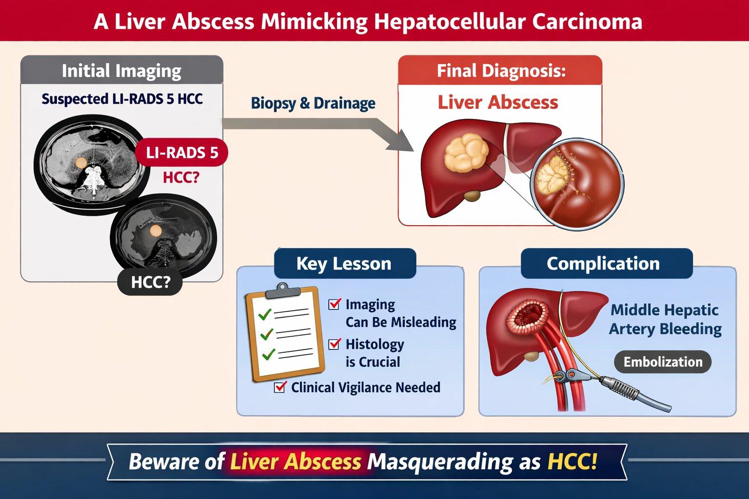
Background: Liver abscesses represent an atypical yet potentially life-threatening complication of bacterial, fungal, protozoal, and helminthic infections. Frequently, the clinical findings associated with liver abscesses are nonspecific, necessitating a reliance on imaging for diagnosis. It is uncommon for a liver abscess to radiographically resemble a malignant liver tumor such as hepatocellular carcinoma (HCC). Here, we present the case of a 45-year-old male who was initially diagnosed with HCC (BCLC C) but was subse-quently found to have a liver abscess following biopsy.
Case Presentation: A male patient, 45, presented with stiffness and pain in the right upper abdomen. He complained of nausea and vomiting since 10 days before admission as well. All supportive imaging suggested a diagnosis of HCC. A liver abscess was detected during a biopsy. A liver ultrasound-guided FNAB showcased chronic, suppurative in-flammation with negative acid-fast bacilli on Ziehl-Neelsen staining. The patient sub-sequently developed a complication of middle hepatic artery bleeding and underwent immediate embolization.
Discussion: In fact, a liver abscess can be the initial manifestation of HCC. Patients tend to have a poorer prognosis because the diagnosis of a liver abscess often delays the discovery of the underlying HCC. Radiographically, liver abscesses range from well-circumscribed cystic lesions with an enhancing rim to heterogeneously enhancing mass-like lesions, which are sometimes indistinguishable from liver neoplasms. However, it is so scarce that a liver abscess may radiographically mimic HCC.
Conclusion: Assessing liver abscess is somewhat complicated since the symptoms vary a lot. Therefore, a correct and exact diagnosis entail a combination of more comprehensive clinical and supporting examinations.
Keywords:
liver abscess
; hepatocellular carcinoma
; hepatoma
; HCC
Copyright: This open access article is published under a Creative Commons CC BY 4.0 license, which permit the free download, distribution, and reuse, provided that the author and preprint are cited in any reuse.


