
Submitted:
30 March 2026
Posted:
31 March 2026
You are already at the latest version
Abstract
Freeze-drying of Platelet-rich plasma and its effect on wound healing: An in vivo pilot case study. Himanshu Bansal1,2*, Alnkrita Bansal1,2, Irfan Khan2, Anupama Bansal1, Shahnawaz Hussein Khan3, Jerry Leon4, Mustafa al Maini5, Matias Fernandez Viña6 1Mother Cell Spinal Injury and Stem Cell Research, Anupam Hospital, Rudrapur, Uttarakhand, India 2Revita Life Sciences, Rudrapur, Uttarakhand, India 3Department of regenerative medicine, Lovinium Biotech, Asturias, Spain 4PMR Advance Health Institute Mayaguez, Puerto Rico, USA 5Mafraq Hospital, Sheikh Shakhbout Medical City, Abu Dhabi, United Arab Emirates 6Consultant, Stem Cell Therapy, Argentina *Corresponding author Dr. Himanshu Bansal, MD, Consultant Regenerative Medicine, Revita Lifesciences, Rudrapur, India-263153 Phone: 09634501234 Email: hbansal@drhbf.org Abstract Background: Platelet-rich plasma (PRP) is widely used in regenerative medicine because of its high content of growth factors and cytokines that promote tissue repair. However, fresh PRP is limited by the rapid loss of bioactivity, compositional variability, and storage challenges. Freeze-dried PRP (FD-PRP) is a promising alternative with potential for long-term storage and standardized therapeutic efficacy. Objective: This study aimed to evaluate the biological activity, growth factor preservation, and wound healing efficacy of FD-PRP compared with fresh PRP using in vitro and in vivo assays. Methods: PRP from 20 healthy donors was standardized to 5 billion platelets per 4 mL and freeze-dried using proprietary stabilizers. Platelet morphology, aggregation, and growth factor concentrations (PDGF, TGF-β1, VEGF, EGF, bFGF, and IGF-1) were assessed before and after lyophilization. The stability of the samples was monitored over 26 weeks. In vitro wound healing was assessed in human mesenchymal stem cells (MSCs) using scratch assays. A pilot in vivo study evaluated the efficacy of FD-PRP in accelerating wound closure in 10 patients over three weeks. Results: FD-PRP retained 82.05% of platelets post-lyophilization, with preserved morphology and aggregation. Growth factor levels were maintained or increased in FD-PRP, notably TGF-β1 (2.1-fold) and IGF-1 (3.04-fold) levels. Stability testing demonstrated >90% retention of platelets and growth factors over 26 weeks. In vitro, FD-PRP accelerated wound closure (85% at 72 hours) compared to fresh PRP (70%) and saline (30%). In vivo, FD-PRP-treated wounds showed a mean size reduction of 56.17 % %by day 12, which was significantly higher than that in non-treated regions (23.5%). Conclusion: FD-PRP preserves platelet functionality and key growth factors, demonstrates excellent storage stability, and enhances wound healing both in vitro and in vivo. These findings support FD-PRP as a practical and effective alternative to fresh PRP for clinical applications that require standardized, off-the-shelf regenerative therapies. Keywords: Platelet-rich plasma, Freeze-drying, Lyophilization, Wound healing, Growth factors, Regenerative medicine.
Keywords:
platelet-rich plasma
; freeze-drying
; lyophilization
; wound healing
; growth factors
; regenerative medicine
Introduction
Platelet-rich plasma (PRP) is one of the most commonly used biologics in Regenerative Medicine, owing to its high content of growth factors and cytokines, which promote tissue repair and regeneration, revolutionising the therapeutic approaches in Musculoskeletal (MSK) disorders, aesthetics and wound healing (Marx 2001, Gentile, Garcovich et al. 2015). Over the past decade, it has been explored as a new therapeutic middleman for treating chronic degenerative diseases (Ramaswamy Reddy, Reddy et al. 2018).
Platelet activation releases growth factors, such as Platelet Derived Growth Factor(PDGF), Vascular Endothelial Growth Factor(VEGF), Transforming growth factor-α/β (TFF-α/β), Epidermal Growth Factor (EGF), Hepatocyte Growth Factor (HGF), Basic Fibroblast Growth Factor (bFGF), and Insulin-like Growth Factor-1 (IGF-1), and cytokines, which mediate cell proliferation, differentiation, migration, and cellular development (Lubkowska, Dolegowska B Fau - Banfi et al. 2012). PRP therapy has several limitations, including time-dependent cell activity (Roffi, Filardo et al. 2014), rapid loss of bioactivity (Dhurat and Sukesh 2014), and varying platelet and growth factor composition based on patient health status, preparation method, and centrifugation protocol (Anitua, Andia et al. 2004). Fresh PRP preparation involves several steps, increasing the risk of microbial contamination(Sampson, Gerhardt et al. 2008) and RBC contamination, which can trigger cytokine release (Everts, Onishi et al. 2020). Unsatisfactory study results, PRP dosing and standardization, blood manipulation, and sterility are also issues. Additionally, application in clinical settings makes waiting time for patients and standardization of PRP formulations a challenge (Gangaraju, Mahajan et al. 2023).
Freeze-dried PRP (FD-PRP) is a dry powdered form that retains essential growth factors(Andia, Perez-Valle et al. 2020) like VEGF, EGF, TGF-β, and PDGF, similar to fresh PRP (Nakatani, Agata et al. 2017, Shiga, Kubota et al. 2017, McClain and McCarrel 2019). In vitro studies have confirmed FD-PRP’s growth factors profile and biological activation, supporting cell proliferation, migration, and angiogenesis (Huber, Montalvão et al. 2016, McClain and McCarrel 2019, Verma, Kumar et al. 2023). In vivo studies have shown that FD-PRP effectively promotes tissue regeneration in animal models, showing comparable or superior efficacy to fresh PRP in wound healing, bone regeneration, and tendon repair(Huber, Montalvão et al. 2016, Huber, Junior et al. 2019, Koga, Nakatani et al. 2021, Saputro, Rizaliyana et al. 2022, Morimoto, Morio et al. 2025). Lyophilisation and freeze-drying preserve platelet functionality and cytokine concentration, allowing for tailored cytokine release kinetics (Andia, Perez-Valle et al. 2020). The advantages of FD-PRP include bioactivity preservation, standardized platelet and growth factor levels, ease of handling, quick reconstitution, stability, off-the-shelf availability, avoidance of inter-donor variability, no need for venipuncture, higher concentration of growth factors, and reduced cost and manpower (Andia, Perez-Valle et al. 2020). However, there are currently no commercially available freeze-dried PRP products for human clinical use, except PrecisePRP™ (VetStem, Inc.), which is intended for veterinary purposes. This study presents a novel method that improves platelet recovery and extends their biological activity, overcoming previous challenges in FD-PRP research, including low platelet recovery during reconstitution, an incomplete growth factor spectrum, and limited human trials.
To test this hypothesis, we evaluated the approach using a wound healing model and conducted a pilot study in a limited clinical setting. The wound healing process involves four phases: hemostasis, inflammation, proliferation, and tissue remodelling, each requiring the coordinated action of cytokines and growth factors(Guo and Dipietro 2010) (Figure 1). Chronic wounds are impaired due to factors like age, stress, and underlying conditions, highlighting the need for effective translational therapies (Guo and Dipietro 2010, Frykberg and Banks 2015, Wilkinson and Hardman 2020, Bogadi, Uddin et al. 2025). Future research will expand to chronic wound scenarios and translate these findings into effective regenerative therapies.
Materials and Methods
Whole Blood Collection
Peripheral blood was collected from four healthy adult donors (aged 25–35 years) with a normal body mass index (BMI), platelet counts within the reference range, and no history of smoking, chronic disease, or haematological disorders. A total of 60 ml of venous blood per donor was drawn into sterile tubes containing the anticoagulant acid citrate dextrose (ACD) to ensure consistency in PRP quality and growth factor levels for downstream analyses. All donors provided written informed consent in accordance with the guidelines of the Ethics Committee of HB Specialty Hospital and Research Institute, Rudrapur, India.
PRP Preparation
PRP was prepared using a standardized two-step centrifugation protocol (Bansal, Leon et al. 2021). Whole blood (60 ml) was centrifuged to separate the plasma from the red blood cells. Platelet-poor plasma (PPP) was removed, and the lower third of the plasma fraction, which was enriched with platelets, was collected. Platelet counts were analyzed using microscopy, and the platelet concentration was standardized to 5 × 10⁹ in 4 ml PRP (Figure 2).
Freeze-Dried PRP
Platelet concentrates containing 5 × 10⁹ platelets was aliquoted into lyophilization vials. A proprietary stabilization system (stabilizers A and B) was added to the final volume of 5 ml before freezing at –20 °C. Lyophilization was performed using a Delaware freeze-dryer (model Delvac-LYO1550) under vacuum (0.100 mbar, condenser –50 °C). The vials were sealed under vacuum and stored at 4 °C. Freeze-dried PRP (FD-PRP) was reconstituted in sterile saline to its original volume before analysis.
Characterisation of FD-PRP
Platelet Morphology and Count
Platelet morphology and count were assessed using a light microscope and hemocytometer before and after lyophilization to ensure that the freeze-drying process did not negatively impact platelet integrity.
Platelet Function
To evaluate platelet function, platelet aggregation was induced with adenosine diphosphate (ADP, 10 µM) or adrenaline (10 µM) in three tubes: PRP, FD-PRP, and PPP. The absorbance was measured at 615 nm (Tsujino, Isobe et al. 2019), and the change in light transmission over a 6–9-minute period was reflected in the aggregation responses (da Silva, Montalvao et al. 2018). The control samples consisted of PPP. The maximum aggregation (%) was calculated as the peak change in light transmission relative to the PPP.
Growth factor quantification
Growth factor levels in fresh PRP and FD-PRP were quantified using enzyme-linked immunosorbent assay (ELISA) kits (Elabscience, USA) according to the manufacturer’s instructions. Both fresh PRP and FD-PRP were activated with 10% CaCl₂ at a 1:10 ratio to induce growth factor release
Storage stability test
The stability of fresh PRP and FD-PRP samples was assessed after storage at 4 °C for 8 weeks. Stability measurements were performed at 0, 4, and 8 weeks to assess bioactivity retention. Stability was expressed as a percentage relative to the initial measurement, with 100% at week 0. Stability trends were plotted and analyzed to determine the effectiveness of the freeze-drying process in preserving the samples. This study aimed to determine the efficacy of freeze-drying in preserving samples.
In Vitro Wound Healing Assay
Comparison of wound healing efficacy of fresh PRP and reconstituted FD-PRP in human mesenchymal stem cells (huMSCs). The cells were cultured in DMEM-Low glucose (Himedia) supplemented with 10% FBS (Himedia) and 1% antibiotic-antimycotic (Himedia). The scratch assay involved seeding cells in 12-well plates, creating a scratch, washing with PBS, and treatment with fresh PRP and reconstituted FD-PRP. Wound closure was monitored at 0, 12, and 24 h, and the rate of healing was analyzed using image analysis software (Jonkman, Cathcart et al. 2014).
In Vivo Wound Healing Assay
Since the results of the in vitro study were satisfactory, a proof-of-concept study was conducted on three patients to augment wound healing. The study was conducted on patients with a severe case of a wound (n=10). The wounds were approximately 16.6, 15.4, and 13.7 cm in three patients each and were divided into treated and standard care regions. The treatments were applied topically every 3rd day to the wounds and covered with sterile gauze after application. Wound size was measured at specified time points (days 0, 3, 6, 9, and 12) (Horgos, Pop et al. 2023).
Results
Platelet Enrichment and Integrity in FD-PRP
Platelets were markedly enriched in PRP relative to whole blood, with fresh PRP reaching the highest platelet count of 2.65 × 10⁹ platelets/mL (Table 1), corresponding to a maximum enrichment efficiency of 89% across 20 samples (Figure 2A). To standardize the platelet content, 5 × 10⁹ platelets were aspirated from each sample and adjusted with platelet-poor plasma to a uniform concentration of 5 × 10⁹ platelets/mL before freeze-drying.
Reconstituted freeze-dried PRP (FD-PRP) retained a well-preserved platelet morphology (Figure 2 C), as confirmed by light microscopy. The recovery efficiency was high, ranging from 82% to 84%, yielding a mean platelet concentration of 1.02 × 10⁹ platelets/mL (Table 1), indicating an average 18% loss during processing. These findings demonstrate that FD-PRP achieves robust platelet enrichment while maintaining structural integrity after reconstitution (Figure 2B).
Platelet Aggregation Test
PRP showed the highest ADP-induced aggregation (80%), followed by FD-PRP (78%) (Figure 3A and B). The same trend was observed with adrenaline-induced aggregation at 75% and FD-PRP at 72%. Control samples showed minimal aggregation, indicating that although there is a slight decrease in platelet aggregation capacity after freeze-drying, the overall functionality remains largely preserved in PRP preparations. (Table 2)
Growth Factor Concentrations in FD-PRP vs. Fresh PRP
Quantitative analysis of growth factor concentrations revealed that FD-PRP either maintained or enhanced multiple bioactive components compared to fresh PRP (Figure 4a–f; Table 3). PDGF-AB/BB levels were slightly but significantly higher in FD-PRP (~102 ng ml⁻¹) than in fresh PRP (~89 ng ml⁻¹; P < 0.05); (Figure 4a). A similar pattern was observed for TGF-β1, with FD-PRP showing substantially elevated concentrations (~225 ng ml⁻¹) compared to fresh PRP (~145 ng ml⁻¹; P < 0.05); (Figure 4b). In contrast, VEGF levels were not significantly different between the groups (FD-PRP ~1.9 ng ml⁻¹; fresh PRP ~1.5 ng ml⁻¹; P=ns); (Figure 4c), indicating comparable preservation of this angiogenic factor. More pronounced differences were noted for EGF and IGF-1. FD-PRP contained markedly higher EGF concentrations (~2.6 ng ml⁻¹) than fresh PRP (~0.7 ng ml⁻¹;P< 0.001); (Figure 4d). Similarly, IGF-1 levels were significantly elevated in FD-PRP (~660 ng ml⁻¹) compared to fresh PRP (~220 ng ml⁻¹; P < 0.001); (Figure 4f). Basic FGF (bFGF) was also significantly increased in FD-PRP (~1.05 ng ml⁻¹ versus ~0.70 ng ml⁻¹ in fresh PRP; P < 0.05); (Figure 4e). Collectively, these findings demonstrate that freeze-drying does not reduce growth factor content and, for several key mediators (TGF-β1, EGF, IGF-1, PDGF-AB/BB, and bFGF), is associated with significantly higher measurable concentrations compared to fresh PRP, while maintaining comparable VEGF levels.
Storage Stability of FD-PRP
To evaluate the long-term stability of FD-PRP, both platelet counts and growth factor concentrations were monitored over 26 weeks. Platelet quantification revealed sustained preservation over time. At baseline (Week 0), FD-PRP contained 1.02 × 10⁹ platelets per ml (100%). Counts remained largely stable at Week 4 (0.99 × 10⁹ ml⁻¹; 97.05%) and Week 8 (0.97 × 10⁹ ml⁻¹; 95.09%), with only a modest decline observed at Week 26 (0.91 × 10⁹ ml⁻¹; 89.17%) (Table 4) (Figure 5A). These findings indicate high structural stability of platelets within the lyophilized preparation over extended storage. Consistent with platelet counts, the long-term stability of growth factors in FD-PRP was assessed by quantifying growth factor concentrations at baseline (Week 0), Week 4, and Week 26 (Table 5) (Figure 5B i–vi). Overall, most growth factors demonstrated gradual, declines over time, with differential preservation profiles. PDGF-AB/BB levels decreased modestly from 103 ng ml⁻¹ at baseline to 98.7 ng ml⁻¹ at Week 4 and 88 ng ml⁻¹ at Week 26, corresponding to 95.8% and 85.4% stability, respectively (Figure 5B iii). The reduction was not significant at Week 4 but became significant by Week 26. Similarly, TGF-β1 showed high stability, declining from 270.7 ng ml⁻¹ to 264 ng ml⁻¹ at Week 4 (97.5% retention; not significant) and to 243.2 ng ml⁻¹ at Week 26 (89.8% retention; P < 0.01) (Figure 5B iv). IGF-1 exhibited the greatest stability among the measured factors. Concentrations remained largely unchanged at Week 4 (664 to 656 ng ml⁻¹; 98.8% retention; ns) and showed only a modest reduction by Week 26 (621.7 ng ml⁻¹; 93.7% retention; P < 0.05) (Figure 5B ii). In contrast, angiogenic factors displayed more pronounced declines. VEGF decreased from 1.8 ng ml⁻¹ at baseline to 1.5 ng ml⁻¹ at Week 4 (83.3% retention) and 1.1 ng ml⁻¹ at Week 26 (61.1% retention; P < 0.001) (Figure 5B v). EGF showed a similar pattern, falling from 2.2 ng ml⁻¹ to 1.8 ng ml⁻¹ at Week 4 (81.8% retention; P < 0.001) and to 1.2 ng ml⁻¹ at Week 26 (54.5% retention; P < 0.001) (Figure 5B i). bFGF exhibited the greatest relative decline, decreasing from 0.9 ng ml⁻¹ at baseline to 0.7 ng ml⁻¹ at Week 4 (77.8% retention; P < 0.001) and 0.47 ng ml⁻¹ at Week 26 (52.2% retention; P < 0.001) (Figure 5B vi). Collectively, these data indicate that FD-PRP preserves key structural and mitogenic growth factors, particularly IGF-1, TGF-β1, and PDGF-AB/BB, over 26 weeks, whereas VEGF, EGF, and bFGF exhibit more substantial time-dependent reductions.

Table 4.
Storage stability of platelet concentration in freeze-dried platelet-rich plasma (FD-PRP). Platelet concentration was measured at baseline (week 0) and after 4, 8, and 26 weeks of storage. Values are expressed as mean platelet concentration (×10⁹/mL). Stability (%) was calculated relative to baseline values, which were set at 100%.
Table 4.
Storage stability of platelet concentration in freeze-dried platelet-rich plasma (FD-PRP). Platelet concentration was measured at baseline (week 0) and after 4, 8, and 26 weeks of storage. Values are expressed as mean platelet concentration (×10⁹/mL). Stability (%) was calculated relative to baseline values, which were set at 100%.
| Duration (weeks) | FD-PRP | |
| Platelets per ml mean | % | |
| 0 | 1.02 billion | 100 |
| 4 | 0.99 billion | 97.05 |
| 8 | 0.97 billion | 95.09 |
| 26 | 0.91 billion | 89.17 |
Table 5.
Stability of growth factor concentrations in FD-PRP over 26 weeks. The concentrations of the growth factors PDGF AB/BB, TGF-β1, VEGF, EGF, bFGF, and IGF-1 in FD-PRP were measured at baseline (week 0), week 4, and week 26 in independent samples (n=4). Values are reported as mean concentrations (ng/mL). Stability (%) at each time point was calculated relative to the baseline mean using the formula: stability%= (mean at given time point/baseline) × 100.
Table 5.
Stability of growth factor concentrations in FD-PRP over 26 weeks. The concentrations of the growth factors PDGF AB/BB, TGF-β1, VEGF, EGF, bFGF, and IGF-1 in FD-PRP were measured at baseline (week 0), week 4, and week 26 in independent samples (n=4). Values are reported as mean concentrations (ng/mL). Stability (%) at each time point was calculated relative to the baseline mean using the formula: stability%= (mean at given time point/baseline) × 100.
| Growth factor (ng/ml) | Week 0 | Week 4 | Week 26 |
| EGF | 2.2348 ± 0.0652 (100%) | 1.8285 ± 0.0533 (81.82%) | 1.1760 ± 0.0240 (52.62%) |
| IGF-1 | 674.51 ± 19.66 (100%) | 666.39 ± 19.43 (98.79%) | 609.27 ± 12.43 (90.33%) |
| PDGF-AB/BB | 104.63 ± 3.05 (100%) | 100.26 ± 2.92 (95.82%) | 86.24 ± 1.76 (82.43%) |
| TGF-β1 | 274.99 ± 8.02 (100%) | 268.18 ± 7.82 (97.52%) | 238.34 ± 4.86 (86.67%) |
| VEGF | 1.8285 ± 0.0533 (100%) | 1.5238 ± 0.0444 (83.34%) | 1.0780 ± 0.0220 (58.96%) |
| bFGF | 0.9142 ± 0.0267 (100%) | 0.7111 ± 0.0207 (77.78%) | 0.4606 ± 0.0094 (50.38%) |
Table 6.
Weekly wound healing progression in patients receiving FD-PRP, PRP, or standard care. Wound size (cm) and healing percentage were recorded weekly for 4 weeks. Values are expressed as mean ± standard deviation, with healing percentages calculated relative to baseline (week 0). FD-PRP-treated wounds exhibited the fastest reduction in wound area, followed by PRP. In contrast, standard care showed the slowest healing, indicating the superior efficacy of FD-PRP in promoting wound closure.
Table 6.
Weekly wound healing progression in patients receiving FD-PRP, PRP, or standard care. Wound size (cm) and healing percentage were recorded weekly for 4 weeks. Values are expressed as mean ± standard deviation, with healing percentages calculated relative to baseline (week 0). FD-PRP-treated wounds exhibited the fastest reduction in wound area, followed by PRP. In contrast, standard care showed the slowest healing, indicating the superior efficacy of FD-PRP in promoting wound closure.
| Treatment Regime | WEEK 0 | WEEK 1 | WEEK 2 | WEEK 3 | WEEK 4 |
| FD-PRP (Wound Size, cm) |
7.7 ± 0.15 | 6.6 ± 0.18 | 5.8 ± 0.22 | 5.0 ± 0.25 | 3.5 ± 0.30 |
| FD-PRP (Healing %) | 0.0 ± 0.0 | 14.8 ± 2.2 | 26.6 ± 3.0 | 37.5 ± 3.4 | 54.8 ± 4.1 |
| PRP (Wound Size, cm) |
6.8 ± 0.10 | 6.2 ± 0.15 | 5.7 ± 0.20 | 5.0 ± 0.21 | 3.4 ± 0.27 |
| PRP (Healing %) | 0.0 ± 0.0 | 11.2 ± 1.9 | 18.9 ± 2.6 | 26.7 ± 3.1 | 43.5 ± 3.8 |
| Standard Care (Wound Size, cm) |
6.8 ± 0.12 | 6.4 ± 0.18 | 6.0 ± 0.23 | 5.7 ± 0.24 | 4.8 ± 0.28 |
| Standard Care (Healing %) | 0.0 ± 0.0 | 7.6 ± 1.5 | 11.9 ± 2.3 | 17.4 ± 2.7 | 30.2 ± 3.3 |
In-Vitro Wound Healing Assay
In vitro wound healing assays were conducted to evaluate the impact of FD-PRP on cell migration. Microscopic images showed a progressive reduction in the wound gap in all PRP-treated groups compared to that in the FBS group (Figure 6A). Quantitative analysis showed that wound closure was significantly enhanced in the PRP-treated groups in a time-dependent manner. At 12 h, fresh PRP, 5% FD-PRP, and 10% FD-PRP promoted markedly greater closure than FBS, with 10% FD-PRP showing the most pronounced effect (~85%). At 24 h, wound closure reached ~70% in FBS, ~85% in fresh PRP, 5% in FD-PRP, and nearly 100% in 10% FD-PRP. These findings indicate that FD-PRP retains comparable activity to fresh PRP, with 10% FD-PRP exhibiting superior efficacy in promoting wound closure (Figure 6B).
In Vivo Wound Healing
During the 4-week follow-up period, macroscopic assessment revealed progressive wound contraction across all treatment groups (Figure 7A). Representative images illustrate a gradual reduction in wound area and re-epithelialization in PRP-treated (Figure 7A, i, iv, vii), FD-PRP–treated (Figure 7A, ii, v, viii), and standard care wounds (Figure 7A, iii, vi, ix). Notably, wounds treated with FD-PRP demonstrated earlier granulation tissue formation and more pronounced surface coverage compared to the other groups. The kinetics and magnitude of wound closure varied significantly between treatments. FD-PRP induced the most rapid and pronounced response, with mean wound size decreasing from 7.7 ± 0.15 cm at baseline to 3.5 ± 0.30 cm at Week 4, corresponding to 54.8 ± 4.1% closure (Figure 7B, C). In comparison, PRP treatment reduced wound size from 6.8 ± 0.10 cm to 3.4 ± 0.27 cm (43.5 ± 3.8% closure), whereas standard care resulted in a reduction from 6.8 ± 0.12 cm to 4.8 ± 0.28 cm (30.2 ± 3.3% closure) over the same period (Figure 7B,C). Divergence between groups became evident from Week 2 onward, with FD-PRP consistently outperforming both PRP and standard care at subsequent time points (Figure 7B). At Week 4, wound closure in the FD-PRP group was significantly greater than in PRP-treated and control wounds (**P < 0.001; Figure 7C). Collectively, these data demonstrate that FD-PRP accelerates wound contraction and enhances overall wound closure relative to both conventional PRP and standard wound management.
Discussion
PRP is gaining popularity for treating various medical conditions, especially in MSK (Filardo, Kon et al. 2011), cosmetics (Redaelli, Romano et al. 2010), gynaecology, infertility (Dawood and Salem 2018), and chronic wounds, but variations in concentration, growth factor levels, and manufacturing methods can limit its effectiveness (Mishra, Woodall et al. 2009). Consistent techniques and protocols can improve PRP’s acceptance and effectiveness of PRP. Off-the-shelf PRP products with consistent platelet counts and standardized growth factor levels can address the challenges of PRP therapy. FD-PRP offers advantages like longer shelf life, easier storage, and stable growth factor concentrations, making it practical for medical applications (Braun, Kim et al. 2014). Allogenic FD-PRP can reduce waiting times and eliminate the need for autologous blood draws.
FD methodology for (PRP, which shows a moderate reduction in platelet count from whole blood to the final product. FD-PRP had a platelet recovery rate of 82.05%, which was significantly higher than that reported in previous studies. For instance, Sobral et al. (2023) reported 53.4% platelet viability post-lyophilisation, 2023). Pietramaggiori et al. (2006) showed that the use of stabilizers during freeze-drying enhances platelet viability (Pietramaggiori, Kaipainen et al. 2006). Lyophilized PRP maintains higher intact platelet counts, indicating that freeze-drying can effectively preserve platelet integrity(da Silva, Montalvao et al. 2018). The freeze-drying protocol for PRP involves buffer A with 5% EGTA and 5 mM glucose, adjusted to a pH of 6.8–7.4, and 1% Stabilizer A 1% stabilizer B for enhanced cryopreservation. In comparison to the protocols by Pietramaggiori G et al. (Pietramaggiori, Kaipainen et al. 2006) and Pan et al.,(Pan, Yong et al. 2016) our method introduces EGTA and glucose, known to stabilize platelets and maintain energy metabolism, while Stabiliser A protects cells and proteins from freeze-induced damage. The inclusion of stabilizer B prevents protein aggregation and maintains osmotic balance, thereby enhancing the biological efficacy of PRP after freeze-drying.
This study evaluated platelet function using morphological and aggregation tests, particularly for freeze-drying preservation methods. Morphology was assessed using light and electron microscopy, and platelet aggregation tests were performed using ADP and adrenaline. The results showed intact morphology and minimal aggregation in FD and PPP showed minimal aggregation, indicating that platelet functionality remains intact post-lyophilization. This comprehensive evaluation, unlike previous studies, incorporates aggregation analyses, providing a more thorough assessment of the treatment efficacy, making it a more comprehensive evaluation of FD-PRP.
FD-PRP exhibited higher or comparable levels of key growth factors compared to fresh PRP. PDGF AB/BB concentrations increased 1.1-fold (Figure 5A), whereas TGF-β1 concentrations were 2.1 times higher in FD-PRP (Figure 5B). This result for PDGF and TGF is consistent with previous studies, with ranges between 1.5–2.5 and 2–3-fold increases, respectively (Kieb, Sander et al. 2017). VEGF and EGF also showed significant increases, indicating their potential as stable and potent therapeutic agents (Figure 5C, D). Post-lyophilization, lyophilized PRP exhibited varying levels of growth factor preservation. Pietramaggiori et al., found comparable TGF-β and VEGF levels to fresh PRP (Pietramaggiori, Kaipainen et al. 2006), while da Silva et al., found slightly higher concentrations of PDGF-AA and lower VEGF levels in lyophilized PRP (da Silva, Montalvao et al. 2018). The freeze-drying process can alter platelet integrity, making it more susceptible to rupture during rehydration, enhancing degranulation, and robust growth factor release (Pan, Yong et al. 2016, Cecerska-Heryc, Goszka et al. 2022). It also minimizes enzymatic activity in plasma and stabilizes the protein structure of growth factors, resulting in higher growth factor levels compared to fresh PRP (Ledent, Wasteson et al. 1995, Wolkers, Walker et al. 2001, Nakajima, Kawase et al. 2012). Optimizing freeze-drying protocols is crucial for maximizing growth factor retention (Figure 7A-F). Elevated growth factor levels in FD-PRP can be attributed to optimized conditions, stabilizers, platelet integrity preservation, and controlled rehydration, ensuring better preservation and concentration of the growth factors.
The results of this study show that PRP preparations are effective in wound healing, with in vitro results showing improved wound confluency with FD-PRP and fresh PRP compared to FBS. PRP has been successfully used in treating various wound types, including chronic pressure ulcers with 26% reduction in 4 weeks(Knox, Hunt et al. 2006) and diabetic foot ulcers, demonstrating significant reductions in wound size and depth (Sell, Ericksen et al. 2011, Cobos, Aizpuru et al. 2015). In vivo data have shown that FD PRP significantly stimulates angiogenesis and granulation tissue formation in a diabetic mouse model, indicating its potential for effective wound healing (Nakajima, Kawase et al. 2012). In a rabbit model, PRP-treated wounds exhibited smaller areas and increased vascular density compared to controls, indicating enhanced healing dynamics(Ostvar, Shadvar et al. 2015). In a clinical trial involving patients with deep second-degree burns, the FD PRP group achieved a wound closure rate of nearly 80% within three weeks, significantly outperforming the control group (Yeung, Hsieh et al. 2018). Autologous FD-PRP was used in this study, which may offer distinct advantages in terms of safety and efficacy by reducing the risk of immunogenic reactions and ensuring better biocompatibility in certain clinical contexts (Huber, Junior et al. 2019). The in vivo study aimed to demonstrate the safety of FD-PRP despite its limitations. This serves as a foundation for further research into its therapeutic potential. Both in vitro and in vivo studies support the effectiveness of PRP preparations in promoting wound healing, with FD-PRP showing promising outcomes. The study design may not fully address all potential variables.
PRP’s adaptability of PRP as a regeneration agent could transform treatment regimens across medical specialties (Lana, Weglein et al. 2014). Its ability to control inflammation, promote angiogenesis, and synthesize collagen has been recognized (Marx and Garg 2018). PRP therapies have a good safety profile, with few documented side effects, and autologous PRP reduces infection transmission and immunological responses (Foster, Puskas et al. 2009). Future research should focus on innovative delivery strategies, understanding the long-term effects, and optimizing PRP formulations. Large-scale randomized controlled trials are needed to determine the safety and effective (Filardo, Di Matteo et al. 2015). FD-PRP’s superior performance in wound healing assays is due to the preservation and concentration of bioactive components during the freeze-drying process. These components are essential for wound healing and promote cellular proliferation, migration, and angiogenesis. FD-PRP offer practical advantages and are a viable alternative to fresh platelet preparations in therapeutic applications.
Conclusion
The study revealed that FD-PRP offers superior performance and practicality compared to fresh preparations. These formulations preserve the growth factor concentrations, maintain the platelet aggregation capabilities, and accelerate wound healing. Freeze-drying also improves storage stability, ensuring bioactivity over extended periods. These findings suggest that FD-PRP offers a viable, effective, and practical alternative to fresh preparations, with the potential for widespread clinical applications that require long-term storage and consistent therapeutic efficacy.
Author Contributions
H.B. and A.B. designed and conducted the study. A.B. drafted the study protocol. A.B. and A.B. contributed to protocol design, supervised the study, and reviewed the results, including growth factor profiling and data analysis. I.K. conducted and drafted the in vitro cell studies. H.B. and I.K. contributed to writing, review, and editing of the manuscript. J.L. and M.F.V. reviewed the results and the manuscript. H.B. and S.H.K. contributed to funding acquisition and project administration. All authors have read and approved the final version of the manuscript.
Ethics Approval and Consent to Participate
This study (Protocol No. AAH-02-2025, Version 2.0, dated 8 February 2025) was approved by the Institutional Committee for Stem Cell Research and Therapy (ICSCRT), Anupam Hospital, Rudrapur. Written informed consent was obtained from all participants prior to their inclusion in the study. Additionally, all patients provided written informed consent specifically for participation in this research.
Data Availability Statement
The data presented in this study are available on request from the corresponding author.
Consent for Publication
Not applicable.
Acknowledgments
We are grateful to the patients for their kind collaboration. The authors also acknowledge the laboratory staff for their assistance in the collection and processing of samples.
Competing interests
The authors declare that they have no competing interests.
References
- Andia, I., A. Perez-Valle, C. Del Amo and N. Maffulli (2020) “Freeze-Drying of Platelet-Rich Plasma: The Quest for Standardization.” International Journal of Molecular Sciences 21. [CrossRef]
- Anitua, E., I. Andia, B. Ardanza, P. Nurden and A. T. Nurden (2004). “Autologous platelets as a source of proteins for healing and tissue regeneration.” Thromb Haemost 91(1): 4-15. [CrossRef] [PubMed]
- Bansal, H., J. Leon, J. L. Pont, D. A. Wilson, A. Bansal, D. Agarwal and I. Preoteasa (2021). “Platelet-rich plasma (PRP) in osteoarthritis (OA) knee: Correct dose critical for long term clinical efficacy.” Sci Rep 11(1): 3971. [CrossRef] [PubMed]
- Bogadi, S., M. E. Uddin, M. H. Rahman, V. V. Satyanarayana Reddy Karri, R. Begum and S. E. Udeabor (2025). “Wound healing in the modern era: Emerging research, biomedical advances, and transformative clinical approaches.” Journal of Drug Delivery Science and Technology 110: 107058. [CrossRef]
- Braun, H. J., H. J. Kim, C. R. Chu and J. L. Dragoo (2014). “The effect of platelet-rich plasma formulations and blood products on human synoviocytes: implications for intra-articular injury and therapy.” Am J Sports Med 42(5): 1204-1210. [CrossRef]
- Cecerska-Heryc, E., M. Goszka, N. Serwin, M. Roszak, B. Grygorcewicz, R. Heryc and B. Dolegowska (2022). “Applications of the regenerative capacity of platelets in modern medicine.” Cytokine Growth Factor Rev 64: 84-94. [CrossRef]
- Cobos, R., F. Aizpuru, N. Parraza, E. Anitua and G. Orive (2015). “Effectiveness and efficiency of platelet rich plasma in the treatment of diabetic ulcers.” Curr Pharm Biotechnol 16(7): 630-634. [CrossRef]
- da Silva, L. Q., S. A. L. Montalvao, A. D. S. Justo-Junior, J. L. R. Cunha Junior, S. C. Huber, C. C. Oliveira and J. M. Annichino-Bizzacchi (2018). “Platelet-rich plasma lyophilization enables growth factor preservation and functionality when compared with fresh platelet-rich plasma.” Regen Med 13(7): 775-784. [CrossRef]
- Dawood, A. S. and H. A. Salem (2018). “Current clinical applications of platelet-rich plasma in various gynecological disorders: An appraisal of theory and practice.” Clin Exp Reprod Med 45(2): 67-74. [CrossRef]
- Dhurat, R. and M. Sukesh (2014). “Principles and Methods of Preparation of Platelet-Rich Plasma: A Review and Author’s Perspective.” J Cutan Aesthet Surg 7(4): 189-197. [CrossRef]
- Everts, P., K. Onishi, P. Jayaram, J. F. Lana and K. Mautner (2020) “Platelet-Rich Plasma: New Performance Understandings and Therapeutic Considerations in 2020.” International Journal of Molecular Sciences 21 . [CrossRef]
- Filardo, G., B. Di Matteo, A. Di Martino, M. L. Merli, A. Cenacchi, P. Fornasari, M. Marcacci and E. Kon (2015). “Platelet-Rich Plasma Intra-articular Knee Injections Show No Superiority Versus Viscosupplementation: A Randomized Controlled Trial.” Am J Sports Med 43(7): 1575-1582. [CrossRef] [PubMed]
- Filardo, G., E. Kon, R. Buda, A. Timoncini, A. Di Martino, A. Cenacchi, P. M. Fornasari, S. Giannini and M. Marcacci (2011). “Platelet-rich plasma intra-articular knee injections for the treatment of degenerative cartilage lesions and osteoarthritis.” Knee Surg Sports Traumatol Arthrosc 19(4): 528-535. [CrossRef]
- Foster, T. E., B. L. Puskas, B. R. Mandelbaum, M. B. Gerhardt and S. A. Rodeo (2009). “Platelet-rich plasma: from basic science to clinical applications.” Am J Sports Med 37(11): 2259-2272. [CrossRef] [PubMed]
- Frykberg, R. G. and J. Banks (2015). “Challenges in the Treatment of Chronic Wounds.” Advances in Wound Care 4(9): 560-582. [CrossRef] [PubMed]
- Gangaraju, B., P. Mahajan, S. Subramanian, A. Kulkarni and S. Mahajan (2023). “Lyophilized Platelet-rich plasma for the management of thin endometrium and facilitation of in-vitro fertilization.” JBRA Assist Reprod 27(1): 55-59. [CrossRef]
- Gentile, P., S. Garcovich, A. Bielli, M. G. Scioli, A. Orlandi and V. Cervelli (2015). “The Effect of Platelet-Rich Plasma in Hair Regrowth: A Randomized Placebo-Controlled Trial.” Stem Cells Transl Med 4(11): 1317-1323. [CrossRef]
- Guo, S. and L. A. Dipietro (2010). “Factors affecting wound healing.” J Dent Res 89(3): 219-229. [CrossRef]
- Horgos, M. S., O. L. Pop, M. Sandor, I. L. Borza, R. A. Negrean, A. Cote, A.-A. Neamtu, C. Grierosu, L. Sachelarie and A. Huniadi (2023) “Platelets Rich Plasma (PRP) Procedure in the Healing of Atonic Wounds.” Journal of Clinical Medicine 12 . [CrossRef]
- Huber, S. C., J. Junior, L. Q. Silva, S. A. L. Montalvao and J. M. Annichino-Bizzacchi (2019). “Freeze-dried versus fresh platelet-rich plasma in acute wound healing of an animal model.” Regen Med 14(6): 525-534. [CrossRef]
- Huber, S. C., S. A. D. L. Montalvão, L. Q. da Silva, J. L. R. C. Junior, E. V. De Paula and J. M. Annichino-Bizzacchi (2016). “Freeze-Dried Platelet Rich Plasma (PRP) Has Biological Activity in Acute Wound Healing of Animals, Inducing Significantly Angiogenesis.” Blood 128(22): 5030-5030. [CrossRef]
- Jonkman, J. E., J. A. Cathcart, F. Xu, M. E. Bartolini, J. E. Amon, K. M. Stevens and P. Colarusso (2014). “An introduction to the wound healing assay using live-cell microscopy.” Cell Adh Migr 8(5): 440-451. [CrossRef]
- Kieb, M., F. Sander, C. Prinz, S. Adam, A. Mau-Moller, R. Bader, K. Peters and T. Tischer (2017). “Platelet-Rich Plasma Powder: A New Preparation Method for the Standardization of Growth Factor Concentrations.” Am J Sports Med 45(4): 954-960. [CrossRef]
- Knox, R. L., A. R. Hunt, J. C. Collins, M. DeSmet and S. Barnes (2006). “Platelet-rich plasma combined with skin substitute for chronic wound healing: a case report.” J Extra Corpor Technol 38(3): 260-264. [CrossRef]
- Koga, T., Y. Nakatani, S. Ohba, M. Hara, Y. Sumita, K. Nagai and I. Asahina (2021) “Clinical Safety Assessment of Autologous Freeze-Drying Platelet-Rich Plasma for Bone Regeneration in Maxillary Sinus Floor Augmentation: A Pilot Study.” Journal of Clinical Medicine 10 . [CrossRef]
- Lana, J. F., A. Weglein, E. Vicente, A. G. M. Perez, A. A. Rodrigues, Â. C. M. Luzo, M. H. A. Santana and W. D. Belangero (2014). Platelet Rich Plasma and Its Growth Factors: The State of the Art. Platelet-Rich Plasma. J. F. S. D. Lana, M. H. Andrade Santana, W. Dias Belangero and A. C. Malheiros Luzo. Berlin, Heidelberg, Springer Berlin Heidelberg: 1-59.
- Ledent, E., A. Wasteson and G. Berlin (1995). “Growth factor release during preparation and storage of platelet concentrates.” Vox Sang 68(4): 205-209. [CrossRef]
- Lubkowska, A., G. Dolegowska B Fau - Banfi and G. Banfi (2012). “Growth factor content in PRP and their applicability in medicine.” J Biol Regul Homeost Agents(0393-974X (Print)).
- Marx, R. E. (2001). “Platelet-Rich Plasma (PRP): What Is PRP and What Is Not PRP?” Implant Dentistry 10(4). [CrossRef]
- Marx, R. E. and A. K. Garg (2018). Dental and craniofacial applications of platelet-rich plasma, Quintessence Publishing Co, Inc.
- McClain, A. K. and T. M. McCarrel (2019). “The effect of four different freezing conditions and time in frozen storage on the concentration of commonly measured growth factors and enzymes in equine platelet-rich plasma over six months.” BMC Vet Res 15(1): 292. [CrossRef] [PubMed]
- Mishra, A., J. Woodall, Jr. and A. Vieira (2009). “Treatment of tendon and muscle using platelet-rich plasma.” Clin Sports Med 28(1): 113-125. [CrossRef] [PubMed]
- Morimoto, S., F. Morio, Y. Matsumae, M. Nakamura, Y. Nakao, T. Tachibana and T. Iseki (2025). “Clinical outcome of adding injection of freeze-dried platelet-derived factor concentrate to operative treatment accompanied by an early rehabilitation for Achilles tendon rupture in high-level athletes.” Regenerative Therapy 30: 107-111. [CrossRef] [PubMed]
- Nakajima, Y., T. Kawase, M. Kobayashi, K. Okuda, L. F. Wolff and H. Yoshie (2012). “Bioactivity of freeze-dried platelet-rich plasma in an adsorbed form on a biodegradable polymer material.” Platelets 23(8): 594-603.
- Nakatani, Y., H. Agata, Y. Sumita, T. Koga and I. Asahina (2017). “Efficacy of freeze-dried platelet-rich plasma in bone engineering.” Arch Oral Biol 73: 172-178. [CrossRef] [PubMed]
- Ostvar, O., S. Shadvar, E. Yahaghi, K. Azma, A. F. Fayyaz, K. Ahmadi and I. Nowrouzian (2015). “Effect of platelet-rich plasma on the healing of cutaneous defects exposed to acute to chronic wounds: a clinico-histopathologic study in rabbits.” Diagn Pathol 10(1): 85.
- Pan, L., Z. Yong, K. S. Yuk, K. Y. Hoon, S. Yuedong and J. Xu (2016). “Growth Factor Release from Lyophilized Porcine Platelet-Rich Plasma: Quantitative Analysis and Implications for Clinical Applications.” Aesthetic Plast Surg 40(1): 157-163.
- Pietramaggiori, G., A. Kaipainen, J. M. Czeczuga, C. T. Wagner and D. P. Orgill (2006). “Freeze-dried platelet-rich plasma shows beneficial healing properties in chronic wounds.” Wound Repair Regen 14(5): 573-580.
- Ramaswamy Reddy, S. H., R. Reddy, N. C. Babu and G. N. Ashok (2018). “Stem-cell therapy and platelet-rich plasma in regenerative medicines: A review on pros and cons of the technologies.” J Oral Maxillofac Pathol 22(3): 367-374.
- Redaelli, A., D. Romano and A. Marciano (2010). “Face and neck revitalization with platelet-rich plasma (PRP): clinical outcome in a series of 23 consecutively treated patients.” J Drugs Dermatol 9(5): 466-472.
- Roffi, A., G. Filardo, E. Assirelli, C. Cavallo, A. Cenacchi, A. Facchini, B. Grigolo, E. Kon, E. Mariani, L. Pratelli, L. Pulsatelli and M. Marcacci (2014). “Does platelet-rich plasma freeze-thawing influence growth factor release and their effects on chondrocytes and synoviocytes?” Biomed Res Int 2014(1): 692913. [CrossRef]
- Sampson, S., M. Gerhardt and B. Mandelbaum (2008). “Platelet rich plasma injection grafts for musculoskeletal injuries: a review.” Curr Rev Musculoskelet Med 1(3-4): 165-174. [PubMed]
- Saputro, I. D., S. Rizaliyana and D. A. Noverta (2022). “The effect of allogenic freeze-dried platelet-rich plasma in increasing the number of fibroblasts and neovascularization in wound healing.” Ann Med Surg (Lond) 73: 103217. [PubMed]
- Sell, S. A., J. J. Ericksen, T. W. Reis, L. R. Droste, M. B. Bhuiyan and D. R. Gater (2011). “A case report on the use of sustained release platelet-rich plasma for the treatment of chronic pressure ulcers.” J Spinal Cord Med 34(1): 122-127. [CrossRef]
- Shiga, Y., G. Kubota, S. Orita, K. Inage, H. Kamoda, M. Yamashita, T. Iseki, M. Ito, K. Yamauchi, Y. Eguchi, T. Sainoh, J. Sato, K. Fujimoto, K. Abe, H. Kanamoto, M. Inoue, H. Kinoshita, T. Furuya, M. Koda, Y. Aoki, T. Toyone, K. Takahashi and S. Ohtori (2017). “Freeze-Dried Human Platelet-Rich Plasma Retains Activation and Growth Factor Expression after an Eight-Week Preservation Period.” Asian Spine J 11(3): 329-336. [CrossRef]
- Sobral, G. G., E. d. F. L. Catão, D. P. Ribeiro, G. R. Xavier, G. O. A. Pinto, L. F. M. Tobal, L. C. P. Arruda and G. F. Carneiro (2023). “Platelet-rich plasma: lyophilization as an alternative for conservation and transport.” Journal of Equine Veterinary Science 125: 104748. [CrossRef]
- Tsujino, T., K. Isobe, H. Kawabata, H. Aizawa, S. Yamaguchi, Y. Kitamura, H. Masuki, T. Watanabe, H. Okudera, K. Nakata and T. Kawase (2019) “Spectrophotometric Determination of the Aggregation Activity of Platelets in Platelet-Rich Plasma for Better Quality Control.” Dentistry Journal 7 . [CrossRef]
- Verma, R., S. Kumar, P. Garg and Y. K. Verma (2023). “Platelet-rich plasma: a comparative and economical therapy for wound healing and tissue regeneration.” Cell Tissue Bank 24(2): 285-306. [CrossRef] [PubMed]
- Wilkinson, H. N. and M. J. Hardman (2020). “Wound healing: cellular mechanisms and pathological outcomes.” Open Biology 10(9): 200223. [CrossRef] [PubMed]
- Wolkers, W. F., N. J. Walker, F. Tablin and J. H. Crowe (2001). “Human platelets loaded with trehalose survive freeze-drying.” Cryobiology 42(2): 79-87. [CrossRef]
- Yeung, C. Y., P. S. Hsieh, L. G. Wei, L. C. Hsia, L. G. Dai, K. Y. Fu and N. T. Dai (2018). “Efficacy of Lyophilised Platelet-Rich Plasma Powder on Healing Rate in Patients With Deep Second Degree Burn Injury: A Prospective Double-Blind Randomized Clinical Trial.” Ann Plast Surg 80(2S Suppl 1): S66-S69. [CrossRef]
Figure 1.
| Schematic representation of the benefits of freeze-dried platelet-rich plasma (FD-PRP) in wound healing. FD-PRP releases growth factors (PDGF, TGF-β, VEGF, EGF, IGF) that promote angiogenesis, fibroblast proliferation, collagen synthesis, and tissue regeneration, thereby accelerating wound closure.
Figure 1.
| Schematic representation of the benefits of freeze-dried platelet-rich plasma (FD-PRP) in wound healing. FD-PRP releases growth factors (PDGF, TGF-β, VEGF, EGF, IGF) that promote angiogenesis, fibroblast proliferation, collagen synthesis, and tissue regeneration, thereby accelerating wound closure.
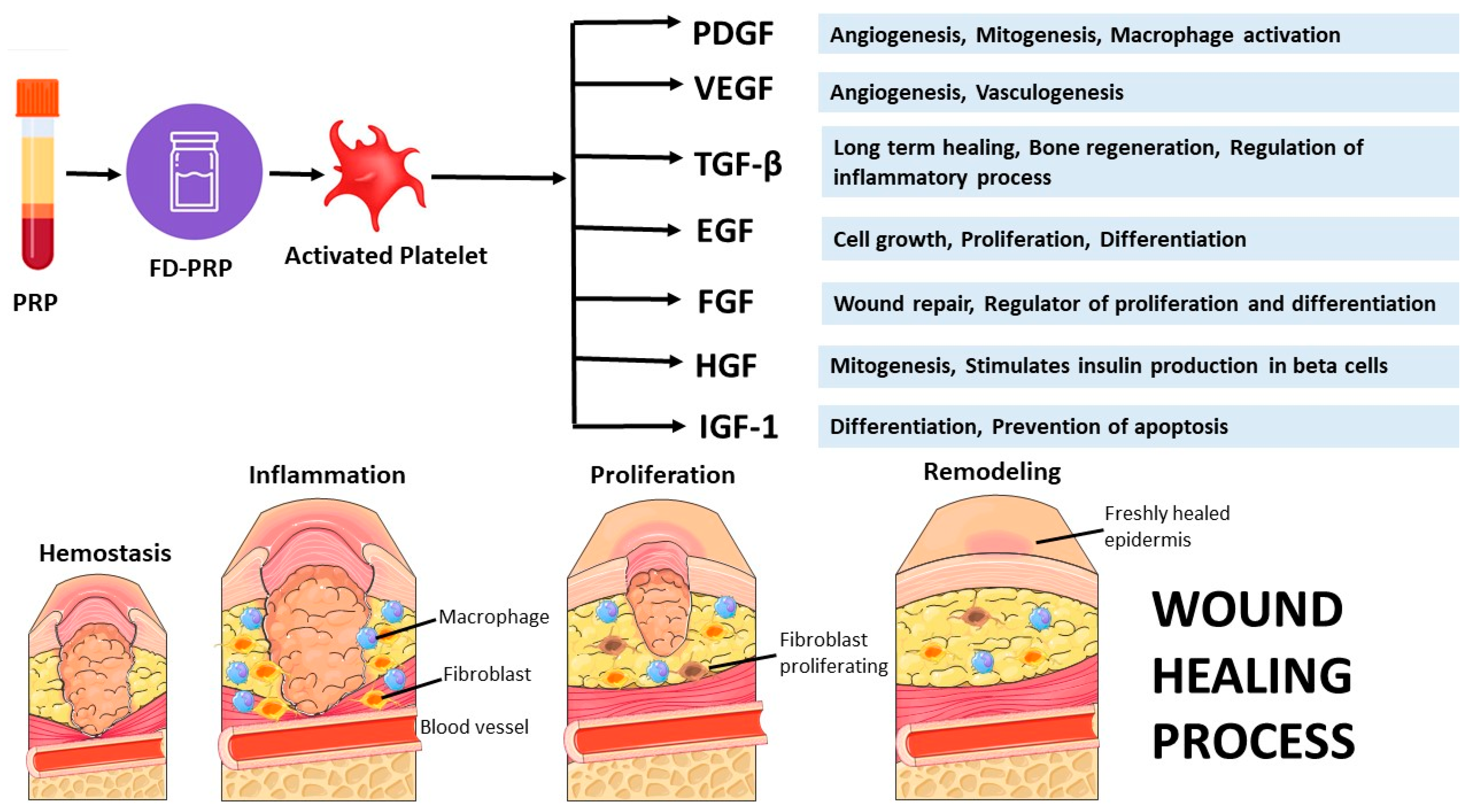
Figure 2.
| Platelet recovery from whole blood and reconstituted FD-PRP. Mean platelet counts in whole blood (baseline) and platelet-rich plasma (PRP), expressed as ×10⁹ mL⁻¹. PRP preparation resulted in a marked enrichment of platelets relative to baseline, indicating efficient recovery from whole blood (n = 20) (A). Platelet counts in FD-PRP and reconstituted FD-PRP (n = 20). Statistical analysis was performed using one-way ANOVA (***P < 0.001) (B). Microscopic images of PRP (i) and FD-PRP (ii) at ×40 magnification (C).
Figure 2.
| Platelet recovery from whole blood and reconstituted FD-PRP. Mean platelet counts in whole blood (baseline) and platelet-rich plasma (PRP), expressed as ×10⁹ mL⁻¹. PRP preparation resulted in a marked enrichment of platelets relative to baseline, indicating efficient recovery from whole blood (n = 20) (A). Platelet counts in FD-PRP and reconstituted FD-PRP (n = 20). Statistical analysis was performed using one-way ANOVA (***P < 0.001) (B). Microscopic images of PRP (i) and FD-PRP (ii) at ×40 magnification (C).
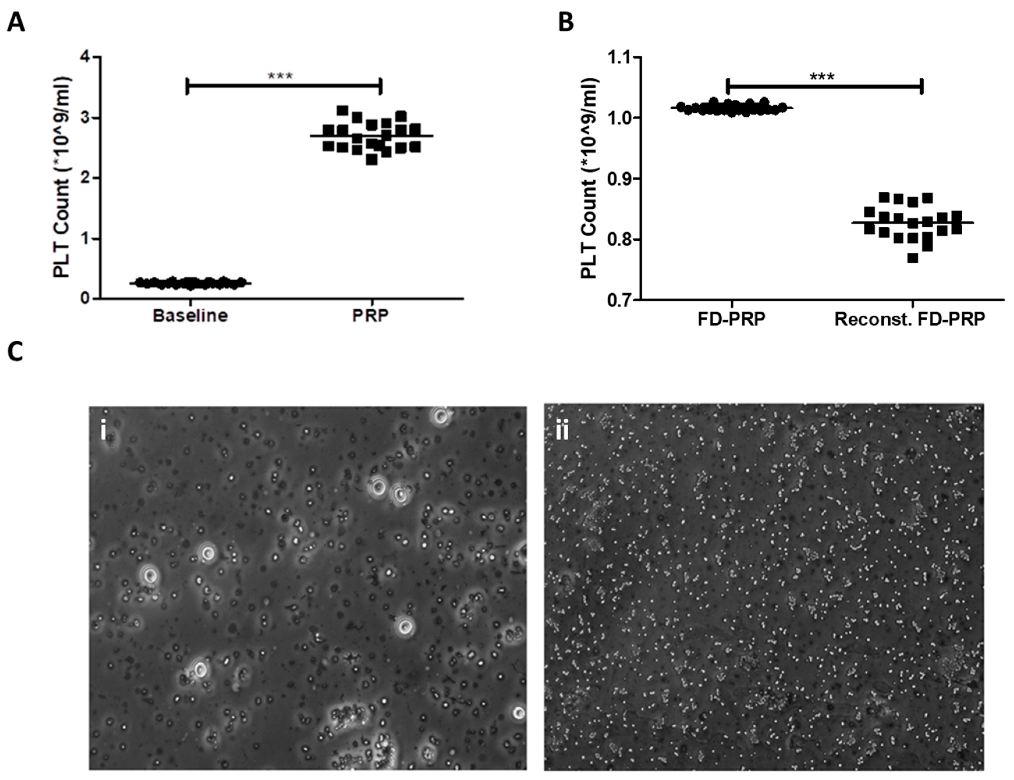
Figure 3.
| Platelet aggregation activity. ADP- and adrenaline-induced aggregation of fresh PRP, FD-PRP, and platelet-poor plasma (PPP). Aggregation (%) is shown as the mean. Minimal loss of aggregation response in FD-PRP demonstrates functional preservation after lyophilization.
Figure 3.
| Platelet aggregation activity. ADP- and adrenaline-induced aggregation of fresh PRP, FD-PRP, and platelet-poor plasma (PPP). Aggregation (%) is shown as the mean. Minimal loss of aggregation response in FD-PRP demonstrates functional preservation after lyophilization.
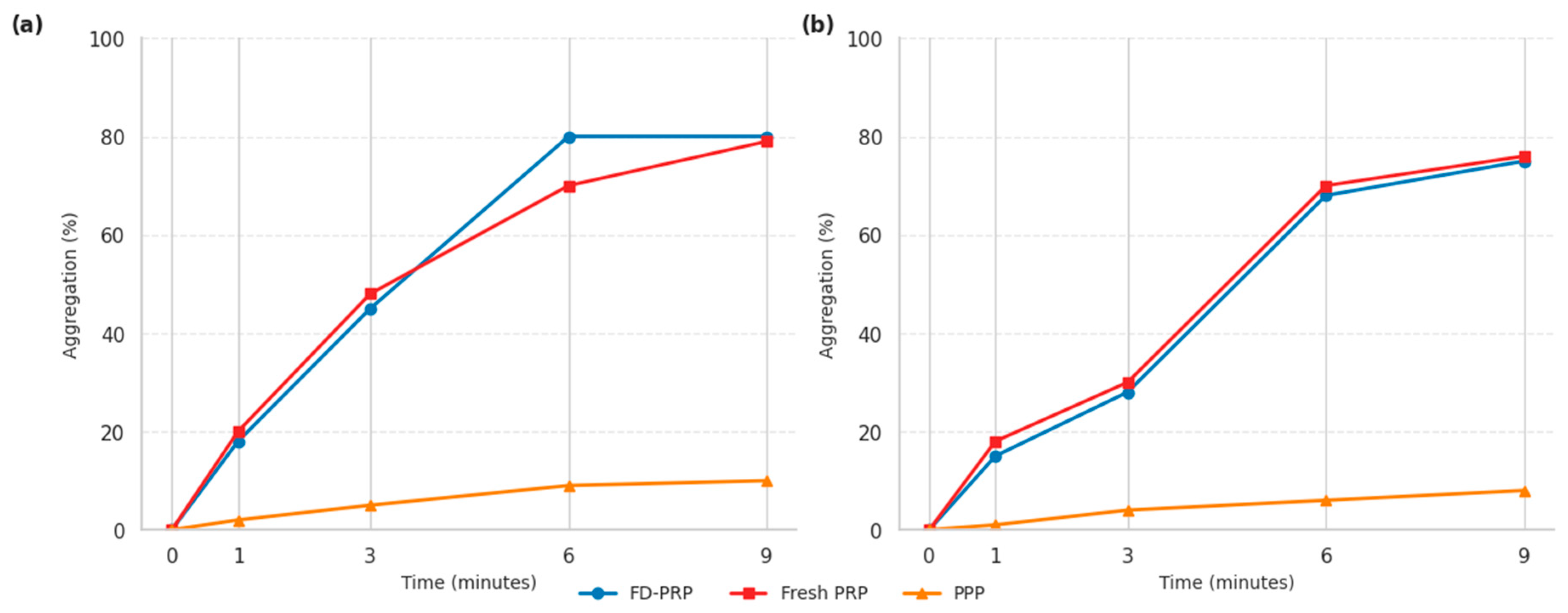
Figure 4.
| Growth factor content of FD-PRP versus fresh PRP. The concentrations of major growth factors (PDGF-AB/BB, TGF-β1, VEGF, EGF, bFGF, and IGF-1) were quantified in FD-PRP and fresh PRP across donors. Bars indicate mean ± s.d.; numbers above bars represent fold changes (FD-PRP / Fresh PRP). FD-PRP retained or enhanced levels of key regenerative factors. Statistical analysis was performed using one-way ANOVA (***P < 0.001).
Figure 4.
| Growth factor content of FD-PRP versus fresh PRP. The concentrations of major growth factors (PDGF-AB/BB, TGF-β1, VEGF, EGF, bFGF, and IGF-1) were quantified in FD-PRP and fresh PRP across donors. Bars indicate mean ± s.d.; numbers above bars represent fold changes (FD-PRP / Fresh PRP). FD-PRP retained or enhanced levels of key regenerative factors. Statistical analysis was performed using one-way ANOVA (***P < 0.001).
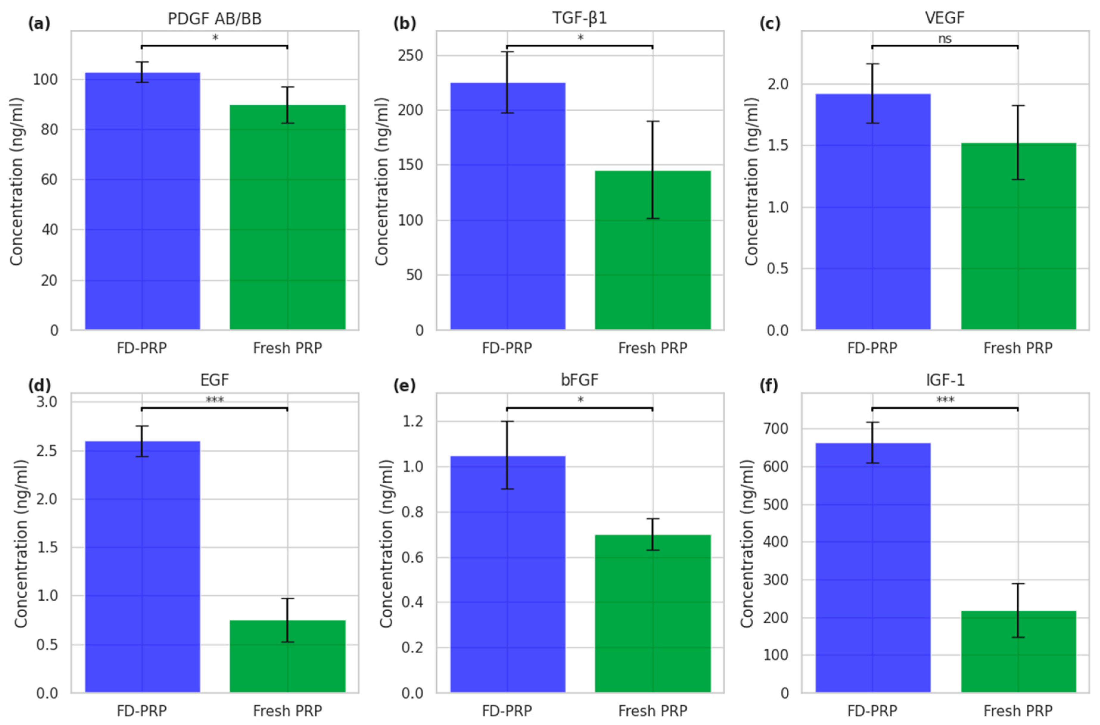
Figure 5.
| Longitudinal changes in platelet count, percentage recovery and growth factor concentrations during storage. Platelet count (billion ml⁻¹; bars, mean ± s.d., n = 3 independent measurements per time point) measured at 0, 4, 8 and 26 weeks of storage (A). Red dashed line indicates percentage platelet recovery relative to baseline (week 0 set to 100%). Statistical significance was assessed by one-way ANOVA followed by multiple-comparison testing against week 0. Concentrations of growth factors measured at week 0, week 4 and week 26 (B): i, EGF; ii, IGF-1; iii, PDGF-AB/BB; iv, TGF-β1; v, VEGF; vi, bFGF. Bars represent mean ± s.d. (n = 3 independent measurements per time point). Statistical analysis was performed using one-way ANOVA with post hoc multiple comparisons. Significance is indicated as ns (not significant), *P < 0.05, **P < 0.01, ***P < 0.001.
Figure 5.
| Longitudinal changes in platelet count, percentage recovery and growth factor concentrations during storage. Platelet count (billion ml⁻¹; bars, mean ± s.d., n = 3 independent measurements per time point) measured at 0, 4, 8 and 26 weeks of storage (A). Red dashed line indicates percentage platelet recovery relative to baseline (week 0 set to 100%). Statistical significance was assessed by one-way ANOVA followed by multiple-comparison testing against week 0. Concentrations of growth factors measured at week 0, week 4 and week 26 (B): i, EGF; ii, IGF-1; iii, PDGF-AB/BB; iv, TGF-β1; v, VEGF; vi, bFGF. Bars represent mean ± s.d. (n = 3 independent measurements per time point). Statistical analysis was performed using one-way ANOVA with post hoc multiple comparisons. Significance is indicated as ns (not significant), *P < 0.05, **P < 0.01, ***P < 0.001.
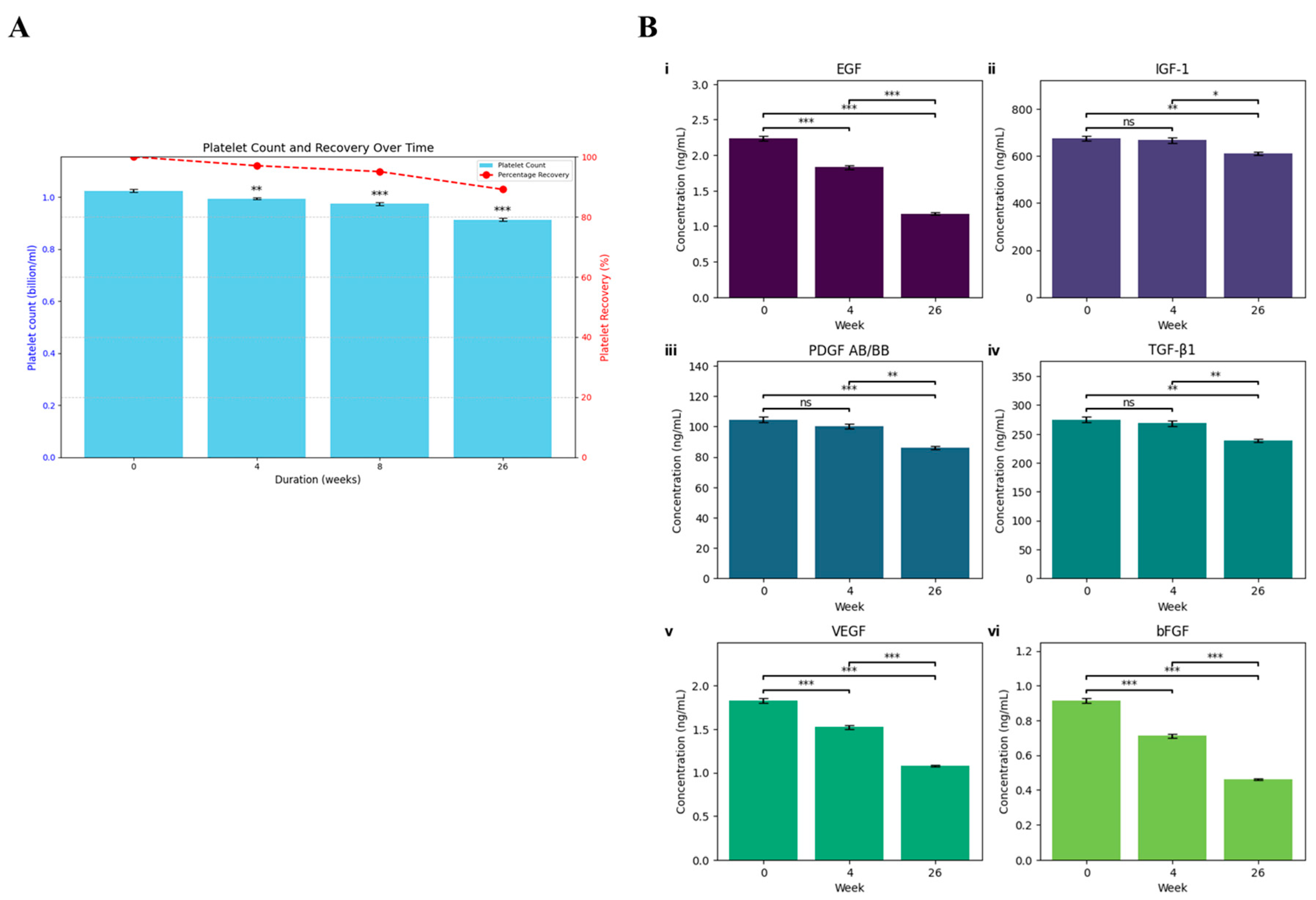
Figure 6.
| In vitro wound-healing assay. Representative images of wound closure at 0, 12, and 24 h in cells treated with FBS, fresh PRP, 5% FD-PRP, or 10% FD-PRP (A). Quantification of wound closure (%) over time showed accelerated migration in the PRP-treated groups, with the highest effect observed with 10% FD-PRP (B). (FBS: Fetal Bovine Serum; PRP: Platelet Rich Plasma; FD-PRP: Freeze-dried Platelet Rich Plasma).
Figure 6.
| In vitro wound-healing assay. Representative images of wound closure at 0, 12, and 24 h in cells treated with FBS, fresh PRP, 5% FD-PRP, or 10% FD-PRP (A). Quantification of wound closure (%) over time showed accelerated migration in the PRP-treated groups, with the highest effect observed with 10% FD-PRP (B). (FBS: Fetal Bovine Serum; PRP: Platelet Rich Plasma; FD-PRP: Freeze-dried Platelet Rich Plasma).
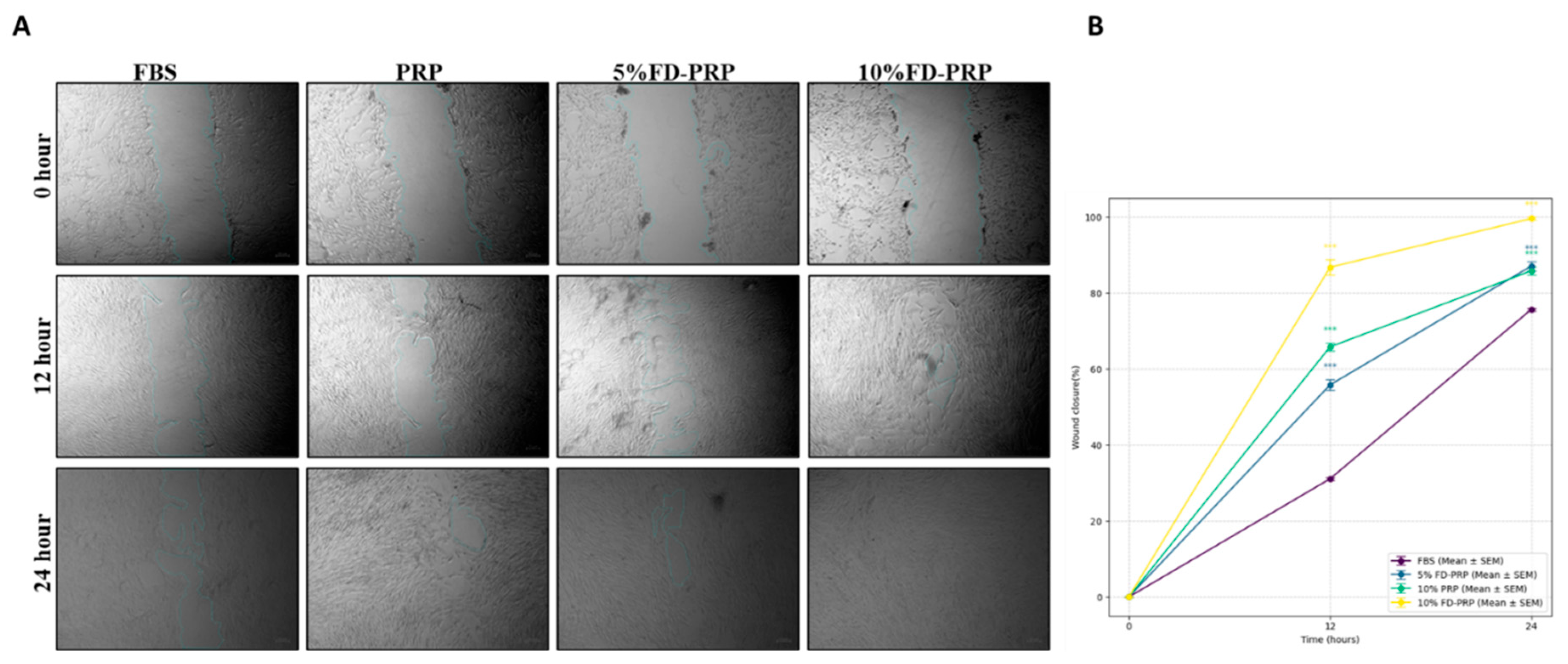
Figure 7.
| FD-PRP enhances in vivo wound healing. Representative images of wounds treated with fresh PRP (i, iv, vii), FD-PRP (ii, v, viii), and standard care (iii, vi, ix) at weeks 0 (i–iii), 2 (iv–vi), and 3 (vii–ix). FD-PRP-treated wounds exhibited visibly faster closure and improved epithelial coverage compared with fresh PRP and standard care (A); Quantitative analysis of wound healing over 4 weeks. Healing percentages were calculated relative to the baseline (week 0) wound size for each patient. Data are presented as mean ± standard deviation (n = 10 patients). FD-PRP-treated wounds showed the fastest reduction in wound area (>60%), outperforming fresh PRP (~50%) and standard care (<40%). Error bars indicate variability between patients, highlighting the superior efficacy of FD-PRP in promoting wound closure (B).
Figure 7.
| FD-PRP enhances in vivo wound healing. Representative images of wounds treated with fresh PRP (i, iv, vii), FD-PRP (ii, v, viii), and standard care (iii, vi, ix) at weeks 0 (i–iii), 2 (iv–vi), and 3 (vii–ix). FD-PRP-treated wounds exhibited visibly faster closure and improved epithelial coverage compared with fresh PRP and standard care (A); Quantitative analysis of wound healing over 4 weeks. Healing percentages were calculated relative to the baseline (week 0) wound size for each patient. Data are presented as mean ± standard deviation (n = 10 patients). FD-PRP-treated wounds showed the fastest reduction in wound area (>60%), outperforming fresh PRP (~50%) and standard care (<40%). Error bars indicate variability between patients, highlighting the superior efficacy of FD-PRP in promoting wound closure (B).
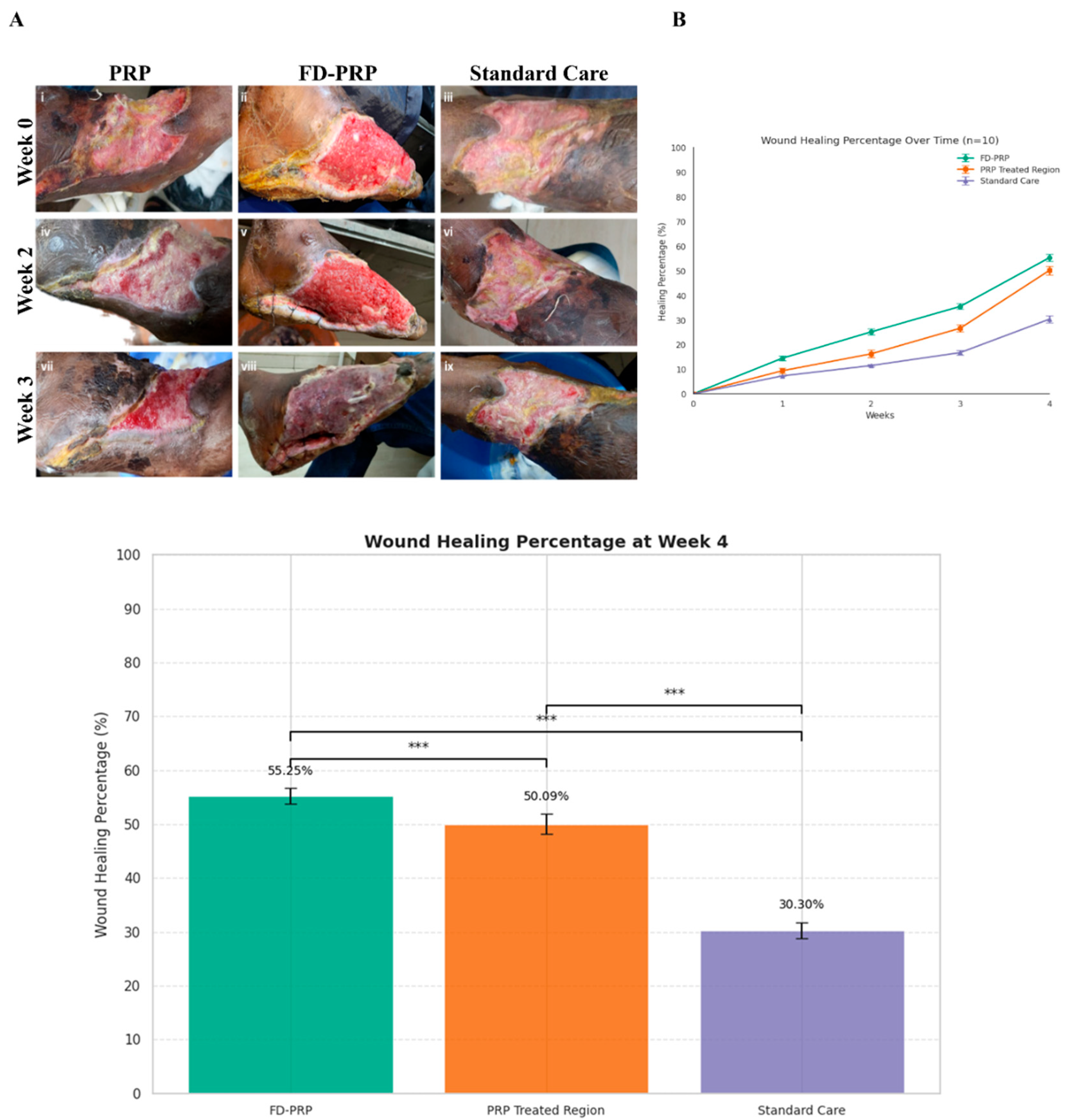
Table 1.
Blood cell counts and platelet recovery. Blood cell counts were measured in whole blood samples (RBC, WBC, and platelets), platelet-rich plasma (PRP) and Freeze-dried PRP (FD-PRP). Platelet recovery (%) was calculated as the proportion of platelets enriched in the PRP relative to the baseline. Values are presented as mean ± standard deviation (s.d.) and range (minimum–maximum). Units are expressed per milliliter (mL) of blood or plasma (n=20 donors).
Table 1.
Blood cell counts and platelet recovery. Blood cell counts were measured in whole blood samples (RBC, WBC, and platelets), platelet-rich plasma (PRP) and Freeze-dried PRP (FD-PRP). Platelet recovery (%) was calculated as the proportion of platelets enriched in the PRP relative to the baseline. Values are presented as mean ± standard deviation (s.d.) and range (minimum–maximum). Units are expressed per milliliter (mL) of blood or plasma (n=20 donors).
| Parameter | Mean ± SD | Range | P-value |
| WBC (×10⁹/L) | 6.15 ± 0.18 | 5.8–6.5 | |
| RBC (×10¹²/L) | 4.70 ± 0.10 | 4.5–4.9 | |
| Hemoglobin (g/dL) | 14.16 ± 0.17 | 13.8–14.5 | |
| Hematocrit (%) | 42.0 ± 1.0 | 40–44 | |
| Baseline platelet count (×10⁹/mL) | 0.26 ± 0.02 | 0.22–0.29 | |
| PRP platelet count (×10⁹/mL) | 2.69 ± 0.22 | 2.31–3.12 | |
| P value platelet baseline count vs PRP | P <0.001 | ||
| PRP recovery (%) | 86.48 ± 2.62 | 78.85–89.88 | |
| FD-PRP platelet count (×10⁹/mL) | 1.016 ± 0.005 | 1.009–1.026 | |
| Reconstituted FD-PRP (×10⁹/mL) | 0.827 ± 0.027 | 0.770–0.869 | |
| P value FD-PRP platelet count vs Reconstituted FD-PRP | P <0.001 | ||
| FD-PRP recovery (%) | 81.40 ± 2.81 | 75.93–86.11 | |
Table 2.
Platelet aggregation responses to agonists in fresh PRP, FD-PRP and PPP. Platelet aggregation induced by adenosine diphosphate (ADP) and adrenaline was assessed in fresh platelet-rich plasma (PRP), freeze-dried platelet-rich plasma (FD-PRP) after reconstitution, and platelet-poor plasma (PPP). Aggregation is expressed as percentage maximal light transmission, with PPP serving as the negative control.
Table 2.
Platelet aggregation responses to agonists in fresh PRP, FD-PRP and PPP. Platelet aggregation induced by adenosine diphosphate (ADP) and adrenaline was assessed in fresh platelet-rich plasma (PRP), freeze-dried platelet-rich plasma (FD-PRP) after reconstitution, and platelet-poor plasma (PPP). Aggregation is expressed as percentage maximal light transmission, with PPP serving as the negative control.
| ADP | Adrenaline | |||||
| Time (min) | FD-PRP | PRP | PPP | FD-PRP | PRP | PPP |
| 0 | 0.0 | 0.0 | 0.0 | 0.0 | 0.0 | 0.0 |
| 1 | 18.0 | 20.0 | 2.0 | 15.0 | 18.0 | 1.0 |
| 3 | 45.0 | 48.0 | 5.0 | 28.0 | 30.0 | 4.0 |
| 6 | 80.0 | 70.0 | 9.0 | 68.0 | 70.0 | 6.0 |
| 9 | 80.0 | 79.0 | 10.0 | 74.0 | 76.0 | 8.0 |
Table 3.
Growth factor concentrations in FD-PRP and fresh PRP. The growth factor concentrations in freeze-dried PRP (FD-PRP) and fresh PRP were measured in four independent donors (D1–D4). Values are presented as mean ± standard deviation (SD) with the observed range. Fold change represents the ratio of the mean FD-PRP to the mean fresh PRP.
Table 3.
Growth factor concentrations in FD-PRP and fresh PRP. The growth factor concentrations in freeze-dried PRP (FD-PRP) and fresh PRP were measured in four independent donors (D1–D4). Values are presented as mean ± standard deviation (SD) with the observed range. Fold change represents the ratio of the mean FD-PRP to the mean fresh PRP.
| Growth factor (ng/mL) | FD-PRP Mean ± SD | Range (FD-PRP) | Fresh PRP Mean ± SD | Range (Fresh PRP) | Fold (FD-PRP / Fresh PRP) |
| PDGF AB/BB | 103 ± 9.5 | 92–115 | 90 ± 10.8 | 78–102 | 1.14 |
| TGF-β1 | 270.7 ± 171 | 143–546 | 125.5 ± 53.7 | 70–180 | 2.16 |
| VEGF | 1.8 ± 0.36 | 1.4–2.2 | 1.5 ± 0.33 | 1.1–1.9 | 1.20 |
| EGF | 2.2 ± 0.88 | 1.4–3.5 | 1.4 ± 0.87 | 0.5–2.5 | 1.57 |
| bFGF | 0.9 ± 0.22 | 0.6–1.1 | 0.75 ± 0.17 | 0.5–0.9 | 1.20 |
| IGF-1 | 664 ± 55 | 610–720 | 218.7 ± 77 | 146–322 | 3.04 |
Disclaimer/Publisher’s Note: The statements, opinions and data contained in all publications are solely those of the individual author(s) and contributor(s) and not of MDPI and/or the editor(s). MDPI and/or the editor(s) disclaim responsibility for any injury to people or property resulting from any ideas, methods, instructions or products referred to in the content. |
© 2026 by the authors. Licensee MDPI, Basel, Switzerland. This article is an open access article distributed under the terms and conditions of the Creative Commons Attribution (CC BY) license (http://creativecommons.org/licenses/by/4.0/).
Copyright: This open access article is published under a Creative Commons CC BY 4.0 license, which permit the free download, distribution, and reuse, provided that the author and preprint are cited in any reuse.